Morphogenesis of rod shaped bacteria
9
1 Legends and figures of chapter: 1 2 Morphogenesis of rod shaped bacteria 3 4 Figure 1. The sacculus determines the shape of the cell. Electron microscopy image of an 5 isolated sacculus of the stalk containing Caulobacter crescentus cell on the left and a 6 sacculus of a dividing Escherichia coli right. The sacculi were isolated and processed for 7 electron microscopy. They were labeled on grid with antibodies against murein and 8 secondary antibodies conjugated to 6 nm gold particles and subsequently stained by uranyl- 9 acetate. The bar equals 200 nm. Courtesy of Waldemar Vollmer and Heinz Schwarz. 10 11 Figure 2. Schematic representation of the elongase. Proteins are shown that are supposed to 12 interact with each other and form a complex responsible for the elongation of the cylindrical 13 part of the cell (A). Because the majority of these interactions have been found by bacterial 14 two hybrid systems or by co-immunoprecipitation, the precise interactions are not 15 established. Therefore alternative interaction cannot be excluded. Instead of a mysterious 16 protein or RodA, the translocase could also be formed by a combination of integral 17 membrane proteins (e.g. MraY and RodA as shown in B or RodA and MreD, not shown). 18 19 Figure 3. Schematic representation of the assembly of the components of the divisome. 20 The boxed proteins represent subcomplexes: 1) the Z-ring with FtsZ-FtsA-ZipA-ZapA which 21 interacts with FtsE/X; 2) the subcomplex FtsQLB which contains an heterodimer of FtsL and 22 FtsB; 3) the subcomplex FtsW-PBP3; 4) PBP1B and FtsN interact with PBP3 and could be 23 part of the subcomplex FtsW-PBP3. FtsK appears at the division site at the same time as 24 FtsQLB and FtsW-PBP3, is back recruited by the complex QLB and interacts with most of 25 these proteins. MurG, PBPs2 and PBP5, AmiC and EnvC are located at the division and are 26 part of the cell division machinery. Dashed line: Interaction detected by using a two-hybrid 27 system (90,91, 92 and unpublished results). Solid lines: interactions detected using different 28 techniques. Courtesy of Benôit Wolff. 29 30 31 Figure 4. Schematic representation of the divisome. The membrane topology and 32 compartmentalization of cell divisome proteins that have been localized at mid cell either by 33 GFP fusions or by immunofluorescence microscopy are shown. Transmembrane helices are 34 represented by cylinders and the cytosolic or periplasmic (above the membrane) domains are 35 sized approximately according to the molecular weight of the protein domains. MraY has 36 been added in analogy to its presence in the elongase but has not yet been localized. EnvA, 37 PBP2 and PBP5 are not shown because, presently information is lacking on their possible 38 interactions with the divisomal proteins. Courtesy of Benôit Wolff. 39 40 Figure 5. Schematic interpretation of murein synthesis in E. coli cells subjected to a D-Cys 41 label-and-chase experiment. Cells incubated in the presence of D-Cys incorporate the amino 42 acid homogeneously over the entire surface of the sacculus. When cells are transferred to D- 43 Cys free medium, newly inserted precursors are devoid of D-Cys, and can be differentiated 44 from preexisting material by immuno-microscopy techniques. The figure depicts the 45 evolution of a newly born cell through two successive generations. At the beginning of the 46 chase period (0 Mass Doubling, MD) the cell wall is heavily and uniformly labelled with D- 47
Transcript of Morphogenesis of rod shaped bacteria

1
Legends and figures of chapter: 1 2 Morphogenesis of rod shaped bacteria 3 4 Figure 1. The sacculus determines the shape of the cell. Electron microscopy image of an 5 isolated sacculus of the stalk containing Caulobacter crescentus cell on the left and a 6 sacculus of a dividing Escherichia coli right. The sacculi were isolated and processed for 7 electron microscopy. They were labeled on grid with antibodies against murein and 8 secondary antibodies conjugated to 6 nm gold particles and subsequently stained by uranyl-9 acetate. The bar equals 200 nm. Courtesy of Waldemar Vollmer and Heinz Schwarz. 10 11 Figure 2. Schematic representation of the elongase. Proteins are shown that are supposed to 12 interact with each other and form a complex responsible for the elongation of the cylindrical 13 part of the cell (A). Because the majority of these interactions have been found by bacterial 14 two hybrid systems or by co-immunoprecipitation, the precise interactions are not 15 established. Therefore alternative interaction cannot be excluded. Instead of a mysterious 16 protein or RodA, the translocase could also be formed by a combination of integral 17 membrane proteins (e.g. MraY and RodA as shown in B or RodA and MreD, not shown). 18 19 Figure 3. Schematic representation of the assembly of the components of the divisome. 20 The boxed proteins represent subcomplexes: 1) the Z-ring with FtsZ-FtsA-ZipA-ZapA which 21 interacts with FtsE/X; 2) the subcomplex FtsQLB which contains an heterodimer of FtsL and 22 FtsB; 3) the subcomplex FtsW-PBP3; 4) PBP1B and FtsN interact with PBP3 and could be 23 part of the subcomplex FtsW-PBP3. FtsK appears at the division site at the same time as 24 FtsQLB and FtsW-PBP3, is back recruited by the complex QLB and interacts with most of 25 these proteins. MurG, PBPs2 and PBP5, AmiC and EnvC are located at the division and are 26 part of the cell division machinery. Dashed line: Interaction detected by using a two-hybrid 27 system (90,91, 92 and unpublished results). Solid lines: interactions detected using different 28 techniques. Courtesy of Benôit Wolff. 29 30 31 Figure 4. Schematic representation of the divisome. The membrane topology and 32 compartmentalization of cell divisome proteins that have been localized at mid cell either by 33 GFP fusions or by immunofluorescence microscopy are shown. Transmembrane helices are 34 represented by cylinders and the cytosolic or periplasmic (above the membrane) domains are 35 sized approximately according to the molecular weight of the protein domains. MraY has 36 been added in analogy to its presence in the elongase but has not yet been localized. EnvA, 37 PBP2 and PBP5 are not shown because, presently information is lacking on their possible 38 interactions with the divisomal proteins. Courtesy of Benôit Wolff. 39 40 Figure 5. Schematic interpretation of murein synthesis in E. coli cells subjected to a D-Cys 41 label-and-chase experiment. Cells incubated in the presence of D-Cys incorporate the amino 42 acid homogeneously over the entire surface of the sacculus. When cells are transferred to D-43 Cys free medium, newly inserted precursors are devoid of D-Cys, and can be differentiated 44 from preexisting material by immuno-microscopy techniques. The figure depicts the 45 evolution of a newly born cell through two successive generations. At the beginning of the 46 chase period (0 Mass Doubling, MD) the cell wall is heavily and uniformly labelled with D-47

2
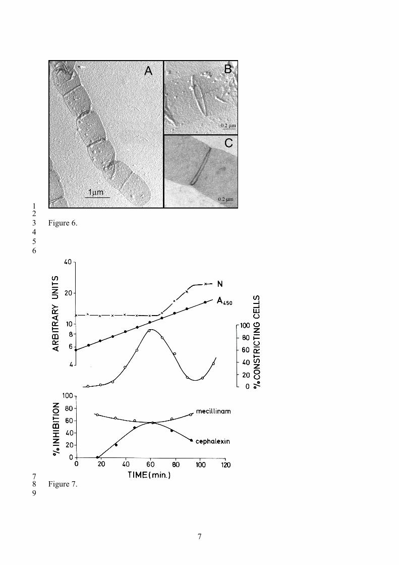
Cys (dark grey). As the cell elongates, insertion of new material causes dilution of D-Cys 1 labelled peptidoglycan in the lateral wall (medium grey) but not in the polar regions. Shortly 2 before the cell starts to constrict, incorporation of precursors in the central region is 3 exacerbated, which results in the creation of a band-like region of all-new D-Cys free murein 4 (white) at the potential division site. As division progresses two new cells are born with a 5 heavily labelled old pole and a new pole devoid of D-Cys (1 MD). The next cell cycle results 6 in a gradual reduction of label in the lateral wall (light grey) and the generation of cells with 7 two unlabeled poles and with one labelled and one unlabeled pole (2 MD). The labelled pole, 8 derived from the original cell, retains a dense labelling and can be traced for at least 5 9 generations. Inhibition of cell division proteins results in filaments characterized by the 10 presence of two densely labelled poles (dark grey) and an homogeneously diluted cylindrical 11 wall which presents a series of regularly spaced bands of unlabeled peptidoglycan at the 12 potential division sites when FtsZ protein is active (FtsQ(Ts)), but not when FtsZ is impaired 13 (FtsZ(Ts)). 14 15 16 Figure 6. SP-rings in purified sacculi from a Salmonella thyphimurium SL1344 amiABC 17 triple deletion mutant. Sacculi purified from exponentially growing cultures were processed 18 for electron microscopy. Sacculi spread on carbon-pioloform-coated cooper grids were 19 stained with 1%(w/v) uranyl acetate and observed under the EM either after carbon-platinum 20 shadowing (A and B) to enhance surface detail, or directly after staining (C). 21 22 Figure 7. Contribution of PBP2 and PBP3 to peptidoglycan synthesis in relation to the cell 23 division cycle. In the upper panel the cell number (N), the optical density (A450), and the 24 percentage of constricting cells in the synhronously growing cell culture of a wild type E. coli 25 strain are shown. In the lower panel is the percentage of radioactive Dap incorporation in the 26 sacculi shown in the presence of either the PBP2 inhibitor mecillinam or the PBP3 inhibitor 27 cephalexin. (Reproduced from reference xx with permission from Elsevier Limited) 28 29 Figure 8. Schematic representation of the Tol-Pal system. The cytoplasmic membrane 30 proteins TolQ and TolR probably form a channel and interact with TolA in the periplasm. Pal 31 is anchored to the outer membrane and interacts strongly with the peptide moiety of the 32 peptidoglycan layer by its carboxy-terminal region. TolB competes with the peptidoglycan 33 peptide side chain for the same region on Pal. In addition to the interaction of Pal with TolB 34 and peptidoglycan, it interacts independently with OmpA and with TolA. 35 36 Figure 9. Schematic representation of the elongation and septal mode of peptidoglycan 37 synthesis in a slice perpendicular to the length axes of the bacterium. Insertion of 38 peptidoglycan precursors in the existing peptidoglycvcan layer is accomplished by the 39 elongases that use the MreB cytoskeletal helix as tracking devise to ensure dispersed lateral 40 extension of the sacculus. Based on the abundance of the elongase subunits between 20-100 41 elongases per cell are expected to be active. After approximately 40% of a generation time 42 the FtsZ-ring is formed and a switch occurs to polar peptidoglycan synthesis by penicillin 43 insensitive peptidoglycan synthesizing machinery. About one fifth of a generation time later 44 the proteins FtsK up to AmiC assemble onto the ring and constriction of the cell is initiated. 45 The proteins that constitute a mature divisome are present in about 50 complexes (here 46 represented by 12) that are predicted to stay attached to the Z-ring during the whole 47

3
constriction process. 1 2

4
1 2 Figure 1. 3
4 Figure 2. 5 6 7 8

5
1 Figure 3. 2
3 Figure 4. 4

6
1 2 Figure 5. 3 4 5
7
1 2 Figure 6. 3 4 5 6
7 Figure 7. 8 9

8
1 Figure 8. 2 3

9
1 Figure 9. 2 3 4