Moraxella sp. Strain C-1 - Journal of Bacteriology - American Society
5
Vol. 170, No. 7 Purification and Properties of Formate Dehydrogenase from Moraxella sp. Strain C-1 YASUHISA ASANO,* TOSHIKAZU SEKIGAWA, HISAMITSU INUKAI, AND AKIKO NAKAZAWA Sagami Chemical Research Center, Nishi-Ohnuma, Sagamihara, Kanagawa 229, Japan Received 6 November 1987/Accepted 25 March 1988 NAD+-dependent formate dehydrogenase was screened in various bacterial strains. Facultative methanol- utilizing bacteria isolated from soil samples, acclimated to a medium containing methanol and formate at pH 9.5, were classified as members of the genus Moraxella. From a crude extract of Moraxella sp. strain C-1, formate dehydrogenase was purified to homogeneity, as judged by disc gel electrophoresis. The enzyme has an isoelectric point of 3.9 and a molecular weight of approximately 98,000. The enzyme is composed of two identical subunits with molecular weights of about 48,000. The apparent Km values for sodium formate and NAD+ were calculated to be 13 mM and 0.068 mM, respectively. Formate dehydrogenases have been known to occur widely in nature. They include NAD+-dependent formate dehydrogenase (EC 1.2.1.2) (15, 23), NADP+-dependent formate dehydrogenase (EC 1.2.1.43) (31), and formate dehydrogenases participating in the electron transport or the synthesis of formate from CO2 with electron acceptors or donors such as ferredoxin, cytochromes, or artificial dyes (EC 1.2.2.1 and 1.2.2.3) (9, 17, 24, 30). While NAD+- dependent formate dehydrogenases from methanol-utilizing yeasts have been well characterized (11, 12, 15, 25), the bacterial enzymes have been purified to homogeneity and their properties have been investigated only in Pseudomonas oxalaticus (21) and Achromobacter parvulus (7, 8). There has been no report of the utilization of methanol or formate by a Moraxella strain. In this study, we screened for a stable NAD+-dependent formate dehydrogenase from methanol- utilizing bacteria, and we purified and characterized the enzyme from Moraxella sp. strain C-1, isolated from soil in an alkaline medium. MATERIALS AND METHODS Materials. Butyl-Toyopearl and high-pressure liquid chro- matography (HPLC) columns G-3000 SW, DEAE-5PW, and Phenyl-5PW were purchased from Tosoh Corp., Tokyo, Japan; Sephadex G-100 and Sephadex G-200 were obtained from Pharmacia, Uppsala, Sweden; alcohol dehydrogenase (Saccharomyces cerevisiae, EC 1.1.1.1) and marker proteins for molecular weight determinations were obtained from Oriental Yeast, Tokyo, Japan; and ampholites were obtained from LKB Produkter AB, Uppsala, Sweden. The membrane filter (Diaflo, PM 30) was obtained from Amicon Corp., Lexington, Mass. Phosphoriboisomerase (spinach, EC 5.3.1.6) and disodium D-ribose-5-phosphate were purchased from Sigma Chemical Co., St. Louis, Mo. 2-(p-Iodophenyl)- 3-(p-nitrophenyl)-5-phenyltetrazolium chloride was pur- chased from Dojin Chemicals, Kumamoto, Japan; Coomas- sie brilliant blue R-250 was purchased from Fluka AG, Buchs, Switzerland; and hydroxyapatite was purchased from Wako Pure Chemicals, Osaka, Japan. Screening of formate dehydrogenase-producing strains. Formate dehydrogenase activities were measured for bacte- rial strains from type culture collections and microorganisms isolated from soil samples. Strains from type culture collec- * Corresponding author. tions included eight strains of six genera of bacteria (28, 29). They were obtained from stock cultures of the Institute for Microbiology, University of Tokyo, Tokyo, Japan; the Northern Regional Research Laboratory, Peoria, Ill.; the National Collection of Industrial Bacteria, Torry Research Station, Aberdeen, Scotland; and our laboratory. Basal medium consisted of 2 g of K2HPO4, 2 g of (NH4)2SO4, 1 g of NaCl, 0.2 g of MgSO4 7H21, 0.5 g of yeast extract (Oriental Yeast), 0.02 pxg of biotin, 4 ,ug of calcium panto- thenate, 20 ,ug of inositol, 4 jig of nicotinic acid, 4 ,ug of thiamine hydrochloride, 2 jig of pyridoxine hydrochloride, 2 ,ug of p-aminobenzoic acid, 2 ,ug of riboflavin, and 0.1 jLg of folic acid in 1 liter of tap water (pH 7.0). Basal medium supplemented with 0.8% (wt/vol) methanol, 1% sodium formate, and 1% sodium carbonate was used as an enrich- ment medium. The pH of the enrichment medium was 9.5. Methanol and sodium carbonate were added after the me- dium was autoclaved. About 100 soil samples from Sagami- hara in the Kanagawa Prefecture, Japan, were suspended in 200 ml of the enrichment medium in a 500-ml Erlenmeyer flask and incubated at 30°C on a rotary shaker. Half of the supernatant was removed every other day, and the same volume of the fresh medium was added. Consumption of methanol in the enrichment medium was monitored by a gas-liquid chromatograph (model 802; Ohkura, Tokyo, Ja- pan) with a glass column of Porapack Q (Waters Associates, Inc., Milford, Mass.). Methanol-utilizing bacteria, including Moraxella sp. strains C-1 and D2b, were cultured aerobically at 30°C for 48 h in enrichment medium supplemented with 0.8% (wt/vol) methanol. TGY medium (5 g of tryptone [Difco Laboratories, Detroit, Mich.], 5 g of yeast extract, 1 g of glucose, and 1 g of K2HPO4 in 1 liter of tap water [pH 7.0]) does not induce formate dehydrogenase. The buffer used in this study was potassium phosphate buffer (pH 7.0), contain- ing 0.1 mM EDTA and 5 mM 2-mercaptoethanol unless otherwise stated. The cells were washed once with physio- logical saline and suspended in 0.1 M buffer. The cells were disrupted for 20 min with a 9-kHz ultrasonic oscillator (Kubota Syoji, Tokyo, Japan). The disrupted cells were centrifuged at 14,000 x g for 20 min. The enzyme activity in the supernatant was measured after dialysis against 0.01 M buffer. Enzyme assay. The enzyme activity was assayed by the reduction of NAD+, monitored at 340 nm with a Hitachi 228A spectrophotometer, with sodium formate as a sub- 3189 JOURNAL OF BACTERIOLOGY, JUlY 1988, p. 3189-3193 0021-9193/88/073189-05$02.00/0 Copyright X) 1988, American Society for Microbiology Downloaded from https://journals.asm.org/journal/jb on 26 January 2022 by 186.236.10.178.
Transcript of Moraxella sp. Strain C-1 - Journal of Bacteriology - American Society

Vol. 170, No. 7
Purification and Properties of Formate Dehydrogenase fromMoraxella sp. Strain C-1
YASUHISA ASANO,* TOSHIKAZU SEKIGAWA, HISAMITSU INUKAI, AND AKIKO NAKAZAWA
Sagami Chemical Research Center, Nishi-Ohnuma, Sagamihara, Kanagawa 229, Japan
Received 6 November 1987/Accepted 25 March 1988
NAD+-dependent formate dehydrogenase was screened in various bacterial strains. Facultative methanol-utilizing bacteria isolated from soil samples, acclimated to a medium containing methanol and formate at pH9.5, were classified as members of the genus Moraxella. From a crude extract of Moraxella sp. strain C-1,formate dehydrogenase was purified to homogeneity, as judged by disc gel electrophoresis. The enzyme has anisoelectric point of 3.9 and a molecular weight of approximately 98,000. The enzyme is composed of twoidentical subunits with molecular weights of about 48,000. The apparent Km values for sodium formate andNAD+ were calculated to be 13 mM and 0.068 mM, respectively.
Formate dehydrogenases have been known to occurwidely in nature. They include NAD+-dependent formatedehydrogenase (EC 1.2.1.2) (15, 23), NADP+-dependentformate dehydrogenase (EC 1.2.1.43) (31), and formatedehydrogenases participating in the electron transport or thesynthesis of formate from CO2 with electron acceptors ordonors such as ferredoxin, cytochromes, or artificial dyes(EC 1.2.2.1 and 1.2.2.3) (9, 17, 24, 30). While NAD+-dependent formate dehydrogenases from methanol-utilizingyeasts have been well characterized (11, 12, 15, 25), thebacterial enzymes have been purified to homogeneity andtheir properties have been investigated only in Pseudomonasoxalaticus (21) and Achromobacter parvulus (7, 8). Therehas been no report of the utilization of methanol or formateby a Moraxella strain. In this study, we screened for a stableNAD+-dependent formate dehydrogenase from methanol-utilizing bacteria, and we purified and characterized theenzyme from Moraxella sp. strain C-1, isolated from soil inan alkaline medium.
MATERIALS AND METHODSMaterials. Butyl-Toyopearl and high-pressure liquid chro-
matography (HPLC) columns G-3000 SW, DEAE-5PW, andPhenyl-5PW were purchased from Tosoh Corp., Tokyo,Japan; Sephadex G-100 and Sephadex G-200 were obtainedfrom Pharmacia, Uppsala, Sweden; alcohol dehydrogenase(Saccharomyces cerevisiae, EC 1.1.1.1) and marker proteinsfor molecular weight determinations were obtained fromOriental Yeast, Tokyo, Japan; and ampholites were obtainedfrom LKB Produkter AB, Uppsala, Sweden. The membranefilter (Diaflo, PM 30) was obtained from Amicon Corp.,Lexington, Mass. Phosphoriboisomerase (spinach, EC5.3.1.6) and disodium D-ribose-5-phosphate were purchasedfrom Sigma Chemical Co., St. Louis, Mo. 2-(p-Iodophenyl)-3-(p-nitrophenyl)-5-phenyltetrazolium chloride was pur-chased from Dojin Chemicals, Kumamoto, Japan; Coomas-sie brilliant blue R-250 was purchased from Fluka AG,Buchs, Switzerland; and hydroxyapatite was purchasedfrom Wako Pure Chemicals, Osaka, Japan.
Screening of formate dehydrogenase-producing strains.Formate dehydrogenase activities were measured for bacte-rial strains from type culture collections and microorganismsisolated from soil samples. Strains from type culture collec-
* Corresponding author.
tions included eight strains of six genera of bacteria (28, 29).They were obtained from stock cultures of the Institute forMicrobiology, University of Tokyo, Tokyo, Japan; theNorthern Regional Research Laboratory, Peoria, Ill.; theNational Collection of Industrial Bacteria, Torry ResearchStation, Aberdeen, Scotland; and our laboratory. Basalmedium consisted of 2 g of K2HPO4, 2 g of (NH4)2SO4, 1 g
of NaCl, 0.2 g of MgSO4 7H21, 0.5 g of yeast extract(Oriental Yeast), 0.02 pxg of biotin, 4 ,ug of calcium panto-thenate, 20 ,ug of inositol, 4 jig of nicotinic acid, 4 ,ug ofthiamine hydrochloride, 2 jig of pyridoxine hydrochloride, 2,ug ofp-aminobenzoic acid, 2 ,ug of riboflavin, and 0.1 jLg offolic acid in 1 liter of tap water (pH 7.0). Basal mediumsupplemented with 0.8% (wt/vol) methanol, 1% sodiumformate, and 1% sodium carbonate was used as an enrich-ment medium. The pH of the enrichment medium was 9.5.Methanol and sodium carbonate were added after the me-
dium was autoclaved. About 100 soil samples from Sagami-hara in the Kanagawa Prefecture, Japan, were suspended in200 ml of the enrichment medium in a 500-ml Erlenmeyerflask and incubated at 30°C on a rotary shaker. Half of thesupernatant was removed every other day, and the samevolume of the fresh medium was added. Consumption ofmethanol in the enrichment medium was monitored by agas-liquid chromatograph (model 802; Ohkura, Tokyo, Ja-pan) with a glass column of Porapack Q (Waters Associates,Inc., Milford, Mass.). Methanol-utilizing bacteria, includingMoraxella sp. strains C-1 and D2b, were cultured aerobicallyat 30°C for 48 h in enrichment medium supplemented with0.8% (wt/vol) methanol. TGY medium (5 g of tryptone [DifcoLaboratories, Detroit, Mich.], 5 g of yeast extract, 1 g ofglucose, and 1 g of K2HPO4 in 1 liter of tap water [pH 7.0])does not induce formate dehydrogenase. The buffer used inthis study was potassium phosphate buffer (pH 7.0), contain-ing 0.1 mM EDTA and 5 mM 2-mercaptoethanol unlessotherwise stated. The cells were washed once with physio-logical saline and suspended in 0.1 M buffer. The cells weredisrupted for 20 min with a 9-kHz ultrasonic oscillator(Kubota Syoji, Tokyo, Japan). The disrupted cells werecentrifuged at 14,000 x g for 20 min. The enzyme activity inthe supernatant was measured after dialysis against 0.01 Mbuffer.Enzyme assay. The enzyme activity was assayed by the
reduction of NAD+, monitored at 340 nm with a Hitachi228A spectrophotometer, with sodium formate as a sub-
3189
JOURNAL OF BACTERIOLOGY, JUlY 1988, p. 3189-31930021-9193/88/073189-05$02.00/0Copyright X) 1988, American Society for Microbiology
Dow
nloa
ded
from
http
s://j
ourn
als.
asm
.org
/jour
nal/j
b on
26
Janu
ary
2022
by
186.
236.
10.1
78.

3190 ASANO ET AL.
strate. The enzyme activity was measured at 25°C in areaction mixture (1.0 ml) containing 100 mM sodium for-mate, 100 mM potassium phosphate buffer (pH 7.5), 2.5 mMNAD+, and the enzyme. A reaction mixture giving a linearchange in absorbance for at least 2 min was used in thekinetic study, and the absorbance change for the initial 5 swas used for the calculation. One unit of the enzyme isdefined as the amount of enzyme that catalyzed the forma-tion of 1 ,umol ofNADH per min. Protein was determined bythe method of Lowry et al. (20) or from the A280.Hydroxypyruvate reductase (26) and 3-hexulose phos-
phate synthase (10) activities were measured as describedpreviously. Formaldehyde was assayed according to themethod of Nash (22).
Purification of formate dehydrogenase from Moraxella sp.strain C-1. Moraxella sp. strain C-1 was cultivated in a 2-literflask containing 600 ml of basal medium with 0.8% (wt/vol)methanol. Cells harvested from 8 lots of 600-ml cultures(13.2 g [wet weight]) were washed and suspended in 200 mlof 0.1 M buffer. All the procedures, except HPLC columnchromatography, were carried out at 0 to 5°C. The cells weredisrupted (2 h total), and the cell debris was removed bycentrifugation at 14,000 x g for 20 min. The enzyme solutionwas fractionated with ammonium sulfate (30 to 60% satura-tion). The active precipitate was dialyzed and applied first ona DEAE-cellulose column (2.6 by 15 cm), equilibrated with0.01 M buffer. Ammonium sulfate was added (30% satura-tion) to the active fractions eluted with 0.1 M buffer, and thefractions were then applied to a Butyl-Toyopearl column (2.6by 9.5 cm), equilibrated with 0.01 M buffer containingammonium sulfate (30% saturation). The active enzymefraction eluted with a linear gradient of ammonium sulfate(30 to 0% saturation) in 0.01 M buffer. The active fractionswere combined, dialyzed, concentrated by ultrafiltration,and then applied to a Sephadex G-100 column (1.7 by 120cm), which had been equilibrated with 0.05 M buffer con-taining 0.1 M NaCl. The enzyme solution concentrated byultrafiltration was then applied to a G-3000 SW column (0.75by 60 cm; Tosoh) in an HPLC system (SP-8700; Tosoh) andwas eluted at a flow rate of 0.5 ml/min at room temperatureby a buffer consisting of 0.1 M potassium phosphate buffer(pH 7.0) with 0.2 M NaCl. The active fractions were com-bined and concentrated by ultrafiltration.
Other methods. The molecular weight of the enzyme wascalibrated by a gel filtration with the G-3000 SW column (2),and the enzyme was eluted as described above. Electropho-resis and calibration of the molecular weight of the subunit ofthe enzyme were carried out on polyacrylamide gels with orwithout sodium dodecyl sulfate (SDS), as described previ-ously (2). Isoelectric focusing (2) and N-terminal amino acidsequencing (1) of the purified enzyme were carried out asdescribed previously. Formate dehydrogenase was stainedfor its activity with a solution containing 2.9 M dioxane, 10mM sodium formate, 0.42 mM NAD+, 0.60 mM 2-(p-iodo-phenyl)-3-(p-nitrophenyl)-5-phenyltetrazolium chloride, and0.33 mM phenazine methosulfate in 0.1 M Tris-hydrochlo-ride (pH 8.5). Formaldehyde was prepared from paraform-aldehyde (16) and standardized by using alcohol dehydroge-nase (4).
RESULTS
Isolation and identification of microorganisms. Two bacte-rial strains, C-1 and D2b, which could utilize methanol orformate as the sole carbon source were isolated from enrich-ment medium after 40 days of acclimation.
The taxonomical characteristics of strain C-1 were asfollows. The cells were short rods (0.8 by 1.5 ,m), occurringin pairs. They were nonmotile, nonsporeforming, and gram-negative.With growth on nutrient agar, the colonies were smooth,
whole, low convex, round, buff, and opaque. Gelatin lique-faction was negative. Starch hydrolysis and casein hydroly-sis were negative. Nitrate reduction and denitrification werenegative. Indole and hydrogen sulfide formations were neg-ative. Catalase and oxidase reactions were positive. Ureaseand DNase reactions were negative. Phenylalanine deami-nase activity was positive. Citrate utilization (Koser, Sim-mons, and Christensen) was negative. The Hugh-Leifsonreaction was aerobic. Pigment production of King A and Bmedia was negative. The growth factor requirement wasnegative. Acid and gas were produced from trehalose,sorbitol, and mannitol. Acid without gas was produced fromsucrose, sorbose, and carboxymethyl cellulose. No acid orgas was produced from arabinose, xylose, glucose, man-nose, fructose, galactose, maltose, lactose, glycerol, starch,raffinose, inulin, ribose, or glycogen. The bacteria assimi-lated the carbon compounds methanol, ethanol, formamide,methylamine, dimethylamine, trimethylamine, formate, glu-cose, arabinose, mannose, mannitol, glycerol, acetate, DL-lactate, succinate, citrate, fumarate, malate, and L-serine.n-Propanol, 3-hydroxypyruvate, oxalate, glycine, betaine,N-acetylglucosamine, maltose, gluconate, caproate, adipate,and phenylacetate were not assimilated. Growth tempera-tures ranged between 6 and 39°C. Growth pHs rangedbetween 6.0 and 10.0. Growth in the presence of 5% NaClwas positive. The bacteria were sensitive to penicillin G,streptomycin, chloramphenicol, tetracycline, novobiocin,and polymyxin G. 3-Hexulose phosphate synthase activity(10) was positive (3.6 mU/mg), and hydroxypyruvate reduc-tase activity (26) was negative.The taxonomical studies of strain C-1 indicate that it
belongs to the genus Moraxella, because the cells exist asrods and because the strain is oxidase and catalase positiveand denitrification negative (5). Although the strain appearedto be close to Moraxella phenylpyruvica, which is charac-terized by phenylalanine deaminase activity, we could notidentify the strain to the species level and tentatively namedit Moraxella sp. strain C-1. This is the first report that aMoraxella sp. utilizes C1 compounds. Deposited strains M.phenylpyruvica IAM 12282, Moraxella bovis IAM 12313,and Moraxella nonliquefaciens IAM 12281 did not utilizemethanol as the sole source of carbon.The bacterial strain D2b had taxonomical characteristics
very similar to those of strain C-1, except that strain D2bproduced no acid or gas from all the sugars tested with strainC-1. D2b was classified as a member of the genus Moraxellaby similar taxonomical studies.
Distribution of bacterial formate dehydrogenase. The totalactivities (units per liter of culture) and the specific activities(units per milligram) detected in the cell extracts of some ofthe methanol-utilizing bacteria were as follows: Pseudomo-nas sp. strain NCIB 9399, 16 and 0.080, respectively; Methy-lobacterium sp. strain NCIB 9686, 3.3 and 0.13; Methylo-bacterium sp. strain NCIB 10611, 5.2 and 0.030;Ancylobacter aquaticus NCIB 10516, 160 and 0.66;Moraxella sp. strain D2b, 45 and 0.18; and Moraxella sp.strain C-1, 68 and 0.13. The strains which showed almost nodetectable enzyme activity included Pseudomonas metha-nica NRRL B-3449, Pseudomonas radiora IAM 12099,Methylobacterium methylotropha NCIB 10510, Protamino-bacter ruber NRRL B-1048, and Paracoccus denitrificans
J. BACTERIOL.
Dow
nloa
ded
from
http
s://j
ourn
als.
asm
.org
/jour
nal/j
b on
26
Janu
ary
2022
by
186.
236.
10.1
78.
FORMATE DEHYDROGENASE FROM A MORAXELLA SP.
TABLE 1. Purification of formate dehydrogenasefrom Moraxella sp. strain C-1
Total Total Sp actPurification step activity protein (U/mg)
(U) (Mg)a (/g
Cell extract 490 1,910 0.257Ammonium sulfate 377 1,240 0.304DEAE-cellulose 264 179 1.47Butyl-Toyopearl 201 48.0 4.19Sephadex G-100 72.0 18.0 4.00G-3000 SW 40.0 6.70 5.97
a The concentration of the puriifed enzyme after the DEAE-cellulose stepwas determined from the A280 by assuming an extinction coefficient (AV7ccm= 10.0).
NRRL B-3784. Although Ancylobacter aquaticus NCIB10516 showed high formate dehydrogenase activity, it lackedheat stability. On incubation of the dialyzed crude extractfrom A. aquaticus in 0.05 M potassium phosphate buffer (pH7.0) for 10 min at 30, 40, and 50°C, the remaining enzymeactivities were 38, 33, and 7%, respectively. Moraxella sp.strain C-1, which could utilize methanol and formate as thesole sources of carbon, exhibited inducible formate dehydro-genase activity when grown on basal medium containing0.8% (wt/vol) methanol (total enzyme activity, 68 U/liter ofculture; specific activity, 0.13 U/mg) and on basal mediumcontaining 1% sodium formate (total enzyme activity, 260 U/liter of culture; specific activity, 0.51 U/mg). Little enzymeactivity was detected when the bacterium was grown onTGY medium (total enzyme activity, 0.6 U/liter of culture;specific activity, 0.001 U/mg). Since Moraxella strains C-1and D2b appeared to produce stable formate dehydroge-nases, they were chosen as likely sources of the enzyme.
Purification of formate dehydrogenase from Moraxella sp.strain C-1. Table 1 summarizes the purification of formatedehydrogenase. The enzyme was purified about 23-fold withan 8.2% yield from the cell extract.
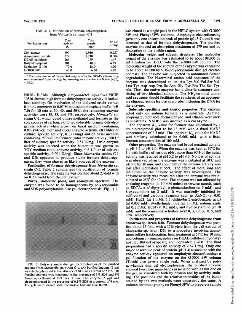
Purity, isoelectric point, and absorption spectrum. Theenzyme was found to be homogeneous by polyacrylamideand SDS-polyacrylamide disc gel electrophoresis (Fig. 1). It
in~~~~~~~~~~~~~~~~~~~~~~~~~~~~~~~~~~~~~~~~~~~~~~~~~~~~~~;
FIG. 1. Polyacrylamide disc gel electrophoresis of the purifiedenzyme from Moraxella sp. strain C-1. (A) Purified enzyme (6 ,g)was electrophoresed in the absence of SDS at a current of 2 mA. (B)Purified enzyme was incubated in the presence of 1% SDS and 3%2-mercaptoethanol at 95°C for 3 min. The enzyme (5 ,ug) was
electrophoresed in the presence of 0.1% SDS at a current of 8 mA.The gels were stained with Coomassie brilliant blue R-250.
was eluted as a single peak in the HPLC system with G-3000SW and Phenyl-5PW columns. Ampholyte electrofocusinggave only one absorption peak of protein (pI, 3.9), and it wasidentical to that of formate dehydrogenase. The purifiedenzyme showed an absorption maximum at 279 nm and noabsorption in the visible region.
Molecular weight and subunit structure. The molecularweight of the enzyme was estimated to be about 98,000 bygel filtration on HPLC with the G-3000 SW column. Themolecular weight of the subunit of the enzyme was estimatedto be about 48,000 by SDS-polyacrylamide disc gel electro-phoresis. The enzyme was subjected to automated Edmandegradation. The N-terminal amino acid sequence of theenzyme was determined to be Ala-Lys-Val-Val-Ser-Val-Leu-Tyr-Asp-Asp -Pro-Ile -Asn- Gly-Tyr-Pro-Thr-Ser-Tyr-Ala. Thus, the native enzyme has a dimeric structure con-sisting of two identical subunits. The NH2-terminal aminoacid sequence should facilitate the synthesis of an appropri-ate oligonucleotide for use as a probe in cloning the DNA forthe enzyme.
Substrate specificity and kinetic properties. The enzymeacted only on sodium formate as a substrate. Acetate,propionate, methanol, formaldehyde, and ethanol were inertas substrates. NADP+ was inactive as a coenzyme.The apparent Km value for formate was calculated by a
double-reciprocal plot to be 13 mM with a fixed NAD+concentration of 2.5 mM. The apparent Km value for NAD+was similarly calculated to be 0.068 mM, with a fixedformate concentration of 100 mM.Other properties. The enzyme had broad maximal activity
at pH 6.5 to pH 9.0. When the enzyme was kept at 30°C for1 h with buffers of various pHs, more than 90% of the initialactivity was retained at pH 5.2 to pH 9.0. No loss of activitywas observed when the enzyme was incubated at 50°C andpH 9.0 for 10 min, and about half of the enzyme activity waslost after incubation at 55°C. The effect of metal ions andinhibitors on the enzyme activity was investigated. Theenzyme activity was measured after the enzyme was prein-cubated at 25°C for 10 min. The enzyme was not affected bychelating reagents (at 10 mM unless otherwise noted), suchas EDTA, a,a'-dipyridyl, o-phenanthroline (at 5 mM), and8-oxyquinoline (at 2 mM). It was markedly inhibited bysulfhydryl and carbonyl reagents such as AgNO3 (at 0.01mM), HgCl2 (at 1 mM), 5,5'-dithio-bis(2-nitrobenzoic acid)(at 0.033 mM), N-ethylmaleimide (at 1 mM), sodium azide(at 0.1 mM), KCN (at 0.1 mM), and hydroxylamine (at 10mM), and the remaining activities were 0, 3, 19, 66, 0, 5, and76%, respectively.
Purification and properties of formate dehydrogenase fromMoraxella sp. strain D2b. Formate dehydrogenase was puri-fied about 15-fold, with a 17% yield from the cell extract ofMoraxella sp. strain D2b by a procedure involving ammo-nium sulfate fractionation, heat treatment at 55°C for 10 min,and column chromatographies on DEAE-cellulose, hydroxy-apatite, Butyl-Toyopearl, and Sephadex G-200. The finalpreparation had a specific activity of 2.67 U/mg. Only onemajor absorption peak of protein (pl, 3.4) associated with theenzyme activity appeared on ampholyte electrofocusing; agel filtration of the enzyme on the G-3000 SW column(Tosoh) also gave a single peak. When analyzed by poly-acrylamide disc gel electrophoresis, the purified enzymeshowed two close main bands associated with a faint one onthe gel, as visualized both by protein and by activity stain-ing. The positions and the relative intensities of the bandsstained by the two methods were apparently the same. Acolumn chromatography on Phenyl-5PW to prepare a sample
3191VOL. 170, 1988
Dow
nloa
ded
from
http
s://j
ourn
als.
asm
.org
/jour
nal/j
b on
26
Janu
ary
2022
by
186.
236.
10.1
78.

3192 ASANO ET AL.
TABLE 2. Comparison of properties of formate dehydrogenases from Moraxella sp. strain C-1, A. parvulus, and P. oxalaticus
Value with enzyme froma:Property
Moraxella sp. strain C-1 A. parvulus P. oxalaticus
Sp act of final prepn (U/mg) 5.97 (at 250C) 11.0 (at 37°C) 42 (at 25°C)Mol wt 98,000 80,000 + 8,000 315,000Mol wt of subunit(s) 48,000 46,000 ± 2,000 100,000 and 59,000No. of subunits 2 2 2 eachpI 3.9pH optimum 6.5 to 9.0 6.0 to 9.0 7.4Inhibition with SH and carbonyl reagents Yes Yes YesApparent Km (mM)Formate 13 15 0.135NAD+ 0.068 0.11 0.105
Prosthetic group None None FMNElectron acceptor(s) NAD+ NAD+ NAD,+ 02, artificial dyesFormation Inducible
a Values for Moraxella sp. strain C-1 are reported in this study. Values for A. parvulus (7, 8) and P. oxalaticus (21) are reported elsewhere.
for N-terminal amino acid sequencing showed that thepurified enzyme was approximately 84% pure. The N-terminal amino acid sequence of the main peak was exactlythe same as that of the enzyme from Moraxella sp. strain C-1from amino acid 1 to amino acid 20. Therefore, the segrega-tion of the enzyme from Moraxella sp. strain D2b in severalactive forms on the polyacrylamide disc gel was probablycaused by an artificial deterioration of the enzyme duringelectrophoresis. The behavior of the enzyme from Moraxellasp. strain D2b in response to heat, pH, and inhibitors wasquite similar to the behavior of the enzyme from Moraxellasp. strain C-1, except that the former enzyme was morestable to heat: no loss of activity was observed on heating at55°C and pH 9.0 for 10 min. The apparent Km values forformate and NAD+ were calculated to be 10.0 mM and 0.18mM, respectively.
DISCUSSION
We have described the bacterial distribution of NAD+-dependent formate dehydrogenase, as well as the purifica-tion and physicochemical properties of the enzymes fromMoraxella sp. strains C-1 and D2b, which were isolated fromsoil samples acclimated to an alkaline medium containingmethanol and formate as the sole carbon sources.Lower formate dehydrogenase activities are generally
detected in methylotrophic bacteria as compared with thosein yeasts (14, 15, 28). Johnson and Quayle (14) detectedformate dehydrogenase activities in the cell extracts ofMethylobacterium sp. strain NCIB 2879 (formerly Protami-nobacter ruber), P. methanica, and Protomonas extorquens(0.11, 0.080, 0.092 U/mg, respectively, at pH 8.4; recalcu-lated from results reported by Johnson and Quayle [14]). Inthis study, we could not find a producer of large amounts ofstable formate dehydrogenase among the deposited methylo-trophic bacteria. Almost no activity was detected in the cellextracts of P. methanica NRRL B-3449, and Protamino-bacter ruber NRRL B-1048. Although Ancylobacter aqua-ticus NCIB 10516 exhibited high total and specific activitiesof formate dehydrogenase in the cell extract, it was easilyinactivated by heat treatments. The enzymes from Morax-ella sp. strains C-1 and D2b were found to be fairly stable toheat, pH, and oxygen.The formate dehydrogenases from facultative or obligate
anaerobes are characterized by their complex subunit struc-ture, extreme sensitivity to oxygen, metal contents, utiliza-tion of various electron acceptors, etc. (9, 17, 18, 24, 30).
Moraxella formate dehydrogenase is markedly differentfrom the formate dehydrogenases of anaerobes and P. oxa-laticus (21), which are chromophoric iron-sulfur enzymesand are similar to those from the pea (23), methylotrophicyeasts (3, 11, 12, 15, 25), and A. parvulus (7, 8), whichcontain no prosthetic group. In Table 2, some enzymatic andphysicochemical properties of formate dehydrogenase fromMoraxella sp. strain C-1 are summarized and compared withthose from A. parvulus and P. oxalaticus. The molecularweight of strain C-1 formate dehydrogenase, as determinedby HPLC analysis, was found to be about 98,000. Theenzyme consists of two identical subunits with molecularweights of about 48,000. The activity of the formate dehy-drogenase was strongly inhibited by heavy metal ions suchas Ag+, Hg2+, 5,5'-dithio-bis(2-nitrobenzoic acid), N-ethyl-maleimide, sodium azide, KCN, and hydroxylamine, sug-gesting that essential sulfhydryl and carbonyl groups exist inthe active site. This observation and the high Km value forformate are the common characteristics of NAD+-depen-dent formate dehydrogenases of methylotrophic microorgan-isms (3, 7, 11, 12, 15, 25), excluding the formate dehydroge-nase of P. oxalaticus, which shows a low Km value (21). Theformate dehydrogenase of Moraxella sp. strain C-1 resem-bles that of A. parvulus with respect to molecular weight,subunit structure, optimum pH, and inhibitors. A singleband of the purified enzyme from Moraxella sp. strain C-1 onpolyacrylamide gel was observed; it has not been observedwith the A. parvulus enzyme (7, 8). The genus Achromo-bacter is not defined in Bergey's Manual of SystematicBacteriology, 1st ed. (5). According to the old criteria (6),A. parvulus is gram negative, and the cells are nonmotilesmall rods measuring 0.1 to 0.2 by 0.3 to 0.5 ,um. A. parvulus1 is a facultatively methylotrophic bacterium (19), althoughthe details of the cultural and biochemical characteristics arenot clear. Our isolates, strains C-1 and D2b, are alsofacultatively methylotrophic, nonmotile, gram-negative rodswhich occur mostly in pairs. This morphological character iscommon in the family Neisseriaceae. Even if the genusAchromobacter is valid now, our isolates should thus beclassified as members of the genus Moraxella.Because the formate dehydrogenases produced by Mor-
axella sp. strains C-1 and D2b are considerably stable andthe enzyme activity per culture is not less than the enzymeactivities of methylotrophic yeasts, they should be useful astools for the regeneration ofNADH in the enzyme-mediatedorganic synthesis of amino acids (1), the enzymatic determi-
J. BACTERIOL.
Dow
nloa
ded
from
http
s://j
ourn
als.
asm
.org
/jour
nal/j
b on
26
Janu
ary
2022
by
186.
236.
10.1
78.

FORMATE DEHYDROGENASE FROM A MORAXELLA SP.
nation of formate (27), and the production of NADH fromNAD+ (13).
ACKNOWLEDGMENTS
We thank K. Kondo, M. Ohmori, and N. Numao for continuousdiscussion and encouragement. We are also grateful to R. Matsu-moto for skillful assistance in the N-terminal amino acid sequencing.
LITERATURE CITED1. Asano, Y., K. Endo, A. Nakazawa, Y. Hibino, N. Okazaki, M.
Ohmori, N. Numao, and K. Kondo. 1987. Bacillus phenylalaninedehydrogenase produced in Escherichia coli: its purification andapplication to L-phenylalanine synthesis. Agric. Biol. Chem. 51:2621-2623.
2. Asane, Y., A. Nakazawa, and K. Endo. 1987. Novel phenylala-nine dehydrogenases from Sporosarcina ureae and Bacillussphaericus: purification and characterization. J. Biol. Chem.262:10346-10354.
3. Avilova, T. V., 0. A. Egorova, L. S. Ioanesyan, and A. M.Egorov. 1985. Biosynthesis, isolation and properties of NAD-dependent formate dehydrogenase from the yeast Candidamethylica. Eur. J. Biochem. 152:657-662.
4. Bernt, E., and H. U. Bergmeyer. 1974. Acetaldehyde: determi-nation with alcohol dehydrogenase from yeast, p. 1506-1509. InH. U. Bergmeyer (ed.), Methods of enzymatic analysis, 2nd ed.,vol. 3. Verlag Chemie, Weinheim, Federal Republic of Ger-many.
5. B0vre, K. 1984. Family VIII. Neisseriaceae Prevot 1933, 119AL,p. 288-309. In N. R. Krieg, and J. G. Holt (ed.), Bergey'smanual of systematic bacteriology, vol. 1. The Williams &Wilkins Co., Baltimore.
6. Breed, R. S. 1957. Genus II. Achromobacter Bergey et al., 1923,p. 300-309. In R. S. Breed, E. G. D. Murray, and N. R. Smith(ed.), Bergey's manual of determinative bacteriology, 7th ed.The Williams & Wilkins Co., Baltimore.
7. Egorov, A. M., T. V. Avilova, M. M. Dikov, V. 0. Popov, Y. V.Rodionov, and I. V. Berezin. 1979. NAD-dependent formatedehydrogenase from methylotrophic bacterium, strain 1: purifi-cation and characterization. Eur. J. Biochem. 99:569-576.
8. Egorov, A. M., V. I. Tishkov, T. V. Avilova, and V. 0. Popov.1982. S-formyl glutathione as a substrate of bacterial formatedehydrogenase. Biochem. Biophys. Res. Commun. 104:1-5.
9. Enoch, H. G., and R. L. Lester. 1982. Formate dehydrogenasefrom Escherichia coli. Methods Enzymol. 89:537-543.
10. Ferenci, T., T. Str0m, and J. R. Quayle. 1974. Purification andproperties of 3-hexulose phosphate synthase and phospho-3-hexuloisomerase from Methylococcus capsulatus. Biochem. J.144:477-486.
11. Fujii, T., and K. Tonomura. 1972. Oxidation of methanol,formaldehyde and formate by a Candida species. Agric. Biol.Chem. 36:2297-2306.
12. Hou, C. T., R. N. Patel, A. I. Laskin, and N. Barnabe. 1982.NAD-linked formate dehydrogenase from methanol-grown Pi-chia pastoris NRRL-Y-7556. Arch. Biochem. Biophys. 216:296-305.
13. Izumi, Y., S. K. Mishra, B. S. Ghosh, Y. Tani, and H. Yamada.1983. NADH production from NAD' using a formate dehydro-genase system with cells of a methanol-utilizing bacterium. J.
Ferment. Technol. 61:135-142.14. Johnson, P. A., and J. R. Quayle. 1964. Microbial growth on Cl
compounds: 6. Oxidation of methanol, formaldehyde and for-mate by methanol-growth Pseudomonas AM 1. Biochem. J. 93:281-290.
15. Kato, N., M. Kano, Y. Tani, and K. Ogata. 1974. Purificationand characterization of formate dehydrogenase in a methanol-utilizing yeast, Kloeckera sp. no. 2201. Agric. Biol. Chem. 38:111-116.
16. Kato, N., N. Miyawaki, and C. Sakazawa. 1982. Oxidation offormaldehyde by resistant yeasts Debaryomyces vanriji andTrichosporon penicillatum. Agric. Biol. Chem. 46:655-661.
17. Kroger, A., E. Winkler, A. Innerhofer, H. Hackenberg, and H.SchAgger. 1979. The formate dehydrogenase involved in elec-tron transport from formate to fumarate in Vibrio succinogenes.Eur. J. Biochem. 94:465-475.
18. Liu, C.-L., and L. E. Mortenson. 1984. Formate dehydrogenaseof Clostridium pasteurianum. J. Bacteriol. 159:375-380.
19. Loginova, N. V., and Y. A. Trotsenko. 1979. Autotrophic growthon methanol by bacteria isolated from activated sludge. FEMSMicrobiol. Lett. 5:239-243.
20. Lowry, 0. H., N. J. Rosebrough, A. L. Farr, and R. J. Randall.1951. Protein measurement with the Folin phenol reagent. J.Bi(l. Chem. 193:265-275.
21. Muller, U., P. Wilinow, U. Ruschig, and T. Hopner. 1978.Formate dehydrogenase from Pseudomonas oxalaticus. Eur. J.Biochem. 83:485-498.
22. Nash, T. 1953. The colorimetric estimation of formaldehyde bymeans of the Hantzsch reaction. Biochem. J. 55:416-421.
23. Ohyama, T., and I. Yamazaki. 1974. Purification and someproperties of formate dehydrogenase. J. Biochem. (Tokyo) 75:1257-1263.
24. Schauer, N. L., and J. G. Ferry. 1986. Composition of thecoenzyme F420-dependent formate dehydrogenase from Metha-nobacterium formicicum. J. Bacteriol. 165:405-411.
25. Schutte, H., J. Flossdorf, H. Sahm, and M.-R. Kula. 1976.Purification and properties of formaldehyde dehydrogenase andformate dehydrogenase from Candida boidinii. Eur. J. Bio-chem. 62:151-160.
26. Strafford, H. A., A. Magaldi, and B. Vennesland. 1954. Theenzymatic reduction of hydroxypyruvic acid to D-glyceric acidin higher plants. J. Biol. Chem. 207:621-629.
27. Triebig, G., and K.-H. Schaller. 1980. A simple and reliableenzymatic assay for the determination of formic acid in urine.Clin. Chim. Acta 108:355-360.
28. Urakami, T., and K. Komagata. 19$1. Electrophoretic compar-ison of enzymes in the gram negative methanol-utilizing bacte-ria. J. Gen. Appl. Microbiol. 27:381-403.
29. Urakami, T., J. Tamaoka, and K. Komagata. 1985. DNA basecomposition and DNA-DNA homologies of methapol-utilizingbacteria. J. Gen. Appl. Microbiol. 31:243-253.
30. Yagi, T. 1979. Purification and properties of cytochrome c-553,an electron acceptor for formate dehydrogenase of Desulfovib-rio vulgaris, Miyazaki. Biochim. Biophys. Acta 548:96-105.
31. Yamamoto, I., T. Saiki, T., S.-M. Liu, an/d L. G. Ljungdahl.1983. Purification and properties of NADP-dependent formatedehydrogenase from Clostridium thermoaceticum, a tungsten-selenium-iron protein. J. Biol. Chem. 259:1826-1832.
VOL. 170, 1988 3193
Dow
nloa
ded
from
http
s://j
ourn
als.
asm
.org
/jour
nal/j
b on
26
Janu
ary
2022
by
186.
236.
10.1
78.