Monte Carlo verification of IMRT dose distributions …lcr.uerj.br/Manual_ABFM/Monte Carlo...
14
Monte Carlo verification of IMRT dose distributions from a commercial treatment planning optimization system This article has been downloaded from IOPscience. Please scroll down to see the full text article. 2000 Phys. Med. Biol. 45 2483 (http://iopscience.iop.org/0031-9155/45/9/303) Download details: IP Address: 152.92.171.92 The article was downloaded on 15/10/2010 at 15:11 Please note that terms and conditions apply. View the table of contents for this issue, or go to the journal homepage for more Home Search Collections Journals About Contact us My IOPscience
-
Upload
truonghanh -
Category
Documents
-
view
221 -
download
0
Transcript of Monte Carlo verification of IMRT dose distributions …lcr.uerj.br/Manual_ABFM/Monte Carlo...

Monte Carlo verification of IMRT dose distributions from a commercial treatment planning
optimization system
This article has been downloaded from IOPscience. Please scroll down to see the full text article.
2000 Phys. Med. Biol. 45 2483
(http://iopscience.iop.org/0031-9155/45/9/303)
Download details:
IP Address: 152.92.171.92
The article was downloaded on 15/10/2010 at 15:11
Please note that terms and conditions apply.
View the table of contents for this issue, or go to the journal homepage for more
Home Search Collections Journals About Contact us My IOPscience
Phys. Med. Biol. 45 (2000) 2483–2495. Printed in the UK PII: S0031-9155(00)12146-3
Monte Carlo verification of IMRT dose distributions from acommercial treatment planning optimization system
C-M Ma, T Pawlicki, S B Jiang, J S Li, J Deng, E Mok, A Kapur, L Xing,L Ma and A L BoyerRadiation Oncology Department, Stanford University School of Medicine, Stanford, CA 94305,USA
E-mail: [email protected]
Received 25 February 2000, in final form 8 May 2000
Abstract. The purpose of this work was to use Monte Carlo simulations to verify the accuracy ofthe dose distributions from a commercial treatment planning optimization system (Corvus, NomosCorp., Sewickley, PA) for intensity-modulated radiotherapy (IMRT). A Monte Carlo treatmentplanning system has been implemented clinically to improve and verify the accuracy of radiotherapydose calculations. Further modifications to the system were made to compute the dose in a patient formultiple fixed-gantry IMRT fields. The dose distributions in the experimental phantoms and in thepatients were calculated and used to verify the optimized treatment plans generated by the Corvussystem. The Monte Carlo calculated IMRT dose distributions agreed with the measurements towithin 2% of the maximum dose for all the beam energies and field sizes for both the homogeneousand heterogeneous phantoms. The dose distributions predicted by the Corvus system, whichemploys a finite-size pencil beam (FSPB) algorithm, agreed with the Monte Carlo simulationsand measurements to within 4% in a cylindrical water phantom with various hypothetical targetshapes. Discrepancies of more than 5% (relative to the prescribed target dose) in the target regionand over 20% in the critical structures were found in some IMRT patient calculations. The FSPBalgorithm as implemented in the Corvus system is adequate for homogeneous phantoms (such asprostate) but may result in significant under- or over-estimation of the dose in some cases involvingheterogeneities such as the air–tissue, lung–tissue and tissue–bone interfaces.
(Some figures in this article are in colour only in the electronic version; see www.iop.org)
1. Introduction
For the last few years, extensive research has been carried out to develop conformal radiotherapyusing computer-controlled linear accelerators equipped with multileaf collimators (MLC)(Boesecke et al 1988, Leibel et al 1992, LoSasso et al 1993, Powlis et al 1993, Mageraset al 1994, Brewster et al 1995, Fraass et al 1995, McShan et al 1995, Yu et al 1995). Morerecently, intensity modulated radiotherapy (IMRT) has been developed (Brahme 1988, Converyand Rosenbloom 1992, Webb 1992, Boyer et al 1997) and implemented (Ling et al 1996,Boyer et al 1998) that uses computer-controlled modulation of x-ray fields by the MLC. It isanticipated that conformal radiotherapy will provide radiation oncologists with a significantlyimproved tool to deliver high doses of ionizing radiation to some tumour sites while reducingdoses to adjacent normal tissue below levels to which they are unavoidably exposed by currentlyavailable techniques. Thus, acute and chronic toxicity associated with treatment of a tumourvolume by radiation may be significantly reduced or delayed for certain sites of malignantpresentations.
0031-9155/00/092483+13$30.00 © 2000 IOP Publishing Ltd 2483
2484 C-M Ma et al
The use of conformal radiotherapy, especially with the IMRT technique, is a majordeparture from the way radiotherapy is currently delivered. Although the use of MLCsprovides the possibility of achieving better dose distributions conformed to tumour targets,it also increases the complexity of treatment. The sequences of leaf movement and theirassociated effects on the dose delivered to the patient may vary significantly depending on theaccelerator and the MLC design. Important factors include the variation of the acceleratorhead scatter component in the MLC-collimated beam (Convery and Webb 1997), the amountof photon leakage through the leaves (Wang et al 1996, Webb 1997, Holmes et al 1997), thescatter from the leaf ends, the ‘tongue and groove’ effect (Chui et al 1994, Wang et al 1996),the effect of back-scattered photons from the moving jaws and MLC leaves on the monitorchamber signal (Hounsell 1998). Traditionally, patient dose calculations in radiotherapy havebeen based on correcting measured dose distributions. New dose calculation algorithms havebeen developed to predict the patient dose from ‘first principles’ using a model of radiationtransport (Mackie et al 1995). Comparisons of the traditional photon algorithms and the newerones have been reviewed by Wong and Purdy (1990), Cunningham and Battista (1995) andMackie et al (1996). Due to the lack of electron transport, the conventional dose calculationalgorithms often failed to predict the dose distribution accurately near inhomogeneities (Mackieet al 1996, Mohan 1997, DeMarco et al 1998, Wang et al 1998, Ma et al 1999). Furthermore,the inverse-planning algorithms for beam optimization have all used approximations to speedup the dose computation that may introduce significant uncertainty in the calculated dosedistributions, especially in the presence of heterogeneities. When simple source models areused in the dose computation, the correlation between the calibrated reference dose and thedose related to a beam segment may be lost. All the above imply a potential problem with theprediction of the dose distributions in a patient for an IMRT treatment.
Oldham and Webb (1997) reported differences in excess of 10% in the absolute dosebetween the optimization dose calculations and measurements (using film) of fields deliveredby a dynamic MLC. The differences were attributed partially to the nonlinearity of dose permonitor unit (MU) for small MU deliveries (the actual dose delivered per MU increased bymore than 10% from MU = 20 to MU = 1). Wang et al (1996) reported that for the MemorialHospital’s dynamic MLC delivery process, the discrepancies between the calculated dose andthe measured dose were in excess of 5% if various effects related to the MLC construction,such as accelerator head scatter, were not properly accounted for. The uncertainty in thedoses calculated by a conventional dose calculation algorithm was 5–10% in the presence ofheterogeneities (Mohan 1997). Our recent Monte Carlo results were consistent with thesefindings (Ma et al 1999).
The purpose of this work was to verify the accuracy of the IMRT dose distributions froma commercial treatment planning optimization system (Corvus, Nomos Corp., Sewickley, PA)using Monte Carlo simulations. We have used the EGS4/BEAM Monte Carlo code system(Nelson et al 1985, Rogers et al 1995a, b) to simulate the clinical photon beams from twolinear accelerators, Varian Clinac 2100C and 2300C/D (Varian Oncology, Palo Alto, CA). TheEGS4/DOSXYZ code (Rogers et al 1995a, Ma et al 1995) was modified to compute the dosein a patient (a three-dimensional phantom built from the CT data) for multiple fixed-gantryfields. Phantom measurements were performed to commission the Monte Carlo system. Thedose distributions in the experimental phantoms and in the patients were calculated and usedto verify the optimized treatment plans generated by the Corvus inverse-planning system. Inthe following sections, we will describe the dose calculations in the inverse-planning systemand the Monte Carlo simulations. We will show the dose distributions for homogeneous andheterogeneous phantoms and discuss the effect of material density and atomic number on thefinal dose calculations.
MC verification of IMRT dose distributions 2485
2. Materials and method
2.1. Treatment planning and dose measurement
The treatment planning optimization system used in this work is the Corvus inverse-planningsystem. The inverse planning process employs a simulated annealing optimization algorithm(Webb 1992). The dose calculation algorithm used for the inverse planning process is a finite-size pencil beam (FSPB) algorithm, which uses predetermined beamlet dose distributions. Thebeamlet distributions were derived from measured dose distributions and normalized to produceconsistent output factors for various field sizes. The patient inhomogeneity correction is madeby ‘stretching’ the beamlet distribution proportionally based on the equivalent pathlength.Some details of the dose calculations have been described by Holmes et al (1998) and Boyeret al (1999). The monitor unit calculations for a ‘step-and-shoot’ leaf sequence algorithmhave been discussed by Boyer et al (1999). The effect of the leaf leakage is accounted forin Corvus by reducing the original beamlet weight by the same amount as the accumulatedleakage for a given leaf sequence. This method works well for those beamlets whose weightsare greater than the estimated leakage. If the beamlet weights are smaller than the leakagethis method will underestimate the dose as any remaining leakage effect cannot be accountedfor and the initial dose calculation does not include any leaf leakage effect (which is unknownbefore a leaf sequence is generated for the field). The Corvus system has been commissionedfor clinical IMRT treatment planning (Xing et al 1999) and used for treating head and neckpatients with IMRT (Boyer et al 1998).
In order to test the Corvus system, inverse plans were computed for various target shapesplaced in the centre of a cylindrical water phantom having a diameter of 30 cm. Comparisonsof the Corvus dose distributions with measurements have been reported in detail by Boyer et al(1999). In this work, we have computed the dose distributions for these plans using MonteCarlo methods (see descriptions below). For completeness, we briefly describe the plans andthe measurements below. Inverse plans were computed for different hypothetical targets withdifferent numbers of beams directed toward the axis of the cylinder at the centre of the target andspaced at equal angles. The treatments were delivered using a dynamic multileaf collimator(Varian Oncology Systems, Palo Alto, CA). The leaf sequences computed within the Nomossoftware were written into files with formats conformed to the requirements of the Variandigital control software. The leaf sequences were delivered using the monitor units calculatedby the Corvus system. The absolute dose delivered by the leaf sequences was measured in the30 cm diameter cylindrical water phantom using a 0.147 cm3 ionization chamber (WellhoferDosimetrie, Schwartzenbruck, Germany) following the AAPM TG-21 protocol (AAPM 1983).No corrections were made for the variation in the chamber displacement effect, which dependson the dose gradient at the measurement point and the chamber diameter. This may introducean up to 2% uncertainty in the measured dose for the 6 mm diameter chamber used (the dosegradient was 5–8% per centimetre at some measurement points). The chamber positioninguncertainty was about 0.1 cm. The overall uncertainty in the measured dose was estimated tobe about 3% (1σ ).
2.2. The Monte Carlo simulation
We have used BEAM and DOSXYZ (Rogers et al 1995a, b, Ma et al 1995) Monte Carlo codesfor the accelerator head simulation and dose calculation in the patient respectively. Both codeswere EGS4 (Electron Gamma Shower version 4, Nelson et al (1985)) user codes, running underthe UNIX operating system, developed through the Omega project for Monte Carlo treatmentplanning dose calculations (Mackie et al 1994). Detailed descriptions of the software can be

2486 C-M Ma et al
found from Rogers et al (1995a, b). A detailed description of the clinical implementation ofthe Monte Carlo code system was given in a previous publication (Ma et al 1999).
Two types of clinical linear accelerators were simulated for the clinical implementationof Monte Carlo dose verification in this work: Varian Clinac 2100C and 2300C/D (VarianOncology Systems, Palo Alto, CA). The dimensions and materials for the acceleratorcomponents were incorporated according to the manufacturer’s specifications. Electronbeams emerging from the vacuum exit window were assumed to be monoenergetic andmonodirectional with a beam radius of 0.1–0.2 cm. These were found to be reasonableassumptions to achieve an acceptable dose calculation accuracy of about 2% of the dosemaximum (Dmax) anywhere in the phantom for clinical radiotherapy applications (Kapur et al1998, Ma 1998, Ma et al 1999). We obtained accurate phase-space data for photon beamswith nominal energies of 4, 6 and 15 MV. The energy cut-offs for electron transport in theaccelerator simulation (ECUT and AE) 700 keV (kinetic + rest mass) and for photon transport(PCUT and AP) 10 keV. The maximum fractional energy loss per electron step (ESTEPE) was0.04. The bremsstrahlung splitting and Russian roulette options were implemented for photonbeam simulations (Rogers et al 1995b). The ICRU recommended compositions and stoppingpower values were used for the materials used in the accelerator simulations (ICRU 1984).The phase-space data were scored at a plane immediately above the photon jaws. The numberof particles in a photon beam file was about 50 million. Field shaping by photon jaws, blocksand the MLC was further simulated using BEAM and the phase-space data could be storedtemporarily or used directly for dose calculations.
The DOSXYZ code was designed for dose calculations in a 3D rectilinear voxel (volumeelement) geometry (Ma et al 1995). Voxel dimensions were completely variable in all threedirections. Every voxel could be assigned to a different material. The cross-section datafor the materials used were available in a pre-processed PEGS4 cross-section data file. Themass density of the material in a DOSXYZ calculation was varied based on the patient’sCT data, although the density effect corrections for the stopping powers of the materialremain unchanged (Ma et al 1999). The voxel dimensions and materials were defined ina DOSXYZ input file together with the transport parameters such as ECUT, PCUT, ESTEPEand the parameters required by PRESTA (Bielajew and Rogers 1988). The phase-spacedata obtained from a BEAM simulation was used as a source input with variable sourcepositions and beam incidence angles. Dose contributions from different beam componentswere selectively calculated based on the particle charge or the LATCH settings specified inthe BEAM simulation. DOSXYZ produced a data file that contained geometry specificationssuch as the number of voxels in all three directions and their boundaries as well as the dosevalues and the associated (1σ ) statistical uncertainties in the individual voxels.
The DOSXYZ code has been modified to read the MLC leaf sequence files for IMRTtreatment. For this study, the Varian dynamic MLC leaf sequence files (G-version) generatedusing a step-and-shoot leaf sequencing algorithm, were used. The monitor units for eachleaf sequence were integrated into a two-dimensional map (grids). The dimensions of themap at the isocentre (100 cm from the virtual point source position) are 40 cm in both the x
and y directions and the grid sizes were 0.1 cm. The monitor units for the open areas wereaccumulated for each of the pixels, while for the closed areas a fraction of the monitor unitswere accumulated based on the measured MLC leakage factor for each beam energy. Thus,the integrated intensity map has included the averaged leaf leakage effect but ignored theinfluence of the leaf shape (tongue and groove) and the variation of the spatial and spectraldistributions due to photon attenuation and scattering in the MLC leaves. This is considered tobe a reasonable approach since, unless the effect of organ motion and patient set-up uncertaintyhas been accounted for properly, the averaged leaf leakage is a more realistic quantity than
MC verification of IMRT dose distributions 2487
the explicit leakage through the leaf and the tongue and groove for estimating the overall dosein a patient during the whole treatment course. The transmission factor for the areas underthe photon jaws was assumed to be zero. The dimensions of the photon jaw opening were setaccording to the actual treatment set-ups. Further modifications were made to simulate severalgantry angles in the same run. The leaf sequence files were read sequentially and the gantryangle was changed automatically after the simulation of a photon field was completed.
During the Monte Carlo simulation, the weight of a phase-space particle was altered basedon the value of the pixel in the intensity map through which the particle was travelling. Toimprove the simulation efficiency, two variance reduction techniques were implemented inthe DOSXYZ code, particle splitting and Russian roulette (Rogers and Bielajew 1990). Theformer was applied to the particles with weight greater than unity and the latter was appliedto the particles with weight smaller than unity. The splitting and Russian roulette processeswere implemented in such a way that the resulting particles would have identical weightingfactors. Using uniform weighting factors will generally improve the statistical uncertainty ofa simulation.
For patient dose calculations, the simulation phantom was built from the patient’s CT datawith up to 128 × 128 × 128 voxels (uniform in any dimensions). The side of a voxel variedfrom 0.2 to 0.4 cm. The EGS4 transport parameters were set to ECUT = AE = 700 keV,PCUT = AP = 10 keV and ESTEPE = 0.04. The number of particle histories simulatedranged from 300 million to 1 billion for up to nine gantry angles for an IMRT treatment. The1σ statistical uncertainty in the dose was generally 1–2% of the Dmax value. The CPU timerequired for an IMRT simulation was 3–15 h on a single Pentium III 450 MHz PC dependingon the beam energy and field sizes.
3. Results
3.1. Comparisons in homogeneous water phantoms
Both the inverse-planning system and the Monte Carlo dose calculation system have beencommissioned for routine clinical applications (Xing et al 1999, Ma 1998, Ma et al 1999).Both systems regenerated the PDD curves and the dose profiles at various depths to within2% of the dose maximum values of the measured beam data for various field sizes and sourceto surface distances (SSD). The dose distributions given by the Corvus system agreed withmeasurements to about 3% in a cylindrical water phantom with various hypothetical targetshapes (Boyer et al 1999). Figure 1 shows the dose distributions in a 30 cm diameter watercylinder irradiated by seven 6 MV intensity-modulated photon beams calculated by Corvusand by the Monte Carlo simulations. The critical structure is in the centre of the water cylinderimmediately next to the C-shaped target. A beamlet size of 1 cm × 1 cm was used in theoptimization calculation. The leaf sequencing algorithm used 20 intensity levels. The isodoselines calculated by both systems agreed within about 4% or a shift in isodose lines within about0.3 cm. These small discrepancies were thought to be partially due to the approximations usedby Corvus in accounting for the effect of MLC leaf leakage. Considering that the average leafleakage is about 1.5% for a 6 MV beam and the total MU required to deliver an IMRT field isabout three times more than that required by a conventional field, the estimated accumulatedleaf leakage beneath a completely closed leaf pair is about 4.5%. Thus, a few per cent of theobserved dose difference may be attributed to the difference in leaf leakage implementationbetween Corvus and Monte Carlo.

2488 C-M Ma et al
(a) (b)
Figure 1. Comparison of 6 MV photon beam (seven co-planar fields at 26, 77, 180, 231, 283 and334◦ gantry angles) dose distributions in a water cylinder calculated by Monte Carlo (a) and Corvus(b). The differences between the Corvus calculations and measurements using an ion chamber areshown for certain locations (arrows).
3.2. Comparisons in CT phantoms
In this work, we have compared the dose distributions calculated by Monte Carlo and theCorvus FSPB algorithm for various treatment sites to identify potential treatment situationsthat may benefit from dose accuracy improvements. In the following examples, we show twotypical IMRT treatment plans computed by the Corvus inverse planning system and verifiedby the Monte Carlo system.
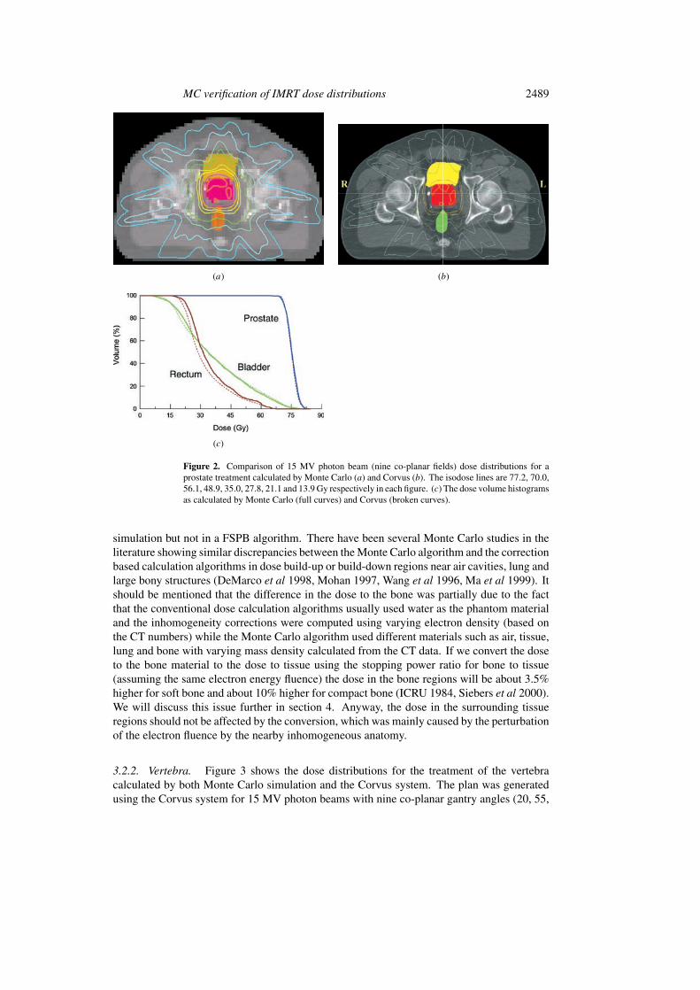
3.2.1. Prostate. To explore the effect of photon and electron transport on IMRT dosecalculations, we show in figure 2 an IMRT prostate treatment plan calculated by Monte Carlosimulation and the Corvus system. The plan was generated using the Corvus system for 15 MVphoton beams with nine gantry angles (20, 60, 100, 140, 220, 260, 300 and 340◦). The beamintensity was modulated using a Varian dynamic MLC with 80 leaves. In both calculations,the isodose lines represent the absolute dose values in the patient. It can be seen that the dosevalues in the target (the prostate) agreed very well between the Corvus calculations and theMonte Carlo simulations. Similar results were found for six other prostate cases compared(not shown). The difference in the average dose to the target volume was 1.6% betweenMonte Carlo and Corvus and the maximum dose difference in the target dose was 3.4%.This confirms that both calculation algorithms can predict dose distributions in homogeneousphantoms accurately. The dose values in the nearby critical structures also agreed to 3–7%of the prescribed target dose. For the case shown in figure 2(c), the maximum difference wasabout 3 Gy in the rectum. However, other cases also showed the maximum differences in thebladder. These differences may be considered to be clinically acceptable.
The dose values in the regions near the bony structures sometimes showed a difference ofa few per cent between the Corvus and the Monte Carlo calculations. These discrepancies maybe partially explained by the effect of electron backscatter from the bone (high atomic numberand high density) to the soft tissue, which was accurately accounted for by the Monte Carlo
MC verification of IMRT dose distributions 2489
(a) (b)
(c)
Figure 2. Comparison of 15 MV photon beam (nine co-planar fields) dose distributions for aprostate treatment calculated by Monte Carlo (a) and Corvus (b). The isodose lines are 77.2, 70.0,56.1, 48.9, 35.0, 27.8, 21.1 and 13.9 Gy respectively in each figure. (c) The dose volume histogramsas calculated by Monte Carlo (full curves) and Corvus (broken curves).
simulation but not in a FSPB algorithm. There have been several Monte Carlo studies in theliterature showing similar discrepancies between the Monte Carlo algorithm and the correctionbased calculation algorithms in dose build-up or build-down regions near air cavities, lung andlarge bony structures (DeMarco et al 1998, Mohan 1997, Wang et al 1996, Ma et al 1999). Itshould be mentioned that the difference in the dose to the bone was partially due to the factthat the conventional dose calculation algorithms usually used water as the phantom materialand the inhomogeneity corrections were computed using varying electron density (based onthe CT numbers) while the Monte Carlo algorithm used different materials such as air, tissue,lung and bone with varying mass density calculated from the CT data. If we convert the doseto the bone material to the dose to tissue using the stopping power ratio for bone to tissue(assuming the same electron energy fluence) the dose in the bone regions will be about 3.5%higher for soft bone and about 10% higher for compact bone (ICRU 1984, Siebers et al 2000).We will discuss this issue further in section 4. Anyway, the dose in the surrounding tissueregions should not be affected by the conversion, which was mainly caused by the perturbationof the electron fluence by the nearby inhomogeneous anatomy.
3.2.2. Vertebra. Figure 3 shows the dose distributions for the treatment of the vertebracalculated by both Monte Carlo simulation and the Corvus system. The plan was generatedusing the Corvus system for 15 MV photon beams with nine co-planar gantry angles (20, 55,

2490 C-M Ma et al
(a) (b)
(c)
Figure 3. Dose distributions for the treatment of the vertebra calculated by Monte Carlo (a) andby Corvus (b) for 15 MV photons (nine co-planar fields). The isodose lines are 17.6, 15.6, 13.7,11.7, 9.8, 7.8, 5.9, 3.9 and 2.0 Gy respectively in each figure. (c) The dose volume histograms ascalculated by Monte Carlo (full curves) and Corvus (broken curves) for the target and the spinalcord.
90, 140, 180, 220, 260, 300 and 340◦). The intensity was modulated using a Varian dynamicMLC with 80 leaves. The prescribed target dose was 18 Gy. The maximum dose in the targetshowed good agreement between Corvus and Monte Carlo (figure 3(c)). The Monte Carlodose distribution showed slightly better target coverage than the Corvus dose distribution (a2 Gy difference in the minimum target dose). Because the cord was sometimes immediatelynext to the target region the maximum cord dose was expected to be equal to or higher than theminimum target dose. This was confirmed by the Monte Carlo simulations (see figure 3(c)).In the regions near large bony structures (such as the cord) differences of more than 20% of theprescribed target dose could be seen between the Corvus calculation (10 Gy) and the MonteCarlo simulation (14 Gy). The difference in the dose to the cord was thought to be due inpart to electron scattering from the surrounding bone, which could not be modelled properlyusing the FSPB algorithm. Another possible reason might be due to the implementation of theheterogeneity and leaf leakage corrections in the FSPB model. Although the photon beamswere optimized to avoid the cord, electrons could reach the cord and the dose to the cord couldbe enhanced due to the high-density material surrounding it and/or photon leaf leakage that isnot included in the dose calculation during the inverse planning process. Further studies are

MC verification of IMRT dose distributions 2491
(a) (b)
(c)
Figure 4. Dose distributions for different tissue types and material densities: (a) tissue and bonewith variable density (thick line) and tissue with unity density (thin line); (b) tissue and bone withvariable density (thick line) and tissue with variable density (thin line); and (c) tissue and bone withvariable density (thick line) and tissue with unity density and bone with 10 g cm−3 density (thinline). The phantom geometry and beam arrangements are the same as in figure 3. The isodoselines are given as 10, 30, 50, 70 and 80% of the prescribed target dose.
needed to understand these differences if more access to the FSPB and leaf sequence algorithmsin the Corvus system is available.
4. Discussion
Several important factors may affect the Monte Carlo calculated dose distributions and theDVH curves. First, the isodose lines and the DVH curves are affected by the materials used inthe patient CT phantom, i.e. whether we plot dose to tissue only or dose to any material (suchas air, tissue or bone). It seems reasonable that our previous experience was based on dose totissue (or dose to water, the difference between the two is within 1%) and therefore the dosevalues should be expressed as dose to tissue. However, it can also be argued that the real doseto the biological material such as bone should be given whenever possible. Only in this waycan the relationship between the ‘old’ practice and new experience be established.
To understand the effect of the conversion of the dose to different materials, we showin figure 4 the dose distributions calculated using Monte Carlo with different materials and
2492 C-M Ma et al
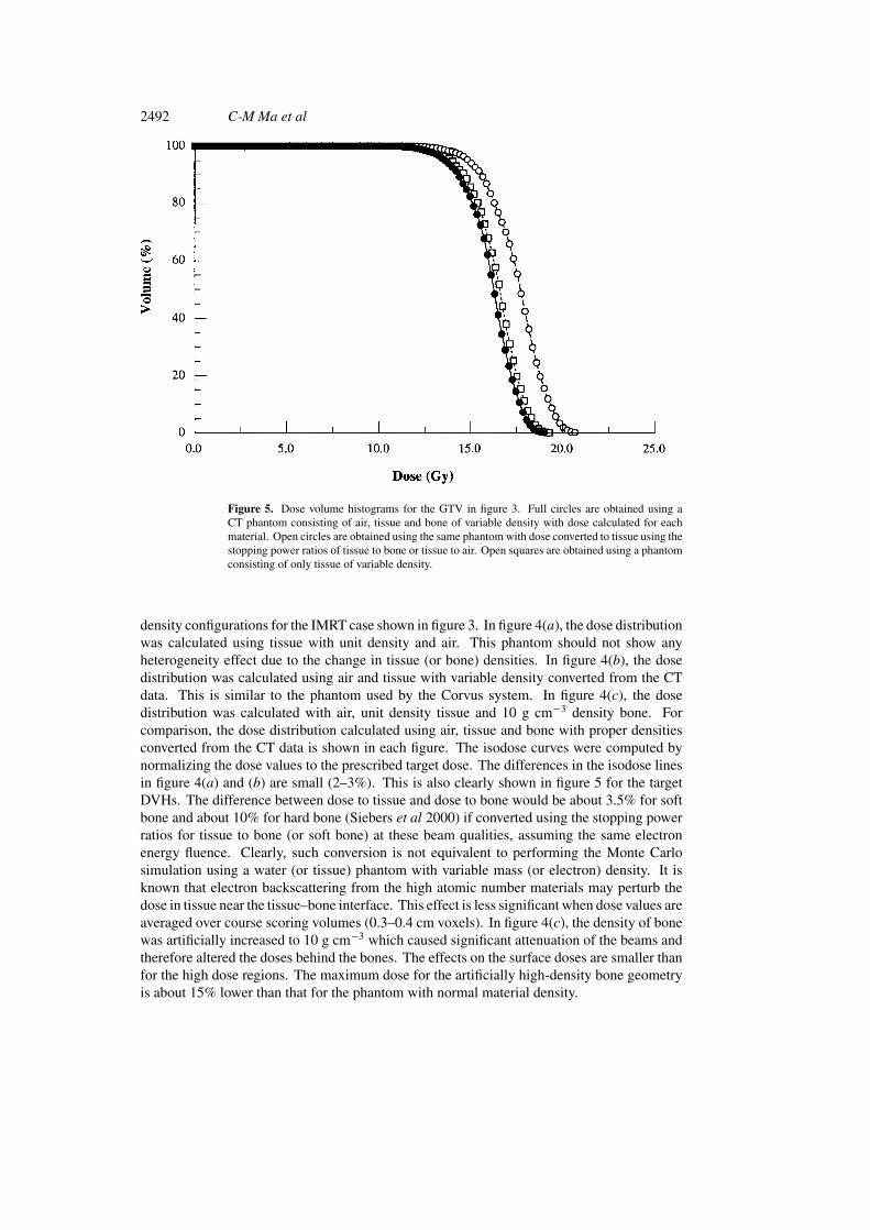
Figure 5. Dose volume histograms for the GTV in figure 3. Full circles are obtained using aCT phantom consisting of air, tissue and bone of variable density with dose calculated for eachmaterial. Open circles are obtained using the same phantom with dose converted to tissue using thestopping power ratios of tissue to bone or tissue to air. Open squares are obtained using a phantomconsisting of only tissue of variable density.
density configurations for the IMRT case shown in figure 3. In figure 4(a), the dose distributionwas calculated using tissue with unit density and air. This phantom should not show anyheterogeneity effect due to the change in tissue (or bone) densities. In figure 4(b), the dosedistribution was calculated using air and tissue with variable density converted from the CTdata. This is similar to the phantom used by the Corvus system. In figure 4(c), the dosedistribution was calculated with air, unit density tissue and 10 g cm−3 density bone. Forcomparison, the dose distribution calculated using air, tissue and bone with proper densitiesconverted from the CT data is shown in each figure. The isodose curves were computed bynormalizing the dose values to the prescribed target dose. The differences in the isodose linesin figure 4(a) and (b) are small (2–3%). This is also clearly shown in figure 5 for the targetDVHs. The difference between dose to tissue and dose to bone would be about 3.5% for softbone and about 10% for hard bone (Siebers et al 2000) if converted using the stopping powerratios for tissue to bone (or soft bone) at these beam qualities, assuming the same electronenergy fluence. Clearly, such conversion is not equivalent to performing the Monte Carlosimulation using a water (or tissue) phantom with variable mass (or electron) density. It isknown that electron backscattering from the high atomic number materials may perturb thedose in tissue near the tissue–bone interface. This effect is less significant when dose values areaveraged over course scoring volumes (0.3–0.4 cm voxels). In figure 4(c), the density of bonewas artificially increased to 10 g cm−3 which caused significant attenuation of the beams andtherefore altered the doses behind the bones. The effects on the surface doses are smaller thanfor the high dose regions. The maximum dose for the artificially high-density bone geometryis about 15% lower than that for the phantom with normal material density.

MC verification of IMRT dose distributions 2493
Although Monte Carlo dose calculations are time-consuming it is possible to use MonteCarlo calculated dose distributions to verify the IMRT treatment plans in order to detect anycases where the FSPB dose calculation algorithm as implemented in Corvus may fail to predictthe dose perturbation effect near inhomogeneities. Monte Carlo simulation may also be useddirectly for IMRT beamlet distribution calculation as a practical solution to this problem. Ourexperiences show that a factor of two to three more Monte Carlo particle histories are neededfor an IMRT treatment simulation compared with a conventional photon treatment simulationto achieve the same statistical uncertainty. This is because more monitor units are needed todeliver intensity modulated photon fields; more particles will be simulated in a Monte Carlocalculation but many of them will be stopped by the MLC leaves. Therefore, the CPU timeper photon history for an IMRT simulation is less than that for a conventional field. Using theexisting computing power (8 CPUs) of the Corvus system, the calculation time for a typical‘inverse plan’ would be increased from the current 0.5–1 h to 2–4 h with two Monte Carlocalculations. The pre-optimization dose calculation will provide the beamlet distributions forthe optimization process, which take into account the effect of the accelerator head scatter andinhomogeneous anatomy of the patient. The post-optimization dose calculation will include theeffects due to leaf leakage, leaf scatter and photon backscatter into the monitor chamber afterthe sequence of MLC leaf movement and jaw positions has been generated. Further studies areunder way on a Monte Carlo dose calculation based inverse planning system (Pawlicki et al1999, Ma et al 2000).
5. Summary
We have implemented a Monte Carlo system for routine radiotherapy treatment planningdose calculations. In our previous publications we have shown that Monte Carlo simulationsagreed with measurements to within 2% for various clinical beam set-ups in homogeneousand heterogeneous phantoms. Based on these results, we have moved one step nearer to usingthe Monte Carlo simulations to verify the IMRT dose distributions computed by the Corvussystem, which employs a FSPB algorithm for beamlet dose calculations, assuming that theMonte Carlo simulations are accurate at the 2% level for patient phantoms built from CT data.
Our results showed that the FSPB algorithm was adequate for most of the IMRT caseswhere the target was not immediately adjacent to the critical structures. However, the FSPBalgorithm may not accurately predict the dose distributions in and near inhomogeneities insome cases. The dose in the target volume calculated by the Corvus system differed from theMonte Carlo results by more than 5%, while the dose to the critical organ differed by morethan 20% of the prescribed target dose for a few cases. This suggests that, for such cases,more accurate dose calculation algorithms than that currently implemented in Corvus shouldbe used for intensity-modulated radiotherapy treatment planning.
Acknowledgments
We would like to acknowledge Varian Oncology Systems, Palo Alto, CA, for providing detailedinformation on the Varian Clinac linear accelerators and Nomos Corp., Sewickley, PA, for theinverse-planning system. We would like to thank our colleagues, Sam Brain, Todd Koumrian,Behrooz Tofighrad and Michael Luxton, for help with the computers and software support.We are grateful to Dr Iwan Kawrakow for the DOSXYZ SHOW program to plot isodosedistributions, and to Jinsheng Li and Michael Luxton for modifications to the program todisplay structures of interest and to compare two isodose distributions on the same plot.

2494 C-M Ma et al
This investigation was supported in part by grants CA78331 from the NIH, BC971292 fromthe DOD, Seed Cycle 1 from the RSNA Research and Education Fund, and a consortiumagreement with the NumeriX, LLC.
References
AAPM 1983 AAPM TG-21, A protocol for the determination of absorbed dose from high-energy photons and electronsMed. Phys. 10 741
Bielajew A F and Rogers D W O 1988 Variance-reduction techniques Monte Carlo Transport of Electrons and Photonsed T M Jenkins, W R Nelson, A Rindi, A E Nahum and D W O Rogers (New York: Plenum) pp 407–19
Boesecke R, Doll J, Bauer B, Schlegel W, Pastyr O and Lorenz M 1988 Treatment planning for conformation therapyusing a multileaf collimator Strahlenther. Onkol. 164 151–4
Boyer A L, Geis P B, Grant W, Kendall R and Carol M 1997 Modulated-beam conformal therapy for head and necktumors Int. J. Radiat. Oncol. Biol. Phys. 39 227–36
Boyer A L, Xing L, Ma C-M, Curran B, Hill R, Holmes T and Bleier A 1999 Theoretical consideration of monitorunit calculations for intensity modulated beam treatment planning (abstract) Med. Phys. 26 187–95
Boyer A L, Xing L, Ma L and Forster K 1998 Verification and delivery of head and neck intensity modulatedradiotherapy Med. Phys. 25 A200–1
Brahme A 1988 Optimal setting of multileaf collimators in stationary beam radiation therapy Strahlenther. Onkol.164 343–50
Brewster L, Mohan R, Mageras G, Burman C, Leibel S and Fuks Z 1995 Three dimensional conformal treatmentplanning with multileaf collimators Int. J. Radiat. Oncol. Biol. Phys. 33 1081–89
Chui C S, LoSasso T and Spirou S 1994 Dose calculations for photon beams with intensity modulation generated bydynamic jaw or multileaf collimators Med. Phys. 21 1237–43
Convery D J and Rosenbloom M E 1992 The generation of intensity-modulated fields for conformal radiotherapy bydynamic collimation Phys. Med. Biol. 37 1359–74
Convery D J and Webb S 1997 Calculation of the distribution of head-scattered radiation in dynamically-collimatedMLC fields Proc. 12th Int. Conf. on the Use of Computers in Radiation Therapy (Salt Lake City, UT) pp 350–3
Cunningham J R and Battista J J 1995 Calculation of dose distributions for x-ray therapy Phys. Canada 51 190–218DeMarco J J, Solberg T D and Smathers J B 1998 A CT-based Monte Carlo simulation tool for dosimetry planning
and analysis Med. Phys. 25 1–11Fraass B A, McShan D L, Kessler M L, Matrone G M, Lewis J D and Weaver T A 1995 A computer-controlled
conformal radiotherapy system. I: overview Int. J. Radiat. Oncol. Biol. Phys. 33 1139–57Holmes T M, Bleier A, Carol M, Curran B, DeNisi J, Hill R, Kania A, Lalonde R, Larson L and Sternick E 1998 The
Corvus dose model revealed (abstract) Med. Phys. 25 144Holmes T M, Bleier A, Carol M, Curran B, Kania A, Lalonde R, Larson L and Sternick E 1997 The effect of MLC
leakage on the calculation and delivery of intensity modulated radiotherapy (abstract) Med. Phys. 24 997Hounsell A R 1998 Monitor chamber backscatter for intensity modulated radiation therapy using multileaf collimators
Phys. Med. Biol. 43 445–54ICRU 1984 Radiation dosimetry: stopping powers for electrons and positrons ICRU Report 37 (Bethesda, MD: ICRU)Kapur A, Ma C-M, Mok E and Findley D 1997 Characterization of small field electron beams for radiotherapy using
Monte Carlo simulations Proc. 12th Int. Conf. on the Use of Computers in Radiation Therapy (Salt Lake City,UT) pp 157–8
Kapur A, Ma C-M, Mok E, Findley D and Boyer A L 1998 Monte Carlo calculations of clinical electron beam outputfactors Phys. Med. Biol. 43 3479–94
Kutcher G J, Mageras G S and Leibel S A 1995 Control, correction and modeling of set-up errors and organ motionSemin. Radiat. Oncol. 5 134–45
Leibel S A, Kutcher G J and Mohan R et al 1992 Three-dimensional conformal radiation therapy at the MemorialSloan-Kettering Cancer Center Semin. Radiat. Oncol. 2 274–89
Ling C C et al 1996 Conformal radiation treatment of prostate cancer using inversely-planned intensity-modulatedphoton beams produced with dynamic multileaf collimation Int. J. Radiat. Oncol. Biol. Phys. 35 730–41
LoSasso T, Chui C S, Kutcher G J, Leibel S A, Fuks Z and Ling C C 1993 The use of multileaf collimators forconformal radiotherapy of carcinomas of the prostate and nasopharynx Int. J. Radiat. Oncol. Biol. Phys. 25161–70
Ma C-M 1998 Characterization of computer simulated radiotherapy beams for Monte Carlo treatment planning Radiat.Phys. Chem. 53 329–44

MC verification of IMRT dose distributions 2495
Ma C-M, Mok E, Kapur A and Findley D 1997 Improvement of small-field electron beam dosimetry by Monte Carlosimulations Proc. 12th Int. Conf. on the Use of Computers in Radiation Therapy (Salt Lake City, UT) pp 159–62
Ma C-M, Mok E, Kapur A, Pawlicki T A, Findley D, Brain S, Forster K and Boyer A L 1999 Clinical implementationof a Monte Carlo treatment planning system for radiotherapy Med. Phys. 26 2133–43
Ma C-M, Pawlicki P, Lee M C, Jiang S B, Li J S, Deng J, Yi B, Mok E and Boyer A L 2000 Energy- and intensity-modulated electron beams for radiotherapy Phys. Med. Biol. 45 2293–311
Ma C-M, Reckwerd P, Holmes M, Rogers D W O and Geiser B 1995 DOSXYZ users manual National ResearchCouncil Report PIRS-0509(B) (Ottawa: NCRC)
Mackie T R, Holmes T W, Reckwerdt P J and Yang J 1995 Tomotherapy: optimized planning and delivery of radiationtherapy Int. J. Imaging Syst. Technol. 6 43–55
Mackie T R, Reckwerdt P, McNutt T, Gehring M and Sanders C 1996 Photon beam dose calculations Teletherapy:Present and Future ed T R Mackie and J R Palta (Madison, WI: Advanced Medical Publishing) pp 103–35
Mackie T R, Reckwerdt P and Papanikolaou N 1995 3-D photon beam algorithms 3-D Radiation Treatment Planningand Conformal Therapy ed J A Purdy and B Emami (Madison, WI: Medical Physics Publishing) pp 201–22
Mackie T R et al 1994 The OMEGA project: comparison among EGS4 electron beam simulation, 3D Fermi–eygescalculations and dose measurements Proc. 11th Int. Conf. on the Use of Computers in Radiation Therapy(Manchester, UK) pp 152–3
Mageras G S et al 1994 Initial clinical experience with computer-controlled conformal radiotherapy using the MM50microtron Int. J. Radiat. Oncol. Biol. Phys. 30 971–8
McShan D L, Fraass B A, Kessler M L, Matrone G M, Lewis J D and Weaver T A 1995 A computer-controlledconformal radiotherapy system. II: sequence processor Int. J. Radiat. Oncol. Biol. Phys. 33 1159–72
Mohan R 1997 Why Monte Carlo? Proc. 12th Int. Conf. on the Use of Computers in Radiation Therapy (Salt LakeCity, UT) pp 16–18
Nelson R, Hirayama H and Rogers D W O 1985 The EGS4 code system Stanford Linear Accelerator Center ReportSLAC-265 (Stanford, CA: SLAC)
Oldham M and Webb S 1997 Intensity-modulated radiotherapy by means of static tomotherapy: a planning andverification study Med. Phys. 24 827–36
Pawlicki T A, Jiang S B, Deng J, Li J S and Ma C-M 1999 Monte Carlo calculated beamlets for photon beam inverseplanning (abstract) Med. Phys. 26 1064–5
Powlis W D, Smith A, Cheng E et al 1993 Initiation of multileaf collimator conformal radiation therapy Int. J. Radiat.Oncol. Biol. Phys. 25 171–9
Rogers D W O and Bielajew A F 1990 Monte Carlo techniques of electrons and photons for radiation dosimetryDosimetry of Ionizing Radiation vol 3, ed K Kase, B E Bjarngard and F H Attix (New York: Academic)pp 427–539
Rogers D W O, Faddegon B A, Ding G X, Ma C M, Wei J S and Mackie T R 1995a BEAM: a Monte Carlo code tosimulate radiotherapy treatment units Med. Phys. 22 503–25
Rogers D W O, Ma C M, Ding G X and Walters B 1995b BEAM users manual National Research Council ReportPIRS-0509(A) (Ottawa: NRC)
Siebers J V, Keall P J, Nahum A E and Mohan R 2000 Converting absorbed dose to medium to absorbed dose to waterfor Monte Carlo based photon beam dose calculations Phys. Med. Biol. 45 983–95
Wang L, Chui C and Lovelock M 1998 A patient-specific Monte Carlo dose-calculation method for photon beamsMed. Phys. 25 867–78
Wang X, Spirou S, LoSasso T, Stein J, Chui C and Mohan R 1996 Dosimetric verification of intensity modulatedfields Med. Phys. 23 317–28
Wong J W and Purdy J A 1990 On the methods of inhomogeneity corrections for photon transport Med. Phys. 17807–14
Webb S 1992 Optimization by simulated annealing of three-dimensional conformal treatment planning for radiationfields defined by multi-leaf collimator: II. Inclusion of two-dimensional modulation of x-ray intensity Phys.Med. Biol. 37 1689–704
——1997 The Physics of Conformal Radiotherapy: Advances in Technology (Bristol: Institute of Physics Publishing)Xing L, Curran B, Hill R, Holmes T, Ma L, Forster K and Boyer A L 1999 Dosimetric verification of a commercial
inverse treatment planning system Phys. Med. Biol. 44 463–78Yu C X, Symons J M, Du M N, Martinez A A and Wong J W 1995 A method for implementing dynamic photon beam
intensity modulation using independent jaws and multileaf collimators Phys. Med. Biol. 40 769–87