Monocyte-Derived Dendritic Cells Vaccine Interactions with Human ...
10
of April 12, 2018. This information is current as Monocyte-Derived Dendritic Cells Vaccine Interactions with Human Analysis of Anthrax and Plague Biowarfare Martien Kapsenberg and Mark Peakman Jennifer S. Allen, Simon C. Wessely, Gareth Griffiths, Anna Skowera, Esther C. de Jong, Joost H. N. Schuitemaker, http://www.jimmunol.org/content/175/11/7235 doi: 10.4049/jimmunol.175.11.7235 2005; 175:7235-7243; ; J Immunol References http://www.jimmunol.org/content/175/11/7235.full#ref-list-1 , 11 of which you can access for free at: cites 30 articles This article average * 4 weeks from acceptance to publication Fast Publication! • Every submission reviewed by practicing scientists No Triage! • from submission to initial decision Rapid Reviews! 30 days* • Submit online. ? The JI Why Subscription http://jimmunol.org/subscription is online at: The Journal of Immunology Information about subscribing to Permissions http://www.aai.org/About/Publications/JI/copyright.html Submit copyright permission requests at: Email Alerts http://jimmunol.org/alerts Receive free email-alerts when new articles cite this article. Sign up at: Print ISSN: 0022-1767 Online ISSN: 1550-6606. Immunologists All rights reserved. Copyright © 2005 by The American Association of 1451 Rockville Pike, Suite 650, Rockville, MD 20852 The American Association of Immunologists, Inc., is published twice each month by The Journal of Immunology by guest on April 12, 2018 http://www.jimmunol.org/ Downloaded from by guest on April 12, 2018 http://www.jimmunol.org/ Downloaded from
Transcript of Monocyte-Derived Dendritic Cells Vaccine Interactions with Human ...

of April 12, 2018.This information is current as
Monocyte-Derived Dendritic CellsVaccine Interactions with Human Analysis of Anthrax and Plague Biowarfare
Martien Kapsenberg and Mark PeakmanJennifer S. Allen, Simon C. Wessely, Gareth Griffiths, Anna Skowera, Esther C. de Jong, Joost H. N. Schuitemaker,
http://www.jimmunol.org/content/175/11/7235doi: 10.4049/jimmunol.175.11.7235
2005; 175:7235-7243; ;J Immunol
Referenceshttp://www.jimmunol.org/content/175/11/7235.full#ref-list-1
, 11 of which you can access for free at: cites 30 articlesThis article
average*
4 weeks from acceptance to publicationFast Publication! •
Every submission reviewed by practicing scientistsNo Triage! •
from submission to initial decisionRapid Reviews! 30 days* •
Submit online. ?The JIWhy
Subscriptionhttp://jimmunol.org/subscription
is online at: The Journal of ImmunologyInformation about subscribing to
Permissionshttp://www.aai.org/About/Publications/JI/copyright.htmlSubmit copyright permission requests at:
Email Alertshttp://jimmunol.org/alertsReceive free email-alerts when new articles cite this article. Sign up at:
Print ISSN: 0022-1767 Online ISSN: 1550-6606. Immunologists All rights reserved.Copyright © 2005 by The American Association of1451 Rockville Pike, Suite 650, Rockville, MD 20852The American Association of Immunologists, Inc.,
is published twice each month byThe Journal of Immunology
by guest on April 12, 2018
http://ww
w.jim
munol.org/
Dow
nloaded from
by guest on April 12, 2018
http://ww
w.jim
munol.org/
Dow
nloaded from

Analysis of Anthrax and Plague Biowarfare VaccineInteractions with Human Monocyte-Derived Dendritic Cells1
Anna Skowera,* Esther C. de Jong,†‡ Joost H. N. Schuitemaker,† Jennifer S. Allen,*Simon C. Wessely,§ Gareth Griffiths,¶ Martien Kapsenberg,†‡ and Mark Peakman2*
The anti-biowarfare anthrax and plague vaccines require repeated dosing to achieve adequate protection. To test the hypothesisthat this limited immunogenicity results from the nature of vaccine interactions with the host innate immune system, we inves-tigated molecular and cellular interactions between vaccines, dendritic cells (DCs), and T cells and explored the potential foradjuvants (pertussis) to boost induction of host immunity. Human monocyte-derived DCs were matured in the presence of vaccinesand analyzed for their ability to induce Th1/Th2 development from naive T cells, expression of cell surface maturation/costimu-lation molecules, and cytokine production. The vaccines showed different behavior patterns. Although the plague vaccine isequivalent to control maturation factors in maturation and stimulation of DCs and induces strong MLR and Th outgrowth, theanthrax vaccine is a poor inducer of DC maturation, as indicated by low levels of HLA-DR, CD86, and CD83 induction andminimal proinflammatory cytokine production. Interestingly, however, anthrax vaccine-treated DCs stimulate Th1 and Th2outgrowth and a limited MLR response. There was no sustained negative modulatory effects of the anthrax vaccine on DCs, andits limited stimulatory effects could be overridden by coculture with pertussis. These results were supported by analysis of anthraxvaccine recall responses in subjects vaccinated using pertussis as an adjuvant, who demonstrate anthrax-specific effector T cellresponses. These data show that the anthrax vaccine is a suboptimal DC stimulus that may in part explain the observation thatit requires repeated administration in vivo and offer a rational basis for the use of complementary DC-maturing adjuvants incombined immunotherapy. The Journal of Immunology, 2005, 175: 7235–7243.
T he advent of vaccine schedules for common childhoodepidemic infectious diseases over recent decades has ledto their eradication in most industrialized societies. More
recently, therefore, the attention of vaccinologists has turned tomore complex challenges, including that of biological warfare andpossible bioterrorism. Among those agents considered to be cate-gory A threats are anthrax (Bacillus anthracis) and plague (Yer-sinia pestis) (1–4). For both agents, vaccines have been in exis-tence for years, but both have limitations (3). The anthrax vaccine,for example, requires doses at 2 and 4 wk, with boosters at 6, 12,and 18 mo and then annually (5). Protection against plague can beachieved after two doses at an interval of 1–4 wk, with boostingevery 6 mo (6, 7). Even under these circumstances, the protectionafforded by vaccines from inhalational anthrax and pneumonicplague may not be ideal (3). The complexity of achieving adequateprotection is further increased by the fact that targets for bioter-
rorism are typically unspecified, resulting in the potential need forrapid induction of protection, possibly against multiple agents.
The goal of vaccine administration is the safe induction of adap-tive immunity to the given pathogen. Ideal vaccines should affordsimilar levels and quality of immunity to those achieved throughthe wild-type infection. Typically, this includes a balance of Th1,Th2, and regulatory T cells, as well as the generation of high-affinity Abs of the appropriate class and long-lived immunologicalmemory (8). The central immune cell responsible for directing themagnitude and quality of an immune response is the dendritic cell(DC).3 DCs are professional APCs, responsible for de novo prim-ing of immune responses and efficiently stimulating memory re-sponses (8–10). The interaction between DCs and naive T cellsdetermines the strength, quality, and breadth of the T and B cellresponse through a variety of molecular mechanisms includinglevels of peptide-MHC complexes, costimulatory molecules, andsecretion of cytokines and chemokines (11).
In peripheral tissues, DCs exist in an immature form that isefficient at taking up and processing Ags (12). After activation,DCs become competent APCs bearing high levels of costimulatorymolecules, presenting MHC-bound antigenic peptides to naive,Ag-specific T lymphocytes that they encounter after migration tothe local lymph node. In their sentinel role, DCs are constitutivelydistributed throughout most tissues of the body, particularly local-ized to those sites that comprise the external barriers, such as skinand mucosal surfaces (8, 13, 14). In this study, the DC is likely tobe one of the first immune cells to encounter the wild-type organ-ism, or its vaccine equivalent, and therefore the outcome of thisencounter is critical to the host immune response.
*Department of Immunobiology, King’s College London, School of Medicine, Guy’sHospital, London, United Kingdom; †Department of Cell Biology and Histology and‡Department of Dermatology, Academic Medical Centre, University of Amsterdam,Amsterdam, The Netherlands; §Department of Psychological Medicine, King’s Col-lege London, Institute of Psychiatry, London, United Kingdom; and ¶Department ofBiology, Biomedical Sciences, Defense Science and Technology Laboratory, PortonDown, Salisbury, United Kingdom
Received for publication October 28, 2004. Accepted for publication September9, 2005.
The costs of publication of this article were defrayed in part by the payment of pagecharges. This article must therefore be hereby marked advertisement in accordancewith 18 U.S.C. Section 1734 solely to indicate this fact.1 This work was supported by U.S. Army Medical Research and Materiel CommandAward DAMD17-02-1-0724.2 Address correspondence and reprint requests to Dr. Mark Peakman, Department ofImmunobiology, King’s College London, School of Medicine, 2nd Floor New Guy’sHouse, Guy’s Hospital, London SE1 9RT, U.K. E-mail address: [email protected]
3 Abbreviations used in this paper: DC, dendritic cell; WCP, whole cell pertussisvaccine; PA, protective Ag; MF, maturation-inducing factor; biowarfare vaccines,anti-biowarfare vaccinations; iDC, immature DC; LF, lethal factor; EF, edema factor;CBA, cytokine bead array; 7-AAD, 7-amino-actinomycin D.
The Journal of Immunology
Copyright © 2005 by The American Association of Immunologists, Inc. 0022-1767/05/$02.00
by guest on April 12, 2018
http://ww
w.jim
munol.org/
Dow
nloaded from

We reasoned that the explanation for the limited immune re-sponse to vaccines such as anthrax and plague could lie in thisinitial interaction between the vaccine and immature DC (iDC). Inthe present study, therefore, we used an in vitro strategy for theexamination of vaccine-DC encounters and subsequent DC matu-rity, secretory capacity, and induction of T cell polarization.Through this approach, we demonstrate that inadequate stimula-tion of DCs by the anthrax vaccine may be a factor underlying itslimited immunogenicity in vivo. In contrast, at high concentra-tions, the plague vaccine is capable of full DC maturation andinduction of effector responses. Adapting our in vitro strategy toexamine the potential for beneficial or negative effects of vaccinecombinations and adjuvants, we were able to show that combina-tions that include powerful adjuvant agents such as whole-cell per-tussis (WCP) can enhance the limited stimulatory effects of theanthrax vaccine, a concept supported by analysis of recall re-sponses in anthrax/WCP vaccinees.
Materials and MethodsWe generated iDCs from five healthy human donors and exposed them tovaccines to examine the major checkpoints in the events leading to T cellpriming in the lymph node. Our overall strategy is shown in Fig. 1. Thisstudy was approved by the local research ethics committee.
Generation of immature monocyte-derived DCs
PBMCs from healthy laboratory donors were isolated by density gradientcentrifugation on Lymphoprep (Nycomed). PBMCs were then layered ontoa Percoll (Amersham Biosciences) gradient, consisting of three densitylayers (1.076, 1.059, and 1.045 g/ml), and centrifuged at 1750 � g for 45min. The light density fraction, containing predominantly monocytes, wasseeded into 24-well culture plates at a density of 0.5 � 106 cells/ml inIMDM (Invitrogen Life Technologies) containing 86 �g/L gentamicin(Sigma-Aldrich), 2 mM L-glutamine, 100 IU/ml penicillin, and 100 �g/mlstreptomycin (Invitrogen Life Technologies) supplemented with 1% FCS(PAA Laboratories). Cytofluorimetric analysis showed that the purificationprocedure yielded �90% pure CD14� cells. After 1 h at 37°C, nonadherentcells were removed, and adherent cells were cultured in IMDM/FCS sup-plemented with IL-4 (250 IU/ml) and GM-CSF (500 IU/ml; both cytokinesfrom Strathmann Biotec) to obtain immature monocyte-derived DCs. Onday 3, the supplemented media was refreshed, and after 6 days, the iDCswere ready for stimulation. iDCs were washed and analyzed by cytofluo-rimetric analysis for CD1a, CD14, and CD3 expression. The mean per-centage (�SD) of CD1a�, CD14�, and CD3� cells was 92% (2.6), 7.5%(2.4), and 2.8% (1.6), respectively, from five experiments.
Vaccine preparations
The United Kingdom human anthrax vaccine consists of a protein precip-itate from the supernatant fluid of cultures of the Sterne strain of B. an-
thracis. The major immunogen is protective Ag (PA), the nontoxic, cell-binding component of the anthrax toxin complex. The vaccine alsocontains lethal factors (LFs) and edema factors (EFs). The concentration ofPA in the vaccine preparation is 1.3–2.2 �g/ml (whole molecule and frag-ments), and the concentration of LF is 0.4–0.7 �g/ml (G. Griffiths and M.Hudson, personal communication; provisional data to be updated and val-idated using GLP functional assays/immunoassays). An EF is present invery low levels below the detection limit of the assay method. This alum-precipitated human anthrax vaccine (product license no. PL1511/0037) wasproduced by The Centre for Applied Microbiology and Research (PortonDown, Salisbury, Wiltshire, U.K.) for the United Kingdom Department ofHealth. In our study, the anthrax vaccine was used over a range of quan-tities, which achieved a PA concentration of 0.13–43 pg/ml (i.e., vaccinediluted between 1/10,000 and 1/30).
The plague vaccine (CSL Limited) consists of a suspension of agar-grown, heat-killed organisms of Y. pestis in saline at 3 � 109 organisms/mland was used in our studies in the range 0.3–100 � 106/ml (i.e., vaccinediluted between 1/10,000 and 1/30). The strains used in this preparationwere obtained from the Haffkine Institute (Mumbai/Bombay, India), andtheir virulence was confirmed by demonstration of lethal effect in rats.
The WCP vaccine consists of killed whole-cell preparation of Bordetellapertussis W28, prepared at The Centre for Applied Microbiology and Re-search to the original Burroughs-Wellcome procedure for the preparationof a single WCP vaccine and provided at a strength of 4 � 1010 organ-isms/ml (product license no. 208/10/99). In our studies, the pertussis vac-cine was used in the concentration range 0.4–4 � 107 organisms/ml (i.e.,vaccine diluted between 1/10,000 and 1/30).
In preliminary studies to examine DC maturation and effector functionby analysis of accessory molecule expression, cytokine production, andcellular toxicity, vaccines were used across the range of concentrationsstated above. Single concentrations were then selected as those at whichoptimal DC activation was observed at 48 h. These selected concentrationswere used in time-course studies (6, 16, 24, 48, and 72 h) and in theexamination of DC interaction with CD40L, MLR, and polarization ofeffector T cell responses.
Examining cytokine potential of iDCs and degree of maturation
Immature DCs were stimulated at a density of 8 � 104 cells/200 �l in96-well plates in IMDM and 1% FCS with either vaccines or various con-trol preparations representing maturation-inducing factors (MFs) IL-1� (10ng/ml), TNF-� (50 ng/ml) (both cytokines from Strathmann Biotec AG),and LPS (100 ng/ml) (Sigma-Aldrich); the Th1-type stimulant IFN-� (1000U/ml; R&D Systems), which drives the development of DCs that promoteTh1 responses; and PGE2 (10�6 M; Sigma-Aldrich), which promotes Th2responses. Supernatants were harvested and stored at �80°C for subse-quent analysis of cytokine secretion using the cytokine bead array (CBA)inflammatory kit assay (BD Biosciences) and a FACSCalibur flow cytom-eter (BD Biosciences) according to the manufacturer’s instructions. Inbrief, we used four bead populations with distinct fluorescence intensities,coated with capture Ab specific for IL-12p70, IL-6, IL-10, and TNF-�proteins. Supernatant samples were incubated with human cytokine capturebeads and stained with PE detection reagent. After incubation for 3 h atroom temperature, samples were washed and acquired using the FACS-Calibur. Data were analyzed using CellQuest and CBA Analysis Software1.1 (both from BD Biosciences). The lower detection limit for IL-12p70was 1.9 pg/ml, for IL-6 was 2.5 pg/ml, for IL-10 was 3.3 pg/ml, and forTNF-� was 3.7 pg/ml.
iDCs were also cultured under identical conditions to examine the ef-fects of vaccines on maturation. DCs were then harvested, washed exten-sively to remove all supplements, and stained for expression of cell-surfacemolecules representing maturation and activation by flow cytometry.Mouse anti-human mAbs against the following molecules were used:CD1a-FITC, CD83-FITC, and CD86-PE (all from Serotec); and HLA-DR-PerCP, CD3-FITC, CD14-FITC, CD19-FITC, and CD56-FITC (all fromBD Biosciences). In addition, cellular toxicity was evaluated using 7-ami-no-actinomycin D (7-AAD; final concentration, 1 �g/ml; Calbiochem). Atleast 5000 events gated on forward and side scatter were analyzed using theCellQuest program (BD Biosciences), with dead (7-AAD�) cells excluded.Corresponding isotype Abs were used to establish the quadrants and mark-ers for analysis.
Effects of vaccines on the T cell stimulatory potential of DCs
Vaccine effects on the T cell stimulatory potential of DCs were examinedusing the MLR. Immature DCs were exposed to vaccines for 48 h at thefollowing concentrations, which had elicited optimal effects on DCs in thepreliminary studies: anthrax at 0.43 pg/ml of PA; plague at 10 � 106
organisms/ml; pertussis at 1 � 106 organisms/ml. DCs were harvested,FIGURE 1. Scheme used to examine checkpoints in DC maturation andgeneration of Th effector cells. mDCs, Mature DCs.
7236 BIOWARFARE VACCINES AND DC INTERACTIONS
by guest on April 12, 2018
http://ww
w.jim
munol.org/
Dow
nloaded from

washed extensively, and cultured at a range of cells/well (100–10,000) in96-well flat-bottom plates in the presence of 50,000/well allo-CD4� Tcells, purified from PBMCs using the Isolation kit II (Miltenyi Biotec).After 5 days, [3H]thymidine (0.5 �Ci/well) was added, cells were har-vested, and proliferation was measured by liquid scintillation spectroscopy.
Effects of vaccines on cytokine production by mature DCs
To examine vaccine effects on cytokine production by mature DCs, iDCswere first matured for 48 h in the presence of vaccines as described above,with the addition of the conventional MFs (IL-1� at 10 ng/ml, TNF-� at 50ng/ml, and LPS at 100 ng/ml final concentrations). In these experiments,vaccines were used at the following concentrations, which had elicitedoptimal effects on DCs in the preliminary studies: anthrax at 0.43 pg/ml ofPA; plague at 10 � 106 organisms/ml; pertussis at 1 � 106 organisms/ml.Mature DCs were then harvested, washed extensively, and cultured (4 �104 cells/well) in 96-well flat-bottom plates in the presence of an equiva-lent number of human CD40L-expressing mouse plasmacytoid cells (J558cells; a gift from Dr. P. Lane, Medical Research Council Center for Im-mune Regulation, University of Birmingham, Birmingham, U.K.) inIMDM containing 10% FCS in a final volume of 200 �l. Supernatants wereharvested after 24 h and stored at �80°C for analysis of cytokine secretionmeasured using the CBA inflammatory kit as described above.
Effects of vaccines on naive Th cell polarization by mature DCs
To examine vaccine effects on naive Th cell polarization by mature DCs,iDCs were first matured for 48 h in the presence of vaccines (at the sameconcentrations as above) supplemented with conventional MFs (IL-1� (10ng/ml), TNF-� (50 ng/ml), and LPS (100 ng/ml)).
Naive (CD4�CD45RA�CD45RO�) cells were purified from PBMCsby negative selection using a CD4�/CD45RO� kit (R&D Systems) andwere �98% pure as assessed by flow cytometry. Naive CD4� T cells (2 �104 cells/200 �l of IMDM with 10% FCS) were cocultured with 5 � 103
DCs matured in the presence of vaccines plus MF and staphylococcal en-terotoxin B (final concentration, 100 pg/ml; Sigma-Aldrich), in 96-wellflat-bottom plates. On day 5 of these cultures, recombinant human IL-2(final concentration, 10 U/ml; Cetus Corporation) was added, and the cul-tures were expanded for the next 9 days. On day 14, the quiescent Th cellswere restimulated with PMA (10 ng/ml; Sigma-Aldrich) and ionomycin (1�g/ml; Sigma-Aldrich) for 6 h, and during the last 5 h, brefeldin A (10�g/ml; Sigma-Aldrich) was present, to detect the intracellular productionof IL-4 and IFN-� using specific mAbs (both from BD Pharmingen) byflow cytometry. Briefly, the cells were fixed in paraformaldehyde (2%;Sigma-Aldrich), permeabilized with saponin (0.1%; Sigma-Aldrich), andlabeled with FITC-conjugated anti-IFN-� mAb (BD Biosciences) and PE-conjugated anti-IL-4 (BD Biosciences). The cells were evaluated by FAC-SCalibur (BD Biosciences). At least 10,000 events gated on forward andside scatter were analyzed using the CellQuest program (BD Biosciences).Corresponding isotype Abs were used to establish the quadrants foranalysis.
Modulation of vaccine effects on DCs by adjuvant
To investigate the potential for effects of anthrax vaccine to be counteredby adjuvant, all of the studies were also conducted in the presence of WCPextract.
ELISPOT analysis of recall T cell responses to the anthraxvaccine
Blood samples were obtained from volunteers attending the Gulf War Ill-ness Research Unit at King’s College London as part of a cross-sectionalstage II immunological analysis of veterans of the 1990–1991 Persian GulfWar, the details of which have been reported previously (15). The studyhad ethical committee approval, and informed consent was obtained fromeach subject. Acquisition for the current study was between 2002 and 2005.In all cases, exposure to the anthrax vaccine with WCP as an adjuvant wasrecorded in personal medical records, and vaccination was conductedas described previously (16) (�www.mod.uk/issues/gulfwar/info/medical/mcm.htm�). Fresh heparinized blood was obtained from 25 veterans and 8healthy control subjects without history of anthrax/anthrax vaccine expo-sure. Cytokine ELISPOT analysis was performed as described previously(17, 18) using U-Cytech kits for IFN-�, IL-2, IL-4, and IL-13 and thefollowing stimuli: medium alone, anthrax vaccine (diluted 1/3000), andtetanus toxoid vaccine (1/1000). Plates were dried, and spots of �80 �mwere counted in a BioReader 3000 (Biosys). Results are reported as spotsper 300,000 cells.
Statistical analysis
Comparisons of cytokine secretion by DCs under different experimentalconditions were made using one-way ANOVA and the Dunn multiple com-parison test. Comparison of ELISPOT responses were made using theMann-Whitney U test. Calculations were made using Prism 4 software, andp values �0.05 were considered significant.
ResultsPhenotypic analysis of DC maturation and cytokine productionin the presence of anthrax and plague vaccines
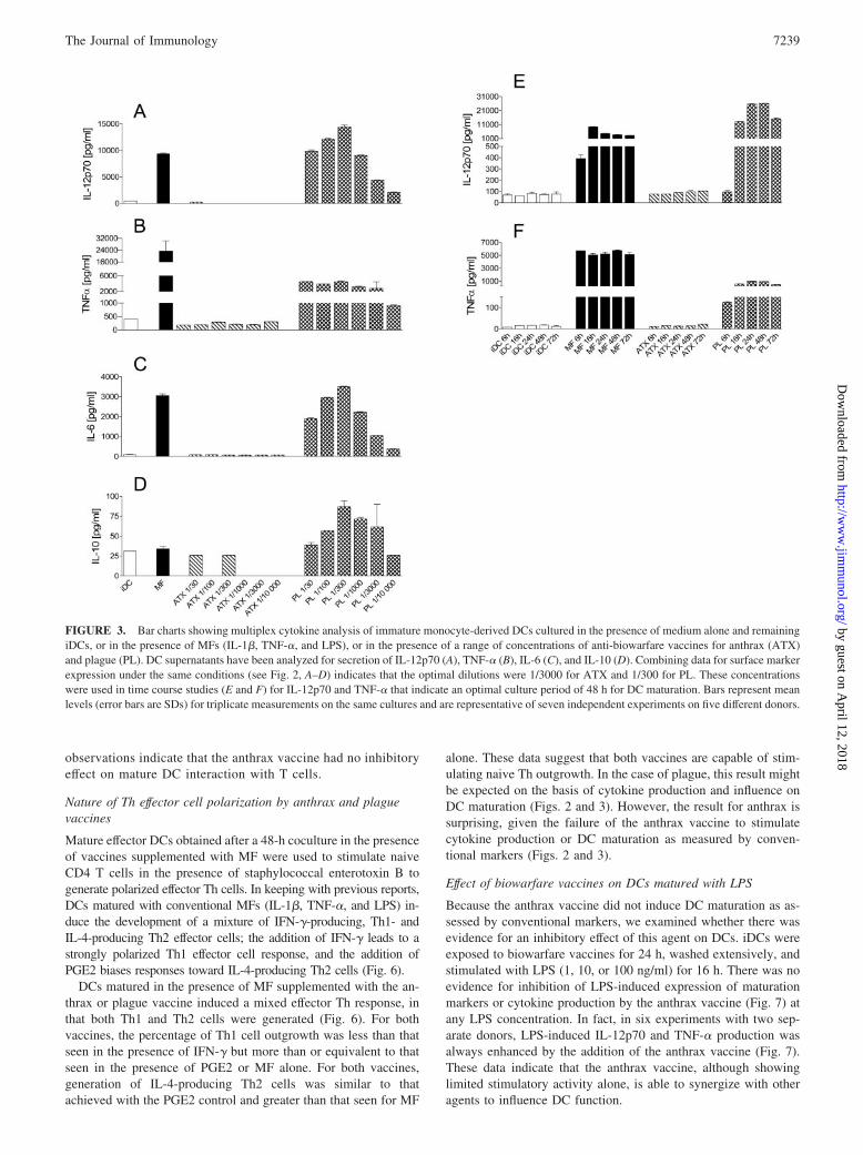
We first studied the direct effects of the anthrax and plague vac-cines on the maturation of monocyte-derived iDCs and their cy-tokine production. DCs cultured in the presence of control prepa-rations comprising TNF-�, IL-1�, and LPS (MF) showedmaturation as expected after 48 h. This was evident from the in-duction of CD83 expression and the marked up-regulation ofHLA-DR and CD86, the key molecules for T cell stimulation (Fig.2, B–D, filled bars), as well as the production of the Th1-stimula-tory cytokine IL-12p70 and proinflammatory cytokines TNF-�,IL-6, and IL-10 (Fig. 3, A–D, filled bars).
Our initial experiments aimed to establish optimal vaccine con-centrations for additional in vitro studies. After 48-h cultures withvaccines, iDCs were assessed for viability, surface molecule ex-pression, and cytokine production. As shown in Fig. 2A, cell via-bility of iDCs was preserved in the presence of a range of con-centrations of the plague vaccine. On the basis that it offeredoptimal expression of accessory molecules (Fig. 2, B–D) and se-cretion of cytokines (Fig. 3, A–D), a dilution of the plague vaccineof 1/300 (equivalent to 107 organisms/ml) was selected for use insubsequent experiments. At this dilution, the plague vaccine wasable to activate iDCs to a similar level, in terms of cytokine pro-duction and expression of CD83, HLA-DR, and CD86, to thatachieved with MF. In contrast, for the anthrax vaccine, cell deathincreased markedly at dilutions of �1/300 (Fig. 2A). At dilutionsbetween 1/300 and 1/10,000, cell death was equivalent to non-treated iDCs, and across this range, surface molecule expressionand cytokine production did not vary greatly (Figs. 2, B–D, and 3,A–D). In subsequent experiments, a dilution of 1/3000 wasselected.
These selected dilutions were used to examine the optimal timecourse for iDC stimulation. iDCs showed optimal cell viability andstimulation, as judged by the balance between surface marker ex-pression, cytokine production, and cell death, at 48 h in the pres-ence of either MF or the biowarfare vaccines (Figs. 2, E–G, and 3,E and F).
Under the optimal conditions that we established, it was evidentthat the two biowarfare vaccines behaved differently in terms ofiDC maturation. Whereas the plague vaccine was able to up-reg-ulate surface molecules and induce cytokine secretion to levelsequivalent to those observed with MF, the anthrax vaccine, in di-rect contrast, induced very low levels of maturation, such that thesurface phenotype and cytokine secretion of DCs cultured in theirpresence resembled the immature state (Figs. 2 and 3). Althoughstimulants such as MF can up-regulate HLA-DR expression bythree to five times, levels of HLA-DR expression on DCs after a48-h stimulation with the anthrax vaccine were unchanged. Sim-ilarly, although MF up-regulated costimulatory molecule (e.g.,CD86) expression by three to eight times, anthrax vaccine-exposedDCs had levels of CD86 expression similar to those observed inthe immature state. Likewise, CD83, a surface molecule charac-teristically appearing on DCs along with the process of maturation,as shown here, increased 4- to 5-fold after induction with MF butwas expressed at very low levels after coculture of iDCs with theanthrax vaccine (Fig. 2D). The anthrax vaccine triggered very little
7237The Journal of Immunology
by guest on April 12, 2018
http://ww
w.jim
munol.org/
Dow
nloaded from

IL-12p70 secretion (Fig. 3A), achieving levels similar to or lessthan unstimulated iDCs and significantly lower than the levelachieved with MF alone ( p � 0.01 compared with mean levels inseven independent experiments on five different donors at an an-thrax vaccine dilution of 1/3000). Likewise, TNF-�, IL-6, andIL-10 were undetectable or were produced in amounts similar to orbelow the level observed for iDCs and significantly lower than inthe presence of MF ( p � 0.01 compared with means from sevenindependent experiments on five different donors).
Overall, these data on the acquisition of T cell-stimulatory mol-ecules and cytokine secretion after coculture with biowarfarevaccines indicate that the anthrax vaccine does not induce DCmaturation or cytokine production when assessed using the con-ventional markers shown here.
Effects of vaccines on the T cell-stimulatory potential of DCs
In light of these findings, we next sought to examine whether DCsexposed to biowarfare vaccines acquired the capacity to stimulateMLR. This was assessed using a fixed number of allogeneic re-sponder CD4� T cells and varying numbers of DCs that had beenexposed to vaccines for 48 h. We found that plague and pertussisvaccines have a high capacity to stimulate allogeneic CD4� T cellsthat is directly comparable to DCs exposed to MF. In contrast, DCs
exposed to the anthrax vaccine stimulated CD4� T cells subopti-mally, but at a level higher than iDCs (Fig. 4A).
CD40L-induced cytokine production by DCs matured in thepresence of vaccines
We next sought to establish the cytokine milieu generated by DCsmatured in the presence of the anthrax and plague vaccines onencountering CD40L-expressing cells as a surrogate for interactionwith lymph node T cells.
Because the anthrax vaccine alone did not induce a matureDC phenotype, and the state of maturity influences the capacityof DCs to drive Th1 or Th2 responses, to perform these exper-iments, DCs were matured in the presence of vaccines as wellas conventional MFs (IL-1�, TNF-�, and LPS). Under theseconditions, maturation in the presence of the anthrax vaccinewas equivalent to that achieved with MF alone (Table I and Fig.5). IL-12p70, TNF-�, IL-6, and IL-10 production reached levelssimilar to or beyond those achieved in the presence of MFalone. However, it is noteworthy that in each of five experi-ments the presence of anthrax induced IL-12p70 production atlevels higher than those seen with MF alone, although thisdifference did not reach statistical significance. Overall, these
FIGURE 2. Optimization of vaccine concentration and time course for detection of DC maturation. Bar charts show flow cytometric analysis ofimmature monocyte-derived DCs cultured in the presence of medium alone and remaining iDCs, or in the presence of MFs (IL-1�, TNF-�, and LPS), orin the presence of a range of concentrations of anti-biowarfare vaccines for anthrax (ATX) and plague (PL). DCs have been labeled with 7-AAD forlive/dead analysis (A), anti-HLA-DR (B), anti-CD86 (C), and anti-CD83 (D). Combining data for cytokine production under the same conditions (see Fig.3, A–D) indicates that the optimal dilutions were 1/3000 for ATX and 1/300 for PL. These concentrations were used in time course studies (E–G) thatindicate an optimal culture period of 48 h for DC maturation. Bars represent mean levels (error bars are SDs) for triplicate measurements on the samecultures and are representative of seven independent experiments on five different donors. MFI, Mean fluorescence intensity.
7238 BIOWARFARE VACCINES AND DC INTERACTIONS
by guest on April 12, 2018
http://ww
w.jim
munol.org/
Dow
nloaded from
observations indicate that the anthrax vaccine had no inhibitoryeffect on mature DC interaction with T cells.
Nature of Th effector cell polarization by anthrax and plaguevaccines
Mature effector DCs obtained after a 48-h coculture in the presenceof vaccines supplemented with MF were used to stimulate naiveCD4 T cells in the presence of staphylococcal enterotoxin B togenerate polarized effector Th cells. In keeping with previous reports,DCs matured with conventional MFs (IL-1�, TNF-�, and LPS) in-duce the development of a mixture of IFN-�-producing, Th1- andIL-4-producing Th2 effector cells; the addition of IFN-� leads to astrongly polarized Th1 effector cell response, and the addition ofPGE2 biases responses toward IL-4-producing Th2 cells (Fig. 6).
DCs matured in the presence of MF supplemented with the an-thrax or plague vaccine induced a mixed effector Th response, inthat both Th1 and Th2 cells were generated (Fig. 6). For bothvaccines, the percentage of Th1 cell outgrowth was less than thatseen in the presence of IFN-� but more than or equivalent to thatseen in the presence of PGE2 or MF alone. For both vaccines,generation of IL-4-producing Th2 cells was similar to thatachieved with the PGE2 control and greater than that seen for MF
alone. These data suggest that both vaccines are capable of stim-ulating naive Th outgrowth. In the case of plague, this result mightbe expected on the basis of cytokine production and influence onDC maturation (Figs. 2 and 3). However, the result for anthrax issurprising, given the failure of the anthrax vaccine to stimulatecytokine production or DC maturation as measured by conven-tional markers (Figs. 2 and 3).
Effect of biowarfare vaccines on DCs matured with LPS
Because the anthrax vaccine did not induce DC maturation as as-sessed by conventional markers, we examined whether there wasevidence for an inhibitory effect of this agent on DCs. iDCs wereexposed to biowarfare vaccines for 24 h, washed extensively, andstimulated with LPS (1, 10, or 100 ng/ml) for 16 h. There was noevidence for inhibition of LPS-induced expression of maturationmarkers or cytokine production by the anthrax vaccine (Fig. 7) atany LPS concentration. In fact, in six experiments with two sep-arate donors, LPS-induced IL-12p70 and TNF-� production wasalways enhanced by the addition of the anthrax vaccine (Fig. 7).These data indicate that the anthrax vaccine, although showinglimited stimulatory activity alone, is able to synergize with otheragents to influence DC function.
FIGURE 3. Bar charts showing multiplex cytokine analysis of immature monocyte-derived DCs cultured in the presence of medium alone and remainingiDCs, or in the presence of MFs (IL-1�, TNF-�, and LPS), or in the presence of a range of concentrations of anti-biowarfare vaccines for anthrax (ATX)and plague (PL). DC supernatants have been analyzed for secretion of IL-12p70 (A), TNF-� (B), IL-6 (C), and IL-10 (D). Combining data for surface markerexpression under the same conditions (see Fig. 2, A–D) indicates that the optimal dilutions were 1/3000 for ATX and 1/300 for PL. These concentrationswere used in time course studies (E and F) for IL-12p70 and TNF-� that indicate an optimal culture period of 48 h for DC maturation. Bars represent meanlevels (error bars are SDs) for triplicate measurements on the same cultures and are representative of seven independent experiments on five different donors.
7239The Journal of Immunology
by guest on April 12, 2018
http://ww
w.jim
munol.org/
Dow
nloaded from

Modulation of DC maturation status and function by adjuvant
Our results indicate that the anthrax vaccine fails to induce DCmaturation or cytokine production as assessed using conventionalmarkers. As a consequence, expansion of effector T cells in vivocould, in theory, be limited or less sustained. These limitationscould theoretically be counteracted through the use of an appro-priate adjuvant. To examine this, we analyzed the adjuvant effectof whole-cell extract of B. pertussis on the different checkpoints ofDC function. Pertussis was selected because it has previously beencoadministered with the anthrax vaccine to enhance immunoge-nicity and protection from potential biological warfare attack.
Using our in vitro approach, we assessed the effects of addingWCP extract to DCs cocultured with the anthrax biowarfare vac-cine. The WCP extract alone proved a powerful stimulator of DCmaturation. At dilutions �1/3000, WCP was toxic to DCs as as-sessed by staining with 7-AAD. However, at a dilution of 1/3000,WCP induced DC maturation (91% CD83� DCs; mean fluores-cence intensity for HLA-DR and CD86 staining was 456 and 189units, respectively). Likewise, WCP diluted 1/3000 induced amean 1955 pg/ml (SD 54) TNF-� and 1260 pg/ml (SD 192) IL-12p70. These markers of DC maturation for WCP were similar tothose obtained with MF alone as shown in Figs. 2 and 3 (filledbars). In addition, WCP appeared to have a strong overall Th1priming effect (Fig. 6).
Using WCP in cocultures with the anthrax vaccine, there was noapparent modulatory effect on DC maturation or cytokine produc-tion by the anthrax vaccine. Thus, iDCs showed appropriate mat-uration in the presence of WCP and the anthrax vaccine combined(Fig. 8). Cytokine production by mature DCs in the combinedpresence of WCP and the anthrax vaccine was similar to that forWCP alone (Fig. 5), whereas the effect of adding WCP to biowarfarevaccines was a polarization of naive CD4 T cells toward Th1 similarto that observed for WCP alone (Fig. 6). These data indicate that the
anthrax vaccine, despite inducing only limited DC maturation whenjudged by conventional markers, does not lead to any sustained in-hibitory effects on DC maturation or effector function.
Detection of recall responses to the anthrax vaccine in a cohortof vaccinees
Our in vitro data suggested that the limited DC stimulationachieved by the anthrax vaccine could be overcome in the presenceof an adjuvant such as WCP. Indeed, this is a strategy used in vivoto enhance the immunogenicity of the anthrax vaccine. To examinethe efficacy of this approach, we next investigated whether recall Tcell responses against the anthrax vaccine could be detected bycytokine ELISPOT in control subjects (anthrax vaccine naive) andvaccinated subjects (military personnel who had received the an-thrax vaccine coadministered with WCP as an adjuvant). Anthrax-specific recall responses were clearly detectable for both Th1 andTh2 cytokines in the vaccinated group but not in the naive controls(Fig. 9). There were significant differences in the number of an-thrax-specific spots between vaccinated and naive individuals forIFN-� ( p � 0.05), IL-2 ( p � 0.01), and IL-13 ( p � 0.0001).
DiscussionIn the present study, we have used existing technologies for ex-amining DC responses to pathogens to develop an in vitro strategyfor examining the interaction between vaccines and DCs. We haveapplied this approach to increase our understanding of the immuneresponse engendered by two vaccine preparations, anthrax andplague, that may be of critical importance in protection from ex-posure to the respective biological warfare agents. Our data showthat the plague vaccine preparation triggers maturation and effectorfunction of DCs. In contrast, using the same measures of DC ac-tivation, the anthrax vaccine invokes minimal maturation and verylimited effector function. In the case of the anthrax vaccine, thismay explain the requirement for repeated dosing to obtain protec-tion in vivo.
The anthrax and plague vaccines are known to be inefficientimmunogens, requiring repeated and frequent administration. As aconsequence, the ability of public health systems to plan for pos-sible bioterrorism is severely impaired. Not only is the degree ofprotection unpredictable, but so is the length of time taken toachieve it, and any possible confounding effects of multiple vac-cinations given in the same short space of time. An in vitro model,in which it is possible to play out interaction between vaccine andiDCs, may therefore prove useful in evaluating vaccine effects.Vaccines could fail, or engender only limited protection, for atleast two possible reasons: either as a result of not stimulating DCmaturation or through the presence of toxins and proteins withinhibitory effects on DC function. The in vitro model can be usedto explore the relative contribution of these scenarios and also toexamine measures that could be used to redress the balance, suchas the use of adjuvants.
Table I. Phenotypic characteristics of DCs exposed to vaccines in thepresence of MFs
Cell TypeMaturationStimulus
Maturation Markers
HLA-DR CD86 CD83
iDC 90 36 6Mature DC MF 243 98 66Mature DC Anthrax � MF 257 150 60Mature DC Plague � MF 318 222 62Mature DC Pertussis � MF 424 306 78
FIGURE 4. MLR of 50,000 allogeneic CD4� T cells to a range of DCnumbers after DC exposure to different vaccine conditions. A, Proliferationof CD4� T cells after coculture with iDC (f), anthrax vaccine (�), plaguevaccine (F), pertussis vaccine (�), and MFs (Œ). The plague and pertussisvaccines give rise to DCs that evoke robust MLR response equivalent toMF alone. MLR response to anthrax vaccine-treated DCs is higher than foriDCs but falls short of that achieved with MF. B, Proliferation of CD4� Tcells after coculture with iDCs (f), MF alone (Œ), and anthrax vaccine pluspertussis (ƒ), or plus MF (�). These data indicate that the anthrax vaccinehas no sustained inhibitory effect on the ability of DCs to induce MLR.Data represent means of triplicates from a single experiment, and the errorbars are SEMs. Data from a single donor are shown and are representativeof data from three independent experiments from three different donors.
7240 BIOWARFARE VACCINES AND DC INTERACTIONS
by guest on April 12, 2018
http://ww
w.jim
munol.org/
Dow
nloaded from

Our studies indicate that during its encounter with iDCs, theanthrax vaccine induces limited maturation or production of keyproinflammatory cytokines. This finding resonates with the recentreport that the anthrax lethal toxin, present in our vaccine prepa-ration, severely impairs DC function through disruption of immunecell MAPK signaling networks (19). In that report, DCs exposed tolethal toxin failed to up-regulate costimulatory molecules or makeproinflammatory cytokines and did not effectively stimulate Ag-specific T cells in vivo. There are some similarities between thesefindings and those made in the present study when DCs were ex-posed to the vaccine, although we were unable to demonstrateinhibition of LPS-induced responses. This may reflect differencesin the preparations used, or the fact that the final concentrations ofPA and LF achieved in our cultures are below those showing clearinhibitory effects in the study by Agrawal et al. (19). An additionalpossibility is that factors present in the anthrax vaccine prepara-tion, but not LF or PA, are responsible for effects on DCs. Thecombination of PA, a nontoxic, cell-binding component of the an-
thrax toxin complex, with EFs produces an edema toxin that in-duces increased intracellular cAMP levels in susceptible cells (20).This inhibits neutrophil phagocytosis (21) and differentially down-regulates LPS-induced production of TNF-� and IL-6 by increas-ing the intracellular cAMP levels in monocytes (22). Additionally,factors that up-regulate cAMP, such as PGE2 (23), cholera toxin(24), and histamine (25), are all known to induce Th2-type re-sponses, and in the present study, anthrax-primed, mature DCsgenerated a predominant Th2-type response.
Clearly, there are important implications both for the generationof effective immunity during encounter with wild-type B. anthra-cis and also for anthrax vaccine design. However, our strategy ofin vitro studies has the capacity to indicate manipulations that mayovercome any undesirable effects. First, we were able to show that,for the most part, the limited DC maturation and cytokine productioninduced by the anthrax vaccine was correctable. For example, DCsmatured in the presence of conventional MFs plus vaccine producedappropriate amounts of some of the major proinflammatory cytokines
FIGURE 5. Bar charts showing cytokine production by DCs exposed at the mature stage for 24 h in the presence of CD40 ligation to various controlstimuli, vaccines, or combinations of vaccines, always in the presence of MFs. IFN-� and PGE2 (plus MF) are used to represent Th1 and Th2 polarizingstimuli, respectively. Bars represent mean (SD) cytokine levels measured in triplicates from cultures obtained in a single representative experiment of fivereplicated studies. ATX, Anthrax; PL, plague.
FIGURE 6. Dot plot flow cytomet-ric analyses of IFN-� (x-axis) vs IL-4(y-axis) staining of T cells expandedand polarized by DCs matured for 48 hunder the conditions shown. The fig-ures in quadrants represent the percent-age of stained cells. Representativedata from one of five different donorsare shown.
7241The Journal of Immunology
by guest on April 12, 2018
http://ww
w.jim
munol.org/
Dow
nloaded from

(TNF-�, IL-6) and were less liable to Th2 polarization when com-pared with vaccine alone. This has important implications for thepossible use of adjuvants to overcome poor immunogenicity. It isalso consistent with the fact that repeated, short-term vaccinationachieves effective host protection, presumably as a result of induc-ing escalating levels of local inflammation that in turn provides DCmaturation signals.
It is of interest that under conditions designed to examine Thcell outgrowth, both the anthrax and plague vaccines promote thegeneration of Th2 effector cells. It is possible that this responseoccurs naturally as a result of exposure to wild-type B. anthracisor Y. pestis organisms, but to date no study on the T cell responseunder these conditions has been conducted. The current literatureis limited to observations on the type of Ab response generated asthe result of natural infection compared with vaccine administra-tion. IgG1 and IgG3 class Abs are generally detected after natural
infection compared with all of the IgG subclasses after vaccineadministration, and it is noteworthy that IgG4 isotype Abs areindicative of a Th2 immune response (26). A final possibility, inthe case of the anthrax vaccine at least, is the effect of alum, aknown Th2-polarizing adjuvant. However, it is known that alumdoes not have direct effects on DC activation (27).
Despite the fact that the anthrax vaccine invoked limited DCmaturation, it was able to induce a predominant Th2 response.This is not typical of iDCs and is more surprising given the highproduction of IL-12p70 after CD40 ligation. It is possible thatTh2-type vaccine effector responses are due to OX40 ligandinteraction, which is known to promote Th2 cells (28, 29), orother factors, which are as yet unknown. Equally possible isthat the anthrax vaccine, by not maturing DCs and not trigger-ing the expression of costimulatory molecules on DCs, resultsin an iDC-like phenotype that is known to produce more IL-12p70 than the mature cells (30).
To counter the limited immunogenicity of the anthrax andplague vaccines, WCP extract was added to the vaccination regi-men as an adjuvant for United Kingdom troops deployed to thefirst Persian Gulf War. Pertussis has a powerful effect on DC mat-uration and activation, with marked proinflammatory cytokine pro-duction and Th1 polarization (29). In our study, examination of theeffect of pertussis on DC-anthrax vaccine interactions showed thatthe limited effects of this biowarfare vaccine could be overcome. Amature DC phenotype was achieved along with good Th1 out-growth. Indeed, our ex vivo cytokine ELISPOT data indicate thatTh1 and Th2 anthrax-specific responses are induced and long-livedin individuals receiving combined anthrax and pertussis vaccines,although we were unable to document the relative benefit of ad-juvant because subjects that received the anthrax vaccine alone arenot available for study. In contrast with the limited effects of the
FIGURE 7. Effect of anthrax biowarfare vaccine on LPS-stimulatedDCs. The graph shows cytokine production by iDCs exposed initially toanthrax and plague vaccines and subsequently stimulated with LPS for24 h. Data are representative of six experiments on two donors. Bars rep-resent the means of triplicates, and error bars represent the SD. ATX,Anthrax; PL, plague vaccine.
FIGURE 8. Effects of WCP in combination with theanthrax vaccine on DC maturation. Histograms of flowcytometric analysis of acquisition of maturation markerson DCs exposed at the immature stage to control stimuli,single vaccine preparations, and combinations of vaccinesfor 48 h are shown. The thin line represents isotype con-trol Ab staining; the bold line represents the designatedmAb. The histograms represent mean fluorescent intensityfor HLA-DR and CD86 and the percentage of positivecells set at the 99th percentile of the isotype control forCD83. Representative data from one of five different do-nors are shown.
7242 BIOWARFARE VACCINES AND DC INTERACTIONS
by guest on April 12, 2018
http://ww
w.jim
munol.org/
Dow
nloaded from

anthrax vaccine, the plague vaccine was efficient in induction ofDC maturation and effector function. It seems unlikely, therefore,that the requirement for repeated multiple administrations of theplague vaccine is explained by an inability to activate the immunesystem via DCs.
In summary, anthrax and plague vaccines are poor immunogensin vivo; in the case of anthrax, this may be a result, at least in part,of its limited ability to trigger the maturation process of iDCs andgenerate an appropriate proinflammatory cytokine milieu, whichare crucial for initiation of effector immune responses. Our studyalso supports, through in vitro and in vivo data, the potential ofDC-activating adjuvants to overcome such effects.
AcknowledgmentsWe are grateful to colleagues at Defense Science and Technology andCAMR (Porton Down, Salisbury, U.K.) for discussions and contributionsto this work.
DisclosuresThe authors have no financial conflict of interest.
References1. O’Toole, T., and T. V. Inglesby. 2000. Facing the biological weapons threat.
Lancet 356: 1128–1129.2. Rotz, L. D., A. S. Khan, S. R. Lillibridge, S. M. Ostroff, and J. M. Hughes. 2002.
Public health assessment of potential biological terrorism agents. Emerg. Infect.Dis. 8: 225–230.
3. Gruchalla, R. S., and J. Jones. 2003. Combating high-priority biological agents:what to do with drug-allergic patients and those for whom vaccination is con-traindicated? J. Allergy Clin. Immunol. 112: 675–682.
4. Goodman, L. 2004. Taking the sting out of the anthrax vaccine. J. Clin. Invest.114: 868–869.
5. Inglesby, T. V., T. O’Toole, D. A. Henderson, J. G. Bartlett, M. S. Ascher,E. Eitzen, A. M. Friedlander, J. Gerberding, J. Hauer, J. Hughes, et al. 2002.
Anthrax as a biological weapon, 2002: updated recommendations for manage-ment. J. Am. Med. Assoc. 287: 2236–2252.
6. Marshall, J. D., Jr., P. J. Bartelloni, D. C. Cavanaugh, P. J. Kadull, andK. F. Meyer. 1974. Plague immunization. II. Relation of adverse clinical reac-tions to multiple immunizations with killed vaccine. J. Infect. Dis. 129(Suppl.):S19–S25.
7. Inglesby, T. V., D. T. Dennis, D. A. Henderson, J. G. Bartlett, M. S. Ascher,E. Eitzen, A. D. Fine, A. M. Friedlander, J. Hauer, J. F. Koerner, et al. 2000.Plague as a biological weapon: medical and public health management. WorkingGroup on Civilian Biodefense. J. Am. Med. Assoc. 283: 2281–2290.
8. Banchereau, J., and R. M. Steinman. 1998. Dendritic cells and the control ofimmunity. Nature 392: 245–252.
9. Steinman, R. M., and M. C. Nussenzweig. 2002. Avoiding horror autotoxicus: theimportance of dendritic cells in peripheral T cell tolerance. Proc. Natl. Acad. Sci.USA 99: 351–358.
10. Hart, D. N. 1997. Dendritic cells: unique leukocyte populations which control theprimary immune response. Blood 90: 3245–3287.
11. Kalinski, P., C. M. Hilkens, E. A. Wierenga, and M. L. Kapsenberg. 1999. T-cellpriming by type-1 and type-2 polarized dendritic cells: the concept of a thirdsignal. Immunol. Today 20: 561–567.
12. Mellman, I., and R. M. Steinman. 2001. Dendritic cells: specialized and regulatedantigen processing machines. Cell 106: 255–258.
13. Banchereau, J., F. Briere, C. Caux, J. Davoust, S. Lebecque, Y. J. Liu,B. Pulendran, and K. Palucka. 2000. Immunobiology of dendritic cells. Annu.Rev. Immunol. 18: 767–811.
14. Liu, Y. J. 2001. Dendritic cell subsets and lineages, and their functions in innateand adaptive immunity. Cell 106: 259–262.
15. Skowera, A., M. Hotopf, E. Sawicka, R. Varela-Calvino, C. Unwin, V. Nikolaou,L. Hull, K. Ismail, A. S. David, S. C. Wessely, and M. Peakman. 2004. Cellularimmune activation in gulf war veterans. J. Clin. Immunol. 24: 66–73.
16. Hotopf, M., A. David, L. Hull, K. Ismail, C. Unwin, and S. Wessely. 2000. Roleof vaccinations as risk factors for ill health in veterans of the Gulf War: crosssectional study. Br. Med. J. 320: 1363–1367.
17. Arif, S., T. I. Tree, T. P. Astill, J. M. Tremble, A. J. Bishop, C. M. Dayan,B. O. Roep, and M. Peakman. 2004. Autoreactive T cell responses show proin-flammatory polarization in diabetes but a regulatory phenotype in health. J. Clin.Invest. 113: 451–463.
18. Schloot, N. C., G. Meierhoff, M. Karlsson Faresjo, P. Ott, A. Putnam,P. Lehmann, P. Gottlieb, B. O. Roep, M. Peakman, and T. Tree. 2003. Compar-ison of cytokine ELISpot assay formats for the detection of islet antigen autore-active T cells: Report of the Third Immunology of Diabetes Society T-Cell Work-shop. J. Autoimmun. 21: 365–376.
19. Agrawal, A., J. Lingappa, S. H. Leppla, S. Agrawal, A. Jabbar, C. Quinn, andB. Pulendran. 2003. Impairment of dendritic cells and adaptive immunity byanthrax lethal toxin. Nature 424: 329–334.
20. Leppla, S. H. 1982. Anthrax toxin edema factor: a bacterial adenylate cyclase thatincreases cyclic AMP concentrations of eukaryotic cells. Proc. Natl. Acad. Sci.USA 79: 3162–3166.
21. O’Brien, J., A. Friedlander, T. Dreier, J. Ezzell, and S. Leppla. 1985. Effects ofanthrax toxin components on human neutrophils. Infect. Immun. 47: 306–310.
22. Hoover, D. L., A. M. Friedlander, L. C. Rogers, I. K. Yoon, R. L. Warren, andA. S. Cross. 1994. Anthrax edema toxin differentially regulates lipopolysaccha-ride-induced monocyte production of tumor necrosis factor � and interleukin-6by increasing intracellular cyclic AMP. Infect. Immun. 62: 4432–4439.
23. Snijdewint, F. G., P. Kalinski, E. A. Wierenga, J. D. Bos, and M. L. Kapsenberg.1993. Prostaglandin E2 differentially modulates cytokine secretion profiles ofhuman T helper lymphocytes. J. Immunol. 150: 5321–5329.
24. Xu-Amano, J., H. Kiyono, R. J. Jackson, H. F. Staats, K. Fujihashi,P. D. Burrows, C. O. Elson, S. Pillai, and J. R. McGhee. 1993. Helper T cellsubsets for immunoglobulin A responses: oral immunization with tetanus toxoidand cholera toxin as adjuvant selectively induces Th2 cells in mucosa associatedtissues. J. Exp. Med. 178: 1309–1320.
25. van der Pouw Kraan, T. C., A. Snijders, L. C. Boeije, E. R. de Groot,A. E. Alewijnse, R. Leurs, and L. A. Aarden. 1998. Histamine inhibits the pro-duction of interleukin-12 through interaction with H2 receptors. J. Clin. Invest.102: 1866–1873.
26. Rodriguez, V., M. Centeno, and M. Ulrich. 1996. The IgG isotypes of specificantibodies in patients with American cutaneous leishmaniasis; relationship to thecell-mediated immune response. Parasite Immunol. 18: 341–345.
27. Sun, H., K. G. Pollock, and J. M. Brewer. 2003. Analysis of the role of vaccineadjuvants in modulating dendritic cell activation and antigen presentation in vitro.Vaccine 21: 849–855.
28. Flynn, S., K. M. Toellner, C. Raykundalia, M. Goodall, and P. Lane. 1998. CD4T cell cytokine differentiation: the B cell activation molecule, OX40 ligand, in-structs CD4 T cells to express interleukin 4 and upregulates expression of thechemokine receptor, Blr-1. J. Exp. Med. 188: 297–304.
29. de Jong, E. C., P. L. Vieira, P. Kalinski, J. H. Schuitemaker, Y. Tanaka,E. A. Wierenga, M. Yazdanbakhsh, and M. L. Kapsenberg. 2002. Microbialcompounds selectively induce Th1 cell-promoting or Th2 cell-promoting den-dritic cells in vitro with diverse th cell-polarizing signals. J. Immunol. 168:1704–1709.
30. Cella, M., D. Scheidegger, K. Palmer-Lehmann, P. Lane, A. Lanzavecchia, andG. Alber. 1996. Ligation of CD40 on dendritic cells triggers production of highlevels of interleukin-12 and enhances T cell stimulatory capacity: T-T help viaAPC activation. J. Exp. Med. 184: 747–752.
FIGURE 9. Recall T cell responses detected by cytokine ELISPOTagainst tetanus toxoid (TT) and anthrax (ATX) vaccines in control subjects(anthrax vaccine naive) and military personnel who had received anthraxvaccine coadministered with WCP as an adjuvant. Bars represent the meannumber of spots counted for each cytokine in triplicate, and error bars areSEM. Anthrax-specific recall responses are clearly detectable for both Th1and Th2 cytokines in the vaccinated group but not in the naive controls.There are significant differences in the number of anthrax-specific spots be-tween vaccinated and naive individuals for IFN-� (�, p � 0.05), IL-2 (��, p �0.01), and IL-13 (���, p � 0.0001). Mean background (medium alone) spotsranged between 0 and 4 and were not different between the groups.
7243The Journal of Immunology
by guest on April 12, 2018
http://ww
w.jim
munol.org/
Dow
nloaded from