
Monitoring the anesthetized patient - Illinois State …€“Increased metabolic production of...
62
Monitoring the anesthetized patient: How anesthesia affects the body By: Jessica Antonicic CVT, VTS (Anesthesia)
Transcript of Monitoring the anesthetized patient - Illinois State …€“Increased metabolic production of...
Monitoring the anesthetized patient: How anesthesia affects the body
By: Jessica Antonicic CVT, VTS (Anesthesia)

Defining Anesthesia
• The word anesthesia originated from the Greek term anaisthaesia, meaning “insensibility”; is used to describe the loss of sensation to the body in part or in its entirety.
• General anesthesia (GA) is defined as drug-induced unconsciousness where CNS depression is controlled but reversible.
• Surgical anesthesia is the state/plane of GA that provides unconsciousness, muscular relaxation, and analgesia sufficient for a painless surgery.
Considerations for Anesthesia
• The dose of anesthesia and the techniques for administration are based on the average healthy animal that is of normal body condition
• Variations in the response to anesthetics results from many different factors including CNS status, metabolic activity, any existing diseases or pathology and the uptake and distribution of the drug.
• There are many factors that influence or modify the uptake, distribution and elimination of anesthetics. Some of these factors include: rate of administration, concentration of anesthetic, physical status, muscular development, adiposity, respiratory/circulatory status, administration of other drugs, and more.
Considerations for Anesthesia
• A thorough pre-anesthetic evaluation should be performed prior to any surgical procedure.
• Some factors to take into consideration are pre-operative heart rate, respiratory rate and effort, temperature, blood pressure, and pain/stress.
• Yes, pain and stress are factors to consider.
• Pre-operative blood tests are important prior to anesthesia

Under Anesthesia
• After induction is performed, a patient should be placed on monitoring equipment.
• As the anesthetist, you should be monitoring the monitor as well as the patient themselves.
• If the monitor seems to good to be true or if it is showing you something unusual, check your patient.
• Key vitals to check first-HR, RR and effort, then everything else
Under Anesthesia
• The vitals that are typically monitored under anesthesia
– Heart rate
– Respiratory rate and effort
– Temperature
– Pulse oximetry
– Blood pressure
– Capnography-if available

The Heart

Heart Rate
• Heart rate is defined as the number of heartbeats per unit time, expressed as beats per minute– For each species the normal range is different
(for awake and anesthetized patients)
– For dogs: on average 90-140bpm awake
– For cats: on average 100-200bpm awake
• What machines do we use to monitor heart rate and rhythm??
Cardiac Output
• The rate at which the heart beats can affect a value called cardiac output (CO)
• Cardiac output is defined as the volume of blood being pumped by the heart (ventricle) per unit time
• It is calculated by a simple formula– CO= HR x SV
– HR= heart rate
– SV= stroke volume

Heart Rate and Rhythm
• Sinus bradycardia : typically defined as a rate that falls below 60bpm in larger dogs, 80 bpm in smaller dogs, and 90-100 bpm in cats– Leads to low CO
– Leads to hypotension and poor tissue perfusion• If goes untreated can lead to renal failure, reduced hepatic
metabolism of drugs, worsening of V/Q mismatch and hypoxemia, delayed recovery, CNS abnormalities (blindness), and eventually arrest
• Before treatment-verify that it is truly bradycardia and not ventricular arrhythmias– Excessive vagal tone can be caused by pharyngeal, laryngeal, or
tracheal stimulation or by visceral inflammation, distension, or
traction.

Sinus Bradycardia
• Causes of peri-operative bradycardia:– Anesthetic overdose
– Opioids
– Alpha-2s
– Excessive vagal tone
– Hypothermia
– Hyperkalemia
– Sick Sinus Syndrome
– AV Block
– Hypoxia

Heart Rate and Rhythm
• Sinus tachycardia can be a sign of an underlying problem
(rate over 160 in dogs, high 200’s for cats)
• It becomes a problem when there is not enough time for diastolic filling
– This decreases CO thus decreasing BP
– The same problems can arise like renal failure, worsening of V/Q mismatch and more

Sinus Tachycardia
• Common causes for sinus tachycardia:– Too light
– Ketamine
– Anticholinergics
– Hypovolemia
– Hyperthermia
– Hypoxemia
– Hypercapnia
– Pain
– Acepromazine

Most common bradyarrhythmia
• Atrioventricular Block
– First degree: rate and rhythm are typically normal
• Hard to differentiate on ECG
– Second degree:
• Mobitz type 1-when there is a progressive delay in the AV transmission prior to blocked P-wave
• Mobitz type 2-when 1 or more P waves are blocked without previous delay
– Third degree or total: complete dissociation between P wave and QRS complex

AV block

Ventricular Premature Contractions
• Can be single or multiple cardiac impulses that come from the ventricles instead of the sinus node.
• On ECG, a wide and bizarre QRS complex is noted with no association to the P wave
• Can be unifocal or multifocal
• An occasional VPC is usually not concerning; when there are runs that start to affect CO and BP then treatment should be initiated
Blood Pressure
• Blood pressure in arteries is frequently assessed under anesthesia, whether it is indirect (Doppler/oscillometric) or direct
• Arterial blood pressure measurement is one of the fastest and most informative means of assessing cardiovascular function
• When it is done correctly and frequently enough, it helps provides an accurate indication of drug effects, surgical events, and hemodynamic trends
• This is modified by almost all drugs used to induce and maintain anesthesia

Arterial Blood Pressure
• Is the key component in determining perfusion pressure and the adequacy of tissue perfusion
• Having a mean pressure greater than 60 mm Hg is generally considered adequate to perfuse tissues
• There are several organ systems that are more sensitive to changes in perfusion pressure and there can be immediate consequences in organ function– Heart– Lung – Kidney– Fetal
Blood Pressure
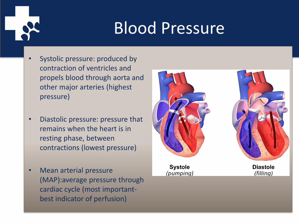
• Systolic pressure: produced by contraction of ventricles and propels blood through aorta and other major arteries (highest pressure)
• Diastolic pressure: pressure that remains when the heart is in resting phase, between contractions (lowest pressure)
• Mean arterial pressure (MAP):average pressure through cardiac cycle (most important-best indicator of perfusion)
Arterial Blood Pressure
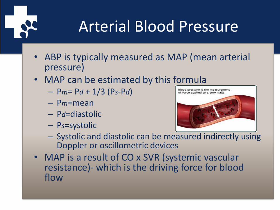
• ABP is typically measured as MAP (mean arterial pressure)
• MAP can be estimated by this formula– Pm= Pd + 1/3 (Ps-Pd)– Pm=mean– Pd=diastolic– Ps=systolic– Systolic and diastolic can be measured indirectly using
Doppler or oscillometric devices
• MAP is a result of CO x SVR (systemic vascular resistance)- which is the driving force for blood flow

Blood Pressure Values
• Normal systolic (90-160 mm Hg)
• Normal diastolic (50-90 mm Hg)
• Normal mean (70-90 mm Hg)
– Values under anesthesia
• Monitor trends-one reading might not be indicative of a problem
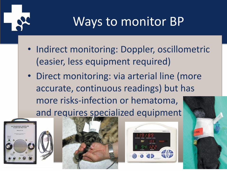
Ways to monitor BP
• Indirect monitoring: Doppler, oscillometric (easier, less equipment required)
• Direct monitoring: via arterial line (more accurate, continuous readings) but has more risks-infection or hematoma, and requires specialized equipment
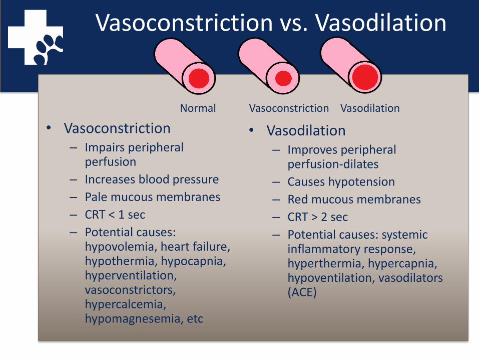
Vasoconstriction vs. Vasodilation
• Vasoconstriction– Impairs peripheral
perfusion
– Increases blood pressure
– Pale mucous membranes
– CRT < 1 sec
– Potential causes: hypovolemia, heart failure, hypothermia, hypocapnia, hyperventilation, vasoconstrictors, hypercalcemia, hypomagnesemia, etc
• Vasodilation– Improves peripheral
perfusion-dilates
– Causes hypotension
– Red mucous membranes
– CRT > 2 sec
– Potential causes: systemic inflammatory response, hyperthermia, hypercapnia, hypoventilation, vasodilators (ACE)
Normal Vasoconstriction Vasodilation

Hypotension
• Hypotension is defined as a MAP lower than 60 mm Hg• Any value lower than 60 can lead to compromised
perfusion of visceral organs and peripheral tissues, leading to ischemia
• If it goes untreated, significant hypotension can lead to renal failure, reduced hepatic metabolism of drugs, worsening of V/Q mismatch and hypoxemia, delayed recovery, neuromuscular complications during recovery, and CNS abnormalities including blindness.
• Eventually, if it continues to worsen, severe hypotension can lead to cardiac and respiratory arrest.

What causes Hypotension?
– Decrease in SVR
– Decrease in blood volume
– Decrease in vascular tone
– Decrease in CO
– Reduction in contractility
– Drugs (inhalants, ace, Propofol, alpha 2s, etc.)-have vasodilating effects
• IPPV
• Gastric distension
• Heart diseases
• Tachycardia
• Ventricular arrhythmias
• Bradycardia– Reduction in stroke volume
– High ETCO2
Hypertension
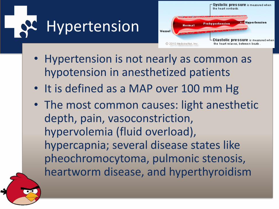
• Hypertension is not nearly as common as hypotension in anesthetized patients
• It is defined as a MAP over 100 mm Hg
• The most common causes: light anesthetic depth, pain, vasoconstriction, hypervolemia (fluid overload), hypercapnia; several disease states like pheochromocytoma, pulmonic stenosis, heartworm disease, and hyperthyroidism

Thermoregulation
• Thermoregulation is the ability of an organism to maintain its body temperature within certain boundaries, even when the surrounding temperature is very different
• The rectum in animals is considered to most accurately reflect the temperature of internal parts
• Thermoregulation is controlled by the preoptic area of the anterior hypothalamus
Anesthesia and Thermoregulation
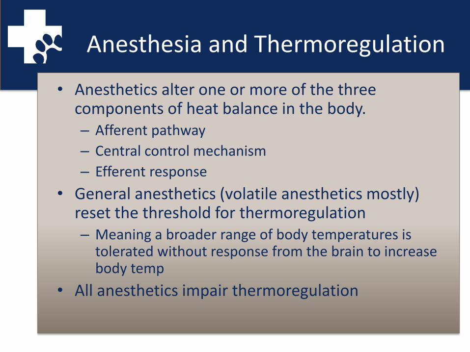
• Anesthetics alter one or more of the three components of heat balance in the body.– Afferent pathway
– Central control mechanism
– Efferent response
• General anesthetics (volatile anesthetics mostly) reset the threshold for thermoregulation– Meaning a broader range of body temperatures is
tolerated without response from the brain to increase body temp
• All anesthetics impair thermoregulation
Hypothermia
• Hypothermia is associated with drug depression of muscular activity, metabolism, and thermostatic regulatory mechanisms
• There are several factors that might add to heat loss– Evaporation of surgical scrub solutions– Infusion of room temp fluids– Contact with cold surgical table/radiology
table/prep table– Evaporation of surface fluid from exposed body
cavity

Hypothermia
• Core body temp down to 96⁰F is typically not detrimental to patients– Recovery should not be prolonged in any noticeable way
• Temp of 90-95⁰F is associated with reduced anesthetic requirements– Recovery should be noticeably prolonged– Some animals will shiver to help raise body temp
• Temp of 82-86⁰F shows marked CNS-depressant effects, no anesthetic is required– Arterial arrhythmias may occur– HR and CO are reduced to 35-40% of normal
function– MAP is about 60% of normal– Oxygen consumption is reduced to 50% of normal
Hypothermia
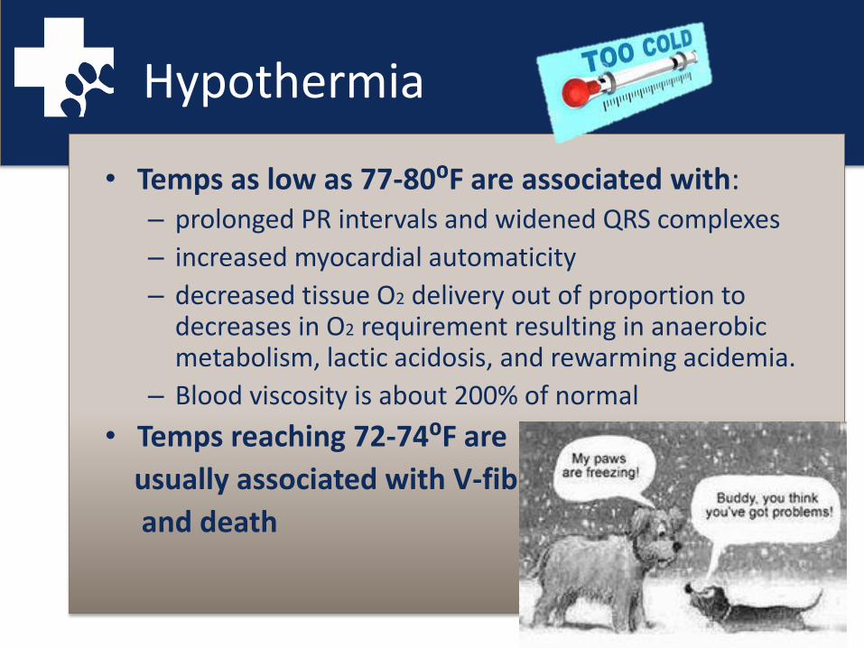
• Temps as low as 77-80⁰F are associated with: – prolonged PR intervals and widened QRS complexes
– increased myocardial automaticity
– decreased tissue O2 delivery out of proportion to decreases in O2 requirement resulting in anaerobic metabolism, lactic acidosis, and rewarming acidemia.
– Blood viscosity is about 200% of normal
• Temps reaching 72-74⁰F are
usually associated with V-fib
and death
Hypothermia
• Potential problems associated with hypothermia
– Coagulation deficiencies
• Impaired platelet function
• Decrease in activity of coagulation pathways
• Increases in fibrinolysis
– Delayed wound healing with moderate hypothermia
– Muscle protein breakdown
– Prolonged recovery
– Slows liver metabolism of drugs
– Shivering-increase in O2 demands
• Anesthesia related consequences of hypothermia:– Hypothermia decreases the
amount of anesthetic required– 5% decrease in MAC
requirement for inhalants with each 1.8⁰F decrease in body temp
– Increases the solubility of volatile anesthetics, which in turn increases the effective dose administered
– Decrease in clearance of anesthetic drugs
– All of these combined can lead to an anesthetic overdose
Hypothermia
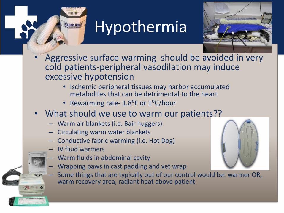
• Aggressive surface warming should be avoided in very cold patients-peripheral vasodilation may induce excessive hypotension
• Ischemic peripheral tissues may harbor accumulated metabolites that can be detrimental to the heart
• Rewarming rate- 1.8⁰F or 1⁰C/hour
• What should we use to warm our patients??– Warm air blankets (i.e. Bair huggers)– Circulating warm water blankets– Conductive fabric warming (i.e. Hot Dog)– IV fluid warmers– Warm fluids in abdominal cavity– Wrapping paws in cast padding and vet wrap– Some things that are typically out of our control would be: warmer OR,
warm recovery area, radiant heat above patient
Fever vs. Hyperthermia
• Fever is a reset thermostat– Caused by the release of endogenous pyrogens
from monocytes in response to infections, tissue damage, or antigen-antibody reactions
• Hyperthermia, without a reset thermostat, is pathological– Not uncommon in large breed dogs wrapped in
layers of drapes on the table– Can be potentiated by surface vasoconstriction,
light levels of anesthesia, and administration of ketamine

Hyperthermia
• How do patients become hyperthermic under anesthesia?
– Excessive external heating-#1
– Decreased loss of body heat through increased insulation, thick hair coats
– Increased metabolic production of heat, including stress related hyperthermia, increased muscle tone, resetting of thermoregulatory process
– Malignant hyperthermia

Hyperthermia
• Hyperthermia of even a couple of degrees increases circulatory work– For most animals this is not detrimental
• Mild hyperthermia, below 104⁰F, does not normally require treatment– This should be determined on a case by case basis
• Moderate hyperthermia-temp up to 108⁰F is when cell damage starts – O2 delivery can not keep up with a racing metabolism
and increased O2 consumption– Cellular hypoxia results with potential to do damage to the
brain, liver, kidneys, and blood
Hyperthermia
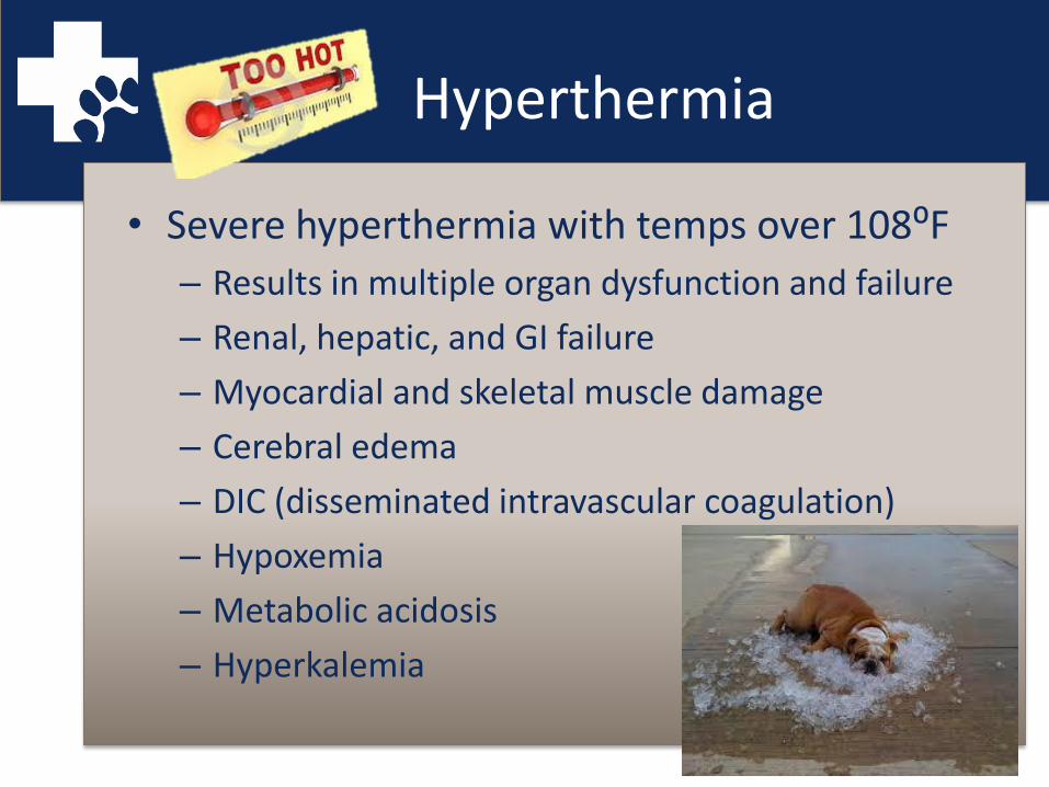
• Severe hyperthermia with temps over 108⁰F
– Results in multiple organ dysfunction and failure
– Renal, hepatic, and GI failure
– Myocardial and skeletal muscle damage
– Cerebral edema
– DIC (disseminated intravascular coagulation)
– Hypoxemia
– Metabolic acidosis
– Hyperkalemia

Post-op Hyperthermia
• Hyperthermia post-operatively can result from increased muscle activity and inappropriate thermoregulation
• Marked hyperthermia can be seen in cats post op – These cats are usually excited or stressed and
recovering from a dissociative anesthetic– With these cats, remove any external heating,
tranquilizers can be administered to reduce anxiety, IV fluids should be continued, and continual monitoring to prevent further increases in temp
Respiratory System Definitions
• Respiration is defined as the total process whereby oxygen is supplied to and used by cells and CO2 is eliminated
• Ventilation is the movement of gas in and out of the alveoli
• Ventilatory movement varies with the metabolic requirements of the patient so it varies with body size, level of activity, temp, and depth of anesthesia
• Pulmonary ventilation is accomplished with the expansion and contraction of the lungs
Breathing Pattern Definitions
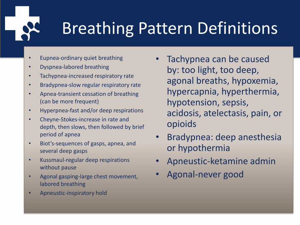
• Eupnea-ordinary quiet breathing
• Dyspnea-labored breathing
• Tachypnea-increased respiratory rate
• Bradypnea-slow regular respiratory rate
• Apnea-transient cessation of breathing (can be more frequent)
• Hyperpnea-fast and/or deep respirations
• Cheyne-Stokes-increase in rate and depth, then slows, then followed by brief period of apnea
• Biot’s-sequences of gasps, apnea, and several deep gasps
• Kussmaul-regular deep respirations without pause
• Agonal gasping-large chest movement, labored breathing
• Apneustic-inspiratory hold
• Tachypnea can be caused by: too light, too deep, agonal breaths, hypoxemia, hypercapnia, hyperthermia, hypotension, sepsis, acidosis, atelectasis, pain, or opioids
• Bradypnea: deep anesthesia or hypothermia
• Apneustic-ketamine admin
• Agonal-never good

Breathing Patterns
Tidal Volume and Dead Space
• Tidal Volume is defined as the volume of air inspired or expired in one breath
– Normal tidal volume is 10-15 ml/kg
• Dead space is the gas in the breathing system that does not participate in oxygen exchange.
– It is composed of gas from equipment dead space and the gas in the connecting airways in the lungs
– It also includes the gas that reaches the alveoli but is not perfused
– As the volume of dead space increases in proportion to tidal volume, the effectiveness of gas exchange will decrease
– Normal dead space for an anesthetized patient should be 2 ml/kg. Numbers exceeding that will create difficultly in maintaining normal
CO2.
– Important because a patient is breathing out expired gas and CO2 and is then re-inhaling that gas every breath
Anesthesia and Respiratory Monitoring
• Maintenance of adequate respiratory function is a prime requirement for safe anesthesia
• Inadequate tissue oxygenation can lead to acute cessation of vital organ function, especially the brain or heart, and lead to an anesthetic death
• Delayed recovery and post-anesthetic renal, hepatic, or cardiac insufficiency all come from inadequate respiratory function while under anesthesia

Anesthesia and Respiratory Monitoring
• Body positioning, concurrent drug use, and pre-anesthetic cardiorespiratory dysfunction (from mild to severe) all affect respiratory function while under anesthesia
• Sedatives, analgesics, anesthetics, and equipment used for inhalant anesthesia may profoundly alter respiration and the ability for the patient to maintain homeostasis on a cellular level

Respiratory Monitoring
• What are the options for respiratory monitoring– Auscultation of the chest– Pulse oximetry– Capnography
• Are there disadvantages of using a monitor?– Yes there are. You need to know what the data means
and how to interpret it. – Equipment can cause dead space issues for small
patients– Increased potential for leaks– Possible spread of contamination with reusable connectors
Patients
• All patients would benefit from respiratory monitoring but there are some that would benefit more than others
• These patients have pre-existing respiratory issues or are undergoing a thoracic surgery; some examples are: – Thoracotomy/thoracoscopy– Diaphragmatic hernia– Possible pneumothorax – Pneumonia– Asthma/heaves– Critically ill (septic or in shock)– Trauma– Brachycephalics
Respiratory Rate
• Breathing rates can vary widely and the only real information it gives is that your patient is breathing-Good to know
– On average: respiratory rate should not fall below 8bpm
• Where it becomes important is when there is a change in the rate or effort
– A change can be a sensitive indicator of an underlying change in the status of the patient
Other definitions
• Hyperventilation=occurs when alveolar ventilation is excessive relative to the metabolic rate-decrease in PaCO2– It may or may not be accompanied by tachypnea
• Hypoventilation=present when alveolar ventilation is small relative to metabolic rate-resulting in increase in PaCO2– It may be accompanied by bradypnea, normal rate, or
tachypnea • Hypoxemia=low oxygen content in the blood• Hypoxia=impaired oxygen delivery; takes into account CO,
perfusion, and O2 extraction by tissues– Most hypoxic patients go undetected and untreated
• Cyanosis=appearance of blue coloring due to excessive concentration of deoxyhemoglobin-2.5-5 g/dl
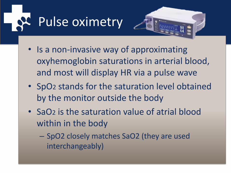
Pulse oximetry
• Is a non-invasive way of approximating oxyhemoglobin saturations in arterial blood, and most will display HR via a pulse wave
• SpO2 stands for the saturation level obtained by the monitor outside the body
• SaO2 is the saturation value of atrial blood within in the body
– SpO2 closely matches SaO2 (they are used interchangeably)
How does it work?
• Works on the principle that oxygenated hemoglobin absorbs red and infrared light at a specific frequencies (660nm for red light and 920-940 for infrared light)– Red is for deoxygenated, Infrared is for oxygenated
• It uses 2 light emitting diodes (LEDs) that pulse red and infrared light through perfused tissue several hundred times per second
• The amount of light absorbed at each wavelength is measured by photodetectors and that data is expressed as a percentage of oxygenated to total hemoglobin

Pulse ox waveforms

Different Types of Hemoglobin
• Oxyhemoglobin= oxygenated hemoglobin• Deoxyhemoglobin=deoxygenated hemoglobin• Carboxyhemoglobin=hemoglobin combined
with carbon monoxide (cannot carry O2)– Absorbs red light like oxyhemoglobin so it can
falsely increase the percentage
• Methemoglobin=when oxidation of iron atom without oxygen converts hemoglobin to methemoglobin (cannot carry O2)– Can absorb light at both wavelengths (660 and
940nm) thus pushing the percentage toward 85%
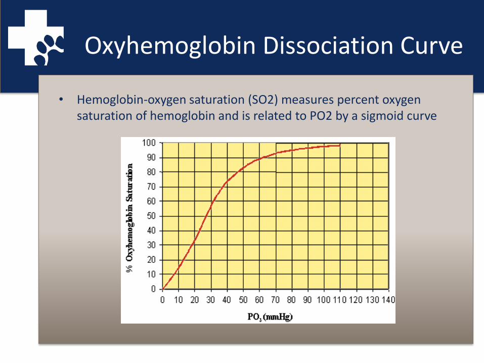
Oxyhemoglobin Dissociation Curve
• Hemoglobin-oxygen saturation (SO2) measures percent oxygen saturation of hemoglobin and is related to PO2 by a sigmoid curve
PaO2 vs. SpO2
• On room air or O2 of 21% the PaO2 should be 100mm Hg which is equivalent to an SpO2 of 98-100%
• A PaO2 of 80 mm Hg to an SpO2 of 95%• A PaO2 of 60 mm Hg to an SpO2 of 90%• A PaO2 of 40 mm Hg to an SpO2 of 75%• On 100% O2 the PaO2 should be 500mm Hg
– A big disadvantage to the pulse ox is that at 100% it does not indicate if the PaO2 is 500 or 100mm Hg

Accuracy and Inaccuracy
• The accuracy of the pulse oximeter is greatest within the range of 80-95% and is determined by the accuracy of the empirical formula that is programmed into the instrument
• What can make it inaccurate?– Dark pigmented skin/tongues– Vasoconstriction– Hypothermia– Hypoperfusion/hypovolemic– Tachycardia– Severe anemia– Hyperbilirubinemia– Ambient light– Oxyglobin
Capnography
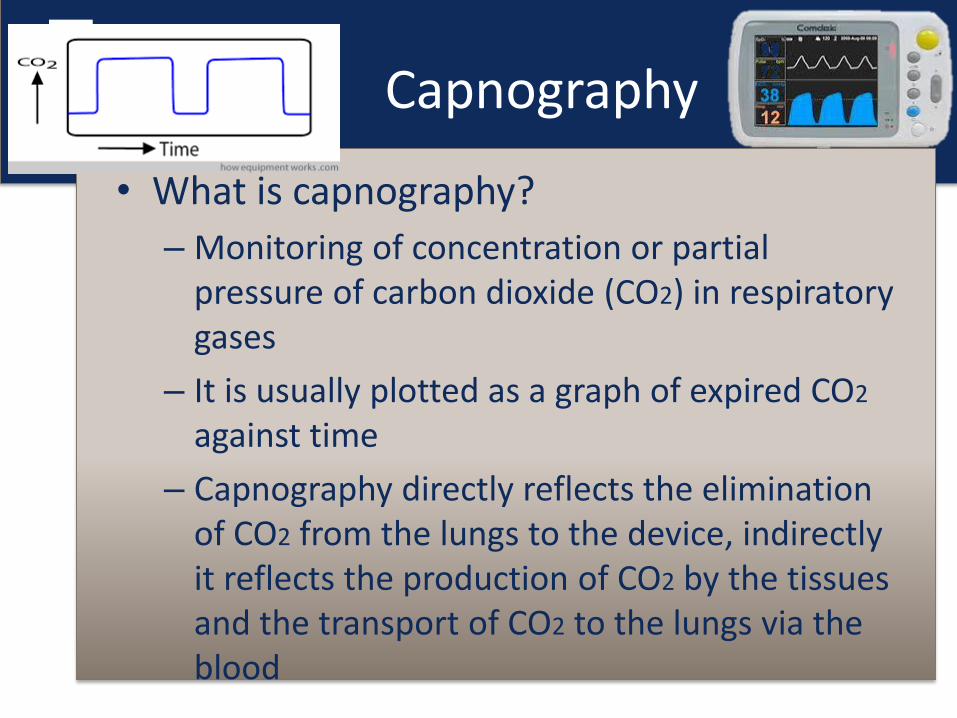
• What is capnography?
– Monitoring of concentration or partial pressure of carbon dioxide (CO2) in respiratory gases
– It is usually plotted as a graph of expired CO2
against time
– Capnography directly reflects the elimination of CO2 from the lungs to the device, indirectly it reflects the production of CO2 by the tissues and the transport of CO2 to the lungs via the blood
How does it work?
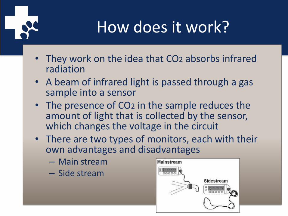
• They work on the idea that CO2 absorbs infrared radiation
• A beam of infrared light is passed through a gas sample into a sensor
• The presence of CO2 in the sample reduces the amount of light that is collected by the sensor, which changes the voltage in the circuit
• There are two types of monitors, each with their own advantages and disadvantages– Main stream– Side stream

Why is it useful?
• There is quite a bit of useful information that can be obtained from capnography– Gives information on respiratory rate for intubated as well as non-
intubated patients
– Shows if the there is an obstruction in the airway
– Hypoventilation or hyperventilation
– Allows evaluation of respiratory depression and any rebreathing that is occurring
– The shape of the curves shows if there are leaks in the circuit, if bronchospasm is present, apnea and more
– One of the earliest and most sensitive signs of cardiovascular collapse or arrest is an abrupt decrease in end-tidal CO2
– Will show if it is an esophageal intubation
– Mechanical error in breathing circuit

Partial Pressure of CO2
• PaCO2 is a measure of the ventilatory status of a patient– Normal ranges are between 35-45 mm Hg– Values may be slightly higher in anesthetized small animals– End tidal CO2 is usually 2-4 mm Hg lower than PaCO2 in
dogs
• A PaCO2 in excess of 60 mm Hg may be associated with excessive respiratory acidosis and represents sufficient hypoventilation to warrant PPV
• A PaCO2 below 20 mm Hg are associated with respiratory alkalosis and a decreased cerebral blood flow that may impair cerebral oxygenation
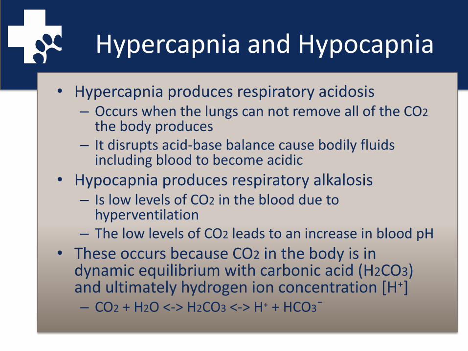
Hypercapnia and Hypocapnia
• Hypercapnia produces respiratory acidosis– Occurs when the lungs can not remove all of the CO2
the body produces– It disrupts acid-base balance cause bodily fluids
including blood to become acidic
• Hypocapnia produces respiratory alkalosis– Is low levels of CO2 in the blood due to
hyperventilation– The low levels of CO2 leads to an increase in blood pH
• These occurs because CO2 in the body is in dynamic equilibrium with carbonic acid (H2CO3) and ultimately hydrogen ion concentration [H⁺]– CO2 + H2O <-> H2CO3 <-> H⁺ + HCO3¯

Any Questions???????
• If you have any additional questions please feel free to send me an email at: