Monitoring of Low Molecular Weight Heparin ...
11
Monitoring of Low Molecular Weight Heparin Thromboprophylaxis with Alternative Methods to Anti-Factor Xa Ölander, Filippa; Schött, Ulf Published in: Anesthesia and Medical Practice Journal DOI: 10.29011/2637-9953.100136 2021 Document Version: Publisher's PDF, also known as Version of record Link to publication Citation for published version (APA): Ölander, F., & Schött, U. (2021). Monitoring of Low Molecular Weight Heparin Thromboprophylaxis with Alternative Methods to Anti-Factor Xa. Anesthesia and Medical Practice Journal, 5(1), 1-9. https://doi.org/10.29011/2637-9953.100136 Total number of authors: 2 Creative Commons License: Unspecified General rights Unless other specific re-use rights are stated the following general rights apply: Copyright and moral rights for the publications made accessible in the public portal are retained by the authors and/or other copyright owners and it is a condition of accessing publications that users recognise and abide by the legal requirements associated with these rights. • Users may download and print one copy of any publication from the public portal for the purpose of private study or research. • You may not further distribute the material or use it for any profit-making activity or commercial gain • You may freely distribute the URL identifying the publication in the public portal Read more about Creative commons licenses: https://creativecommons.org/licenses/ Take down policy If you believe that this document breaches copyright please contact us providing details, and we will remove access to the work immediately and investigate your claim.
Transcript of Monitoring of Low Molecular Weight Heparin ...
LUND UNIVERSITY
PO Box 117221 00 Lund+46 46-222 00 00
Monitoring of Low Molecular Weight Heparin Thromboprophylaxis with AlternativeMethods to Anti-Factor Xa
Ölander, Filippa; Schött, Ulf
Published in:Anesthesia and Medical Practice Journal
DOI:10.29011/2637-9953.100136
2021
Document Version:Publisher's PDF, also known as Version of record
Link to publication
Citation for published version (APA):Ölander, F., & Schött, U. (2021). Monitoring of Low Molecular Weight Heparin Thromboprophylaxis withAlternative Methods to Anti-Factor Xa. Anesthesia and Medical Practice Journal, 5(1), 1-9.https://doi.org/10.29011/2637-9953.100136
Total number of authors:2
Creative Commons License:Unspecified
General rightsUnless other specific re-use rights are stated the following general rights apply:Copyright and moral rights for the publications made accessible in the public portal are retained by the authorsand/or other copyright owners and it is a condition of accessing publications that users recognise and abide by thelegal requirements associated with these rights. • Users may download and print one copy of any publication from the public portal for the purpose of private studyor research. • You may not further distribute the material or use it for any profit-making activity or commercial gain • You may freely distribute the URL identifying the publication in the public portal
Read more about Creative commons licenses: https://creativecommons.org/licenses/Take down policyIf you believe that this document breaches copyright please contact us providing details, and we will removeaccess to the work immediately and investigate your claim.
Anesth Med Pract J, an open access journal
ISSN: 2637-9953
1 Volume 5; Issue 01
Anesthesia and Medical Practice JournalCase Report
Ölander F and Schött U Anesth Med Pract J Res 5: 136.
Monitoring of Low Molecular Weight Heparin Thromboprophylaxis with Alternative Methods to Anti-Factor Xa
Filippa Ölander1, Ulf Schött2*
1Department of Intensive and Perioperative Care, Skane University Hospital, Sweden 2Institution of Clinical Sciences, Medical Faculty, Lund University, Sweden
*Corresponding author: Ulf Schött, Institution of Clinical Sciences, Medical Faculty, Lund University, Sweden
Citation: Ölander F, Schött U (2021) Monitoring of Low Molecular Weight Heparin Thromboprophylaxis with Alternative Methods to Anti-Factor Xa. Anesth Med Pract J Res 5: 136. DOI: 10.29011/2637-9953.100136
Received Date: 29 January, 2021; Accepted Date: 03 February, 2021; Published Date: 08 February, 2021
DOI: 10.29011/2637-9953.100136
AbstractIntroduction: Initiating low molecular weight heparin (LMWH) thromboprophylaxis too early in patients with traumatic brain injury increases the risk of intracranial bleeding. Therefore, it is important to monitor LMWH and asses when it is safe to initiate. The aim of this research was to study alternative monitoring methods for LMWH than the standard method anti-factor Xa (anti-FXa), and to investigate the peak anti-FXa level. We hoped to answer “How do rotational thromboelastometry (ROTEM) and Sonoclot change at different LMWH concentrations added in vitro to blood from intensive care patients? How do point of care parameters change after subcutaneous LMWH administration on healthy volunteers with consecutive measurements to catch the peak effect?”.
Methods: Different concentrations of enoxaparin were added in vitro to citrated whole blood from fifteen intensive care patients. The first ten patients’ coagulation was analysed with ROTEM using the INTEM and NATEM assays, and the last five with Sonoclot with a kaolin activator and ROTEM INTEM. Previously collected data was used from nine healthy volunteers that had received subcutaneous enoxaparin. Citrated blood samples were collected before and after LMWH initiation and analysed with Sonoclot kaolin and a chromogenic anti-FXa-assay. Friedman test, Dunn’s multiple comparison test and Spearman’s correlation test were performed.
Results: ROTEM INTEM CT, CFT, A10 and MCF were significantly affected with increasing in vitro enoxaparin from 0.4 anti-FXa concentration. ROTEM NATEM CT was also prolonged at this LMWH concentration. Sonoclot’s parameters didn’t significantly change with increasing in vitro enoxaparin. The peak of in vivo LMWH was reached after 2 to 4 hours with a variation of peak anti-FXa between 0.3-0.5 IU/mL.
Conclusions: ROTEM INTEM CT performed best and was prolonged at anti-FXa from 0.4-0.6 IU/mL. ROTEM INTEM should be tested in neurointensive care to increase the safety of LMWH thromboprophylaxis and possibly to individualize the dosage.
IntroductionVenous thromboembolism (VTE) is a condition where
clots are formed in the veins. Most cases of VTE are deep vein thrombosis (DVT), most commonly located in the lower limbs. The clot will obstruct the blood flow in its area resulting in swelling, pain and discoloration of the skin. If the DVT clot breaks free and migrates it will become an embolism, most commonly and seriously a pulmonary embolism (PE). This is life threatening and is therefore very important to prevent.
Intensive care and neurointensive care patients are at a greater risk of developing VTE [1]. One group of these patients are
the ones with traumatic brain injury (TBI), which is a major cause of death globally amongst young adults and youths [2]. A common complication of TBI is intracerebral hemorrhages (ICH). ICH patients usually have an altered coagulation profile and increased coagulation after bleeding has ended. These patients are usually immobilised for a long time, which may lead to blood stasis in the deep veins [3], further increasing the risk for thromboembolism.
Moreover, patients with ICH have damaged blood vessels in the brain. In some TBI patients, the blood vessels are even damaged in other parts of the body. The damaged endothelium lead to inflammation, act as prothrombic surfaces and decrease their release of fibrinolytic proteins. The concentration of acute
Citation: Ölander F, Schött U (2021) Monitoring of Low Molecular Weight Heparin Thromboprophylaxis with Alternative Methods to Anti-Factor Xa. Anesth Med Pract J Res 5: 136. DOI: 10.29011/2637-9953.100136
2 Volume 5; Issue 01
Anesth Med Pract J, an open access journal
ISSN: 2637-9953
phase reactants rises in response to inflammation. As a result, the blood gets more hypercoagulative. All these factors explain why TBI patients have an increased risk of developing VTE [4]. Without prophylaxis, it is estimated that 20 % of severe TBI patients develop DVT. Therefore, it is recommended to use both mechanical and pharmacological thromboprophylaxis [2,3].
Low molecular weight heparin (LMWH) is one of the most commonly used pharmacological thromboprophylaxis to prevent VTE in intensive care patients [1]. There are many different types of LMWH, but they all indirectly inhibit factor Xa in the common coagulation pathway, and directly inhibit factor IIa in varying degrees. Factor Xa cleaves factor II to its active form factor IIa, also called thrombin. When these factors are inhibited, the conversion of fibrinogen to fibrin will be disrupted as well as clot formation. Monitoring LMWH is recommended when it is important with accurate dosing, as with TBI patients. The reason for this is if LMWH is given too early, the patients have an increased risk for expansion of the ICH [2]. Despite that, there is no clear evidence that shows when it is safe to start LMWH. Some studies recommend initiating LMWH between 24 to 72 hours after injury, while performing consecutive computed tomography on the patients to avoid starting LMWH in patients with an expanding ICH [2,5,6]. One of these studies, concluded that LMWH should be started 48 to 72 h after trauma after at least two computed tomography scans, as opposed to 24 to 72 hours [6]. Other studies have compared early (< 72 h) and late (≥ 72 h) LMWH initiation. The patients that started LMWH treatment early had a decreased risk of DVT and PE without increasing the risk of ICH progression [7-9].
Current guidelines from Society of Critical Care Medicine and Neurocritical Care recommend starting LMWH in patients with stable hematomas and no ongoing coagulopathy within 48 hours after hospital admission along with mechanical devices [10]. Additionally, this approach is recommended by the updated TBI guidelines from the United Kingdom Brain Trauma Foundation [11]. The gold standard for monitoring LMWH treatment is by measuring its’ anti-factor Xa (anti-FXa) activity in the patient’s plasma. The peak level of anti-FXa has been estimated to be reached 3 to 5 hours after administration [4,12]. This is important to study, since anti-FXa’s peak levels should not exceed the recommendations. That would lead to an increased risk of bleeding complications.
Rotational thromboelastometry (ROTEM) and Sonoclot are viscoelastic point of care (POC) analyses measuring the entire process of blood coagulation. They reflect hypo- and hypercoagulation better than standard laboratory coagulation tests [13,14]. One study identified perioperative hypercoagulable ROTEM parameters using ROTEM assays INTEM and EXTEM in 10 out of 333 non-cardiac surgery patients who developed DVT after surgery even after pharmacological prophylaxis. This
indicates that preoperative ROTEM analyses can be used to detect patients with an increased risk for postoperative thromboembolic complications [15].
Plasma analysis of anti-FXa must be analysed in a central laboratory, but with the POC devices the blood can be analysed in patient-near laboratories. This leads to faster turnaround times [16]. Therefore, it is interesting to study if such POC devices can be used to monitor LMWH instead of the plasma anti-FXa assay. A previous study evaluated if two types of LMWH, enoxaparin and tinzaparin, added in vitro in different concentrations had dose-dependent effects on ROTEM INTEM clotting time (CT). It was found that the ROTEM CT was increasingly prolonged by both types of LMWH at 0.5, 1.0 and 1.5 anti-FXa IU/mL LMWH concentrations [17]. However, this area needs further research to test its’ clinical significance to guide optimal LMWH dosages in different clinical situations. The in vitro doses reflect the in vivo peak LMWH concentrations after subcutaneous thromboprophylaxis and the concentrations achieved with therapeutic LMWH treatments of actual thromboembolism [17].
There has also been research about anticoagulant monitoring with Sonoclot. A study using the anticoagulant fondaparinux that inhibits factor Xa, evaluated if Sonoclot could be used for in vitro monitoring. Nilsson et al. found that the Sonoclot parameter clot rate significantly decreased at increasing doses of fondaparinux, indicating a potential clinical value [18]. This has also been shown with ROTEM [19].
The aim of this research is to study alternative monitoring methods for LMWH in the thromboprophylactic anti-FXa concentration range, e.g. 0-0.5-0.6 IU/mL, and compare these with the standard method anti-FXa. Moreover, we want to further investigate the peak anti-FXa level and when it occurs after subcutaneous injection.
Questions
How do viscoelastic POC coagulation analyses ROTEM and Sonoclot change at different LMWH concentrations added in vitro to blood from intensive care patients? How do POC parameters change after subcutaneous LMWH administration on healthy volunteers with consecutive measurements to catch the peak effect?
Material and Methods
Study population
Fifteen patients, admitted to the intensive care unit (ICU), Skane University Hospital, Lund, were included. Additionally, data that my supervisor previously had collected on nine healthy volunteers was included in this study. All study participants gave consent before any blood was sampled and were aged eighteen or over. The Regional Ethical Review Board in Lund (registration
Citation: Ölander F, Schött U (2021) Monitoring of Low Molecular Weight Heparin Thromboprophylaxis with Alternative Methods to Anti-Factor Xa. Anesth Med Pract J Res 5: 136. DOI: 10.29011/2637-9953.100136
3 Volume 5; Issue 01
Anesth Med Pract J, an open access journal
ISSN: 2637-9953
numbers 2010/482 and DNR 2017/636) approved the study.
Blood sampling
Blood from intensive care patients was sampled from an arterial catheter with a continuous flushing and sampling membrane. This way of drawing blood eliminates the need to dispose of blood samples. Blood was collected in citrated tubes (Becton Dickison (BD), Plymouth, UK, Vacutainer tubes, 2.7 mL, 0.109 sodium citrate) for whole blood ROTEM and Sonoclot analyses. For the data that my supervisor previously had collected, venous blood samples were drawn from the brachial vein through an indwelling peripheral venous catheter. Blood was sampled before administrating LMWH, and 1, 2, 3, 4 and 5 hours after LMWH initiation. Afterwards, the samples were used for Sonoclot and chromogenic anti-FXa analyses.
LMWH for in vitro dose-response
The LMWH used for the in vitro experiments was enoxaparin (Klexane, Sanofi-Aventis, Guildford, UK). Enoxaparin was diluted with isotonic saline (9 mg/mL NaCl: Fresenius Kabi, Bad Homburg, Germany) in an Eppendorf tube. Then 20, 40, and 60 µl of the dilution was added to the blood, leading to a plasma concentration of 0.2, 0.4 and 0.6 anti-FXa IU/mL.
LMWH for in vivo peak LMWH monitoring
For the in vivo experiments previously performed by my supervisor, the LMWH enoxaparin was used. Nine healthy volunteers received a standard subcutaneous 40 mg dose of enoxaparin.
ROTEM
ROTEM is a viscoelastic POC coagulation device that measures the clot’s physical property. Different reagents can be used in ROTEM. For this study, the NATEM and INTEM assays were used. NATEM represents the native coagulation and INTEM represents the intrinsic coagulation pathway [20]. Both NATEM and INTEM were used on the first ten patients, and on the last five only INTEM was performed. In the NATEM assay, 20 µl of 0.2 Molar (M) CaCl2 (StarTEM reagent) was added to a fixed cup of the ROTEM device. With a new pipette, 300 µl of citrated whole blood was added to the cup. ROTEM has a rotating pin that is suspended into the blood sample. As the blood begins to clot, the rotating pin’s movement is registered which generates a graph that displays the changes in viscoelasticity of the clotting process [16]. In the INTEM assay, 20 µl of 0.2 M CaCl2 (StarTEM reagent) and 20 µl of the InTEM reagent were added to the fixed cup before adding 300 µl of citrated whole blood.
The variables acquired from the NATEM and INTEM assays were as followed: Clotting time (CT), the time from when the reagent is added to the blood until the start of the clotting process. Clotting formation time (CFT), how long it takes for a clot to get
from a thickness of 2 mm to 20 mm. AA (α-angle), the speed of the clotting process. A10, the amplitude after 10 minutes. Maximum clot firmness (MCF), the maximum amplitude in the test and corresponds to maximal strength of the fibrin clot [16]. The normal values for NATEM: CT, 300-1000 s; CFT, 150-700 s; AA, 30-70; MCF, 40-65 mm. The normal values for INTEM: CT, 100-240 s; CFT, 30-110 s; AA, 70-83; A10, 44-66 mm; MCF, 50-72 mm.
Sonoclot
Sonoclot is a viscoelastic POC coagulation device, like ROTEM. The device has an oscillating plastic probe that is suspended into the blood sample and measures the viscoelastic drag impedance that the developing clot impose [16]. We used the kaolin activated test in this study and performed it on the last five patients. 500 µl of citrated whole blood was pipetted into an Eppendorf tube, then 20 µl of 0.25 M CaCl2 was added to the blood. After each tube had been mixed, 360 µl of the blood and CaCl2 solution was pipetted into the cuvette of the Sonoclot, and the analysis was started. My supervisor had previously collected Sonoclot data on healthy volunteers before and after giving subcutaneous LMWH to healthy volunteers. The blood was analysed before initiating LMWH, and 1, 2, 3, 4 and 5 hours after administering LMWH. The Sonoclot analysis was performed using the same method as described above. The variables obtained from the Sonoclot were as followed: Activated clotting time (ACT), the time from the beginning of the test until the start of a fibrin formation. Clot rate (CR), the rate of the fibrin formation and clot development [16]. The normal values for Sonoclot kaolin activated test: ACT, 97-178 s; CT, 15-33 units/min.
Coefficient of variation for ROTEM
The coefficient of variation (CV) has been calculated for ROTEM and Sonoclot kaolin in previous research [21,22]. However, it has not been done on LMWH affected blood. Therefore, we wanted to calculate it for blood samples with 0.6 anti-FXa IU/mL in this study to compare the ROTEM INTEM and NATEM CVs from those from non-anticoagulated blood. We analysed citrated whole blood with 0.6 anti-FXa IU/mL from two patients using ROTEM NATEM and INTEM. The first patient’s blood sample was analysed eight times using the INTEM assay, and the other patient’s sample was analysed seven times with the NATEM assay. Both assays were performed the same way as described earlier.
Coefficient of variation for Sonoclot – not performed
With Sonoclot the test variability is much higher than for ROTEM [21], but with a rebuilt and computerized Sonoclot Coagulation AnalyzerTM used in the present study CVs has decreased to the same level as for ROTEM [16]. We did not have enough blood samples to perform CV estimation for Sonoclot at LMWH concentration anti-FXa 0.6 IU/mL.
Citation: Ölander F, Schött U (2021) Monitoring of Low Molecular Weight Heparin Thromboprophylaxis with Alternative Methods to Anti-Factor Xa. Anesth Med Pract J Res 5: 136. DOI: 10.29011/2637-9953.100136
4 Volume 5; Issue 01
Anesth Med Pract J, an open access journal
ISSN: 2637-9953
Chromogenic anti-FXa testing for in vivo LMWH
The plasma anti-FXa level of the healthy volunteers’ blood samples was measured using a chromogenic substrate (Upjohn (Pharmacia), New York, NY, USA) in order to monitor LMWH’s peak effect.
Statistical Analysis
GraphPad Prism was used for the statistical analyses and figures. As our data is nonparametric and paired, we used Friedman test and Dunn’s multiple comparison test to determine any significant differences when increasing the anti-FXa concentration using ROTEM and Sonoclot, and when monitoring in vivo LMWH using Sonoclot.
Spearman’s correlation test was used to calculate
correlations between different parameters. This test is also used on nonparametric data.ResultsROTEM with increasing in vitro anti-FXa concentrations
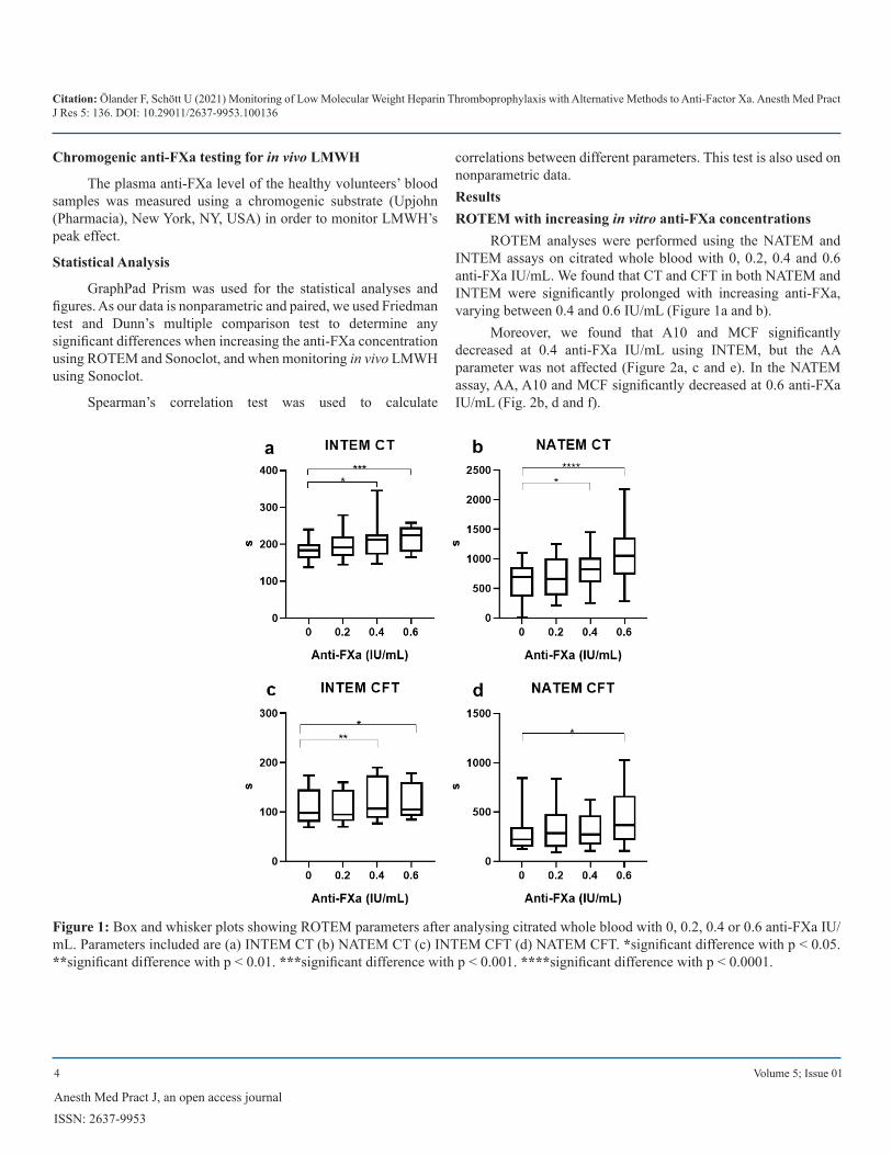
ROTEM analyses were performed using the NATEM and INTEM assays on citrated whole blood with 0, 0.2, 0.4 and 0.6 anti-FXa IU/mL. We found that CT and CFT in both NATEM and INTEM were significantly prolonged with increasing anti-FXa, varying between 0.4 and 0.6 IU/mL (Figure 1a and b).
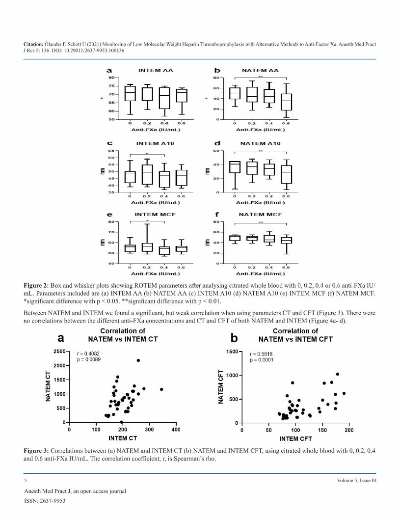
Moreover, we found that A10 and MCF significantly decreased at 0.4 anti-FXa IU/mL using INTEM, but the AA parameter was not affected (Figure 2a, c and e). In the NATEM assay, AA, A10 and MCF significantly decreased at 0.6 anti-FXa IU/mL (Fig. 2b, d and f).
Figure 1: Box and whisker plots showing ROTEM parameters after analysing citrated whole blood with 0, 0.2, 0.4 or 0.6 anti-FXa IU/mL. Parameters included are (a) INTEM CT (b) NATEM CT (c) INTEM CFT (d) NATEM CFT. *significant difference with p < 0.05. **significant difference with p < 0.01. ***significant difference with p < 0.001. ****significant difference with p < 0.0001.
Citation: Ölander F, Schött U (2021) Monitoring of Low Molecular Weight Heparin Thromboprophylaxis with Alternative Methods to Anti-Factor Xa. Anesth Med Pract J Res 5: 136. DOI: 10.29011/2637-9953.100136
5 Volume 5; Issue 01
Anesth Med Pract J, an open access journal
ISSN: 2637-9953
Figure 2: Box and whisker plots showing ROTEM parameters after analysing citrated whole blood with 0, 0.2, 0.4 or 0.6 anti-FXa IU/mL. Parameters included are (a) INTEM AA (b) NATEM AA (c) INTEM A10 (d) NATEM A10 (e) INTEM MCF (f) NATEM MCF. *significant difference with p < 0.05. **significant difference with p < 0.01.
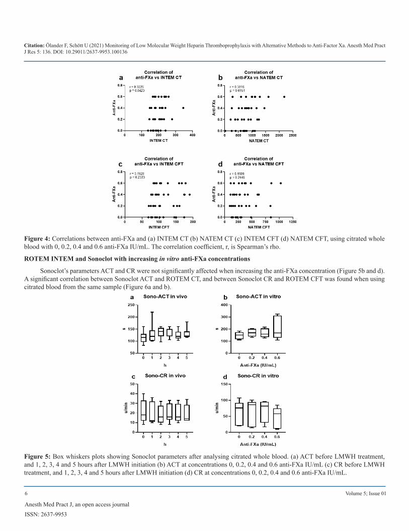
Between NATEM and INTEM we found a significant, but weak correlation when using parameters CT and CFT (Figure 3). There were no correlations between the different anti-FXa concentrations and CT and CFT of both NATEM and INTEM (Figure 4a- d).
Figure 3: Correlations between (a) NATEM and INTEM CT (b) NATEM and INTEM CFT, using citrated whole blood with 0, 0.2, 0.4 and 0.6 anti-FXa IU/mL. The correlation coefficient, r, is Spearman’s rho.
Citation: Ölander F, Schött U (2021) Monitoring of Low Molecular Weight Heparin Thromboprophylaxis with Alternative Methods to Anti-Factor Xa. Anesth Med Pract J Res 5: 136. DOI: 10.29011/2637-9953.100136
6 Volume 5; Issue 01
Anesth Med Pract J, an open access journal
ISSN: 2637-9953
Figure 4: Correlations between anti-FXa and (a) INTEM CT (b) NATEM CT (c) INTEM CFT (d) NATEM CFT, using citrated whole blood with 0, 0.2, 0.4 and 0.6 anti-FXa IU/mL. The correlation coefficient, r, is Spearman’s rho.
ROTEM INTEM and Sonoclot with increasing in vitro anti-FXa concentrations
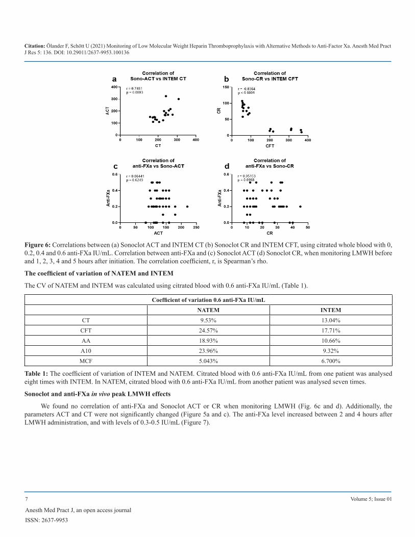
Sonoclot’s parameters ACT and CR were not significantly affected when increasing the anti-FXa concentration (Figure 5b and d). A significant correlation between Sonoclot ACT and ROTEM CT, and between Sonoclot CR and ROTEM CFT was found when using citrated blood from the same sample (Figure 6a and b).
Figure 5: Box whiskers plots showing Sonoclot parameters after analysing citrated whole blood. (a) ACT before LMWH treatment, and 1, 2, 3, 4 and 5 hours after LMWH initiation (b) ACT at concentrations 0, 0.2, 0.4 and 0.6 anti-FXa IU/mL (c) CR before LMWH treatment, and 1, 2, 3, 4 and 5 hours after LMWH initiation (d) CR at concentrations 0, 0.2, 0.4 and 0.6 anti-FXa IU/mL.
Citation: Ölander F, Schött U (2021) Monitoring of Low Molecular Weight Heparin Thromboprophylaxis with Alternative Methods to Anti-Factor Xa. Anesth Med Pract J Res 5: 136. DOI: 10.29011/2637-9953.100136
7 Volume 5; Issue 01
Anesth Med Pract J, an open access journal
ISSN: 2637-9953
Figure 6: Correlations between (a) Sonoclot ACT and INTEM CT (b) Sonoclot CR and INTEM CFT, using citrated whole blood with 0, 0.2, 0.4 and 0.6 anti-FXa IU/mL. Correlation between anti-FXa and (c) Sonoclot ACT (d) Sonoclot CR, when monitoring LMWH before and 1, 2, 3, 4 and 5 hours after initiation. The correlation coefficient, r, is Spearman’s rho.
The coefficient of variation of NATEM and INTEM
The CV of NATEM and INTEM was calculated using citrated blood with 0.6 anti-FXa IU/mL (Table 1).
Coefficient of variation 0.6 anti-FXa IU/mL
NATEM INTEM
CT 9.53% 13.04%
CFT 24.57% 17.71%
AA 18.93% 10.66%
A10 23.96% 9.32%
MCF 5.043% 6.700%
Table 1: The coefficient of variation of INTEM and NATEM. Citrated blood with 0.6 anti-FXa IU/mL from one patient was analysed eight times with INTEM. In NATEM, citrated blood with 0.6 anti-FXa IU/mL from another patient was analysed seven times.
Sonoclot and anti-FXa in vivo peak LMWH effects
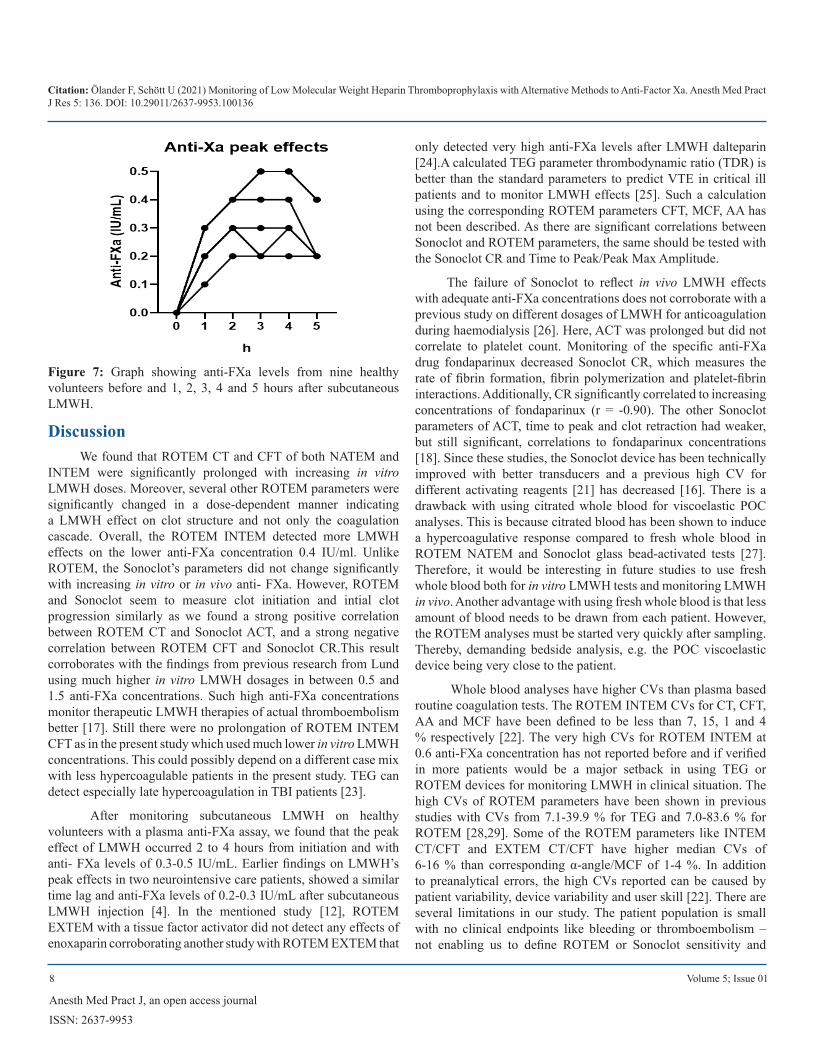
We found no correlation of anti-FXa and Sonoclot ACT or CR when monitoring LMWH (Fig. 6c and d). Additionally, the parameters ACT and CT were not significantly changed (Figure 5a and c). The anti-FXa level increased between 2 and 4 hours after LMWH administration, and with levels of 0.3-0.5 IU/mL (Figure 7).
Citation: Ölander F, Schött U (2021) Monitoring of Low Molecular Weight Heparin Thromboprophylaxis with Alternative Methods to Anti-Factor Xa. Anesth Med Pract J Res 5: 136. DOI: 10.29011/2637-9953.100136
8 Volume 5; Issue 01
Anesth Med Pract J, an open access journal
ISSN: 2637-9953
Figure 7: Graph showing anti-FXa levels from nine healthy volunteers before and 1, 2, 3, 4 and 5 hours after subcutaneous LMWH.
DiscussionWe found that ROTEM CT and CFT of both NATEM and
INTEM were significantly prolonged with increasing in vitro LMWH doses. Moreover, several other ROTEM parameters were significantly changed in a dose-dependent manner indicating a LMWH effect on clot structure and not only the coagulation cascade. Overall, the ROTEM INTEM detected more LMWH effects on the lower anti-FXa concentration 0.4 IU/ml. Unlike ROTEM, the Sonoclot’s parameters did not change significantly with increasing in vitro or in vivo anti- FXa. However, ROTEM and Sonoclot seem to measure clot initiation and intial clot progression similarly as we found a strong positive correlation between ROTEM CT and Sonoclot ACT, and a strong negative correlation between ROTEM CFT and Sonoclot CR.This result corroborates with the findings from previous research from Lund using much higher in vitro LMWH dosages in between 0.5 and 1.5 anti-FXa concentrations. Such high anti-FXa concentrations monitor therapeutic LMWH therapies of actual thromboembolism better [17]. Still there were no prolongation of ROTEM INTEM CFT as in the present study which used much lower in vitro LMWH concentrations. This could possibly depend on a different case mix with less hypercoagulable patients in the present study. TEG can detect especially late hypercoagulation in TBI patients [23].
After monitoring subcutaneous LMWH on healthy volunteers with a plasma anti-FXa assay, we found that the peak effect of LMWH occurred 2 to 4 hours from initiation and with anti- FXa levels of 0.3-0.5 IU/mL. Earlier findings on LMWH’s peak effects in two neurointensive care patients, showed a similar time lag and anti-FXa levels of 0.2-0.3 IU/mL after subcutaneous LMWH injection [4]. In the mentioned study [12], ROTEM EXTEM with a tissue factor activator did not detect any effects of enoxaparin corroborating another study with ROTEM EXTEM that
only detected very high anti-FXa levels after LMWH dalteparin [24].A calculated TEG parameter thrombodynamic ratio (TDR) is better than the standard parameters to predict VTE in critical ill patients and to monitor LMWH effects [25]. Such a calculation using the corresponding ROTEM parameters CFT, MCF, AA has not been described. As there are significant correlations between Sonoclot and ROTEM parameters, the same should be tested with the Sonoclot CR and Time to Peak/Peak Max Amplitude.
The failure of Sonoclot to reflect in vivo LMWH effects with adequate anti-FXa concentrations does not corroborate with a previous study on different dosages of LMWH for anticoagulation during haemodialysis [26]. Here, ACT was prolonged but did not correlate to platelet count. Monitoring of the specific anti-FXa drug fondaparinux decreased Sonoclot CR, which measures the rate of fibrin formation, fibrin polymerization and platelet-fibrin interactions. Additionally, CR significantly correlated to increasing concentrations of fondaparinux (r = -0.90). The other Sonoclot parameters of ACT, time to peak and clot retraction had weaker, but still significant, correlations to fondaparinux concentrations [18]. Since these studies, the Sonoclot device has been technically improved with better transducers and a previous high CV for different activating reagents [21] has decreased [16]. There is a drawback with using citrated whole blood for viscoelastic POC analyses. This is because citrated blood has been shown to induce a hypercoagulative response compared to fresh whole blood in ROTEM NATEM and Sonoclot glass bead-activated tests [27]. Therefore, it would be interesting in future studies to use fresh whole blood both for in vitro LMWH tests and monitoring LMWH in vivo. Another advantage with using fresh whole blood is that less amount of blood needs to be drawn from each patient. However, the ROTEM analyses must be started very quickly after sampling. Thereby, demanding bedside analysis, e.g. the POC viscoelastic device being very close to the patient.
Whole blood analyses have higher CVs than plasma based routine coagulation tests. The ROTEM INTEM CVs for CT, CFT, AA and MCF have been defined to be less than 7, 15, 1 and 4 % respectively [22]. The very high CVs for ROTEM INTEM at 0.6 anti-FXa concentration has not reported before and if verified in more patients would be a major setback in using TEG or ROTEM devices for monitoring LMWH in clinical situation. The high CVs of ROTEM parameters have been shown in previous studies with CVs from 7.1-39.9 % for TEG and 7.0-83.6 % for ROTEM [28,29]. Some of the ROTEM parameters like INTEM CT/CFT and EXTEM CT/CFT have higher median CVs of 6-16 % than corresponding α-angle/MCF of 1-4 %. In addition to preanalytical errors, the high CVs reported can be caused by patient variability, device variability and user skill [22]. There are several limitations in our study. The patient population is small with no clinical endpoints like bleeding or thromboembolism – not enabling us to define ROTEM or Sonoclot sensitivity and
Citation: Ölander F, Schött U (2021) Monitoring of Low Molecular Weight Heparin Thromboprophylaxis with Alternative Methods to Anti-Factor Xa. Anesth Med Pract J Res 5: 136. DOI: 10.29011/2637-9953.100136
9 Volume 5; Issue 01
Anesth Med Pract J, an open access journal
ISSN: 2637-9953
specificity to detect these clinical complications. The in vitro testing with use of citrated blood does not necessarily reflect the in vivo haemostasis and real effect of an in vivo administered LMWH thromboprophylactic dose. The anti-FXa levels in our study with volunteers using a chromogenic assay were much higher than in clinical neurointensive care patients presented in a previous study from our centre [12]. The reason for this is unclear but could possibly be related to less absorption of subcutaneously administered drugs in critical ill patients. The high variability during LMWH anticoagulation has not been reported before and heavily influences the reported ROTEM changes after increasing doses of LMWH.
ConclusionROTEM INTEM CT performed best and was prolonged
at anti-FXa from 0.4-0.6 IU/mL. ROTEM INTEM should be tested in neurointensive care to increase the safety of LMWH thromboprophylaxis and possibly used to individualize the dosage. This should preferably be used with calculated parameters like the TDR.
References1. Ejaz A, Ahmed MM, Tasleem A, Rafay Khan Niazi M, Ahsraf MF, et
al. (2018) Thromboprophylaxis in Intensive Care Unit Patients: A Literature Review. Cureus. 10: e3341.
2. Haddad SH, Arabi YM. (2012) Critical care management of severe traumatic brain injury in adults. Scand J Trauma Resusc Emerg Med. 20:12.
3. Kaufman HH, Satterwhite T, McConnell BJ, Costin B, Borit A, et al. (1983) Deep vein thrombosis and pulmonary embolism in head injured patients. Angiology. 34: 627-638.
4. Lindström O, Thomas O, Strandberg K, Rundgren M, Ekelius Cederberg D et al. (2018) Thrombin Generation Assay for Evaluation of Thromboprophylaxis in Neurointensive Care. Int J Cerebrovasc Dis Stroke. 1: 1-10.
5. Minshall CT, Eriksson EA, Leon SM, Doben AR, McKinzie BP, et al. (2011) Safety and efficacy of heparin or enoxaparin prophylaxis in blunt trauma patients with a head abbreviated injury severity score >2. J Trauma. 71: 396-399.
6. Dudley RR, Aziz I, Bonnici A, Saluja RS, Lamoureux J, et al. (2010) Early venous thromboembolic event prophylaxis in traumatic brain injury with low-molecular- weight heparin: risks and benefits. J Neurotrauma. 27: 2165-2172.
7. Byrne JP, Mason SA, Gomez D, Hoeft C, Subacius H, et al. (2016) Timing of Pharmacologic Venous Thromboembolism Prophylaxis in Severe Traumatic Brain Injury: A Propensity-Matched Cohort Study. J Am Coll Surg. 223: 621-631.e5.
8. Koehler DM, Shipman J, Davidson MA, Guillamondegui O. (2011) Is early venous thromboembolism prophylaxis safe in trauma patients with intracranial hemorrhage. J Trauma. 70: 324-329.
9. Jamjoom AA, Jamjoom AB. (2013) Safety and efficacy of early pharmacological thromboprophylaxis in traumatic brain injury: systematic review and meta-analysis. J Neurotrauma. 30: 503-511.
10. Nyquist P, Bautista C, Jichici D, Burns J, Chhangani S, et al. (2016) Prophylaxis of Venous Thrombosis in Neurocritical Care Patients: An Evidence-Based Guideline: A Statement for Healthcare Professionals from the Neurocritical Care Society. Neurocrit Care. 24: 47-60.
11. Carney N, Totten AM, O’Reilly C, Ullman JS, Hawryluk GW, et al. (2017) Guidelines for the Management of Severe Traumatic Brain Injury, Fourth Edition. Neurosurgery. 80: 6-15.
12. Wei MY, Ward SM. (2015) The Anti-Factor Xa Range For Low Molecular Weight Heparin Thromboprophylaxis. Hematol Rep. 7: 5844.
13. Johansson PI, Sorensen AM, Perner A, Welling KL, Wanscher M, et al. (2011) Disseminated intravascular coagulation or acute coagulopathy of trauma shock early after trauma? An observational study. Crit Care. 15: R272.
14. Miao W, Zhao K, Deng W, Teng J. (2018) Coagulation Factor Hyperfunction After Subarachnoid Hemorrhage Induces Deep Venous Thrombosis. World Neurosurg. 110: e46-e52.
15. Hincker A, Feit J, Sladen RN, Wagener G. (2014) Rotational thromboelastometry predicts thromboembolic complications after major non-cardiac surgery. Crit Care. 18: 549.
16. Ganter MT, Hofer CK. (2008) Coagulation monitoring: current techniques and clinical use of viscoelastic point-of-care coagulation devices. Anesth Analg. 106: 1366-1375.
17. 17.Thomas O, Larsson A, Tynngard N, Schott U. (2015) Thromboelastometry versus free- oscillation rheometry and enoxaparin versus tinzaparin: an in-vitro study comparing two viscoelastic haemostatic tests’ dose-responses to two low molecular weight heparins at the time of withdrawing epidural catheters from ten patients after major surgery. BMC Anesthesiol. 15:170.
18. Nilsson CU, Engstrom M. (2017) Monitoring fondap arinux with the Sonoclot. Blood Coagul Fibrinolysis. 18: 619-622.
19. Engstrom M, Rundgren M, Schott U. (2010) An evaluation of monitoring possibilities of argatroban using rotational thromboelastometry and activated partial thromboplastin time. Acta Anaesthesiol Scand. 54: 86-91.
20. Rossetto V, Spiezia L, Senzolo M, (2013) Rodriguez-Castro KI, Maggiolo S, Simioni P. Whole blood rotation thromboelastometry (ROTEM(R)) profiles in subjects with non- neoplastic portal vein thrombosis. Thromb Res. 132: e131-4.
21. Ekback G, Carlsson O, Schott U. (1999) Sonoclot coagulation analysis: a study of test variability. J Cardiothorac Vasc Anesth. 13: 393-397.
22. Nilsson CU, Strandberg K, Reinstrup P. (2018) Warfarin monitoring with viscoelastic haemostatic assays, thrombin generation, coagulation factors and correlations to Owren and Quick prothrombin time. Scand J Clin Lab Invest. 78: 358-364.
23. Massaro AM, Doerfler S, Nawalinski K, Michel B, Driscoll N, Ju C, et al. (2015) Thromboelastography defines late hypercoagulability after TBI: a pilot study. Neurocrit Care. 22: 45-51.
24. Feuring M, Wehling M, Schultz A. (2011) Dalteparin dose-dependently increases ROTEM((R)) thrombelastography parameters only at supratherapeutic anti-factor Xa levels: an in vitro study. Clin Exp Pharmacol Physiol. 38: 783-786.
25. Tartamella F, Vassallo MC, Berlot G, Grassi P, Testa F. (2016) Thromboelastographic predictors of venous thromboembolic events in critically ill patients: are we missing something? Blood Coagul Fibrinolysis. 27: 804-811.
26. Schott U, Nilsson LG, Broman M, Engstrom M. (2010) Monitoring of low molecular weight heparin anticoagulation during haemodialysis with a Sonoclot Analyzer. Perfusion. 25: 191-196.
27. Silverberg E, Tornqvist F, Kander T, Bengzon J, Solomon C, et al. (2017) Comparison of citrated and fresh whole blood for viscoelastic coagulation testing during elective neurosurgery. Thromb Res. 156: 73-79.
Citation: Ölander F, Schött U (2021) Monitoring of Low Molecular Weight Heparin Thromboprophylaxis with Alternative Methods to Anti-Factor Xa. Anesth Med Pract J Res 5: 136. DOI: 10.29011/2637-9953.100136
10 Volume 5; Issue 01
Anesth Med Pract J, an open access journal
ISSN: 2637-9953
28. Theusinger OM, Nurnberg J, Asmis LM, Seifert B, Spahn DR. (2010) Rotation thromboelastometry (ROTEM) stability and reproducibility over time. Eur J Cardiothorac Surg. 37: 677-683.
29. Kitchen DP, Kitchen S, Jennings I, Woods T, (2010) Walker I. Quality assurance and quality control of thrombelastography and rotational Thromboelastometry: the UK NEQAS for blood coagulation experience. Semin Thromb Hemost. 36: 757-763.