Monica Acciarresi, nata a Fermo il 23 Maggio 1977 ...eprints.gla.ac.uk/117439/1/117439.pdf ·...
21
Paciaroni, M. et al. (2016) Prognostic value of trans-thoracic echocardiography in patients with acute stroke and atrial fibrillation: findings from the RAF study. Journal of Neurology, 263(2), pp. 231-237. (doi:10.1007/s00415-015-7957-3) There may be differences between this version and the published version. You are advised to consult the publisher’s version if you wish to cite from it. http://eprints.gla.ac.uk/117439/ Deposited on: 02 September 2016 Enlighten – Research publications by members of the University of Glasgow http://eprints.gla.ac.uk
Transcript of Monica Acciarresi, nata a Fermo il 23 Maggio 1977 ...eprints.gla.ac.uk/117439/1/117439.pdf ·...

Paciaroni, M. et al. (2016) Prognostic value of trans-thoracic echocardiography in patients with acute stroke and atrial fibrillation: findings from the RAF study. Journal of Neurology, 263(2), pp. 231-237. (doi:10.1007/s00415-015-7957-3) There may be differences between this version and the published version. You are advised to consult the publisher’s version if you wish to cite from it.
http://eprints.gla.ac.uk/117439/
Deposited on: 02 September 2016
Enlighten – Research publications by members of the University of Glasgow http://eprints.gla.ac.uk

1
Prognostic value of trans-thoracic echocardiography in patients with acute stroke
and atrial fibrillation: findings from the RAF study
Maurizio Paciaroni MD1, Giancarlo Agnelli MD1, Nicola Falocci, PhD1, Valeria Caso MD,
PhD1, Cecilia Becattini MD1, Simona Marcheselli MD2, Christina Rueckert MD3,
Alessandro Pezzini MD4, Loris Poli MD4, Alessandro Padovani MD, PhD4, Laszló Csiba
MD5, Lilla Szabó MD5, Sung-Il Sohn MD, PhD6, Tiziana Tassinari MD7, Azmil H Abdul-
Rahim MD8, Patrik Michel, PD-MER9, Maria Cordier MD9, Peter Vanacker MD10, Suzette
Remillard MD9, Andrea Alberti MD1, Michele Venti MD, PhD1, Monica Acciarresi MD1,
Cataldo D’Amore MD1, Maria Giulia Mosconi MD1, Umberto Scoditti MD11, Licia Denti
MD12, Giovanni Orlandi MD13, Alberto Chiti MD13, Gino Gialdini MD13, Paolo Bovi MD14,
Monica Carletti MD14, Alberto Rigatelli MD14, Jukka Putaala MD15, Turgut Tatlisumak
MD15,16, Luca Masotti MD17, Gianni Lorenzini MD17, Rossana Tassi MD18, Francesca
Guideri MD18, Giuseppe Martini MD18, Georgios Tsivgoulis MD19,20,21, Kostantinos
Vadikolias MD19, Chrissoula Liantinioti MD21, Francesco Corea MD, PhD22, Massimo Del
Sette MD23, Walter Ageno MD24, Maria Luisa De Lodovici MD25, Giorgio Bono MD25,
Antonio Baldi MD26, Sebastiano D’Anna MD26, Simona Sacco MD27, Antonio Carolei27,
Cindy Tiseo MD27, Davide Imberti MD28, Dorjan Zabzuni MD28, Boris Doronin MD29, Vera
Volodina MD29, Domenico Consoli MD30, Franco Galati MD30, Alessio Pieroni MD31,
Danilo Toni MD, PhD31, Serena Monaco MD32, Mario Maimone Baronello MD32, Kristian
Barlinn MD33, Lars-Peder Pallesen MD33, Jessica Kepplinger MD33, Ulf Bodechtel MD33,
Johannes Gerber MD33, Dirk Deleu, MD34, Gayane Melikyan MD34, Faisal Ibrahim MD34,
Naveed Akhtar MD34, Kennedy R Lees MD8
1Stroke Unit and Division of Cardiovascular Medicine, University of Perugia, Italy. These authors take responsibility for all aspects of the reliability and freedom from bias of the data presented and their discussed interpretation. 2Neurologia d'urgenza e Stroke Unit, Istituto Clinico Humanitas, Rozzano, Milano, Italy. These authors take responsibility for all aspects of the reliability and freedom from bias of the data presented and their discussed interpretation. 3Abteilung für Neurologie, Oberschwabenklinik gGmbH, Ravensburg, Germany. These authors take responsibility for all aspects of the reliability and freedom from bias of the data presented and their discussed interpretation.

2
4Department of Clinical and Experimental Sciences, Neurology Unit, University "Health and Wealth" of Brescia, Italy. These authors take responsibility for all aspects of the reliability and freedom from bias of the data presented and their discussed interpretation. 5Stroke Unit, University of Debrecen, Hungary. These authors take responsibility for all aspects of the reliability and freedom from bias of the data presented and their discussed interpretation. 6Department of Neurology, Keimyung University School of Medicine, Daegu, South Korea. These authors take responsibility for all aspects of the reliability and freedom from bias of the data presented and their discussed interpretation. 7Stroke Unit-Department of Neurology, Santa Corona Hospital, Pietra Ligure (Savona), Italy. These authors take responsibility for all aspects of the reliability and freedom from bias of the data presented and their discussed interpretation. 8Medical School and Institute of Cardiovascular and Medical Sciences, University of Glasgow, Glasgow, United Kingdom. These authors take responsibility for all aspects of the reliability and freedom from bias of the data presented and their discussed interpretation. 9Centre Cérébrovasculaire, Service de Neurologie, Département des Neurosciences Cliniques Centre Hopitalier Universitaire Vaudois, Lausanne (Switzerland). These authors take responsibility for all aspects of the reliability and freedom from bias of the data presented and their discussed interpretation. 10Department of Neurology, Born Bunge Institute, Antwerp University Hospital, Antwerp, Belgium. These authors take responsibility for all aspects of the reliability and freedom from bias of the data presented and their discussed interpretation. 11Stroke Unit, Neuroscience Department, University of Parma, Italy. These authors take responsibility for all aspects of the reliability and freedom from bias of the data presented and their discussed interpretation. 12Stroke Unit - Dipartimento Geriatrico Riabilitativo – University of Parma, Italy 13Clinica Neurologica – Azienda Ospedaliero-Universitaria, Pisa, Italy. These authors take responsibility for all aspects of the reliability and freedom from bias of the data presented and their discussed interpretation. 14SSO Stroke Unit, UO Neurologia, DAI di Neuroscienze, AOUI Verona, Italy. These authors take responsibility for all aspects of the reliability and freedom from bias of the data presented and their discussed interpretation. 15Department of Neurology, Helsinki University Central Hospital, Helsinki, Finland. These authors take responsibility for all aspects of the reliability and freedom from bias of the data presented and their discussed interpretation. 16Institute of Neuroscience and Physiology, Sahlgrenska Academy at University of Gothenburg and Department of Neurology, Sahlgrenska University Hospital, Gothenburg, Sweden. 17Department of Internal Medicine, Cecina Hospital, Cecina, Livorno, Italy. These authors take responsibility for all aspects of the reliability and freedom from bias of the data presented and their discussed interpretation. 18Stroke Unit, AOU Senese, Siena, Italy. These authors take responsibility for all aspects of the reliability and freedom from bias of the data presented and their discussed interpretation. 19Department of Neurology, Democritus University of Thrace, University Hospital of Alexandroupolis, Greece. These authors take responsibility for all aspects of the reliability and freedom from bias of the data presented and their discussed interpretation. 20International Clinic Research Center, St. Anne’s University Hospital Brno, Brno, Czech Republic 21Second Department of Neurology, “Attikon” Hospital, University of Athens, School of Medicine, Athens, Greece 22UO Gravi Cerebrolesioni, San Giovanni Battista Hospital, Foligno. These authors take responsibility for all aspects of the reliability and freedom from bias of the data presented and their discussed interpretation. 23Stroke Unit, Department of Neurology, Sant'Andrea Hospital, La Spezia, Italy. These authors take responsibility for all aspects of the reliability and freedom from bias of the data presented and their discussed interpretation. 24Department of Internal Medicine, Insubria University, Varese, Italy. These authors take responsibility for all aspects of the reliability and freedom from bias of the data presented and their discussed interpretation. 25Stroke Unit, Neurology, Insubria University, Varese, Italy. These authors take responsibility for all aspects of the reliability and freedom from bias of the data presented and their discussed interpretation. 26Stroke Unit, Ospedale di Portogruaro, Portogruaro (Venice), Italy. These authors take responsibility for all aspects of the reliability and freedom from bias of the data presented and their discussed interpretation. 27Department of Neurology, University of L’Aquila, Italy. These authors take responsibility for all aspects of the reliability and freedom from bias of the data presented and their discussed interpretation. 28Department of Internal Medicine, Ospedale Civile di Piacenza, Italy. These authors take responsibility for all aspects of the reliability and freedom from bias of the data presented and their discussed interpretation. 29Municipal Budgetary Healthcare Institution of Novosibirsk. City Clinical Hospital #1. Novosibirsk (Russia) 30Stroke Unit, Jazzolino Hospital, Vibo Valentia, Italy. These authors take responsibility for all aspects of the reliability and freedom from bias of the data presented and their discussed interpretation. 31Department of Neurology and Psychiatry, Sapienza University of Rome, Italy. These authors take responsibility for all aspects of the reliability and freedom from bias of the data presented and their discussed interpretation. 32Stroke Unit, Ospedale Civico, Palermo. These authors take responsibility for all aspects of the reliability and freedom from bias of the data presented and their discussed interpretation.

3
33Department of Neurology, Dresden University Stroke Center, Dresden, Germany. These authors take responsibility for all aspects of the reliability and freedom from bias of the data presented and their discussed interpretation. 34Neurology, Hamad Medical Corporation, Doha, Qatar. These authors take responsibility for all aspects of the reliability and freedom from bias of the data presented and their discussed interpretation. Corresponding author: Maurizio Paciaroni Stroke Unit and Division of Internal and Cardiovascular Medicine University of Perugia, Santa Maria della Misericordia Hospital Via G. Dottori 1, Perugia 06100 – Italy Email: [email protected] Tel and fax: ++39(0)75.5782765 Cover title: echocardiography in patients with acute stroke and atrial fibrillation Key words: acute stroke, atrial fibrillation, echocardiography, outcome Tables: 3 Figures: 2 World count: 3896 Disclosures M. Paciaroni has received honoraria as a member of the speaker bureaus for Sanofi-Aventis, Boehringer Ingelheim, Bayer and Pfizer. G. Agnelli has received honoraria as a member of the speaker bureau for Boehringer Ingelheim and Bayer. C. Becattini has received honoraria as a member of the speaker bureau for Bristol Meyer Squibb and Bayer. P. Michel has received a research grant from the Swiss National Science Foundation and the Swiss Heart Foundation; he has also received speaker fees from Bayer, Boehringer Ingelheim, Covidien, St. Jude Medical as well as honoraria for his advisory relationships with Pierre-Fabre, Bayer, Bristol Meyer Squibb, Amgen, and Boehringer Ingelheim. J. Putaala has received honoraria for lectures on atrial fibrillation and anticoagulants for Orion Pharma, Bristol Meyer Squibb, Pfizer, Bayer, and Boehringer Ingelheim. T. Tatlisumak has received honoraria for his consultancy and advisory relationships with Lundbeck and Boehringer Ingelheim. G. Tsivgoulis has received research support from the European Regional Development Fund, Project St. Anne´s University Hospital, Brno, International Clinical Research Center (FNUSA-ICRC) (No. CZ.1.05/1.1.00/02.0123). D. Toni has received honoraria as a member of the speaker bureaus and advisory boards of Boehringer Ingelheim and Bayer. The remaining Authors report no conflicts of interest. The Authors report that no funding has been received for this study.
4
Abstract
Background and purposes: Anticoagulant therapy is recommended for the secondary
prevention of stroke in patients with atrial fibrillation (AF). The identification of patients
at high risk for early recurrence, which are potential candidates to prompt
anticoagulation, is crucial to justify the risk of bleeding associated with early
anticoagulant treatment. The aim of this study was to evaluate in patients with acute
ischemic stroke and AF the association between findings at trans-thoracic
echocardiography (TTE) and 90 day recurrence.
Methods: In consecutive patients with acute ischemic stroke and AF, TTE was performed
within 7 days from hospital admission. Study outcomes were recurrent ischemic
cerebrovascular events (stroke or TIA) and systemic embolism.
Results: 854 patients (mean age 76.3±9.5 years) underwent a TTE evaluation; 63
patients (7.4%) had at least a study outcome event. Left atrial thrombosis was present in
11 patients (1.3%) among whom 1 had recurrent ischemic event. Left atrial enlargement
was present in 548 patients (64.2%) among whom 51 (9.3%) had recurrent ischemic
events. The recurrence rate in the 197 patients with severe left atrial enlargement was
11.7%. On multivariate analysis, the presence of atrial enlargement (OR=2.13; 95% CI
1.06-4.29, p=0.033) and CHA2DS2-VASc score (OR 1.22; 95% CI 1.04-1.45, p=0.018, for
each point increase) were correlated with ischemic recurrences.
Conclusion: In patients with AF-associated acute stroke, left atrial enlargement is an
independent marker of recurrent stroke and systemic embolism. The risk of recurrence is
accounted for by severe atrial enlargement. TTE-detected left atrial thrombosis is
relatively uncommon.

5
Background
Atrial fibrillation (AF) is the most common cardiac arrhythmia and is associated with an
elevated risk of ischemic stroke and systemic thromboembolism. After an acute stroke,
patients with AF have a high risk of early recurrence that is about 8% within 90 days
from the acute event (1).
Anticoagulant therapy is effective for the secondary prevention of stroke in patients with
AF. The balance between the risk of recurrence and bleeding associated to anticoagulant
therapy for any given patient remains unclear. The identification of patients at high risk
for early recurrence, which are potential candidates to prompt anticoagulation, is crucial
to justify the risk of bleeding associated with early anticoagulant treatment.
Echocardiography, transthoracic (TTE) or less commonly transesophageal (TEE), is
included in the work-up of patients with AF and stroke. In these patients,
echocardiography detected left atrial thrombosis supports early anticoagulation (2).
Likewise, in patients with acute stroke, left atrial enlargement, has been associated with
the risk of stroke recurrence (3).
The aim of this study was to evaluate the potential association between TTE findings in
the early stage of cerebral ischemia (within 7 days from hospital admission) and the risk
of 90-day thromboembolic recurrence in patients with acute ischemic stroke and AF.
Methods
Patients
Data for this analysis were extracted from the database of a prospective multicentre
study which enrolled consecutive patients with acute stroke and AF (the RAF study) (1).
This study, carried out between January 2012 and March 2014, enrolled 1,029
consecutive patients in 29 Stroke Units from Europe and Asia.
On admission, the severity of acute stroke was assessed using the National Institutes of
Health Stroke Scale (NIHSS); all investigators were certified about the use of this scale.

6
AF was classified as paroxysmal (episodes terminating spontaneously within 7 days),
persistent (episodes lasting more than 7 days requiring pharmacologic and/or electrical
stimulation), or permanent (persisting for more than 1 year, either because cardioversion
failed or was not attempted).
Risk factors
Data on known stroke risk factors were collected as reported in the main paper (1).
A cerebral computed tomography (CT) or magnetic resonance (MR) was performed on
admission in all patients to exclude intracranial hemorrhage. A second cerebral CT scan
or MR was performed 48-72 h from stroke onset. The sites and sizes of the qualifying
infarcts were determined based on standard templates (4,5) as follows: (a) small, when a
lesion was ≤1.5 cm in the anterior or posterior circulation, (b) medium, when a lesion
was in a cortical superficial branch of middle cerebral artery [MCA], in the MCA deep
branch, in the internal border zone territories, in a cortical superficial branch of posterior
cerebral artery [PCA], in the PCA branch or in a cortical superficial branch of the anterior
cerebral artery [ACA]), (c) large anterior, when a lesion involved the complete territory of
MCA, PCA, or ACA, in 2 cortical superficial branches of MCA, in a cortical superficial
branch of MCA associated to the MCA deep branch, or in more than 1 artery territory [eg,
MCA associated to ACA territories]), (d) large posterior, when a lesion was ≥1.5 cm in the
brain stem or cerebellum (6).
Echocardiogram evaluation
TTE was performed within 7 days form stroke onset by a local cardiologist using a
standardized protocol. Patients were imaged in the left lateral decubitus. Images were
obtained using a 3.5 MHz transducer, at a depth of 16 cm in the parasternal (standard
long- and short-axis images) and apical views (standard long-axis, 2- and 4-chamber
images). Standard 2-dimensional and color Doppler data, triggered to the QRS complex,

7
were saved in cine loop format. Pulsed and continuous wave Doppler data were also
stored digitally.
TTE analysis included the presence of: 1) intra-cardiac thrombus; 2) left atrial
enlargement; 3) mitral valve stenosis or regurgitation; 4) aortic valve stenosis or
regurgitation; 5) tricuspidal valve stenosis or regurgitation; 6) dilated cardiomyopathy
with left ventricular ejection ≤40%. Presence of pacemaker, biological or mechanical valve
(mitral or aortic) was also assessed.
The presence of an intra-cardiac thrombus was diagnosed by the presence of an echo-
dense mass distinct from the endocardium in any cardiac cavity (7).
Left atrial enlargement was defined following the American Society of Echocardiography
guidelines measuring the left atrial diameter or volume taking into account the difference
between sexes (8). The severity of atrial enlargement was defined according the
definitions described in table 1.
Mitral, aortic and tricuspid stenoses or regurgitations were dichotomized as absent or
present (mild/moderate/severe) following the definitions of published guidelines (9,10).
Evaluation of outcome
Patients were followed-up prospectively by office or telephone interviews. Study outcome
was the composite of recurrent ischemic cerebrovascular events (stroke or TIA) and
symptomatic systemic embolisms. Recurrent ischemic stroke was defined as the sudden
onset of a new focal neurological deficit of vascular origin in a site consistent with the
territory of a major cerebral artery and after exclusion of cerebral hemorrhage at
neuroimaging. TIA was defined as a transient episode of neurological dysfunction caused
by focal brain ischemia without acute infarction. Systemic embolism was defined as an
acute vascular occlusion of an extremity or organ confirmed by imaging, surgery, or
autopsy. The diagnosis of systemic embolism was independently adjudicated by vascular
surgeons.

8
Statistical analysis
Differences in the characteristics of patients with or without outcome events were tested
using χ-square test. Correlations between TTE characteristics and risk of recurrent
ischemic events were sought by multiple logistic regression analysis. The variables
included in the model other than the TTE characteristics were the type of AF, CHA2DS2-
VASc score, cardiovascular risk factors, reperfusion therapy, the severity of stroke on
admission according to NIHSS score, the presence of pacemaker, biological or
mechanical valve, anti-thrombotic treatment and size of the lesion.
The relationship between the recurrent ischemic events and the TTE characteristics,
correlated to outcome on multivariate analysis, were also explored with Cox proportional
hazard models. The Cox model provides an estimate of the effect of TTE characteristics
on recurrent ischemic events after adjusting for: lesion sizes, age, sex, type of AF, NIHSS
on admission, anticoagulant treatment, the presence of pacemaker and the histories of
hypertension, diabetes, previous stroke/TIA and congestive heart failure.
Results
Overall, 1,037 consecutive patients were included in the study. Of these, 8 patients were
excluded for incomplete data while 1,029 patients were included in the analysis; of these,
854 patients had a TTE evaluation. The 175 patients did not undergo TTE evaluation for
the following reasons: 34 (19.4%) early death, 86 (49.2%) severe stroke and 55 (31.4%)
for unknown reasons. The characteristics of the patients with and without TTE
evaluation are summarized in table 2. Patients without TTE evaluation were older, with a
more severe stroke and with more cardiovascular risk factors.
Mitral valve disease was present in 360 patients (42.2%; 275 regurgitations and 85
stenosis) while aortic valve disease was present in 230 patients (24.9%; regurgitation in
47 and stenosis in 183). In 168 patients (19.7%), mitral valve disease was associated

9
with aortic valve disease. Intra-cardiac thrombosis, all in left atrium, was present in 11
patients (1.3%) while atrial enlargement was present in 548 patients (64.2%). Atrial
enlargement as mild in 145 patients (16.9%), moderate in 203 patients (23.8%) and
severe in 197 (23.1%).
Within 90 days, 63 patients (7.4%) of those who underwent TTE had at least a study
outcome event: 49 recurrent ischemic stroke, 8 TIA and 6 symptomatic systemic
embolism. Three patients had two study outcome events. The TTE characteristics of the
patients with and without outcome event are summarized in table 3. Atrial enlargement
was present in 81% of the patients with recurrent ischemic events. The risk of outcome
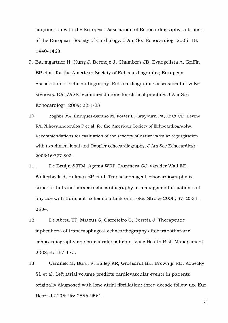
event increased with the severity of atrial enlargement (p for trend: 0.012) (Fig.1). The
recurrence rate in the 197 patients with severe left atrial enlargement was 11.7%.
Therapy with anticoagulants after the index stroke event was performed in 80.5% of the
patients without atrial enlargement, in 79.3% with mild atrial enlargement, in 77.6%
with moderate atrial enlargement and in 77.1% of the patients with severe atrial
enlargement (p for trend: 0.78).
On multivariate analysis, the presence of atrial enlargement (OR=2.13; 95% CI 1.06-
4.29, p=0.033) and CHA2DS2-VASc score (OR 1.22; 95% CI 1.04-1.45, p=0.018, for each
point increase) were correlated with ischemic recurrent event. Small ischemic lesion was
inversely correlated with ischemic recurrent event (OR 0.55; 95% CI 0.30-1.00, p=0.05).
The presence of pacemaker showed an OR 2.10; 95% CI 0.97-4.60, p=0.06. When in the
model the variable atrial enlargement was introduced as mild, moderate or severe, only
the presence of severe atrial enlargement was correlated with an outcome event (OR:
2.05; 95% CI 1.08-2.87, p=0.027). All the remaining TTE characteristics were not
correlated with outcome event.
Figure 2 shows the adjusted analysis using the Cox regression model that evidences the
different risk for an ischemic outcome event according to the severity of atrial

10
enlargement within 90 days. This analysis, was adjusted for age, sex, CHA2DS2-VASc
score, lesion size, anticoagulant therapy and NIHSS score on admission.
Discussion
In this prospective study in patients with acute stroke and AF, TTE showed a left atrial
thrombus in about 1% and an atrial enlargement in 65% of the patients. The presence of
atrial enlargement was associated with a higher risk of an ischemic recurrence within 90
days. This association was accounted for by severe atrial enlargement.
In clinical practice, for the fear of an early recurrence, TTE is usually performed in acute
phase of stroke to detect a thrombus and to start anticoagulant therapy earlier. This
study showed the futility of acute TTE for detection of intra-cardiac thrombosis. About
17% of the patients did not undergo TTE in the acute phase of stroke and these patients
were older and with more severe neurological deficit. Therefore, it is possible that in
these patients, the rate of intra-cardiac thrombus was higher. However, these are not the
typical patients in whom anticoagulation would be started promptly.
Several studies demonstrated that TEE is superior to TTE for identification of a cardiac
embolic source in patients with TIA or stroke (11,12). De Brujn et al. (11), in patients
with acute stroke, found a thrombus in 17% of the patients using TEE compared to 2%
using TTE. Thus, in patients with acute stroke and AF, TEE is probably more useful to
detect intra-cardiac thrombosis. However, this examination in patients with acute stroke
is not easy.
Our study showed that atrial enlargement, especially severe, was independently
associated with early stroke recurrence. Several studies found that moderate to severe
left atrial enlargement is an independent marker of recurrent stroke of embolic subtypes
in patients with ischemic stroke even in patients without evidence of AF (3,13).
Furthermore, atrial dilatation is correlated with more severe stroke (14).

11
An association between ischemic recurrent events and the presence of a valve prosthesis,
biological or mechanical, was not found but the number of patients with valve prosthesis
was low (17 patients with biological prosthesis and 40 with mechanical prosthesis).
This study has several limitations and strengths. The evaluation of TTE and the
adjudication of the outcome events were not centralized. Not all the patients had TTE
examination and this might have introduced some selection bias.
Strengths of our study include the prospective and multicentre design with an adequate
sample size of consecutive patients, making the results robust and generalizable.
Furthermore, we collected data on a wide range of potentially confounding risk factors,
allowing us to estimate the independent effects of TTE characteristics evaluated.
In summary, in patients with AF-associated acute stroke, atrial enlargement is an
independent marker of recurrent stroke and systemic embolism. Left atrial thrombosis is
relatively uncommon. Future studies are needed to evaluate whether left atrial
enlargement could be used to drive prompt anticoagulant therapy in patients with acute
stroke and AF to reduce the risk of recurrence.

12
References:
1. Paciaroni M, Agnelli G, Falocci N, Caso V, Becattini C et al. Early
Recurrence and Cerebral Bleeding in Patients With Acute Ischemic Stroke
and Atrial Fibrillation: Effect of Anticoagulation and Its Timing: The RAF
Study. Stroke. 2015;46:2175-2182.
2. Douen AG, Sabin M, Pageau N. Thrombus detection by echocardiography in
patients with acute ischemic stroke and chronic or new-onset atrial
fibrillation. J Stroke Cerebrovasc Dis 2008; 17: 208-211.
3. Yaghi S, Moon YP, Mora-McLaughlin C, Willey JZ, Cheung K, Di Tullio MR et al.
Left atrial enlargement and stroke recurrence: the northern Manhattan stroke
study. Stroke. 2015;46:1488-93.
4. Tatu L, Moulin T, Bogousslavsky J, Duvemoy H. Arterial territories of the human
brain: cerebral hemispheres. Neurology. 1998;50:1699 –1708.
5. Tatu L, Moulin T, Bogousslavsky J, Duvemoy H. Arterial territories of the human
brain: brainstem and cerebellum. Neurology. 1996;47: 1125–1135.
6. Paciaroni M, Agnelli G, Corea F, Ageno W, Alberti A, Lanari A, et al. Early
hemorrhagic transformation of brain infarction: rate, predictive factors, and
influence on clinical outcome: results of a prospective multicenter study.
Stroke. 2008;39:2249-56.
7. Beppu S, Park YD, Sakakibara H, Nagata S, Nimura Y. Clinical features of
intra-cardiac thrombus based on echocardiographic observation. Jpn Circ
J 1984; 48: 75-82.
8. Lang RM, Bierig M, Devereux RB, Flachskampf FA, Foster E, Pellikka PA et
al. Recommendations for chamber quantification: a report from the
American Society of Echocardiography’s Guidelines and Standard
Committee and the Chamber Quantification Writing Group, Developed in
13
conjunction with the European Association of Echocardiography, a branch
of the European Society of Cardiology. J Am Soc Echocardiogr 2005; 18:
1440-1463.
9. Baumgartner H, Hung J, Bermejo J, Chambers JB, Evangelista A, Griffin
BP et al. for the American Society of Echocardiography; European
Association of Echocardiography. Echocardiographic assessment of valve
stenosis: EAE/ASE recommendations for clinical practice. J Am Soc
Echocardiogr. 2009; 22:1-23
10. Zoghbi WA, Enriquez-Sarano M, Foster E, Grayburn PA, Kraft CD, Levine
RA, Nihoyannopoulos P et al. for the American Society of Echocardiography.
Recommendations for evaluation of the severity of native valvular regurgitation
with two-dimensional and Doppler echocardiography. J Am Soc Echocardiogr.
2003;16:777-802.
11. De Bruijn SFTM, Agema WRP, Lammers GJ, van der Wall EE,
Wolterbeek R, Holman ER et al. Transesophageal echocardiography is
superior to transthoracic echocardiography in management of patients of
any age with transient ischemic attack or stroke. Stroke 2006; 37: 2531-
2534.
12. De Abreu TT, Mateus S, Carreteiro C, Correia J. Therapeutic
inplications of transesophageal echocardiography after transthoracic
echocardiography on acute stroke patients. Vasc Health Risk Management
2008; 4: 167-172.
13. Osranek M, Bursi F, Bailey KR, Grossardt BR, Brown jr RD, Kopecky
SL et al. Left atrial volume predicts cardiovascular events in patients
originally diagnosed with lone atrial fibrillation: three-decade follow-up. Eur
Heart J 2005; 26: 2556-2561.

14
14. Kim TW, Jung SW, Song IU, Koo J, Choi HS, Lee KS et al. Left atrial
dilatation is associated with severe ischemic stroke in men with non-
valvular atrial fibrillation. J Neurol Sci 2015; 354: 97-102.

15
Figure legends
Figure 1. The risk of an ischemic outcome event according to the severity of atrial
enlargement (p for trend: 0.012).
Figure 2. The cumulative risk of an ischemic outcome event according to the
severity of atrial enlargement using Cox regression model.

1
Table1. Definitions of left atrial enlargement according to the severity (Lang et al, 2005)
Women
Men
Reference Range
Mildly abnormal
Moderately abnormal
Severely abnormal
Reference Range
Mildly abnormal
Moderately abnormal
Severely abnormal
Atrial dimension LA diameter, cm 2.7-3.8 3.9-4.2 4.3-4.6 ≥4.7 3.0-4.0 4.1-4.6 4.7-5.2 ≥5.2 LA diameter/BSA,cm/m2
1.5-2.3
2.4-2.6
2.7-2.9
≥3.0
1.5-2.3
2.4-2.6
2.7-2.9
≥3.0
Atrial area LA area, cm2
≤20 20-30 30-40 >40 ≤20 20-30 30-40 >40
Atrial volumes LA volume, mL 22-52 53-62 63-72 ≥73 18-58 59-68 69-78 ≥79 LA volume/BSA,mL/m2
22±6
29-33
34-39
≥40
22±6
29-33
34-39
≥40
LA: left atrial
BSA: body surface area in m2

2
Table 2. Characteristics of the patients with or without TTE evaluation.
*Transient Ischemic attack
**Congestive heart failure
***Myocardial infarction
****Stroke/TIA/systemic embolism
Total (n=1029)
With TTE (n=854)
Without TTE (n=175)
p
Age (mean, years) 77.2 ± 9.5 76.3 ± 9.5 81.4 ± 8.4 0.0001 NHISS (mean) 9.2 ±7.3 8.9 ± 7.0 10.7 ± 9.0 0.002 Sex male 468 (45.5%) 398 (46.6%) 70 (40.0%) 0.1 Diabetes mellitus 264 (25.6%) 221 (26.0%) 43 (24.8%) 0.7 Statins 260 (25.3%) 213 (25.2%) 47 (27.3%) 0.5 Hipertension 821 (79.8%) 676 (79.8%) 145 (83.8%) 0.2 Hyperlipidemia 332 (32.3%) 282 (33.4%) 50 (29.2%) 0.3 History stroke/TIA* 266 (25.8%) 205 (24.3%) 61 (35.5%) 0.04 Smoking 190 (18.5%) 158 (18.7%) 32 (18.5%) 0.5 Alcoholism 68 (6.6%) 57 (6.7%) 11 (6.4%) 1.0 History of CHF** 193 (18.7%) 167 (19.6%) 26 (15.0%) 0.2 History of MI*** 166 (16.1%) 142 (16.8) 24 (13.9%) 0.4 Paroxysmal AF 364 (35.4%) 316 (37.0%) 48 (27.9%) 0.023 Permanent AF 473 (46.0%) 385 (45.1%) 88 (51.2%) 0.1 Persistent AF 191 (18.6%) 155 (18.2%) 36 (20.9%) 0,3 Pacemaker 85 (8.3%) 70 (8.2%) 15 (8.7%) 0.8 rtPA 184 (17.9%) 158 (18.5%) 26 (15.1%) 0.3 Intra-arterial revascularization
46 (4.5%)
43 (5.1%)
3 (1.7%)
0.067
CHA2DS2-VASc 0 17 (1.6%) 16 (1.9%) 1 (0.6%) 0.053 1 54 (5.2%) 51 (6.0%) 3 (1.7%) 2 91 (8.8%) 82 (9.6%) 9 (5.1%) 3 200 (19.4%) 167 (19.5%) 33 (18.9%) 4 243 (23.6%) 203 (23.8%) 40 (22.9%) 5 206 (20.0%) 161 (18.8%) 45 (25.7%) 6 129 (12.5%) 104 (12.2%) 25 (14.3%) 7 66 (6.4%) 51 (6.0%) 15 (8.6%) 8-9 23 (2.2) 19 (2.2%) 4 (2.3%) Therapy with anticoagulants after index stroke
766 (74.4%)
673 (78.8%)
93 (53.1%)
0.0001
Recurrent ischemic event****
77 (7.6%) 63 (7.4%) 14 (8.0%) 0.7
Disability at 90 days (mRS≥3)
510 (49.5%) 403 (47.6%) 107 (61.8%) 0.001

3
Table 3. TTE characteristics in patients with and without outcome event
Total
(n=854) Without outcome event
(n =791) With outcome event
(n= 63)
p
Atrial enlargement 548 (64.2%) 497 (62.8%) 51 (81.0%) 0.004 Mild 145 (16.9%) 133 (16.9%) 12 (19.0%) 0.6
Moderate 203 (23.8%) 187 (23.7%) 16 (25.4) 0.7 Severe 197 (23.1%) 174 (22.0%) 23 (36.5%) 0.013
Intracardiac thrombus 11 (1.3%) 10 (1.3%) 1 (1.6%) 0.5 Cardiomyopathy* 99 (11.6%) 89 (11.3%) 10 (15.9) 0.3 Mitral disease 360 (42.2%) 323 (40.1%) 37 (58.7%) 0.021 Aortic disease 230 (26.9%) 205 (25.9%) 25 (39.7%) 0.026 Tricuspidal disease 213 (24.9%) 189 (23.9%) 24 (38.1%) 0.016 Biological aortic valve 10 (1.2%) 10 (1.2%) 0 1.0 Mechanical aortic valve 17 (2.0%) 15 (1.8%) 2 (3.0%) 0.3 Biological mitral valve 7 (0.8%) 7 (0.8%) 0 1.0 Mechanical mitral valve 23 (2.7%) 20 (2.4%) 3 (4.5%) 0.2
*left ventricular ejection ≤40%


![MD11 - Poclain · PDF fileHydraulic motors MD11 POCLAIN HYDRAULICS CHARACTERISTICS 8 837 ... 33 [44] Motor inertia ... NF E22141; 24 teeth; module 2,5](https://static.fdocuments.in/doc/165x107/5abcdf017f8b9a24028e5829/md11-poclain-motors-md11-poclain-hydraulics-characteristics-8-837-33-44.jpg)