Molluscan Success Molluscan Success.pdf · 11 Molluscan Success This mai·ine nudibmnch...
23
11 Molluscan Success This mai·ine nudibmnch (Chromodoris kuniei) is a member ofone ofthe most succsful animal pla. Members of the Mollusca have ated to nr every habitat on the earth. This cbapter describ the markable diversity and aptations ofmembers oftbis plum. 11. 1 EVOLUTIONARY PERSPECTIVE LEARNING OUTCOMES 1. Give examples of, and describe the prevalence of, members of the phylum Mollusca. 2. Discuss the relationship of the Mollusca to other animal phyla. Octopuses, squids, and cuttlefish (the cephalopods) are some of the invertebrate world's most adept predators. Predatory lifestyles have resulted in the evolu- tion of large brains (by invertebrate standards), complex sensory structures (by any standards), rapid locomotion, grasping tentacles, and tearing mouthparts. In spite of these adaptations, cephalopods rarely make up a major component of any community. Once numbering about 9,000 species, the class Cephalopoda now includes only about 550 species (figure 11.1). Zoologists do not know why the cephalopods have declined so dramatically. Vertebrates may have outcompeted cephalopods because the vertebrates were also making their appearance in prehistoric seas, and some ve1tebrates (e.g., bony fish) acquired active, predatory lifestyles. This evolutionary decline has not been the case for all molluscs. Overall, this group has been very successful. If success is measured by numbers of species, the molluscs are twice as successful as ve1tebrates! The vast majority of the nearly 100,000 living species of molluscs belongs to two classes: Gastropoda, the snails and slugs; and Bivalvia, the clams and their close relatives. Molluscs are triploblastic, as are all the remaining animals covered in this text. In addition, they are the first animals described in this text that possess a coelom, although the coelom of molluscs is only a small cavity (the pericardia! cav- ity) surrounding the heart, nephridia, and gonads. A coelom is a body cavity that arises in mesoderm and is lined by a sheet of mesoderm called the peritoneum (see figure 7.11 c). Relationships to Other Animals Molluscs are members of the Lophotrochozoa. Their lophotrochozoan relatives include the annelids, along with other phyla discussed in chapter 10 (seepages xvi-xvii of this textbook). 11.1 11.2 11.3 11.4 11.5 11.6 11.7 11.8 11.9 11.10 11.11 Chapter Outline Evolutiona Perspective Relationships to Other Animals Molluscan Characteristics Class Gastropoda Torsion Shell Coiling Locomotion Feeding and Digestion Other Maintenance Functions Reproduction and Development Gastropod Diversity Class Bivalvia Shell and Associated Structures Gas change, Filter Feeding, and Digestion Other Maintenance Functions Reproduction and Development Bivalve Diversity Class Cephalopoda Shell Locomotion Feeding and Digestion Other Maintenance Functions Leaing Reproduction and Development Class Polyplacophora Class Scaphopoda Class Monoplacophora Class Solenogastres Class Caudofoveata Fuher Phylogenetic Considerations
Transcript of Molluscan Success Molluscan Success.pdf · 11 Molluscan Success This mai·ine nudibmnch...
11
Molluscan Success
This mai·ine nudibmnch (Chromodoris kuniei) is a member of one of the most
successful animal phyla. Members of the Mollusca have adapted to nearly
every habitat on the earth. This cbapter describes the remarkable diversity and
adaptations of members of tbis phylum.
11. 1 EVOLUTIONARY PERSPECTIVE
LEARNING OUTCOMES
1. Give examples of, and describe the prevalence of, members of the phylumMollusca.
2. Discuss the relationship of the Mollusca to other animal phyla.
Octopuses, squids, and cuttlefish (the cephalopods) are some of the invertebrate world's most adept predators. Predatory lifestyles have resulted in the evolution of large brains (by invertebrate standards), complex sensory structures (by any standards), rapid locomotion, grasping tentacles, and tearing mouthparts. In spite of these adaptations, cephalopods rarely make up a major component of any community. Once numbering about 9,000 species, the class Cephalopoda now includes only about 550 species (figure 11.1).
Zoologists do not know why the cephalopods have declined so dramatically. Vertebrates may have outcompeted cephalopods because the vertebrates were also making their appearance in prehistoric seas, and some ve1tebrates (e.g., bony fish) acquired active, predatory lifestyles.
This evolutionary decline has not been the case for all molluscs. Overall, this group has been very successful. If success is measured by numbers of species, the molluscs are twice as successful as ve1tebrates! The vast majority of the nearly 100,000 living species of molluscs belongs to two classes: Gastropoda, the snails and slugs; and Bivalvia, the clams and their close relatives.
Molluscs are triploblastic, as are all the remaining animals covered in this text. In addition, they are the first animals described in this text that possess a coelom, although the coelom of molluscs is only a small cavity (the pericardia! cavity) surrounding the heart, nephridia, and gonads. A coelom is a body cavity that arises in mesoderm and is lined by a sheet of mesoderm called the peritoneum (see
figure 7.11 c).
Relationships to Other Animals
Molluscs are members of the Lophotrochozoa. Their lophotrochozoan relatives include the annelids, along with other phyla discussed in chapter 10 (see pages xvi-xvii
of this textbook).
11.1
11.2
11.3
11.4
11.5
11.6
11.7
11.8
11.9
11.10
11.11
Chapter Outline Evolutionary Perspective
Relationships to Other Animals Molluscan Characteristics Class Gastropoda
Torsion Shell Coiling Locomotion Feeding and Digestion Other Maintenance Functions Reproduction and Development Gastropod Diversity
Class Bivalvia Shell and Associated Structures Gas Exchange, Filter Feeding,
and Digestion Other Maintenance Functions Reproduction and Development Bivalve Diversity
Class Cephalopoda Shell Locomotion Feeding and Digestion Other Maintenance Functions Learning Reproduction and Development
Class Polyplacophora Class Scaphopoda Class Monoplacophora Class Solenogastres Class Caudofoveata Further Phylogenetic Considerations
198 CHAPTER ELEVEN
�, _ __:::i 'f ,. , ..
Prottsts
FIGURE 11.1
Basal Phyla
Lophotrochozoa
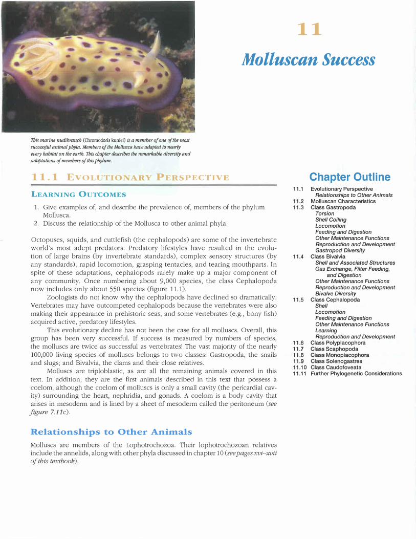
Evolutionary Relationships of Molluscs to Other Animals. The figure shows one interpretation of the relationship of the Mollusca to other members of the animal kingdom. The relationships depicted here are based on evidence from developmental and molecular biology. Molluscs are placed within the Lophotrochozoa along with the Annelida, Plathyhelminthes, Rotifera, and others (see pages xvi-xvii). The phylum Mollusca includes nearly 100,000 living species, including members of the class Cephalopoda-some of the invertebrates' most adept predators. The blue-ringed octopus (Hapalochlaena) is shown here. Four species of Hapalochlaena are found in the Pacific Ocean from Japan to Australia. This ve1y small octopus has a ve1y powerful venom containing tetrodotoxin, the same found in cone snails and pufferfish. The venom causes respiratory paralysis and is powerful enough to kill a human.
SECTION REVIEW 11.1
Members of the phylum Mollusca include octopuses and their relatives, snails, bivalves, and others. They are members of the Lophmrochozoa with evoluliona1 y Lie:,; Lu Ll1e Aundida and other lophotrochozoans.
How do the molluscs illustrate the idea that complexity
does not always ensure evolutionary success?
11 .. 2 LLU
CHARACTE .s
LEARNING OUTCOME
1. Hypothesize about the structure of a hypothetical newlydiscovered class of molluscs.
Molluscs range in size and body form from the largest of all invertebrates, the giant squid (.Architeuthis), measuring 18 m
in length, to the smallest garden slug, less than 1 cm long. In spite of this diversity, the phylum Mollusca (mol-lus'kah) (L. molluscus, soft) is not difficult to characterize (table 11.1).
Characteristics of the phylum Mollusca include:
1. Body of two parts: head-foot and visceral mass2. Mantle that secretes a calcareous shell and covers the
3. Mantle cavity functions in excretion, gas exchange,elimination of digestive wastes, and release ofreproductive products
4. Bilateral symmetry5. Trochophore larvae, spiral cleavage, and schizocoelous
coelom formation6. Coelom reduced to cavities surrounding the heart,
nephridia, and gonads7. Open circulatory system in all but one class
( Cephalopoda)8. Radula usually present and used in scraping food
The body of a mollusc has two main regions-the head-foot and the visceral mass (figure 11.2). The head-foot is elongate with an anterior head, containing the mouth and certain nervous and senso1y structures, and an elongate foot, used for attachment and locomotion. The visceral mass contains the organs of digestion, circulation, reproduction, and excretion
and is positioned dorsal to the head-foot.
T/\H!.F, i 1 �
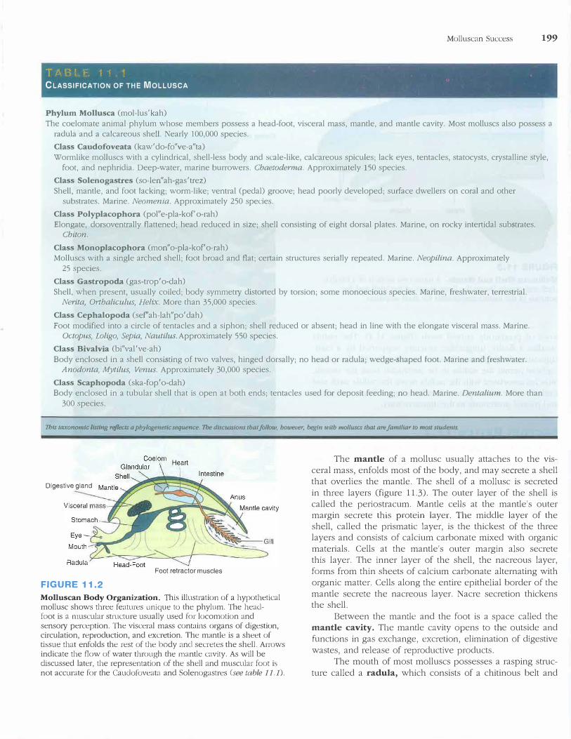
CLASSIFICATION OF THE MOLLUSCA
Phylum Mollusca (mol-lus'kah)
Molluscan Success 199
The coelomate animal phylum whose members possess a head-foot, visceral mass, mantle, and mantle cavity. Most molluscs also possess a radula and a calcareous she!L Nearly 100,000 species.
Class Caudofoveata (kaw' do-fo"ve-a"ta)
Wormlike molluscs with a cylindrical, shell-less body and scale-like, calcareous spicules; lack eyes, tentacles, statocysts, crystalline style, foot, and nephridia. Deep-water, marine burrowers. Chaetoderma. Approximately 150 species.
Class Solenogastres (so-len"ah-gas'trez)
Shell, mantle, and foot lacking; worm-like; ventral (pedal) groove; head poorly developed; surface dwellers on coral and other substrates. Marine. Neomenia. Approximately 250 species.
Class Polyplacophora (pol"e-pla-kof' o-rah)
Elongate, dorsoventrally flattened; head reduced in size; shell consisting of eight dorsal plates. Marine, on rocky intertidal substrates. Chiton.
Class Monoplacophora (mon"o-pla-kof'o-rah) Molluscs with a single arched shell; foot broad and flat; certain structures serially repeated. Marine. Neopilina. Approximately
25 species.
Class Gastropoda (gas-trop'o-dah)
Shell, when present, usually coiled; body symmetty distorted by torsion; some monoecious species. Marine, freshwater, terrestrial. Nerita, Ot1haliculus, Helix. More than 35,000 species.
Class Cephalopoda (sef'ah-lah"po'dah)
Foot modified into a circle of tentacles and a siphon; shell reduced or absent; head in line with the elongate visceral mass. Marine. Octopus, Loligo, Sepia, Nautilus. Approximately 550 species.
Class Bivalvia (bi"val've-ah)
Body enclosed in a shell consisting of two valves, hinged dorsally; no head or radula; wedge-shaped foot. Marine and freshwater. Anodonta, Mytilus, Venus. Approximately 30,000 species.
Class Scaphopoda (ska-fop'o-dah) Body enclosed in a tubular shell that is open at both ends; tentacles used for deposit feeding; no head. Marine. Dentalium. More than
300 species.
Ibis ta.xonomfc listing reflects a phylogenetic sequence. 1be discussions that follow, however, begin with molluscs that are Jamfliar to most students
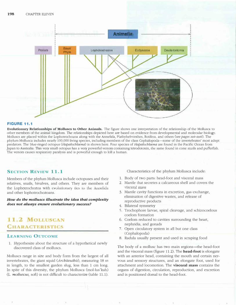
Digestive gland
Foot retractor muscles
FIGURE 11.2
Molluscan Body Organization. This illustration of a hypothetical mollusc shows three features unique to the phylum. The head-foot is a muscular structure usually used for locomotion and senso1y perception. The visceral mass contains organs of digestion, circulation, reproduction, and excretion. The mantle is a sheet of tissue that enfolds the rest of the body and secretes the shell. Arrows indicate the flow of water through the mantle cavity. As will be discussed later, the representation of the shell and muscular foot is not accurate for the Cauclofoveata and Solenogastres (see table 11.1).
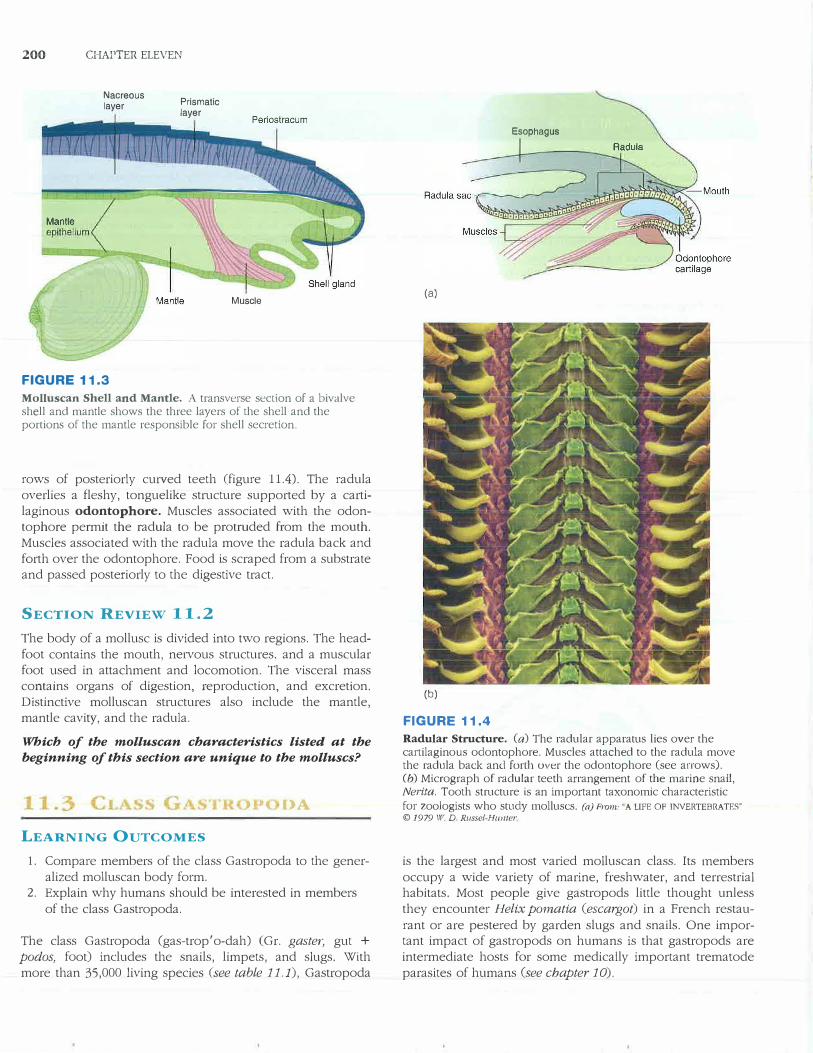
The mantle of a mollusc usually attaches to the visceral mass, enfolds most of the body, and may secrete a shell that overlies the mantle. The shell of a mollusc is secreted in three layers (figure 11.3). The outer layer of the shell is called the periostracum. Mantle cells at the mantle's outer margin secrete this protein layer. The middle layer of the shell, called the prismatic layer, is the thickest of the three layers and consists of calcium carbonate mixed with organic materials. Cells at the mantle's outer margin also secrete this layer. The inner layer of the shell, the nacreous layer, forms from thin sheets of calcium carbonate alternating with organic matter. Cells along the entire epithelial border of the mantle secrete the nacreous layer. Nacre secretion thickens the shell.
Between the mantle and the foot is a space called the mantle cavity. The mantle cavity opens to the outside and functions in gas exchange, excretion, elimination of digestive wastes, and release of reproductive products.
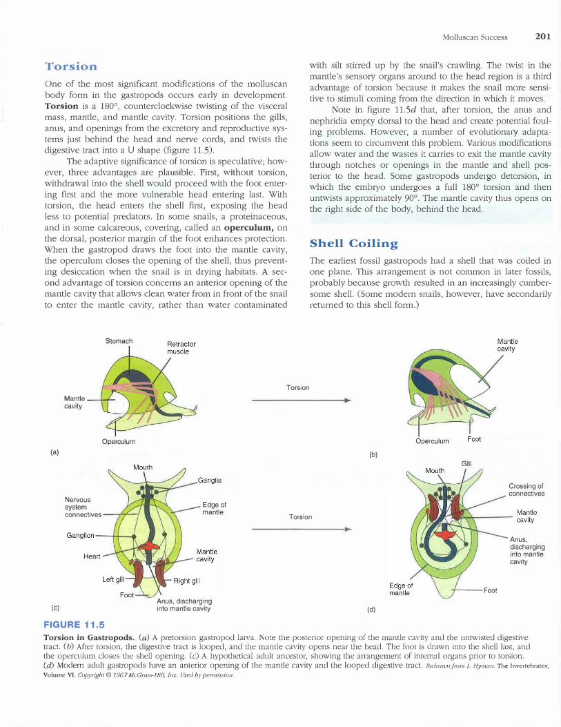
The mouth of most molluscs possesses a rasping struc
ture called a radu]a, which consists of a chitinous belt and
200 CHAPTER ELEVEN
Nacreous layer
FIGURE 11.3
Prismatic layer
Muscle
Molluscan Shell and Mantle. A transverse section of a bivalve shell and mantle shows the three layers of the shell and the portions of the mantle responsible for shell secretion.
rows of posteriorly curved teeth (figure 11.4). The radula
overlies a fleshy, tonguelike structure supported by a carti
laginous odontophore. Muscles associated with the odontophore permit the radula to be protruded from the mouth.
Muscles associated with the radula move the radula back and
forth over the odontophore. Food is scraped from a substrate and passed posteriorly to the digestive tract.
SECTION REVIEW 11.2
The body of a mollusc is divided into two regions. The head
foot contains the mouth, nervous structures, and a muscular foot used in attachment and locomotion. The visceral mass contains organs of digestion, reproduction, and excretion.
Distinctive molluscan structures also include the mantle, mantle cavity, and the radula.
Which of the molluscan characteristics listed at the
beginning of this section are unique to the molluscs?
DA
LEARNING OUTCOMES
1. Compare members of the class Gastropoda to the gener
alized molluscan body form.2. Explain why humans should be interested in members
of the class Gastropoda.
The class Gastropoda (gas-trop'o-dah) (Gr. gaster, gut +
podos, foot) includes the snails, limpets, and slugs. With
more than 35,000 living species (see table 11.1), Gastropoda
(a)
(b)
FIGURE 11.4
Esophagus
Radular Structure. (a) The radular apparatus lies over the cartilaginous odontophore. Muscles attached to the radula move the radula back and forlh over the odontophore (see arrows). (b) Micrograph of radular teeth arrangement of the marine snail,Nerita. Tooth structure is an important taxonomic characteristicfor zoologists who study molluscs. (a) From:. A LIFE OF INVERTEBRATES"
© 1979 \Ii'. D. Russel-Hu/lier
is the largest and most varied molluscan class. Its members
occupy a wide variety of marine, freshwater, and terrestrial habitats. Most people give gastropods little thought unless
they encounter Helix pomatia (escargot) in a French restau
rant or are pestered by garden slugs and snails. One important impact of gastropods on humans is that gastropods are
intermediate hosts for some medically important trematode
parasites of humans (see chapter 10).
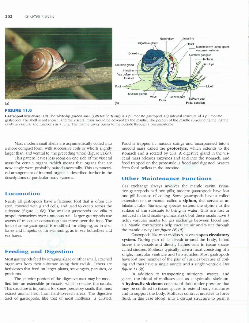
Torsion
One of the most significant modifications of the molluscan
body form in the gastropods occurs early in development.
Torsion is a 180° , counterclockwise twisting of the visceralmass, mantle, and mantle cavity. Torsion positions the gills,
anus, and openings from the excretory and reproductive systems just behind the head and ne1ve cords, and twists the digestive tract into a U shape (figure 11.5).
The adaptive significance of torsion is speculative; however, three advantages are plausible. First, without torsion, withdrawal into the shell would proceed with the foot enter
ing first and the more vulnerable head entering last. With torsion, the head enters the shell first, exposing the head less to potential predators. In some snails, a proteinaceous,
and in some calcareous, covering, called an operculum, on the dorsal, posterior margin of the foot enhances protection. When the gastropod draws the foot into the mantle cavity, the operculum closes the opening of the shell, thus prevent
ing desiccation when the snail is in d1ying habitats. A second advantage of torsion concerns an anterior opening of the mantle cavity that allows clean water from in front of the snail
to enter the mantle cavity, rather than water contaminated
Stomach
Molluscan Success 201
with silt stirred up by the snail's crawling. The twist in the
mantle's sensory organs around to the head region is a third
advantage of torsion because it makes the snail more sensitive to stimuli coming from the direction in which it moves.
Note in figure 11.Sd that, after torsion, the anus and
nephridia empty dorsal to the head and create potential fouling problems. However, a number of evolutiona1y adaptations seem to circumvent this problem. Various modifications allow water and the wastes it carries to exit the mantle cavity through notches or openings in the mantle and shell pos
terior to the head. Some gastropods undergo detorsion, in which the embryo undergoes a full 180° torsion and thenuntwists approximately 90°. The mantle cavity thus opens onthe right side of the body, behind the head.
Shell Coiling
The earliest fossil gastropods had a shell that was coiled in one plane. This arrangement is not common in later fossils,
probably because growth resulted in an increasingly cumber
some shell. (Some modern snails, however, have secondarily returned to this shell form.)
Torsion
(a)
(c)
Mantle ---i-cavity
Nervous system
Operculum
connectives _ __,....,__,
Heart
FIGURE 11.5
Edge of mantle
Anus, discharging into mantle cavity
Torsion
(b)
(d)
Operculum
GIii
Crossing of connectives
Anus, discharging into mantle cavity
Torsion in Gastropods. (a) A pretorsion gastropod larva. Note the posterior opening of the mantle cavity and the untwisted digestive tract. (b) After torsion, the digestive tract is looped, and the mantle cavity opens near the head. The foot is drawn into the shell last, and the operculum closes the shell opening. (c) A hypothetical adult ancestor, showing the arrangement of internal organs prior to torsion. (d) Modern adult gastropods have an anterior opening of the mantle cavity and the looped digestive tract. Redrawn.Jiwn L Hyman, The Invertebrates,
Volume VI. Copyrigbt © 1967 McGraw-Hi//, Inc. Used by permission,
202 CHAPTER ELEVEN
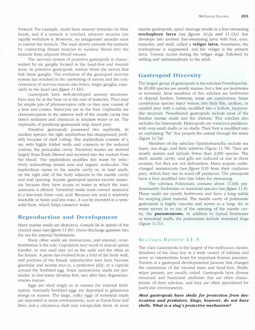
(a)
FIGURE 11.6
Nephridium
Albumen gland ,--.1�-=��WIntestine -1--._.,-
Foot
Penis (b)
Intestine
Mantle cavity (lung) opens via pneumostome
Cerebral ganglion
Eye
Gastropod Structure. (a) The white-lip garden snail (Cepaea hortensis) is a pulmonate gastropod. (b) Internal structure of a pulmonate gastropod. The shell is not shown, and the visceral mass would be covered by the mantle. The portion of the mantle surrounding the mantle cavity is vascular and functions as a lung. The mantle cavity opens to the outside through a pneumostome.
Most modern snail shells are asymmetrically coiled into a more compact form, with successive coils or whorls slightly larger than, and ventral to, the preceding whorl (figure ll.6a).
This pattern leaves less room on one side of the visceral mass for certain organs, which means that organs that are now single were probably paired ancestrally. This asymmetrical arrangement of internal organs is described further in the descriptions of particular body systems.
Locomotion
Nearly all gastropods have a flattened foot that is often ciliated, covered with gland cells, and used to creep across the substrate (figure ll.6b). The smallest gastropods use cilia topropel themselves over a mucous trail. Larger gastropods use waves of muscular contraction that move over the foot. The foot of some gastropods is modified for clinging, as in abalones and limpets, or for swimming, as in sea butterflies and sea hares.
Feeding and Digestion
Most gastropods feed by scraping algae or other small, attached organisms from their substrate using their radula. Others are herbivores that feed on larger plants, scavengers, parasites, or predators.
The anterior portion of the digestive tract may be modified into an extensible proboscis, which contains the radula. This structure is important for some predato1y snails that must extract animal flesh from hard-to-reach areas. The digestive tract of gastropods, like that of most molh1scs, is cili1tJ�c;l.
Food is trapped in mucous strings and incorporated into a mucoid mass called the protostyle, which extends to the stomach and is rotated by cilia. A digestive gland in the visceral mass releases enzymes and acid into the stomach, and food trapped on the protostyle is freed and digested. Wastes form fecal pellets in the intestine.
Other Maintenance Functions
Gas exchange always involves the mantle cavity. Primitive gastropods had two gills; modern gastropods have lost one gill because of coiling. Some gastropods have a rolled extension of the mantle, called a siphon, that se1ves as an inhalant tube. Burrowing species extend the siphon to the surface of the substrate to bring in water. Gills are lost or reduced in land snails (pulmonates), but these snails have a richly vascular mantle for gas exchange between blood and air. Mantle contractions help circulate air and water through the mantle cavity (see figure 26.14).
Gastropods, like most molluscs, have an open circulatory system. During part of its circuit around the body, blood leaves the vessels and directly bathes cells in tissue spaces called sinuses. Molluscs typically have a heart consisting of a single, muscular ventricle and two auricles. Most gastropods have lost one member of the pair of auricles because of coiling and thus have a single auricle and a single ventricle (see figure 11.6b).
In addition to transporting nutrients, wastes, and gases, the blood of molluscs acts as a hydraulic skeleton. A hydraulic skeleton consists of fluid under pressure that may be confined to tissue spaces to extend body structures and to support the body. Molluscs contract muscles to force fluid, in this qse. bl99�, into a distant structure to push it
forward. For example, snails have sensory tentacles on their heads, and if a tentacle is touched, retractor muscles can rapidly withdraw it. However, no antagonistic muscles exist to extend the tentacle. The snail slowly extends the tentacle by contracting distant muscles to squeeze blood into the tentacle from adjacent blood sinuses.
The ne1vous system of primitive gastropods is characterized by six ganglia located in the head-foot and visceral mass. In primitive gastropods, torsion twists the ne1ves that link these ganglia. The evolution of the gastropod ne1vous system has resulted in the untwisting of ne1ves and the concentration of ne1vous tissues into fewer, larger ganglia, especially in the head (see figure 11.6h).
Gastropods have well-developed sensory structures. Eyes may be at the base or at the end of tentacles. They may be simple pits of photoreceptor cells or they may consist of a lens and cornea. Statocysts are in the foot. Osphradia are chemoreceptors in the anterior wall of the mantle cavity that detect sediment and chemicals in inhalant water or air. The osphradia of predat01y gastropods help detect prey.
Primitive gastropods possessed two nephridia. In modern species, the right nephridium bas disappeared, probably because of shell coiling. The nephridium consists of a sac with highly folded walls and connects to the reduced coelom, the pericardia! cavity. Excretory wastes are derived largely from fluids filtered and secreted into the coelom from the blood. The nephridium modifies this waste by selectively reabsorbing certain ions and organic molecules. The nephridium opens to the mantle cavity or, in land snails, on the right side of the body adjacent to the mantle cavity and anal opening. Aquatic gastropod species excrete ammonia because they have access to water in which the toxic ammonia is diluted. Terrestrial snails must convert ammonia to a less-toxic form-uric acid. Because uric acid is relatively insoluble in water and less toxic, it can be excreted in a semisolid form, which helps conse1ve water.
Reproduction and Development
Many marine snails are dioecious. Gonads lie in spirals of the visceral mass (seefigure 11.6b). Ducts discharge gametes into the sea for external fertilization.
Many other snails are monoecious, and internal, crossfertilization is the rule. Copulation may result in mutual sperm transfer, or one snail may act as the male and the other as the female. A penis has evolved from a fold of the body wall, and portions of the female reproductive tract have become glandular and secrete mucus, a protective jelly, or a capsule around the fertilized egg. Some monoecious snails are protandric in that testes develop first, and after they degenerate, ovaries mature.
Eggs are shed singly or in masses for external fertilization. Internally fertilized eggs are deposited in gelatinous strings or masses. The large, yolky eggs of terrestrial snails are deposited in moist environments, such as forest-floor leaf litter, and a calcareous shell may encapsulate them. In most
Molluscan Success 203
marine gastropods, spiral cleavage results in a free-swimming trochophore larva (see figures 10.2a and 11.13a) that develops into another free-swimming larva with foot, eyes,
tentacles, and shell, called a veliger larva. Sometimes, the trochophore is suppressed, and the veliger is the prima1y laiva. Torsion occurs during the veliger stage, followed by settling and metamorphosis to the adult.
Gastropod Diversity
The largest group of gastropods is the subclass Prosobranchia. Its 20,000 species are mostly marine, but a few are freshwater or terrestrial. Most members of this subclass are herbivores or deposit feeders; however, some are carnivorous. Some carnivorous species inject venom into their fish, mollusc, or annelid prey with a radula modified into a hollow, harpoonlike structure. Prosobranch gastropods include most of the familiar marine snails and the abalone. This subclass also includes the heteropods. Heteropods are voracious predators, with ve1y small shells or no shells. Their foot is modified into an undulating "fin" that propels the animal through the water (figure 11.7a).
Members of the subclass Opisthobranchia include sea hares, sea slugs, and their relatives (figure 11.7b). They are mostly marine and include fewer than 2,000 species. The shell, mantle cavity, and gills are reduced or lost in these animals, but they are not defenseless. Many acquire undischarged nematocysts (see figure 9.9) from their cnidarian
prey, which they use to ward off predators. The pteropods have a foot modified into thin lobes for swimming.
The subclass Pulmonata contains about 17,000 predominantly freshwater or terrestrial species (see figure 11.6).
These snails are mostly herbivores and have a long radula
for scraping plant material. The mantle cavity of pulmonate gastropods is highly vascular and serves as a lung. Air or water moves in or out of the opening of the mantle cavity, the pneumostome. In addition to typical freshwater or terrestrial snails, the pulmonates include terrestrial slugs (figure 11.7c).
S1•:C'flON REVIEW 11.3
The class Gastropoda is the largest of the molluscan classes. Members of the class live in a wide variety of habitats and serve as intermediate hosts for important human parasites. Torsion is a gastropod developmental process that changes the orientation of the visceral mass and head-foot. Shells, when present, are usually coiled. Gastropods have diverse structural and functional attributes that are often characteristic of their subclass, and they are often specialized for particular environments.
Most gastropods have shells for protection from des
iccation and predators. Slugs, however, do not have
shells. What is a slug's protective mechanism?
204 CHAPTER ELEVEN
(a)
(b)
(c)
FIGURE 11.7
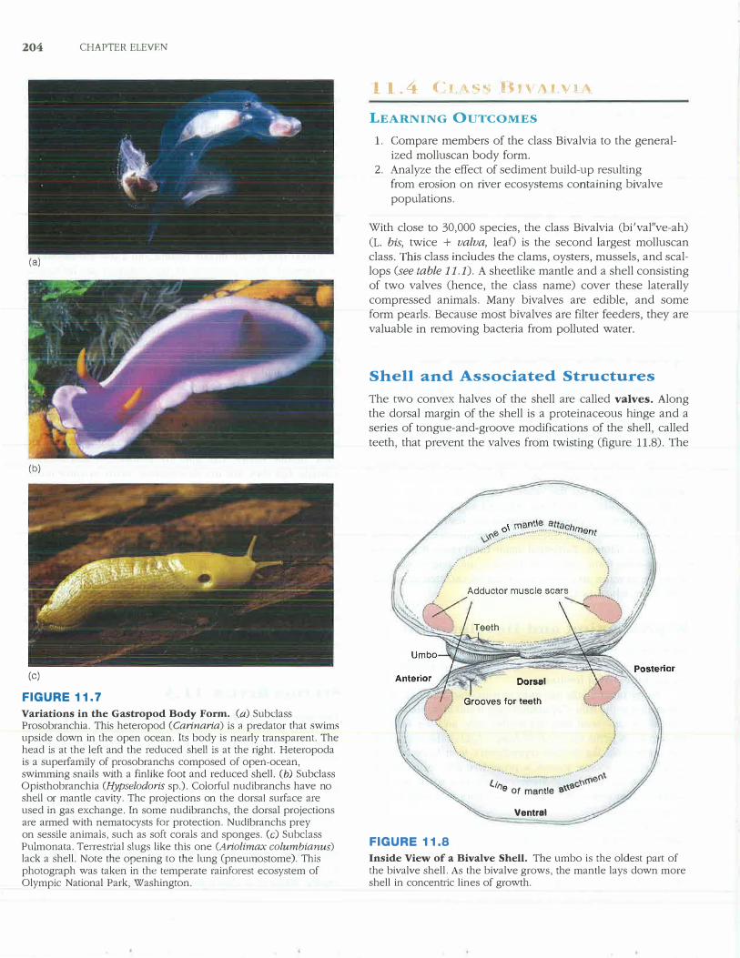
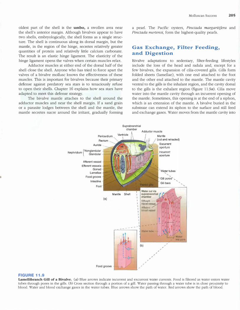
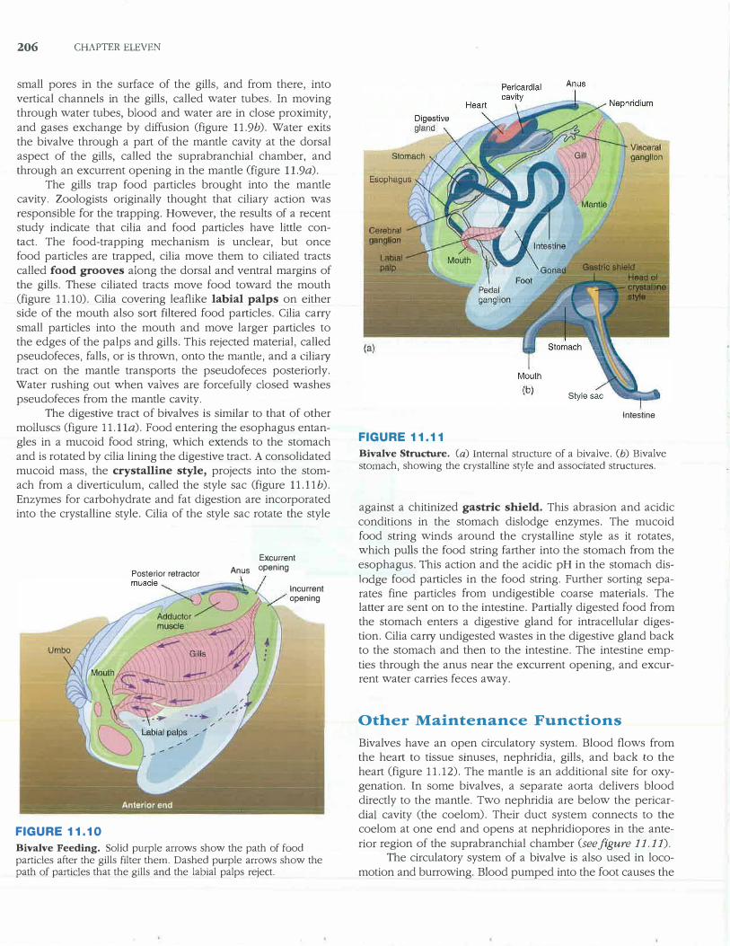
Variations in the Gastropod Body Form. (a) Subclass Prosobranchia. This heteropod (Carinaria) is a predator that swims upside down in the open ocean. Its body is nearly transparent. The head is at the left and the reduced shell is at the right. Heteropoda is a superfamily of prosobranchs composed of open-ocean, swimming snails with a finlike foot and reduced shell. (b) Subclass Opisthobranchia (Hypselodoris sp.). Colorful nuclibranchs have no shell or mantle cavity. The projections on the dorsal surface are used in gas exchange. In some nudibranchs, the dorsal projections are armed with nematocysts for protection. Nudlbranchs prey on sessile animals, such as soft corals and sponges. (c) Subclass Pulmonata. Terrestrial slugs like this one (Ariolimax columbianus) lack a shell. Note the opening to the lung (pneumostome). This photograph was taken in the temperate rainforest ecosystem of Olympic National Park, Washington.
l l ./-1 ,�, , ... � BtV/\1 VIA
LEARNING OUTCOMES
1. Compare members of the class Bivalvia to the general
ized molluscan body form.2. Analyze the effect of sediment build-up resulting
from erosion on river ecosystems containing bivalve
populations.
With close to 30,000 species, the class Bivalvia (bi'val"ve-ah)
(L. bis, twice + valva, leaf) is the second largest molluscan class. This class includes the clams, oysters, mussels, and scallops (see table 11.1). A sheetlike mantle and a shell consisting of two valves (hence, the class name) cover these laterally
compressed animals. Many bivalves are edible, and some form pearls. Because most bivalves are filter feeders, they are
valuable in removing bacteria from polluted water.
Shell and Associated Structures
The two convex halves of the shell are called valves. Along the dorsal margin of the shell is a proteinaceous hinge and a series of tongue-and-groove modifications of the shell, called
teeth, that prevent the valves from twisting (figure 11.8). The
(
FIGURE 11.8
..
...�.,�-............ _ ........ � ... -
............. , •.•. •·· 0(\\ l.ir, ,ac'n(t'
e of mantle a\\
Ventral
Inside View of a Bivalve Shell. The umbo is the oldest part of the bivalve shell. As the bivalve grows, the mantle lays down more shell in concentric lines of growth.
•
oldest part of the shell is the umbo, a swollen area near the shell's anterior margin. Although bivalves appear to have two shells, embryologically, the shell forms as a single structure. The shell is continuous along its dorsal margin, but the mantle, in the region of the hinge, secretes relatively greater quantities of protein and relatively little calcium carbonate. The result is an elastic hinge ligament. The elasticity of the hinge ligament opens the valves when certain muscles relax.
Adductor muscles at either end of the dorsal half of the shell close the shell. Anyone who has tried to force apart the valves of a bivalve mollusc knows the effectiveness of these muscles. This is important for bivalves because their primary
defense against predatory sea stars is to tenaciously refuse to open their shells. Chapter 16 explains how sea stars have adapted to meet this defense strategy.
The bivalve mantle attaches to the shell around the adductor muscles and near the shell margin. If a sand grain or a parasite lodges between the shell and the mantle, the mantle secretes nacre around the irritant, gradually forming
Molluscan Success 205
a pearl. The Pacific oysters, Pinctada margaritifera and Pinctada mertensi, form the highest-quality pearls.
Gas Exchange, Filter Feeding, and Digestion
Bivalve adaptations to sedenta1y, filter-feeding lifestyles include the loss of the head and radula and, except for a few bivalves, the expansion of cilia-covered gills. Gills form folded sheets (lamellae), with one end attached to the foot and the other end attached to the mantle. The mantle cavity ventral to the gills is the inhalant region, and the cavity dorsal to the gills is the exhalant region (figure 11.9a). Cilia move water into the mantle cavity through an incurrent opening of the mantle. Sometimes, this opening is at the end of a siphon, which is an extension of the mantle. A bivalve buried in the substrate can extend its siphon to the surface and still feed and exchange gases. Water moves from the mantle cavity into
Suprabranchial chamber
FIGURE 11.9
Pericardium
Auricle _
N h 'd' { Nonglandular ep n ium Glandular
Afferent vessel Efferent vessels
Gonad Lamellae
Food grooveIntestine
Foot
(a)
Food groove
Adductor muscle Mantle
Water out via 1 suprabranchlal
chamber
Efferent-.,..�...:. llloodllessel Affi;trent / lllo,ll)d yq$,Slll
---- - - I
I Wallirlul>& I
,
(b)
,I'
/
,I'
,
/
/
/
/
,,
,
'
Lamellibranch Gill of a Bivalve. (a) Blue arrows indicate incurrent and excurrent water currents. Food is filtered as water enters water tubes through pores in the gills. (b) Cross section through a portion of a gill. Water passing through a water tube is in close proximity to blood. Water and blood exchange gases in the water tubes. Blue arrows show the path of water. Red arrows show the path of blood.
206 CHAPTER ELEVEN
small pores in the surface of the gills, and from there, into vertical channels in the gills, called water tubes. In moving through water tubes, blood and water are in close proximity, and gases exchange by diffusion (figure 11.9b). Water exits the bivalve through a part of the mantle cavity at the dorsal aspect of the gills, called the suprabranchial chamber, and through an excurrent opening in the mantle (figure 11.9a).
The gills trap food particles brought into the mantle cavity. Zoologists originally thought that ciliary action was responsible for the trapping. However, the results of a recent study indicate that cilia and food particles have little contact. The food-trapping mechanism is unclear, but once food particles are trapped, cilia move them to ciliated tracts called food grooves along the dorsal and ventral margins of the gills. These ciliated tracts move food toward the mouth (figure 11.10). Cilia covering leaflike labial palps on either side of the mouth also sort filtered food particles. Cilia carry small particles into the mouth and move larger particles to the edges of the palps and gills. This rejected material, called pseudofeces, falls, or is thrown, onto the mantle, and a cilia1y tract on the mantle transports the pseudofeces posteriorly. Water rushing out when valves are forcefully closed washes pseudofeces from the mantle cavity.
The digestive tract of bivalves is similar to that of other molluscs (figure 11.lla). Food entering the esophagus entangles in a mucoid food string, which extends to the stomach and is rotated by cilia lining the digestive tract. A consolidated mucoid mass, the crystalline style, projects into the stomach from a diverticulum, called the style sac (figure 11.llb). Enzymes for carbohydrate and fat digestion are incorporated into the crystalline style. Cilia of the style sac rotate tl1e style
Posterior retractor
FIGURE 11.10
Excurrent
Anus opening
lncurrent opening
Bivalve Feeding. Solid purple arrows show the path of food particles after the gills filter them. Dashed purple arrows show the path of particles that the gills and the labial palps reject.
Intestine
FIGURE 11.11
Bivalve Structure. (a) Internal structure of a bivalve. Cb) Bivalve stomach, showing the crystalline style and associated structures.
against a chitinized gastric shield. This abrasion and acidic conditions in the stomach dislodge enzymes. The mucoid food string winds around the crystalline style as it rotates, which pulls the food string farther into the stomach from the esophagus. This action and the acidic pH in the stomach dislodge food particles in the food string. Further sorting separates fine particles from undigestible coarse materials. The latter are sent on to the intestine. Partially digested food from the stomach enters a digestive gland for intracellular digestion. Cilia cany undigested wastes in the digestive gland back to the stomach and then to the intestine. The intestine emp
ties through the anus near the excurrent opening, and excurrent water carries feces away.
Other Maintenance Functions
Bivalves have an open circulatory system. Blood flows from the heart to tissue sinuses, nephridia, gills, and back to the heart (figure 11.12). The mantle is an additional site for oxygenation. In some bivalves, a separate aorta delivers blood directly to the mantle. Two nephridia are below the pericardia! cavity (the coelom). Their duct system connects to the coelom at one end and opens at nephridiopores in the ante
rior region of the su prabranchial chamber (see figure 11. 11).
The circulatory system of a bivalve is also used in locomotion and burrowing. Blood pumped into the foot causes the
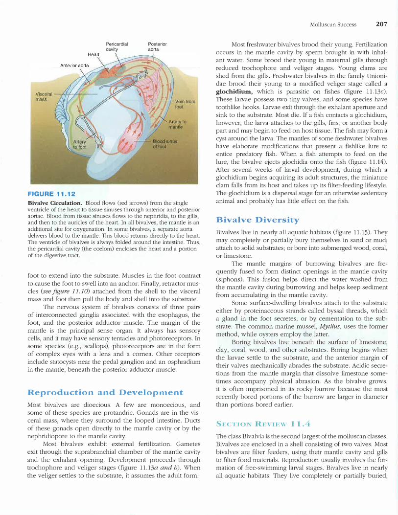
FIGURE 11.12
Pericardia! cavity
Posterior aorta
Bivalve Circulation. Blood flows (red arrows) from the single ventricle of the heart to tissue sinuses through anterior and posterior aortae. Blood from tissue sinuses flows to the nephridia, to the gills, and then to the auricles of the heart. In all bivalves, the mantle is an additional site for oxygenation. In some bivalves, a separate aorta delivers blood to the mantle. This blood returns directly to the heart. The ventricle of bivalves is always folded around the intestine. Thus, the pericardia! cavity (the coelom) encloses the heart and a portion of the digestive tract.
foot to extend into the substrate. Muscles in the foot contract
to cause the foot to swell into an anchor. Finally, retractor mus
cles (see figure 11. 10) attached from the shell to the visceral
mass and foot then pull the body and shell into the substrate.
The nervous system of bivalves consists of three pairs
of interconnected ganglia associated with the esophagus, the
foot, and the posterior adductor muscle. The margin of the
mantle is the principal sense organ. It always has sensory
cells, and it may have sensory tentacles and photoreceptors. In some species (e.g., scallops), photoreceptors are in the form
of complex eyes with a lens and a cornea. Other receptors
include statocysts near the pedal ganglion and an osphradium in the mantle, beneath the posterior adductor muscle.
Reproduction and Development
Most bivalves are dioecious. A few are monoecious, and
some of these species are protandric. Gonads are in the vis
ceral mass, where they surround the looped intestine. Ducts
of these gonads open directly to the mantle cavity or by the
nephridiopore to the mantle cavity.
Most bivalves exhibit external fertilization. Gametes
exit through the suprabranchial chamber of the mantle cavity
and the exhalant opening. Development proceeds through
trochophore and veliger stages (figure 11.13a and b). When
the veliger settles to the substrate, it assumes the adult form.
Molluscan Success 207
Most freshwater bivalves brood their young. Fertilization
occurs in the mantle cavity by sperm brought in with inhal
ant water. Some brood their young in maternal gills through
reduced trochophore and veliger stages. Young clams are
shed from the gills. Freshwater bivalves in the family Unioni
dae brood their young to a modified veliger stage called a
glochidium, which is parasitic on fishes (figure 11.13c).
These la1vae possess two tiny valves, and some species have
toothlike hooks. Larvae exit through the exhalant aperture and
sink to the substrate. Most die. If a fish contacts a glochidium,
however, the larva attaches to the gills, fins, or another body
part and may begin to feed on host tissue. The fish may form a
cyst around the larva. The mantles of some freshwater bivalves
have elaborate modifications that present a fishlike lure to
entice predato1y fish. When a fish attempts to feed on the
lure, the bivalve ejects glochidia onto the fish (figure 11.14).
After several weeks of laival development, during which a
glochidium begins acquiring its adult structures, the miniature
clam falls from its host and takes up its filter-feeding lifestyle.
The glochidium is a dispersal stage for an otherwise sedentary
animal and probably has little effect on the fish.
Bivalve Diversity
Bivalves live in nearly all aquatic habitats (figure 11.15). They
may completely or partially bury themselves in sand or mud;
attach to solid substrates; or bore into submerged wood, coral,
or limestone.
The mantle margins of burrowing bivalves are fre
quently fused to form distinct openings in the mantle cavity
(siphons). This fusion helps direct the water washed from
the mantle cavity during burrowing and helps keep sediment
from accumulating in the mantle cavity.
Some surface-dwelling bivalves attach to the substrate
either by proteinaceous strands called byssal threads, which
a gland in the foot secretes, or by cementation to the sub
strate. The common marine mussel, Mytilus, uses the former
method, while oysters employ the latter.
Boring bivalves live beneath the surface of limestone, clay, coral, wood, and other substrates. Boring begins when
the larvae settle to the substrate, and the anterior margin of
their valves mechanically abrades the substrate. Acidic secre
tions from the mantle margin that dissolve limestone some
times accompany physical abrasion. As the bivalve grows,
it is often imprisoned in its rocky burrow because the most
recently bored portions of the burrow are larger in diameter
than portions bored earlier.
SECTION Rt•:vrnw 11.4
The class Bi val via is the second largest of the molluscan classes.
Bivalves are enclosed in a shell consisting of two valves. Most
bivalves are filter feeders, using their mantle cavity and gills
to filter food materials. Reproduction usually involves the for
mation of free-swimming larval stages. Bivalves live in nearly
all aquatic habitats. They live completely or partially buried,
208 CHAPTER ELEVEN
Anterior adductor muscle
(b)
Digestive gland Intestine
FIGURE 11.13
(a)
::" �--�,'·,, ,,., .-
'i,:4 . I , '' 1'' ,11:, ... �.:./rr. ,,
.,.,, 11',·A ....... •�\\' ,,,,,, ' •• \,!l-'••l...'1 I
u -' •�1h·-
/Velum
Mouth
/Foot
(c)
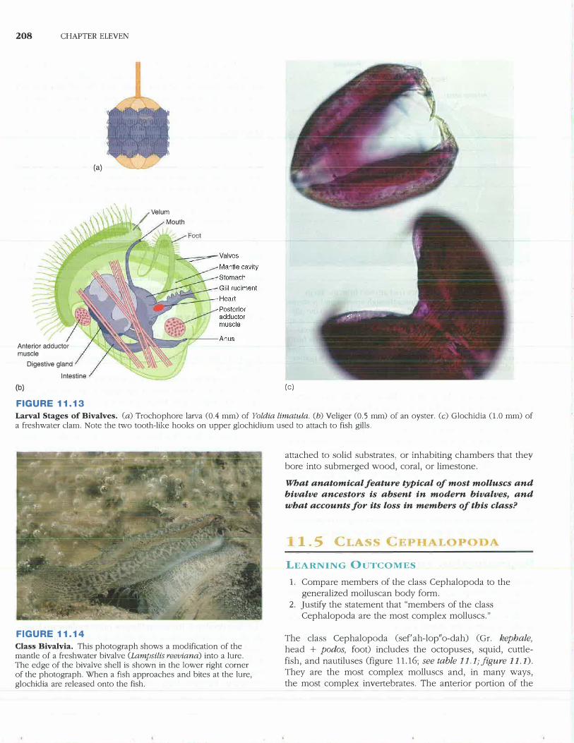
Larval Stages of Bivalves. (a) Trochophore larva (0.4 mm) of Yoldia limatula. (b) Veliger (0.5 mm) of an oyster. (c) Glochidia (1.0 mm) of a freshwater clam. Note the two tooth-like hooks on upper glochidium used to attach to fish gills.
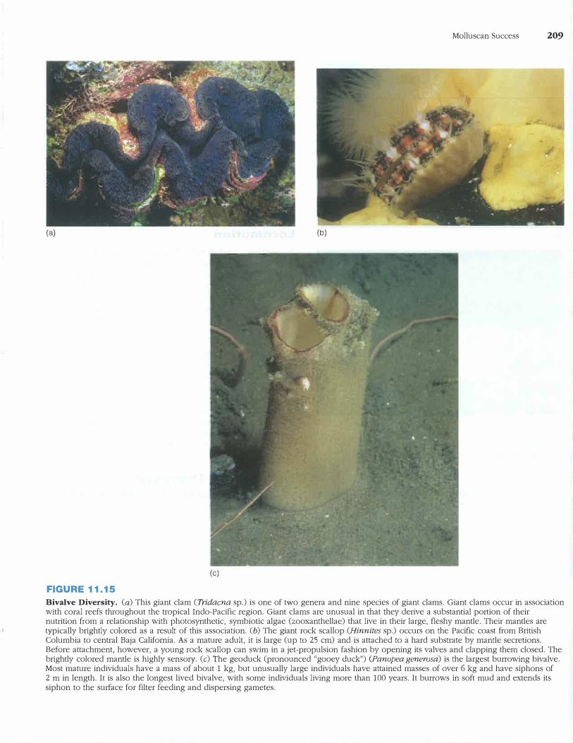
FIGURE 11.14
Class Bivalvia. This photograph shows a modification of the mantle of a freshwater bivalve (Lampsilis reeviana) into a lure. The edge of the bivalve shell is shown in the lower right corner of the photograph. When a fish approaches and bites at the lure, glochidia are released onto the fish.
attached to solid substrates, or inhabiting chambers that they bore into submerged wood, coral, or limestone.
What anatomical feature typical of most molluscs and
bivalve ancestors is absent in modern bivalves, and
what accounts for its loss in members of this class?
1.5 CLAS
LEARNING OUTCOMES
1. Compare members of the class Cephalopoda to thegeneralized molluscan body form.
2. Justify the statement that "members of the classCephalopoda are the most complex molluscs."
The class Cephalopoda (sef' ah-lop" o-dah) ( Gr. kephale,
head + podos, foot) includes the octopuses, squid, cuttlefish, and nautiluses (figure 11.16; see table 11.1; figure 11.1).
They are the most complex molluscs and, in many ways, the most complex invertebrates. The anterior portion of the
(a)
(c)
FIGURE 11.15
(b)
Molluscan Success 209
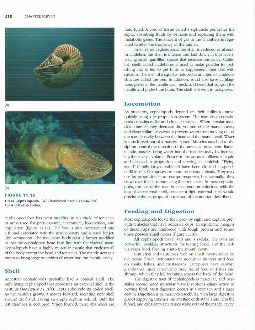
Bivalve Diversity. (a) This giant clam (Tridacna sp.) is one of two genera and nine species of giant clams. Giant clams occur in association with coral reefs throughout the tropical Indo-Pacific region. Giant clams are unusual in that they derive a substantial portion of their nutrition from a relationship with photosynthetic, symbiotic algae (zooxanthellae) that live in their large, fleshy mantle. Their mantles are typically brightly colored as a result of this association. (b) The giant rock scallop (Hinnites sp.) occurs on the Pacific coast from British Columbia to central Baja California. As a mature adult, it is large (up to 25 cm) and is attached to a hard substrate by mantle secretions. Before attachment, however, a young rock scallop can swim in a jet-propulsion fashion by opening its valves and clapping them closed. The brightly colored mantle is highly sensory. (c) The geoduck (pronounced "gooey duck") (Panopea generosa) is the largest burrowing bivalve. Most mature individuals have a mass of about 1 kg, but unusually large individuals have attained masses of over 6 kg and have siphons of 2 m in length. It is also the longest lived bivalve, with some individuals living more than 100 years. It burrows in soft mud and extends its siphon to the surface for filter feeding and dispersing gametes.
210 CH APTER ELEVEN
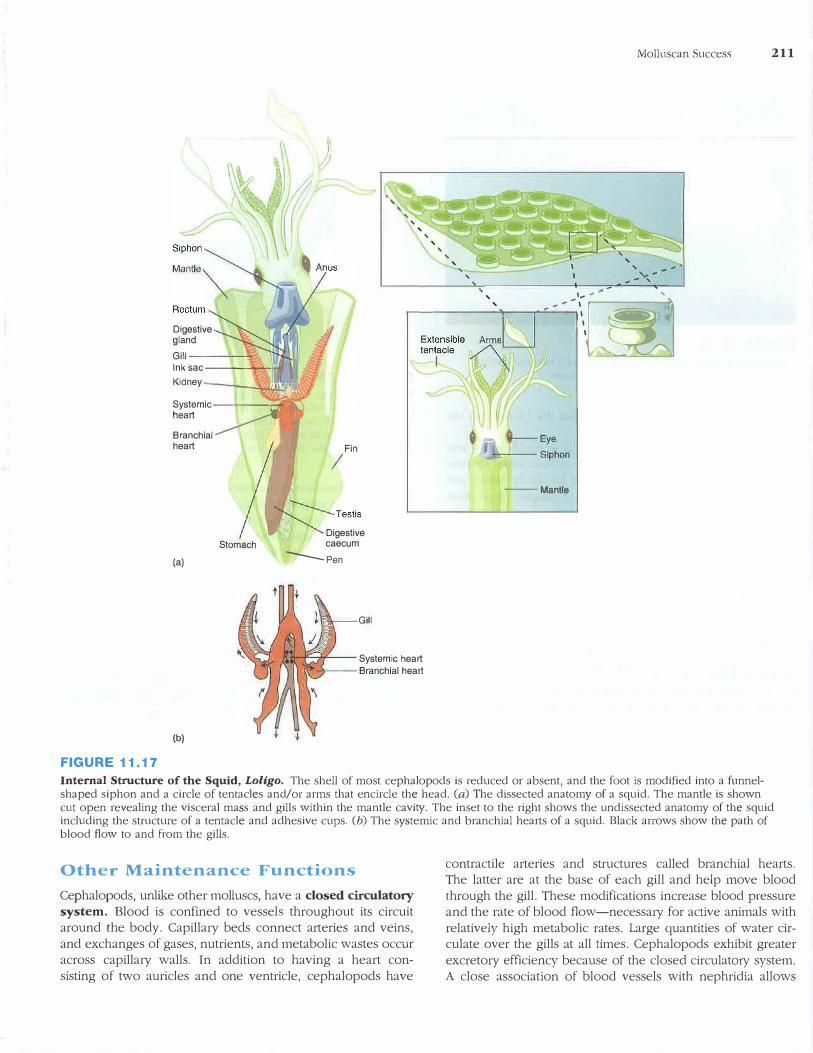
(a)
(b)
FIGURE 11.16
Class Cephalopoda. (a) Chambered nautilus (Nautilus). (b) A cuttlefish (Sepia).
cephalopod foot has been modified into a circle of tentacles or arms used for prey capture, attachment, locomotion, and copulation (figure 11.17). The foot is also incorporated into a funnel associated with the mantle cavity and is used for jetlike locomotion. The molluscan body plan is further modified in that the cephalopod head is in line with the visceral mass. Cephalopods have a highly muscular mantle that encloses all of the body except the head and tentacles. The mantle acts as a pump to bring large quantities of water into the mantle cavity.
Shell
Ancestral cephalopods probably had a conical shell. The only living cephalopod that possesses an external shell is the nautilus (see figure 11.16a). Septa subdivide its coiled shell. As the nautilus grows, it moves forward, secreting new shell
around itself and leaving an empty septum behind. Only the last chamber is occupied. When formed, these chambers are
fluid filled. A cord of tissue called a siphuncle perforates the septa, absorbing fluids by osmosis and replacing them with metabolic gases. The amount of gas in the chambers is regulated to alter the buoyancy of the animal.
In all other cephalopods, the shell is reduced or absent. In cuttlefish, the shell is internal and laid down in thin layers, leaving small, gas-filled spaces that increase buoyancy. Cuttlefish shell, called cuttlebone, is used to make powder for polishing and is fed to pet birds to supplement their diet with calcium. The shell of a squid is reduced to an internal, chitinous structure called the pen. In addition, squid also have cartilaginous plates in the mantle wall, neck, and head that support the mantle and protect the brain. The shell is absent in octopuses.
Locomotion
As predators, cephalopods depend on their ability to move quickly using a jet-propulsion system. The mantle of cephalopods contains radial and circular muscles. When circular muscles contract, they decrease the volume of the mantle cavity and close collarlike valves to prevent water from moving out of the mantle cavity between the head and the mantle wall. Water is thus forced out of a narrow siphon. Muscles attached to the siphon control the direction of the animal's movement. Radial mantle muscles bring water into the mantle cavity by increasing the cavity's volume. Posterior fins act as stabilizers in squid and also aid in propulsion and steering in cuttlefish. "Flying squid" (family Onycoteuthidae) have been clocked at speeds of 30 km/hr. Octopuses are more sedentary animals. They may use jet propulsion in an escape response, but normally, they crawl over the substrate using their tentacles. In most cephalopods, the use of the mantle in locomotion coincides with the loss of an external shell, because a rigid external shell would preclude the jet-propulsion method of locomotion described.
Feeding and Digestion
Most cephalopods locate their prey by sight and capture prey with tentacles that have adhesive cups. In squid, the margins of these cups are reinforced with tough protein and sometimes possess small hooks (figure 11.18).
All cephalopods have jaws and a radula. The jaws are powerful, beaklike structures for tearing food, and the radula rasps food, forcing it into the mouth cavity.
Cuttlefish and nautiluses feed on small invertebrates on the ocean floor. Octopuses are nocturnal hunters and feed on snails, fishes, and crustaceans. Octopuses have salivary glands that inject venom into prey. Squid feed on fishes and shrimp, which they kill by biting across the back of the head.
The digestive tract of cephalopods is muscular, and peristalsis (coordinated muscular waves) replaces ciliary action in moving food. Most digestion occurs in a stomach and a large cecum. Digestion is primarily extracellular, with large digestive glands supplying enzymes. An intestine ends at the anus, near the funnel, and exhalant water carries wastes out of the mantle cavity.
'
' ' ' ' ' ' ' '"""'" '
Molluscan Success 211
, .
Digestive gland Gill-----
'
..----------
'
-
'
....... --.--'-'---' ...... -\.� lnk sac---"--_.,.....,_:;#f Kidney -�;---'.!,.::t/.:!.Yl,I'
Systemic , heart � Branchial heart
Stomach
Fin I
Digestive caecum
(a) ----Pen
(b)
FIGURE 11.17
GIii
Systemic heart Branchial heart
---Mantle
Internal Strncture of the Squid, Loligo. The shell of most cephalopods is reduced or absent, and the foot is modified into a funnelshaped siphon and a circle of tentacles and/or arms that encircle the head. (a) The dissected anatomy of a squid. The mantle is shown cut open revealing the visceral mass and gills within the mantle cavity. The inset to the right shows the undissected anatomy of the squid including the structure of a tentacle and adhesive cups. (b) The systemic and branchial hearts of a squid. Black arrows show the path of blood flow to and from the gills.
Other Maintenance Functions
Cephalopods, unlike other molluscs, have a closed circulatory
system. Blood is confined to vessels throughout its circuit around the body. Capilla1y beds connect aneries and veins,
and exchanges of gases, nutrients, and metabolic wastes occur across capilla1y walls. In addition to having a heart con
sisting of two auricles and one ventricle, cephalopods have
contractile arteries and structures called branchial hearts. The latter are at the base of each gill and help move blood
through the gill. These modifications increase blood pressure and the rate of blood flow-necessa1y for active animals with
relatively high metabolic rates. Large quantities of water circulate over the gills at all times. Cephalopods exhibit greater
excretory efficiency because of the closed circulat01y system. A close association of blood vessels with nephridia allows
212 CHAPTER ELEVEN
FIGURE 11.18
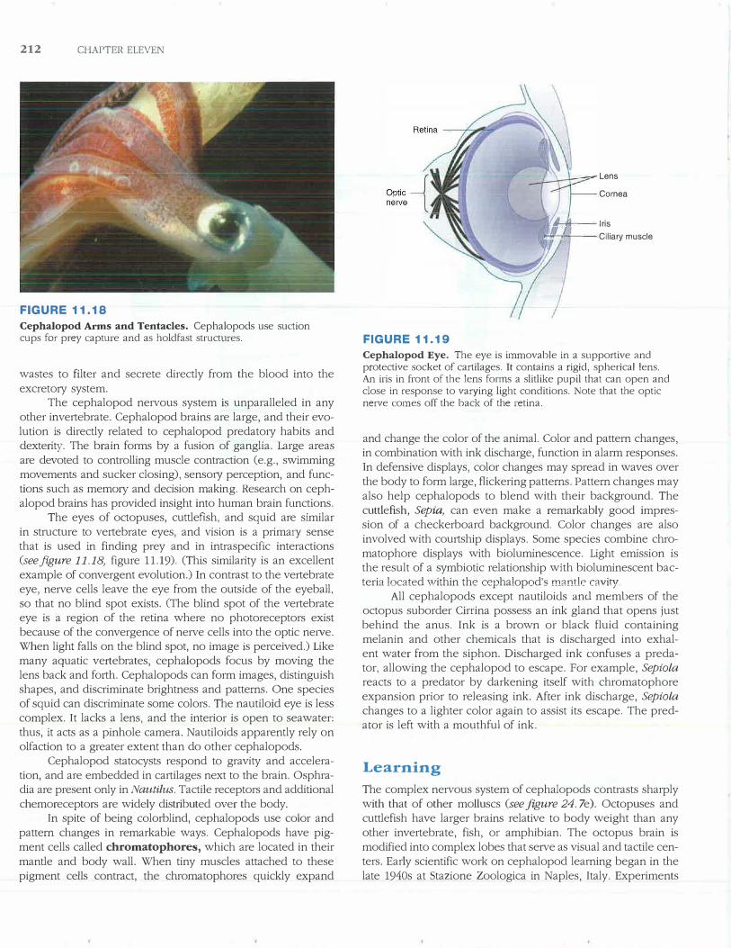
Cephalopod Arms and Tentacles. Cephalopods use suction cups for prey capture and as holdfast structures.
wastes to filter and secrete directly from the blood into the
excretory system. The cephalopod nervous system is unparalleled in any
other invertebrate. Cephalopod brains are large, and their evolution is directly related to cephalopod predatory habits and dexterity. The brain forms by a fusion of ganglia. Large areas are devoted to controlling muscle contraction (e.g., swimming movements and sucker closing), sensory perception, and functions such as memmy and decision making. Research on cephalopod brains has provided insight into human brain functions.
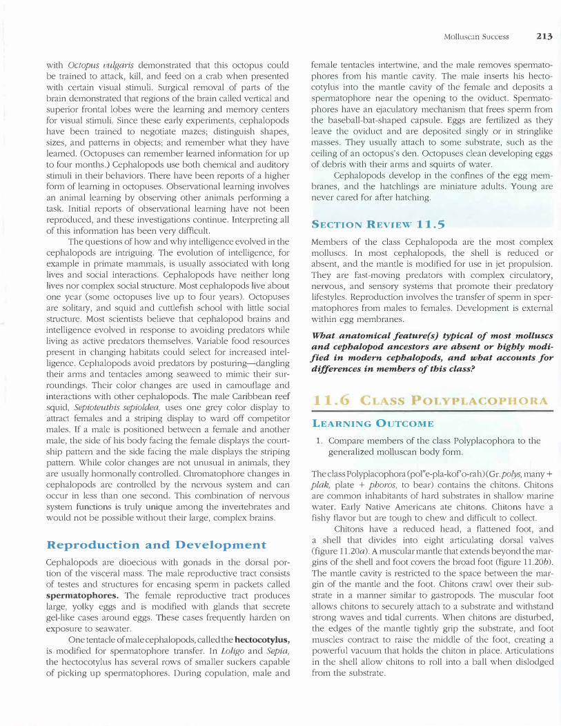
The eyes of octopuses, cuttlefish, and squid are similar
in structure to vertebrate eyes, and vision is a prima1y sense that is used in finding prey and in intraspecific interactions (see.figure 11.18, figure 11.19). (This similarity is an excellent example of convergent evolution.) In contrast to the vertebrate eye, nerve cells leave the eye from the outside of the eyeball, so that no blind spot exists. (The blind spot of the vertebrate eye is a region of the retina where no photoreceptors exist because of the convergence of nerve cells into the optic ne1ve. When light falls on the blind spot, no image is perceived.) Like many aquatic vertebrates, cephalopods focus by moving the lens back and forth. Cephalopods can form images, distinguish shapes, and discriminate brightness and patterns. One species of squid can discriminate some colors. The nautiloid eye is less complex. It lacks a lens, and the interior is open to seawater: thus, it acts as a pinhole camera. Nautiloids apparently rely on olfaction to a greater extent than do other cephalopods.
Cephalopod statocysts respond to gravity and acceleration, and are embedded in cartilages next to the brain. Osphradia are present only in Nautilus. Tactile receptors and additional
chemoreceptors are widely distributed over the body. In spite of being colorblind, cephalopods use color and
pattern changes in remarkable ways. Cephalopods have pigment cells called chromatophores, which are located in their
mantle and body wall. When tiny muscles attached to these pigment ce11s contract, the chromatophores quickly expand
FIGURE 11.19
Lens
Cornea
1!1--++---lris
_.-1-;--- Ciliary muscle
Cephalopod Eye. The eye is immovable in a supportive and protective socket of cartilages. It contains a rigid, spherical lens. An iris in front of the lens forms a slitlike pupil that can open and close in response to varying light conditions. Note that the optic nerve comes off the back of the retina.
and change the color of the animal. Color and pattern changes, in combination with ink discharge, function in alarm responses. In defensive displays, color changes may spread in waves over the body to form large, flickering patterns. Pattern changes may also help cephalopods to blend with their background. The cuttlefish, Sepia, can even make a remarkably good impression of a checkerboard background. Color changes are also involved with courtship displays. Some species combine chromatophore displays with bioluminescence. Light emission is the result of a symbiotic relationship with bioluminescent bacteria located ,vithin the cephalopod's mantle cavity
All cephalopods except nautiloids and members of the octopus suborder Cirrina possess an ink gland that opens just behind the anus. Ink is a brown or black fluid containing melanin and other chemicals that is discharged into exhalent water from the siphon. Discharged ink confuses a predator, allowing the cephalopod to escape. For example, Sepia/a
reacts to a predator by darkening itself with chromatophore expansion prior to releasing ink. After ink discharge, Sepiola
changes to a lighter color again to assist its escape. The predator is left with a mouthful of ink.
Learning
The complex nervous system of cephalopods contrasts sharply with that of other molluscs (see figure 24. 7e). Octopuses and cuttlefish have larger brains relative to body weight than any other invertebrate, fish, or amphibian. The octopus brain is modified into complex lobes that serve as visual and tactile centers. Early scientific work on cephalopod learning began in the late 1940s at Stazione Zoologica in Naples, Italy. Experiments
with Octopus uulgaris demonstrated that this octopus could be trained to attack, kill, and feed on a crab when presented
with certain visual stimuli. Surgical removal of parts of the brain demonstrated that regions of the brain called vertical and superior frontal lobes were the learning and memo1y centers for visual stimuli. Since these early experiments, cephalopods have been trained to negotiate mazes; distinguish shapes, sizes, and patterns in objects; and remember what they have learned. (Octopuses can remember learned information for up to four months.) Cephalopods use both chemical and audito1y stimuli in their behaviors. There have been repo1ts of a higher form of learning in octopuses. Observational learning involves an animal learning by observing other animals performing a task. Initial reports of obse1vational learning have not been reproduced, and these investigations continue. Interpreting all of this information has been ve1y difficult.
The questions of how and why intelligence evolved in the cephalopods are intriguing. The evolution of intelligence, for example in primate mammals, is usually associated with long lives and social interactions. Cephalopods have neither long lives nor complex social structure. Most cephalopods live about one year (some octopuses live up to four years). Octopuses are solita1y, and squid and cuttlefish school with little social structure. Most scientists believe that cephalopod brains and intelligence evolved in response to avoiding predators while living as active predators themselves. Variable food resources present in changing habitats could select for increased intelligence. Cephalopods avoid predators by posturing-dangling their arms and tentacles among seaweed to mimic their surroundings. Their color changes are used in camouflage and interactions with other cephalopods. The male Caribbean reef squid, Sepioteuthis sepioldea, uses one grey color display to attract females and a striping display to ward off competitor males. If a male is positioned between a female and another male, the side of his body facing the female displays the couttship pattern and the side facing the male displays the striping pattern. While color changes are not unusual in animals, they are usually hormonally controlled. Chromatophore changes in cephalopods are controlled by the ne1vous system and can occur in less than one second. This combination of ne1vous system functions is truly unique among the inve1tebrates and would not be possible without their large, complex brains.
Reproduction and Developm.ent
Cephalopods are dioecious with gonads in the dorsal portion of the visceral mass. The male reproductive tract consists of testes and structures for encasing sperm in packets called spermatophores. The female reproductive tract produces large, yolky eggs and is modified with glands that secrete gel-like cases around eggs. These cases frequently harden on exposure to seawater.
One tentacle of male cephalopods, called the hectocotylus,
is modified for spermatophore transfer. In Latigo and Sepia,
the hectocotylus has several rows of smaller suckers capable of picking up spermatophores. During copulation, male and
Molluscan Success 213
female tentacles intertwine, and the male removes spermatophores from his mantle cavity. The male inserts his hectocotylus into the mantle cavity of the female and deposits a spermatophore near the opening to the oviduct. Spermato
phores have an ejaculat01y mechanism that frees sperm from the baseball-bat-shaped capsule. Eggs are fertilized as they leave the oviduct and are deposited singly or in stringlike masses. They usually attach to some substrate, such as the ceiling of an octopus's den. Octopuses clean developing eggs of debris with their arms and squirts of water.
Cephalopods develop in the confines of the egg membranes, and the hatchlings are miniature adults. Young are never cared for after hatching.
SECTION REVIEW 11.5
Members of the class Cephalopoda are the most complex molluscs. In most cephalopods, the shell is reduced or absent, and the mantle is modified for use in jet propulsion.
They are fast-moving predators with complex circulato1y, ne1vous, and senso1y systems that promote their predato1y lifestyles. Reproduction involves the transfer of sperm in spermatophores from males to females. Development is external within egg membranes.
What anatomical feature(s) typical of most molluscs
and cepbalopod ancestors are absent or highly modi
fied in modern cepbalopods, and what accounts for
differences in members of this class?
l ORA
LEARNING OUTCOME
1. Compare members of the class Polyplacophora to thegeneralized molluscan body form.
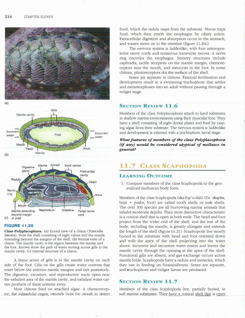
The class Polyplacophora (pol"e-pla-kof' o-rah) (Gr.polys, many+ plak, plate + phoros, to bear) contains the chitons. Chitons are common inhabitants of hard substrates in shallow marine water. Early Native Americans ate chitons. Chitons have a fishy flavor but are tough to chew and difficult to collect.
Chitons have a reduced head, a flattened foot, and a shell that divides into eight articulating dorsal valves (figure 11.20a). A muscular mantle that extends beyond the margins of the shell and foot covers the broad foot (figure 11.2Gb).
The mantle cavity is restricted to the space between the margin of the mantle and the foot. Chitons crawl over their substrate in a manner similar to gastropods. The muscular foot
allows chitons to securely attach to a substrate and withstand strong waves and tidal currents. When chitons are disturbed, the edges of the mantle tightly grip the substrate, and foot
muscles contract to raise the middle of the foot, creating a powerful vacuum that holds the chiton in place. Articulations
in the shell allow chitons to roll into a ball when dislodged from the substrate.
214 COAPTER ELEVEN
(a)
=
lncurrent water
=
(b)
GIiis
Mantle
=
Excurrent =water
Mantle Gonad Shell valves
Mantle extending beyond margin
(c) of shell
FIGURE 11.20
Pericardial
Pedal nerve cord
Anus
Class Polyplacophora. (a) Dorsal view of a chiton (Tonicella
lineata). Note the shell consisting of eight valves and the mantle extending beyond the margins of the shell. (b) Ventral view of a chiton. The mantle cavity is the region between the mantle and the foot. Arrows show the path of water moving across gills in the mantle cavity. (c) Internal structure of a chiton.
A linear series of gills is in the mantle cavity on each side of the foot. Cilia on the gills create water currents that enter below the anterior mantle margins and exit posteriorly. The digestive, excretory, and reproductive tracts open near the exhalant area of the mantle cavity, and exhalant water carries products of these systems away.
Most chitons feed on attached algae. A chemorecep_tor,_tlli:_subradular_mgan, xt nds Frcm1 Lh n� uth to detect
food, which the radula rasps from the substrate. Mucus traps food, which then enters the esophagus by cilia1y action. Extracellular digestion and absorption occur in the stomach, and wastes move on to the intestine (figure 11.20c).
The nervous system is ladderlike, with four anteroposterior nerve cords and numerous transverse nerves. A nerve ring encircles the esophagus. Sensory structures include osphradia, tactile receptors on the mantle margin, chemoreceptors near the mouth, and statocysts in the foot. In some chitons, photoreceptors dot the surface of the shell.
Sexes are separate in chitons. External fertilization and development result in a swimming trochophore that settles and metamorphoses into an adult without passing through a veliger stage.
SECTION REVIEW 11.6
Members of the class Polyplacophora attach to hard substrates in shallow marine environments using their muscular foot. They have a shell consisting of eight dorsal plates and feed by rasping algae from their substrate. The nervous system is ladderlike and development is external with a trochophore larval stage.
What features of members of the class Polyplacophora
(if any) would be considered atypical of molluscs in
general?
11. 7 CLASS SCAPHOPODA
LEARNING OUTCOME
1. Compare members of the class Scaphopoda to the gen-eralized molluscan body form.
Members of the class Scaphopoda (ska-fop'o-dah) (Gr. skaphe,
boat + podos, foot) are called tooth shells or tusk shells. The over 300 species are all burrowing marine animals that inhabit moderate depths. Their most distinctive characteristic is a conical shell that is open at both ends. The head and foot project from the wider end of the shell, and the rest of the body, including the mantle, is greatly elongate and extends the length of the shell (figure 11.21). Scaphopods live mostly buried in the substrate with head and foot oriented down and with the apex of the shell projecting into the water above. Incurrent and excurrent water enters and leaves the mantle cavity through the opening at the apex of the shell. Functional gills are absent, and gas exchange occurs across mantle folds. Scaphopods have a radula and tentacles, which they use in feeding on foraminiferans. Sexes are separate, and trochophore and veliger larvae are produced.
SECTION REVIEW 11. 7
Members of the class Scaphopoda live, partially buried, in soft marine _sll_bstr_ates. Th�y have a_ conical shell_tli.Jlct is Qgen ____ _
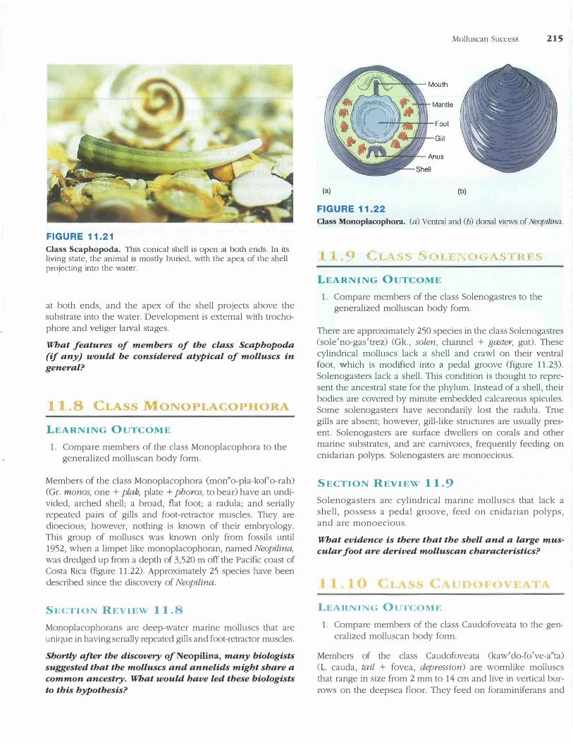
FIGURE 11.21
Class Scaphopoda. This conical shell is open at both ends. In its living state, the animal is mostly buried, with the apex of the shell projecting into the water.
at both ends, and the apex of the shell projects above the substrate into the water. Development is external with trochophore and veliger larval stages.
What features of members of the class Scaphopoda
(if any J would be considered atypical of molluscs in
general?
1 .8 Cl.A ONOPLACOPI ORA
LEARNING OUTCOME
1. Compare members of the class Monoplacophora to thegeneralized molluscan body form.
Members of the class Monoplacophora (mon"o-pla-kof' o-rah) (Gr. monos, one + plak, plate + phoros, to bear) have an undivided, arched shell; a broad, flat foot; a radula; and serially repeated pairs of gills and foot-retractor muscles. They are dioecious; however, nothing is known of their embryology. This group of molluscs was known only from fossils until 1952, when a limpet like monoplacophoran, named Neopilina,
was dredged up from a depth of 3,520 m off the Pacific coast of Costa Rica (figure 11.22). Approximately 25 species have been described since the discove1y of Neopilina.
SECTION REVIEW 11.8
Monoplacophorans are deep-water marine molluscs that are unique in having serially repeated gills and foot-retractor muscles.
Shortly after the discovery ofNeopilina, many biologists
suggested that the molluscs and annelids might share a
common ancestry. What would have led these biologists
to this hypothesis?
(a)
FIGURE 11.22
Molluscan Success 215
(b)
Class Monoplacophora. (a) Ventral and (b) dorsal views of Neopilina.
LEARNING OUTCOME
1. Compare members of the class Solenogastres to thegeneralized molluscan body form.
There are approximately 250 species in the class Solenogastres (sole'no-gas'trez) (Gk., solen, channel + gaster, gut). These cylindrical molluscs lack a shell and crawl on their ventral foot, which is modified into a pedal groove (figure 11.23). Solenogasters lack a shell. This condition is thought to represent the ancestral state for the phylum. Instead of a shell, their bodies are covered by minute embedded calcareous spicules. Some solenogasters have secondarily lost the radula. True gills are absent; however, gill-like structures are usually present. Solenogasters are surface dwellers on corals and other marine substrates, and are carnivores, frequently feeding on cnidarian polyps. Solenogasters are monoecious.
SECTION REVIEW 11.9
Solenogasters are cylindrical marine molluscs that lack a shell, possess a pedal groove, feed on cnidarian polyps, and are monoecious.
What evidence is there that the shell and a large mus
cular foot are derived molluscan characteristics?
I I I 0 l I ( J 0
LEARNING OUTCOM E
1. Compare members of the class Caudofoveata to the gen-eralized molluscan body form.
Members of the class Caudofoveata (kaw'clo-fo've-a"ta) (L. caucla, tail + fovea, depression) are wormlike molluscs that range in size from 2 mm to 14 cm and live in vertical burrows on the deepsea floor. They feed on foraminiferans and
216 CHAPTER ELEVEN
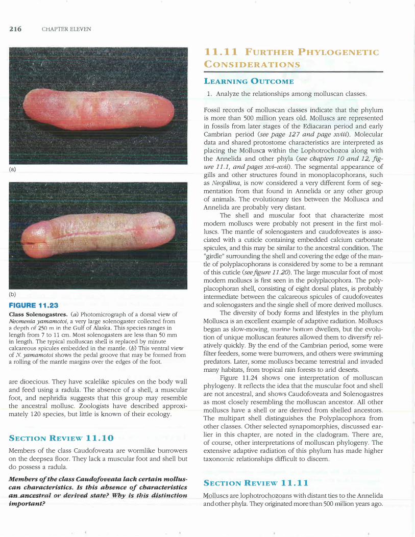
(a)
(b)
FIGURE 11.23
Class Solenogastres. (a) Photomicrograph of a dorsal view of Neomenia yamamotoi, a very large solenogaster collected from 3 depth nf 250 m in the Gulf of Alaska. This species ranges in length from 7 to 11 cm. Most solenogasters are less than 50 mm in length. The typical molluscan shell is replaced by minute calcareous spicules embedded in the mantle. (b) This ventral view of N. yamamotoi shows the pedal groove that may be formed from a rolling of the mantle margins over the edges of the foot.
are dioecious. They have scalelike spicules on the body wall and feed using a radula. The absence of a shell, a muscular foot, and nephridia suggests that this group may resemble the ancestral mollusc. Zoologists have described approximately 120 species, but little is known of their ecology.
SECTION REVIEW 11.10
Members of the class Caudofoveata are wormlike burrowers on the deepsea floor. They lack a muscular foot and shell but do possess a radula.
Members of the class Caudofoveata lack certain mollus
can characteristics. Is this absence of characteristics
a:n_anus.tral 01· derived, st4te? Why is this d;istinctio·n
important?
11.11 FURTHER PHYLOGENETIC
CONSIDERATIONS
LEARNING OUTCOME
1. Analyze the relationships among molluscan classes.
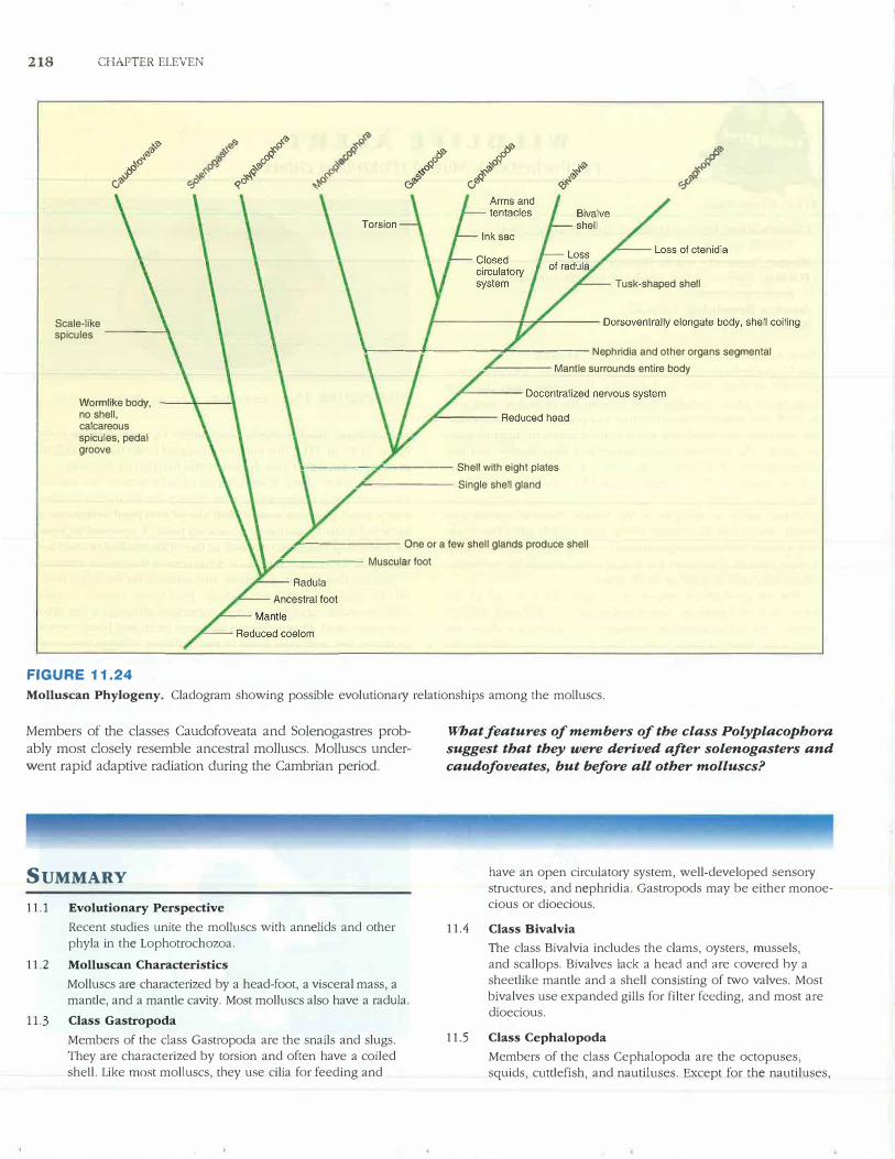
Fossil records of molluscan classes indicate that the phylum is more than 500 million years old. Molluscs are represented in fossils from later stages of the Ediacaran period and early Cambrian period (see page 127 and page xviii). Molecular data and shared protostome characteristics are interpreted as placing the Mollusca within the Lophotrochozoa along with the Annelida and other phyla (see chapters 10 and 12, figure 11.1, and pages xvi-xvii). The segmental appearance of gills and other structures found in monoplacophorans, such as Neopilina, is now considered a ve1y different form of segmentation from that found in Annelida or any other group of animals. The evolutionary ties between the Mollusca and Annelida are probably veiy distant.
The shell and muscular foot that characterize most modern molluscs were probably not present in the first molluscs. The mantle of solenogasters and caudofoveates is associated with a cuticle containing embedded calcium carbonate spicules, and this may be similar to the ancestral condition. The "girdle" surrounding the shell and covering the edge of the mantle of polyplacophorans is considered by some to be a remnant of this cuticle (see figure 11.20). The large muscular foot of most modern molluscs is first seen in the polyplacophora. The polyplacophoran shell, consisting of eight dorsal plates, is probably intermediate between the calcareous spicules of caudofoveates and solenogasters and the single shell of more derived molluscs.
The diversity of body forms and lifestyles in the phylum Mollusca is an excellent example of adaptive radiation. Molluscs began as slow-moving, marine hottom dwellers, but the evolution of unique molluscan features allowed them to diversify relatively quickly. By the end of the Cambrian period, some were filter feeders, some were burrowers, and others were swimming predators. Later, some molluscs became terrestrial and invaded many habitats, from tropical rain forests to arid deserts.
Figure 11.24 shows one interpretation of molluscan phylogeny. It reflects the idea that the muscular foot and shell are not ancestral, and shows Caudofoveata and Solenogastres as most closely resembling the molluscan ancestor. All other molluscs have a shell or are derived from shelled ancestors. The multipart shell distinguishes the Polyplacophora from other classes. Other selected synapomorphies, discussed earlier in this chapter, are noted in the cladogram. There are, of course, other interpretations of molluscan phylogeny. The extensive adaptive radiation of this phylum has made higher taxonomic relationships difficult to discern.
SECTION REVIEW 11.11
MQlluscs are_lophotrochozoans with distant ties to the Annelida and other phyla. They originated more than 500 million years ago.
Molluscan Success 217
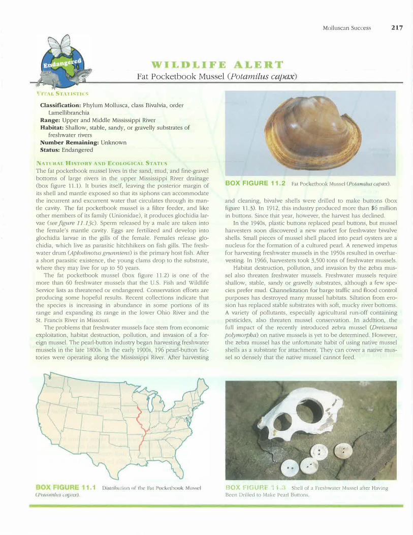
Fat Pocketbook Mussel (Potamilus cc,pax
Classification: Phylum Mollusca, class Bivalvia, order Lamellibranchia
Range: Upper and Middle Mississippi River Habitat: Shallow, stable, sandy, or gravelly substrates of
freshwater rivers Number Remaining: Unknown Status: Endangered
Nl\l'llHAL HISTORY ANU Eco1,0GICAL STATUS
The fat pocketbook mussel lives in the sand, mud, and fine-gravel bottoms of large rivers in the upper Mississippi River drainage (box figure 11.1). It buries itself, leaving the posterior margin of its shell and mantle exposed so that its siphons can accommodate the incurrent and excurrent water that circulates through its mantle cavity. The fat pocketbook mussel is a filter feeder, and like other members of its family (Unionidae), it produces glochidia larvae (see.figure 11.13c). Sperm released by a male are taken into the female's mantle cavity. Eggs are fertilized and develop into glochidia larvae in the gills of the female. Females release glochidia, which live as parasitic hitchhikers on fish gills. The freshwater drum (Aplodinotus grunniens) is the prima1y host fish. After a short parasitic existence, the young clams drop to the substrate, where they may live for up to 50 years.
The fat pocketbook mussel (box figure 11.2) is one of the more than 60 freshwater mussels that the U.S. Fish and Wildlife Service lists as threatened or endangered. Conservation efforts are producing some hopeful results. Recent collections indicate that the species is increasing in abundance in some portions of its range and expanding its range in the lower Ohio River and the St. Francis River in Missouri.
The problems that freshwater mussels face stem from economic exploitation, habitat destruction, pollution, and invasion of a foreign mussel. The pearl-button industty began harvesting freshwater mussels in the late 1800s. In the early 1900s, 196 pearl-button factories were operating along the Mississippi River. After harvesting
( P//Utlllfltt Wj)ll.\'J
1.1 I 1strib,1uor1 uf 1lic.: l'u1 Puckl'lbook Mus,el
BOX FIGURE 11.2 Fat Pocketbook Mussel (Potamilus capax).
and cleaning, bivalve shells were drilled to make buttons (box figure 11.3). In 1912, this industry produced more than $6 million in buttons. Since that year, however, the harvest has declined.
In the 1940s, plastic buttons replaced pearl buttons, but mussel harvesters soon discovered a new market for freshwater bivalve shells. Small pieces of mussel shell placed into pearl oysters are a nucleus for the formation of a cultured pearl. A renewed impetus for harvesting freshwater mussels in the 1950s resulted in overharvesting. In 1966, ha1vesters took 3,500 tons of freshwater mussels.
Habitat destruction, pollution, and invasion by the zebra mussel also threaten freshwater mussels. Freshwater mussels require shallow, stable, sandy or gravelly substrates, although a few species prefer mud. Channelization for barge traffic and flood control purposes has destroyed many mussel habitats. Siltation from erosion has replaced stable substrates with soft, mucky river bottoms. A variety of pollutants, especially agricultural run-off containing pesticides, also threaten mussel conservation. In addition, the full impact of the recently introduced zebra mussel (Dreissena
polymotpha) on native mussels is yet to be determined. However, the zebra mussel has the unfortunate habit of using native mussel shells as a substrate for attachment. They can cover a native mussel so densely that the native mussel cannot feed.
BOX FIGURF l ·1 .:'\ Shell of a Freshwater Mussel after Having Been Drilled to Make Pearl Buttons
218 CHAPTER ELEVEN
w'I, ,.0 �0<!, �'I>
�o 0� � o'I> o'I>
& �
<I,
[J>t/j c,O�
�o�o
�o f:-,� rfr'li 00 il' �o
� �'Ii J'(IJ
�o
ci1,-s
a:,'>'e
�cj-.:,;, �o,;; ;;; c}� .,:,.'Ii (/>� 0'1, <o� �
Tusk-shaped shell
Scale-like spicules ____ ..,.
---------�------Nephridia and other organs segmental
Wormlike body, no shell, calcareous spicules, pedal groove
----- Mantle surrounds entire body
_____ __,.c._ ____ Shell with eight plates
.,... ______ Single shell gland
--------- One or a few shell glands produce shell
------- Muscular foot
FIGURE 11.24
Molluscan Phylogeny. Cladogram showing possible evolutiona1y relationships among the molluscs.
Members of the classes Caudofoveata and Solenogastres prob
ably most closely resemble ancestral molluscs. Molluscs under
went rapid adaptive radiation during the Cambrian period.
SUMMARY
11.1 Evolutionary Perspective
Recent studies unite the molluscs with annelids and other phyla in the Lophotrochozoa.
11.2 Molluscan Characteristics
Molluscs are characterized by a head-foot, a visceral mass, a mantle, and a mantle cavity. Most molluscs also have a radula.
11.3 Class Gastropoda
Members of the class Gastropoda are the snails and slugs. They are characterized by torsion and often have a coiled
shell. Like most molluscs, they use cilia for feeding and
What features of members of the class Polyplacophora
suggest that they were derived after solenogasters and
caudofoveates, but before all other molluscs?
have an open circulatory system, well-developed sensory structures, and nephridia. Gastropods may be either monoecious or dioecious.
11. 4 Class Bivalvia
The class Bivalvia includes the clams, oysters, mussels, and scallops. Bivalves lack a head and are covered hy a sheetlike mantle and a shell consisting of two valves. Most bivalves use expanded gills for filter feeding, and most are dioecious.
11.5 Class Cephalopoda
Members of the class Cephalopoda are the octopuses, squids, cuttlefish, and nautiluses. Except for the nautiluses,
they have a reduced shell. The anterior portion of their
foot has been modified into a circle of tentacles. Cepha
lopocls have a closed circulatmy system and highly devel
oped nervous and sensmy systems. They are efficient
predators.
11.6 Class Polyplacophora
Members of the class Polyplacophora are the chitons. They
have a muscular foot for attachment to solid substrates and
a shell consisting of eight dorsal plates.
11. 7 Class Scaphopoda
Members of the class Scaphopocla are the tooth shells or
tusk shells. Their shell is open at both ends and the apex
of the shell extends to the surface of the substrate as these
animals move through soft marine sediments.
11.8 Class Monoplacophora
Members of the class Monoplacophora have an uncliviclecl arched shell and a broad, flat foot. They live at great
depths in the Pacific Ocean and were known only as
fossils until 1952.
11.9 Class Solenogastres
Solenogasters live associated with coral, where they feed
on coral polyps. They lack a shell and muscular foot. Their foot is
modified into a ventral pedal groove.
11.10 Class Caudofoveata
Members of the class Cauclofoveata are burrowers in
deep-water marine substrates. They lack a shell and
muscular foot.
11.11 Further Phylogenetic Considerations
Molluscs are lophotrochozoans with distant ties to the
Annelida. The cauclofoveates and solenogasters are
probably most similar to the mollusc ancestor. Adaptive
radiation in the molluscs has resulted in their presence in
most ecosystems of the ea1th.
CONCEPT REVIEW QUESTIONS
1. All of the following may be functions of the mantle cavity of
molluscs, except one. Select the exception.
a. Gas exchange
b. Serving as a hydrostatic skeleton
c. Excretion
d. Elimination of digestive wastes
e. Release of reproductive products
2. Members of all of the following classes possess an open
circulato1y system, except one. Select the exception.
a. Bivalvia
b. Gastropoda
c. Cephalopoda
cl. Polyplacophora
Molluscan Success 219
3. In the gastropods, food is trapped in a mucoicl mass called
the - - - - ----� which extends to the stomach
and is rotated by cilia.
a. style
b. protostyle
c. c1ystalline style
cl. raclula
e. oclontophore
4. Which of the following is the correct sequence of structures
encountered by food entering the mantle cavity of a bivalve?
a. Incurrent opening, gill filaments, food groove, labial palps,
mouth
b. Incurrent opening, labial palps, food groove, gill filaments,
mouth
c. Incurrent opening, food groove, gill filaments, labial palps,
mouth
cl. Incurrent opening, gill filaments, labial palps, food groove,
mouth
5. All of the following statements regarding the evolution of the
Mollusca are true except one. Select the exception.
a. Gastropods and cephalopods are more closely related to
each other than to other mollusc groups.
b. Solenogastres and Cauclofoveata are the classes whose
members most closely resemble the mollusc ancestor.
c. The muscular foot and shell were characteristics of the mol
lusc ancestor.
cl. Molluscs are lophotrochozans and distantly related to the
Annelida.
ANALYSIS AND APPLICATION
QUESTIONS
1. Compare and contrast the hydraulic skeletons of molluscs with
the hydrostatic skeletons of cnidarians and pseuclocoelomates.
2. Review the functions of body cavities presented in chapter 7.
Which of those functions, if any, apply to the coelom of molluscs?
3. Students often confuse torsion and shell coiling. Describe each
and its effect on gastropod structure and function.
4. Bivalves are often used as indicators of environmental quality.
Based on your knowledge of bivalves, describe why bivalves
are useful environmental indicator organisms.
mconnect jZOOLOGY
Enhance your study of this chapter with study tools and practice
tests. Also ask your instructor about the resources available through
Connect, including a media-rich eBook, interactive learning tools,
and animations.