
Molecular Typing and Antimicrobial Susceptibility of ... · %JTFBTFT JO "TJBO "RVBDVMUVSF 7*...
17
Molecular Typing and Antimicrobial Susceptibility of Vibrio parahaemolyticus Strains R. P. MALUPING 1,2 , C. R. LAVILLA-PITOGO 3 , J.L. ROMALDE 4 and K. KROVACEK 1 1 Department of Biomedical Sciences and Veterinary Public Health, Faculty of Veterinary Medicine and Animal Science, SLU, Box 7036 750 07 Uppsala, Sweden 2 Department of Veterinary Paraclinical Sciences, College of Veterinary Medicine, University of the Philippines-Los Baños, 4031 College, Laguna, Philippines 3 Aquaculture Department, Southeast Asian Fisheries Development Center, Tigbauan, 5021 Iloilo, Philippines 4 Departamento de Microbiologia y Parasitologia, Facultad de Biologia, Universidad de Santiago, 15782 Santiago de Compostela, Spain ABSTRACT The aim of the present study was to use three PCR-based techniques for the analysis of genetic variability among Vibrio parahaemolyticus strains isolated from the Philippines. Seventeen strains of V. parahaemolyticus isolated from shrimps and from the environments where these shrimps are being cultivated were analyzed by RAPD, ERIC and REP-PCR. Antimicrobial susceptibility of these strains to selected compounds was investigated using broth microdilution method. Results of this work and analysis of similarity among strains using Dice coefficient and unweighted average pair group method have demonstrated genetic variability within the V. parahaemolyticus strains. The RAPD, ERIC and REP- PCR were found to be suitable typing methods for V. parahaemolyticus. They have good discriminative ability and can be used as rapid means of comparing these strains for epidemiological investigation. However, the REP-PCR analysis yielded a relatively small number of products suggesting that the REP sequences may not be widely distributed in the V. parahaemolyticus genome. Results of antimicrobial susceptibility revealed that resistance among the strains was rare. In conclusion, RAPD, ERIC and REP-PCR techniques are useful methods for molecular typing of V. parahaemolyticus strains. To our knowledge this is the first study of this kind carried out on V. parahaemolyticus strains isolated from the Philippines. Maluping, R.P., Lavilla-Pitogo, C.R., Romalde, J.L. and Krovacek, K. 2008. Molecular typing and antimicrobial susceptibility of Vibrio parahaemolyticus strains, pp. 451-468. In Bondad- Reantaso, M.G., Mohan, C.V., Crumlish, M. and Subasinghe, R.P. (eds.). Diseases in Asian Aquaculture VI. Fish Health Section, Asian Fisheries Society, Manila, Philippines. Corresponding author: R.P. Maluping, [email protected]
Transcript of Molecular Typing and Antimicrobial Susceptibility of ... · %JTFBTFT JO "TJBO "RVBDVMUVSF 7*...
Molecular Typing and Antimicrobial Susceptibility of Vibrio parahaemolyticus Strains
R. P. MALUPING 1,2, C. R. LAVILLA-PITOGO3, J.L. ROMALDE4 and K. KROVACEK1
1Department of Biomedical Sciences and Veterinary Public Health, Faculty of Veterinary Medicine and Animal Science, SLU, Box 7036
750 07 Uppsala, Sweden2Department of Veterinary Paraclinical Sciences, College of Veterinary Medicine,
University of the Philippines-Los Baños, 4031 College, Laguna, Philippines3Aquaculture Department, Southeast Asian Fisheries Development Center,
Tigbauan, 5021 Iloilo, Philippines4Departamento de Microbiologia y Parasitologia, Facultad de Biologia,
Universidad de Santiago, 15782 Santiago de Compostela, Spain
ABSTRACT
The aim of the present study was to use three PCR-based techniques for the analysis of genetic variability among Vibrio parahaemolyticus strains isolated from the Philippines. Seventeen strains of V. parahaemolyticus isolated from shrimps and from the environments where these shrimps are being cultivated were analyzed by RAPD, ERIC and REP-PCR. Antimicrobial susceptibility of these strains to selected compounds was investigated using broth microdilution method. Results of this work and analysis of similarity among strains using Dice coefficient and unweighted average pair group method have demonstrated genetic variability within the V. parahaemolyticus strains. The RAPD, ERIC and REP-PCR were found to be suitable typing methods for V. parahaemolyticus. They have good discriminative ability and can be used as rapid means of comparing these strains for epidemiological investigation. However, the REP-PCR analysis yielded a relatively small number of products suggesting that the REP sequences may not be widely distributed in the V. parahaemolyticus genome. Results of antimicrobial susceptibility revealed that resistance among the strains was rare. In conclusion, RAPD, ERIC and REP-PCR techniques are useful methods for molecular typing of V. parahaemolyticus strains. To our knowledge this is the first study of this kind carried out on V. parahaemolyticus strains isolated from the Philippines.
Maluping, R.P., Lavilla-Pitogo, C.R., Romalde, J.L. and Krovacek, K. 2008. Molecular typing and antimicrobial susceptibility of Vibrio parahaemolyticus strains, pp. 451-468. In Bondad-Reantaso, M.G., Mohan, C.V., Crumlish, M. and Subasinghe, R.P. (eds.). Diseases in Asian Aquaculture VI. Fish Health Section, Asian Fisheries Society, Manila, Philippines.
Corresponding author: R.P. Maluping, [email protected]
INTRODUCTION
Vibrio parahaemolyticus belongs to the expanding group of water and food-borne pathogens. It is widely distributed in the aquatic environment and is increasingly regarded as an important pathogen of shrimp causing significant economic problems within the aquaculture industry worldwide (Lightner, 1996; Goarant et al., 1999). In recent years, V. parahaemolyticus has been implicated as an opportunistic pathogen, mainly causing gastroenteritis in humans due to consumption of contaminated seafood (DePaola et al., 2003). These isolates tend to possess the tdh or trh genes which have only been identified in a minority of the environmental isolates. Therefore, due to increasing reports of disease outbreaks in humans and with the great economic loss in aquaculture, the correct identification and classification of this organism is of great importance.
The development of molecular biology has allowed the typing of organisms on the basis of analysis of their nucleic acids. Molecular typing techniques are specifically useful for microbial epidemiological purposes as they can give information on the genetic relatedness of strains, the source of infection and detection of particularly virulent strains, as well as the study of the geographical and host distribution of possible variants of a specific pathogen (Olive and Bean, 1999). Included are several PCR-based methods such as the Random Amplified Polymorphic DNA PCR (RAPD-PCR), Enterobacterial Repetitive Intergenic Consencus Sequence PCR (ERIC-PCR) and Repetitive Extragenic Palindromic PCR (REP-PCR). The RAPD method involves the use of short random sequence primers, usually 9 to 10 nucleotides long, and low-stringency primer annealing conditions to amplify arbitrary fragments of template DNA. The single primer anneals anywhere on the genome where a near-complementary sequence exists, and amplification occurs if two priming sites are sufficiently close (Welsh and McClelland, 1990). On the other hand, ERIC and REP-PCR methods, respectively, utilize primers complementary to specific sequences in the bacterial genome. ERIC sequences are 126 bp long and appear to be restricted to transcribed regions of the genome, whereas the REP sequences consist of a highly conserved 33 bp inverted repeat sequence (Versalovic et al., 1991).
The main aim of this study was to use and compare three PCR-based techniques for the analysis of genetic variability among V. parahaemolyticus strains isolated from the Philippines. These strains were recovered from shrimp (Penaeus monodon) and from the environments where these shrimps are being cultivated. RAPD-PCR, ERIC-PCR and REP-PCR were used to establish the DNA fingerprints of V. parahaemolyticus strains, with the aim of evaluating the applicability of these techniques in epidemiological studies. In addition, the antimicrobial susceptibility profile and the presence of extended-spectrum β-lactamases (ESBLs) among V. parahaemolyticus strains were determined. To our knowledge this is the first study of this kind carried out on V. parahaemolyticus strains isolated from the Philippines.

MATERIALS AND METHODS
Bacterial strainsSeventeen V. parahaemolyticus strains were analyzed in this study including a strain from the USA and two American Type Culture Collection (ATCC) reference strains. These strains, identified by phenotypic characterisation using classical tube and plate tests (West and Colwell, 1984) and API 20E system (BioMerieux, France), matched perfectly the V. parahaemolyticus species description. In addition, identification was confirmed by DNA hybridisation using a thermolabile haemolysin (tlh) gene probe (McCarthy et al. 1999). The sources of these strains are shown in Table 1. Stock cultures were maintained frozen at -80ºC in tryptone soy broth (Difco, Madrid, Spain) with 15% glycerol.
Strain Code Origin Year DonorPhillipine isolatesMF-0107-1 Creek water 2001 SEAFDECMF-0107-4 Pond water 2001 SEAFDECMF-0107-7 Reservoir water 2001 SEAFDECCLM3-0108 Crab haemolymph 2001 SEAFDECIA3 Pond water 2002 SEAFDECIA13 Creek water 2002 SEAFDECIA16 Creek water 2002 SEAFDECIM12 Source water 2002 SEAFDEC03S1a-04-5 Rearing water of P. monodon 2003 SEAFDEC03S3-01-2 P. monodon 2003 SEAFDEC03S3-01-4 P. monodon 2003 SEAFDEC03S3-07-2 Crab haemolymph 2003 SEAFDEC03S3-07-5 P. monodon 2003 SEAFDEC03S3-07-6 P. monodon 2003 SEAFDECReference strainsIV – 86 Chesapeake Bay, USA 1986 R.R. ColwellATCC 43996 Cockles, England 1972 ATCCATCC 17802T Shirasu food poisoning, Japan 1951 ATCC
Table 1. The sources of V. parahaemolyticus strains used in this study.
SEAFDEC, Southeast Asian Fisheries Development Center, Tigbauan (Philippines)(Dr. C. Lavilla-Pitogo collection); R.R. Colwell, Center of Marine Biotechnology, University of Maryland Biotechnology Institute, Baltimore (MD), USA; ATCC, American Type Culture Collection, Manassas (VA), USA.
Isolation of DNAIsolation of DNA was performed using InstaGene Matrix (Bio-Rad, Madrid, Spain). Strains were routinely grown on Trypticase soy agar (TSA, Oxoid Ltd., Madrid, Spain) plates with 1% NaCl at 25ºC for 24 to 48 hr, after which colonies were scraped off and suspended in 1 ml of autoclaved water and centrifuged at 13,200 x g for 1 min. The supernatants were
removed and the remaining pellets were resuspended in 200µl of InstaGene Matrix and incubated at 56ºC for 30 min. They were then vortexed at high speed for 10 sec and boiled in a water bath for 8 min. The lysates were vortexed again at high speed and centrifuged at 13,200 x g for 3 min. The InstaGene DNA preparations were stored at -20ºC until used for PCR amplifications.
RAPD-PCR typingThe RAPD-PCR amplifications were performed using Ready-to-Go RAPD analysis beads (Amersham Biosciences) as previously described (Romalde et al., 1999). Six distinct random 10-mer primers (Amersham Pharmacia Biotech) were used: primers P1 (GGTGCGGGAA), P2 (GTTTCGCTCC), P3 (GTAGACCCGT), P4 (AAGAGCCCGT), P5 (AACGCGCAAC), and P6 (CCCGTCAGCA). Amplifications were performed in a T-Gradient thermocycler (Biometra) programmed as follows: an initial denaturation step at 95ºC for 5 min followed by 30 cycles of denaturation (95ºC for 1 min), annealing (35ºC for 1 min), and extension (72ºC for 2 min), with a final extension step at 72ºC for 5 min.
ERIC-PCR typingThe ERIC-PCR amplifications were performed using Ready-to-Go PCR analysis beads (Amersham Biosciences). A pair of 22-mer primers (Sigma): ERIC 1R (5’-ATG TAA GCT CCT GGG GAT TCA C-3’) and ERIC 2 (5’-AAG TAA GTG ACT GGG GTG AGC G-3’) were used as previously reported by Versalovic et al. (1991). After bead rehydratation, each 25 µl ERIC-PCR reaction mixture contained 2.5 U of Taq polymerase, 10 mM Tris-HCl (pH 9.0), 50 mM KCl, 1.5 mM MgCl2, 200 µM each deoxynucleoside triphosphate, 20 ng/µl of the respective primer, and 1 µl of DNA solution. Amplifications were carried out in a T-Gradient thermocycler using an initial denaturation step at 95ºC for 5 min followed by 35 cycles of denaturation (92ºC for 45 secs), annealing (52ºC for 1 min), and extension (70ºC for 10 min), with a final extension step at 70ºC for 20 min.
REP-PCR typingThe same Ready-to-Go PCR analysis beads were used for REP-PCR amplifications. For REP-PCR the following 18-mer primers (Sigma) were utilized: REP 1D (5’-NNN RCG YCG NCA TCM GGC-3’) and REP 2D (5’-RCG YCT TAT CMG GCC TAC -3’), where M is A or C, R is A or G, Y is C or T, and N is any nucleotide (Stern et al., 1984). Each 25 µl REP-PCR reaction mixture contained the same components as with ERIC-PCR mixture except that here REP primers were used. Amplifications were also performed in a T-Gradient thermocycler programmed as follows: an initial denaturation step at 95ºC for 7 min followed by 35 cycles of denaturation (92ºC for 45 secs), annealing (40ºC for 1 min), and extension (72ºC for 8 min), with a final extension step at 72ºC for 15 min.
Gel electrophoresisThe RAPD, ERIC and REP-PCR products were separated by electrophoresis on a 1.5% agarose gel and stained with ethidium bromide (Bio-Rad). A negative control, consisting

of the same reaction mixture but with distilled water instead of DNA template DNA, was included in each run. The gels were photographed under UV light. A 50- to 2000-bp ladder (Sigma, St. Louis, Mo.) was used as a molecular mass marker.
To determine significant differences in the patterns, the reproducibility of results was assessed by repetition of at least three independent RAPD, ERIC and REP-PCR assays.
Computer data analysisAll the gels were also scanned and the images were captured by a Gel Doc-2000 gel documentation system (Bio-Rad). The data analysis was performed by using Diversity Database software (Bio-Rad), and the computed similarities among strains were estimated by means of the Dice coefficient (Sd) (Dice, 1945). Dendrograms were produced on the basis of the unweighted average pair group method (UPGMA) (Sneath and Sokal, 1973).
Annealing temperature gradient-PCRTo confirm the presence of ERIC and REP sequences in the genome of the investigated strains, representative samples were subjected to PCR amplification using 10 different annealing temperature gradients. The method used was patterned with the work of Gillings and Holley (1997). If ERIC and REP sequences are not present on the genome, bands will fail to amplify at higher temperatures, specifically the Tm of ERIC and REP primers.
For ERIC-PCR amplifications the annealing temperature gradient ranged from 52.3 to 65.6ºC, whereas for REP-PCR a temperature gradient between 40.5 and 63.4ºC was employed.
ANTIMICROBIAL SUSCEPTIBILITY TESTING
A broth microdilution method (VetMIC, National Veterinary Institute (SVA), Uppsala, Sweden) was used for susceptibility testing of the 14 V. parahaemolyticus strains isolated from the Philippines. The standards of the National Committee for Clinical Laboratory Standards (NCCLS) M31-A were followed with some modifications (NCCLS, 1999). The direct inoculum method, with Mueller-Hinton broth (MHB, Difco, MD, USA) as test medium, was used and the microdilution panels were incubated at 28C for 18 h. For isolates that did not grow, 2% NaCl was added to MHB. Escherichia coli ATCC 25922 was included as a quality control strain
Minimum inhibitory concentration (MIC) was registered as the lowest concentration of an antimicrobial agent completely inhibiting bacterial growth. As no criteria were available for these bacteria, the Swedish Veterinary Antimicrobial Resistance Monitoring (SVA, Uppsala, Sweden) susceptibility criterion for enterobacteriaceae was used (SVARM, 2002). The MIC results were interpreted using the following resistance breakpoints (mg/L): ampicillin: >8; ceftiofur: >2; gentamicin: >8; neomycin: >8; streptomycin: >32; enrofloxacin: >0.5; florfenicol: >16; oxytetracycline: >8; and trimethoprim-sulphamethoxazole (TMPS) : >2/38.

Detection of extended-spectrum β-lactamases (ESBLs)Strains resistant to ampicillin (n=12) were tested for the presence of ESBLs by combination disc diffusion method. Discs of cefpodoxime (10 g), ceftazidime (30 g), cefotaxime (30 g) and imipenem (10 g) were used in the detection, along with discs in combination with clavulanic acid: cefpodoxime (10/1 g), ceftazidime (30/10 g), cefotaxime (30/10 g) (Oxoid, Hampshire, England). The tests were conducted according to NCCLS standard M100-S9 with some modifications (NCCLS, 1995). Mueller-Hinton agar (Oxoid, Hampshire, England) was used and the plates were incubated at 28C for 18 h. An ESBLs-producing organism was confirmed if there was a >5 mm increase in zone diameter for the antimicrobial agent tested in combination with clavulanic acid compared with the zone when tested without clavulanic acid.
RESULTS
RAPD fingerprintingRAPD analysis of the V. parahaemolyticus strain MF-0107-1 was initially performed using each of the six primers provided in the commercial kit. Only two of them, oligonucleotides P4 and P6, generated reproducible patterns with an appropriate number of amplified products suitable for an accurate analysis. Analysis with the remaining primers did not provide a good amplification of the strain or only gave a small number of PCR products. Primers P4 and P6 were then used to analyse all the 17 strains of V. parahaemolyticus. A set of reproducible bands produced for a particular primer was defined as a pattern or profile. The reproducibility of the RAPD method was tested by repeating the RAPD assays at least three times for each primer tested. Results revealed that apart from some minor variations in the band intensity, no significant differences were observed between the profiles obtained, which demonstrated the reproducibility of the method.
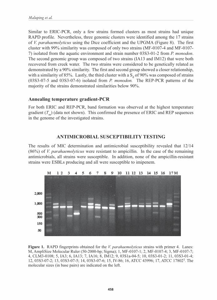
The nine distinct patterns obtained with primer 4 , designated RAPD types A to I exhibited between 2 and 6 bands ranging from 300 to 1,500 bp in size (Figs 1 and 5). Three strains recovered from water were shown to form RAPD profile A, while three isolates from P. monodon and the type strain ATCC 17802 comprised profile F. Reference strain ATCC 43996 clustered together with three isolates from different sources forming profile G. The remaining profiles contained a unique strain. Regardless of the RAPD profile, all the strains shared a common band at 750 bp (Figure 1).
When primer 6 was used, six distinct patterns were observed and were designated RAPD types A to F, comprising of two to four bands with sizes ranging between 300 and 2,000 bp (Figures 2 and 6). Eight strains from aquatic environment formed RAPD types A and D, while four strains from P. monodon formed profile F. Profile E was composed of two strains coming from aquatic environment and from P. monodon. Finally, profiles B and C contained a unique strain. Using primer 6, all strains showed a common band at approximately 600 bp (Figure 2).
The results of the analysis of similarity among the different profiles with the Diversity database software employing the Sd and the UPGMA allowed us to identify at least 3 genetic groups or clusters (I-III), with at least 70% similarity level. Thus, with primer
4, the three genetic groups were defined at Sd values of 70% for cluster I (profiles A, C, H, I), 99% for cluster II (profile F), and 90% for cluster III (patterns B and G) (Figure 5). At 80% similarity level all the strains in cluster I were from aquatic environments. The second cluster was composed of two strains from P. monodon that are genetically identical sharing the same common bands at 750, 800 and 1,000 bp. Lastly cluster III was composed of strains coming from both aquatic environment and P. monodon. The RAPD patterns of all remaining strains were diverse, with similarities below 70% and were considered genetically unrelated by this method.
However, when primer 6 was used three genomic clusters with Sd values ranging from 85 to 99% were formed (Figure 6). Cluster I has an 85% similarity and was formed by RAPD profiles D and E. Majority of the strains (4/7) from this cluster were recovered from aquatic environment. Clusters II (profile A) and III (profile F) were composed of strains that have an identical RAPD type. Both clusters had a 99% similarity. Cluster II was composed of strains coming from aquatic environments whereas cluster III was composed of strains recovered from affected P. monodon. In addition, genetic clusters I and II showed a close relationship with a similarity of 80%. Lastly, RAPD types B and C were more diverse with similarities below 50% and were considered genetically unrelated to any of the three clusters.
ERIC-PCR fingerprintingThe fingerprints of V. parahaemolyticus strains consisted of 6 to 8 amplification bands, ranging in size from 50 to 2,500 bp (Figure 3). All strains were typeable by ERIC-PCR. The same fingerprints were observed when ERIC-PCR was repeated at least three times, demonstrating the reproducibility of this technique. Almost each strain gave a different ERIC profile although a common band was observed in all 17 strains around 500 bp (Figure 3). Analysing the similarity among the different profiles with the Diversity database software identified three genomic clusters; although there are only few strains that formed these clusters as majority of the strains were genetically unrelated (Figure 7). The first group included strains CLM3-0108 and reference strain ATCC 43996 with 85% similarity. The second cluster consisted of strains IA13 and 03S1a-04-5 with a Sd of 90%. Both strains were recovered from the aquatic environment. Lastly, the third cluster with 80% similarity was composed of strains MF-0107-4 and 03S3-01-2 recovered from pond water and P. monodon, respectively. The remaining strains were distinct with similarity of lower than 80%.
REP-PCR fingerprintingAnalysis of the strains with REP-PCR yielded one to four bands depending on the strains (Figure 4). The size of these bands ranged from 150 to 2,000 bp and it was evident that the majority of the strains exhibited patterns with small number of REP-PCR products. As with ERIC-PCR, each strain gave a REP-PCR profile that was different from each other. However, all strains demonstrated a common band at approximately 400 bp (Figure 4). The REP-PCR amplifications were also repeated at least three times. Results revealed that some of the minor light amplification bands were inconsistent making the analysis more difficult.
Similar to ERIC-PCR, only a few strains formed clusters as most strains had unique RAPD profile. Nevertheless, three genomic clusters were identified among the 17 strains of V. parahaemolyticus using the Dice coefficient and the UPGMA (Figure 8). The first cluster with 99% similarity was composed of only two strains (MF-0107-4 and MF-0107-7) isolated from the aquatic environment and strain number 03S3-01-2 from P. monodon. The second genomic group was composed of two strains (IA13 and IM12) that were both recovered from creek water. The two strains were considered to be genetically related as demonstrated by a 90% similarity. The first and second group showed a closer relationship, with a similarity of 85%. Lastly, the third cluster with a Sd of 90% was composed of strains (03S3-07-5 and 03S3-07-6) isolated from P. monodon. The REP-PCR patterns of the majority of the strains demonstrated similarities below 90%.
Annealing temperature gradient-PCRFor both ERIC and REP-PCR, band formation was observed at the highest temperature gradient (Tm) (data not shown). This confirmed the presence of ERIC and REP sequences in the genome of the investigated strains.
ANTIMICROBIAL SUSCEPTIBILITY TESTING
The results of MIC determination and antimicrobial susceptibility revealed that 12/14 (86%) of V. parahaemolyticus were resistant to ampicillin. In the case of the remaining antimicrobials, all strains were susceptible. In addition, none of the ampicillin-resistant strains were ESBLs producing and all were susceptible to imipenem.
Figure 1. RAPD fingerprints obtained for the V. parahaemolyticus strains with primer 4. Lanes: M, AmpliSize Molecular Ruler (50-2000-bp; Sigma); 1, MF-0107-1; 2, MF-0107-4; 3, MF-0107-7; 4, CLM3-0108; 5, IA3; 6, IA13; 7, IA16; 8, IM12; 9, 03S1a-04-5; 10, 03S3-01-2; 11, 03S3-01-4; 12, 03S3-07-2; 13, 03S3-07-5; 14, 03S3-07-6; 15, IV-86; 16, ATCC 43996; 17, ATCC 17802T. The molecular sizes (in base pairs) are indicated on the left.
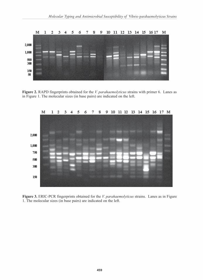
Figure 2. RAPD fingerprints obtained for the V. parahaemolyticus strains with primer 6. Lanes as in Figure 1. The molecular sizes (in base pairs) are indicated on the left.
Figure 3. ERIC-PCR fingerprints obtained for the V. parahaemolyticus strains. Lanes as in Figure 1. The molecular sizes (in base pairs) are indicated on the left.

Figure 4. REP-PCR fingerprints obtained for the V. parahaemolyticus strains. Lanes as in Figure 1. The molecular sizes (in base pairs) are indicated on the left.
Figure 5. Dendrogram established by the Diversity Database software package (Bio-Rad) using the Dice similarity coefficient and UPGMA on the basis of the RAPD profiles of V. parahaemolyticus strains obtained with primer 4.
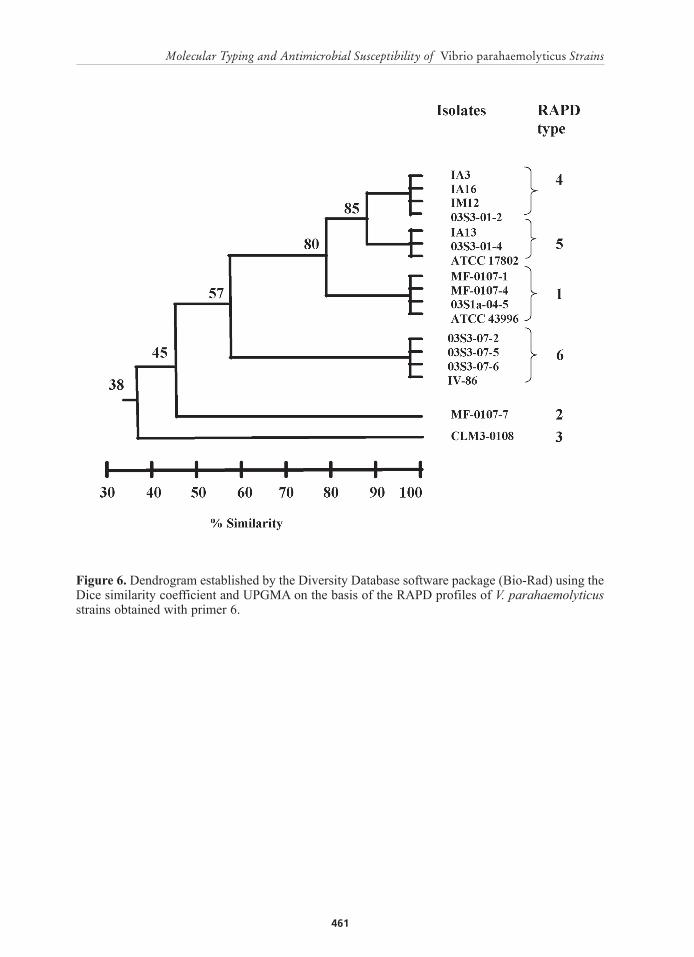
Figure 6. Dendrogram established by the Diversity Database software package (Bio-Rad) using the Dice similarity coefficient and UPGMA on the basis of the RAPD profiles of V. parahaemolyticus strains obtained with primer 6.
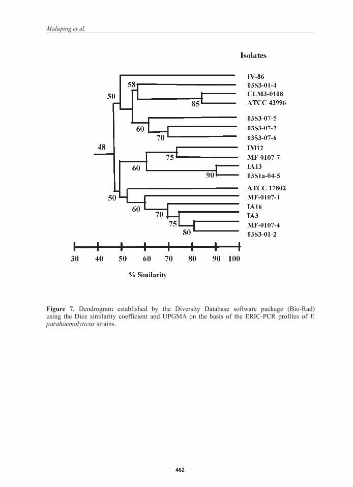
Figure 7. Dendrogram established by the Diversity Database software package (Bio-Rad) using the Dice similarity coefficient and UPGMA on the basis of the ERIC-PCR profiles of V. parahaemolyticus strains.
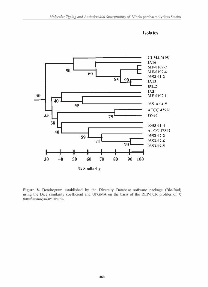
Figure 8. Dendrogram established by the Diversity Database software package (Bio-Rad) using the Dice similarity coefficient and UPGMA on the basis of the REP-PCR profiles of V. parahaemolyticus strains.

DISCUSSION
Results of several epidemiological studies on V. parahaemolyticus infections have supported the genetic similarity between clinical and environmental strains (Kelly and Stroh, 1998; Marshall et al., 1999), revealing that V. parahaemolyticus acquired infections in humans were only reported when the organism could be detected in the local environment (Kelly and Stroh, 1998). This was particularly true when the water temperatures were greater than 14ºC. This is of great importance in tropical countries such as the Philippines, wherein environmental temperatures are usually high.
Results of RAPD fingerprinting with the appropriate primers revealed that some of the patterns obtained could be related to the origin of the strains, which indicated its potential use in epidemiological studies of this organism. Analysis of the similarity of the RAPD patterns using the Dice coefficient and UPGMA has revealed the high genetic variation among strains. As shown in the dendrograms, only few strains clustered with a similarity of 90 to 99%. In a study done by Szczuka and Kaznowski (2004), strains with similarities below 90% were considered genetically unrelated. We could say that based on similarity using the Dice coefficient, our V. parahaemolyticus strains are genetically diverse wherein majority have similarity coefficient of below 90%. This result agreed with other studies confirming the genetic diversity among V. parahaemolyticus strains (Goarant et al., 1999; Sudheesh et al., 2002). It was found that, regardless of the RAPD profiles observed, all strains showed a common band of 750 bp or 600 bp using both P4 and P6 respectively. These fragments would be favourable traits for the development of genetic amplification and hybridization assays for diagnostic purpose (Dalla Valle et al., 2002), which can be important to verify V. parahaemolyticus strains that are relatively difficult to identify.
Both ERIC and REP methods gave almost a unique profile for each strain. Comparison of the similarities among the different patterns using Sd and UPGMA confirmed the genetic heterogeneity among V. parahaemolyticus strains, as the majority of the strains gave a similarity value below 90%. Although these two methods did not allow the establishment of well defined genetic clusters because of their high discriminatory power such techniques could be useful to follow the spreading of bacterial strain responsible for a particular outbreak. In addition, a common band of 500 bp and 400 bp was shown by all the strains using ERIC and REP PCR methods, respectively. This could be useful for the tracing of the point source of infection.
As shown by Marshall et al. (1999), Sudheesh et al. (2002) and Szczuka and Kaznowski (2004), it was found that both RAPD and ERIC-PCR gave more reproducible results compared with REP-PCR. The presence of the repeatable fingerprints in ERIC and REP-PCR suggested the presence of these repetitive consensus sequences (ERIC and REP) in V. parahaemolyticus. However, Gillings and Holley (1997) have revealed that besides the presence of ERIC, the formation of fingerprints could be due to random amplification. Our results showed band amplifications at higher temperatures (Tm of ERIC and REP) confirming the presence of these sequences on the genome of V. parahaemolyticus strains.
Antibiotics resistance among V. parahaemolyticus strains was less prevalent in this study. The only resistance observed was to ampicillin (12/14). This finding supported
several reports on the increasing -lactam resistance among vibrios from different sources (Zanetti et al., 2001; Molina-Aja et al., 2002). This study also determined that V. parahaemolyticus strains were susceptible to the majority of antimicrobials tested, indicated by the susceptibility of all the strains to ceftiofur, aminoglycosides, florfenicol, TMPS, enrofloxacin and oxytetracycline. However, an increasing number of cases of resistance to these antimicrobials have been reported (Molina-Aja et al., 2002).
Extended-spectrum -lactamases (ESBLs) are enzymes that mediate resistance to extended-spectrum (third-generation) cephalosporins. The presence of ESBLs was reported in enterobacteriaceae and Pseudomonas aeruginosa (Carter et al., 2000; Gheldre et al., 2003). In this study, production of ESBLs among V. parahaemolyticus strains could not be demonstrated.
ACKNOWLEDGEMENTS
This work was supported in part by Grant AGL2003-09307-C02-00 from the Ministerio de Ciencia y Tecnología (Spain). R.P. Maluping thanks the Fish Health Section of the Asian Fisheries Society (FHS-AFS) for the Student Travel Award, the Federation of European Microbiological Societies (FEMS) for the research fellowship to Spain and the Swedish Foundation for International Cooperation in Research and Higher Education (STINT) for the postgraduate scholarship. We are also greatful to Dr. Angelo DePaola of the Gulf Coast Seafood Laboratory, US Food and Drug Administration (FDA), for verifying the strains by the tlh gene probe.
REFERENCES
Carter, M.W., Oakton, K.J., Warner, M. and Livermore, D.M. 2000. Detection of extended-spectrum -lactamases in Klebsiella with the Oxoid combination disk method. Journal of Clinical Microbiology 38:4228-4232.
Dalla Valle, L., Zanella, L., Belvedere, P. and Colombo, L. 2002. Use of random amplification to develop a PCR detection method for the causative agent of fish pasteurellosis, Photobacterium damselae subsp. Piscicida (Vibrionaceae). Aquaculture 207:187-202.
DePaola, A., Ulaszek, J., Kaysner, C.A., Tenge, B.J., Nordstrom, J.L., Well, J., Puhr, N. and Gendel, S.M. 2003. Molecular, serological, and virulence characteristics of Vibrio parahaemolyticus isolated from environmental, food, and clinical sources in North America and Asia. Applied and Environmental Microbiology 69:3999-4005.
Dice L.R. 1945. Measures of the amount of ecological association between species. Ecology 26:297.
Gheldre, Yde, Avesani, V., Berhin, C., Delmee, M. and Glupczynski. 2003. Evaluation of Oxoid combination discs for detection of extended-spectrum -lactamases. Journal of Antimicrobial Agents and Chemotherapy 52:591-597.
Gillings, M. and Holley, M. 1997. Repetitive element PCR fingerprinting (rep-PCR) using enterobacterial repetitive intergenic consencus (ERIC) primers is not necessarily directed at ERIC elements. Letters in Applied Microbiology 25:17-21.
Goarant, C., Merien, F., Berthe, F., Mermoud, I. and Perolat, P. 1999. Arbitrarily primed PCR to type Vibrio spp. pathogenic for shrimp. Applied and Environmental Microbiology 9:1145-1151.
Kelly, M.T. and Stroh, E.M.D. 1988. Temporal relationship of Vibrio parahaemolyticus in patients and the environment. Journal of Clinical Microbiology 26:1754-1756.
Lightner, D.V. 1996. A handbook of pathology and diagnostic procedures for diseases of penaeid shrimps. Baton Rouge: World Aquaculture Society.
Marshall, S., Clark, C.G., Wang, G., Mulvey, M., Kelly, M.T. and Johnson, W.M. 1999. Comparison of molecular methods for typing Vibrio parahaemolyticus. Journal of Clinical Microbiology 37:2473-2478.
McCarthy, S.A., DePaola, A., Cook, D.W., Kaysner, C.A. and Hill, W.E. 1999. Evaluation of alkaline phosphatase- and digoxigenin-labelled probes for detection of the thermolabile hemolysin (tlh) gene of Vibrio parahaemolyticus. Letters in Applied Microbiology 28:66-70.
Molina-Aja, A., Gasca, A.G., Grobois, A.A., Mejia, C.B., Roque, A. and Gil, B.G. 2002. Plasmid profiling and antibiotic resistance of Vibrio strains isolated from cultured penaeid shrimp. FEMS Microbiology Letters 213:7-12.
National Committee for Clinical Laboratory Standards. 1999. Performance standards for antimicrobial disk and dilution susceptibility test for bacteria isolated from animals. Approved standard M31-A. National Committee for Clinical Laboratory Standards, Wayne, PA.
National Committee for Clinical Laboratory Standards. 1999. Performance standards for antimicrobial susceptibility testing. Approved standard M100-S9. National Committee for Clinical Laboratory Standards, Wayne, PA.
Olive, D.M. and Bean, P. 1999. Principles and applications of methods for DNA-based typing of microbial organisms. Journal of Clinical Microbiology 37: 1661-1669.
Romalde, J.L., Magariños, B., Villar, C., Barja, J.L. and Toranzo, A.E. 1999. Genetic analysis of turbot pathogenic Streptococcus parauberis strains by ribotyping and random amplified polymorphic DNA. FEMS Microbiology Letters 179:297-304.
Sneath, P.H.A. and Sokal, R.R. 1973. Numerical taxonomy. The principles and practice of numerical classification. San Francisco: W.H. Freeman.
Stern, M.J., Ames, G.F.L., Smith, N.H., Robinson, E.C. and Higgins, C.F. 1984. Repetitive extragenic palindromic sequences: a major component of the bacterial genome. Cell 37:1015-1026.
Sudheesh, P.S., Jie, K. and Xu, H. 2002. Random amplified polymorphic DNA-PCR typing of Vibrio parahaemolyticus and V. alginolyticus isolated from cultured shrimps. Aquaculture 207:11-17.
SVARM. 2002. Swedish Veterinary Antimicrobial Resistance Monitoring. Eds. Bengtsson, B, Greko, C. & Wallén C. National veterinary Institute, Uppsala Sweden. ISSN 1650-6332.
Szczuka, E. and Kaznowski, A. 2004. Typing of clinical and environmental Aeromonas sp. Strains by random amplified polymorphic DNA PCR, repetitive extragenic palindromic PCR, and enterobacterial repetitive intergenic consensus sequence PCR. Journal of Clinical Microbiology 42:220-228.
Versalovic, J., Koeuth, T. and Lupski, J.R. 1991. Distribution of repetitive DNA sequences in eubacteria and application to fingerprinting of bacterial genomes. Nucleic Acids Research 19:6823-6831.
Welsh, J. and McClelland, M. 1990. Fingerprinting genomes using PCR with arbitrary primers. Nucleic Acids Research 18:7213-7218.
West, P.A. and Colwell, R.R. 1984. Identification and clssification of vibrionaceae-an overview, pp. 285-363. In Colwell, R.R, (ed.).Vibrios in the environment. New York: John Wiley and Sons.
Wong, H. and Lin, C. 2001. Evaluation of typing of Vibrio parahaemolyticus by three PCR methods using specific primers. Journal of Clinical Microbiology 39: 4233-4240.
Zanetti, S., Spanu, T., Deriu, A., Romano, L., Sechi, L.A. and Fadda, G. 2001. In vitro susceptibility of Vibrio spp. isolated from the environment. Journal of Antimicrobial Agents and Chemotherapy 17:407-409.


