Molecular treatment of colon cancer - European Review for ... · Molecular treatment of colon...
7
4860 Introduction Colon cancer ranks the fourth most common cancer worldwide. The incidence of colon can- cer kept increasing in the past several decades 1 . In China, although colon cancer has a relatively low incidence in males aged 40-70 years old, re- cent epidemic studies have reported an increasing tendency in terms of the incidence rates of the malignancy, especially in patients who are under 30 years old 2 . At the stage, surgery is the main treatment for colon cancer. However, for different reasons, there are several patients who are not su- itable for surgery and thus require chemotherapy treatment. Also, chemotherapy is still required after surgery in some cases at times. 5-FU is the standard drug for chemotherapy of colon cancer. However, there are some negative aspects such as slow drug pharmacokinetics into tumor lesion and an uneven distribution 3 , which largely affect the chemotherapy efficacy against colon cancer. Therefore, it is significant to develop a novel stra- tegy for effective delivery of 5-FU into tumor cel- ls in order to generate the treatment efficacy of the anti-tumor drug 4 . Drug delivery into tumor lesion by blood sup- ply system is a critical step of anti-tumor therapy, as appropriate blood supply can reduce tumor cell viability potentially by elevating blood oxygen le- vel, and thus inhibit further growth of tumors 5 . Therefore, the improvement of tumor blood sup- ply has become a new strategy in terms of the treatment of colon cancer. Dopamine, a classical blood vessel transmitter/drug, has been known to inhibit tumor survival 6 . Studies have suggested Abstract. – OBJECTIVE: To investigate the an- ti-tumor effect and potential mechanism of do- pamine combined with 5-FU in colon cancer. MATERIALS AND METHODS: Babl/c F1 gen- eration male mice (N = 60) were inoculated with mouse C26 colon cancer cells below the pit to construct colon cancer model. Tumor-bear- ing mice were then divided into 4 groups (N = 8 each): model control group, dopamine group, 5-FU group and dopamine combined with 5-FU group. Dopamine (100 mg/kg/d) was intraperito- neally injected into mice. The tumor-suppress- ing rate was calculated. Serum VEGF concentra- tion was gained. Tumor tissues were subjected to HE staining. KLF2 expression was determined by immunohistochemical (IHC) staining. In vitro cul- tured C26 cells which treated with dopamine and cell apoptosis were analyzed by flow cytometry. RESULTS: Compared with model control group, all treatment groups showed significant- ly decreased tumor weight and volume (p < 0.01), increased tumor necrosis (p < 0.05), reduced serum VEGF concentration (p < 0.05), and en- hanced KLF2 expression in microvessels (p < 0.05). Combined treatment in terms of dopamine combined with 5-FU had the most pronounced effect compared with both dopamine and 5-FU treatment groups individually. Dopamine single t 5-FU and 5-FU groups showed a similar propor- tion of viable C26 cells (p > 0.05). CONCLUSIONS: Dopamine exerts anti-tumor effects by modulating tumor vascular homeo- stasis through the KLF2 signaling pathway, and potentiates the treatment efficacy of anti-tumor drug 5-FU. Our study discovered clinical signif- icance concerning the novelty of therapeutic strategy against colon cancer. Key Words: Colon cancer, Dopamine, KLF2, Tumor blood supply. European Review for Medical and Pharmacological Sciences 2017; 21: 4860-4866 S. CHEN 1 , Y.-M. ZHANG 2 , Y.-G. ZHANG 1 , J. JIA 1 , R.-W. ZHU 2 1 Wenzhou Hospital of Integrated Traditional Chinese and Western Medicine, Wenzhou, Zhejiang, P.R. China 2 Department of General Surgery, Wenzhou Hospital of Integrated Traditional Chinese and Western Medicine, Wenzhou, Zhejiang, P.R. China Corresponding Author: Yiming Zhang, MD; e-mail: [email protected] Potentiation of treatment efficacy against colon cancer of dopamine via elevating KLF2 expression in tumor vascular endothelial cells
Transcript of Molecular treatment of colon cancer - European Review for ... · Molecular treatment of colon...

4860
Introduction
Colon cancer ranks the fourth most common cancer worldwide. The incidence of colon can-cer kept increasing in the past several decades1. In China, although colon cancer has a relatively low incidence in males aged 40-70 years old, re-cent epidemic studies have reported an increasing tendency in terms of the incidence rates of the malignancy, especially in patients who are under 30 years old2. At the stage, surgery is the main treatment for colon cancer. However, for different reasons, there are several patients who are not su-itable for surgery and thus require chemotherapy treatment. Also, chemotherapy is still required after surgery in some cases at times. 5-FU is the standard drug for chemotherapy of colon cancer. However, there are some negative aspects such as slow drug pharmacokinetics into tumor lesion and an uneven distribution3, which largely affect the chemotherapy efficacy against colon cancer. Therefore, it is significant to develop a novel stra-tegy for effective delivery of 5-FU into tumor cel-ls in order to generate the treatment efficacy of the anti-tumor drug4.
Drug delivery into tumor lesion by blood sup-ply system is a critical step of anti-tumor therapy, as appropriate blood supply can reduce tumor cell viability potentially by elevating blood oxygen le-vel, and thus inhibit further growth of tumors5. Therefore, the improvement of tumor blood sup-ply has become a new strategy in terms of the treatment of colon cancer. Dopamine, a classical blood vessel transmitter/drug, has been known to inhibit tumor survival6. Studies have suggested
Abstract. – OBJECTIVE: To investigate the an-ti-tumor effect and potential mechanism of do-pamine combined with 5-FU in colon cancer.
MATERIALS AND METHODS: Babl/c F1 gen-eration male mice (N = 60) were inoculated with mouse C26 colon cancer cells below the pit to construct colon cancer model. Tumor-bear-ing mice were then divided into 4 groups (N = 8 each): model control group, dopamine group, 5-FU group and dopamine combined with 5-FU group. Dopamine (100 mg/kg/d) was intraperito-neally injected into mice. The tumor-suppress-ing rate was calculated. Serum VEGF concentra-tion was gained. Tumor tissues were subjected to HE staining. KLF2 expression was determined by immunohistochemical (IHC) staining. In vitro cul-tured C26 cells which treated with dopamine and cell apoptosis were analyzed by flow cytometry.
RESULTS: Compared with model control group, all treatment groups showed significant-ly decreased tumor weight and volume (p < 0.01), increased tumor necrosis (p < 0.05), reduced serum VEGF concentration (p < 0.05), and en-hanced KLF2 expression in microvessels (p < 0.05). Combined treatment in terms of dopamine combined with 5-FU had the most pronounced effect compared with both dopamine and 5-FU treatment groups individually. Dopamine single t 5-FU and 5-FU groups showed a similar propor-tion of viable C26 cells (p > 0.05).
CONCLUSIONS: Dopamine exerts anti-tumor effects by modulating tumor vascular homeo-stasis through the KLF2 signaling pathway, and potentiates the treatment efficacy of anti-tumor drug 5-FU. Our study discovered clinical signif-icance concerning the novelty of therapeutic strategy against colon cancer.
Key Words: Colon cancer, Dopamine, KLF2, Tumor blood supply.
European Review for Medical and Pharmacological Sciences 2017; 21: 4860-4866
S. CHEN1, Y.-M. ZHANG2, Y.-G. ZHANG1, J. JIA1, R.-W. ZHU2
1Wenzhou Hospital of Integrated Traditional Chinese and Western Medicine, Wenzhou, Zhejiang, P.R. China2Department of General Surgery, Wenzhou Hospital of Integrated Traditional Chinese and Western Medicine, Wenzhou, Zhejiang, P.R. China
Corresponding Author: Yiming Zhang, MD; e-mail: [email protected]
Potentiation of treatment efficacy against colon cancer of dopamine via elevating KLF2 expression in tumor vascular endothelial cells

Molecular treatment of colon cancer
4861
that dopamine stabilizes tumor blood vessels by up-regulating the expression of KLF2, a zinc fin-ger transcription factor in tumor endothelial cel-ls7. However, the effect and mechanism of dopa-mine in combination with 5-FU in colon cancer has not been investigated in the past. In this stu-dy, we constructed a colon cancer mouse model and evaluated the therapeutic effect of dopamine in combination with 5-FU. We also explored the molecular mechanism by which dopamine exer-ted the anti-tumor effect.
Materials and Methods
MaterialsA total of 60 male Babl/c F1 generation mice
weighted 18±2 g were purchased from Sitaike Ani-mal Corp (Shanghai, China) and raised in an SPF grade Animal Center at Wenzhou Hospital of Inte-grated Traditional Chinese and Western Medicine. Mouse C26 colon cancer cell line was purchased from Chinese Academy of Science (Shanghai, Chi-na). Dopamine (>98% purity) was purchased from Sigma-Aldrich (St. Louis, MO, USA). 5-FU (lot No. 20070802) was purchased from Haipu Biote-ch (Nanjing, China). Mouse VEGF ELISA kit was purchased from Biosources (Camarillo, CA, USA). Goat anti-mouse CD34 monoclonal antibody was purchased from Sigma-Aldrich (St. Louis, MO, USA). The animal study was approved by the Ani-mal Ethics Committee of Wenzhou Hospital of Inte-grated Traditional Chinese and Western Medicine.
Cell CultureC26 cells were cultured in 1640 medium con-
taining 10% fetal bovine serum and 1% strep-tomycin/penicillin at 37°C, 5% CO2 incubator. C26 cells (passage 4) were divided into 4 groups which are: control group, dopamine (1 mg/mL) group, 5-FU (0.5 mg/mL), and dopamine com-bined with 5-FU group. After 7-days incubation, cell apoptosis was analyzed by flow cytometry.
Construction of Mouse Colon Cancer Model and Dopamine Interference
C26 cells at exponential phase were inoculated below the mouse pit (5 × 10⁶ cells per animal). After 7-10 days, C26 cells formed visible tu-mors. C26 tumor tissues were homogenized into 1-mm3 pieces. Under sterile conditions, C26 cell suspension was inoculated below the mouse pit using a 1-ml syringe. Three passages were per-formed as described above. Stable mouse colon
cancer model was successfully constructed when the tumor body reached 100 mm3. Colon cancer mice were randomly divided into 4 groups (N = 8 each) which are: model control group, dopamine group, 5-FU group and dopamine combined with 5-FU group. For dopamine and dopamine combi-ned with 5-FU groups, dopamine (100 mg/kg/d) was injected daily into the peritoneal cavity for 1 week. For 5-FU and dopamine combined with 5-FU groups, 5-FU (1 mg/kg/d) was injected daily into the tail vein of mice for 1 week. The model group received a daily intraperitoneal injection of an equal volume of saline. At day 8, mice were sacrificed by cervical dislocation. Blood samples were collected, and colon cancer lesion was re-sected. Tumor length, width, and weight were measured. Tumor specimen was fixed in 30% for-maldehyde and cut into 4-μm sections.
Calculation of Tumor-Suppressing Rate The tumor volume was calculated as previou-
sly described in last section7. Tumor volume (TV) = ab2/2, where a and b stand for tumor length and width, respectively. Using the model control group as reference, the tumor volume suppressing rate and tumor weight suppressing rate in all three treatment groups was calculated as follows: tu-mor volume suppressing rate = (1-average tumor volume in drug treatment group / average tumor volume in model control group) × 100%, and tu-mor weight suppressing rate = (1- average tumor weight in drug treatment group / average tumor weight in model control group) × 100%.
ELISA Blood samples were collected from orbital vein
plexus. Serum was collected after the completion of centrifugation. VEGF expression was measu-red using VEGF ELISA kit following the manual instruction. A standard curve was plotted by a software. The serum VEGF concentration in all samples was calculated.
Hematoxylin and Eosin (HE) StainingTissue cubes were fixed in 40 g/L 30% formal-
dehyde solution, embedded in paraffin, cut into 4-μm slices and observed under an inverted mi-croscope (100 ×). Regions with tumor necrosis were collected. Necrosis area in three randomly selected visual fields (200 ×) was analyzed in a double-blinded manner. Image pro software was used to process images. A score was given based on the necrosis area: 0 for 5%, 1 for 5%-25%, 2 for 25%-50%, and 3 for >50%.

S. Chen, Y.-M. Zhang, Y.-G. Zhang, J. Jia, R.-W. Zhu
4862
Immunohistochemical (IHC) Analysis of KLF2 Expression in Tumor Microvessels
Paraffin-embedded sections were dewaxed with xylene and dehydrated with ethanol. Slices were processed to remove endogenous peroxida-se, followed by antigen retrieval, serum blocking, antibody binding, DAB staining, rising, hema-toxylin staining, HCl-ethanol immersing, gra-dient ethanol dehydration, xylene immersing and coverslip mounting. Goat anti-mouse KLF2 mo-noclonal antibody (1:50) biotin-labeled secondary IgG (1:200), and horseradish peroxidase-labeled streptavidin (1:200) were also utilized. KLF2-po-sitive microvessels in dark brown color were counted under an inverted microscope.
Flow CytometryC26 cells were collected after 7-days cell cultu-
re and cell apoptosis was analyzed by flow cyto-metry. According to manufacture’s instructions, C26 cells were prepared into 105/mL cell suspen-sion. Cell suspension, reaction buffer, and FI-TC-Annexin V reagent were mixed at a ratio of 250:50:1, and incubated at room temperature for 15 minutes in the dark. C26 cells were assayed for phosphatidylserine expression under the wa-
velength of 625 nm. Caspase-3 activity was me-asured using caspase-3 kit following the manual. Briefly, C26 cells were lysed and incubated with chromogenic substrate at 37°C for 30 minutes. The absorbance at 490 nm was measured by flow cytometry.
Statistical AnalysisSPSS 17.0 software (SPSS Inc., Chicago, IL,
USA) was used for data analysis. Measurement data were analyzed by Student’s t-test. Statistical significance was defined as p < 0.05.
Results
Dopamine Inhibited Colon Cancer and Potentiated the Treatment Efficacy of 5-FU
The tumor volume of subcutaneous transplant tumors was determined at 7 days after model pre-paration. Although there was no significant diffe-rence in pretreatment tumor volume amongst all groups, tumor lesion size in all treatment group was significantly lower compared with model control group (p < 0.01, Table I, Figure 1 and 2). The tumor weight suppressing rates in dopamine, 5-FU and dopamine combined with 5-FU groups were 45.8%, 60.3%, and 85.6%, respectively, and tumor volume suppressing rates were 51.2%, 62.8%, and 81.6%, respectively, suggesting that dopamine exerted anti-tumor effects against co-
Table I. Tumor weight, volume and suppression rate in all groups of rats (N=8, mean±SD).
Group Weight (g) Weight suppression rate (%) Volume (cm3) Volume suppression rate (%) Model control 0.73±0.22 - 1.00±0.17 -Dopamine 0.40±0.22 45.8 0.50±0.30 51.25-FU 0.29±0.09 60.3 0.40±0.12 62.8Dopamine + 5-FU 0.11±0.06 84.6 0.16±0.09 81.6
Figure 1. Tumor weight and volume suppression rates of all drug treatment groups. A, Model control group; B, Do-pamine group; C, 5-FU group; D, Dopamine + 5-FU group. b, p < 0.01 compared to model control group.
Figure 2. Rat tumor volume. A, Model control group; B, Do-pamine group; C, 5-FU group; D, Dopamine + 5-FU group.

Molecular treatment of colon cancer
4863
lon cancer. More importantly, dopamine poten-tiated the treatment efficacy of the classical drug 5-FU.
Dopamine Increased Tumor Tissue Necrosis of Colon Cancer
Upon the completion of 7-days treatment, the tumor tissue necrosis was evaluated by HE-stai-ning. No necrosis was observed in model control group. In contrast, all treatment groups exhibi-ted moderate to severe necrosis with cell nuclear atrophy, deformation, rupture, or sparse arrange-ment (Figure 3). The necrosis score in dopami-ne combined with 5-FU group was significantly higher compared with both dopamine and 5-FU
groups (p < 0.05, Table II), indicating that dopa-mine potentially facilitated colon cancer necrosis.
Dopamine Regulated VEGF to Stabilize the Tumor Vessel Status
The serum VEGF concentration in all treat-ment groups was significantly lower compared with model control group (p < 0.01). The VEGF suppressing rates in dopamine, 5-FU, and dopa-mine combined with 5-FU groups were 15.8%, 34.2%, and 44.8%, respectively (Table III). The-se results indicated that exogenous dopamine injection had regulated the expression of VEGF to stabilize the tumor vessel status in colon cancer, and thus potentiated the efficacy of 5-FU.
Figure 3. Tumor body necrosis of all drug treatment groups of rats (X400). A, Model control group; B, Dopamine group; C, 5-FU group; D, Dopamine + 5-FU group.
Table II. Tumor necrosis of all drug treatment groups.
Necrosis score of all groups (N)
Group 0 1 2 3 Total Model control 13 11 0 0 11Dopamine 7 13 3 1 255-FU 2 7 12 3 44Dopamine + 5-FU 1 2 10 11 55
S. Chen, Y.-M. Zhang, Y.-G. Zhang, J. Jia, R.-W. Zhu
4864
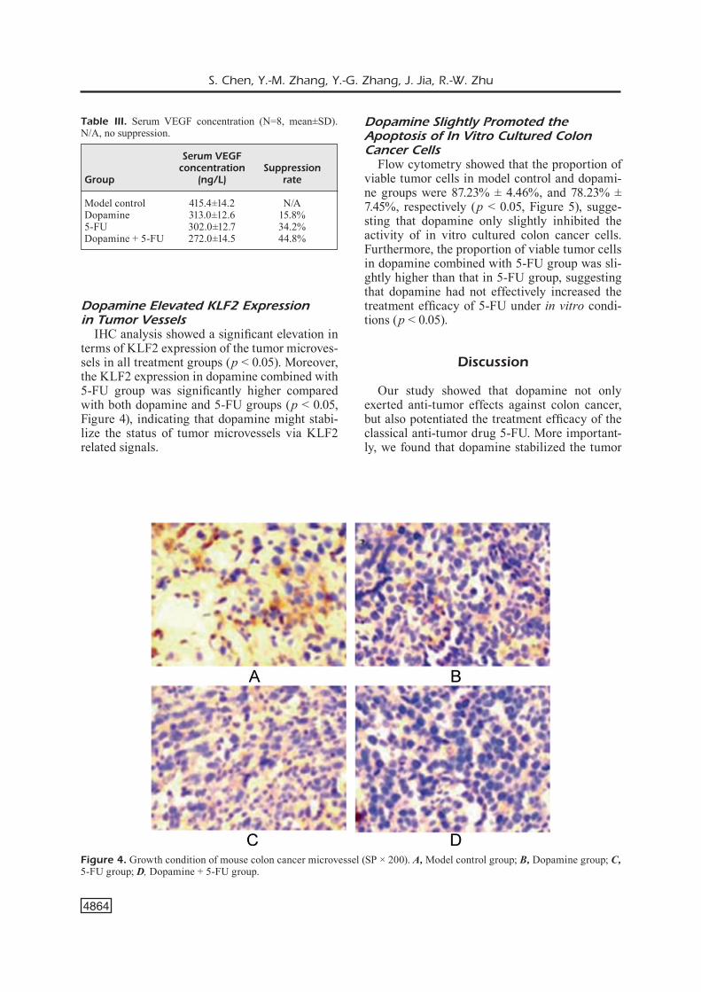
Dopamine Elevated KLF2 Expression in Tumor Vessels
IHC analysis showed a significant elevation in terms of KLF2 expression of the tumor microves-sels in all treatment groups (p < 0.05). Moreover, the KLF2 expression in dopamine combined with 5-FU group was significantly higher compared with both dopamine and 5-FU groups (p < 0.05, Figure 4), indicating that dopamine might stabi-lize the status of tumor microvessels via KLF2 related signals.
Dopamine Slightly Promoted the Apoptosis of In Vitro Cultured Colon Cancer Cells
Flow cytometry showed that the proportion of viable tumor cells in model control and dopami-ne groups were 87.23% ± 4.46%, and 78.23% ± 7.45%, respectively (p < 0.05, Figure 5), sugge-sting that dopamine only slightly inhibited the activity of in vitro cultured colon cancer cells. Furthermore, the proportion of viable tumor cells in dopamine combined with 5-FU group was sli-ghtly higher than that in 5-FU group, suggesting that dopamine had not effectively increased the treatment efficacy of 5-FU under in vitro condi-tions (p < 0.05).
Discussion
Our study showed that dopamine not only exerted anti-tumor effects against colon cancer, but also potentiated the treatment efficacy of the classical anti-tumor drug 5-FU. More important-ly, we found that dopamine stabilized the tumor
Table III. Serum VEGF concentration (N=8, mean±SD). N/A, no suppression.
Serum VEGF concentration SuppressionGroup (ng/L) rate Model control 415.4±14.2 N/ADopamine 313.0±12.6 15.8%5-FU 302.0±12.7 34.2%Dopamine + 5-FU 272.0±14.5 44.8%
Figure 4. Growth condition of mouse colon cancer microvessel (SP × 200). A, Model control group; B, Dopamine group; C, 5-FU group; D, Dopamine + 5-FU group.

Molecular treatment of colon cancer
4865
vessel status via up-regulating KLF2 expression in tumor microvessels, which means there’s po-tential treatment by targeting the facilitator of drug delivery into tumors through blood supply.
As an important catecholamine neurotransmit-ter, dopamine plays pluripotent biological fun-ctions including recognition, emotion, appetite and motor reflex. More importantly, dopamine plays a crucial role in the maintenance of tissue blood supply8-10. Studies have suggested that mo-derately increased blood supply in tumor tissue can manage tumor cell viability and improve drug concentration in tumor region due to the re-ason that tumor cells are more responsive under hypoxic condition11. Although the anti-tumor ef-fects of dopamine have been previously reported, the mechanism underlying the anti-tumor effects of dopamine remains unknown. Our paper sug-gested that the anti-tumor effects of dopamine might be associated with its ability to stabilize the tumor blood supply.
Dopamine has been confirmed to have anti-tu-mor effects12,13. In this work, we found that do-pamine significantly inhibited colon cancer cell viability, and facilitated tumor necrosis. We also found that dopamine affected tumor blood sup-ply via modulating VEGF expression, which is in consistency with previous studies regarding the effect of dopamine on tumor blood supply14,15. More importantly, our study revealed that dopa-mine potentiated the treatment efficacy of the classical drug 5-FU, suggesting that the combined therapy of 5-FU and dopamine is promising in the treatment of colon cancer.
Further, we investigated the regulatory me-chanism of dopamine in blood supply of colon cancer. KLF2, a crucial factor that helps to stabi-lize microvessels, has been proven as an impor-tant downstream target of the dopamine-related signal pathway15,16. Previous researches17,18 have demonstrated the regulatory effects of dopami-ne on KLF2 in colon cancer. Down-regulation of KLF2 frequently accelerates tumor cell pro-liferation, which results in tumor tissue aggra-vation19,20. The mechanism involves the tran-sition of blood vessel homeostasis which leads to tumor growth, including internal hypoxia of tumors, activation of oncogene and impeding of drugs into tumors21. In this study, we found that KL2 expression was significantly incre-ased in colon cancer, and has been effectively suppressed in tumor microvessels in all treat-ment groups afterward. Moreover, dopamine treatment effectively decreased serum VEGF level in colon cancer mice. Meanwhile, that the experience also indicated that dopamine slightly promoted the cell apoptosis of in vitro cultured colon cancer cells, and had not improved the tre-atment efficacy of 5-FU in these cells, indicating that dopamine required blood supply system to exert its anti-tumor effects as well as its ability to potentiate the 5-FU. All together, we belie-ved that dopamine inhibited tumor growth and potentiated the treatment efficacy of 5-FU via stabilizing tumor microvessels and enhancing blood drug concentration inside tumors.
Conclusions
In summary, dopamine not only exerts anti-tu-mor effects, but also potentiates the treatment ef-ficacy of 5-FU via stabilizing tumor microvessels through KLF2 signaling. The combined therapy of dopamine and 5-FU might provide a novel the-rapeutic strategy against colon cancer. In the me-antime, we expect further clinical investigations to validate our findings.
AcknowledgmentsThis work was supported by Wenzhou Hospital of In-tegrated Traditional Chinese and Medicine, Municipal Science and Technology Bureau (No. Y20150250).
Conflict of interestThe authors declare no conflicts of interest.
Figure 5. Tumor cell survival ratio of in vitro cultured mouse colon cancer cells after drug treatment. A, Model control group; B, Dopamine group; C, 5-FU group; D, Do-pamine + 5-FU group. *, p < 0.05 compared to group A; #, p < 0.05 compared to group C.

S. Chen, Y.-M. Zhang, Y.-G. Zhang, J. Jia, R.-W. Zhu
4866
References
1) Sharma P, mccleeS SF, aFaq F, Pomegranate for pre-vention and treatment of cancer: an update. Mole-cules 2017; 22. pii: E177.
2) Zhu J, Tan Z, holliS-hanSen K, Zhang Y, Yu c, li Y. epidemiological trends in colorectal cancer in China: an ecological study. Dig Dis Sci. 2017; 62: 235-243.
3) Sharma Sh, ThulaSingam S, nagaraJan S. Terpenoids as anti-colon cancer agents - A comprehensive review on its mechanistic perspectives. Eur J Pharmacol 2017; 795: 169-178.
4) Wang X, Yang Y, huYcKe mm. Microbiome-driven carcinogenesis in colorectal cancer: Models and mechanisms. Free Radic Biol Med 2017; 105: 3-15.
5) Zhan h, JagTiani T, liang JF. A new targeted de-livery approach by functionalizing drug nanocry-stals through polydopamine coating. Eur J Pharm Biopharm 2017; 114: 221-229.
6) chauveT n, romano n, laFonT c, guillou a, galiberT e, bonneFonT X, le TiSSier P, Fedele m, FuSco a, mol-lard P, couTrY n. Complementary actions of dopa-mine D2 receptor agonist and anti-vegf therapy on tumoral vessel normalization in a transgenic mouse model. Int J Cancer. 2017; 140: 2150-2161.
7) chaKroborTY d, SarKar c, Yu h, Wang J, liu Z, da-SguPTa PS, baSu S. Dopamine stabilizes tumor blood vessels by up-regulating angiopoietin 1 expres-sion in pericytes and Kruppel-like factor-2 expres-sion in tumor endothelial cells. Proc Natl Acad Sci U S A. 2011; 108: 20730-20735.
8) rehmani n, ZaFar a, ariF h, hadi Sm, Wani aa. Cop-per-mediated DNA damage by the neurotransmit-ter dopamine and L-DOPA: a pro-oxidant mecha-nism. Toxicol In Vitro 2017; 40: 336-346.
9) deKanT W, Scialli ar, PloTZKe K, Klaunig Je. Biological relevance of effects following chronic administration of octamethylcyclotetrasiloxane (D4) in Fischer 344 rats. Toxicol Lett 2017; 279 Suppl 1: 42-53.
10) chen X, corTeZ-Jugo c, choi gh, bJornmalm m, dai Y, Yoo PJ, caruSo F. Patterned poly(dopamine) Fil-ms for enhanced cell adhesion. Bioconjug Chem 2017; 28: 75-80.
11) ding J, Xie m, lian Y, Zhu Y, Peng P, Wang J, Wang l, Wang K. Long noncoding RNA HOXA-AS2 re-presses P21 and KLF2 expression transcription
by binding with EZH2, LSD1 in colorectal cancer. Oncogenesis 2017; 6: e288.
12) Khodeer S, era T. Identifying the biphasic role of calcineurin/NFAT signaling enables replacement of Sox2 in somatic cell reprogramming. Stem Cel-ls 2017; 35: 1162-1175.
13) mao qq, chen JJ, dong l, Zhong l, Sun X. Krup-pel-like factor 2 suppresses growth and invasion of gastric cancer cells in vitro and in vivo. J Biol Regul Homeost Agents 2016; 30: 703-712.
14) Fang J, Sun cc, gong c. Long noncoding RNA XIST acts as an oncogene in non-small cell lung cancer by epigenetically repressing KLF2 expression. Bio-chem Biophys Res Commun 2016; 478: 811-817.
15) nie F, Yu X, huang m, Wang Y, Xie m, ma h, Wang Z, de W, Sun m. Long noncoding RNA ZFAS1 pro-motes gastric cancer cells proliferation by epige-netically repressing KLF2 and NKD2 expression. Oncotarget 2017; 8: 38227-38238.
16) boguSZ am, bagg a. Genetic aberrations in small B-cell lymphomas and leukemias: molecular pa-thology, clinical relevance and therapeutic tar-gets. Leuk Lymphoma 2016; 57: 1991-2013.
17) Zou K, lu X, Ye K, Wang c, You T, chen J. Krup-pel-like factor 2 promotes cell proliferation in he-patocellular carcinoma through up-regulation of c-myc. Cancer Biol Ther 2016; 17: 20-26.
18) Zang c, nie Fq, Wang q, Sun m, li W, he J, Zhang m, lu Kh. Long non-coding RNA LINC01133 re-presses KLF2, P21 and E-cadherin transcription through binding with EZH2, LSD1 in non small cell lung cancer. Oncotarget 2016; 7: 11696-11707.
19) haddocK mg. Intraoperative radiation therapy for colon and rectal cancers: a clinical review. Radiat Oncol 2017; 12: 11.
20) ohguchi h, hideShima T, bhaSin mK, gorgun gT, SanTo l, cea m, Samur mK, mimura n, SuZuKi r, Tai YT, carraSco rd, raJe n, richardSon Pg, munShi nc, harigae h, Sanda T, SaKai J, anderSon Kc. The KDM3A-KLF2-IRF4 axis maintains myeloma cell survival. Nat Commun 2016; 7: 10258.
21) demolli S, doebele c, doddaballaPur a, lang v, FiSSlThaler b, chavaKiS e, vinciguerra m, Sciacca S, henSchler r, hecKer m, SavanT S, auguSTin hg, Ka-luZa d, dimmeler S, boon ra. MicroRNA-30 media-tes anti-inflammatory effects of shear stress and KLF2 via repression of angiopoietin 2. J Mol Cell Cardiol 2015; 88: 111-119.