
Molecular similarity between myelodysplastic form of chronic ... · Molecular similarity between...
9
Molecular similarity between myelodysplastic form of chronic myelomonocytic leukemia and refractory anemia with ring sideroblasts Gelsi-Boyer, V., Cervera, N., Bertucci, F., Brecqueville, M., Finetti, P., Murati, A., Arnoulet, C., Mozziconacci, M. J., Mills, K. I., Cross, N. C. P., Vey, N., & Birnbaum, D. (2013). Molecular similarity between myelodysplastic form of chronic myelomonocytic leukemia and refractory anemia with ring sideroblasts. HAEMATOLOGICA, 98(4), 576-583. https://doi.org/10.3324/haematol.2012.071506 Published in: HAEMATOLOGICA Document Version: Publisher's PDF, also known as Version of record Queen's University Belfast - Research Portal: Link to publication record in Queen's University Belfast Research Portal Publisher rights Copyright© 2013 Ferrata Storti Foundation. This is an open access Creative Commons Attribution-NonCommercial License (https://creativecommons.org/licenses/by-nc/4.0/), which permits use, distribution and reproduction for non-commercial purposes, provided the author and source are cited. General rights Copyright for the publications made accessible via the Queen's University Belfast Research Portal is retained by the author(s) and / or other copyright owners and it is a condition of accessing these publications that users recognise and abide by the legal requirements associated with these rights. Take down policy The Research Portal is Queen's institutional repository that provides access to Queen's research output. Every effort has been made to ensure that content in the Research Portal does not infringe any person's rights, or applicable UK laws. If you discover content in the Research Portal that you believe breaches copyright or violates any law, please contact [email protected]. Download date:28. Jun. 2020
Transcript of Molecular similarity between myelodysplastic form of chronic ... · Molecular similarity between...

Molecular similarity between myelodysplastic form of chronicmyelomonocytic leukemia and refractory anemia with ring sideroblasts
Gelsi-Boyer, V., Cervera, N., Bertucci, F., Brecqueville, M., Finetti, P., Murati, A., Arnoulet, C., Mozziconacci, M.J., Mills, K. I., Cross, N. C. P., Vey, N., & Birnbaum, D. (2013). Molecular similarity between myelodysplasticform of chronic myelomonocytic leukemia and refractory anemia with ring sideroblasts. HAEMATOLOGICA,98(4), 576-583. https://doi.org/10.3324/haematol.2012.071506
Published in:HAEMATOLOGICA
Document Version:Publisher's PDF, also known as Version of record
Queen's University Belfast - Research Portal:Link to publication record in Queen's University Belfast Research Portal
Publisher rightsCopyright© 2013 Ferrata Storti Foundation. This is an open access Creative Commons Attribution-NonCommercial License(https://creativecommons.org/licenses/by-nc/4.0/), which permits use, distribution and reproduction for non-commercial purposes, providedthe author and source are cited.
General rightsCopyright for the publications made accessible via the Queen's University Belfast Research Portal is retained by the author(s) and / or othercopyright owners and it is a condition of accessing these publications that users recognise and abide by the legal requirements associatedwith these rights.
Take down policyThe Research Portal is Queen's institutional repository that provides access to Queen's research output. Every effort has been made toensure that content in the Research Portal does not infringe any person's rights, or applicable UK laws. If you discover content in theResearch Portal that you believe breaches copyright or violates any law, please contact [email protected].
Download date:28. Jun. 2020
Articles Myelodysplastic Syndromes
576 haematologica | 2013; 98(4)
Introduction
Chronic myelomonocytic leukemia (CMML) is a malignanthematologic disease of the elderly characterized by peripher-al blood monocytosis, overproduction of bone marrowmonocytes with dysplasia of one or more lineages, and lessthan 20% of blasts in the bone marrow. Its prognosis is poorwith a median survival of 12-18 months and a 15-20% risk oftransformation into acute myeloid leukemia (AML).1,2 CMMLis classified by the World Health Organization (WHO) intothe myelodysplastic/myeloproliferative neoplasms and,based on the number of blasts, subclassified into CMML1and CMML2 (5-9% and 10-19%, respectively).3 Likemyelodysplastic syndromes (MDS), CMML shows dysplasticfeatures that reflect ineffective hematopoiesis; however, dys-plasia is associated with bone marrow proliferation.4,5
Because of this duality, CMML had been separated into amyeloproliferative form (MP-CMML) and a myelodysplasticform (MD-CMML) based on a semi-arbitrary threshold of 13x 109/L for peripheral white blood cell (WBC).6 However, dueto its lack of impact on outcome, this separation is not includ-
ed in the WHO classification.3 Yet, the prognosis of MD-CMML but not MP-CMML may be evaluated by the interna-tional prognostic scoring system, underlining a similarity ofMD-CMML with MDS. Moreover, even if, given the limitedtreatments currently available, MD and MP-CMMLs havesimilar outcome, this situation may change with the adventof new therapies, in which case they would each need to berecognized separately. Because CMML has both dysplastic and proliferative fea-
tures it is likely that the disease is heterogeneous. We wantedto determine whether these MD and MP features may haveany relevant molecular basis that may help classify andunderstand CMML. To this aim, we established the geneexpression profiles and the mutational status of CMML andcompared them to those of MDS.
Design and Methods
Patients and samplesWe selected 53 CMML and 32 MDS bone marrow (BM) samples
previously studied by array-comparative genome hybridization
©2013 Ferrata Storti Foundation. This is an open-access paper. doi:10.3324/haematol.2012.071506*VG-B and NC contributed equally to this work.The online version of this article has a Supplementary Appendix.Manuscript received on June 4, 2012. Manuscript accepted on September 21, 2012.Correspondence: [email protected]
Chronic myelomonocytic leukemia is similar to but a separate entity from both myeloproliferative neoplasms andmyelodysplastic syndromes, and shows either myeloproliferative or myelodysplastic features. We ask whether thisdistinction may have a molecular basis. We established the gene expression profiles of 39 samples of chronicmyelomonocytic leukemia (including 12 CD34-positive) and 32 CD34-positive samples of myelodysplastic syn-dromes by using Affymetrix microarrays, and studied the status of 18 genes by Sanger sequencing and array-com-parative genomic hybridization in 53 samples. Analysis of 12 mRNAS from chronic myelomonocytic leukemiaestablished a gene expression signature of 122 probe sets differentially expressed between proliferative and dysplas-tic cases of chronic myelomonocytic leukemia. As compared to proliferative cases, dysplastic cases over-expressedgenes involved in red blood cell biology. When applied to 32 myelodysplastic syndromes, this gene expression sig-nature was able to discriminate refractory anemias with ring sideroblasts from refractory anemias with excess ofblasts. By comparing mRNAS from these two forms of myelodysplastic syndromes we derived a second geneexpression signature. This signature separated the myelodysplastic and myeloproliferative forms of chronicmyelomonocytic leukemias. These results were validated using two independent gene expression data sets. Wefound that myelodysplastic chronic myelomonocytic leukemias are characterized by mutations in transcription/epi-genetic regulators (ASXL1, RUNX1, TET2) and splicing genes (SRSF2) and the absence of mutations in signalinggenes. Myelodysplastic chronic myelomonocytic leukemias and refractory anemias with ring sideroblasts share acommon expression program suggesting they are part of a continuum, which is not totally explained by their similarbut not, however, identical mutation spectrum.
Molecular similarity between myelodysplastic form of chronicmyelomonocytic leukemia and refractory anemia with ring sideroblastsVéronique Gelsi-Boyer,1,2,3,* Nathalie Cervera,1,* François Bertucci,1,3 Mandy Brecqueville,1 Pascal Finetti,1Anne Murati,1,2 Christine Arnoulet,2 Marie-Joelle Mozziconacci,1,2 Ken I. Mills,4 Nicholas C. P. Cross,5 Norbert Vey,3,6and Daniel Birnbaum1
1Centre de Recherche en Cancérologie de Marseille; Laboratoire d’Oncologie Moléculaire; UMR1068 Inserm; Institut Paoli-Calmettes; Marseille, France; 2Département de BioPathologie, Institut Paoli-Calmettes, Marseille, France; 3Faculté de Médecine,Aix-Marseille Université, Marseille, France; 4Centre for Cancer Research and Cell Biology, Queens University Belfast, UK; 5Faculty ofMedicine, University of Southampton, UK and Wessex Regional Genetics Laboratory, Salisbury, UK; and 6Départementd’Hématologie, Institut Paoli-Calmettes, Marseille, France
ABSTRACT
(aCGH) and sequencing of candidate genes.7,8 According to theFrench-American-British (FAB)6 and WHO3 classifications, theCMML series was made up of 31 MP and 22 MD cases (OnlineSupplementary Table S1) and the MDS panel 8 refractory anemia(RA) with ring sideroblasts (RARS), 13 RA with excess of blaststype 1 (RAEB1) and 11 RAEB type 2 (RAEB2). CMML and MDScases selected for gene expression profiling were collected at thetime of diagnosis or in therapeutic abstention; none had beentreated. All patients signed an informed consent for research andthe study was approved by our institutional review board("Comité d'Orientation Scientifique" of the Institut Paoli-Calmettes).
CD34 enrichmentSamples were enriched in CD34-positive (CD34+) cells for 12
CMML and 32 MDS cases. Leukocytes were obtained after bonemarrow red cell lysis and washing with PBS, and labeled withmagnetic bead-conjugated anti-CD34 monoclonal antibody(AC34 MicroBead; Miltenyi Biotec, Auburn, CA, USA). CD34+
hematopoietic stem cell populations were then purified through aminiMACS magnetic cell separation column (Miltenyi Biotec).
RNA/DNA extractionRNAs and DNAs were extracted from whole BM CMML sam-
ples. After BM aspiration, a red cell lysis was carried out, followedby rinses with PBS. Leukocytes were processed immediately orcryopreserved at –80°C at the sample bank of the Institute andprocessed later. DNA and RNA were extracted using NucleobondRNA/DNA kit from Macherey-Nagel (Düren, Germany) as recom-mended by the supplier. RNA from CD34+ cells were similarlyextracted using Nucleobond RNA/DNA kit from Macherey-Nagel.
Sequencing of 18 candidate genesMutations in ASXL1 (exon 12), CBL (exons 8, 9), DNMT3A
(exons 15-23), EZH2 (all exons), FLT3 (exons 14, 15, 20), IDH1/2(exons 4), JAK2 (exon 14), NF1 (exons 1-50), N/KRAS (exons 1, 2),PTPN11 (exons 3, 11) ), RUNX1 (exons 1-8), SF3B1 (exons 12-16),SUZ12 (exons 14-16), SRSF2 (exon 2), TET2 (exons 3-11),U2AF35/U2AF1 (exons 2, 6), and ZRSR2 (exons 1-11) were ana-lyzed using BM DNA as previously described.7-11
Gene expression profilingGene expression profiles of 39 CMML (out of the 53) and 32
MDS (all from CD34+ cells) mRNAs were established. Among the39 CMML cases, 37 were studied as BM (10 of these were alsostudied as CD34+) and 2 as CD34+ RNAs. In other words, 10CMML samples were studied as both CD34+ and whole BMRNAs, and 2 as CD34+ only (12 CD34+ in total). RNA quality and purity were assessed with Agilent Bioanalyzer
2100 (Agilent Technologies, Palo Alto, CA, USA). Preparation ofcRNA, hybridizations onto Affymetrix U133 Plus 2.0 humanoligonucleotide microarrays, washes and detection were carriedout as recommended by the supplier and as previously described.12
Data were analyzed by the Robust Multichip Average (RMA)method in R using Bioconductor and associated package, as previ-ously described.12 Before analysis, a first filtering process removedfrom the data set the probe sets with low and poorly measuredexpression as defined by an expression value inferior to 100 units,thus retaining 19,730 probe sets in the 12 CMML CD34+ data setand 23,515 probe sets in the 32 MDS CD34+ data set.Before hierarchical clustering, a second filter, based on the inten-
sity of standard deviation (SD >0.5), was applied and retained9179 probe sets in the 37 CMML from the whole BM data set,12,660 probe sets in the 12 CMML CD34+ data set, and 11,623probe sets in the 32 MDS CD34+ data set. Filtered data were then
log2-transformed and submitted to the Cluster program using datamedian-centered on genes, Pearson’s correlation as similarity met-rics and centroid linkage clustering. Results were shown using theTreeView program.Supervised analyses identified and ranked genes that discrimi-
nate two groups of samples. A discriminating score (DS) was cal-culated for each of the 19,730 probe sets for the 12 CMML and ofthe 23,515 probe sets for the 32 MDS.13 A ‘leave-one-out’ (LOO)cross-validation procedure was applied to estimate the accuracy ofprediction of the signature and the validity of the supervisedanalysis. Functional processes and pathways were identified usingIngenuity software (Ingenuity Systems, Redwood City, CA, USA).To test the performance of our signature on independent panels,
we analyzed publicly-available external data sets14,15 collectedfrom NCBI/Genbank GEO database (series entry GES4619 andentry GES15061). Gene set enrichment analysis (GSEA) was car-ried out as reported.16 Fisher’s exact test was used when appropri-ate. All statistical tests were two-sided at the 5% level of signifi-cance. All statistical analyses were carried out in R (2.8.0) and itsassociated packages.
Results
Gene expression analysis separates MD- from MP-CMML cases
We first determined the gene expression profiles of 37BM CMML samples. Unsupervised analysis identified twoclusters (S1 and S2) including 17 and 20 cases, respectively(Online Supplementary Figure S1). S1 and S2 cases did notcorrelate with clinical or hematologic data and were notseparated according to MD/MP features. We next deter-mined the gene expression profiles of 12 available RNAsfrom CD34+ CMML samples (5 MD and 7 MP).Hierarchical clustering separated the 12 samples into twoclusters (Online Supplementary Figure S2). The two clustersdiffered (Fisher’s exact test, P=0.04) in terms of leukocyto-sis and overlapped the MP/MD definition: the left clustercontained 4 of 5 MD-CMML cases whereas the right clus-ter comprised 6 of 7 MP-CMML cases (black boxes). Thisshowed that the MD/MP distinction has a molecular basisat the transcriptional level on a whole-genome scale.
MD-CMML over-expresses genes involved in red bloodcell biology as compared to MP-CMMLTo understand this MD/MP difference, we compared
the gene expression profiles from the 5 MD-CMML sam-ples to those of the 7 MP-CMML samples in a supervisedanalysis. A total of 122 probe sets (corresponding to 96unique genes and 6 ESTs; Online Supplementary Table S2)were differentially expressed between the two forms. Theaccuracy of prediction and validity of our procedure wascross validated by LOO with overall accuracy of 92%(Fisher’s exact test, P=0.015) with high sensitivity andspecificity (86% and 100%; only one MP-CMML wasmisplaced) and with a theoretical number of false positiveof 30.Among the 122 probe sets, 61 were up-regulated and 61
were down-regulated in the MP samples (the top 20 up-regulated genes are listed in the Online Supplementary TableS3). Inspection of the list (hereafter called MD/MP CMMLgene expression signature or CMML GES) showed thatup-regulated genes in MD-CMML belonged to pathwaysand cell processes found in red blood cells: they encoded
Molecular similarity between MD-CMML and RARS
haematologica | 2013; 98(4) 577
enzymes involved in heme synthesis (ALAS2, HMBS,FECH), glycophorins (GYPA, GYPB), globins (HBA1, HBB,HBM), and proteins associated with blood groups (RHD,RHCE) and erythrocyte differentiation (TRIM10).Ingenuity analysis of this GES confirmed that the most rel-evant over-expressed genes in MD-CMML cases wereinvolved in erythropoiesis (data not shown). Down-regulat-ed genes in MD-CMML included ZCCHC11/TUT4, PHC1and BMI1.
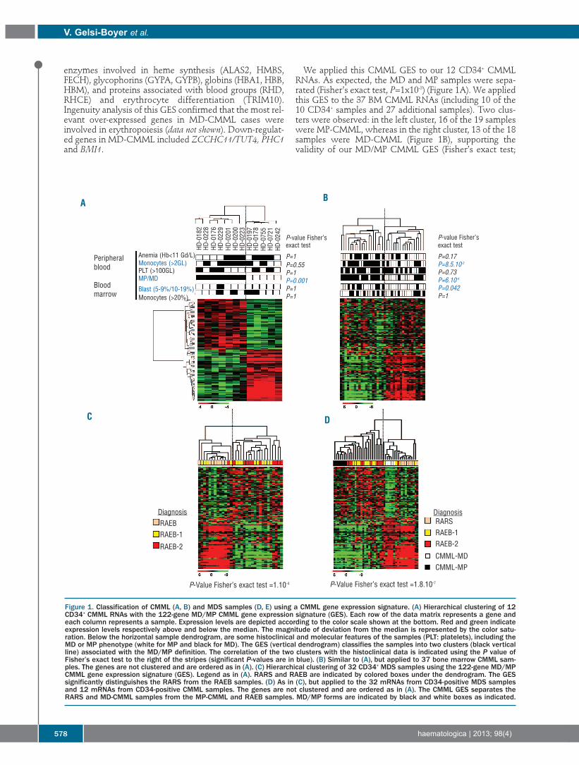
We applied this CMML GES to our 12 CD34+ CMMLRNAs. As expected, the MD and MP samples were sepa-rated (Fisher’s exact test, P=1x10⁻³) (Figure 1A). We appliedthis GES to the 37 BM CMML RNAs (including 10 of the10 CD34+ samples and 27 additional samples). Two clus-ters were observed: in the left cluster, 16 of the 19 sampleswere MP-CMML, whereas in the right cluster, 13 of the 18samples were MD-CMML (Figure 1B), supporting thevalidity of our MD/MP CMML GES (Fisher’s exact test;
V. Gelsi-Boyer et al.
578 haematologica | 2013; 98(4)
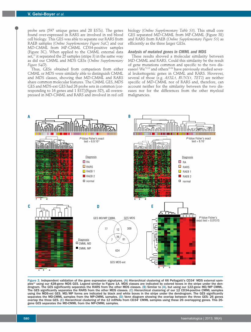
Figure 1. Classification of CMML (A, B) and MDS samples (D, E) using a CMML gene expression signature. (A) Hierarchical clustering of 12CD34+ CMML RNAs with the 122-gene MD/MP CMML gene expression signature (GES). Each row of the data matrix represents a gene andeach column represents a sample. Expression levels are depicted according to the color scale shown at the bottom. Red and green indicateexpression levels respectively above and below the median. The magnitude of deviation from the median is represented by the color satu-ration. Below the horizontal sample dendrogram, are some histoclinical and molecular features of the samples (PLT: platelets), including theMD or MP phenotype (white for MP and black for MD). The GES (vertical dendrogram) classifies the samples into two clusters (black verticalline) associated with the MD/MP definition. The correlation of the two clusters with the histoclinical data is indicated using the P value ofFisher’s exact test to the right of the stripes (significant P-values are in blue). (B) Similar to (A), but applied to 37 bone marrow CMML sam-ples. The genes are not clustered and are ordered as in (A). (C) Hierarchical clustering of 32 CD34+ MDS samples using the 122-gene MD/MPCMML gene expression signature (GES). Legend as in (A). RARS and RAEB are indicated by colored boxes under the dendrogram. The GESsignificantly distinguishes the RARS from the RAEB samples. (D) As in (C), but applied to the 32 mRNAs from CD34-positive MDS samplesand 12 mRNAs from CD34-positive CMML samples. The genes are not clustered and are ordered as in (A). The CMML GES separates theRARS and MD-CMML samples from the MP-CMML and RAEB samples. MD/MP forms are indicated by black and white boxes as indicated.
A B
DC
Peripheral blood
Blood marrow
DiagnosisRAEBRAEB-1
RAEB-2
DiagnosisRARSRAEB-1RAEB-2
CMML-MDCMML-MP
Anemia (Hb<11 Gd/L)Monocytes (>2GL)PLT (>100GL)MP/MD
Blast (5-9%/10-19%)Monocytes (>20%)
P-Value Fisher’s exact test =1.10-4 P-Value Fisher’s exact test =1.8.10-7
P-value Fisher’sexact test
P=1P=0.55P=1P=0.001P=1P=1
P-value Fisher’sexact test
P=0.17P=8.5.10-3
P=0.73P=6.10-4
P=0.042P=1
HD-018
2HD
-022
8HD
-017
6HD
-022
9HD
-020
1HD
-020
0HD
-022
3HD
-019
7HD
-017
8
HD-072
1HD
-024
2
HD-075
5
P=6x10⁻⁴). Using GSEA, we confronted the gene expres-sion profiles of our 37 BM CMML mRNAs to the 122 genesignature. We found a significant enrichment in the redcell genes of this signature in the MD-CMML samples(Enrichment Score=0.76; Normalized EnrichmentScore=2.08; FDR q-value<0.01).
CMML gene expression signature classifies MDS samples Overexpression of genes involved in red cell biology has
been observed in previous gene expression analyses ofRARS samples.14,17 When applied to our 32 CD34+ MDSsamples, the CMML GES perfectly separated RARS fromRAEB samples (Fisher’s exact test, P=1x10⁻⁴, Figure 1C).When the CMML GES was applied to the pool of 12CMML and 32 MDS CD34+ samples the MD-CMMLsclustered with the RARS samples and the MP-CMMLswith the RAEB samples (Figure 1D) (Fisher’s exact test,P=1.8x10⁻⁷). These results showed that MD-CMML andRARS share gene similar expression programs.
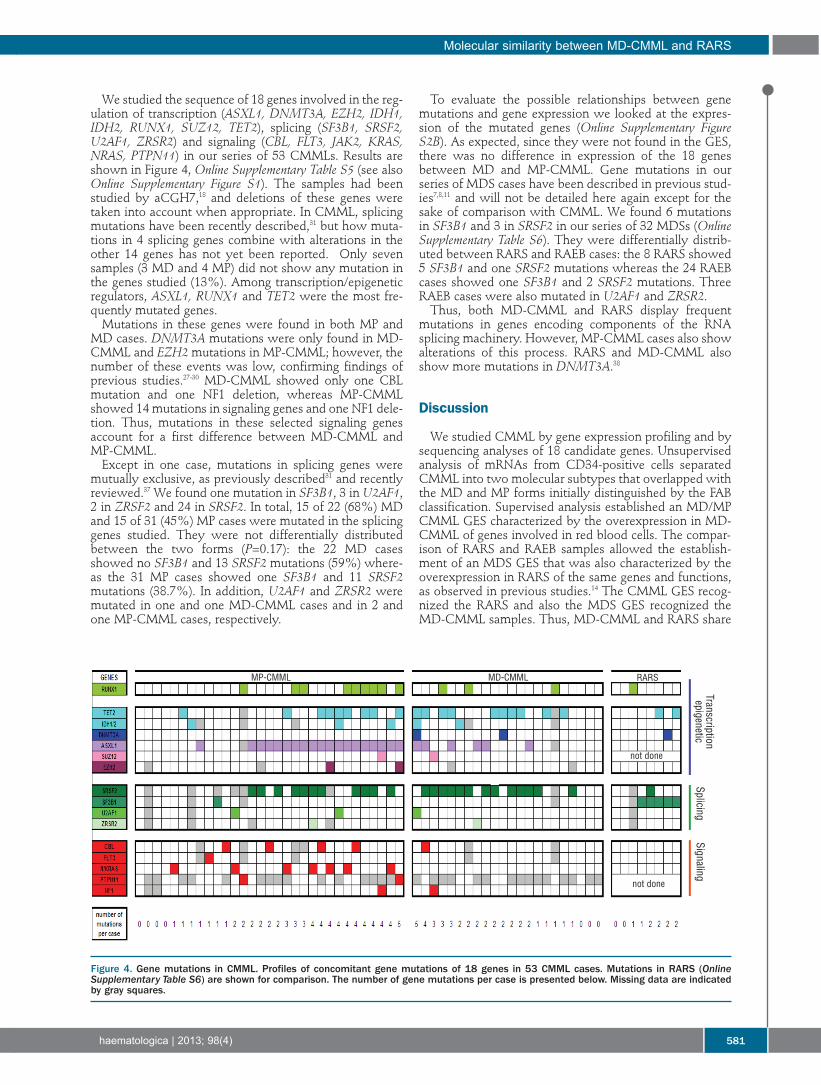
MDS gene expression signature classifies CMML samplesThen we derived an MDS GES by comparing the gene
expression profiles of our 8 RARS to those of our 24 RAEBsamples. A total of 428 probe sets (295 unique genes and25 ESTs; Online Supplementary Table S4) were differentiallyexpressed between RARS and RAEB (hereafter calledMDS GES). The accuracy of prediction and validity of ourprocedure was cross-validated by LOO with overall accu-racy of 78% (Fisher’s exact test, P=6x10⁻⁴) with high sensi-tivity and specificity (72% and 100%, respectively) andwith a theoretical number of false positive of 5.A total of 304 probe sets were up-regulated and 124
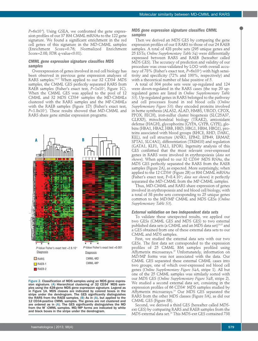
were down-regulated in the RARS cases (the top 20 up-regulated genes are listed in Online Supplementary TableS3). Up-regulated genes in RARS belonged to the pathwayand cell processes found in red blood cells (OnlineSupplementary Figure S3): they encoded proteins involvedin heme synthesis (ALAS2, ALAD, HMBS, UROD, CPOX,PPOX, FECH), iron-sulfur cluster biogenesis (SLC25A37,GLRX5), mitochondrial biology (TRAK2), antioxidantdefense (HAGH), glycophorins (GYPA, GYPB, GYPE), glo-bins (HBA1, HBA2, HBB, HBD, HBG1, HBM, HBQ1), pro-teins associated with blood groups (RHCE, RHD, DARC,KEL), red cell structure (ANK1, EPB42, EPB49, ERMAP,SPTA1, SLC4A1), differentiation (TRIM10) and regulation(GATA1, KLF1, TAL1, EPOR). Ingenuity analysis of thisGES confirmed that the most relevant over-expressedgenes in RARS were involved in erythropoiesis (data notshown). When applied to our 32 CD34+ MDS RNAs, theMDS GES perfectly separated the RARS from the RAEBsamples (Figure 2A), as expected. More surprisingly, whenapplied to the 12 CD34+ (Figure 2B) or BM CMML mRNAs(Fisher’s exact test, P=8.4.10-4; data not shown) it perfectlyseparated the MD-CMML from the MP-CMML samples.Thus, MD-CMML and RARS share expression of genes
involved in erythropoiesis and red blood cell biology, witha total of 38 probe sets corresponding to 25 unique genescommon to the MD/MP CMML and MDS GESs (OnlineSupplementary Table S3).
External validation on two independent data sets To validate these unexpected results, we applied our
two GESs (CMML GES and MDS GES) to two externalpublished data sets (a CMML and an MDS data set)14,15 anda GES obtained from one of these external data sets to ourCMML and MDS samples.First, we studied the external data sets with our two
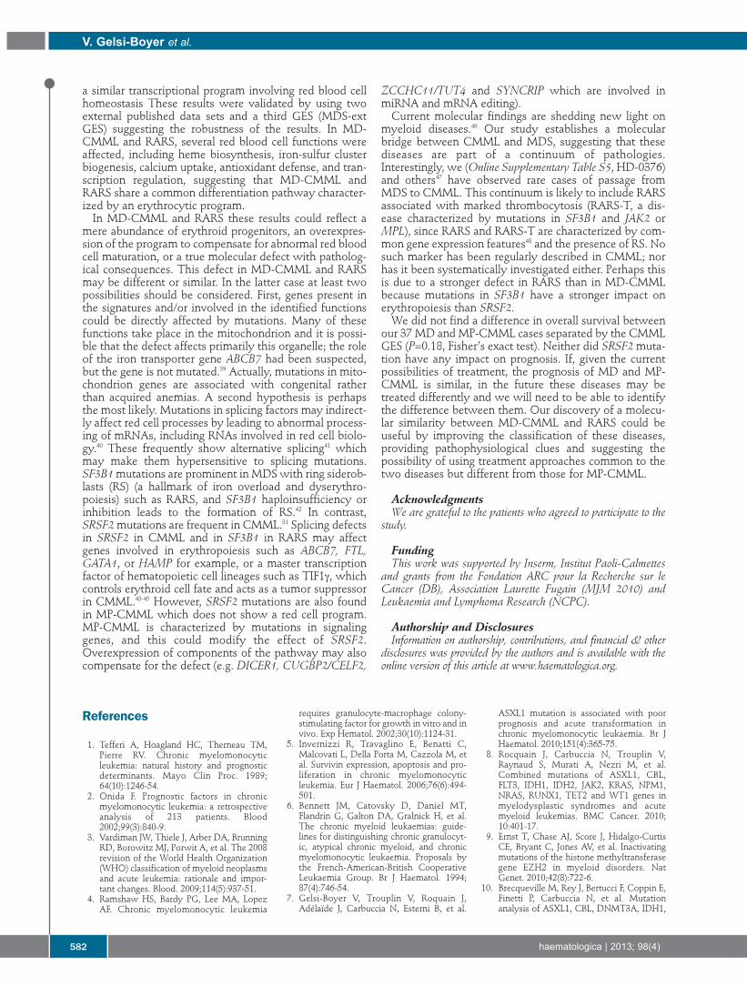
GESs. The first data set corresponded to the expressionprofiles of 25 CMML BM samples profiled usingAffymetrix microarrays.15 Unfortunately, information onMD/MP forms was not associated with the data. OurCMML GES separated these external CMML cases intotwo groups, one of which over-expressed red blood cellgenes (Online Supplementary Figure S4A, stripe 1). All butone of the 25 CMML samples was similarly sorted withour MDS GES (Online Supplementary Figure S4B, stripe 2).We studied a second external data set, consisting in theexpression profiles of 66 CD34+ MDS samples studied byAffymetrix microarrays.14 Our MDS GES separated theRARS from the other MDS classes (Figure 3A), as did ourCMML GES (Figure 3B).Second, we derived a third GES (hereafter called MDS-
ext GES) by comparing RARS and RAEB samples from theMDS external data set.14 This MDS-ext GES contained 738
Molecular similarity between MD-CMML and RARS
haematologica | 2013; 98(4) 579
Figure 2. Classification of MDS samples using an MDS gene expres-sion signature. (A) Hierarchical clustering of 32 CD34+ MDS sam-ples using the 428-gene MDS gene expression signature. Legend asin Figure 1A. MDS classes are indicated by colored boxes in thestripe under the dendrogram. The GES significantly distinguishesthe RARS from the RAEB samples. (B) As in (A), but applied to the12 CD34-positive CMML samples. The genes are not clustered andare ordered as in (A). The GES significantly distinguishes the MDfrom the M¨-CMML samples. MD/MP forms are indicated by whiteand black boxes in the stripe under the dendrogram.
BA
Diagnosis
RARS
RAEB-1
RAEB-2
Diagnosis
CMML-MD
CMML-MP
P-Value Fisher’s exact test =2.8.10-4 P-Value Fisher’s exact test =0.001
probe sets (597 unique genes and 28 ESTs). The genesfound over-expressed in RARS are involved in red bloodcell biology. This GES was able to separate our RARS fromRAEB samples (Online Supplementary Figure S4C) and ourMD-CMML from MP-CMML CD34-positive samples(Figure 3C). When applied to the CMML external dataset,15 it separated the 25 samples (stripe 3) in the same wayas did our CMML and MDS GESs (Online SupplementaryFigure S4D).Thus, GESs obtained from comparison from either
CMML or MDS were similarly able to distinguish CMMLand MDS classes, showing that MD-CMML and RARSshare common molecular features. The CMML GES, MDSGES and MDS-ext GES had 26 probe sets in common (cor-responding to 16 genes and 1 EST)(Figure 3D), all overex-pressed in MD-CMML and RARS and involved in red cell
biology (Online Supplementary Table S3). This small coreGES separated MD-CMML from MP-CMML (Figure 3E)and RARS from RAEB (Online Supplementary Figure S5) asefficiently as the three larger GESs.
Analysis of mutated genes in CMML and MDS These results showed a molecular similarity between
MD-CMML and RARS. Could this similarity be the resultof gene mutations common and specific to the two dis-eases? We7,8,18 and others19-36 have previously studied sever-al leukemogenic genes in CMML and RARS. However,several of those (e.g. ASXL1, RUNX1, TET2) are neitherspecific of MD-CMML nor of RARS and, therefore, canaccount neither for the similarity between the two dis-eases nor for the differences from the other myeloidmalignancies.
V. Gelsi-Boyer et al.
580 haematologica | 2013; 98(4)
Figure 3. Independent validation of the gene expression signatures. (A) Hierarchical clustering of 66 Pellagatti’s CD34+ MDS external sam-ples14 using our 428-gene MDS GES. Legend similar to Figure 1A. MDS classes are indicated by colored boxes in the stripe under the den-drogram. The GES significantly separates the RARS from the other MDS classes. (B) Similar to (A), but using our 122-gene MD/MP CMML.The GES significantly separates the RARS from the other MDS classes. (C) Hierarchical clustering of our 12 CD34-positive CMML samplesusing the MDS-ext GES. MD/MP forms are indicated by black and white boxes in the stripe under the dendrogram. The GES significantlyseparates the MD-CMML samples from the MP-CMML samples. (D) Venn diagram showing the overlap between the three GES: 26 genesoverlap the three GES. (E) Hierarchical clustering of the 12 mRNAs from CD34+ CMML samples using these 26 overlapping genes. This 26-gene GES separates the MD-CMML from the MP-CMML samples.
A B
D EC
Diagnosis
RA
RARS
RAEB 1
RAEB 2
normal
Diagnosis
RA
RARS
RAEB 1
RAEB 2
normal
DiagnosisCMML MD
CMML MP
GES MD/MP CMML GES MDS
GES MDS-ext
77 30912
267 81
624
P-Value Fisher’s exacttest = 6.9.10-5
P-Value Fisher’s exact test = 0.01515
P-Value Fisher’s exacttest = 8.10-7
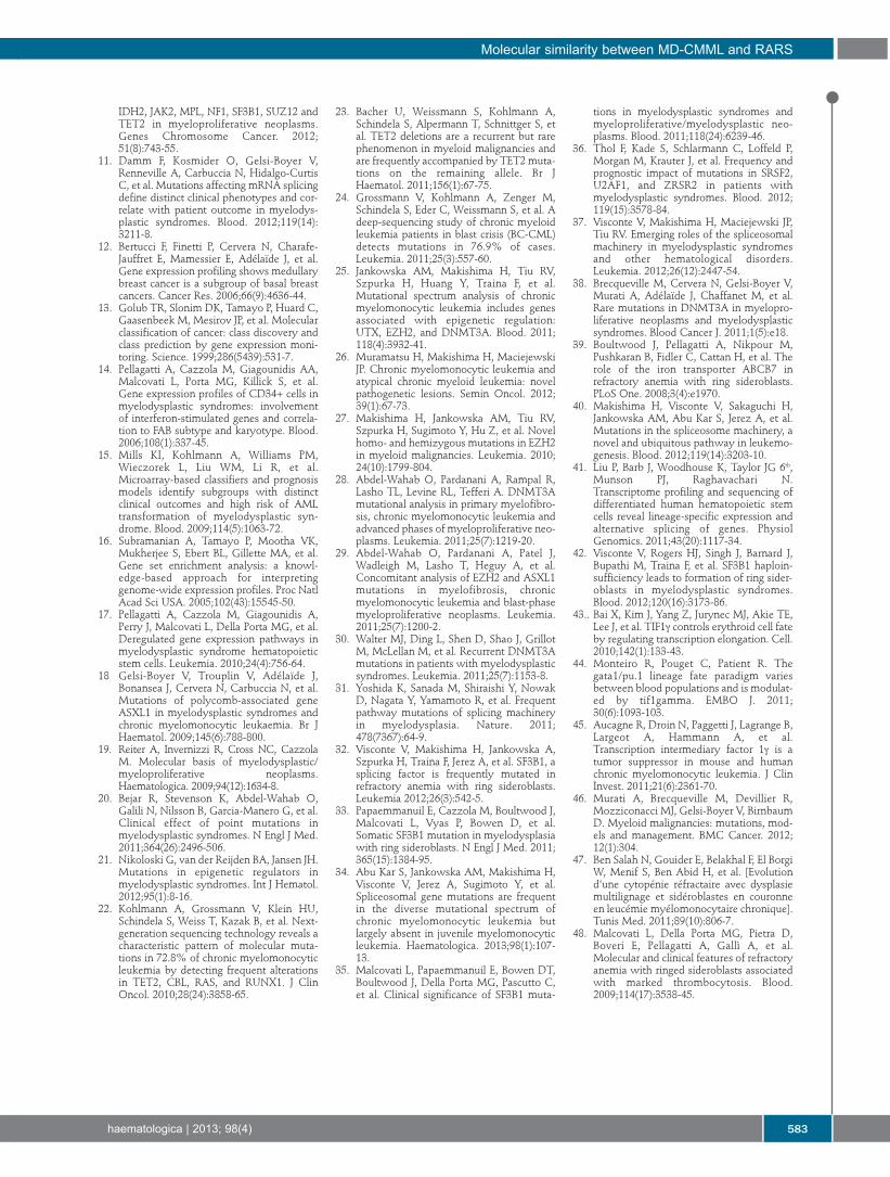
We studied the sequence of 18 genes involved in the reg-ulation of transcription (ASXL1, DNMT3A, EZH2, IDH1,IDH2, RUNX1, SUZ12, TET2), splicing (SF3B1, SRSF2,U2AF1, ZRSR2) and signaling (CBL, FLT3, JAK2, KRAS,NRAS, PTPN11) in our series of 53 CMMLs. Results areshown in Figure 4, Online Supplementary Table S5 (see alsoOnline Supplementary Figure S1). The samples had beenstudied by aCGH7,18 and deletions of these genes weretaken into account when appropriate. In CMML, splicingmutations have been recently described,31 but how muta-tions in 4 splicing genes combine with alterations in theother 14 genes has not yet been reported. Only sevensamples (3 MD and 4 MP) did not show any mutation inthe genes studied (13%). Among transcription/epigeneticregulators, ASXL1, RUNX1 and TET2 were the most fre-quently mutated genes. Mutations in these genes were found in both MP and
MD cases. DNMT3A mutations were only found in MD-CMML and EZH2mutations in MP-CMML; however, thenumber of these events was low, confirming findings ofprevious studies.27-30 MD-CMML showed only one CBLmutation and one NF1 deletion, whereas MP-CMMLshowed 14 mutations in signaling genes and one NF1 dele-tion. Thus, mutations in these selected signaling genesaccount for a first difference between MD-CMML andMP-CMML. Except in one case, mutations in splicing genes were
mutually exclusive, as previously described31 and recentlyreviewed.37 We found one mutation in SF3B1, 3 in U2AF1,2 in ZRSF2 and 24 in SRSF2. In total, 15 of 22 (68%) MDand 15 of 31 (45%) MP cases were mutated in the splicinggenes studied. They were not differentially distributedbetween the two forms (P=0.17): the 22 MD casesshowed no SF3B1 and 13 SRSF2 mutations (59%) where-as the 31 MP cases showed one SF3B1 and 11 SRSF2mutations (38.7%). In addition, U2AF1 and ZRSR2 weremutated in one and one MD-CMML cases and in 2 andone MP-CMML cases, respectively.
To evaluate the possible relationships between genemutations and gene expression we looked at the expres-sion of the mutated genes (Online Supplementary FigureS2B). As expected, since they were not found in the GES,there was no difference in expression of the 18 genesbetween MD and MP-CMML. Gene mutations in ourseries of MDS cases have been described in previous stud-ies7,8,11 and will not be detailed here again except for thesake of comparison with CMML. We found 6 mutationsin SF3B1 and 3 in SRSF2 in our series of 32 MDSs (OnlineSupplementary Table S6). They were differentially distrib-uted between RARS and RAEB cases: the 8 RARS showed5 SF3B1 and one SRSF2 mutations whereas the 24 RAEBcases showed one SF3B1 and 2 SRSF2 mutations. ThreeRAEB cases were also mutated in U2AF1 and ZRSR2.Thus, both MD-CMML and RARS display frequent
mutations in genes encoding components of the RNAsplicing machinery. However, MP-CMML cases also showalterations of this process. RARS and MD-CMML alsoshow more mutations in DNMT3A.38
Discussion
We studied CMML by gene expression profiling and bysequencing analyses of 18 candidate genes. Unsupervisedanalysis of mRNAs from CD34-positive cells separatedCMML into two molecular subtypes that overlapped withthe MD and MP forms initially distinguished by the FABclassification. Supervised analysis established an MD/MPCMML GES characterized by the overexpression in MD-CMML of genes involved in red blood cells. The compar-ison of RARS and RAEB samples allowed the establish-ment of an MDS GES that was also characterized by theoverexpression in RARS of the same genes and functions,as observed in previous studies.14 The CMML GES recog-nized the RARS and also the MDS GES recognized theMD-CMML samples. Thus, MD-CMML and RARS share
Molecular similarity between MD-CMML and RARS
haematologica | 2013; 98(4) 581
Figure 4. Gene mutations in CMML. Profiles of concomitant gene mutations of 18 genes in 53 CMML cases. Mutations in RARS (OnlineSupplementary Table S6) are shown for comparison. The number of gene mutations per case is presented below. Missing data are indicatedby gray squares.
Transcriptionepigenetic
SplicingSignaling
not done
not done
MP-CMML MD-CMML RARS
a similar transcriptional program involving red blood cellhomeostasis These results were validated by using twoexternal published data sets and a third GES (MDS-extGES) suggesting the robustness of the results. In MD-CMML and RARS, several red blood cell functions wereaffected, including heme biosynthesis, iron-sulfur clusterbiogenesis, calcium uptake, antioxidant defense, and tran-scription regulation, suggesting that MD-CMML andRARS share a common differentiation pathway character-ized by an erythrocytic program. In MD-CMML and RARS these results could reflect a
mere abundance of erythroid progenitors, an overexpres-sion of the program to compensate for abnormal red bloodcell maturation, or a true molecular defect with patholog-ical consequences. This defect in MD-CMML and RARSmay be different or similar. In the latter case at least twopossibilities should be considered. First, genes present inthe signatures and/or involved in the identified functionscould be directly affected by mutations. Many of thesefunctions take place in the mitochondrion and it is possi-ble that the defect affects primarily this organelle; the roleof the iron transporter gene ABCB7 had been suspected,but the gene is not mutated.39 Actually, mutations in mito-chondrion genes are associated with congenital ratherthan acquired anemias. A second hypothesis is perhapsthe most likely. Mutations in splicing factors may indirect-ly affect red cell processes by leading to abnormal process-ing of mRNAs, including RNAs involved in red cell biolo-gy.40 These frequently show alternative splicing41 whichmay make them hypersensitive to splicing mutations.SF3B1mutations are prominent in MDS with ring siderob-lasts (RS) (a hallmark of iron overload and dyserythro-poiesis) such as RARS, and SF3B1 haploinsufficiency orinhibition leads to the formation of RS.42 In contrast,SRSF2mutations are frequent in CMML.31 Splicing defectsin SRSF2 in CMML and in SF3B1 in RARS may affectgenes involved in erythropoiesis such as ABCB7, FTL,GATA1, or HAMP for example, or a master transcriptionfactor of hematopoietic cell lineages such as TIF1γ, whichcontrols erythroid cell fate and acts as a tumor suppressorin CMML.43-45 However, SRSF2 mutations are also foundin MP-CMML which does not show a red cell program.MP-CMML is characterized by mutations in signalinggenes, and this could modify the effect of SRSF2.Overexpression of components of the pathway may alsocompensate for the defect (e.g. DICER1, CUGBP2/CELF2,
ZCCHC11/TUT4 and SYNCRIP which are involved inmiRNA and mRNA editing).Current molecular findings are shedding new light on
myeloid diseases.46 Our study establishes a molecularbridge between CMML and MDS, suggesting that thesediseases are part of a continuum of pathologies.Interestingly, we (Online Supplementary Table S5, HD-0376)and others47 have observed rare cases of passage fromMDS to CMML. This continuum is likely to include RARSassociated with marked thrombocytosis (RARS-T, a dis-ease characterized by mutations in SF3B1 and JAK2 orMPL), since RARS and RARS-T are characterized by com-mon gene expression features48 and the presence of RS. Nosuch marker has been regularly described in CMML; norhas it been systematically investigated either. Perhaps thisis due to a stronger defect in RARS than in MD-CMMLbecause mutations in SF3B1 have a stronger impact onerythropoiesis than SRSF2.We did not find a difference in overall survival between
our 37 MD and MP-CMML cases separated by the CMMLGES (P=0.18, Fisher’s exact test). Neither did SRSF2muta-tion have any impact on prognosis. If, given the currentpossibilities of treatment, the prognosis of MD and MP-CMML is similar, in the future these diseases may betreated differently and we will need to be able to identifythe difference between them. Our discovery of a molecu-lar similarity between MD-CMML and RARS could beuseful by improving the classification of these diseases,providing pathophysiological clues and suggesting thepossibility of using treatment approaches common to thetwo diseases but different from those for MP-CMML.
AcknowledgmentsWe are grateful to the patients who agreed to participate to the
study.
FundingThis work was supported by Inserm, Institut Paoli-Calmettes
and grants from the Fondation ARC pour la Recherche sur leCancer (DB), Association Laurette Fugain (MJM 2010) andLeukaemia and Lymphoma Research (NCPC).
Authorship and DisclosuresInformation on authorship, contributions, and financial & other
disclosures was provided by the authors and is available with theonline version of this article at www.haematologica.org.
V. Gelsi-Boyer et al.
582 haematologica | 2013; 98(4)
References
1. Tefferi A, Hoagland HC, Therneau TM,Pierre RV. Chronic myelomonocyticleukemia: natural history and prognosticdeterminants. Mayo Clin Proc. 1989;64(10):1246-54.
2. Onida F. Prognostic factors in chronicmyelomonocytic leukemia: a retrospectiveanalysis of 213 patients. Blood2002;99(3):840-9.
3. Vardiman JW, Thiele J, Arber DA, BrunningRD, Borowitz MJ, Porwit A, et al. The 2008revision of the World Health Organization(WHO) classification of myeloid neoplasmsand acute leukemia: rationale and impor-tant changes. Blood. 2009;114(5):937-51.
4. Ramshaw HS, Bardy PG, Lee MA, LopezAF. Chronic myelomonocytic leukemia
requires granulocyte-macrophage colony-stimulating factor for growth in vitro and invivo. Exp Hematol. 2002;30(10):1124-31.
5. Invernizzi R, Travaglino E, Benatti C,Malcovati L, Della Porta M, Cazzola M, etal. Survivin expression, apoptosis and pro-liferation in chronic myelomonocyticleukemia. Eur J Haematol. 2006;76(6):494-501.
6. Bennett JM, Catovsky D, Daniel MT,Flandrin G, Galton DA, Gralnick H, et al.The chronic myeloid leukaemias: guide-lines for distinguishing chronic granulocyt-ic, atypical chronic myeloid, and chronicmyelomonocytic leukaemia. Proposals bythe French-American-British CooperativeLeukaemia Group. Br J Haematol. 1994;87(4):746-54.
7. Gelsi-Boyer V, Trouplin V, Roquain J,Adélaïde J, Carbuccia N, Esterni B, et al.
ASXL1 mutation is associated with poorprognosis and acute transformation inchronic myelomonocytic leukaemia. Br JHaematol. 2010;151(4):365-75.
8. Rocquain J, Carbuccia N, Trouplin V,Raynaud S, Murati A, Nezri M, et al.Combined mutations of ASXL1, CBL,FLT3, IDH1, IDH2, JAK2, KRAS, NPM1,NRAS, RUNX1, TET2 and WT1 genes inmyelodysplastic syndromes and acutemyeloid leukemias. BMC Cancer. 2010;10:401-17.
9. Ernst T, Chase AJ, Score J, Hidalgo-CurtisCE, Bryant C, Jones AV, et al. Inactivatingmutations of the histone methyltransferasegene EZH2 in myeloid disorders. NatGenet. 2010;42(8):722-6.
10. Brecqueville M, Rey J, Bertucci F, Coppin E,Finetti P, Carbuccia N, et al. Mutationanalysis of ASXL1, CBL, DNMT3A, IDH1,
IDH2, JAK2, MPL, NF1, SF3B1, SUZ12 andTET2 in myeloproliferative neoplasms.Genes Chromosome Cancer. 2012;51(8):743-55.
11. Damm F, Kosmider O, Gelsi-Boyer V,Renneville A, Carbuccia N, Hidalgo-CurtisC, et al. Mutations affecting mRNA splicingdefine distinct clinical phenotypes and cor-relate with patient outcome in myelodys-plastic syndromes. Blood. 2012;119(14):3211-8.
12. Bertucci F, Finetti P, Cervera N, Charafe-Jauffret E, Mamessier E, Adélaïde J, et al.Gene expression profiling shows medullarybreast cancer is a subgroup of basal breastcancers. Cancer Res. 2006;66(9):4636-44.
13. Golub TR, Slonim DK, Tamayo P, Huard C,Gaasenbeek M, Mesirov JP, et al. Molecularclassification of cancer: class discovery andclass prediction by gene expression moni-toring. Science. 1999;286(5439):531-7.
14. Pellagatti A, Cazzola M, Giagounidis AA,Malcovati L, Porta MG, Killick S, et al.Gene expression profiles of CD34+ cells inmyelodysplastic syndromes: involvementof interferon-stimulated genes and correla-tion to FAB subtype and karyotype. Blood.2006;108(1):337-45.
15. Mills KI, Kohlmann A, Williams PM,Wieczorek L, Liu WM, Li R, et al.Microarray-based classifiers and prognosismodels identify subgroups with distinctclinical outcomes and high risk of AMLtransformation of myelodysplastic syn-drome. Blood. 2009;114(5):1063-72.
16. Subramanian A, Tamayo P, Mootha VK,Mukherjee S, Ebert BL, Gillette MA, et al.Gene set enrichment analysis: a knowl-edge-based approach for interpretinggenome-wide expression profiles. Proc NatlAcad Sci USA. 2005;102(43):15545-50.
17. Pellagatti A, Cazzola M, Giagounidis A,Perry J, Malcovati L, Della Porta MG, et al.Deregulated gene expression pathways inmyelodysplastic syndrome hematopoieticstem cells. Leukemia. 2010;24(4):756-64.
18 Gelsi-Boyer V, Trouplin V, Adélaïde J,Bonansea J, Cervera N, Carbuccia N, et al.Mutations of polycomb-associated geneASXL1 in myelodysplastic syndromes andchronic myelomonocytic leukaemia. Br JHaematol. 2009;145(6):788-800.
19. Reiter A, Invernizzi R, Cross NC, CazzolaM. Molecular basis of myelodysplastic/myeloproliferative neoplasms.Haematologica. 2009;94(12):1634-8.
20. Bejar R, Stevenson K, Abdel-Wahab O,Galili N, Nilsson B, Garcia-Manero G, et al.Clinical effect of point mutations inmyelodysplastic syndromes. N Engl J Med.2011;364(26):2496-506.
21. Nikoloski G, van der Reijden BA, Jansen JH.Mutations in epigenetic regulators inmyelodysplastic syndromes. Int J Hematol.2012;95(1):8-16.
22. Kohlmann A, Grossmann V, Klein HU,Schindela S, Weiss T, Kazak B, et al. Next-generation sequencing technology reveals acharacteristic pattern of molecular muta-tions in 72.8% of chronic myelomonocyticleukemia by detecting frequent alterationsin TET2, CBL, RAS, and RUNX1. J ClinOncol. 2010;28(24):3858-65.
23. Bacher U, Weissmann S, Kohlmann A,Schindela S, Alpermann T, Schnittger S, etal. TET2 deletions are a recurrent but rarephenomenon in myeloid malignancies andare frequently accompanied by TET2 muta-tions on the remaining allele. Br JHaematol. 2011;156(1):67-75.
24. Grossmann V, Kohlmann A, Zenger M,Schindela S, Eder C, Weissmann S, et al. Adeep-sequencing study of chronic myeloidleukemia patients in blast crisis (BC-CML)detects mutations in 76.9% of cases.Leukemia. 2011;25(3):557-60.
25. Jankowska AM, Makishima H, Tiu RV,Szpurka H, Huang Y, Traina F, et al.Mutational spectrum analysis of chronicmyelomonocytic leukemia includes genesassociated with epigenetic regulation:UTX, EZH2, and DNMT3A. Blood. 2011;118(4):3932-41.
26. Muramatsu H, Makishima H, MaciejewskiJP. Chronic myelomonocytic leukemia andatypical chronic myeloid leukemia: novelpathogenetic lesions. Semin Oncol. 2012;39(1):67-73.
27. Makishima H, Jankowska AM, Tiu RV,Szpurka H, Sugimoto Y, Hu Z, et al. Novelhomo- and hemizygous mutations in EZH2in myeloid malignancies. Leukemia. 2010;24(10):1799-804.
28. Abdel-Wahab O, Pardanani A, Rampal R,Lasho TL, Levine RL, Tefferi A. DNMT3Amutational analysis in primary myelofibro-sis, chronic myelomonocytic leukemia andadvanced phases of myeloproliferative neo-plasms. Leukemia. 2011;25(7):1219-20.
29. Abdel-Wahab O, Pardanani A, Patel J,Wadleigh M, Lasho T, Heguy A, et al.Concomitant analysis of EZH2 and ASXL1mutations in myelofibrosis, chronicmyelomonocytic leukemia and blast-phasemyeloproliferative neoplasms. Leukemia.2011;25(7):1200-2.
30. Walter MJ, Ding L, Shen D, Shao J, GrillotM, McLellan M, et al. Recurrent DNMT3Amutations in patients with myelodysplasticsyndromes. Leukemia. 2011;25(7):1153-8.
31. Yoshida K, Sanada M, Shiraishi Y, NowakD, Nagata Y, Yamamoto R, et al. Frequentpathway mutations of splicing machineryin myelodysplasia. Nature. 2011;478(7367):64-9.
32. Visconte V, Makishima H, Jankowska A,Szpurka H, Traina F, Jerez A, et al. SF3B1, asplicing factor is frequently mutated inrefractory anemia with ring sideroblasts.Leukemia 2012;26(3):542-5.
33. Papaemmanuil E, Cazzola M, Boultwood J,Malcovati L, Vyas P, Bowen D, et al.Somatic SF3B1 mutation in myelodysplasiawith ring sideroblasts. N Engl J Med. 2011;365(15):1384-95.
34. Abu Kar S, Jankowska AM, Makishima H,Visconte V, Jerez A, Sugimoto Y, et al.Spliceosomal gene mutations are frequentin the diverse mutational spectrum ofchronic myelomonocytic leukemia butlargely absent in juvenile myelomonocyticleukemia. Haematologica. 2013;98(1):107-13.
35. Malcovati L, Papaemmanuil E, Bowen DT,Boultwood J, Della Porta MG, Pascutto C,et al. Clinical significance of SF3B1 muta-
tions in myelodysplastic syndromes andmyeloproliferative/myelodysplastic neo-plasms. Blood. 2011;118(24):6239-46.
36. Thol F, Kade S, Schlarmann C, Loffeld P,Morgan M, Krauter J, et al. Frequency andprognostic impact of mutations in SRSF2,U2AF1, and ZRSR2 in patients withmyelodysplastic syndromes. Blood. 2012;119(15):3578-84.
37. Visconte V, Makishima H, Maciejewski JP,Tiu RV. Emerging roles of the spliceosomalmachinery in myelodysplastic syndromesand other hematological disorders.Leukemia. 2012;26(12):2447-54.
38. Brecqueville M, Cervera N, Gelsi-Boyer V,Murati A, Adélaïde J, Chaffanet M, et al.Rare mutations in DNMT3A in myelopro-liferative neoplasms and myelodysplasticsyndromes. Blood Cancer J. 2011;1(5):e18.
39. Boultwood J, Pellagatti A, Nikpour M,Pushkaran B, Fidler C, Cattan H, et al. Therole of the iron transporter ABCB7 inrefractory anemia with ring sideroblasts.PLoS One. 2008;3(4):e1970.
40. Makishima H, Visconte V, Sakaguchi H,Jankowska AM, Abu Kar S, Jerez A, et al.Mutations in the spliceosome machinery, anovel and ubiquitous pathway in leukemo-genesis. Blood. 2012;119(14):3203-10.
41. Liu P, Barb J, Woodhouse K, Taylor JG 6th,Munson PJ, Raghavachari N.Transcriptome profiling and sequencing ofdifferentiated human hematopoietic stemcells reveal lineage-specific expression andalternative splicing of genes. PhysiolGenomics. 2011;43(20):1117-34.
42. Visconte V, Rogers HJ, Singh J, Barnard J,Bupathi M, Traina F, et al. SF3B1 haploin-sufficiency leads to formation of ring sider-oblasts in myelodysplastic syndromes.Blood. 2012;120(16):3173-86.
43.. Bai X, Kim J, Yang Z, Jurynec MJ, Akie TE,Lee J, et al. TIF1γ controls erythroid cell fateby regulating transcription elongation. Cell.2010;142(1):133-43.
44. Monteiro R, Pouget C, Patient R. Thegata1/pu.1 lineage fate paradigm variesbetween blood populations and is modulat-ed by tif1gamma. EMBO J. 2011;30(6):1093-103.
45. Aucagne R, Droin N, Paggetti J, Lagrange B,Largeot A, Hammann A, et al.Transcription intermediary factor 1γ is atumor suppressor in mouse and humanchronic myelomonocytic leukemia. J ClinInvest. 2011;21(6):2361-70.
46. Murati A, Brecqueville M, Devillier R,Mozziconacci MJ, Gelsi-Boyer V, BirnbaumD. Myeloid malignancies: mutations, mod-els and management. BMC Cancer. 2012;12(1):304.
47. Ben Salah N, Gouider E, Belakhal F, El BorgiW, Menif S, Ben Abid H, et al. [Evolutiond'une cytopénie réfractaire avec dysplasiemultilignage et sidéroblastes en couronneen leucémie myélomonocytaire chronique].Tunis Med. 2011;89(10):806-7.
48. Malcovati L, Della Porta MG, Pietra D,Boveri E, Pellagatti A, Gallì A, et al.Molecular and clinical features of refractoryanemia with ringed sideroblasts associatedwith marked thrombocytosis. Blood.2009;114(17):3538-45.
Molecular similarity between MD-CMML and RARS
haematologica | 2013; 98(4) 583