
Molecular Nanoprobes for Multiphotonics as New tools for ...
39
Molecular Molecular Nanoprobes Nanoprobes for for Multiphotonics Multiphotonics as as New New tools tools for for Bioimaging Bioimaging 2 nd Annual Symposium on Integrating Nanotechnology with Cell Biology and Neuroscience The University of New Mexico, August 22 nd 2008 UNM ICNCBN-IGERT
Transcript of Molecular Nanoprobes for Multiphotonics as New tools for ...
Molecular Molecular NanoprobesNanoprobes for for MultiphotonicsMultiphotonics as as New New toolstools for for BioimagingBioimaging
2nd Annual Symposium on IntegratingNanotechnology with Cell Biology and Neuroscience
The University of New Mexico, August 22nd 2008 UNM ICNCBN-IGERT

@ 488 nm @ 1032 nm
• intrinsic 3-D resolution
Fluorescence excited by :
1 photon 2 photons
Two-Photon Excited Fluorescence(TPEF or 2PEF)
⇒ Advantages in biological imaging :∝ I2∝ I
• increased penetration in tissues
• reduced photodamage• reduced background fluorescence
MultiphotonicsMultiphotonics: Two: Two-- photon absorption (TPA) photon absorption (TPA) ⇒⇒ TwoTwo--photon excitedphoton excited--fluorescence (TPEF)fluorescence (TPEF)
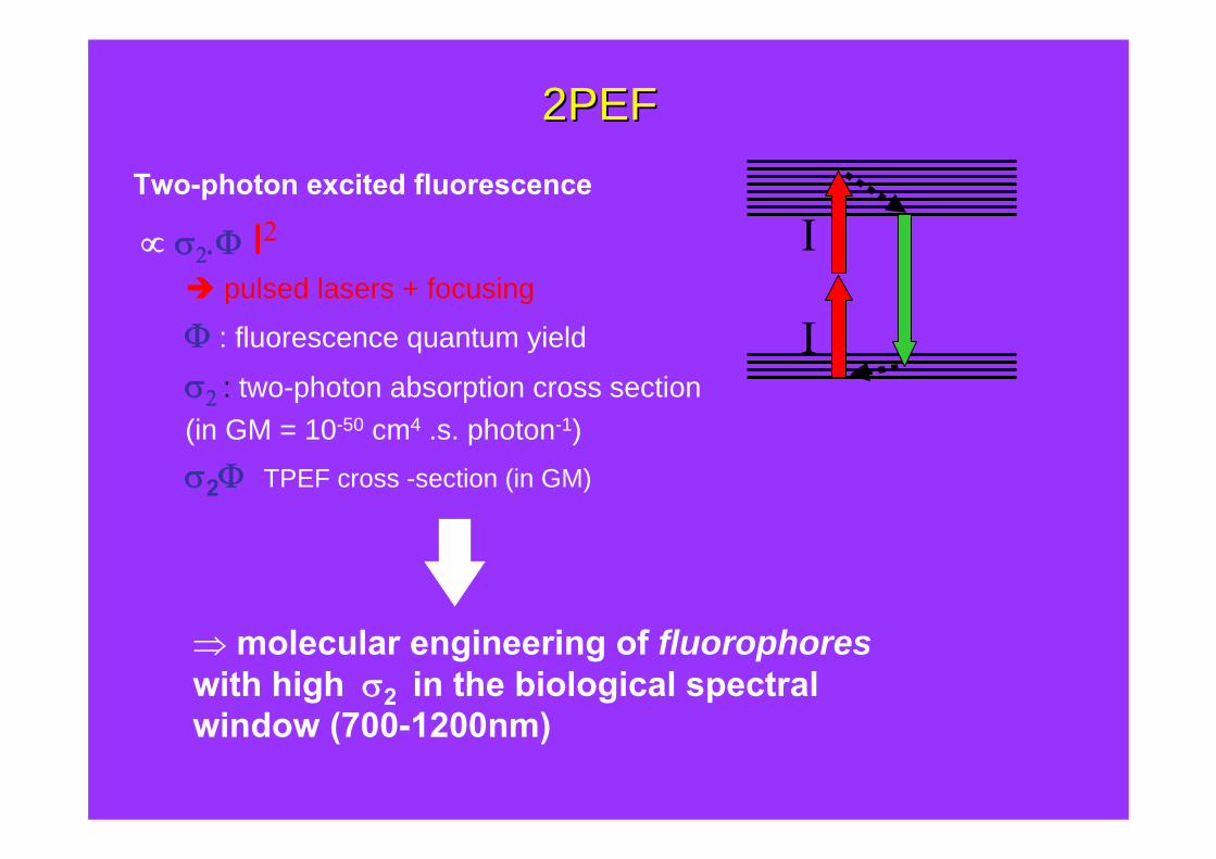
2PEF2PEFTwo-photon excited fluorescence
∝ σ2.Φ I2
pulsed lasers + focusing
Φ : fluorescence quantum yield
σ2 : two-photon absorption cross section(in GM = 10-50 cm4 .s. photon-1)σ2Φ TPEF cross -section (in GM)
⇒ molecular engineering of fluorophoreswith high σ2 in the biological spectral window (700-1200nm)
I
I

Fluorescent Markers and Probes for 2PEF imagingFluorescent Markers and Probes for 2PEF imaging
- Endogenous biological chromophores:NADH, riboflavins, retinol : 10-5 < σ2.Φ < 1 GM
- Classical one-photon fluorophores :DAPI : σ2.Φ < 1 GMCoumarin 307, Bodipy : σ2.Φ < 20 GMFluorescein : σ2.Φ < 40 GM

MOLECULAR ENGINEERING FOR MOLECULAR ENGINEERING FOR MULTIPHOTONIC BIOIMAGINGMULTIPHOTONIC BIOIMAGING :
examples and applicationsWhat is needed ?
• high fluorescence quantum yield• very large TPA cross-sections in the target spectral range• without residual one-photon absorption
⇒ 3D imaging
⇒ enhanced sensitivity
⇒ reduced photodamage
⇒ selective photo-addressing

IncreaselengthD/A strength type of spacer
Molecular engineering of quadrupoles
π co n jug a te dsys te m
D π co n jug a te dsys te m
Dθ
Adjust θ
Chain
SolubilityAffinity
• TPA enhancement• spectral tuning• (photo)stability
J. Chem. Phys. 2000, 113, 3951

R2N NR2
R2N NR2
X = NR2,: X = OMe
2.4 nm
2.3 nm
NonNon XX
NonNon SO2OctOctO2S
NonNon SO2CF3F3CO2S
NonNon NH2H2N
2.4 nm
2.4 nm
2.3 nm
2.3 nm
Non Non
Non Non NR2NonNonR2N
Non Non
SSR2N NR2
Non NonX X
R2N NR2
R2N NR2
X = NR2, : X = SO2R
3.8 nm
3.6 nm
4.5 nm
3.7 nm
3.7 nm
Non Non
NR2
3.6 nm
R2N
RodRod--likelike and bananeand banane--shapedshaped quadrupolarquadrupolar fluorophoresfluorophores
Chem. Eur. J. 2007, 13, 1481
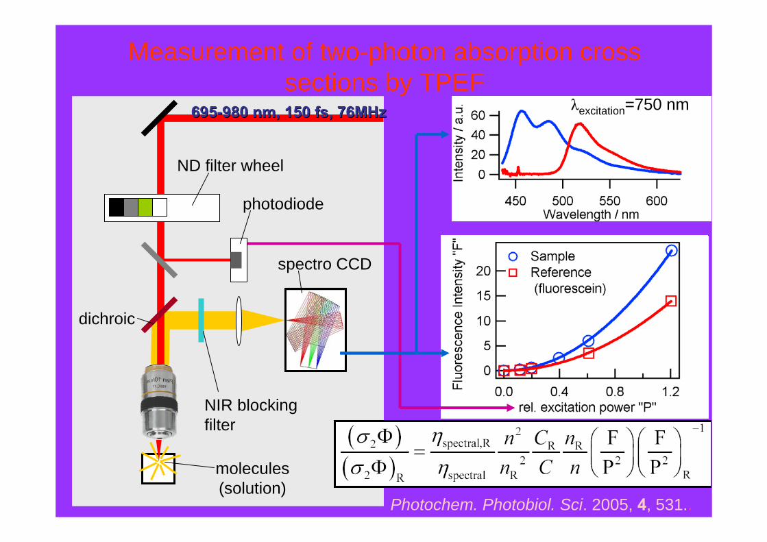
ND filter wheel
photodiode
molecules(solution)
dichroic
NIR blockingfilter
spectro CCD
695695--980 nm, 150 980 nm, 150 fsfs, 76MHz, 76MHz λexcitation=750 nm
Photochem. Photobiol. Sci. 2005, 4, 531..
Measurement of two-photon absorption cross sections by TPEF

major TPA amplification in the NIR fluorescence is maintained
Molecular optimization of quadrupolar fluorophores
Fluorophore λabsmax
(nm)
Φ τ
(ns)
σ2*
(GM)
2
RHex2N
431 0.850.83 2115
2
R
Bu2N
R R
2
R
SOct2N
429
470
0.780.73
0.470.79
3470
5480
*at 705 nm (toluene)
Chem. Eur J, 2007, 13, 1481-1498.

APPLICATION: 3-D BIOIMAGING APPLICATION: 3-D BIOIMAGING
TPEF cross-sectional image of a GUV labeled with BAQ1.The giant unilamellar vesicles (GUV) were prepared from 1,2-dioleoyl-sn-glycero-3-phosphocholine (DOPC)
Angew. Chem., Int. Ed. 2001, 40, 2098
@ 750 nm
polarheadgroup
hydrophobicchain
lipids
bolaamphiphilic fluorophore
hydrophobic part hydrophilicpart
N NOHHO
N NOHHO
N NOHHO
HO OH
l = 30 Å
l = 35 Å
N+ N+
SO3--O3S
N+ N+
From model lipidic membranes…..

APPLICATION: 3-D BIOIMAGING APPLICATION: 3-D BIOIMAGING
C.R. Physique, 2002, 3, 439.
TPEF image of LLC-PK1 cells* labeled with with BAQ1, (excitation at 740 nm with less than 1mW excitation power)
To non-damaging cell imaging

FROM IMAGING TO SENSINGTowards Medium Responsive Two-Photon
Nanoprobes
⇒⇒sensitive twosensitive two--photon pH probes in the NIRphoton pH probes in the NIR
⇒⇒⇒ sensitive twosensitive twosensitive two---photon photon photon micropolaritymicropolaritymicropolarity probesprobesprobes
⇒⇒⇒ fast voltage probesfast voltage probesfast voltage probes

Recent examples of TP probes
NNC
N
OO
OO
B. R. Cho and coworkers, JOC, 2004, 5749-5751
Metal-ion sensor(Ca2+, Ba2+, Mg2+, Na+, K+)
J. W. Perry and coworkers, JACS, 2004, 12, 9291-9306
N
N
OO
OO
CN
NC
Metal-ion sensor(Mg2+)
H+ sensors ⇒ 2-photon pH probes ?
CL4

Diapositive 13
CL4 TP probes for metal-ions have been recently synthesized. This fluorophore with crown ether is a sensor for Mg2+.This another one is based on the same design and can sense different ions :…..At last, this quadrupole is sensitive to the pH of solution.céline; 27/06/2005
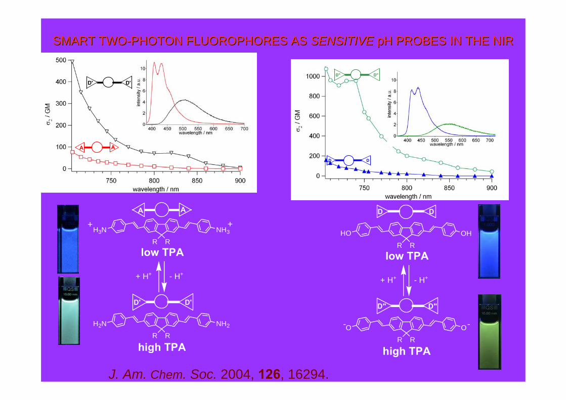
SMART TWOSMART TWO--PHOTON FLUOROPHORES AS PHOTON FLUOROPHORES AS SENSITIVESENSITIVE pH PROBES IN THE NIRpH PROBES IN THE NIR
J. Am. Chem. Soc. 2004, 126, 16294.
AA
high TPA
low TPAR R
NH3H3N
+ H+ - H+
R R
NH2H2N
D' D'
+ +
high TPA
low TPAR R
OHHO
D D
+ H+ - H+
R R
OO- -
D" D"
D D
D" D"
AA
D' D'
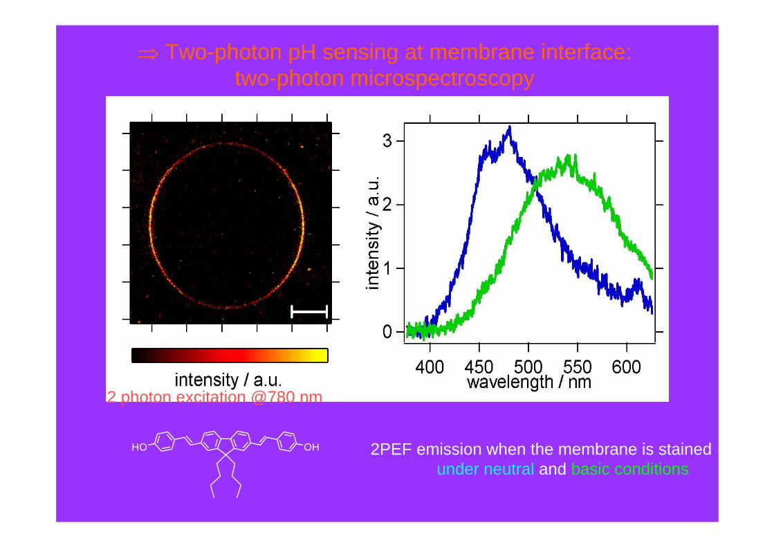
⇒ Two-photon pH sensing at membrane interface:two-photon microspectroscopy
OHHO
2 photon excitation @780 nm
2PEF emission when the membrane is stainedunder neutral and basic conditions
Towards Medium Responsive Two-Photon Nanoprobes
Towards sensitive twoTowards sensitive twoTowards sensitive two---photon pH probes in the NIRphoton pH probes in the NIRphoton pH probes in the NIR
Towards sensitive twoTowards sensitive twoTowards sensitive two---photon photon photon micropolaritymicropolaritymicropolarity probesprobesprobes
Towards fast voltage sensitive probeTowards fast voltage sensitive probe
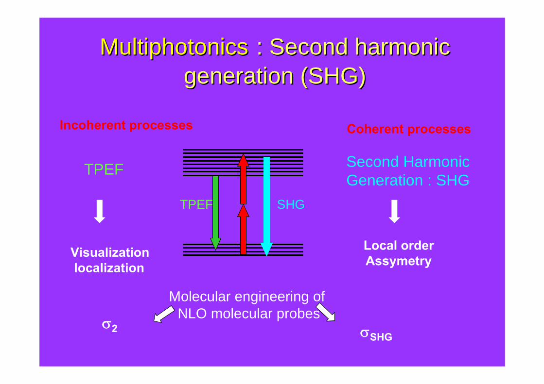
Incoherent processes
TPEF
Coherent processes
Second HarmonicGeneration : SHG
Visualizationlocalization
Local orderAssymetry
TPEF SHG
σ2 σSHG
Molecular engineering of NLO molecular probes
MultiphotonicsMultiphotonics : Second : Second harmonicharmonicgenerationgeneration (SHG)(SHG)

SHG requires asymmetric sourceD Aπ
Amphiphilic Push-pull Chromophores
lipophilic tails
π−conjugated system AD
hydrophilic headgroups
objectives : from static imaging of cells to dynamic imaging of membrane processes
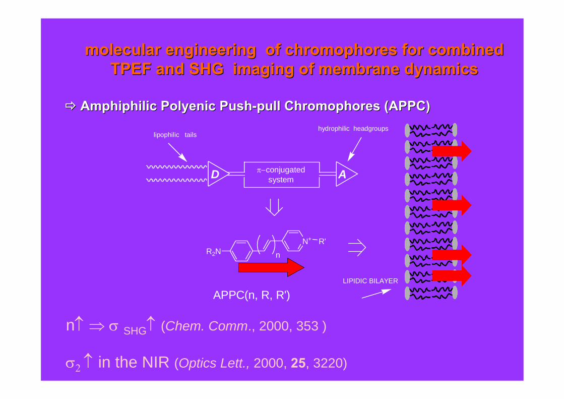
n↑ ⇒ σ SHG↑ (Chem. Comm., 2000, 353 )
σ2 ↑ in the NIR (Optics Lett., 2000, 25, 3220)
molecularmolecular engineering of engineering of chromophores for combined chromophores for combined TPEF and SHG imaging of membrane dynamicsTPEF and SHG imaging of membrane dynamics
AmphiphilicAmphiphilic PolyenicPolyenic PushPush--pull Chromophores (APPC)pull Chromophores (APPC)
lipophilic tails
N+
R2NR'
π−conjugated system AD
n
hydrophilic headgroups
LIPIDIC BILAYER
APPC(n, R, R')

Wavelength (nm)400 450 500 550 600 650
Pow
er (a
.u.)
0.0
0.5
1.0
430 435 440 445 450S
HG
Pow
er (a
.u.)
0.0
0.5
1.0
fluorescence
SHG
SHG 2PEF

APPLICATIONS: NON-LINEAR IMAGING OF BIOLOGICAL CELLS
APPLICATIONS: NON-LINEAR IMAGING OF BIOLOGICAL CELLS
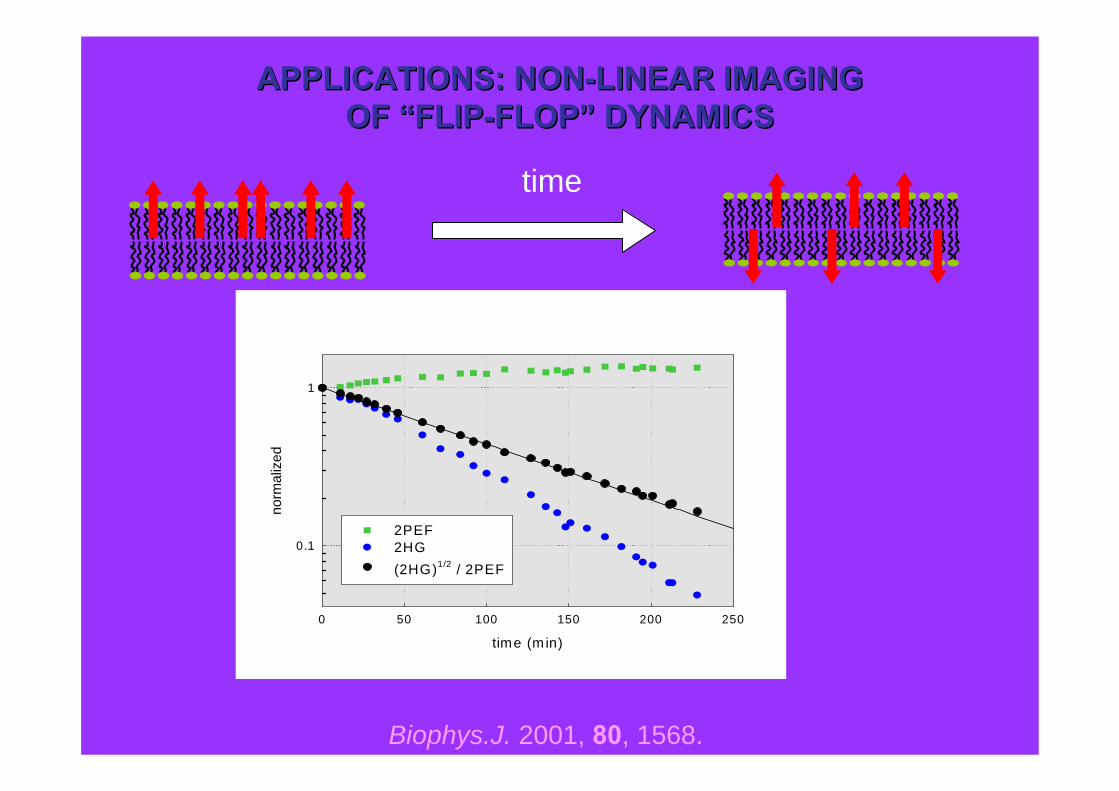
Simultaneous TPEF and SHG cross-sectional images of isolated Ncad1 cells labeled with an amphiphilicpush-pull polyenic chromophore. Internalized dye molecules become randomly oriented in the cytoplasm
and generate no SHG, whereas they continue to generate fluorescence
Biophys. J. 2001, 80, 1568.N+
Bu2N
SO3-
3
@ 880 nm
time (min)
0 50 100 150 200 250
norm
aliz
ed
0.1
1
2PEF 2HG (2HG)1/2 / 2PEF
time
Biophys.J. 2001, 80, 1568.
APPLICATIONS: NONAPPLICATIONS: NON--LINEAR IMAGINGLINEAR IMAGINGOF OF ““FLIPFLIP--FLOPFLOP”” DYNAMICSDYNAMICS
SHG
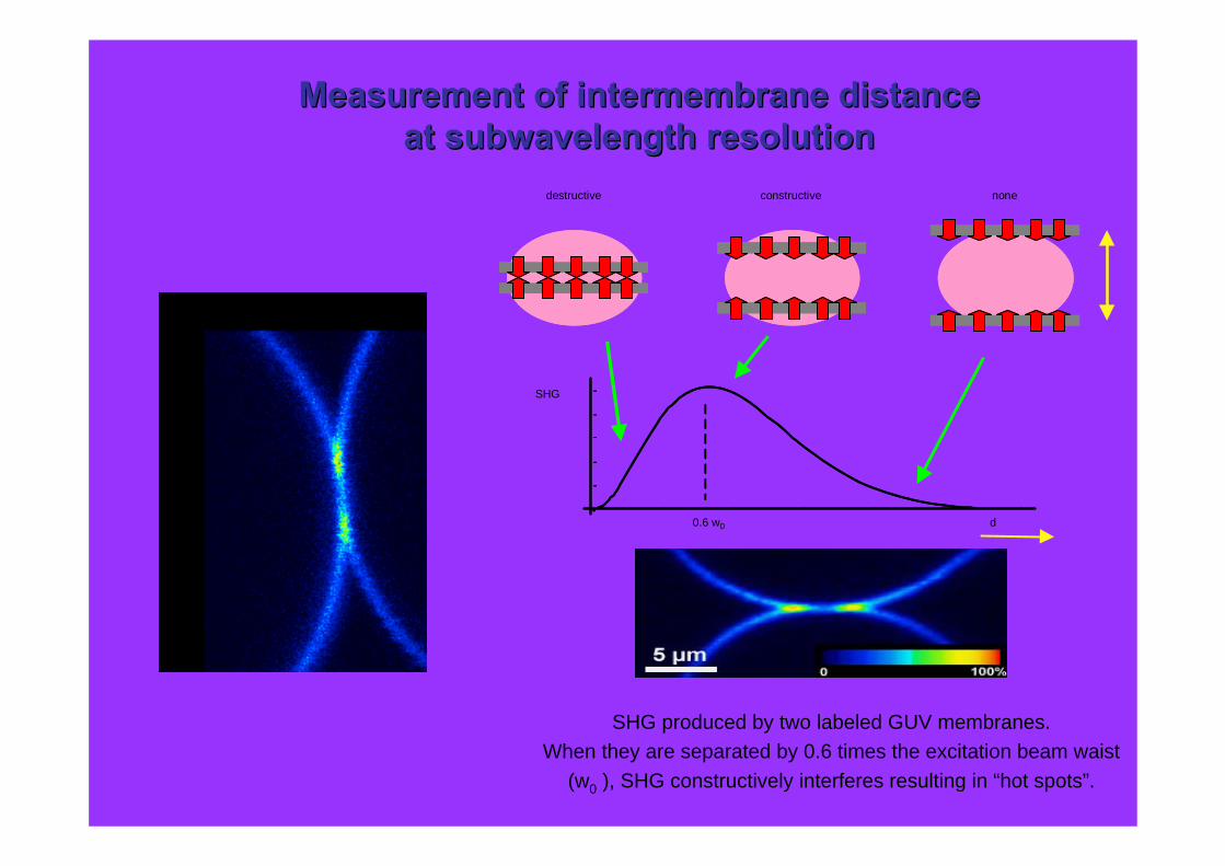
0.6 w0 d
destructive constructive none
SHG produced by two labeled GUV membranes.When they are separated by 0.6 times the excitation beam waist
(w0 ), SHG constructively interferes resulting in “hot spots”.
Measurement of Measurement of intermembraneintermembrane distancedistanceat at subwavelengthsubwavelength resolutionresolution

E
(Lewis, Loew, 1993)
SHG for membrane voltage sensing ?SHG for membrane voltage sensing ?
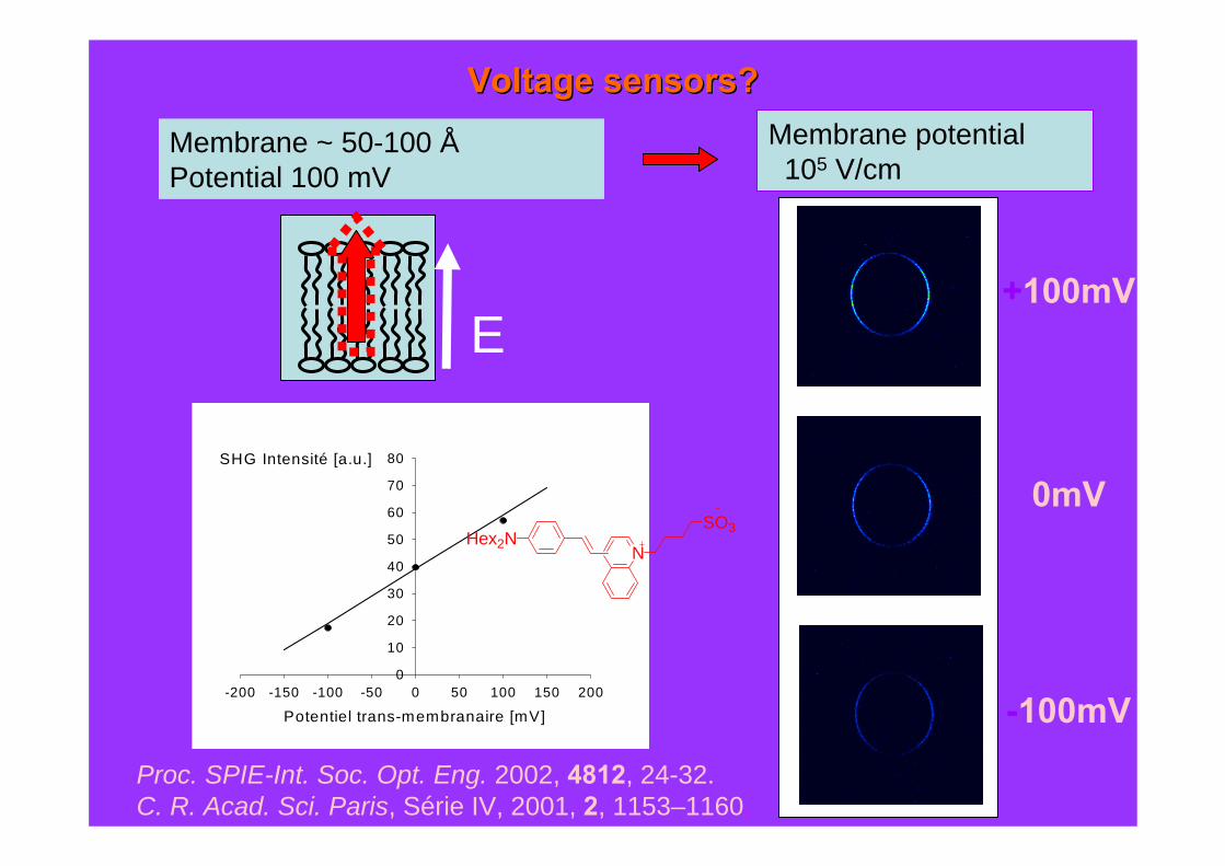
Membrane ~ 50-100 ÅPotential 100 mV
Membrane potential105 V/cm
Proc. SPIE-Int. Soc. Opt. Eng. 2002, 4812, 24-32.C. R. Acad. Sci. Paris, Série IV, 2001, 2, 1153–1160.
E
Voltage sensors?Voltage sensors?
Potentiel trans-membranaire [mV]-200 -150 -100 -50 0 50 100 150 200
SHG Intensité [a.u.]
0
10
20
30
40
50
60
70
80
+100mV
0mV
-100mV
NHex2N
SO3

SHG probes for imaging of neuronal activity
J. Neuroscience, 2004, 24, 999
Imaging ~400 µm deep into intact neurons ⇒ No photodamage
λ = 940 nm
NHex2N SO3-
Optically recording of action potentials⇒ linear dependence of SHG on E⇒ 0.833 ms temporal resolution⇒ 0.6 µm spatial resolution
Collab. W. Webb

quantum dots (QDs) ⇒ bright nanoobjects
⇒ tuneability, photostability
⇒ very large one (ε Φ) and two-photon
(σ2Φ) brilliance*
* W. W. Webb et al, Science, 2003, 300, 1434.
but toxicity, clearance, degradation ?…
Soft substitutes for semiconductor QD's ?

Soft All-Organic Alternative to QD's ?• biocompatibility / degradability• environmental friendly
quantum dots (QDs) ⇒ bright nanoobjects
⇒ tuneability, photostability
⇒ very large one (ε Φ) and two-photon
(σ2Φ) brilliance*
but toxicity, clearance, degradation ?…
Soft substitutes for semiconductor QD's ?
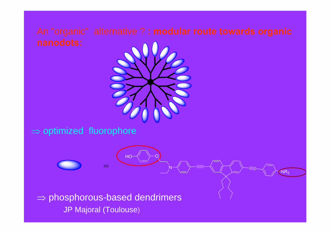
An "organic" alternative ? : modular route towards organic nanodots:
⇒ optimized fluorophore
NR2N
OHO
=
⇒ phosphorous-based dendrimersJP Majoral (Toulouse)

NanodotsNanodots : : ModularModular approachapproach
NPN
P NP
NPN
P NP
NPN
P NP
G1 (n = 12) G2 (n = 24) G3 (n = 48)
Controlled Grafting of Optimized Fluorophores on a branched (dendritic) platform
⇒ nano-object of controlled size, geometry, number of flurophores.
Chem. Commun. 2006, 915-917; New J. Chem., 2007, 31, 1354-1367.
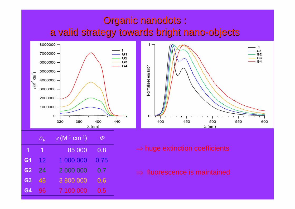
Organic Organic nanodotsnanodots ::a valid strategy towards bright a valid strategy towards bright nanonano--objectsobjects
7 100 0003 800 0002 000 0001 000 000
85 000
ε (M-1 cm-1)
964824121
nF
0.5G4
0.6G3
0.7G2
0.75G1
0.81
Φ
⇒ huge extinction coefficients
⇒ fluorescence is maintained

Organic Organic nanodotsnanodots ::a valid strategy towards bright a valid strategy towards bright nanonano--objectsobjects
"super" bright nano-objects : record brilliance : ε . Φ
− ΦF > 50 % (QD 30-50 %)− ε → 7 000 000 M-1 cm-1
PCT Int. Appl. 2007, WO 2007080176
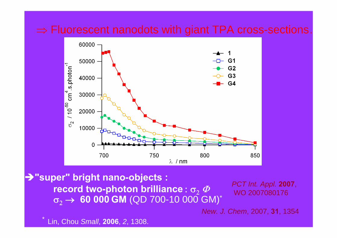
⇒ Fluorescent nanodots with giant TPA cross-sections…
New. J. Chem, 2007, 31, 1354
"super" bright nano-objects : record two-photon brilliance : σ2 Φσ2 → 60 000 GM (QD 700-10 000 GM)*
* Lin, Chou Small, 2006, 2, 1308.
PCT Int. Appl. 2007,WO 2007080176
Lin, Chou Small, 2006, 2, 1308-1313
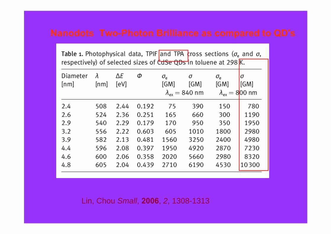
Nanodots Two-Photon Brilliance as compared to QD's

⇒biocompatible biphotonic contrast agents for in vivo imaging
Two-photon imaging of the vascular network in the dorsal part of the rat olfactory bulb. Vessels were labeled after injecting intravenously a small bolus of 500 µM G2 nanodots in water. The image was taken at ~200 µm depth. No obvious toxic effects were observed during the experiment
Excitation @ 710 nmDetection @ 440 nm
Angew. Chem. Int. Ed., 2006, 45, 4645.
Collab. S. Charpak,L. Moreaux(INSERM, Paris Descartes)
+
+
+++++
++
+
+
+
+
++ + + +
++ +
++ + + +
+
++
+
++
+++++
+
+
+
+
+
σ2 = 130 GMλem = 440 nm

APQ 145 (λex : 860 nm)
0
1
2
3
4
5
400 450 500 550 600 650
λem (nm)
ΔF/
F (A
. U.)
2-photon in vivo small animal imaging
Xenopus laevis (stage 53)Gaëlle Recher, François Tiaho (Rennes)
σ2 = 1000 GMλem = 530 nm
SPIE Proc. 2008, 7040, 704006-704017.
Nanodot 2-photon tracer, 0.1 pmoleExcitation : 860 nmEmission 530 nm
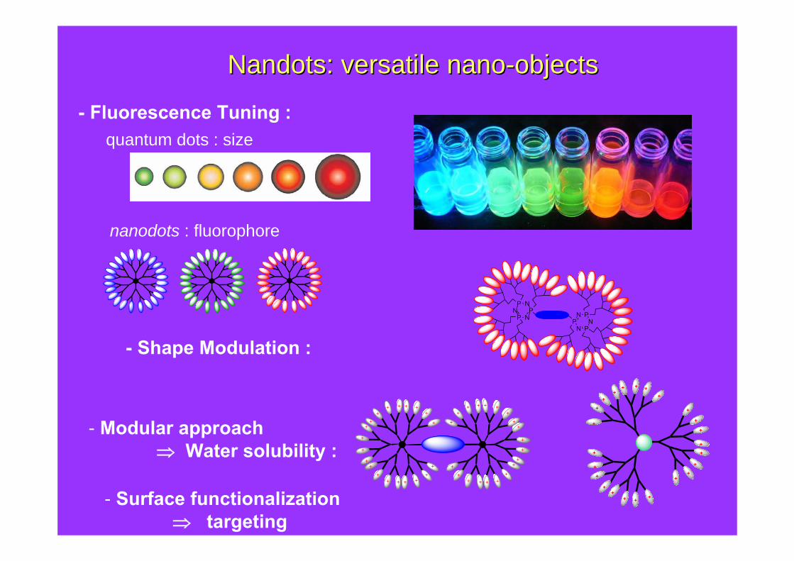
NandotsNandots: versatile : versatile nanonano--objectsobjects
- Fluorescence Tuning :
- Shape Modulation :
- Modular approach ⇒ Water solubility :
quantum dots : size
nanodots : fluorophore
+
+
+++++
++
+
+
+
+
++ + + +
++ +
++ + + +
+
++
+
++
+++++
+
+
+
+
+
NPN
P NP
N PN
PNP
++++
++
+
+
+
+
+
++
++
+
+
+
+
++
+ + +- Surface functionalization⇒ targeting
Molecular and Supramolecular Photonics(Rennes)
Laurent PorrèsMarina CharloCéline Le DroumaguetCédric RouxelAnne-Claire RobinJean-Marie Vabre
Dr Olivier MonginDr Claudine KatanDr. Martinus H. V. Werts
Plate-forme d’imagerie PIXEL (Rennes)
LCC (Toulouse)Jean-Pierre MajoralAnne-Marie CaminadeThatavarathy Rama KrishnaAnna Pla-Quintana
Neurophysiologie et NouvellesMicroscopies (Paris)Laurent MoreauxJerome MertzSerge Charpak
Cornell UniversityD. DombeckW.W. Webb
SCANING (Rennes)François Tiaho, Gaëlle Recher
ACKNOWLEDGMENTSACKNOWLEDGMENTS