Molecular Mechanisms of RNA Interference · 2017-07-16 · RNA Interference Ross C. Wilson2 and...
26
Molecular Mechanisms of RNA Interference Ross C. Wilson 2 and Jennifer A. Doudna 1,2,3,4 1 Howard Hughes Medical Institute, 2 Department of Molecular and Cell Biology, 3 Department of Chemistry, University of California, Berkeley, California 94720; email: [email protected] 4 Physical Biosciences Division, Lawrence Berkeley National Laboratory, Berkeley, California 94720 Annu. Rev. Biophys. 2013. 42:217–39 The Annual Review of Biophysics is online at biophys.annualreviews.org This article’s doi: 10.1146/annurev-biophys-083012-130404 Copyright c 2013 by Annual Reviews. All rights reserved Keywords miRNA, siRNA, RNAi, dsRNA, Argonaute, Dicer, RISC, ribonucleoproteins, crystallography Abstract Small RNA molecules regulate eukaryotic gene expression during devel- opment and in response to stresses including viral infection. Specialized ribonucleases and RNA-binding proteins govern the production and ac- tion of small regulatory RNAs. After initial processing in the nucleus by Drosha, precursor microRNAs (pre-miRNAs) are transported to the cy- toplasm, where Dicer cleavage generates mature microRNAs (miRNAs) and short interfering RNAs (siRNAs). These double-stranded products as- semble with Argonaute proteins such that one strand is preferentially se- lected and used to guide sequence-specific silencing of complementary target mRNAs by endonucleolytic cleavage or translational repression. Molecular structures of Dicer and Argonaute proteins, and of RNA-bound complexes, have offered exciting insights into the mechanisms operating at the heart of RNA-silencing pathways. 217 Annu. Rev. Biophys. 2013.42:217-239. Downloaded from www.annualreviews.org by Portland State University on 07/02/13. For personal use only.
Transcript of Molecular Mechanisms of RNA Interference · 2017-07-16 · RNA Interference Ross C. Wilson2 and...

BB42CH10-Doudna ARI 6 April 2013 15:48
Molecular Mechanisms ofRNA InterferenceRoss C. Wilson2 and Jennifer A. Doudna1,2,3,4
1Howard Hughes Medical Institute, 2Department of Molecular and Cell Biology, 3Departmentof Chemistry, University of California, Berkeley, California 94720; email: [email protected] Biosciences Division, Lawrence Berkeley National Laboratory, Berkeley,California 94720
Annu. Rev. Biophys. 2013. 42:217–39
The Annual Review of Biophysics is online atbiophys.annualreviews.org
This article’s doi:10.1146/annurev-biophys-083012-130404
Copyright c© 2013 by Annual Reviews.All rights reserved
Keywords
miRNA, siRNA, RNAi, dsRNA, Argonaute, Dicer, RISC,ribonucleoproteins, crystallography
Abstract
Small RNA molecules regulate eukaryotic gene expression during devel-opment and in response to stresses including viral infection. Specializedribonucleases and RNA-binding proteins govern the production and ac-tion of small regulatory RNAs. After initial processing in the nucleus byDrosha, precursor microRNAs (pre-miRNAs) are transported to the cy-toplasm, where Dicer cleavage generates mature microRNAs (miRNAs)and short interfering RNAs (siRNAs). These double-stranded products as-semble with Argonaute proteins such that one strand is preferentially se-lected and used to guide sequence-specific silencing of complementary targetmRNAs by endonucleolytic cleavage or translational repression. Molecularstructures of Dicer and Argonaute proteins, and of RNA-bound complexes,have offered exciting insights into the mechanisms operating at the heart ofRNA-silencing pathways.
217
Ann
u. R
ev. B
ioph
ys. 2
013.
42:2
17-2
39. D
ownl
oade
d fr
om w
ww
.ann
ualr
evie
ws.
org
by P
ortla
nd S
tate
Uni
vers
ity o
n 07
/02/
13. F
or p
erso
nal u
se o
nly.

BB42CH10-Doudna ARI 6 April 2013 15:48
nt: nucleotide(s)
Argonaute: a proteincapable of bindingshort ssRNAs and, insome cases, cleaving abound complementarystrand
Contents
A BIOLOGICAL VIEW OF RNA INTERFERENCE . . . . . . . . . . . . . . . . . . . . . . . . . . . . . 218Small Regulatory RNAs in Cellular Function and Dysfunction . . . . . . . . . . . . . . . . . . . . 218Biogenesis and Action of miRNA and siRNA. . . . . . . . . . . . . . . . . . . . . . . . . . . . . . . . . . . . . 219
ACTIVITY, STRUCTURE, AND INTERACTIONS OF miRNAPATHWAY PROTEINS. . . . . . . . . . . . . . . . . . . . . . . . . . . . . . . . . . . . . . . . . . . . . . . . . . . . . . . 221Microprocessor . . . . . . . . . . . . . . . . . . . . . . . . . . . . . . . . . . . . . . . . . . . . . . . . . . . . . . . . . . . . . . . . . 221Dicer . . . . . . . . . . . . . . . . . . . . . . . . . . . . . . . . . . . . . . . . . . . . . . . . . . . . . . . . . . . . . . . . . . . . . . . . . . 223dsRBPs . . . . . . . . . . . . . . . . . . . . . . . . . . . . . . . . . . . . . . . . . . . . . . . . . . . . . . . . . . . . . . . . . . . . . . . . 226Argonaute . . . . . . . . . . . . . . . . . . . . . . . . . . . . . . . . . . . . . . . . . . . . . . . . . . . . . . . . . . . . . . . . . . . . . . 228GW-PABP Interface. . . . . . . . . . . . . . . . . . . . . . . . . . . . . . . . . . . . . . . . . . . . . . . . . . . . . . . . . . . . 232C3PO. . . . . . . . . . . . . . . . . . . . . . . . . . . . . . . . . . . . . . . . . . . . . . . . . . . . . . . . . . . . . . . . . . . . . . . . . . 233
FRONTIERS IN RNAi BIOPHYSICS . . . . . . . . . . . . . . . . . . . . . . . . . . . . . . . . . . . . . . . . . . . . 233Strand Selection . . . . . . . . . . . . . . . . . . . . . . . . . . . . . . . . . . . . . . . . . . . . . . . . . . . . . . . . . . . . . . . . 233P Bodies . . . . . . . . . . . . . . . . . . . . . . . . . . . . . . . . . . . . . . . . . . . . . . . . . . . . . . . . . . . . . . . . . . . . . . . 234Kinetics of Repression, Decay, and RISC Turnover . . . . . . . . . . . . . . . . . . . . . . . . . . . . . . 234
A BIOLOGICAL VIEW OF RNA INTERFERENCE
Small Regulatory RNAs in Cellular Function and Dysfunction
The discovery of RNA interference (RNAi) revolutionized our understanding of gene regulationby revealing an array of related pathways in which small, ∼20- to 30-nucleotide (nt) noncodingRNAs and their associated proteins control the expression of genetic information (7). In processesthat are widespread in plants and animals (80), each small RNA associates with an Argonaute familyprotein to form a sequence-specific, gene-silencing ribonucleoprotein with specificity conferredby base-pairing between the small (guide) RNA and its target mRNA.
RNAi was initially discovered in the form of a single microRNA (miRNA) in the Caenorhabditiselegans genome (48). Eventually such miRNAs were found to be widespread: As much as 5% ofthe human genome is dedicated to encoding and producing the >1,000 miRNAs that regulateat least 30% of our genes (38, 58). RNAi is charged with controlling vital processes such as cellgrowth, tissue differentiation, heterochromatin formation, and cell proliferation. Accordingly,RNAi dysfunction is linked to cardiovascular disease, neurological disorders, and many types ofcancer (53). RNAi pathways transcend mere expansion of the gene regulation toolkit: They confera qualitative change in the way cellular networks are managed. This prompts consideration of thefascinating possibility that we owe our sentience to small RNAs, as the number of miRNAs presentin a genome appears to correlate with the complexity of the organism (3).
In contrast to miRNA, short interfering RNA (siRNA) typically describes exogenous syntheticor viral inducers of RNAi. Such small RNAs have been used in biomedical research to great effect,allowing selective repression of genes of interest. Furthermore, immense effort has been exerted inhopes of developing siRNA-based therapies to combat genetic or viral disease. Although the fieldshows great promise, challenges with delivery as well as harmful off-target effects have preventedany such drug from reaching the market so far (14). In parallel with this effort, advances inmechanistic understanding of these pathways have been impressive, providing increasing insightsinto the molecular structures and activities responsible for small RNA–mediated genetic control.
218 Wilson · Doudna
Ann
u. R
ev. B
ioph
ys. 2
013.
42:2
17-2
39. D
ownl
oade
d fr
om w
ww
.ann
ualr
evie
ws.
org
by P
ortla
nd S
tate
Uni
vers
ity o
n 07
/02/
13. F
or p
erso
nal u
se o
nly.

BB42CH10-Doudna ARI 6 April 2013 15:48
RNA-inducedsilencing complex(RISC): the minimalRNA-inducedsilencing complexcontains an Argonauteprotein bound to aguide strand; theeffector of silencing
Microprocessor:the nuclear proteincomplex of Drosha andDGCR8; responsiblefor cleavage ofpri-miRNAs intopre-miRNAs
dsRBD:double-strandedRNA-binding domain
Dicer: anendoribonucleaseresponsible forcleaving long dsRNAsand/or pre-miRNAs todsRNA duplexes of aspecific length
dsRBP:double-strandedRNA-binding protein
RISC-loadingcomplex (RLC): acomplex composed ofat least Argonaute,Dicer, and a dsRBPsuch as TRBP;responsible forgenerating diceddsRNA and loading itonto Argonaute
TRBP: TARRNA-binding protein
Guide strand: ssRNAof ∼20–30 nt thatbase-pairs withcomplementarytargets, thus providingthe specificity ofsilencing processes
Such insights will be essential to future success in harnessing RNAi and related processes for boththerapeutics and genome engineering.
This review focuses primarily on structural and mechanistic studies of the human miRNApathway. In many cases, macromolecular structures and interactions that are part of miRNApathways are analogous to those responsible for siRNA activities. In contrast, the key molecularplayers involved in the germ line–specific PIWI-interacting RNA (piRNA) pathway have remainedso mysterious that little biophysical study has been possible until very recently. Emerging structuresof the endoribonuclease Zucchini provide a foundation for mechanistic study of piRNA biogenesis(35, 66, 91).
Biogenesis and Action of miRNA and siRNA
The three pathways of RNAi (miRNA, siRNA, piRNA) share a common mode of action: Theminimal effector is a ribonucleoprotein complex comprising an Argonaute family protein boundto a single-stranded ∼20- to 30-nt RNA that grants specificity via base-pairing interactions withthe gene target. In miRNA and siRNA pathways, this is known as the RNA-induced silenc-ing complex (RISC) and it drives silencing of a target mRNA via degradation and/or transcrip-tional repression (Figure 1). The cellular origins of miRNA and siRNA are somewhat disparate:miRNAs are derived from the genome, whereas siRNAs may be endogenous or arise via viralinfection or other exogenous sources (7). Another key difference occurs in the double-strandedRNA (dsRNA) precursors of each: siRNA duplexes feature perfect base-pairing, whereas miRNAhelices contain mismatches and more extended terminal loops. Despite their differing origins,these processing pathways converge once either type of RNA assembles into the RISC.
Typically, the genesis of a miRNA occurs in the nucleus with a transcript known as a primarymiRNA (pri-miRNA); such transcripts are at least 1,000 nt long, containing single or clustereddouble-stranded hairpins that bear single-stranded 5′- and 3′-terminal overhangs and ∼10-ntdistal loops (75). A pri-miRNA is cropped by the microprocessor complex, comprising Drosha,an RNase III family enzyme, and DiGeorge syndrome critical region gene 8 (DGCR8), a proteincontaining two double-stranded RNA-binding domains (dsRBDs) (42). DGCR8 recognizes thepri-miRNA’s junction of stem and single-stranded RNA, which likely helps position Drosha forthe endonucleolytic cleavage it performs on the stem ∼11 base pairs (bp) from the junction (31).The resulting ∼65- to 70-nt precursor miRNA (pre-miRNA) associates with transport facilitatorsExportin-5 and RanGTP and is exported to the cytoplasm (54).
In the cytoplasm, the processing pathways converge for endogenous miRNAs and for typi-cally exogenous siRNAs. Both types of RNAi precursors are trimmed down to a dsRNA duplexof the appropriate size for loading onto an Argonaute protein; this is typically performed by aDicer enzyme. Dicers are large endoribonucleases containing a helicase domain and an internallydimerized pair of RNase III domains, but this composition can vary between organisms (80). Be-fore or after they are excised, introns may serve as viable substrates for microprocessor or Dicer,acting as pri- or pre-miRNAs, respectively (42). In plants, dicing precedes methylation of the3′-terminal nucleotide’s 2′-hydroxyl by the methyltransferase HEN1 (34). The resulting dsRNAis a duplex of 21- to 25-nt strands, bearing a 2-nt overhang at each 3′ terminus and a phosphategroup at each recessed 5′ terminus (78). With this enzymatic step and the subsequent loading ofduplex onto Argonaute, Dicer may be aided by a dsRNA-binding protein (dsRBP) such as TARRNA-binding protein (TRBP). These three proteins (Dicer, Argonaute, and a dsRBP) constitutea minimal RISC-loading complex (RLC), which is responsible for generating diced dsRNA andloading it onto Argonaute (61). Once the dsRNA helix is presented to Argonaute, the 3′ terminusand 5′ phosphate of the guide strand are bound by the protein’s PAZ (named for its presence
www.annualreviews.org • Mechanisms of RNAi 219
Ann
u. R
ev. B
ioph
ys. 2
013.
42:2
17-2
39. D
ownl
oade
d fr
om w
ww
.ann
ualr
evie
ws.
org
by P
ortla
nd S
tate
Uni
vers
ity o
n 07
/02/
13. F
or p
erso
nal u
se o
nly.
BB42CH10-Doudna ARI 6 April 2013 15:48
ssRNA:single-stranded RNA
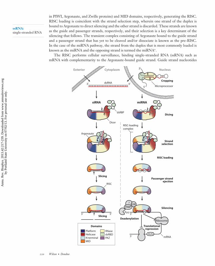
in PIWI, Argonaute, and Zwille proteins) and MID domains, respectively, generating the RISC.RISC loading is coincident with the strand selection step, wherein one strand of the duplex isbound to Argonaute to direct silencing and the other strand is discarded. These strands are knownas the guide and passenger strands, respectively, and their selection is a key determinant of thesilencing that follows. The transient complex consisting of Argonaute bound to the guide strandand a passenger strand that has yet to be cleaved and/or dissociate is known as the pre-RISC.In the case of the miRNA pathway, the strand from the duplex that is most commonly loaded isknown as the miRNA and the opposing strand is termed the miRNA∗.
The RISC performs cellular surveillance, binding single-stranded RNA (ssRNA) such asmRNA with complementarity to the Argonaute-bound guide strand. Guide strand nucleotides
5'
Dicer
Argonaute
dsRBP
mRNA
Dicing
Strandselection
RISC loading
Passenger strandejection
Silencing
Deadenylation
Translationalrepression
RNase
dsRBD
PAZ
Platform
Helicase
N terminal
MID
Domains
GW
Ribosome
PABP
RISC-loading
complex
Cytoplasm NucleusExterior
RISC
Cropping
siRNA miRNA
A(n)
Slicing
Slicing
?
AUG
5'
5'
3'
3'
5' 3'
3' 5'
MicroprocessordsRNA3'
3'
5'
220 Wilson · Doudna
Ann
u. R
ev. B
ioph
ys. 2
013.
42:2
17-2
39. D
ownl
oade
d fr
om w
ww
.ann
ualr
evie
ws.
org
by P
ortla
nd S
tate
Uni
vers
ity o
n 07
/02/
13. F
or p
erso
nal u
se o
nly.

BB42CH10-Doudna ARI 6 April 2013 15:48
Seed sequence:positions 2–8 of theguide stand; a regionwhose targetcomplementarity iscritical in determiningthe efficacy of silencing
aa: amino acid
2–6 constitute the seed sequence and initialize binding to the target. This binding need notinvolve perfect complementarity, and the extent of base-pairing influences how the subsequent si-lencing transpires. In cases of perfect complementarity, target cleavage can occur if the Argonautepresent bears catalytic activity (as is true for just one of the four human AGO proteins, Ago2). TheRISC can also induce nonendonucleolytic translational repression before or after initiation; thismay be followed by deadenylation and degradation. Additional cellular machinery is responsiblefor the processes downstream of target binding, and the Argonaute-binding protein GW182 is akey mediator in recruiting these additional components to the RISC and in localizing silencingactivity to cytoplasmic loci known as processing (P) bodies (20). These complicated gene-silencingmechanisms were initially unclear and appeared daunting, but a recent surge of information hasshed light on major pathways and is reviewed in Reference 22.
ACTIVITY, STRUCTURE, AND INTERACTIONS OF miRNAPATHWAY PROTEINS
Microprocessor
Before their export to the cytoplasm for further processing and silencing, nuclear pri-miRNAsmust be recognized and cleaved. These respective tasks are carried out by the proteins Droshaand DGCR8 (known as Pasha in invertebrates), which together constitute the microprocessorcomplex. As with Dicer, Drosha proteins belong to the RNase III family, whose members containa dimeric active site (60). The minimal endonucleolytic active site is composed of two ∼100-amino-acid (aa) RNase III domains; in bacterial or yeast (class 1) enzymes, these domains resultfrom homodimerization of two identical protomers. In contrast, the pair of RNase III domainscomposing the active sites of Drosha (class 2) and Dicer (class 3) enzymes are provided as anadjacent pair on the same chain, forming a pseudodimer. Regardless of a given protein’s class,the most C-terminal RNase III domain is followed by a ∼70-aa dsRBD, which confers sequence-nonspecific dsRNA binding. All three classes of RNase III enzymes utilize each catalytic domain tocleave a single strand of the dsRNA substrate, working in unison to produce new dsRNA terminibearing 2-nt 3′-terminal overhangs and 5′-terminal phosphate groups (60).
In the context of the microprocessor complex, Drosha performs cleavage consistent with itsidentity as an RNase III enzyme (49). The portion of the protein N-terminal to its paired catalyticdomains is typical of a class 2 RNase III in that it contains a proline-rich region, but the function
←−−−−−−−−−−−−−−−−−−−−−−−−−−−−−−−−−−−−−−−−−−−−−−−−−−−−−−−−−−−−−−−−−−−−−−−−Figure 1The siRNA (left) and miRNA (right) pathways of RNA interference. Protein domain architecture isapproximated in the illustrations here, and domain coloring is maintained in subsequent figures. For clarity,one dsRBD has been omitted from each of the Dicer and microprocessor illustrations. The siRNA pathway(left) begins with Dicer’s cleavage of dsRNA of exogenous or nuclear origin. The resulting siRNA duplex isloaded onto Argonaute by the RISC-loading complex, which comprises Dicer, a dsRBP protein such asTRBP, and an Argonaute protein. The passenger strand ( gray) is cleaved and ejected. The guide strand (red )remains bound to Argonaute, forming the RISC. The RISC binds to complementary target sequences (black)and silences them via the slicing activity of Argonaute. In the miRNA pathway (right), a primary miRNAtranscript is cropped by the microprocessor complex, which consists of the endonuclease Drosha bound toDGCR8. The resulting pre-miRNA is exported to the cytoplasm, where dicing and RISC loading take placeas in the siRNA pathway. Passenger strand ( gray) ejection may take place without concomitant cleavage, andthe guide strand (red ) drives silencing as part of the mature RISC. In cases of a catalytically inactiveArgonaute or partial complementarity to the target (black), silencing takes place via pre- or post-initiationtranslational repression and deadenylation followed by degradation of the mRNA.
www.annualreviews.org • Mechanisms of RNAi 221
Ann
u. R
ev. B
ioph
ys. 2
013.
42:2
17-2
39. D
ownl
oade
d fr
om w
ww
.ann
ualr
evie
ws.
org
by P
ortla
nd S
tate
Uni
vers
ity o
n 07
/02/
13. F
or p
erso
nal u
se o
nly.

BB42CH10-Doudna ARI 6 April 2013 15:48
dsRBD1 dsRBD2
Hsa DGCR8
WW
Hsa DroshaP R+S RNase IIIa RNase IIIb dsRBD
b
a1 1374 aa
Figure 2The microprocessor complex. (a) Domain structures of the human microprocessor constituents Drosha andDGCR8. Regions rich in proline (P), arginine and serine (R+S), or tryptophan (W) are labeled. (b) Thecrystal structure of the DGCR8 core (PDB ID: 2YT4). Models of dsRNA are oriented on the basis of typicaldsRBD binding and show that the DGCR8 core cannot use both dsRBDs to bind a single pre-miRNA unlessextensive deformation of the helix occurs.
of this portion remains unknown (Figure 2a). Drosha’s C-terminal dsRBD is canonical in itssequence and tertiary structure (64) and is required for pri-miRNA processing in vivo (2) butdoes not appear to play a substantial role in substrate binding or recognition in vitro (101), tasksinstead performed by DGCR8 (31). Accordingly, Drosha cleaves pri-miRNAs indiscriminately inthe absence of DGCR8 (27). DGCR8’s dsRNA binding is carried out by a pair of dsRBDs in itsC-terminal half; its N terminus contains a structurally characterized, heme-binding dimerizationdomain (79) and is otherwise of unknown structure and function, though its tryptophan-richregion has been hypothesized to interact with the proline-rich region of Drosha’s N terminus (27)(Figure 2a).
The structure of the DGCR8 core. The crystal structure of DGCR8’s dsRBD pair revealedtwo domains, each adopting the traditional fold: an α/β sandwich with the terminal α-helicesresting atop a three-stranded β-sheet (Figure 2b). These two domains are surrounded by severaladditional secondary structural elements that contribute to a well-ordered quaternary structure(the DGCR8 core), orienting the two dsRBDs with respect to each other without disruptingeither individual structure from the canonical geometry. The DGCR8 domains were crystallizedin the absence of RNA, yet concomitant binding studies via electrophoretic mobility shift assay(EMSA) and fluorescence resonance energy transfer (FRET) revealed that the dsRBDs interactwith dsRNA but not with ssRNA. Although the dsRNA affinity is anticipated for a pair of orthodoxdsRBDs, the lack of affinity for ssRNA is somewhat surprising because the DGCR8-binding siteis a junction between a helical stem and two flanking strands of ssRNA (stem-ssRNA junction)(Figure 1). This binding site is inferred on the basis of cleavage assays that reveal DGCR8 tobe the factor responsible for correctly positioning and orienting the microprocessor for Drosha-catalyzed cleavage ∼11 bp from the stem-ssRNA junction (31), but it remains unclear which regionof DGCR8 or Drosha is responsible for recognition of the flanking ssRNA.
222 Wilson · Doudna
Ann
u. R
ev. B
ioph
ys. 2
013.
42:2
17-2
39. D
ownl
oade
d fr
om w
ww
.ann
ualr
evie
ws.
org
by P
ortla
nd S
tate
Uni
vers
ity o
n 07
/02/
13. F
or p
erso
nal u
se o
nly.

BB42CH10-Doudna ARI 6 April 2013 15:48
Without detectable affinity for ssRNA, it is unknown how DGCR8 is so reliably positioned atthe stem-ssRNA junction instead of binding promiscuously along the pri-miRNA stem. Curiously,the only reported evidence for ssRNA-specific binding by any microprocessor component impli-cates the serine/arginine-rich region of Drosha’s N terminus (102), though this probably explainsthe microprocessor’s preference for a distal ∼10-nt loop on the pri-miRNA substrate rather thanaddresses DGCR8’s positioning at the stem-ssRNA junction.
Higher-order assemblies between the microprocessor and pri-miRNA. Efforts are underway to model the active state of the microprocessor complex interacting with its pri-miRNAsubstrates. The two dsRBDs of the DGCR8 core offer their RNA-binding surfaces such thatthey cannot simultaneously bind a pri-miRNA without a major distortion from A-form geometry(Figure 2b); FRET-based evidence for such bending has been reported, but the possibilityremains that the DGCR8 core’s two domains bind to separate pri-miRNAs (81). Evidencefor such multimeric assembly is provided by an electron tomography study of the associationbetween DGCR8 and a pri-miRNA, which revealed a ∼370-kDa particle consistent with a singlepri-miRNA bound to six DGCR8 protomers or four pri-miRNA:DGCR8 heterodimers, amongother configurations (23). Such larger assemblies might be expected because many miRNAsoriginate from pri-miRNAs clustered on a single transcript.
Recent cellular imaging studies have precisely tracked the nuclear localization of Drosha andDGCR8 over time, revealing that they colocalize simultaneously on unspliced, intronic pri-miRNA and that Drosha tends to dissociate after cleavage, whereas DGCR8 is likely to remainbound to the processed pre-miRNAs before their nuclear export (2). The former finding buildson an earlier report that the microprocessor associates with the spliceosome, providing mountingevidence that cropping takes place before splicing is complete (40).
Dicer
The Dicer family of class 3 RNase III enzymes is vital to siRNA and miRNA pathways. Theseproteins generate dsRNAs suitable for loading onto an Argonaute protein, a process in whichDicer may participate as part of the RLC. Dicer’s active center is derived from bacterial class 1RNase III enzymes, which comprise an RNase III domain with a C-terminal dsRBD (80). Bacterialenzymes dimerize to achieve concerted cleavage of both strands of a dsRNA substrate. Class 3Dicer enzymes accomplish similar dsRNA cleavage using a pseudodimer of RNase III domains ona single polypeptide with a single C-terminal dsRBD (60). N-terminal to these paired active sitesis a PAZ domain, which recognizes the dsRNA end characteristic of RNAi intermediates (57)(Figure 3a). The PAZ and RNase III domains act together as a molecular ruler to mete out dicedstrands of RNA appropriate for a given organism’s silencing machinery, as discussed in greaterdepth below.
A distinguishing characteristic of Dicer is the superfamily 2 helicase domain often found at itsN terminus. This ≥600-aa domain belongs to the eukaryotic RIG-I family of helicases, which ischaracterized by a bilobal architecture comprising DExD/H and helicase C regions, the formerof which is expected to bind and potentially hydrolyze ATP (104). The function of this helicasedomain is still being defined in the case of humans, whereas the helicase domain can recognizeeither miRNA or siRNA precursors (see below) in the Dicer enzymes of Drosophila. In some casesDicer’s helicase domain also contains a binding site for a dsRBP such as TRBP in humans.
Dicer’s domain structure varies between organisms, yet its principal dicing function is pre-served. Giardia intestinalis Dicer lacks a helicase domain (59), and the budding yeast Kluyveromycespolysporus equivalent is further pared down, lacking a PAZ domain and bearing an RNase III
www.annualreviews.org • Mechanisms of RNAi 223
Ann
u. R
ev. B
ioph
ys. 2
013.
42:2
17-2
39. D
ownl
oade
d fr
om w
ww
.ann
ualr
evie
ws.
org
by P
ortla
nd S
tate
Uni
vers
ity o
n 07
/02/
13. F
or p
erso
nal u
se o
nly.

BB42CH10-Doudna ARI 6 April 2013 15:48
RNase IIIa/b
(Ruler) dsRBD HelicasePlatform
23 nt
PAZ
Platform
RNase IIIa
RNase IIIb
NTD
Kpo dicer
Gin dicer
Hsa dicer
1 1924 aa
RNase IIIaPlatform RulerDUF PAZHelicase dsRBDRNase IIIb
d
cb
a
PAZ
Figure 3The diverse architecture of the Dicer family. (a) Domain structures of Dicer enzymes from humans (Hsa),Giardia intestinalis (Gin), and Kluyveromyces polysporus (Kpo). The domain of unknown function (DUF) and theanticipated ruler domain are indicated for the human enzyme. K. polysporus Dicer bears a single RNase IIIdomain and an N-terminal domain (NTD) that mediates dimerization. (b) Dicer’s measurement and cleavageof pre-miRNA as illustrated by the G. intestinalis crystal structure (PDB ID: 2FFL). Purple spheres representerbium atoms present in the crystal, which reflect the position of the Mg2+ ions critical to RNase III enzymecatalysis. In this model, the 2-nt 3′-terminal overhang of a dsRNA substrate is docked onto Dicer’s PAZ domain,orienting it for two cleavage events in the active site 65 A away, generating a 25-nt duplex. (c) The inside-outmechanism of K. polysporus Dicer (PDB ID: 3RV0). This enzyme lacks a PAZ domain and uses neighboringmolecules to measure its product. This model depicts two homodimers of the enzyme, each bearing a singleRNase III domain, contacting each other to measure a 23-nt product. The resulting dsRNA originates fromthe center of the substrate duplex, in contrast to the end-derived products generated by PAZ-containingDicers. (d ) The global architecture of human Dicer. Homologous domains of known structure have beendocked onto a segmented electron microscopic map (PDB IDs: 4A36, 2KOU, 2FFL, and 3C4T). The helicasedomain resembles a clamp and is optimally oriented to guide an incoming dsRNA substrate (black and white coil )toward the RNase IIIa/b active center and PAZ domain. Relative to the G. intestinalis enzyme, a ruler domainis inserted and the PAZ domain is reoriented with respect to the RNase III catalytic center. These changeslikely relate to the fact that human Dicer products are four nucleotides shorter than those of G. intestinalis.
domain (96) (Figure 3a). These helicase-lacking enzymes may have lost dsRBP binding capabilityas well, considering the aforementioned interface. In a case of specialization, Drosophila employsa pair of Dicer enzymes and corresponding dsRBPs to create parallel processing pathways forsiRNAs and miRNAs (9). Plants such as Arabidopsis thaliana take such specialization further withfour Dicer-like proteins (DCL1–4): DCL1 for generation of 21-nt miRNAs and DCL2–4 forprocessing of siRNAs of 22, 24, or 21 nt, respectively (74).
Dicing activity. Dicer’s principal function is to recognize dsRNA precursors from the RNAipathway and sever both strands to generate dsRNAs of a specific length, typically 21–25 nt (4).Recognition and cleavage of a generic segment of dsRNA helix can be expected of any enzyme
224 Wilson · Doudna
Ann
u. R
ev. B
ioph
ys. 2
013.
42:2
17-2
39. D
ownl
oade
d fr
om w
ww
.ann
ualr
evie
ws.
org
by P
ortla
nd S
tate
Uni
vers
ity o
n 07
/02/
13. F
or p
erso
nal u
se o
nly.

BB42CH10-Doudna ARI 6 April 2013 15:48
system bearing a pair of RNase III domains. These dimerized domains bear two active sites anda flat, positively charged surface that can accommodate a long RNA helix. Each active site bearsfour acidic residues that coordinate two Mg2+ ions used in phosphodiester hydrolysis of each RNAstrand (26, 84) (Figure 3b). The dsRNA terminus characteristic of RNAi pathway precursors (a2-nt overhang on the 3′ terminus and a phosphate-bearing 5′ terminus) can be recognized by theDicer’s PAZ domain, as was revealed by a crystal structure of human Argonaute’s similar PAZdomain bound to one such RNA terminus (see below) (57).
The RNA-measuring component of Dicer’s function was revealed upon structure determina-tion of the G. intestinalis version of the protein, wherein the PAZ domain is located 65 A from thecatalytic center (59) (Figure 3b). This distance corresponds to the length spanned by ∼25 bp ofdsRNA, which is consistent with the 25- to 27-nt products generated by G. intestinalis Dicer. Thedistance between the end-binding PAZ domain and the RNase III cleavage sites is determinedby structural orientation of the domains; these domains likely undergo rearrangement in otherDicer incarnations to produce the observed products of differing lengths. G. intestinalis Dicer lacksthe typical N-terminal helicase and C-terminal dsRBD domains, suggesting that these elementsfulfill roles supplementary to dicing (59). This finding is consistent with the observation that atruncated form of human Dicer lacking helicase and dsRBD domains retained dicing activity (55).More recent studies show that the dsRBD can rescue activity in a form of human Dicer lackingits PAZ domain, though the resulting products vary in length as might be expected (56).
Also lacking a helicase domain is Dicer of the budding yeast K. polysporus, Dcr1, which representsan extremely pared down version of the enzyme that nevertheless retains its ability to specifyproduct size in the absence of a PAZ domain (Figure 3a). Its single RNase III domain dimerizesto cleave dsRNA; despite this similarity to class 1 bacterial enzymes, K. polysporus Dcr1 remainsclassified with other class 3 enzymes owing to similarity in sequence and active site composition.A crystal structure and concomitant mechanistic studies revealed that Dcr1 dimers cleave dsRNAat precise intervals by abutting each other along the helix and measuring the product based on thedistance (23 nt) occupied by the protein structure between the neighboring pairs of active sites (96)(Figure 3c). This mechanism contrasts starkly to that of canonical Dicers, which measure froma dsRNA’s terminus; K. polysporus Dcr1 begins at an arbitrary point within a dsRNA and worksoutward in 23-nt steps. Its mode of action is predicated on slow substrate release, which was indeedreported and is likely facilitated by one or both of the enzyme’s two C-terminal dsRBDs (96).
The role of Dicer’s helicase domain in RNA recognition. The function of Dicer’s helicasedomain is best understood in the case of Drosophila Dicer-2. Dicer-2 is specialized to processsiRNA precursors, which are longer, perfectly paired dsRNAs; Dicer-1 handles the shorter andimperfect pre-miRNA hairpins (9). The canonical helicase domain of Dicer-2 consumes ATP totranslocate the enzyme with respect to a long dsRNA, enabling processive generation of manysiRNA duplexes from a single substrate helix (9). Furthermore, this domain facilitates processingof dsRNA substrates that lack the characteristic 2-nt 3′-terminal overhang (97). The divergentDicer-1 helicase domain contains a helicase C moiety but lacks the DExH/D lobe, providing anexplanation for its inability to translocate along dsRNA. This modification does not completelyabolish RNA binding: The domain has been implicated in recognition of the ssRNA loops char-acteristic of pre-miRNA hairpins, thus contributing to the enzyme’s specificity (90).
The parallel processing pathways for siRNA and miRNA are mirrored by a duplication ofRNAi machinery in Drosophila, leading to differing, specialized helicase domains. But in humans,a single Dicer is responsible for handling both types of precursors. How does the single humanDicer recognize and process both types of substrates? Has the human helicase domain evolvedgreater adaptability in substrate specificity or has it experienced a partial loss of function? These
www.annualreviews.org • Mechanisms of RNAi 225
Ann
u. R
ev. B
ioph
ys. 2
013.
42:2
17-2
39. D
ownl
oade
d fr
om w
ww
.ann
ualr
evie
ws.
org
by P
ortla
nd S
tate
Uni
vers
ity o
n 07
/02/
13. F
or p
erso
nal u
se o
nly.

BB42CH10-Doudna ARI 6 April 2013 15:48
questions remain to be fully resolved, but it seems that a loss of function has taken place since thehuman domain appears better adapted for the miRNA pathway. Unlike for Drosophila Dicer-2,RNA processing for human Dicer is ATP independent; thus, the helicase domain is unlikely tobe involved in translocation of long dsRNA (103). Two biochemical reports implicate it in aidingmiRNA processing, probably via binding of the pre-miRNA loop (56, 82).
A recent electron microscopic (EM) study elegantly employed extensive tagging and dele-tion of domains to deduce for the first time the global domain orientation of human Dicer (46)(Figure 3d ). One surprising observation was the reorientation of the RNase III nuclease core withrespect to the PAZ domain when comparing the structure of human Dicer to that of G. intestinalisDicer (59). This global reorganization likely stems from the 4-nt (one-third of a dsRNA helicalturn) length difference between the products of the two enzymes and the differing associatedgeometric requirements for cleavage (46). Similar rearrangements might be expected in Dicersproducing products with other lengths. Data interpretation was bolstered by structure determi-nation of the helicase RIG-I, which provided a model that was readily positioned into the EMdensity for the related helicase domain of human Dicer (43). The EM reconstruction of the elon-gated human Dicer revealed a helicase that is distal from the PAZ domain, with the paired RNaseIII active sites resting in between. This architecture is consistent with both known functions ofthe helicase domain. For pre-miRNAs, the PAZ domain recognizes the 2-nt overhang and thehelicase recognizes the hairpin’s loop on the other end. For long dsRNA substrates, the helicasedomain can feed the helix toward the catalytic center to generate multiple siRNAs. Accordingly, aconcomitant EM reconstruction of the siRNA-specific Drosophila Dicer-2 revealed an architectureremarkably similar to that of human Dicer (46).
Dicer’s interactions with other proteins. As part of the RLC, Dicer associates with an Arg-onaute protein and a dsRBP such as TRBP in humans. The mechanistic implications of these inter-actions for the processes of strand selection and RISC loading are discussed below. The dsRBPs ofRNAi tend to comprise three dsRBDs connected by long, flexible linkers. The third such domainof TRBP interacts with a region of Dicer’s helicase located between the DExD/H and helicase Clobes (13). The interaction with Argonaute takes place between a portion of that protein’s PIWIdomain and a region within the RNase III domain closer to the N terminus of Dicer (76, 85).The conserved Argonaute-interacting site on Dicer is present only in vertebrates, so Argonautebinding must be absent or manifested differently in other nonvertebrate RNAi systems (76).
A solution structure of the DUF283 domain of A. thaliana DCL4 revealed a noncanonicaldsRBD fold that interacts with the second dsRBD of DRB4, a dsRBP homolog of TRBP (74).DRB4 contains two dsRBDs but lacks a predicted third C-terminal dsRBD typically found indsRBPs of RNAi pathways. This third dsRBD is commonly implicated in interactions with Dicer’shelicase, so A. thaliana appears to have evolved a different strategy for recognition between thetwo proteins. Indeed, the paralogous Arabidopsis protein pair DCL1 and DRB1 (also known asHYL1) similarly interact via their respective DUF283 and second dsRBP domains (74).
dsRBPs
Dicing and RISC loading are aided to varying degrees by dsRBPs, which associate with a Dicerprotein and typically comprise two or three dsRBDs. The dsRBD is widespread and typicallyrecognizes dsRNA on the basis of its A-form helical shape with moderate to high affinity and ina sequence-nonspecific manner (17). These proteins typically contain a C-terminal noncanonicaldsRBD dedicated to protein interaction instead of dsRNA binding. TRBP is the best character-ized dsRBP of the human pathway, and its 366-aa polypeptide bears three ∼70-aa dsRNA-binding
226 Wilson · Doudna
Ann
u. R
ev. B
ioph
ys. 2
013.
42:2
17-2
39. D
ownl
oade
d fr
om w
ww
.ann
ualr
evie
ws.
org
by P
ortla
nd S
tate
Uni
vers
ity o
n 07
/02/
13. F
or p
erso
nal u
se o
nly.

BB42CH10-Doudna ARI 6 April 2013 15:48
dsRBD2
Hsa TRBP
dsRBD3dsRBD2dsRBD11 345 aa
b
a
166
165
214
215
188
Figure 4The structure of dsRNA-binding proteins. (a) The domain structure of human TRBP, a typical dsRBP ofthe RNAi pathway. The well-folded dsRBDs are separated by flexible ∼70-aa linkers, lending the protein abeads-on-a-string quality. The first two domains bind dsRNA and the third instead binds to Dicer. (b) Thecrystal structure of the second dsRBD of TRBP in complex with dsRNA (PDB ID: 3ADL). The protein usesthree interfaces to recognize successive portions of minor, major, and minor groove along one face of a helix.
domains separated by two ∼70-aa stretches of disordered linker (Figure 4a). Resembling beadson a string, this domain architecture suggests that the protein may be involved in a dsRNA handoffbetween Dicer and Argonaute (92), but detailed mechanistic evidence is scarce. RNAi pathwaydsRBPs have been implicated in strand selection (8), stabilization of the miRNA-generating com-plex (72), tuning of mature miRNA length (25, 47), and segregation of siRNAs into distinctpathways (32, 68).
Functional coupling between Dicers and dsRBPs. The multiplicity and diversity of dsRBPsimpede facile prediction of their roles in RNAi pathways. A. thaliana represents an extreme case,bearing five dsRBPs serially named DRB1–5. Although these might be expected to generally pairoff with the four Dicer-like proteins DCL1–4, such pairings are observed only for DRB1/DCL1,which produces miRNAs, and for DRB4/DCL4, which processes siRNAs (12). DRB2, DRB3, andDRB5 seem somewhat redundant, whereas DCL2 and DCL3 appear to function unhindered in theabsence of a dsRBP partner (12). Drosophila harbors a slightly more streamlined system in whichDicer-1 pairs with the PB isoform of dsRBP Loquacious (Loqs-PB) to process pre-miRNAs andDicer-2 binds either R2D2 or Loqs-PD to process siRNAs of exogenous or endogenous origin,respectively (32).
The human system includes a solitary Dicer and two dsRBPs, TRBP and PACT, that playpoorly delineated roles in small RNA processing (50). Although it is tempting to speculate thatone human dsRBP is responsible for siRNA while the other tends to miRNA, such evidence has yetto be found. Human Dicer’s interaction with both TRBP and PACT is complicated because eachof the two dsRBPs has been reported to homodimerize and to form heterodimers with the other(45). These dimerization interactions have been localized to the atypical yet conserved C-terminaldsRBD of each protein, which also recognizes Dicer (13). Until it is determined whether any ofthese binding interfaces are mutually exclusive, there are at least two plausible scenarios: Dicer can
www.annualreviews.org • Mechanisms of RNAi 227
Ann
u. R
ev. B
ioph
ys. 2
013.
42:2
17-2
39. D
ownl
oade
d fr
om w
ww
.ann
ualr
evie
ws.
org
by P
ortla
nd S
tate
Uni
vers
ity o
n 07
/02/
13. F
or p
erso
nal u
se o
nly.
BB42CH10-Doudna ARI 6 April 2013 15:48
bind to TRBP or PACT interchangeably, or Dicer can bind to TRBP and PACT simultaneouslyat distinct sites.
Structure and RNA recognition of dsRBPs. Structural study of dsRBPs has been fruitful,although not particularly illuminating regarding mechanism. Structures have been reported forthe first two dsRBDs from TRBP, dsRBD1 and dsRBD2, with the latter bound to dsRNA (98). TheRNA-bound structure adopts an α/β sandwich that embodies the binding mode characteristic ofa canonical dsRBP: The A-form helical geometry of dsRNA is recognized using protein loops andhelices to bind the phosphate backbone at three points: two regions of minor groove surroundinga major groove along one face of the helix (17) (Figure 4b). RNA recognition is shape based andnot sequence specific, as might be expected in consideration of the requirements of the system.The implications of this mode of recognition for strand selection are discussed below. Proteinsequence differences subtly tune the dissociation constants of isolated dsRBD1 and dsRBD2 to220 and 113 nM, respectively, and their collaborative affinity is 0.24 nM in the context of thefull-length protein (98). Similar variability in affinity is likely to be found on other RNAi pathwaydsRBDs with little or no deviation from the canonical fold. No structural information is availablefor the noncanonical RNAi dsRBDs, which employ unknown means to recognize Dicer and/orother noncanonical dsRBDs.
Argonaute
The functional lynchpin of all RNAi pathways is an Argonaute family protein bound to a strandof silencing RNA, which forms the minimal effector complex known as the RISC. The RISCperforms cellular surveillance, silencing ssRNA sequences complementary to its bound guidestrand. Argonaute proteins are found in bacteria, archaea, and eukaryotes. Whereas the formertwo groups contain Argonautes of poorly defined function (80), their eukaryotic counterparts haveevolved into two clades with distinct functions. Proteins of the AGO clade mediate cytosolic genesilencing while bound to siRNAs or miRNAs, and PIWI clade proteins interact with piRNAs tomanage mobile genetic elements of the germ line (29). Humans have four proteins of the formerclade, Ago1–4. Of these four, only Ago2 exhibits slicer activity: the endonucleolytic cleavage ofbound target ssRNA (37). Such activity is not required for gene silencing; human AGO proteinscan accomplish regulation by binding a target, repressing its translation, and recruiting cellularmachinery to induce deadenylation and mRNA decay (36).
The key functions of Argonaute are recognition of guide strand termini, target cleavage, andrecruitment of other proteins involved in silencing. Eukaryotic Argonaute proteins adopt a bilobalarchitecture, with each lobe containing either the N-terminal (or simply N) and PAZ domains orthe MID and PIWI domains (Figure 5a,b). A series of illuminating crystal structures has providedthe initial details of how individual domains function in isolation, ultimately clarifying how theywork together in the context of a competent RISC complete with guide strand and target ssRNA.Via their similarity, structures of ternary complexes between RISC and target have shed light onthe approximate structure of the pre-RISC awaiting passenger strand dissociation.
Recognition of guide strand termini by Argonaute. The first Argonaute fragment structuresrevealed that the PAZ domain is responsible for recognizing 2-nt 3′-terminal overhangs, a findingwith functional implications for PAZ-containing Dicer enzymes as well. The domain providesa pocket to accommodate 2 nt of a 3′ terminus while making less extensive contacts with the 5′
terminus present in many dsRNAs of RNAi pathways (52, 57, 99) (Figure 5c). Side chains ofthe PAZ domain partake in extensive polar interactions with the bound RNA’s buried phosphate
228 Wilson · Doudna
Ann
u. R
ev. B
ioph
ys. 2
013.
42:2
17-2
39. D
ownl
oade
d fr
om w
ww
.ann
ualr
evie
ws.
org
by P
ortla
nd S
tate
Uni
vers
ity o
n 07
/02/
13. F
or p
erso
nal u
se o
nly.

BB42CH10-Doudna ARI 6 April 2013 15:48
group and sugar hydroxyls, though there are no specific contacts to the bound 2′-OH groups,consistent with RNAi being tolerant of such modifications (11, 57). The terminal nucleobase ispartially buried in a hydrophobic cavity and the edges of the terminal two bases remain exposedto solvent, indicative of sequence-nonspecific recognition (57).
In contrast to the indiscriminate 3′-terminal binding, 5′ termini tend to bear a particularnucleotide depending on the identity of the associated Argonaute protein (41). Crystal struc-tures of eukaryotic Argonaute MID domains, some in complex with the four different nucleosidemonophosphates mimicking the 5′ end of miRNAs, revealed specific contacts between a rigid loopin the MID domain and the nucleobase of UMP or AMP (5, 24) (Figure 5d ). The electrostaticenvironment generated by the functional groups of this loop is incompatible with CMP or GMPbinding, a result consistent with NMR titration experiments showing that binding affinities ofUMP (0.12 mM) and AMP (0.26 mM) are 30-fold lower than those measured for either CMP(3.6 mM) or GMP (3.3 mM) (24). The MID domain also bears two invariant lysines that recognizethe 5′-terminal phosphate present in silencing RNAs (6). A crystal structure of an Archaeoglobusfulgidus PIWI enzyme provided the first information on the conformation of a guide strand boundin a MID domain (71). The RNA is bound such that the 5′-terminal base is kinked and buriedand unavailable for base-pairing, as observed in the guide-bound structure of human Ago2 (51)(Figure 5b). This finding provides an explanation for previous biochemical observations that5′-terminal base identity is unimportant in target recognition (51).
Target recognition, slicer activity, and conformational changes. A series of structures char-acterizing full-length archaeal Argonautes has provided crucial details on the protein’s catalyticmechanism, its interaction with a guide strand to form a competent RISC, and the orientation ofternary complexes formed in the presence of the passenger strand or a target ssRNA. A crystalstructure of Argonaute from Pyrococcus furiosus revealed that the PIWI domain adopts an RNaseH–like fold, implicating it as the catalytic domain and site of slicer activity (83). Similar to therequirements for RNaseH activity, RISC-catalyzed RNA cleavage requires divalent metal ionsand yields a 5′ product, which has a free 3′ hydroxyl group, and a 3′ product, which carries a 5′
phosphate group. Crystal structures of Thermus thermophilus Argonaute bound to a DNA approx-imating the guide strand and an RNA target show a PIWI domain bearing a catalytic triad ofaspartic acid residues that coordinates a pair of magnesium ions at an appropriate distance fromthe target strand’s cleavage site (94) (Figure 5f ). This catalytic triad DDX (where X is Asp or His)motif was initially identified in catalytically active Argonautes, is absent from those lacking sliceractivity, and appears to contrast with the catalytic tetrad DEDD motif characteristic of RNase Henzymes (65). Curiously, the second magnesium ion could be observed only when crystallizationconditions were adjusted from 50 to 80 mM Mg2+.
Despite the B-form propensity of the DNA guide present in the T. thermophilus structures,the guide-target duplex adopts the A-form geometry typical of a dsRNA double helix; a dsRNAduplex bound by A. fulgidus PIWI also adopts the A-form architecture (70). When T. thermophilusArgonaute is bound to a 12-nt target, the 3′ terminus of the guide remains bound by the PAZdomain; in contrast, binding to a 15-nt or longer target induces release of the guide’s end from thePAZ domain in order to accommodate the two strands’ extension beyond a full turn (11 nt) of theA-form helix (94) (Figure 5e). Concomitant slicing assays suggest that the 3′ terminus of the guidestrand must be released from the PAZ domain for cleavage to take place, which is consistent withthe structural observations: Duplexes longer than a single turn can be oriented correctly for targetstrand cleavage only if the 3′ terminus of the guide strand is released. This finding supports thepreviously proposed two-state model and refutes the fixed-end model that anticipated both terminiof the guide strand remaining anchored during all stages of target recognition (94). Functional
www.annualreviews.org • Mechanisms of RNAi 229
Ann
u. R
ev. B
ioph
ys. 2
013.
42:2
17-2
39. D
ownl
oade
d fr
om w
ww
.ann
ualr
evie
ws.
org
by P
ortla
nd S
tate
Uni
vers
ity o
n 07
/02/
13. F
or p
erso
nal u
se o
nly.

BB42CH10-Doudna ARI 6 April 2013 15:48
dispensability of 3′ terminus binding is further shown by mutations of the PAZ domain that havelittle effect on slicing assays, which contrasts with the loss of cleavage observed upon mutation ofMID domain residues that recognize the 5′ terminus (95). Cleavage assays also demonstrate thatbulges inserted into the guide’s seed region prevent cleavage, whereas slicing can proceed in thepresence of similar bulges in the same region of the target strand (93).
The conformation of the guide strand in the absence of target is of mechanistic significance.In crystal structures of guide-loaded Argonaute, the nucleic acid is bound in a central basic tractthat spans the two lobes of the protein. The seed portion (positions 2–6) is typically well-ordered,while electron density has not been observed for nucleotides past position 10, with the exceptionof a PAZ-bound terminal nucleotide (19, 65, 77, 95). Recent eukaryotic Argonaute structuresbearing a traditional guide strand of RNA reveal a preordered single helix in A-form geometry(19, 77) (Figure 5b). The thermodynamic repercussions of an ordered guide strand were exploredin a series of isothermal titration calorimetry experiments in the context of A. fulgidus PIWI (69).
977977
10601060
10461046
974974
11981198
7
309309
292292
310310
3'
TrpTrp
PIWIPIWI
977
9851013
1060
1045
1046
974
1198
512
512Inactive
Active
MID MID
PIWI PIWIPAZ
PAZN N
Guide
Target
546
478
10
11
129
660
529
533
570
545
MID
314
309
292
313310
5'
3'
PAZ
MID
PAZ
N terminal
7
65 3'
5'
43
2
Helix 71
PIWI
1 859 aaPAZ
Hsa Ago2
MID PIWIN
b c
e
d
f hg i
a
Trp
230 Wilson · Doudna
Ann
u. R
ev. B
ioph
ys. 2
013.
42:2
17-2
39. D
ownl
oade
d fr
om w
ww
.ann
ualr
evie
ws.
org
by P
ortla
nd S
tate
Uni
vers
ity o
n 07
/02/
13. F
or p
erso
nal u
se o
nly.

BB42CH10-Doudna ARI 6 April 2013 15:48
The study showed that a guide strand held in a helical conformation can increase the affinityfor target up to ∼300-fold by decreasing the entropic cost that would otherwise be associatedwith ordering of the guide. This essentially increases the effective melting temperature and thepropensity for binding between guide and target, which is of particular importance in considerationof the prevalence of mismatches in the miRNA pathway.
Along with the reorientation of nucleic acid components upon RISC’s binding of a target,the Argonaute protein has been observed or postulated to undergo its own rearrangements. Acomparison of T. thermophilus Argonaute bound to a 10-nt versus a 21-nt guide shows moder-ate yet global reorientation of the protein (95). The 10-nt guide-bound conformation is likelysimilar to the unavailable free structure, and the observed motion is presumed to be necessaryto accommodate a full-length guide strand. The two lobes shift conformation again when a tar-get is bound to the T. thermophilus protein, widening to allow formation of a double helix inthe tract that previously housed only the guide strand (93). Still another protein conformation issampled when the guide-target duplex becomes long enough to induce 3′ terminus release fromthe PAZ domain as discussed above (Figure 5e); this involves a pivoting of the PAZ domainand a substantial shift of the PIWI domain loops dubbed L1 and L2 (65, 94) (Figure 5g). Thesignificance of the L2 rearrangement became apparent upon determination of the structure ofK. polysporus Argonaute, which bore a postrearrangement conformation with a glutamate residueof L2 positioned appropriately to act as a fourth catalytic residue in conjunction with the afore-mentioned three aspartic acids (65). This observation can be extended to reveal mechanistic detailsof the T. thermophilus enzyme: Upon sufficient base-pairing between guide and target to releasethe 3′ terminus from the PAZ domain, L2 shifts and deposits the final moiety of a catalytic tetrad,apparently acting as a trigger for catalysis (Figure 5h). Thus, catalytically active Argonaute pro-teins indeed bear a DEDD (or DEDH in the case of human Ago2) catalytic tetrad as initiallyexpected on the basis of the homology between the PIWI domain and RNase H enzymes, and
←−−−−−−−−−−−−−−−−−−−−−−−−−−−−−−−−−−−−−−−−−−−−−−−−−−−−−−−−−−−−−−−−−−−−−−−−−−−−−−−−−−−−−−−−−−Figure 5Snapshots of RISC in action. (a) The domain structure of Argonaute is relatively well conserved in the AGO clade. The PIWI cladeproteins lack the N and PAZ domains. (b) The crystal structure of human Ago2 bound to an RNA guide strand (PDB ID: 4EI1). Theseed region (nt 2–6) is prearranged in A-form geometry while the downstream portions of the guide strand cannot be modeled owing todisorder, as is typical in the absence of a target strand. Helix 7 is observed to rest against guide strand bases 6 and 7, distorting theirgeometry and providing an apparent barrier to target binding that is presumably circumvented via a conformational change. (c) ThePAZ domain of human Ago2 recognizes the 3′-terminal 2-nt overhang typical of helices involved in RNAi (PDB ID: 1SI3). Conservedresidues contacting the 3′ terminus are represented as sticks and are labeled. A hydrophobic pocket receives the terminal nucleobase.Nucleotides from the 5′ terminus can be seen at the bottom right, making only slight contact with the PAZ domain. (d ) The MIDdomain is responsible for recognition of a phosphorylated 5′ terminus (PDB ID: 3LUJ). This UMP-bound crystal structure reveals thepolar contacts that drive phosphate recognition as well as the base-specific contacts that grant the MID domain its preference for a5′-terminal A or U. (e) A pair of Thermus thermophilus crystal structures illustrate a conformational change that results upon extensivebase-pairing between guide and target strands (left, PDB ID: 3DLH; right, PDB ID: 3HM9). The RISC binding to a 19-nt target strandallows formation of an A-form helix that induces release of the guide strand’s 3′ terminus from the PAZ domain along with a drasticopening of the two Argonaute lobes. The target-bound model shows that the N domain blocks formation of a longer helix. ( f ) In thecatalytic center of T. thermophilus Argonaute’s PIWI domain, three aspartic acid residues coordinate a pair of Mg2+ ions for cleavage ofthe target strand, shown as white sticks with nucleotides numbered in gray (PDB ID: 3HVR). ( g) In T. thermophilus the target-inducedconformation change also involves reorientation of L2, which contains a glutamic acid residue. If target binding is incomplete or absent,the inactive state ( yellow) is sampled. In the target-bound state (brown), the glutamic acid is deposited adjacent to the aforementionedcatalytic triad, completing a tetrad typical of RNase H enzymes (bound to a 12-nt target and inactive, PDB ID: 3HO1; bound to a19-nt target and active, PDB ID: 3HM9). (h) The preordered catalytic tetrad observed in the absence of target strand in Kluyveromycespolysporus Argonaute (PDB ID: 4F1N). Residue 1013 here corresponds to T. thermophilus residue 512. (i ) The PIWI domain of humanAgo2 bears two tryptophan-binding sites that complement the side chain geometry expected from a GW protein binding partner (PDBID: 4EI1). Free tryptophan was present in the crystallization conditions and the pair of bound amino acids is represented as sticks.
www.annualreviews.org • Mechanisms of RNAi 231
Ann
u. R
ev. B
ioph
ys. 2
013.
42:2
17-2
39. D
ownl
oade
d fr
om w
ww
.ann
ualr
evie
ws.
org
by P
ortla
nd S
tate
Uni
vers
ity o
n 07
/02/
13. F
or p
erso
nal u
se o
nly.
BB42CH10-Doudna ARI 6 April 2013 15:48
may undergo conformational change in response to target binding to facilitate selective catalyticactivity (65).
The K. polysporus structure revealed an architecture allowing unobstructed formation of anextended guide-target duplex; this is in contrast to the T. thermophilus enzyme, where the Ndomain would sterically clash with any such duplex longer than 16 bp (65). The eukaryotic enzymepermits this by a rotation of the N domain. In the case of the human Ago2 structure, a potentialsteric clash is introduced: Helix 7 abuts and slightly reorients the seventh nucleobase of the guidestrand, and its observed location would preclude target strand binding (77) (Figure 5b). Helix7 has been proposed to facilitate passenger strand release and/or to perform readout of miRNAtarget recognition that would be coupled to its necessary reorientation upon target binding (77).
Interactions between Argonaute and other proteins. The interaction between humanArgonaute and Dicer can be revisited in light of recent structural progress. As discussed above,the 58-aa PIWI-box was previously identified as the minimal portion of Argonaute sufficient forDicer binding (85). It is now apparent that the PIWI-box constitutes a three-stranded β-sheetfrom the middle of the PIWI domain. Interestingly, most of this subdomain is occluded in thecontext of the full protein, leaving few reasonable options for intermolecular contacts with Dicer.However, if a ∼10-aa stretch (bearing four proline residues) of Argonaute’s N terminus werepeeled away from the surface of the PIWI-box, it would reveal a much larger potential bindinginterface. Continued structural study is necessary to verify the nature of an interaction that isprobably vital for RISC loading.
Three of the four human AGO proteins lack slicing activity, yet they remain capable of induc-ing robust translational repression. This is brought about by Argonaute recruiting glycine- andtryptophan-rich GW proteins that are components of the P body wherein mRNAs are degraded.One such protein, GW182, contains multiple binding sites for Ago2 (86). The crystal structure ofhuman Ago2 was determined in the presence of free tryptophan, revealing two pockets that eachbind a tryptophan molecule primarily via hydrophobic interactions (77) (Figure 5i). Both the 24 Aspacing between pockets and the orientation of the carboxyl and amino groups of the bound aminoacids are consistent with the geometry plausible for a pair of tryptophans in the context of a proteinsequence with 8–14 aa between them, as is commonly found in GW proteins (77). This findinglikely reveals the mechanism by which the PIWI domain of human Ago2 serves as an interactionplatform for additional components of the RNAi machinery, as previously suspected (88).
GW-PABP Interface
The target-bound RISC binds GW proteins, which in turn form complexes localizing othercellular machinery to enact silencing. One such GW protein–induced recruitment is of poly(A)-binding proteins (PABPs). PABP binds the 3′ poly(A) tail of mRNA and recruits PAIP1 and eIF4Gto promote translation initiation via mRNA circularization (21). The competing PABP interactionwith a GW protein likely inverts these effects, inhibiting translation initiation and inducing de-adenylation, followed by message degradation. The interaction between the GW proteinTNRC6C and polyadenylate-binding protein 1 (PABPC1) has been characterized, revealingrecognition of a disordered region of a DUF domain from TNRC6 by the α-helical C-terminaldomain of PABPC1 (39) (Figure 6a). The TNRC6C-interacting surface of PABPC1 overlaps withthat used to bind PAIP1, suggesting the interactions are mutually exclusive. Concomitant assays inmammalian cell extracts demonstrated that mutations at the TNRC6C-PABPC1 interface impairmRNA deadenylation, providing evidence for this interaction’s role in miRNA-mediated silencing(39).
232 Wilson · Doudna
Ann
u. R
ev. B
ioph
ys. 2
013.
42:2
17-2
39. D
ownl
oade
d fr
om w
ww
.ann
ualr
evie
ws.
org
by P
ortla
nd S
tate
Uni
vers
ity o
n 07
/02/
13. F
or p
erso
nal u
se o
nly.

BB42CH10-Doudna ARI 6 April 2013 15:48
Trax
Translin
TNRC6C
PABPC1
ba
Figure 6Molecular assemblies in RNAi. (a) The interface between the C-terminal portion of PABPC1 and the DUFdomain of TNRC6C, a GW protein (PDB ID: 2X04). Interacting side chains are shown as sticks. (b) Thehuman endonuclease C3PO comprises hetero- and homodimers of Trax ( yellow) and/or Translin ( green andteal ). The teal-colored Translin homodimer in the foreground is partially cut away to reveal the side chainsof Trax’s active site, rendered as sticks. In this crystal structure, the complex adopts a hollow, egg-like shapewith no obvious means for its passenger strand substrate to access the enclosed active sites (PDB ID: 3PJA).
C3PO
Once pre-RISC has been assembled, Argonaute’s slicing activity can promote passenger stranddissociation. This dissociation and subsequent RISC activation are inhibited when slicing is in-hibited either by Argonaute mutations, via protecting modifications to the passenger strand, orby Argonautes that lack catalytic activity (62, 63). RISC activation is promoted by the exonucleaseC3PO, which binds, nicks, and subsequently degrades the passenger strand of pre-RISC (100).C3PO comprises a multimeric assembly of Trax and Translin protomers. Each protomer adoptsthe same fold, but only Trax bears endonuclease activity. Recent crystal structures contain as-semblies of Trax:Translin protomers in a ratio of 2:6 or 2:4 depending on the use of full-lengthor slightly truncated constructs (87, 100) (Figure 6b). Curiously, the observed C3PO complexesform hollow egg-shaped enclosures, with an inner surface that bears the residues responsible forcatalysis as well as an extensive positively charged surface that likely binds ssRNA (87, 100). Thisarchitecture poses a mystery: How does the passenger strand substrate reach the inaccessible Traxactive sites? It has been proposed that the substrate encounters the catalytic core either via a gap-generating conformational change of the multimeric complex, or by a partial dissociation of theassembly. Observations from native mass spectrometry of the C3PO complex support the latterpossibility; diverse assemblies were detected, including Trax:Translin ratios of 6:1, 6:2, 5:2, 5:3,and 4:3 (87).
FRONTIERS IN RNAi BIOPHYSICS
Strand Selection
Some of the most recalcitrant questions regarding RNAi pertain to the mechanisms of strandselection. Although the sequence-level tendencies are well documented, it remains unclear howguide and passenger strands are distinguished at the molecular level (33). Argonaute’s MID domain
www.annualreviews.org • Mechanisms of RNAi 233
Ann
u. R
ev. B
ioph
ys. 2
013.
42:2
17-2
39. D
ownl
oade
d fr
om w
ww
.ann
ualr
evie
ws.
org
by P
ortla
nd S
tate
Uni
vers
ity o
n 07
/02/
13. F
or p
erso
nal u
se o
nly.

BB42CH10-Doudna ARI 6 April 2013 15:48
binds the 5′-terminal nucleobase selectively, generating a strong preference that varies dependingon the Argonaute and can be detected empirically by deep sequencing of small RNAs (33, 41). Inaddition, the RNAi duplex bearing the 5′ terminus and participating in less thermodynamicallystable base-pairing is more likely to be loaded as the guide strand. The orientation of dicing doesnot determine guide strand identity (67, 73), and it has been reported that an RNAi dsRBP can actas an asymmetry sensor (18, 89) and/or as a functional bridge between Dicer and Argonaute (10).Interestingly, it has been difficult to assign the precise mechanisms responsible for the detectionof thermodynamic asymmetry, probably because it is a dynamic process involving nuanced actionby multiple proteins.
P Bodies
P bodies are cytoplasmic foci of RNA-driven silencing (reviewed in Reference 44) and are home to ahost of proteins that cooperate to degrade an mRNA that has been targeted by RISC. These regionsare defined by a sprawling network of protein-protein interactions, with a structural glimpseprovided by recent insight into the interfaces between GW proteins and PABPs or Argonauteproteins (39, 77). Because the structures of many individual RNAi pathway proteins are nowknown, future structural biology efforts must focus on the binding sites between the players andthe mechanisms by which they colocalize.
Kinetics of Repression, Decay, and RISC Turnover
An mRNA targeted by a nonslicing RISC is subject to translational repression, deadenylation,and exonucleolytic degradation, but the timing, mechanistic coupling, and relative importance ofthese processes have not been determined (reviewed in Reference 15). In the wake of cell-basedstudies implicating mRNA decay as the primary mode of silencing based on 12- and 32-h timepoints (30), two similar studies, each using multiple 2-h time points, demonstrated for the firsttime that translational repression precedes deadenylation and decay of a targeted miRNA (1, 16).These studies revealed the importance of kinetics in the silencing pathway. Impending studieswill be especially illuminating if they are able to improve on the 2-h resolution used in recentexperiments and/or track single molecules instead of population kinetics.
Another unresolved kinetic question pertains to the lifetime of the RISC complex. While RISC-protected small RNAs were initially thought to have half-lives on the order of days, recent reportshave shown these RNAs to be substrates for degradation by enzymes such as XRN2 (reviewed inReference 28). The lifetime (and determinants thereof ) of a given intact RISC will have profoundrepercussions on understanding miRNA function and successful design of siRNA-based therapies,so this area of inquest is fertile ground for biophysical study.
SUMMARY POINTS
1. The central macromolecules and interactions of the RNAi pathway have been identified,with increasing mechanistic insight provided by structural models.
2. A wealth of Argonaute structures has revealed the protein-RNA interactions central toRNAi.
3. The molecular architecture of Dicer is known, elucidating how different Dicers produceproducts of varying lengths.
234 Wilson · Doudna
Ann
u. R
ev. B
ioph
ys. 2
013.
42:2
17-2
39. D
ownl
oade
d fr
om w
ww
.ann
ualr
evie
ws.
org
by P
ortla
nd S
tate
Uni
vers
ity o
n 07
/02/
13. F
or p
erso
nal u
se o
nly.

BB42CH10-Doudna ARI 6 April 2013 15:48
FUTURE ISSUES
1. What is the architecture of the microprocessor complex and how does it facilitate recog-nition of the stem-ssRNA junction?
2. Human Dicer lacks a high-resolution structure, and there are no RNA-bound Dicerstructures available for any organism.
3. How does the RISC-loading complex perform the subtle task of strand selection?
4. What are the structural details of key protein-protein interfaces such as Argonaute–GWprotein, Argonaute-Dicer, Dicer-dsRBP, and Drosha-DGCR8?
5. What are the spatial and kinetic properties of silencing processes downstream of RISCassembly?
DISCLOSURE STATEMENT
The authors are not aware of any affiliations, memberships, funding, or financial holdings thatmight be perceived as affecting the objectivity of this review.
ACKNOWLEDGMENTS
This work is dedicated to Carolen Koleszar, former mentor to R.W. The authors are gratefulto Mary Anne Kidwell, Ho Young Lee, and Cameron Noland for critical comments on themanuscript. We appreciate the contributions to Figure 3 made by Pick-Wei Lau and Ian MacRae.We thank Mark Glover and Stephen Chaulk for helpful communication. Support from the NIH toR.W. is gratefully acknowledged (F32GM096689). J.D. is an investigator of the Howard HughesMedical Institute.
LITERATURE CITED
1. Bazzini AA, Lee MT, Giraldez AJ. 2012. Ribosome profiling shows that miR-430 reduces translationbefore causing mRNA decay in zebrafish. Science 336(6078):233–37
2. Bellemer C, Bortolin-Cavaille M-L, Schmidt U, Jensen SMR, Kjems J, et al. 2012. Microprocessordynamics and interactions at endogenous imprinted C19MC microRNA genes. J. Cell. Sci. 125(Pt.11):2709–20
3. Berezikov E. 2011. Evolution of microRNA diversity and regulation in animals. Nat. Rev. Genet.12(12):846–60
4. Bernstein E, Caudy AA, Hammond SM, Hannon GJ. 2001. Role for a bidentate ribonuclease in theinitiation step of RNA interference. Nature 409(6818):363–66
5. Boland A, Huntzinger E, Schmidt S, Izaurralde E, Weichenrieder O. 2011. Crystal structure of theMID-PIWI lobe of a eukaryotic Argonaute protein. Proc. Natl. Acad. Sci. USA 108(26):10466–71
6. Boland A, Tritschler F, Heimstadt S, Izaurralde E, Weichenrieder O. 2010. Crystal structure and ligandbinding of the MID domain of a eukaryotic Argonaute protein. EMBO Rep. 11(7):522–27
7. Carthew RW, Sontheimer EJ. 2009. Origins and mechanisms of miRNAs and siRNAs. Cell 136(4):642–55
8. Castanotto D, Sakurai K, Lingeman R, Li H, Shively L, et al. 2007. Combinatorial delivery of small inter-fering RNAs reduces RNAi efficacy by selective incorporation into RISC. Nucleic Acids Res. 35(15):5154–64
9. Cenik ES, Fukunaga R, Lu G, Dutcher R, Wang Y, et al. 2011. Phosphate and R2D2 restrict the substratespecificity of Dicer-2, an ATP-driven ribonuclease. Mol. Cell 42:172–84
www.annualreviews.org • Mechanisms of RNAi 235
Ann
u. R
ev. B
ioph
ys. 2
013.
42:2
17-2
39. D
ownl
oade
d fr
om w
ww
.ann
ualr
evie
ws.
org
by P
ortla
nd S
tate
Uni
vers
ity o
n 07
/02/
13. F
or p
erso
nal u
se o
nly.

BB42CH10-Doudna ARI 6 April 2013 15:48
10. Chendrimada TP, Gregory RI, Kumaraswamy E, Norman J, Cooch N, et al. 2005. TRBP recruits theDicer complex to Ago2 for microRNA processing and gene silencing. Nature 436(7051):740–44
11. Chiu Y-L, Rana TM. 2003. siRNA function in RNAi: a chemical modification analysis. RNA 9(9):1034–48
12. Curtin SJ, Watson JM, Smith NA, Eamens AL, Blanchard CL, Waterhouse PM. 2008. The roles ofplant dsRNA-binding proteins in RNAi-like pathways. FEBS Lett. 582(18):2753–60
13. Daniels SM, Melendez-Pena CE, Scarborough RJ, Daher A, Christensen HS, et al. 2009. Characteriza-tion of the TRBP domain required for Dicer interaction and function in RNA interference. BMC Mol.Biol. 10(1):38
14. Davidson BL, McCray PB. 2011. Current prospects for RNA interference-based therapies. Nat. Rev.Genet. 12(5):329–40
15. Djuranovic S, Nahvi A, Green R. 2011. A parsimonious model for gene regulation by miRNAs. Science331(6017):550–53
16. Djuranovic S, Nahvi A, Green R. 2012. miRNA-mediated gene silencing by translational repressionfollowed by mRNA deadenylation and decay. Science 336(6078):237–40
17. Doyle M, Jantsch MF. 2002. New and old roles of the double-stranded RNA-binding domain. J. Struct.Biol. 140(1–3):147–53
18. Eamens AL, Smith NA, Curtin SJ, Wang M-B, Waterhouse PM. 2009. The Arabidopsis thaliana double-stranded RNA binding protein DRB1 directs guide strand selection from microRNA duplexes. RNA15(12):2219–35
19. Elkayam E, Kuhn C-D, Tocilj A, Haase AD, Greene EM, et al. 2012. The structure of human Argonaute-2 in complex with miR-20a. Cell 150:100–10
20. Eulalio A, Huntzinger E, Izaurralde E. 2008. GW182 interaction with Argonaute is essential for miRNA-mediated translational repression and mRNA decay. Nat. Struct. Mol. Biol. 15(4):346–53
21. Fabian MR, Sonenberg N. 2012. The mechanics of miRNA-mediated gene silencing: a look under thehood of miRISC. Nat. Struct. Mol. Biol. 19(6):586–93
22. Fabian MR, Sonenberg N, Filipowicz W. 2010. Regulation of mRNA translation and stability bymicroRNAs. Annu. Rev. Biochem. 79:351–79
23. Faller M, Toso D, Matsunaga M, Atanasov I, Senturia R, et al. 2010. DGCR8 recognizes primarytranscripts of microRNAs through highly cooperative binding and formation of higher-order structures.RNA 16(8):1570–83
24. Frank F, Sonenberg N, Nagar B. 2010. Structural basis for 5′-nucleotide base-specific recognition ofguide RNA by human AGO2. Nature 465(7299):818–22
25. Fukunaga R, Han BW, Hung J-H, Xu J, Weng Z, Zamore PD. 2012. Dicer partner proteins tune thelength of mature miRNAs in flies and mammals. Cell 151(3):533–46
26. Gan J, Tropea JE, Austin BP, Court DL, Waugh DS, Ji X. 2006. Structural insight into the mechanismof double-stranded RNA processing by ribonuclease III. Cell 124(2):355–66
27. Gregory RI, Yan K-P, Amuthan G, Chendrimada T, Doratotaj B, et al. 2004. The microprocessorcomplex mediates the genesis of microRNAs. Nature 432(7014):235–40
28. Großhans H, Chatterjee S. 2011. MicroRNases and the regulated degradation of mature animalmiRNAs. Adv. Exp. Mol. Biol. 700:140–55
29. Gunawardane LS, Saito K, Nishida KM, Miyoshi K, Kawamura Y, et al. 2007. A slicer-mediated mech-anism for repeat-associated siRNA 5′ end formation in Drosophila. Science 315(5818):1587–90
30. Guo H, Ingolia NT, Weissman JS, Bartel DP. 2010. Mammalian microRNAs predominantly act todecrease target mRNA levels. Nature 466(7308):835–40
31. Han J, Lee Y, Yeom K-H, Nam J-W, Heo I, et al. 2006. Molecular basis for the recognition of primarymicroRNAs by the Drosha-DGCR8 complex. Cell 125(5):887–901
32. Hartig JV, Forstemann K. 2011. Loqs-PD and R2D2 define independent pathways for RISC generationin Drosophila. Nucleic Acids Res. 39(9):3836–51
33. Hu H, Yan Z, Xu Y, Hu H, Menzel C, et al. 2009. Sequence features associated with microRNA strandselection in humans and flies. BMC Genomics 10(1):413
34. Huang Y, Ji L, Huang Q, Vassylyev DG, Chen X, Ma J-B. 2009. Structural insights into mechanisms ofthe small RNA methyltransferase HEN1. Nature 461(7265):823–27
236 Wilson · Doudna
Ann
u. R
ev. B
ioph
ys. 2
013.
42:2
17-2
39. D
ownl
oade
d fr
om w
ww
.ann
ualr
evie
ws.
org
by P
ortla
nd S
tate
Uni
vers
ity o
n 07
/02/
13. F
or p
erso
nal u
se o
nly.

BB42CH10-Doudna ARI 6 April 2013 15:48
35. Ipsaro JJ, Haase AD, Knott SR, Joshua-Tor L, Hannon GJ. 2012. The structural biochemistry of Zucchiniimplicates it as a nuclease in piRNA biogenesis. Nature 491(7423):279–83
36. Jackson RJ, Standart N. 2007. How do microRNAs regulate gene expression? Sci. STKE 2007(367):re137. Janowski BA, Huffman KE, Schwartz JC, Ram R, Nordsell R, et al. 2006. Involvement of AGO1 and
AGO2 in mammalian transcriptional silencing. Nat. Struct. Mol. Biol. 13(9):787–9238. Jinek M, Doudna JA. 2009. A three-dimensional view of the molecular machinery of RNA interference.
Nature 457(7228):405–1239. Jinek M, Fabian MR, Coyle SM, Sonenberg N, Doudna JA. 2010. Structural insights into the human
GW182-PABC interaction in microRNA-mediated deadenylation. Nat. Struct. Mol. Biol. 17(2):238–4040. Kataoka N, Fujita M, Ohno M. 2009. Functional association of the microprocessor complex with the
spliceosome. Mol. Cell. Biol. 29(12):3243–5441. Kawamata T, Tomari Y. 2010. Making RISC. Trends Biochem. Sci. 35(7):368–7642. Kim Y-K, Kim VN. 2007. Processing of intronic microRNAs. EMBO J. 26(3):775–8343. Kowalinski E, Lunardi T, McCarthy AA, Louber J, Brunel J, et al. 2011. Structural basis for the activation
of innate immune pattern-recognition receptor RIG-I by viral RNA. Cell 147(2):423–3544. Kulkarni M, Ozgur S, Stoecklin G. 2010. On track with P-bodies. Biochem. Soc. Trans. 38(1):24245. Laraki G, Clerzius G, Daher A, Melendez-Pena C, Daniels S, Gatignol A. 2008. Interactions between
the double-stranded RNA-binding proteins TRBP and PACT define the Medipal domain that mediatesprotein-protein interactions. RNA Biol. 5(2):92–103
46. Lau P-W, Guiley KZ, De N, Potter CS, Carragher B, MacRae IJ. 2012. The molecular architecture ofhuman Dicer. Nat. Struct. Mol. Biol. 19(4):436–40
47. Lee HY, Doudna JA. 2012. TRBP alters human precursor microRNA processing in vitro. RNA18(11):2012–19
48. Lee RC, Feinbaum RL, Ambros V. 1993. The C. elegans heterochronic gene lin-4 encodes small RNAswith antisense complementarity to lin-14. Cell 75(5):843–54
49. Lee Y, Ahn C, Han J, Choi H, Kim J, et al. 2003. The nuclear RNase III Drosha initiates microRNAprocessing. Nature 425(6956):415–19
50. Lee Y, Hur I, Park S-Y, Kim Y-K, Suh MR, Kim VN. 2006. The role of PACT in the RNA silencingpathway. EMBO J. 25(3):522–32
51. Lewis BP, Burge CB, Bartel DP. 2005. Conserved seed pairing, often flanked by adenosines, indicatesthat thousands of human genes are microRNA targets. Cell 120(1):15–20
52. Lingel A, Simon B, Izaurralde E, Sattler M. 2004. Nucleic acid 3′-end recognition by the Argonaute2PAZ domain. Nat. Struct. Mol. Biol. 11(6):576–77
53. Lu M, Zhang Q, Deng M, Miao J, Guo Y, et al. 2008. An analysis of human microRNA and diseaseassociations. PLoS ONE 3(10):e3420
54. Lund E, Dahlberg JE. 2006. Substrate selectivity of exportin 5 and Dicer in the biogenesis of microRNAs.Cold Spring Harb. Symp. Quant. Biol. 71:59–66
55. Ma E, MacRae IJ, Kirsch JF, Doudna JA. 2008. Autoinhibition of human Dicer by its internal helicasedomain. J. Mol. Biol. 380(1):237–43
56. Ma E, Zhou K, Kidwell MA, Doudna JA. 2012. Coordinated activities of human Dicer domains inregulatory RNA processing. J. Mol. Biol. 422:466–76
57. Ma J-B, Ye K, Patel DJ. 2004. Structural basis for overhang-specific small interfering RNA recognitionby the PAZ domain. Nature 429(6989):318–22
58. MacFarlane L-A, Murphy PR. 2010. MicroRNA: biogenesis, function and role in cancer. Curr. Genomics11(7):537–61
59. MacRae IJ. 2006. Structural basis for double-stranded RNA processing by Dicer. Science 311(5758):195–98
60. MacRae IJ, Doudna JA. 2007. Ribonuclease revisited: structural insights into ribonuclease III familyenzymes. Curr. Opin. Struct. Biol. 17(1):138–45
61. MacRae IJ, Ma E, Zhou M, Robinson CV, Doudna JA. 2008. In vitro reconstitution of the humanRISC-loading complex. Proc. Natl. Acad. Sci. USA 105(2):512–17
62. Maiti M, Lee H-C, Liu Y. 2007. QIP, a putative exonuclease, interacts with the Neurospora Argonauteprotein and facilitates conversion of duplex siRNA into single strands. Genes Dev. 21(5):590–600
www.annualreviews.org • Mechanisms of RNAi 237
Ann
u. R
ev. B
ioph
ys. 2
013.
42:2
17-2
39. D
ownl
oade
d fr
om w
ww
.ann
ualr
evie
ws.
org
by P
ortla
nd S
tate
Uni
vers
ity o
n 07
/02/
13. F
or p
erso
nal u
se o
nly.

BB42CH10-Doudna ARI 6 April 2013 15:48
63. Matranga C, Tomari Y, Shin C, Bartel DP, Zamore PD. 2005. Passenger-strand cleavage facilitatesassembly of siRNA into Ago2-containing RNAi enzyme complexes. Cell 123(4):607–20
64. Mueller GA, Miller MT, Derose EF, Ghosh M, London RE, Hall TMT. 2010. Solution structure ofthe Drosha double-stranded RNA-binding domain. Silence 1(1):2
65. Nakanishi K, Weinberg DE, Bartel DP, Patel DJ. 2012. Structure of yeast Argonaute with guide RNA.Nature 486:368–74
66. Nishimasu H, Ishizu H, Saito K, Fukuhara S, Kamatani MK, et al. 2012. Structure and function ofZucchini endoribonuclease in piRNA biogenesis. Nature 491(7423):284–87
67. Noland CL, Ma E, Doudna JA. 2011. siRNA repositioning for guide strand selection by human Dicercomplexes. Mol. Cell 43(1):110–21
68. Okamura K, Robine N, Liu Y, Liu Q, Lai EC. 2011. R2D2 organizes small regulatory RNA pathwaysin Drosophila. Mol. Cell. Biol. 31(4):884–96
69. Parker JS, Parizotto EA, Wang M, Roe SM, Barford D. 2009. Enhancement of the seed-target recogni-tion step in RNA silencing by a PIWI/MID domain protein. Mol. Cell 33(2):204–14
70. Parker JS, Roe SM, Barford D. 2004. Crystal structure of a PIWI protein suggests mechanisms forsiRNA recognition and slicer activity. EMBO J. 23(24):4727–37
71. Parker JS, Roe SM, Barford D. 2005. Structural insights into mRNA recognition from a PIWI domain-siRNA guide complex. Nature 434(7033):663–66
72. Paroo Z, Ye X, Chen S, Liu Q. 2009. Phosphorylation of the human microRNA-generating complexmediates MAPK/Erk signaling. Cell 139(1):112–22
73. Preall JB, He Z, Gorra JM, Sontheimer EJ. 2006. Short interfering RNA strand selection is independentof dsRNA processing polarity during RNAi in Drosophila. Curr. Biol. 16(5):530–35
74. Qin H, Chen F, Huan X, Machida S, Song J, Yuan YA. 2010. Structure of the Arabidopsis thalianaDCL4 DUF283 domain reveals a noncanonical double-stranded RNA-binding fold for protein-proteininteraction. RNA 16(3):474–81
75. Saini HK, Griffiths-Jones S, Enright AJ. 2007. Genomic analysis of human microRNA transcripts. Proc.Natl. Acad. Sci. USA 104(45):17719–24
76. Sasaki T, Shimizu N. 2007. Evolutionary conservation of a unique amino acid sequence in human DICERprotein essential for binding to Argonaute family proteins. Gene 396(2):312–20
77. Schirle NT, MacRae IJ. 2012. The crystal structure of human Argonaute2. Science 336(6084):1037–4078. Schwarz DS, Hutvagner G, Du T, Xu Z, Aronin N, Zamore PD. 2003. Asymmetry in the assembly of
the RNAi enzyme complex. Cell 115(2):199–20879. Senturia R, Faller M, Yin S, Loo JA, Cascio D, et al. 2010. Structure of the dimerization domain of
DiGeorge critical region 8. Protein Sci. 19(7):1354–6580. Shabalina SA, Koonin EV. 2008. Origins and evolution of eukaryotic RNA interference. Trends Ecol.
Evol. 23(10):578–8781. Sohn SY, Bae WJ, Kim JJ, Yeom K-H, Kim VN, Cho Y. 2007. Crystal structure of human DGCR8
core. Nat. Struct. Mol. Biol. 14(9):847–5382. Soifer HS, Sano M, Sakurai K, Chomchan P, Saetrom P, et al. 2008. A role for the Dicer helicase domain
in the processing of thermodynamically unstable hairpin RNAs. Nucleic Acids Res. 36(20):6511–2283. Song J-J, Smith SK, Hannon GJ, Joshua-Tor L. 2004. Crystal structure of Argonaute and its implications
for RISC slicer activity. Science 305(5689):1434–3784. Sun W, Pertzev A, Nicholson AW. 2005. Catalytic mechanism of Escherichia coli ribonuclease III: kinetic
and inhibitor evidence for the involvement of two magnesium ions in RNA phosphodiester hydrolysis.Nucleic Acids Res. 33(3):807–15
85. Tahbaz N, Kolb FA, Zhang H, Jaronczyk K, Filipowicz W, Hobman TC. 2004. Characterization of theinteractions between mammalian PAZ PIWI domain proteins and Dicer. EMBO Rep. 5(2):189–94
86. Takimoto K, Wakiyama M, Yokoyama S. 2009. Mammalian GW182 contains multiple Argonaute-binding sites and functions in microRNA-mediated translational repression. RNA 15(6):1078–89
87. Tian Y, Simanshu DK, Ascano M, Diaz-Avalos R, Park AY, et al. 2011. Multimeric assembly and bio-chemical characterization of the Trax-translin endonuclease complex. Nat. Struct. Mol. Biol. 18(6):658–64
238 Wilson · Doudna
Ann
u. R
ev. B
ioph
ys. 2
013.
42:2
17-2
39. D
ownl
oade
d fr
om w
ww
.ann
ualr
evie
ws.
org
by P
ortla
nd S
tate
Uni
vers
ity o
n 07
/02/
13. F
or p
erso
nal u
se o
nly.

BB42CH10-Doudna ARI 6 April 2013 15:48
88. Till S, Lejeune E, Thermann R, Bortfeld M, Hothorn M, et al. 2007. A conserved motif in Argonaute-interacting proteins mediates functional interactions through the Argonaute PIWI domain. Nat. Struct.Mol. Biol. 14(10):897–903
89. Tomari Y. 2004. A protein sensor for siRNA asymmetry. Science 306(5700):1377–8090. Tsutsumi A, Kawamata T, Izumi N, Seitz H, Tomari Y. 2011. Recognition of the pre-miRNA structure
by Drosophila Dicer-1. Nat. Struct. Mol. Biol. 18(10):1153–5891. Voigt F, Reuter M, Kasaruho A, Schulz EC, Pillai RS, Barabas O. 2012. Crystal structure of the primary
piRNA biogenesis factor Zucchini reveals similarity to the bacterial PLD endonuclease Nuc. RNA18(12):2128–34
92. Wang H-W, Noland C, Siridechadilok B, Taylor DW, Ma E, et al. 2009. Structural insights into RNAprocessing by the human RISC-loading complex. Nat. Struct. Mol. Biol. 16(11):1148–53
93. Wang Y, Juranek S, Li H, Sheng G, Tuschl T, Patel DJ. 2008. Structure of an Argonaute silencingcomplex with a seed-containing guide DNA and target RNA duplex. Nature 456(7224):921–26
94. Wang Y, Juranek S, Li H, Sheng G, Wardle GS, et al. 2009. Nucleation, propagation and cleavage oftarget RNAs in Ago silencing complexes. Nature 461(7265):754–61
95. Wang Y, Sheng G, Juranek S, Tuschl T, Patel DJ. 2008. Structure of the guide-strand-containingargonaute silencing complex. Nature 456(7219):209–13
96. Weinberg DE, Nakanishi K, Patel DJ, Bartel DP. 2011. The inside-out mechanism of Dicers frombudding yeasts. Cell 146(2):262–76
97. Welker NC, Maity TS, Ye X, Aruscavage PJ, Krauchuk AA, et al. 2011. Dicer’s helicase domain dis-criminates dsRNA termini to promote an altered reaction mode. Mol. Cell 41(5):589–99
98. Yamashita S, Nagata T, Kawazoe M, Takemoto C, Kigawa T, et al. 2010. Structures of the first and seconddouble-stranded RNA-binding domains of human TAR RNA-binding protein. Protein Sci. 20(1):118–30
99. Yan KS, Yan S, Farooq A, Han A, Zeng L, Zhou M-M. 2003. Structure and conserved RNA binding ofthe PAZ domain. Nature 426(6965):468–74
100. Ye X, Huang N, Liu Y, Paroo Z, Huerta C, et al. 2011. Structure of C3PO and mechanism of humanRISC activation. Nat. Struct. Mol. Biol. 18(6):650–57
101. Zeng Y, Cullen BR. 2005. Efficient processing of primary microRNA hairpins by Drosha requiresflanking nonstructured RNA sequences. J. Biol. Chem. 280(30):27595–603
102. Zeng Y, Yi R, Cullen BR. 2005. Recognition and cleavage of primary microRNA precursors by thenuclear processing enzyme Drosha. EMBO J. 24(1):138–48
103. Zhang H, Kolb FA, Brondani V, Billy E, Filipowicz W. 2002. Human Dicer preferentially cleavesdsRNAs at their termini without a requirement for ATP. EMBO J. 21(21):5875–85
104. Zou J, Chang M, Nie P, Secombes CJ. 2009. Origin and evolution of the RIG-I like RNA helicase genefamily. BMC Evol. Biol. 9:85
www.annualreviews.org • Mechanisms of RNAi 239
Ann
u. R
ev. B
ioph
ys. 2
013.
42:2
17-2
39. D
ownl
oade
d fr
om w
ww
.ann
ualr
evie
ws.
org
by P
ortla
nd S
tate
Uni
vers
ity o
n 07
/02/
13. F
or p
erso
nal u
se o
nly.

BB42-Frontmatter ARI 13 April 2013 7:12
Annual Review ofBiophysics
Volume 42, 2013Contents
Doing Molecular Biophysics: Finding, Naming, and Picturing SignalWithin ComplexityJane S. Richardson and David C. Richardson � � � � � � � � � � � � � � � � � � � � � � � � � � � � � � � � � � � � � � � � � � � � � � � � 1
Structural Biology of the ProteasomeErik Kish-Trier and Christopher P. Hill � � � � � � � � � � � � � � � � � � � � � � � � � � � � � � � � � � � � � � � � � � � � � � � � � � � �29
Common Folds and Transport Mechanisms of SecondaryActive TransportersYigong Shi � � � � � � � � � � � � � � � � � � � � � � � � � � � � � � � � � � � � � � � � � � � � � � � � � � � � � � � � � � � � � � � � � � � � � � � � � � � � � � � � � � � � �51
Coarse-Graining Methods for Computational BiologyMarissa G. Saunders and Gregory A. Voth � � � � � � � � � � � � � � � � � � � � � � � � � � � � � � � � � � � � � � � � � � � � � � � � �73
Electrophysiological Characterization of MembraneTransport ProteinsChristof Grewer, Armanda Gameiro, Thomas Mager, and Klaus Fendler � � � � � � � � � � � � � � �95
Entropy-Enthalpy Compensation: Role and Ramifications inBiomolecular Ligand Recognition and DesignJohn D. Chodera and David L. Mobley � � � � � � � � � � � � � � � � � � � � � � � � � � � � � � � � � � � � � � � � � � � � � � � � � � � 121
Molecular Mechanisms of Drug Action: An Emerging ViewJames M. Sonner and Robert S. Cantor � � � � � � � � � � � � � � � � � � � � � � � � � � � � � � � � � � � � � � � � � � � � � � � � � � � 143
The Underappreciated Role of Allostery in the Cellular NetworkRuth Nussinov, Chung-Jung Tsai, and Buyong Ma � � � � � � � � � � � � � � � � � � � � � � � � � � � � � � � � � � � � � 169
Structural Insights into the Evolution of the Adaptive Immune SystemLu Deng, Ming Luo, Alejandro Velikovsky, and Roy A. Mariuzza � � � � � � � � � � � � � � � � � � � � � 191
Molecular Mechanisms of RNA InterferenceRoss C. Wilson and Jennifer A. Doudna � � � � � � � � � � � � � � � � � � � � � � � � � � � � � � � � � � � � � � � � � � � � � � � � � � 217
Molecular Traffic Jams on DNAIlya J. Finkelstein and Eric C. Greene � � � � � � � � � � � � � � � � � � � � � � � � � � � � � � � � � � � � � � � � � � � � � � � � � � � � 241
vii
Ann
u. R
ev. B
ioph
ys. 2
013.
42:2
17-2
39. D
ownl
oade
d fr
om w
ww
.ann
ualr
evie
ws.
org
by P
ortla
nd S
tate
Uni
vers
ity o
n 07
/02/
13. F
or p
erso
nal u
se o
nly.

BB42-Frontmatter ARI 13 April 2013 7:12
Advances, Interactions, and Future Developments in the CNS, Phenix,and Rosetta Structural Biology Software SystemsPaul D. Adams, David Baker, Axel T. Brunger, Rhiju Das, Frank DiMaio,
Randy J. Read, David C. Richardson, Jane S. Richardson,and Thomas C. Terwilliger � � � � � � � � � � � � � � � � � � � � � � � � � � � � � � � � � � � � � � � � � � � � � � � � � � � � � � � � � � � � � � 265
Considering Protonation as a Posttranslational ModificationRegulating Protein Structure and FunctionAndre Schonichen, Bradley A. Webb, Matthew P. Jacobson, and Diane L. Barber � � � � � 289
Energy Functions in De Novo Protein Design: Current Challengesand Future ProspectsZhixiu Li, Yuedong Yang, Jian Zhan, Liang Dai, and Yaoqi Zhou � � � � � � � � � � � � � � � � � � � � � 315
Quantitative Modeling of Bacterial Chemotaxis: Signal Amplificationand Accurate AdaptationYuhai Tu � � � � � � � � � � � � � � � � � � � � � � � � � � � � � � � � � � � � � � � � � � � � � � � � � � � � � � � � � � � � � � � � � � � � � � � � � � � � � � � � � � � � 337
Influences of Membrane Mimetic Environments on MembraneProtein StructuresHuan-Xiang Zhou and Timothy A. Cross � � � � � � � � � � � � � � � � � � � � � � � � � � � � � � � � � � � � � � � � � � � � � � � � 361
High-Speed AFM and Applications to Biomolecular SystemsToshio Ando, Takayuki Uchihashi, and Noriyuki Kodera � � � � � � � � � � � � � � � � � � � � � � � � � � � � � � � � 393
Super-Resolution in Solution X-Ray Scattering and Its Applications toStructural Systems BiologyRobert P. Rambo and John A. Tainer � � � � � � � � � � � � � � � � � � � � � � � � � � � � � � � � � � � � � � � � � � � � � � � � � � � � � 415
Molecular Basis of NF-κB SignalingJohanna Napetschnig and Hao Wu � � � � � � � � � � � � � � � � � � � � � � � � � � � � � � � � � � � � � � � � � � � � � � � � � � � � � � � � 443
Regulation of Noise in Gene ExpressionAlvaro Sanchez, Sandeep Choubey, and Jane Kondev � � � � � � � � � � � � � � � � � � � � � � � � � � � � � � � � � � � � 469
Evolution in MicrobesEdo Kussell � � � � � � � � � � � � � � � � � � � � � � � � � � � � � � � � � � � � � � � � � � � � � � � � � � � � � � � � � � � � � � � � � � � � � � � � � � � � � � � � � � 493
Protein Structure Determination by Magic-Angle Spinning Solid-StateNMR, and Insights into the Formation, Structure, and Stability ofAmyloid FibrilsGemma Comellas and Chad M. Rienstra � � � � � � � � � � � � � � � � � � � � � � � � � � � � � � � � � � � � � � � � � � � � � � � � � 515
Structural Studies of RNase PAlfonso Mondragon � � � � � � � � � � � � � � � � � � � � � � � � � � � � � � � � � � � � � � � � � � � � � � � � � � � � � � � � � � � � � � � � � � � � � � � � � 537
On the Universe of Protein FoldsRachel Kolodny, Leonid Pereyaslavets, Abraham O. Samson, and Michael Levitt � � � � � � 559
viii Contents
Ann
u. R
ev. B
ioph
ys. 2
013.
42:2
17-2
39. D
ownl
oade
d fr
om w
ww
.ann
ualr
evie
ws.
org
by P
ortla
nd S
tate
Uni
vers
ity o
n 07
/02/
13. F
or p
erso
nal u
se o
nly.

BB42-Frontmatter ARI 13 April 2013 7:12
Torque Measurement at the Single-Molecule LevelScott Forth, Maxim Y. Sheinin, James Inman, and Michelle D. Wang � � � � � � � � � � � � � � � � 583
Modeling Gene Expression in Time and SpacePau Rue and Jordi Garcia-Ojalvo � � � � � � � � � � � � � � � � � � � � � � � � � � � � � � � � � � � � � � � � � � � � � � � � � � � � � � � � � 605
Mechanics of Dynamin-Mediated Membrane FissionSandrine Morlot and Aurelien Roux � � � � � � � � � � � � � � � � � � � � � � � � � � � � � � � � � � � � � � � � � � � � � � � � � � � � � � 629
Nanoconfinement and the Strength of BiopolymersTristan Giesa and Markus J. Buehler � � � � � � � � � � � � � � � � � � � � � � � � � � � � � � � � � � � � � � � � � � � � � � � � � � � � � 651
Solid-State NMR of Nanomachines Involved in PhotosyntheticEnergy ConversionA. Alia, Francesco Buda, Huub J.M. de Groot, and Jorg Matysik � � � � � � � � � � � � � � � � � � � � � � 675
Index
Cumulative Index of Contributing Authors, Volumes 38–42 � � � � � � � � � � � � � � � � � � � � � � � � � � � 701
Errata
An online log of corrections to Annual Review of Biophysics articles may be found athttp://biophys.annualreviews.org/errata.shtml
Contents ix
Ann
u. R
ev. B
ioph
ys. 2
013.
42:2
17-2
39. D
ownl
oade
d fr
om w
ww
.ann
ualr
evie
ws.
org
by P
ortla
nd S
tate
Uni
vers
ity o
n 07
/02/
13. F
or p
erso
nal u
se o
nly.

![What is RNA Interference [RNAi]](https://static.fdocuments.in/doc/165x107/55354dc34a79596c038b469f/what-is-rna-interference-rnai.jpg)