Molecular Mechanisms of Drug Resistance in...
25
Annu Rev. Biochem. 19%. 65:215-39 Copyright 0 1996 by Annual Reviews Inc. All rights reserved MOLECULAR MECHANISMS OF DRUG RESISTANCE IN MYCOBACTERIUM TUBERCULOSIS John S. Blanchard Department of Biochemistry, Albert Einstein College of Medicine, Bronx, New York 10461 KEY WORDS: drug resistance, tuberculosis, antibacterial, drug action ABSTRACT In spite of forty years of effective chemotherapy for tuberculosis, the molecular mechanisms of antibacterial compounds in Mycobacterium tuberculosis have only recently been revealed. Broad spectrum antibacterials, including strepto- mycin, rifampicin, and fluoroquinolones have been demonstrated to act on the same targets in M. tuberculosis as they do in E. coli. Resistance to these agents results from single mutagenic events that lead to amino acid substitutions in their target proteins. The mechanisms of action of the unique antitubercular drugs, including isoniazid, ethambutol, and pyrazinamide have also recently been de- fined. Resistance to isoniazid can be caused either by mutations in the karC-en- coded catalase-peroxidase, the enzyme responsible for drug activation, or by the molecular target, the inhA-encoded long chain enoyl-ACP reductase. Ethambutol appears to block specifically the biosynthesis of the arabinogalactan component of the mycobacterial cell envelope, and pyrazinamide has no known target. With the resurgence of tuberculosis and the appearance of strains which are multiply resistant to the above compounds, present tuberculosis chemotherapies are threat- ened. New approaches to the treatment of multi drug-resistant tuberculosis are needed. CONTENTS INTRODUCTION ........................................................ RESISTANCE TO INHIBITORS OF PROTEIN SYNTHESIS .................... Streptomycin und Related Inhibitors ....................................... RESISTANCE TO INHIBITORS OF NUCLEIC ACID SYNTHESIS. .............. Rifampicin ............................................................ Fluoroquinolones ...................................................... 216 219 219 22 1 22 1 22 3 215 0066-41 54/96/0701 -021 5$08.00 Annual Reviews www.annualreviews.org/aronline Annu. Rev. Biochem. 1996.65:215-239. Downloaded from arjournals.annualreviews.org by WIB6080 - Universitat Zu Kiel on 04/12/10. For personal use only.
Transcript of Molecular Mechanisms of Drug Resistance in...
Annu Rev. Biochem. 19%. 65:215-39 Copyright 0 1996 by Annual Reviews Inc. All rights reserved
MOLECULAR MECHANISMS OF DRUG RESISTANCE IN MYCOBACTERIUM TUBERCULOSIS
John S. Blanchard Department of Biochemistry, Albert Einstein College of Medicine, Bronx, New York 10461
KEY WORDS: drug resistance, tuberculosis, antibacterial, drug action
ABSTRACT
In spite of forty years of effective chemotherapy for tuberculosis, the molecular mechanisms of antibacterial compounds in Mycobacterium tuberculosis have only recently been revealed. Broad spectrum antibacterials, including strepto- mycin, rifampicin, and fluoroquinolones have been demonstrated to act on the same targets in M. tuberculosis as they do in E. coli. Resistance to these agents results from single mutagenic events that lead to amino acid substitutions in their target proteins. The mechanisms of action of the unique antitubercular drugs, including isoniazid, ethambutol, and pyrazinamide have also recently been de- fined. Resistance to isoniazid can be caused either by mutations in the karC-en- coded catalase-peroxidase, the enzyme responsible for drug activation, or by the molecular target, the inhA-encoded long chain enoyl-ACP reductase. Ethambutol appears to block specifically the biosynthesis of the arabinogalactan component of the mycobacterial cell envelope, and pyrazinamide has no known target. With the resurgence of tuberculosis and the appearance of strains which are multiply resistant to the above compounds, present tuberculosis chemotherapies are threat- ened. New approaches to the treatment of multi drug-resistant tuberculosis are needed.
CONTENTS INTRODUCTION . . . . . . . . . . . . . . . . . . . . . . . . . . . . . . . . . . . . . . . . . . . . . . . . . . . . . . . . RESISTANCE TO INHIBITORS OF PROTEIN SYNTHESIS ....................
Streptomycin und Related Inhibitors . . . . . . . . . . . . . . . . . . . . . . . . . . . . . . . . . . . . . . . RESISTANCE TO INHIBITORS OF NUCLEIC ACID SYNTHESIS. . . . . . . . . . . . . . .
Rifampicin . . . . . . . . . . . . . . . . . . . . . . . . . . . . . . . . . . . . . . . . . . . . . . . . . . . . . . . . . . . . Fluoroquinolones . . . . . . . . . . . . . . . . . . . . . . . . . . . . . . . . . . . . . . . . . . . . . . . . . . . . . .
216 219 219 22 1 22 1 22 3
215
0066-41 54/96/0701 -021 5$08.00
Annual Reviewswww.annualreviews.org/aronline
Ann
u. R
ev. B
ioch
em. 1
996.
65:2
15-2
39. D
ownl
oade
d fr
om a
rjou
rnal
s.an
nual
revi
ews.
org
by W
IB60
80 -
Uni
vers
itat Z
u K
iel o
n 04
/12/
10. F
or p
erso
nal u
se o
nly.
216 BLANCHARD
RESISTANCE TO INHIBITORS OF CELL WALL SYNTHESIS . . . . . . . . . . . . . . . . . Isoniazid and Ethionamide, .............................................. Elhambutol ...........................................................
RESISTANCE TO OTHER ANTITUBERCULARS ............................. Pyrazinamide .........................................................
ANTIBIOTIC RESISTANCE AND TUBERCULOSIS TREATMENT. . . . . . . . . . . . . . Future Studies and Solutions. . . . . . . . . . . . . . . . . . . . . . . . . . . . . . . . . . . . . . . . . . . . .
225 225 230 232 232 233 235
INTRODUCTION
Tuberculosis is the world’s leading cause of mortality owing to an infectious bacterial agent, Mycobacterium tuberculosis. Between 2 and 3 million people will die from the disease this year, some 8-10 million new cases will be reported, and estimates put the total number of infected individuals at 1700 million (1). Tuberculosis has an extraordinary impact on the economies of the developing world because the disease generally strikes individuals in their prime working years. Prior to World War I1 and the development of modem antibiotic therapy, the disease was fatal in 5 0 4 0 % of all cases. However, the discovery of the antibacterial and antitubercular properties of streptomycin in 1944 (2), and both isoniazid and pyrazinamide in 1952 (3,4), led to effective chemotherapies that decreased tuberculosis mortality rates both in the United States and worldwide. This trend continued from 1953, when national surveil- lance programs were instituted, to the mid-l980s, when the number of new reported cases began to increase in the United States. From 1985 to 1992, 52,000 excess cases of tuberculosis have been reported nationally. What is the cause of the resurgence of this ancient disease? Several explanations have been offered (1): The appearance of the AIDS virus in the early 1980s is certainly a contributing factor. The prevalence of tuberculosis in HIV-infected individu- als is high, and the largest increases in tuberculosis nationally and internation- ally have occurred in urban areas that have a high percentage of HIV-infected individuals. Correspondingly, tuberculosis is one of the most common oppor- tunistic infections in AIDS patients. Another reason for the increase is the deteriorating social structure within major national urban areas. Increases in the homeless population and declining health care structures and national surveillance have also contributed to the rise of tuberculosis. In many cases physician complacency in patient monitoring of tuberculosis chemotherapy can result in the appearance of drug-resistant tuberculosis.
Although the emergence of drug resistance is a serious health concern ( 5 , 6), the appearance of multi drug-resistant strains of M. tuberculosis is particu- larly disturbing (1, 7), since few drugs are effective against tuberculosis. Among the broad spectrum antibacterials used against bacterial infection, only streptomycin and related protein-synthesis inhibitors, rifampicin, and fluoro- quinolones are effective against M. tuberculosis in vitro and in vivo. Resistance
Annual Reviewswww.annualreviews.org/aronline
Ann
u. R
ev. B
ioch
em. 1
996.
65:2
15-2
39. D
ownl
oade
d fr
om a
rjou
rnal
s.an
nual
revi
ews.
org
by W
IB60
80 -
Uni
vers
itat Z
u K
iel o
n 04
/12/
10. F
or p
erso
nal u
se o
nly.
DRUG-RESISTANT TUBERCULOSIS 217
to each of these agents has been reported, and drug resistance is encountered even in patients who have never been treated with the drug. Such primary resistance, as opposed to resistance acquired as a result of incomplete or ineffective drug therapy, is particularly disturbing, as it suggests that noso- comial (hospital-acquired) infection with MDR-TB may be increasingly com- mon. Resistance to other atypical antibacterials, including ethambutol, pyra- zinamide, and especially isoniazid, has also been reported. No doubt the extremely long period of tuberculosis chemotherapy is a major element in the initial acquisition of resistance to antitubercular drugs. The modem, standard “short-course” therapy (8,9) for tuberculosis involves the treatment of patients with a four-drug combination of isoniazid, rifampicin, ethambutol, and pyraz- inamide for two months, followed by treatment with a combination of isoniazid and rifampicin for an additional four months. This combination therapy must be strictly followed to prevent drug resistance and relapse, and direct obser- vation of patient compliance is the most reliable way to ensure effective treatment and prevent the acquisition of resistance (10).
Tuberculosis chemotherapy is complicated by the slow-growing nature of the bacillus. M. tuberculosis has a doubling time of - 24 hr (1 l), compared to 2-3 hr for the fast-growing saprophytic mycobacteria, Mycobacterium smeg- matis. This growing time increases the length of time required for chemother- apy and makes drug susceptibility determinations time-consuming (6-8 weeks). Tuberculosis chemotherapy is also complicated by the metabolic ac- tivity of the bacteria and its cellular localization. Mitchison (8) has suggested that pathogenic mycobacterial populations be divided into four components: actively metabolizing and rapidly growing, semidormant in an acidic intracel- lular environment, semidormant in a nonacidic intracellular environment, and dormant. The latter categories appear to be unique properties of mycobacterial infections, with the organisms able to remain quiescent for years, or decades. Effective tuberculosis chemotherapy must include early bactericidal action against rapidly growing organisms (to both reduce the time of infectiousness and shorten the treatment period) and subsequent sterilization of the semidor- mant and dormant populations of bacilli. Certain drugs are clearly more effec- tive against some of these subpopulations. Isoniazid, rifampicin, streptomycin, and ethambutol all individually exhibit rapid bactericidal action against ac- tively metabolizing organisms (12). Pyrazinamide appears to be most effective against semidormant bacilli in acidic intracellular environments, and rifam- picin can be effective against semidormant bacilli in nonacidic environments. Whether any single drug is effective against dormant bacilli is not clear (for exception, see 13, 14), but combinations of the above-mentioned drugs appear to result in effective sterilization of even dormant populations when treatment is continued for the suggested six-month regimen.
An additional level of complexity in antimycobacterial drug treatment re-
Annual Reviewswww.annualreviews.org/aronline
Ann
u. R
ev. B
ioch
em. 1
996.
65:2
15-2
39. D
ownl
oade
d fr
om a
rjou
rnal
s.an
nual
revi
ews.
org
by W
IB60
80 -
Uni
vers
itat Z
u K
iel o
n 04
/12/
10. F
or p
erso
nal u
se o
nly.
2 18 BLANCHARD
sults from the extraordinary difference in susceptibility of various mycobac- terial species to specific drugs. M . tuberculosis and M . bovis BCG (an attenu- ated strain used for vaccination against tuberculosis worldwide) are exquisitely susceptible to isoniazid, whereas M. smegmatis and the Mycobacterium avium- intruceflulure complex (MAC) are 100-1OOO times less susceptible to isonia- zid. These problems are considered in detail in a recent review (12). Through- out the remainder of this article, the focus is on mechanisms of action and resistance to clinically useful antitubercular drugs.
Considering the enormous impact tuberculosis has had on humanity for the past five millenia and the forty years since chemotherapy was shown to be effective, the fact that so little was known about the mechanism of action of any antitubercular drug is surprising. The process of antibacterial drug discov- ery has been intimately linked to tuberculosis chemotherapy: Examples are the discovery of prontosil, and the subsequent identification of sulfanilamide an- tibiotics in 1936, and the discovery of streptomycin in 1944. In spite of this long association, only in the past several years have researchers begun to understand the molecular mechanisms of action and resistance of antitubercular compounds. Certainly some of the delay has been due to the highly pathogenic nature of the organism and the lack of basic biochemical knowledge of the mycobacteria. Mycobacteria are members of the order Actinomycetales which includes the genera Rhodococcus, Corynebacteria, and Nocardia. They are gram-positive eubacteria that are distinguished by the high G+C content of their DNA (6&70%) and unusual cell walls. In addition to a slightly atypical peptidoglycan (15, 16), these bacteria have a unique polysaccharide compo- nent, arabinogalactan (17). To this polysaccharide are covalently attached long, branched lipids, termed mycolic acids, which can be from 40 to 90 carbons long. The structure of the cell wall of the mycobacteria has been recently reviewed in this series (1 8), and this subject will be discussed below when cell wall inhibitors are presented.
Bacteria develop resistance to drugs via a limited number of mechanisms (1 9), some of which apply to mycobacterial drug resistance (20-23). Mutations in the enzymes that either activate antimycobacterial drugs or are themselves the target of drug action are most commonly observed in M. tuberculosis. Drug inactivation mechanisms that result in resistance have only been described for p-lactam antibiotics in fast-growing mycobacteria (24, 25), and this has been of limited clinical interest because P-lactams are not used in the treatment of tuberculosis. No drug efflux mechanisms have yet been described that can account for drug resistance in M. tuberculosis, although diffusion and transport into mycobacterial cells is an extremely important variable in drug activity. Lastly, episomal or transposon-mediated transfer of resistance genes into M. tuberculosis has never been demonstrated, although this is a common mecha- nism for the acquisition of drug resistance in other bacteria. Although both in
Annual Reviewswww.annualreviews.org/aronline
Ann
u. R
ev. B
ioch
em. 1
996.
65:2
15-2
39. D
ownl
oade
d fr
om a
rjou
rnal
s.an
nual
revi
ews.
org
by W
IB60
80 -
Uni
vers
itat Z
u K
iel o
n 04
/12/
10. F
or p
erso
nal u
se o
nly.

DRUG-RESISTANT TUBERCULOSIS 219
vivo- and in vitrdeveloped resistance to antimycobacterial drugs has been documented since the introduction of these drugs, their targets and mechanisms of resistance in M. tuberculosis at the molecular level have all been described in the past several years (26).
RESISTANCE TO INHIBITORS OF PROTEIN SYNTHESIS
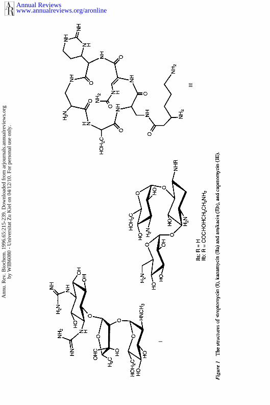
Streptomycin and Related Inhibitors Streptomycin (Figure 1) was first shown to be an effective antitubercular drug in 1944 (2). The liquid Minimum Inhibitory Concentration (MIC) of strepto- mycin against M. tuberculosis has been reported to be 0.4-1.5 pg /d (12), making it one of the most effective early antitubercular drugs. The antibacterial activities of streptomycin, and related aminoglycosides, are due to the inhibi- tion of prokaryotic protein translation. Specifically, initiation of mRNA trans- lation appears to be inhibited, although translational accuracy is also affected (19). A common mechanism of resistance to aminoglycoside antibiotics in other bacteria is drug inactivation via acetylation (19). However, this mecha- nism of resistance has not been reported in Mycobacterium tuberculosis. In- stead two classes of mutations account for some 80% of the high-level strepto- mycin resistance in M. tuberculosis (27). The first consists of point mutations in the ribosomal S12 protein (28, 29), encoded by the rpsL gene, resulting in single-amino acid replacements (30-32). These mutants account for two thirds of the resistant mutations. Mapping of these mutations revealed that all muta- tions occurred in highly conserved regions of the gene encoding one of two critical lysine residues (K43 and K88). In all cases, either K88 was converted to an arginine residue or K43 was converted to either an arginine or threonine residue (3 1). Corresponding mutations result in streptomycin-resistant E. coli (33).
The second class of mutations, which account for the remaining third of the streptomycin-resistance conferring mutations, occur on the 16s rRNA, are encoded in the rrs locus, and are thought to interact with the ribosomal S12 protein. Mutations in M. tuberculosis have been mapped to two regions, the 530 loop and the 915 region. In the 530 loop, C>T transitions at positions 491, 512, and 516 are observed, as is an A S transversion at position 513 (these correspond to positions 501,522,526, and 523 in E. coli 16s rRNA). Mutations at corresponding positions have been mapped in streptomycin-resistant E. coli mutants, including the invariant C513 residue. A single A>G mutant has been mapped to position 913 (30). In contrast to E. coli, little is known of the biochemical basis of the interaction between streptomycin and either the S 12 protein or the 16s rRNA of M . tuberculosis. However, it seems likely that the mutations result in similar changes in the M . tuberculosis ribosome and that
Annual Reviewswww.annualreviews.org/aronline
Ann
u. R
ev. B
ioch
em. 1
996.
65:2
15-2
39. D
ownl
oade
d fr
om a
rjou
rnal
s.an
nual
revi
ews.
org
by W
IB60
80 -
Uni
vers
itat Z
u K
iel o
n 04
/12/
10. F
or p
erso
nal u
se o
nly.
I" z Y P 8
0 I
I O
Annual Reviewswww.annualreviews.org/aronline
Ann
u. R
ev. B
ioch
em. 1
996.
65:2
15-2
39. D
ownl
oade
d fr
om a
rjou
rnal
s.an
nual
revi
ews.
org
by W
IB60
80 -
Uni
vers
itat Z
u K
iel o
n 04
/12/
10. F
or p
erso
nal u
se o
nly.
DRUG-RESISTANT TUBERCULOSIS 221
S12 mutants decrease the binding of the drug. Several recent reviews expand on this discussion (27, 33).
Resistance to streptomycin appears in 5.7% of all cases reported in the United States (3, making streptomycin resistance the third most commonly encountered drug resistance after isoniazid (9.1%) and pyrazinamide (5.8%). Cross-resistance in M. tuberculosis to either related aminoglycosides, kanamy- cin and amikacin, or cyclic peptide protein synthesis inhibitors, such as cap- reomycin, (Figure 1) has never been observed (34-38). This lack of cross- resistance suggests that these compounds may be substituted for streptomycin in those cases where resistance has been determined. Since all of these com- pounds must be administered by injection, whether streptomycin and related inhibitors of bacterial protein synthesis will continue to be considered as first-line antituberculars in short-course chemotherapies is not clear.
RESISTANCE TO INHIBITORS OF NUCLEIC ACID SYNTHESIS
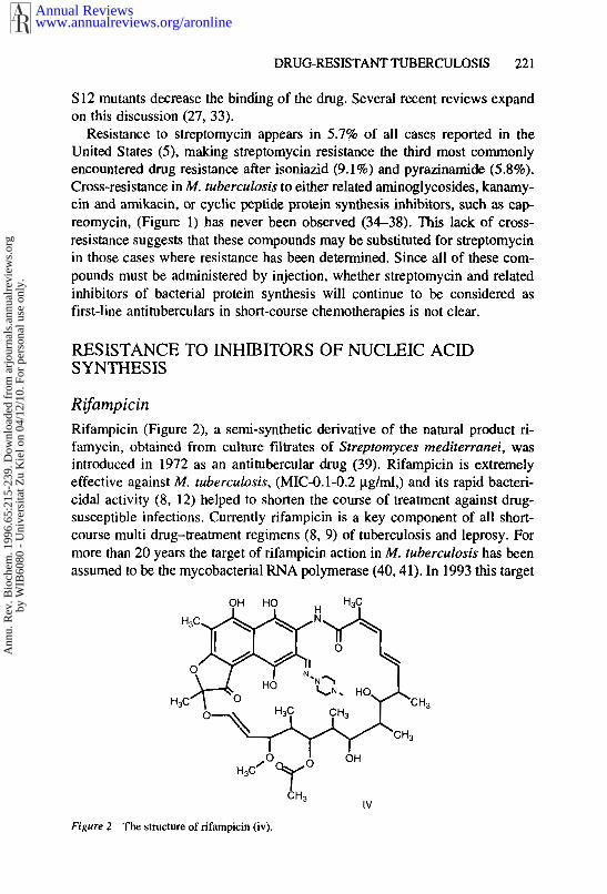
Rifampicin Rifampicin (Figure 2), a semi-synthetic derivative of the natural product ri- famycin, obtained from culture filtrates of Streptomyces mediterranei, was introduced in 1972 as an antitubercular drug (39). Rifampicin is extremely effective against M , tuberculosis, (MIC-0.1-0.2 pglml,) and its rapid bacteri- cidal activity (8, 12) helped to shorten the course of treatment against drug- susceptible infections. Currently rifampicin is a key component of all short- course multi drug-treatment regimens (8, 9) of tuberculosis and leprosy. For more than 20 years the target of rifampicin action in M . tuberculosis has been assumed to be the mycobacterial RNA polymerase (40,41). In 1993 this target
Figure 2 The structure of rifampicin (iv).
Annual Reviewswww.annualreviews.org/aronline
Ann
u. R
ev. B
ioch
em. 1
996.
65:2
15-2
39. D
ownl
oade
d fr
om a
rjou
rnal
s.an
nual
revi
ews.
org
by W
IB60
80 -
Uni
vers
itat Z
u K
iel o
n 04
/12/
10. F
or p
erso
nal u
se o
nly.
222 BLANCHARD
T G KP? C G C A T v I IT CTG AGC CAA TTC ATG GAC CAG AAC AAC CCG CTG TCG GGG TTG ACC CAC AAG CGC CGA CTG TCG GCG CTG
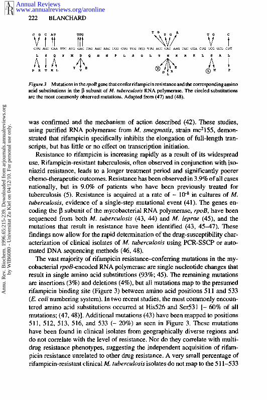
Figure 3 Mutations in the rpoB gene that confer rifampicin resistance and the corresponding amino acid substitutions in the fi subunit of M. tuberculosis RNA polymerase. The circled substitutions are the most commonly observed mutations. Adapted from (47) and (48).
was confirmed and the mechanism of action described (42). These studies, using purified RNA polymerase from M. smegmatis, strain mc2155, demon- strated that rifampicin specifically inhibits the elongation of full-length tran- scripts, but has little or no effect on transcription initiation.
Resistance to rifampicin is increasing rapidly as a result of its widespread use. Rifampicin-resistant tuberculosis, often observed in conjunction with iso- niazid resistance, leads to a longer treatment period and significantly poorer chemo-therapeutic outcomes. Resistance has been observed in 3.9% of all cases nationally, but in 9.0% of patients who have been previously treated for tuberculosis (5). Resistance is acquired at a rate of - lo-* in cultures of M. tuberculosis, evidence of a single-step mutational event (41). The genes en- coding the p subunit of the mycobacterial RNA polymerase, rpoB, have been sequenced from both M. tuberculosis (43, 44) and M. leprae (43 , and the mutations that result in resistance have been identified (43, 4547). These findings now allow for the rapid determination of the drug-susceptibility char- acterization of clinical isolates of M. tuberculosis using PCR-SSCP or auto- mated DNA sequencing methods (46, 48).
The vast majority of rifampicin resistance-conferring mutations in the my- cobacterial rpoBencoded RNA polymerase are single nucleotide changes that result in single amino acid substitutions (93%; 45). The remaining mutations are insertions (3%) and deletions (4%), but all mutations map to the presumed rifampicin binding site (Figure 3) between amino acid positions 51 1 and 533 (E. coli numbering system). In two recent studies, the most commonly encoun- tered amino acid substitutions occurred at His526 and Ser531 [- 60% of all mutations; (47,48)]. Additional mutations (43) have been mapped to positions 51 1, 512, 513, 516, and 533 (- 20%) as seen in Figure 3. These mutations have been found in clinical isolates from geographically diverse regions and do not correlate with the level of resistance. Nor do they correlate with multi- drug resistance phenotypes, suggesting the independent acquisition of rifam- picin resistance unrelated to other drug resistance. A very small percentage of rifampicin-resistant clinical M. tuberculosis isolates do not map to the 51 1-533
Annual Reviewswww.annualreviews.org/aronline
Ann
u. R
ev. B
ioch
em. 1
996.
65:2
15-2
39. D
ownl
oade
d fr
om a
rjou
rnal
s.an
nual
revi
ews.
org
by W
IB60
80 -
Uni
vers
itat Z
u K
iel o
n 04
/12/
10. F
or p
erso
nal u
se o
nly.
DRUG-RESISTANT TUBERCULOSIS 223
region of RNA polymerase, but may be present in the carboxy terminal region of the protein.
The increasing occurrence of rifampicin-resistant M. tuberculosis, especially in individuals not previously treated for tuberculosis (3, is cause for significant concern. Few other antitubercular compounds are as rapidly effective as ri- fampicin. Newer synthetic derivatives (rifabutin and rifapentine MICs = 0.03- 0.06 and 0.01-0.06) do not appear to be substantially more effective than rifampicin (49) and exhibit cross-resistance with rifampicin. Although the target and mechanisms of action and resistance of rifampicin in M. tuberculosis have been well documented in the past several years, more potent new analogs to replace rifampicin have not been developed.
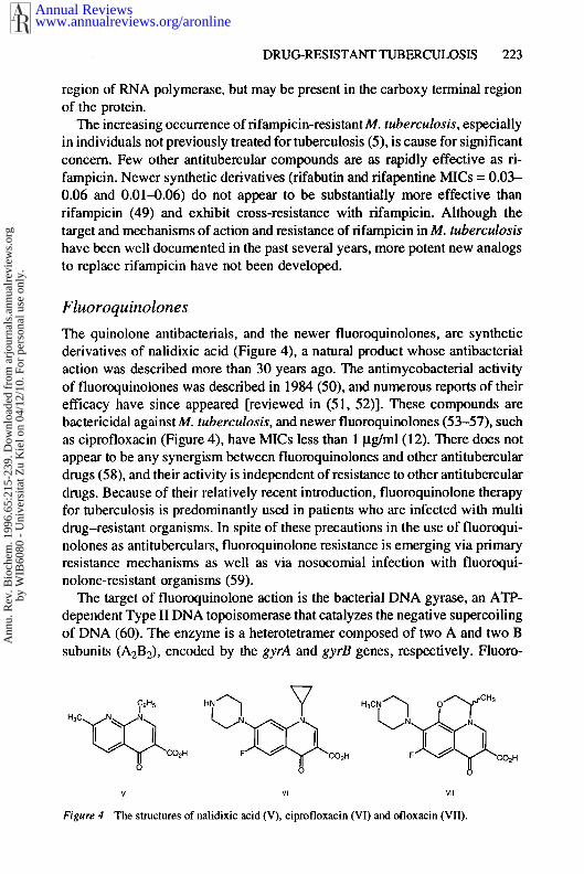
Fluoroquinolones The quinolone antibacterials, and the newer fluoroquinolones, are synthetic derivatives of nalidixic acid (Figure 4), a natural product whose antibacterial action was described more than 30 years ago. The antimycobacterial activity of fluoroquinolones was described in 1984 (50), and numerous reports of their efficacy have since appeared [reviewed in (51, 52)]. These compounds are bactericidal against M. tuberculosis, and newer fluoroquinolones (53-57), such as ciprofloxacin (Figure 4), have MICs less than 1 pg/ml(l2). There does not appear to be any synergism between fluoroquinolones and other antitubercular drugs (58), and their activity is independent of resistance to other antitubercular drugs. Because of their relatively recent introduction, fluoroquinolone therapy for tuberculosis is predominantly used in patients who are infected with multi drug-resistant organisms. In spite of these precautions in the use of fluoroqui- nolones as antituberculars, fluoroquinolone resistance is emerging via primary resistance mechanisms as well as via nosocomial infection with fluoroqui- nolone-resistant organisms (59).
The target of fluoroquinolone action is the bacterial DNA gyrase, an ATP- dependent Type I1 DNA topoisomerase that catalyzes the negative supercoiling of DNA (60). The enzyme is a heterotetramer composed of two A and two B subunits (A2B2), encoded by the gyrA and gyrB genes, respectively. Fluoro-
Figure 4 The structures of nalidixic acid (V), ciprofloxacin (VI) and ofloxacin (VII).
Annual Reviewswww.annualreviews.org/aronline
Ann
u. R
ev. B
ioch
em. 1
996.
65:2
15-2
39. D
ownl
oade
d fr
om a
rjou
rnal
s.an
nual
revi
ews.
org
by W
IB60
80 -
Uni
vers
itat Z
u K
iel o
n 04
/12/
10. F
or p
erso
nal u
se o
nly.

224 BLANCHARD
C T C + /,.E G G C G A C G C C T C G A T C T A C G A C A G/C C Nucleotide
Figure 5 Mutations in the gyrA gene that confer fluoroquinolone resistance and the corresponding amino acid substitutions in the DNA gyrase. Solid lines represent mutations observed in M. tuberculosis and dotted lines represent mutations mapped in both M. tuberculosis and M. smegmatis. Adapted from (61) and (62).
quinolones bind to the gyrase, inhibiting supercoiling and subsequent processes dependent on DNA topology such as replication and transcription. The cloning and sequencing of the gyrA and gyrB genes from M. tuberculosis has allowed the quinolone binding site to be identified and the mutations that confer resistance to be mapped (61). As in other bacteria, these mutations cluster in a small region - 40 residues amino-terminal to the catalytic tyrosine (Y 122 in E. colq involved in DNA strand scission. In both in vitro-selected ciproflox- acin-resistant strains, and resistant clinical isolates, single-amino acid substi- tutions for residues 88-94 (equivalent to residues 81-87 in E. coli) were identified in these strains (Figure 5). These single-amino acid substitutions lead to a - 10-fold increase in the MIC for ciprofloxacin (104).
In a parallel study, mutations in the gyrA gene of M. smegmatis were obtained by selection for ofloxacin-resistant strains (62). Two amino acid substitutions were observed in these studies, AWV and D94G, which could account for low level resistance, comparable to the levels of resistance ob- served in M. tuberculosis. However, a second round of selection at higher ofloxacin levels yielded double mutants that displayed high-level resistance to fluoroquinolones. These double mutants contained substitutions at both posi- tions 90 and 94, A90V/D94G, suggesting that the accumulation of specific mutations in the presumptive quinolone binding site had cumulative effects on the MIC for fluoroquinolones (62). These results suggest that fluoroquinolone susceptibility must be continuously monitored in the treated patient population to prevent low-level fluoroquinolone-resistant strains from acquiring additional mutations that will result in high-level resistance.
Other mechanisms of fluoroquinolone resistance in mycobacteria have been proposed in the literature to quantitatively account for MIC values including non-gyrA mutations (59). Additional mechanisms of resistance include changes
Annual Reviewswww.annualreviews.org/aronline
Ann
u. R
ev. B
ioch
em. 1
996.
65:2
15-2
39. D
ownl
oade
d fr
om a
rjou
rnal
s.an
nual
revi
ews.
org
by W
IB60
80 -
Uni
vers
itat Z
u K
iel o
n 04
/12/
10. F
or p
erso
nal u
se o
nly.
DRUG-RESISTANT TUBERCULOSIS 225
in cell wall permeability or active quinolone efflux pumping. The detailed interactions between target and drug remain unknown; however, newer fluoro- quinolone derivatives such as sparfloxacin (MIC = 0.2 pg/ml; 63) appear to be even more potent antimycobacterial compounds than ciprofloxacin and ofloxacin, promising better therapeutic results against multi drug-resistant tuberculosis.
RESISTANCE TO INHIBITORS OF CELL WALL SYNTHESIS
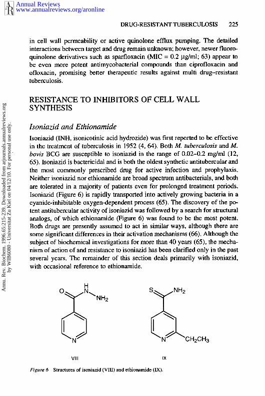
Isoniazid and Ethionamide Isoniazid (INH, isonicotinic acid hydrozide) was first reported to be effective in the treatment of tuberculosis in 1952 (4, 64). Both M. tuberculosis and M. bovis BCG are susceptible to isoniazid in the range of 0.02-0.2 mglml (12, 65). Isoniazid is bactericidal and is both the oldest synthetic antitubercular and the most commonly prescribed drug for active infection and prophylaxis. Neither isoniazid nor ethionamide are broad spectrum antibacterials, and both are tolerated in a majority of patients even for prolonged treatment periods. Isoniazid (Figure 6) is rapidly transported into actively growing bacteria in a cyanide-inhibitable oxygendependent process (65). The discovery of the po- tent antitubercular activity of isoniazid was followed by a search for structural analogs, of which ethionamide (Figure 6) was found to be the most potent. Both drugs are presently assumed to act in similar ways, although there are some significant differences in their activation mechanisms (66). Although the subject of biochemical investigations for more than 40 years (65), the mecha- nism of action of and resistance to isoniazid has been clarified only in the past several years. The remainder of this section deals primarily with isoniazid, with occasional reference to ethionamide.
H
CHzCH3
Vlll IX
Figure 6 Structures of isoniazid (VIII) and ethionamide (a).
Annual Reviewswww.annualreviews.org/aronline
Ann
u. R
ev. B
ioch
em. 1
996.
65:2
15-2
39. D
ownl
oade
d fr
om a
rjou
rnal
s.an
nual
revi
ews.
org
by W
IB60
80 -
Uni
vers
itat Z
u K
iel o
n 04
/12/
10. F
or p
erso
nal u
se o
nly.
226 BLANCHARD
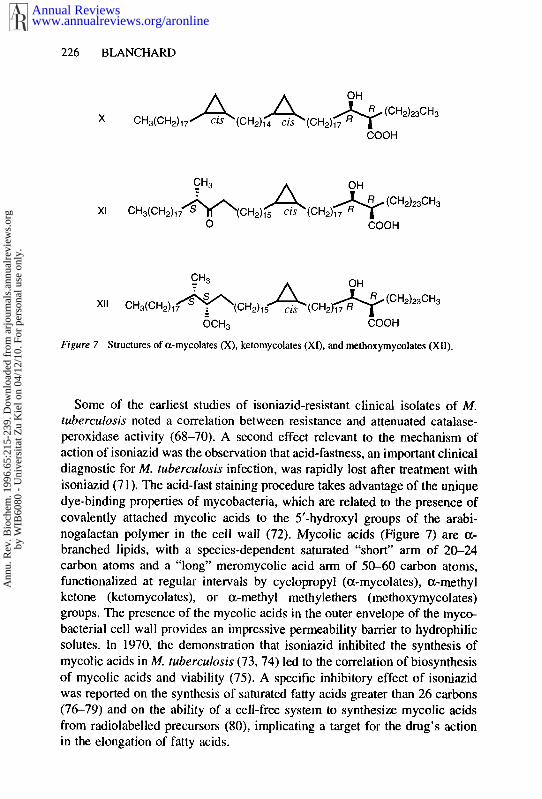
Figure 7 Structures of a-mycolates (X), ketomycolates (XI), and methoxymycolates (XII).
Some of the earliest studies of isoniazid-resistant clinical isolates of M. tuberculosis noted a correlation between resistance and attenuated catalase- peroxidase activity (68-70). A second effect relevant to the mechanism of action of isoniazid was the observation that acid-fastness, an important clinical diagnostic for M. tuberculosis infection, was rapidly lost after treatment with isoniazid (7 1). The acid-fast staining procedure takes advantage of the unique dye-binding properties of mycobacteria, which are related to the presence of covalently attached mycolic acids to the 5’-hydroxyl groups of the arabi- nogalactan polymer in the cell wall (72). Mycolic acids (Figure 7) are a- branched lipids, with a species-dependent saturated “short” arm of 20-24 carbon atoms and a “long” meromycolic acid arm of 50-60 carbon atoms, functionalized at regular intervals by cyclopropyl (a-mycolates), a-methyl ketone (ketomycolates), or a-methyl methylethers (methoxymycolates) groups. The presence of the mycolic acids in the outer envelope of the myco- bacterial cell wall provides an impressive permeability bamer to hydrophilic solutes. In 1970, the demonstration that isoniazid inhibited the synthesis of mycolic acids in M. tuberculosis (73,74) led to the correlation of biosynthesis of mycolic acids and viability (75). A specific inhibitory effect of isoniazid was reported on the synthesis of saturated fatty acids greater than 26 carbons (76-79) and on the ability of a cell-free system to synthesize mycolic acids from radiolabelled precursors (SO), implicating a target for the drug’s action in the elongation of fatty acids.
Annual Reviewswww.annualreviews.org/aronline
Ann
u. R
ev. B
ioch
em. 1
996.
65:2
15-2
39. D
ownl
oade
d fr
om a
rjou
rnal
s.an
nual
revi
ews.
org
by W
IB60
80 -
Uni
vers
itat Z
u K
iel o
n 04
/12/
10. F
or p
erso
nal u
se o
nly.
DRUG-RESISTANT TUBERCULOSIS 227
Genetic and molecular biological approaches have been recently employed to identify the mechanism of activation of isoniazid, its enzymatic target, and mechanisms of resistance. As described above, the attenuation of catalase activity was correlated with resistance to isoniazid in M. tuberculosis. In 1992, the katC gene, encoding the mycobacterial catalase-peroxidase, was cloned from M. tuberculosis (81, 81a). Transformation of the wild-type gene into either M. smegmatis or E. coli, which are naturally less susceptible to isoniazid (82) or isoniazid-resistant strains of M. tuberculosis, sensitized these organisms to the drug (83). Additional evidence shows that - 50% of isoniazid-resistant clinical isolates of M. tuberculosis had deletions or missense mutations within the katC gene (84-86). These results suggested that isoniazid was a prodrug that requires the katC gene product for activation. Earlier studies had shown that peroxidases could react with isoniazid to generate a number of oxidized products similar to those observed in vivo (87), and the M. tuberculosis cata- lase-peroxidase has recently been shown to be capable of oxidizing isoniazid to an electrophilic species (88). The target for the activated form of isoniazid remained unknown.
Selection for single-step spontaneous mutants of M. smegmatis mc2155 resistant to isoniazid yielded a strain, mc265 1, which was an order of magnitude less susceptible to isoniazid, but retained wild-type catalase-peroxidase activ- ity. This strain was similarly resistant to ethionamide, but exhibited wild-type susceptibility to other antitubercular drugs. Subcloning and sequencing of a 3 kilobase DNA fragment, which could be transfected into wild-type strains to yield isoniazid- and ethionamide-resistant transfectants, revealed two open reading frames, one of which was identified as inhA (89). Comparison of wild-type and mutant genes revealed a single nucleotide difference, in which a T+G transversion resulted in the substitution of an alanine residue for serine 94. Using the cell-free mycolic acid synthesizing system, extracts of mc265 1 were shown to exhibit substantially reduced sensitivity to isoniazid, suggesting the inhA-encoded protein was involved in fatty acid elongation and mycolic acid biosynthesis (89).
Expression of the wild-type M. tuberculosis H37Rv inhA-encoded protein in E. coli allowed the facile purification of the protein. The purified, recombinant protein exhibited an appropriate molecular weight, and the amino terminal sequence exactly matched that predicted by the gene se- quence. The protein bound NADH tightly (Kd= 2 pM) and stoichiometrically and was shown to catalyze the reduction of A2-trans-enoyl thioesters of either CoA, or preferentially, acyl carrier protein (ACP). In a homologous series of enoyl-CoA substrates, the enzyme exhibited a marked preference for long chain (C16-C20) substrates (90). The S94A mutant was similarly expressed, purified, and characterized. The enzyme exhibited statistically indistinguish- able maximum velocities and K,,, values for fatty acyl substrates compared
Annual Reviewswww.annualreviews.org/aronline
Ann
u. R
ev. B
ioch
em. 1
996.
65:2
15-2
39. D
ownl
oade
d fr
om a
rjou
rnal
s.an
nual
revi
ews.
org
by W
IB60
80 -
Uni
vers
itat Z
u K
iel o
n 04
/12/
10. F
or p
erso
nal u
se o
nly.
228 BLANCHARD
to the wild-type enzyme, but a 5-8 fold higher K , value for NADH. Kinetic studies supported a random kinetic mechanism and a rate-limiting hydride transfer of the 4s hydrogen of NADH to the C3 position of the bound enoyl thioester.
Both the wild-type and S94A mutant of the M. tuberculosis H37Rv enoyl reductase-NADH complexes were crystallized, and their three-dimensional structures were determined to 2.2 and 2.7 A resolution, respectively (91). The overall fold of the enzyme is reminiscent of a “Rossmann-fold” of alternating parallel p strands and a helices (92). The p sheet is composed of two half sheets: a very regular first half sheet composed of three p strands connected by three a helices and an unusual second half sheet composed of two elongated (p4 and p5) and two short p strands and two elongated a helices (a4 and a5). These elements of the second half sheet generated a large cavity over the p sheet fold which is lined with hydrophobic and aromatic side chains. NADH is bound at the carboxyl termini of the p strands, and the nicotinamide ring of NADH penetrates the hydrophobic cavity, which is the presumed fatty acid binding site. Serine 94 is positioned near the PN atom of bound NADH, and the side chain hydroxyl is hydrogen bonded to a water molecule, which is in turn hydrogen bonded to one of the oxygen atoms of the PN atom of NADH and the backbone carbonyl of Gly14. These interactions are disrupted in the S94A mutant and can account for the observed higher K,,, value for NADH exhibited by the mutant enoyl reductase.
Together, the studies on the M. ruberculosis kutG-encoded catalase-peroxi- dase and in&-encoded enoyl reductase provide a revealing, but complicated, picture of the mechanism of action of (Figure 8) and mechanisms of resistance to isoniazid (65, 93, 94). Isoniazid is a prodrug that must be activated by reaction with the mycobacterial catalase-peroxidase. Mutations in the catalase- peroxidase which generate inactive enzyme will fail to activate the prodrug and lead to high-level isoniazid resistance. This mechanism is the one most commonly encountered for the acquisition of isoniazid resistance in clinical isolates of M. tuberculosis (95, 96). Isoniazid is also a potent inhibitor of the mycobacterial catalase-peroxidase (97), and a secondary effect of drug admini- stration may be to enhance the susceptibility of bacteria to the toxic effects of reactive oxygen, especially HzOz (98,99) generated in the macrophage phago- lysosomal compartments.
Although isoniazid clearly does not bind to the inhA-encoded enoyl reduc- tase (90), the catalase-peroxidase-activated drug does (66), and its binding is correlated with inhibition of the reductase activity. Drug binding occurs pre- dominantly, or exclusively, to the reductase-NADH binary complex, providing a reasonable explanation for the resistance to isoniazid exhibited by organisms expressing the S94A mutant enzyme. The intracellular levels of NADH in M. tuberculosis are low [estimated at < 10 pM; (67)], and thus the wild-type
Annual Reviewswww.annualreviews.org/aronline
Ann
u. R
ev. B
ioch
em. 1
996.
65:2
15-2
39. D
ownl
oade
d fr
om a
rjou
rnal
s.an
nual
revi
ews.
org
by W
IB60
80 -
Uni
vers
itat Z
u K
iel o
n 04
/12/
10. F
or p
erso
nal u
se o
nly.

DRUG-RESISTANT TUBERCULOSIS 229
Annual Reviewswww.annualreviews.org/aronline
Ann
u. R
ev. B
ioch
em. 1
996.
65:2
15-2
39. D
ownl
oade
d fr
om a
rjou
rnal
s.an
nual
revi
ews.
org
by W
IB60
80 -
Uni
vers
itat Z
u K
iel o
n 04
/12/
10. F
or p
erso
nal u
se o
nly.
230 BLANCHARD
enzyme is present as the drug inhibitablebinary complex. The mutant enzyme, because of its lower affinity for NADH, would be only partially saturated with NADH at these concentrations, and would not bind or be inhibited by the drug. Although the residue(s) of the enoyl reductase that interact, or react, with the activated form of isoniazid are not known with certainty, the protective effects of added substrates (66) suggest that residues in the active site are most likely modified by the drug.
The mechanism of action of ethionamide is almost certainly similar to that of isoniazid, including a requirement for prodrug activation. Whereas muta- tions in the enoyl reductase generate resistance to ethionamide, isoniazid-re- sistant strains containing mutations in the catalase-peroxidase gene remain susceptible to ethionamide. A second enzyme that activates ethionamide must be present in mycobacteria to convert this prodrug into a form capable of binding to, and presumably inhibiting, the inh4encoded enoyl reductase, because ethionamide does not bind to the reductase (90).
Lastly, resistance due to the overexpression of the enoyl reductase has been demonstrated in vitro (89), and the majority of isoniazid-resistant clinical isolates of M. tuberculosis that map to the inhA locus have mutations in the promoter region of the gene (21). This finding suggests that this common bacterial-resistance mechanism is operative in the mycobacteria.
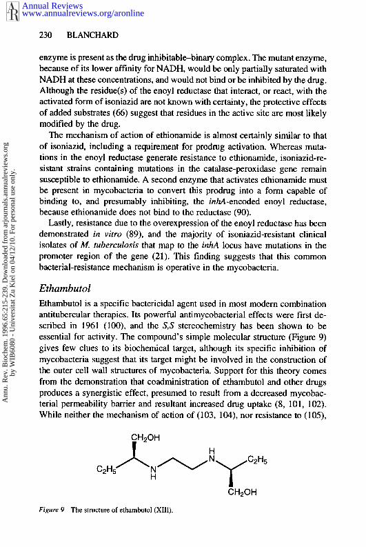
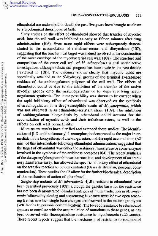
Ethambutol Ethambutol is a specific bactericidal agent used in most modern combination antitubercular therapies. Its powerful antimycobacterial effects were first de- scribed in 1961 (loo), and the S,S stereochemistry has been shown to be essential for activity. The compound’s simple molecular structure (Figure 9) gives few clues to its biochemical target, although its specific inhibition of mycobacteria suggest that its target might be involved in the construction of the outer cell wall structures of mycobacteria. Support for this theory comes from the demonstration that coadministration of ethambutol and other drugs produces a synergistic effect, presumed to result from a decreased mycobac- terial permeability barrier and resultant increased drug uptake (8, 101, 102). While neither the mechanism of action of (103, 104), nor resistance to (105),
CH20H
Figure 9 The structure of ethambutol (XIII).
Annual Reviewswww.annualreviews.org/aronline
Ann
u. R
ev. B
ioch
em. 1
996.
65:2
15-2
39. D
ownl
oade
d fr
om a
rjou
rnal
s.an
nual
revi
ews.
org
by W
IB60
80 -
Uni
vers
itat Z
u K
iel o
n 04
/12/
10. F
or p
erso
nal u
se o
nly.
DRUG-RESISTANT TUBERCULOSIS 23 1
ethambutol are understood in detail, the past five years have brought us closer to a biochemical description of both.
Early studies on the effect of ethambutol showed that transfer of mycolic acids into the cell wall was inhibited as early as fifteen minutes after drug administration (106). Even more rapid effects were subsequently demon- strated in the accumulation of trehalose mono- and dimycolates (107), suggesting that the biochemical target was indeed involved in the construction of the outer envelope of the mycobacterial cell wall (108). The structure and composition of the outer cell wall of M. fuberculosis is still under active investigation, although substantial progress has been made in the past decade [reviewed in (ls)]. The evidence shows clearly that mycolic acids are specifically attached to the 5’-hydroxyl groups of the terminal D-arabinose residues of the arabinogalactan polymer of the cell wall. The effects of ethambutol could be due to the inhibition of the transfer of the active mycolyl groups onto the arabinogalactan or to steps involving arabi- nogalactan synthesis. The latter possibility was shown to be correct when the rapid inhibitory effect of ethambutol was observed on the synthesis of arabinogalactan in a drug-susceptible strain of M . smegmatis, which was not observed in an ethambutol-resistant strain (109). The inhibition of arabinogalactan biosynthesis by ethambutol could account for the accumulation of mycolic acids and their trehalose esters, as well as the effects on cell wall permeability.
More recent results have clarified and extended these studies. The identifi- cation of PD-arabinofuranosyl- 1-monophosphodecaprenol as the major inter- mediate in the biosynthesis of arabinogalactan, and the rapid accumulation (4 min) of this intermediate following ethambutol administration, suggested that the target of ethambutol was either the arabinosyl transferase or some enzyme involved in the synthesis of the arabinose acceptor (104). The recent synthesis of the decaprenylphosphoarabinose intermediate, and development of an arabi- nosyltransferase assay, has allowed the specific inhibitory effect of ethambutol on the transfer reaction to be demonstrated (Besra & Brennan, personal com- munication). These studies should allow for the further biochemical description of the mechanism of action of ethambutol.
Single-step mutants of M . tuberculosis H3,Ra resistant to ethambutol have been described previously (108), although the genetic basis for the resistance has not been demonstrated. Similar strategies of mutant selection in M. smeg- matis followed by cloning and sequencing have now revealed two open reach- ing frames in which single base changes are observed in the mutant genotypes (WR Jacobs Jr, personal communication). The level of resistance to ethambutol appears to correlate with the accumulation of mutations in these genes, as has been observed with fluoroquinolone resistance in mycobacteria (vide supra). These recent reports suggest that the mechanism of resistance to ethambutol
Annual Reviewswww.annualreviews.org/aronline
Ann
u. R
ev. B
ioch
em. 1
996.
65:2
15-2
39. D
ownl
oade
d fr
om a
rjou
rnal
s.an
nual
revi
ews.
org
by W
IB60
80 -
Uni
vers
itat Z
u K
iel o
n 04
/12/
10. F
or p
erso
nal u
se o
nly.
232 BLANCHARD
in mycobacteria will soon be made clear with the aid of this powerful combi- nation of genetic and biochemical methods.
RESISTANCE TO OTHER ANTITUBERCULARS
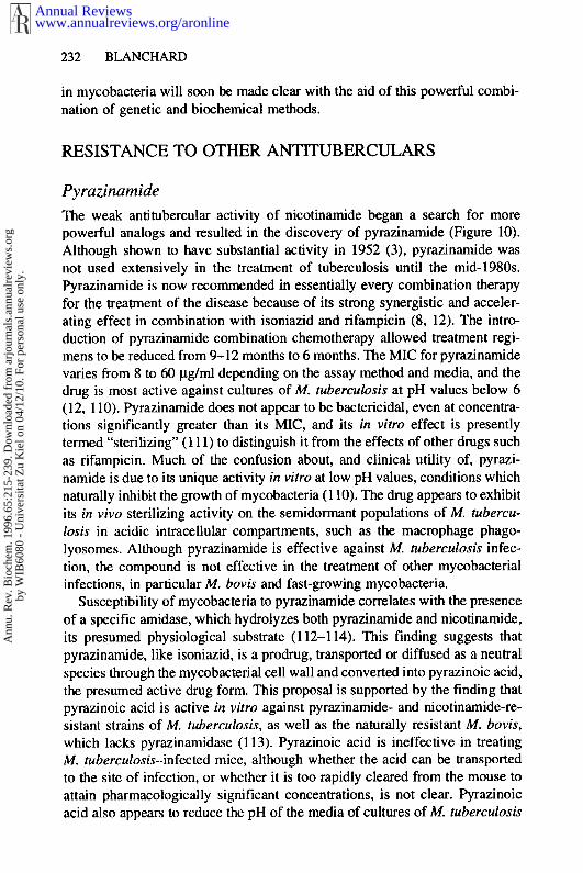
Pyrazinamide The weak antitubercular activity of nicotinamide began a search for more powerful analogs and resulted in the discovery of pyrazinamide (Figure 10). Although shown to have substantial activity in 1952 (3), pyrazinamide was not used extensively in the treatment of tuberculosis until the mid-1980s. Pyrazinamide is now recommended in essentially every combination therapy for the treatment of the disease because of its strong synergistic and acceler- ating effect in combination with isoniazid and rifampicin (8, 12). The intro- duction of pyrazinamide combination chemotherapy allowed treatment regi- mens to be reduced from 9-12 months to 6 months. The MIC for pyrazinamide varies from 8 to 60 pglml depending on the assay method and media, and the drug is most active against cultures of M. tuberculosis at pH values below 6 (12, 1 10). Pyrazinamide does not appear to be bactericidal, even at concentra- tions significantly greater than its MIC, and its in vitro effect is presently termed “sterilizing” (1 1 1) to distinguish it from the effects of other drugs such as rifampicin. Much of the confusion about, and clinical utility of, pyrazi- namide is due to its unique activity in vitro at low pH values, conditions which naturally inhibit the growth of mycobacteria (1 10). The drug appears to exhibit its in vivo sterilizing activity on the semidormant populations of M. tubercu- losis in acidic intracellular compartments, such as the macrophage phago- lyosomes. Although pyrazinamide is effective against M. tuberculosis infec- tion, the compound is not effective in the treatment of other mycobacterial infections, in particular M . bovis and fast-growing mycobacteria.
Susceptibility of mycobacteria to pyrazinamide correlates with the presence of a specific amidase, which hydrolyzes both pyrazinamide and nicotinamide, its presumed physiological substrate (1 12-1 14). This finding suggests that pyrazinamide, like isoniazid, is a prodrug, transported or diffused as a neutral species through the mycobacterial cell wall and converted into pyrazinoic acid, the presumed active drug form. This proposal is supported by the finding that pyrazinoic acid is active in vitro against pyrazinamide- and nicotinamide-re- sistant strains of M. tuberculosis, as well as the naturally resistant M . bovis, which lacks pyrazinamidase (1 13). Pyrazinoic acid is ineffective in treating M. tuberculosis-infected mice, although whether the acid can be transported to the site of infection, or whether it is too rapidly cleared from the mouse to attain pharmacologically significant concentrations, is not clear. Pyrazinoic acid also appears to reduce the pH of the media of cultures of M. tuberculosis
Annual Reviewswww.annualreviews.org/aronline
Ann
u. R
ev. B
ioch
em. 1
996.
65:2
15-2
39. D
ownl
oade
d fr
om a
rjou
rnal
s.an
nual
revi
ews.
org
by W
IB60
80 -
Uni
vers
itat Z
u K
iel o
n 04
/12/
10. F
or p
erso
nal u
se o
nly.
DRUG-RESISTANT TUBERCULOSIS 233
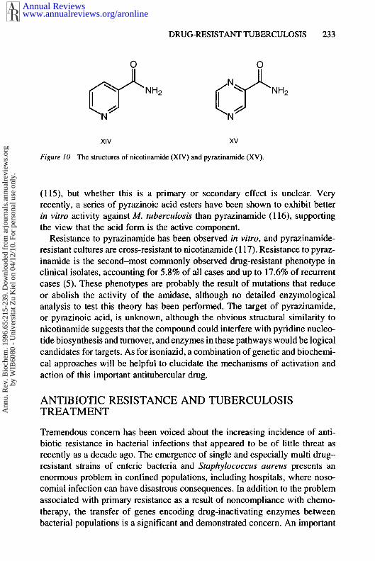
XIV xv
Figure 10 The structures of nicotinamide (XIV) and pyrazinamide (XV).
(1 15), but whether this is a primary or secondary effect is unclear. Very recently, a series of pyrazinoic acid esters have been shown to exhibit better in vitro activity against M. tuberculosis than pyrazinamide (1 16), supporting the view that the acid form is the active component.
Resistance to pyrazinamide has been observed in vitro, and pyrazinamide- resistant cultures are cross-resistant to nicotinamide (1 17). Resistance to pyraz- inamide is the second-most commonly observed drug-resistant phenotype in clinical isolates, accounting for 5.8% of all cases and up to 17.6% of recurrent cases (5). These phenotypes are probably the result of mutations that reduce or abolish the activity of the amidase, although no detailed enzymological analysis to test this theory has been performed. The target of pyrazinamide, or pyrazinoic acid, is unknown, although the obvious structural similarity to nicotinamide suggests that the compound could interfere with pyridine nucleo- tide biosynthesis and turnover, and enzymes in these pathways would be logical candidates for targets. As for isoniazid, a combination of genetic and biochemi- cal approaches will be helpful to elucidate the mechanisms of activation and action of this important antitubercular drug.
ANTIBIOTIC RESISTANCE AND TUBERCULOSIS TREATMENT
Tremendous concern has been voiced about the increasing incidence of anti- biotic resistance in bacterial infections that appeared to be of little threat as recently as a decade ago. The emergence of single and especially multi drug- resistant strains of enteric bacteria and Staphylococcus aureus presents an enormous problem in confined populations, including hospitals, where noso- comial infection can have disastrous consequences. In addition to the problem associated with primary resistance as a result of noncompliance with chemo- therapy, the transfer of genes encoding drug-inactivating enzymes between bacterial populations is a significant and demonstrated concern. An important
Annual Reviewswww.annualreviews.org/aronline
Ann
u. R
ev. B
ioch
em. 1
996.
65:2
15-2
39. D
ownl
oade
d fr
om a
rjou
rnal
s.an
nual
revi
ews.
org
by W
IB60
80 -
Uni
vers
itat Z
u K
iel o
n 04
/12/
10. F
or p
erso
nal u
se o
nly.
234 BLANCHARD
example of the general problem is the recent appearance of vancomycin resis- tance in clinical isolates of methicillin-resistant enterococci (1 18).
The appearance of multi drug-resistant strains of M. tuberculosis shares many of the societal and clinical problems of general bacterial antibiotic resistance and generates some unique concerns. Drug-resistant M . tuberculosis strains are initially the result of noncompliance with chemotherapeutic regi- mens and selective genetic pressure. Because of the extremely infectious nature of the organism and its ability to be transferred via aerosols from infected to noninfected individuals, multi drug-resistant organisms are now being detected in previously untreated tuberculosis patients (5). Patients are often treated with ineffective drug combinations for long periods before susceptibility screens can be analyzed, further complicating treatment and jeopardizing therapeutic outcomes.
Because of the unique nature and antibacterial properties of antitubercular drugs, and the lack of evidence to date for episomal transfer of resistancecon- ferring genes from the general bacterial population to M . tuberculosis, multi drug-resistant tuberculosis remains a treatable infectious disease for the ma- jority of cases. In large part this high level of treatability is due to the early recognition that combinations of at least three or four drugs were required for both effective sterilization and the prevention of the acquisition of resistance commonly observed in monotherapy. Unfortunately, the most commonly en- countered resistance is against the two most effective antitubercular com- pounds, isoniazid and rifampicin. As discussed above, these are the two drugs which are most bactericidal against rapidly growing organisms, and rifampicin appears to be effective against semi-dormant organisms that reactivate and become metabolically active. Rifampicin has no effective homologs for which cross-resistance is not encountered, and ethionamide is only a modestly effec- tive therapeutic substitute for isoniazid in cases in which isoniazid-resistance is due to mutations in the catalase-peroxidase gene.
Other broad spectrum antibacterials, which have significant activity against mycobacteria, can be substituted for isoniazid or rifampicin. The fluoroqui- nolones, including ciprofloxacin, ofloxacin, and the newer sparfloxacin, are extremely effective antituberculars. Although fluoroquinolone resistance has been reported in clinical isolates of M . tuberculosis, the compounds remain effective in the treatment of isoniazid- and rifampicin-resistant tuberculosis. The macrolide antibiotics such as erythromycin and clarithromycin exhibit antimycobacterial activity.The latter has significant activity against M . d u m infections in HIV-infected individuals (1 19), and ethambutol has a pronounced synergistic effect on clarithromycin administration (102).
New drug discovery is hampered by the fact that although tuberculosis remains the world's leading cause of human mortality among infectious diseases, the vast majority of disease occurs in the undeveloped nations which
Annual Reviewswww.annualreviews.org/aronline
Ann
u. R
ev. B
ioch
em. 1
996.
65:2
15-2
39. D
ownl
oade
d fr
om a
rjou
rnal
s.an
nual
revi
ews.
org
by W
IB60
80 -
Uni
vers
itat Z
u K
iel o
n 04
/12/
10. F
or p
erso
nal u
se o
nly.
DRUG-RESISTANT TUBERCULOSIS 235
have limited resources with which to address these health concerns. In contrast, newly diagnosed cases of tuberculosis in the developed nations number - 25,000, and chemotherapy is successful in 95% of compliant cases. By far the most effective way to ensure a decrease in primary resistance, acquired resistance, and relapse is to institute directly observed combination chemotherapy (IO), even amongst individuals harboring multi drug-resistant strains. Drug-resistant phenotypes of M. tuberculosis appear to be uncorre- lated with virulence (120). Thus the solution to the problem of drug-resistant tuberculosis is clear, but the disease remains both a societal and scientific problem.
Future Studies and Solutions The challenge to the scientific and pharmacologic community is to eliminate the comparative lack of modern biochemical and genetic information about mycobacteria in general. M. tuberculosis, in particular, is not an organism whose large scale culture is achievable in any but a handful of laboratories. The recent development of strains of the nonpathogenic fast-growing M. smeg- matis that can be genetically manipulated (121, 122) has allowed researchers to begin classical genetic studies in this organism. These approaches have clarified the mechanism of action of, and resistance to, both rifampicin (42) and isoniazid (89). The biochemical transformations occurring in mycobacteria during the acquisition of drug resistance are generally inferred, rather than demonstrated, and tremendous progress should be made in this area in the next decade.
The serendipitous discovery of isoniazid, ethionamide, and ethambutol has now provided clues into critical and unique biosynthetic pathways in myco- bacteria. The chemical simplicity of these molecules, and a decade of synthetic endeavors after their initial discovery, suggests that more potent analogs, prepared by classic organic synthesis or novel combinatorial synthetic meth- ods, will be hard to find. Given what is now known about the mechanism of action of isoniazid, the discovery of homologs that are both actively accumu- lated and oxidatively activated, and still inhibit long chain fatty acid elongation processes involved in outer envelope biosynthesis, seems unlikely. However, as the mechanism of action of these drugs is clarified, and their molecular targets identified, more rational mechanism-based and structure-based ap- proaches to inhibitor design will be possible. The synthesis and clinical evalu- ation of any lead compound is a long and expensive process, and the worldwide distribution of tuberculosis in the developing nations may discourage such investments.
Alternative solutions that should be examined include the revaluation of existing antibacterials. The example of fluoroquinolone inhibitors of DNA
Annual Reviewswww.annualreviews.org/aronline
Ann
u. R
ev. B
ioch
em. 1
996.
65:2
15-2
39. D
ownl
oade
d fr
om a
rjou
rnal
s.an
nual
revi
ews.
org
by W
IB60
80 -
Uni
vers
itat Z
u K
iel o
n 04
/12/
10. F
or p
erso
nal u
se o
nly.
236 BLANCHARD
~ ~~ ~ ~ ~ ~
Any Annual Review chapter, as well as any article cited in an Annual Review chapter, may be purchased from the Annual Reviews Preprints and Reprints service.
1-800-347-8007; 415-259-5017; email: [email protected]
gyrase was discussed above. A second example could include the p-lactam inhibitors of peptidoglycan biosynthesis. Mycobacteria are naturally insensi- tive to P-lactams, because of their extremely hydrophobic cell wall (123) and the presence of both periplasmic penicillin-binding proteins (124) and an active P-lactamase (25, 123). However, the combined administration of P-lac- tams and P-lactamase inhibitors has recently been shown to be effective in inhibiting the growth of mycobacteria (125-127). Given their oral availability and favorable toxicology profile, the thousands of Plactams that have been synthesized seem worthy of re-examination, in combination with inhibitors of the mycobacterial p-lactamases. The recently demonstrated bactericidal activ- ity of nitroimidazoles such as metronidazole (13, 14) against dormant popu- lations, which are poorly treated in present drug regimens, represents a poten- tial new chemotherapeutic addition. Ultimately, the only solution to the prob- lems with tuberculosis chemotherapy and the explosion of multi-drug resis- tance is patient surveillance and compliance. We have beaten the scourge of tuberculosis once before, but we must remain vigilant in this present day cat-and-mouse game.
ACKNOWLEDGMENTS
This work was supported by NIH grants GM-33449 and AI-33696. I would like to thank Drs. Lincoln Miller, Vem Schramm, and Thomas Shrader for helpful comments and suggestions on the manuscript, and Ms. Lisa Idi for wordprocessing assistance.
Literature Cited
1. Bloom BR, Murray CJL. 1992. Science 257: 105544
2. Schatz A, Waksman SA. 1944. Proc. SOC. Exp. Bioi. Med. 57:244-48
3. Kushner S, Dalalian H, Sanjuro JL, Bach FL, Satir SR, et al. 1952. J. Am. Chem. SOC. 743617-26 Middlebrook G. 1952. Am. Rev. Tuberc. 65: 7 65-67 Bloch AB, Cauthen GM, Onorato IM, Dansbury KG, Kelly GD, et al. 1994. J . Am. Med. Assoc. 271565-71
6. Frieden TR, Sterling T, Pablos-Mendez A, Kilburn 10, Cauthen GM, et al. 1993. N . Engl. J. Med. 328521-26
7. Heym B, Honore N, Truffot-Pernot C, Banerjee A, Schurra C, et al. 1994. Lancer 344:293-98
4.
5 .
8. Mitchison DA. 1985. Tubercle 66:219- 26
9. Stratton MA, Reed MT. 1986. Clin. Pharm. 5:977-87
10. Weis SE, Slocum PC, Blais FX, King B, Nunn M, et al. 1994. N . Engi. J . Med. 330: 1229-30
11. Hiriyanna KT, Ramakrishnan T. 1986. Arch. Microbiol. 144: 1 0 5 9
12. Heifets LB. 1994. Semin. Respir. Infect.
13. Ashtekar DR, Costa-Perira R, Nagrajan K, Vishvanathan N, Bhatt AD, Rittel W. 1993. Antimicrob. Agents Chemo- ther. 37:183-86
14. Wayne LG, Sramek HA. 1994. An- rimicrob. Agenrs Chemother. 38:2054- 58
984-103
Annual Reviewswww.annualreviews.org/aronline
Ann
u. R
ev. B
ioch
em. 1
996.
65:2
15-2
39. D
ownl
oade
d fr
om a
rjou
rnal
s.an
nual
revi
ews.
org
by W
IB60
80 -
Uni
vers
itat Z
u K
iel o
n 04
/12/
10. F
or p
erso
nal u
se o
nly.
15.
16.
17.
18.
19.
20.
21.
2 2.
23.
24.
25.
26.
27.
28.
29.
30.
31.
32.
33.
34.
35.
3 6.
37.
38.
39.
40.
41.
DRUG
Rastoggi N. 1991. Res. Microbiol. 142: 464-76 Rastoggi N, Barrow WW. 1994. Res. Microbiol. 145:243-52 Daffe M, McNeil M, Brennan PJ. 1993. Carbohydr. Res. 249383-98 Brennan PJ. Nikaido H. 1995. Annu. Rev. Biochem. 6429-63 Benveniste R, Davies J. 1973. Annu. Rev. Biochem. 42471-506 Cole ST. 1994. Immunobiology 191: 584-85 Moms S, Bai GH, Suffys P. Portillo- Gomez L, Fairchok M, et al. 1995. J. Infect. Dis. 17 1:954-60 Rastoggi N, David HL. 1993. Res. Mi- crobiol. 144:13343 Suzuki AE, Inamine JM. 1994. Res. Microbiol. 145:210-13 Eum HM, Yap0 A, Petit JF. 1978. Eur. J . Biochem 8697-103 Fattorini L. Orefici G, Jin SH, Scardaci G, Amicosante G, et al. 1992. Antimi- crob. Agents Chemother. 3 6 1068-72 Zhang Y, Young D. 1994. J. Antimicrob. Chemother. 34:313-19 Honore N, Cole ST. 1994. Antimicrob. Agents Chemother. 3823842 Douglass J, Steyn LM. 1993. J. Infect. Dis. 167: 1505-6 Yamada T, Nagata A, Ono Y, Suzuki Y, Yamanouchi T. 1985. Antimicrob. Agents Chemother. 27921-24 Finken M, Kirschner P, Meier A, Wrede A, Bottger EC. 1993. Mol. Microbiol. 9: 123946 Meier A, Kirschner P, Bange FC, Vogel U, Bottger EC. 1994. Antimicrob. Agents Chemother. 38:228-33 Nair J, Rouse DA, Bai G-H, Moms SL. 1993. Mol. Microbiol. 10521-27 Bottger EC. 1994. Trends Microbiol. 2:416-21 Heifets LB, Lindholm-Levy PJ. 1989. Antimicrob. Agents Chemother. 33:
Hoffner SE, Kallenius G. 1988. Eur. J. Clin. Microbiol. Infect. Dis. 7: 188- 90 McClatchy JK, Kanes W, Davidson PT, Moulding TS. 1977. Tubercle 5829-34 Tsukamura M, Mizuno S. 1975. J. Gen. Microbiol. 88269-74 Tsukamura M, Mizuno S. 1980. Micro- biol. Immunol. 24~777-87 Woodley CL, Kilburn JO, David HL, Silcox VA. 1972. Antimicrob. Agents Chemother. 2:24549 Siddiqi SH, Aziz A, Reggiardo Z, Mid- dlebrook G. 1981. J. Clin. Parhol. 34: 927-29 Tsukamura M. 1972. Tubercle 53: 1 11- 17
1298-301
-RESISTANT TUBERCULOSIS 237
42. Levin ME, Hatfull GF. 1993. Mol. Mi- crobiol. 8:277-85
43. Donnabella MV, Martiniuk E Kinnev
48.
49.
50.
51.
52. 53.
54.
55.
5 6.
57.
58.
59.
D, Bacado M, Bonk S, et al. 1994. A;. J. Respir. Cell Mol. Biol. 11:63943
44. Miller LP, Crawford JT. Shinnick TM. 1994. Antimicrob. Agents Chemother.
45. Honore N, Cole ST. 1993. Antimicrob. Agents Chemother. 37:414-18
46. Telenti A, Imboden P, Marchesi F, Lowrie D, Cole S, et al. 1993. Lancer
47. Williams DL, Waguespack C, Eisenach K, Crawford JT, Portaels F, et al. 1994. Antimicrob. Agents Chemother. 38: 2380-86 Kapur V, Li L L , Iordanescu S, Hamrick MR, Wanger A, et al. 1994. J. Clin. Microbiol. 3 2 1095-98 Heifets LB, Lindholm-Levy PJ, Flory MA. 1990. Am. Rev. Respir. Dis. 141: 626-30 Gay JD. DeYoung DR, Roberts GD. 1984. Antimicrob. Agents Chemother. 26:94-96 Leysen DC, Haemers A, Pattyn SR. 1989. Antimicrob. Agents Chemother.
Stratton C. 1992. Clin Ther. 14:348-75 Klopman G. Wang S, Jacobs MR, Ba- jaksouzian S, Edmonds K, et al. 1993. Antimicrob. Agents Chemother. 37: 1799-806 Klopman G, Wang S, Jacobs MR, Ellner JJ. 1993. Antimicrob. Agents Chemo- ther. 37:1807-15 Klopman G, Li JY, Wang S, Pearson AJ, Chang K, et al. 1994. Antimicrob. Agents Chemother. 38: 1794-802 Piersimoni C, Morbiducci V, Bornigia S, DeSio G, Scalise G. 1992. Am. Rev. Respir. Dis. 146 1445-47 Truffot-Pernot C, Ji B, Grosset J. 1991. Tubercle 7257-64 Marinis E, Legakis NJ. 1985. J. Antimi- crob. Chemother. 16527-30
38:805-11
341~647-50
33~1-5
60.
61.
62.
63.
64.
Sullivan EA, Kreiswirth BN, Palumbo L, Kapur V, Musser JM, et al. 1995. Lancet 345: 1148-50 Wang JC. 1991. J . Biol. Chem. 266: 665962 Takiff HE, Salazar L, Guerrero C, Philipp W, Huang WM, et al. 1994. Antimicrob. Agents Chemother. 38:773- 80 Revel V, Cambau E, Jarlier V, Souga- koff W. 1994. Antimicrob. Agents Chemother. 38: 1991-96 Lalande V, Truffot-Pernot C, Paccaly- Moulin A, Grosset J, Ji B. 1993. An- timicrob. Agents Chemother. 37:407-13 Bernstein J, Lott WA, Steinberg BA,
Annual Reviewswww.annualreviews.org/aronline
Ann
u. R
ev. B
ioch
em. 1
996.
65:2
15-2
39. D
ownl
oade
d fr
om a
rjou
rnal
s.an
nual
revi
ews.
org
by W
IB60
80 -
Uni
vers
itat Z
u K
iel o
n 04
/12/
10. F
or p
erso
nal u
se o
nly.
238 BLANCHARD
65.
66.
67.
68.
69.
7 0.
71.
72.
73.
74.
75.
76.
77.
7 8.
79.
80.
81.
81a.
8 2.
83.
8 4.
85.
86.
87.
88.
89.
Yale HL. 1952. Am. Rev. Tuberc. 65: 357-64 Youatt J. 1969. Am. Rev. Respir. Dis. 99:72949 Johnsson K, King DS, Schultz PG. 1995. J. Am. Chem. Soc. 117:5009-10 Gopinathan KP, Sirsi M, Ramakrkhnan T. 1963. Biochem. J. 8744-48 Middlebrook G. 1954. Am. Rev. Tuberc.
Middlebrook G, Cohn ML, Schaefer WB. 1954. Am. Rev. Tuberc. 70852-72 Winder FG. 1960. Am. Rev. Respir. Dis. 8 1 368-78 Koch-Weser D, Ebert RH, Barclay WR, Lee VS. 1953. J. Lab. Clin. Med. 42: 828-29 McNeil M, Daffe M, Brennan PJ. 1991. J. Biol. Chem. 266:13217-23 Winder FG, Collins PB. 1970. J. Gen. Microbiol. 63:41-48 Winder FG, Collins PB, Rwney SA. 1970. Biochem. J. 117:P27 Takayama K, Wang L, David HL. 1972. Antimicrob. Agents Chemother. 2:29-35 Davidson LA, Takayama K. 1979. Antimicrob. Agents Chemother. 16: 104-5 Kikuchi S, Takeuchi T, Yasui M, Ka- saka T, Kolaitukudy PE. 1989. Agric. Biol. Chem. 53:1689-98 Takayama K, Schnoes HK, Armstrong EL, Boyle RW. 1975. J. Lipid Res.
Wang L, Takayama K. 1972. Antimi- cmb. Agents Chemother. 2:43841 Quemard A, Lacave C, Laneelle G. 1991. Antimicrob. Agents Chemother. 35: 1035-39 Zhang Y, Heym B, Allen B, Young D, Cole S. 1992. Nature 358591-93 Heym B, Zhang Y, Poulet S, Young D, Cole ST. 1993. J. Bacteriol. 1754255- 59 Rosner JL. 1993. Antimicrob. Agents Chemother. 37:225 1-53 Zhang Y, Garbe T, Young D. 1993. Mol. Microbiol. 8521 -24 Cockerill FR, Uhl JR, Temesgen Z, Zhang Y, Stockman L, et al. 1995. J. Infect. Dis. 17 1:24045 Heym B, Cole ST. 1992. Res. Microbiol.
Heym B, Alzari PM, Honore N, Cole ST. 1995. Mol. Microbiol. 15:235-45 Shoeb HA, Bowman BU, Ottolenghi AC, Merola AJ. 1985. Antimicrob. Agents Chemother. 27:399403 Johnsson K, Schultz PG. 1994. J. Am. Chem. SOC. l16:7425-26 Banerjee A, Dubnau E, Quemard A, Balasubramanian V, Um KS, et a]. 1994. Science 263:227-30
69:47 1 -72
16:308-17
143:721-30
90.
91.
92.
93.
94.
95.
96.
97.
98.
99.
100.
101.
102.
103.
104.
105.
106.
107.
108.
109.
110.
111.
112.
113.
Quemard A, Sacchettini JC, Dessen A, Jacobs WR Jr, Blanchard JS. 1995. Bio- chemistry 34823-1 Dessen A, Quemard A, Blanchard JS, Jacobs WR Jr, Sacchettini JC. 1995. Science 24: 1638-4 1 Rossmann MG, Liljas A, Branden C-I, Banaszak LJ. 1975. Enzymes 1 IA:61- 102 Rouse DA, Moms SL. 1995. Infect. Immun. 63: 1427-33 Zhang Y, Young D. 1993. Trends M i - crobiol. 1:109-13 Altamirano M, Marostenmaki J, Wong A, FitzGerald M, Black WA, Smith JA. 1994. J. Infect. Dis. 169:1162-65 Stoeckle MY, Guan L, Riegler N, Weitzman I, Kreiswirth B. 1993. J. In- fect. Dis. 168:1063-65 Marcinkeviciene J, Magliozm RS, Blanchard JS. 1995. J. Biol. Chem. 270: 22290-95 Jackett PS, Aber VR, Lowrie DB. 1978. J. Gen. Microbiol. 104:3745 Jackett PS, Aber VR, Mitchison DA, Lowrie DB. 1981. Br. J. Exp. Pathol. 62:34-40 Thomas JP, Baughn CO, Wilkinson RG, Shepherd RG. 1961. Am. Rev. Respir.
Rastoggi N, Goh KS, David HL. 1990. Antimicrob. Agents Chemother. 34: 2061-64 Rastoggi N, Goh KS, Labrousse V. 1992. Anfimicrob. Agents Chemother. 36:2843-46 Silve G, Valero-Guillen P, Quemard A, DuPont M-A, Daffe M, Laneelle G. 1993. Antimicrob. Apents Chemother.
D ~ s . 83~891-93
57:i536-38 "
Wolucka BA, McNeil MR, de Hoff- mann E, Choinacki T, Brennan PJ. 1994. J. Biol. Chem. 269:23228-335 Schroder KH, Hensel I. 1970. Antibiot. Chemother. 16:302-4 Takayama K, Armstrong EL, Kunugi KA, Kilburn JO. 1979. Antimicrob. Agents Chemother. 1624042 Kilburn 10, Takayama K. 1981.Antimi- cmb. Agents Chemother. 2 0 4 0 1 4 Sareen M, Khuller GK. 1990. Antimi- cmb. Agents Chemother. 341773-76 Takayama K, Kilburn JO. 1989. Antimi- crob. Agents Chemother. 33: 1493-99 Salfinger M, Heifets LB. 1988. Antimi- crob. Agents Chemother. 3210024 Heifets LB, Lindholm-Levy PJ. 1992. Am. Rev. Respir. Dis. 145:1223-25 Butler WR, Kilburn 10. 1983. Antimi- cmb. Agents Chemother. 2 4 W 1 Konno K, Feldmann FM, McDermott W. 1967. Am. Rev. Respir. Dis. 95:461- 69
Annual Reviewswww.annualreviews.org/aronline
Ann
u. R
ev. B
ioch
em. 1
996.
65:2
15-2
39. D
ownl
oade
d fr
om a
rjou
rnal
s.an
nual
revi
ews.
org
by W
IB60
80 -
Uni
vers
itat Z
u K
iel o
n 04
/12/
10. F
or p
erso
nal u
se o
nly.
114.
115.
116.
117.
118. 119.
120.
121.
DRUG-RESISTANT TUBERCULOSIS 239
Machaness GB. 1956. Am. Rev. Tuberc.
Heifets LB, FIory MA, Lindholm-Levy PJ. 1989. Anrimicrob. Apenrs Chemo-
74~7 18-28
" rher. 33: 1252-54 Yamamoto S, Toida I, Watanabe N, Ura T. 1995. Anrimicrob. Agents Chemorher. 39:2988-91 Have1 A, Tmka L, Kuska J. 1%4-1965. Chemotherapy 9: 168-75 Walsh CT. 1993. Science 261:308-9 Ruf B, Schurmann D, Mauch H, Jautzke G, Fehrenbach FJ, et a]. 1992. Infection 20:267-72 Ordway DJ, Sonnenberg MG, Donahue SA, Belisle JT, Orme IM. 1995. Infecr. Immun. 63:741-43 Shinnick TM, King CH, QUiM FD. 1995. Am. J. Med Sci. 309:92-98
122. Snapper SB, Melton RE, Mustafa S. Kieser T, Jacobs WR. 1990. Mol. Mi- cmbiol. 4:1911-19
123. Jarlier VL, Gutmann L, Nikaido H. 1991. Antimicrob. Agenrs Chemorher. 35: 1937-39
124. Basu J, Chattopadhyay R, K u d u M, Chakrabati P. 1992. J. Bucreriol. 174
125. Prabhakaran K, Hanis EB, Randhawa B, Hastings RC. 1992. Microbios 72: 1 3 7-42
126. Prabhakaran K, Harris EB, Randhawa B, Adams LB, Williams DL, et al. 1993. Micmbios 76251-61
127. Zhang Y, Steingrube VA, Wallace RJ. 1992. Am. Rev. Respir. Dis. 145:657- 60
4829-32
Annual Reviewswww.annualreviews.org/aronline
Ann
u. R
ev. B
ioch
em. 1
996.
65:2
15-2
39. D
ownl
oade
d fr
om a
rjou
rnal
s.an
nual
revi
ews.
org
by W
IB60
80 -
Uni
vers
itat Z
u K
iel o
n 04
/12/
10. F
or p
erso
nal u
se o
nly.