MOLECULAR MECHANISMS FOR REGULATION OF … · MOLECULAR MECHANISMS FOR REGULATION OF GENE...
180
Virginia Commonwealth University VCU Scholars Compass eses and Dissertations Graduate School 2009 MOLECULAR MECHANISMS FOR REGULATION OF GENE EXPRESSION BY LYSOPHOSPHATIDIC ACID IN OVARIAN CARCINOMA CELLS REGINA OYESANYA Virginia Commonwealth University Follow this and additional works at: hp://scholarscompass.vcu.edu/etd Part of the Biochemistry, Biophysics, and Structural Biology Commons © e Author is Dissertation is brought to you for free and open access by the Graduate School at VCU Scholars Compass. It has been accepted for inclusion in eses and Dissertations by an authorized administrator of VCU Scholars Compass. For more information, please contact [email protected]. Downloaded from hp://scholarscompass.vcu.edu/etd/1689
Transcript of MOLECULAR MECHANISMS FOR REGULATION OF … · MOLECULAR MECHANISMS FOR REGULATION OF GENE...
Virginia Commonwealth UniversityVCU Scholars Compass
Theses and Dissertations Graduate School
2009
MOLECULAR MECHANISMS FORREGULATION OF GENE EXPRESSION BYLYSOPHOSPHATIDIC ACID IN OVARIANCARCINOMA CELLSREGINA OYESANYAVirginia Commonwealth University
Follow this and additional works at: http://scholarscompass.vcu.edu/etd
Part of the Biochemistry, Biophysics, and Structural Biology Commons
© The Author
This Dissertation is brought to you for free and open access by the Graduate School at VCU Scholars Compass. It has been accepted for inclusion inTheses and Dissertations by an authorized administrator of VCU Scholars Compass. For more information, please contact [email protected].
Downloaded fromhttp://scholarscompass.vcu.edu/etd/1689

School of Medicine Virginia Commonwealth University
This is to certify that the dissertation prepared by Regina Adenike Oyesanya entitled MOLECULAR MECHANISMS FOR REGULATION OF GENE EXPRESSION
BY LYSOPHOSPHATIDIC ACID IN OVARIAN CARCINOMA CELLS has been approved by her committee as satisfactory completion of the dissertation requirement for
the degree of Doctor of Philosophy Xianjun Fang, PhD, Director of Dissertation, School of Medicine
Tomasz Kordula, PhD, Committee Member, School of Medicine
Matthew Beckman, PhD, Committee Member, School of Medicine
Youngman Oh, PhD, Committee Member, School of Medicine
Deborah Lebman, PhD, Committee Member, School of Medicine
Sarah Spiegel, PhD, Chair, Department of Biochemistry and Molecular Biology
Jerome F. Strauss, III, M.D. PhD, Dean, School of Medicine
Dr. F. Douglas Boudinot, Dean of the School of Graduate Studies April 14th 2009

© Regina Adenike Oyesanya 2009
All Rights Reserved

MOLECULAR MECHANISMS FOR REGULATION OF GENE EXPRESSION BY
LYSOPHOSPHATIDIC ACID IN OVARIAN CARCINOMA CELLS
A dissertation submitted in partial fulfillment of the requirements for the degree of Doctor of Philosophy at Virginia Commonwealth University.
by
REGINA ADENIKE OYESANYA Bachelor of Science, University of Agriculture, Abeokuta, Ogun State, Nigeria, 2002
Director: XIANJUN FANG ASSISTANT PROFESSOR, BIOCHEMISTRY AND MOLECULAR BIOLOGY
Virginia Commonwealth University Richmond, Virginia
April, 2009

ii
ACKNOWLEDGEMENTS
To God be the glory, great things He has done! With a thankful heart, I give God
all the praise for all He has helped me accomplished. Throughout graduate school, I
enjoyed good health, peace, provision and so much more; because He ordained it so. I am
very thankful to my advisor, Dr. Xianjun (Frank) Fang, for such a tremendous opportunity
to do research under his tutelage. I appreciate his guidance and direction during the course
of my dissertation. Frank taught me when I needed to be taught; and let me make my
mistakes as I explored science with an independent mind.
I would like to acknowledge my committee members: Drs. Tomasz Kordula,
Matthew Beckman, Youngman Oh and Deborah Lebman. Your criticisms were insightful;
your suggestions were very helpful. Thank you. Special thanks to Dr Beckman in whose
lab I had volunteered prior to being accepted into the PhD program in Biochemistry and
Molecular Biology, during which time I picked up useful basic techniques for later use in
my dissertation projects.
My profound gratitude goes to my parents, Emmanuel and Enoh Oridupa, for
affording me a sound educational background; and to my late father-in-law, Pa Thomas
Oyesanya whose kind support was pivotal to my study in the United States. With love,
respect, appreciation and admiration, I acknowledge my husband, Dr Olufemi (Mine)
Oyesanya. Darling, forget it, I would not have done this without you! I love you so much.
iii
I am very grateful to my son and daughter, Olufemi and Tolulope. You are my
special blessings. Thank you for providing so much joy and laughter when graduate school
days were tough; having you to come home to everyday was the best gift I ever had. My
siblings (‘Kitan, ‘Joke and ‘Tola), in-laws, relatives and friends are well appreciated and
thanked for their prayers and encouragements as I completed my graduate program. I
acknowledge my church family as well for their prayers and support.
To the whole members of Dr Fang group, both past and present: Dr Yuanda Song,
Dr Zendra (Pei-Lun) Lee, Jing (Helen) Chen, Suzanne Greenbaum, Jinhua (Denise) Wu,
Abir Mukherjee, and David Dang - life in the lab would have been boring without you
guys. Thank you for offering the needed distractions on those “bad western blot” days
(LOL). I acknowledge my other colleagues and the entire Department of Biochemistry and
Molecular Biology at VCU.
iv
Table of Contents Page
Acknowledgements ............................................................................................................. ii
List of Tables ...................................................................................................................viii
List of Figures .................................................................................................................... ix
List of Abbreviations ........................................................................................................ xii
Abstract ........................................................................................................................... xvii
Chapter
1 INTRODUCTION............................................................................................... 20
1.0 Overview .............................................................................................. 20
1.1 Metabolism of LPA .............................................................................. 22
1.2 LPA receptors and signal transduction................................................. 24
1.3 Role of LPA in tumor biology.............................................................. 26
1.4 Regulation of gene expression.............................................................. 27
1.5 Regulation of transcriptional activation ............................................... 29
1.5.1 Activation of AP-1 family of transcription factors...................... 32
1.5.2 Activation of C/EBP.................................................................... 35
1.5.3 Activation of NF-κB.................................................................... 39

v
1.6 Crosstalk between GPCRs and RTKs .................................................. 43
2 Regulation of Cyclooxygenase-2 expression by LPA: a paradigm of LPA
induced gene expression ..................................................................................... 45
2.0 Abstract................................................................................................. 45
2.1 Introduction .......................................................................................... 46
2.2 Materials and Methods ......................................................................... 49
2.3 Results .................................................................................................. 58
2.3.1 LPA induces Cox-2 protein expression in ovarian cancer cells
.............................................................................................................. 58
2.3.2 LPA induces PGE2 production and AA release........................... 61
2.3.3 The LPA1, LPA2 and LPA5 receptors mediate LPA-induced Cox-2
gene expression..................................................................................... 62
2.3.4 LPA-induced Cox-2 gene expression does not depend on Gi, ERK
or p38.................................................................................................... 66
2.3.5 The effects of LPA involves both transcriptional activation and
post-transcriptional enhancement of Cox-2 mRNA stability ............... 67
2.3.6 LPA induces transcriptional activation of Cox-2 via C/EBP ...... 70
2.3.7 The mRNA binding protein HuR associates with and stabilizes
Cox-2 mRNA in LPA-treated cells ...................................................... 74
2.4 Discussion ................................................................................................. 79
3 Mechanisms for activation of C/EBP-β by LPA............................................... 82
3.0 Abstract................................................................................................. 82
vi
3.1 Introduction .......................................................................................... 83
3.2 Materials and Methods ......................................................................... 85
3.3 Results .................................................................................................. 91
3.3.1 LPA induces phosphorylation and protein expression of C/EBP-β .. 91
3.3.2 LPA induces C/EBP-β binding and transcriptional activities ........... 95
3.3.3 LPA stimulates activation of C/EBP-β through a regulatory mechanism
integrating GPCR signal(s) and a permissive activity of RTK ............ 97
3.3.4 RTK-dependent activation of C/EBP-β mediates induction of Cox-2
gene expression and other LPA-target genes ..................................... 100
3.4 Discussion........................................................................................... 107
4 Differential requirement of RTK for LPA-induced activation of G protein
signaling cascades and transcription factors .................................................. 110
4.0 Abstract............................................................................................... 110
4.1 Introduction ........................................................................................ 111
4.2 Materials and Methods ....................................................................... 113
4.3 Results ................................................................................................ 119
4.3.1 Activation of AP-1 proteins by LPA ......................................... 119
4.3.2 Requirement of EGFR or alternate RTK for LPA-induced
activation of AP-1............................................................................... 123
4.3.3 EGFR independent activation of NF-κB by LPA ..................... 126
4.3.4 G protein cascades mediating LPA-induced AP-1 and NF-κB
activation ........................................................................................... 129
vii
4.3.5 Differential requirement of EGFR for activation of G protein
signaling cascades............................................................................... 132
4.3.6 Essential roles of EGFR in multiple biological responses to LPA
............................................................................................................ 135
4.4 Discussion........................................................................................... 138
5 General Discussion ............................................................................................ 141
References ....................................................................................................................... 149
viii
List of Tables Page
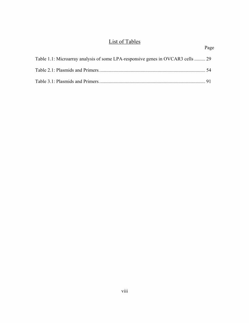
Table 1.1: Microarray analysis of some LPA-responsive genes in OVCAR3 cells ......... 29
Table 2.1: Plasmids and Primers....................................................................................... 54
Table 3.1: Plasmids and Primers....................................................................................... 91
ix
List of Figures Page
Figure 1.1: Metabolism of bioactive LPA ........................................................................ 24
Figure 1.2: Representation of transcription factors binding response elements in the human
Cox-2, IL-6 and uPA promoters ....................................................................................... 32
Figure 1.3: Isoforms of C/EBP-β ...................................................................................... 37
Figure 2.1: LPA induces expression of Cox-2 protein various ovarian cancer cell lines. 58
Figure 2.2: LPA is preferred in the induction of Cox-2 expression in ovarian cancer cell
lines ................................................................................................................................... 60
Figure 2.3: LPA induces release of PGE2 in ovarian cancer cell line............................... 61
Figure 2.4: Involvement of arachidonic acid (AA) and Cox-2 enzyme activity in PGE2
production ......................................................................................................................... 63
Figure 2.5: LPA receptor subtypes of the Edg family are expressed in ovarian cancer cells
........................................................................................................................................... 64
Figure 2.6: LPA stimulates Cox-2 expression through LPA1, LPA2 and LPA5 but not LPA3
or PPARγ .......................................................................................................................... 65
Figure 2.7: Mechanism of LPA-induced Cox-2 gene expression is Gi and ERK
independent ...................................................................................................................... 67
Figure 2.8: LPA induces both Cox-2 mRNA levels and posttranscriptional enhancement of
Cox-2 mRNA stability ...................................................................................................... 68
Figure 2.9: Effect of LPA on Cox-2 expression involves transcriptional activation of Cox-2
promoter ............................................................................................................................ 69

x
Figure 2.10: LPA induces transcriptional activation of Cox-2 through C/EBP
independently of AP-1 or NF-κB...................................................................................... 71
Figure 2.11: Inhibition of AP-1 or NF-κB does not interfere with LPA-induced Cox-2 gene
expression.......................................................................................................................... 73
Figure 2.12: C/EBP-β is crucial for transcriptional activation of Cox-2 expression by LPA
........................................................................................................................................... 75
Figure 2.13: HuR binds to Cox-2 mRNA, contributing to the sustained induction of Cox-2
by LPA .............................................................................................................................. 78
Figure 3.1: LPA induces phosphorylation and expression of C/EBP-β in ovarian cancer
cells ................................................................................................................................... 93
Figure 3.2: Induction of C/EBP-β expression by LPA is a consequence of new protein
synthesis ............................................................................................................................ 94
Figure 3.3: LPA activates binding and transcriptional activities of C/EBP-β .................. 96
Figure 3.4: RTK agonists weakly induce C/EBP-β activity in ovarian cancer cells ........ 97
Figure 3.5: EGFR inhibitor blocks LPA-induced activation of C/EBP-β ........................ 98
Figure 3.6: RTK-dependent activation of C/EBP-β mediates induction of Cox-2 and IL-6
......................................................................................................................................... 102
Figure 3.7: LPA activation of uPA involves C/EBP-β and requires EGFR kinase activity
......................................................................................................................................... 105
Figure 3.8: Schematic representation of LPA GPCR-RTK crosstalk in activation of
transcription factors and gene expression ....................................................................... 108

xi
Figure 4.1: Activation of AP-1 expression, binding and transcriptional activity by LPA
......................................................................................................................................... 121
Figure 4.2: Comparison of LPA to RTK agonists in activation of AP-1........................ 122
Figure 4.3: Requirement of RTK for LPA-induced activation of AP-1 ......................... 124
Figure 4.4: Effects of overexpression of dominant negative EGFR on LPA-induced AP-1
protein expression ........................................................................................................... 125
Figure 4.5: EGFR-independent activation of NF-κB by LPA ........................................ 127
Figure 4.6: G proteins cascades mediating LPA-induced AP-1 and NF-κB activation.. 130
Figure 4.7: Differential effects of EGFR inhibition on Ras and Rho activation by LPA.133
Figure 4.8: EGFR-independent activation of Gq signaling pathway by LPA................. 134
Figure 4.9: Role of EGFR in LPA-mediated biological responses................................. 136
Figure 5.1: Hypothetical model of activation of G proteins and transcription factors by
LPA ................................................................................................................................. 147
xii
List of Abbreviations
AA Arachidonic acid
Ab Antibody
AP-1 Activator Protein-1
ATF Activating transcription factor
ATP Adenosine triphosphate
ATX Autotaxin
Bcl10 B-cell CLL lymphoma 10
BSA Bovine serum albumin
bZIP Basic leucine zipper
C-terminus Carboxyl terminus
C/EBP CCAAT enhancer binding protein
CamK Camodulin kinase
cAMP Cyclic adenosine monophosphate
CARMA3 CARD and MAGUK domain-containing protein 3
cDNA Complementary deoxyribonucleic acid
ChIP Chromatin immunoprecipitation
CMV Cytomegalovirus
Cox Cyclooxygenase
CRE cAMP response element
xiii
DMEM Dulbecco’s modified eagle medium
DN Dominant negative
DNA Deoxyribonucleic acid
DTT Dithiothreitol
EDG Endothelial differentiation gene
EDTA Ethylenediaminetetraacetic acid
EGF Epidermal growth factor
EGFR Epidermal growth factor receptor
EIA Enzyme Immunoassay
ELISA Enzyme-linked immuno sorbent assay
EMSA Electromobility shift assay
ERK Extracellular signal-regulated kinsae
FBS Fetal bovine serum
Fra Fos-related antigen
G Guanine nucleotide
GAPDH Glyceraldehyde 3-phosphate dehydrogenase
GFP Green fluorescent protein
GPCR G protein coupled receptor
GSK-3 Glycogen synthase kinase-3
h hour
HDAC Histone deacetylase
xiv
HGF Hepatocyte growth factor
IGF Insulin-like growth factor
IκB Inhibitor of kappa B
IKK Inhibitor of kappa B kinase
IL Interleukin
JAK Janus kinase
JNK c-Jun N-terminal kinase
kDa Kilo Dalton
LAP Liver activating protein
LIP Liver inhibiting protein
LPA Lysophosphatidic acid
MALT-1 Mucosa associated lymphoid tissue lymphoma translocation gene 1
MAPK Mitogen-activated protein kinase
min minute
NEMO NF-κB essential modulator
NF-κB Nuclear factor-kappa light chain enhancer of B cells
N-terminus Amino- terminus
PAF Platelet-activating factor
PAGE Polyacrylamide gel electrophoresis
PAP Potato alkaline phosphatase
PCR Polymerase chain reaction
xv
PGE2 Prostaglandin E2
PI3K Phosphoinositol 3-kinase
PKC Protein kinase C
PKD Protein kinase D
PPAR Peroxisome proliferator-activated receptor
PTX Pertussis toxin
RHD Rel homology domain
RLU Relative luciferase unit
RNA Ribonucleic acid
rpm Revolutions per minute
RSK p90 ribosomal S6 kinase
RTK Receptor tyrosine kinase
RT-PCR Reverse transcription polymerase chain reaction
S1P Sphingosine-1-phosphate
SAPK Stress-activated protein kinase
SDS Sodium dodecyl sulfate
siRNA Small interfering ribo nucleic acid
TBK1 TANK-binding kinase 1
TK Thymidine kinase
TNF-α Tumor necrosis factor alpha
TRE 12-O-tetradecanoylphorbol-13-acetate (TPA) responsive element

xvi
uPA Urokinase plasminogen activator
UTR Untranslated region
VEGF Vascular endothelial growth factor
WT wildtype
Y Tyrosine
xvii
Abstract
MOLECULAR MECHANISMS FOR REGULATION OF GENE EXPRESSION BY
LYSOPHOSPHATIDIC ACID IN OVARIAN CARCINOMA CELLS
By Regina Adenike Oyesanya, PhD
A dissertation submitted in partial fulfillment of the requirements for the degree of Doctor of Philosophy at Virginia Commonwealth University.
Virginia Commonwealth University, 2009
Major Director: XIANJUN FANG ASSISTANT PROFESSOR, BIOCHEMISTRY AND MOLECULAR BIOLOGY
Lysophosphatidic acid (LPA) is a potent bioactive phospholipid mediator that
functions through multiple G protein couple receptors (GPCRs). LPA is elevated in ascites
of ovarian cancer patients and is involved in growth, survival and metastasis of ovarian
cancer cells. Gene promoter analyses revealed that some LPA-target genes share similar
sets of binding sites for prominent transcription factors posing the possibility of a general
mechanism for activation of their expression by LPA. Detailed investigation of the

xviii
mechanisms of regulation of cyclooxygenase 2 (Cox-2), a paradigm of LPA-regulated
genes, showed that LPA robustly upregulated the expression of Cox-2 in ovarian cancer
cells through multiple receptors. LPA induced rapid increase in Cox-2 mRNA and
significantly enhanced the stability of Cox-2 transcript with the support of mRNA binding
protein HuR. The effects of LPA on Cox-2 transcriptional activation include essential
involvement of transcription factor, C/EBP-β. Further studies on mechanisms of activation
of C/EBP-β demonstrated that LPA increased phosphorylation, binding and transcriptional
activities of C/EBP-β. In addition, activation of C/EBP-β and LPA-target genes required
contribution from EGFR. This novel crosstalk between LPA GPCRs and EGFR in
mediating transcription factors activation was further explored by investigating the
mechanisms of activation of AP-1 and NF-κB by LPA. Activation of AP-1 family of
proteins by LPA relied heavily on basal inputs from EGFR as inhibition of EGFR kinase
activity with AG1478 caused significant loss of LPA-induced AP-1 expression, binding
and transcription activities. Although HGF and other agonists of RTK only weakly
stimulate LPA-target genes and transcription factors in ovarian cancer cells, costimulation
with HGF in the presence of AG1478 restored LPA signals to both C/EBP-β and AP-1.
This suggests an obligatory role for a RTK in LPA-induced transcriptional activation, not
necessarily inputs from EGFR. Interestingly, inhibition of EGFR with AG1478 did not
interfere with LPA-induced NF-κB activation. Pharmacological inhibition and molecular
targeting revealed that only a subset of G proteins participate in the crosstalk between LPA
receptors and EGFR. Collectively, these results demonstrate the presence of at least two

xix
signals downstream of LPA receptors: one dependent on basal RTK activity and another
mediated directly by LPA GPCRs.

20
CHAPTER ONE
INTRODUCTION
1.0 OVERVIEW
Cancer is a disease of uncontrolled cell growth. Cancer cells often possess the
ability to invade adjacent tissues through the release of substances or molecules that can
degrade the tissue material. Some of these malignant cells may also spread to distant sites
in the body via the blood or lymph, a process known as metastasis. All aspects of cancer
development, survival and progression are strongly anchored on abnormal gene
expression, a consequence of bypass of critical points of gene regulation. Many cancer
types originate from cells that possess gene mutations, deletions or amplifications. As
such, certain cellular functions including activation of transcription factors or
posttranslational modifications of synthesized proteins become highly enhanced. Tumor-
suppressing genes are inactivated while growth-promoting oncogenes are continually
turned on, giving the cells new properties, particularly growth advantages. Major
mechanisms responsible for the enhanced growth of malignant cells include reduce
dependence on growth factor, insensitivity to growth inhibition, evasion from programmed
cell death (also called apoptosis), and unlimited growth potentials. The adverse effects of
cancer on patients are therefore mainly due to tumor burden from increased cell number

21
from the dysregulated cell growth. Most cancer therapies target the reduction of cell
number and the prevention of further accumulation of tumor cells.
Ovarian cancer remains the leading cause of death from gynecological cancer and
the fifth leading cause of death from cancer in women. According to the American Cancer
Society, there are an estimated 21,650 new cases of ovarian cancer and 15,520 deaths due
to this malignancy in the United States in 2008 [1]. Ovarian epithelial carcinoma is the
most common type of ovarian cancer. This type includes endometrioid carcinoma, serous
carcinoma, mucinous carcinomas, clear cell carcinoma and borderline tumor. Ovarian
cancer is classified into four stages (Stages I-IV). Most tumor markers are insufficient to
detect ovarian cancer at early Stages I and II because they either lack sensitivity and
specificity for ovarian cancer or are not elevated until advanced stages of the disease.
Hence, ovarian cancer has been termed a “silent killer” due to of the lack of symptoms or
accurate tumor markers at the early stages leading to poor prognosis.
At the advanced Stages III and IV, ovarian cancer is often characterized by
extensive intraperitoneal distribution of tumors and formation of large volumes of ascitic
fluid. The ascitic fluid from ovarian cancer patients contains the ovarian tumor cells and a
broad range of potent growth factors including lysophosphatidic acid (LPA) [2, 3]. The
levels of LPA in the plasma of ovarian cancer patients, including those at early stages, are
significantly higher than those in normal controls [2, 4]. Up to 80 μM LPA has been found
in the ascitic fluid of ovarian cancer patients [4-6]. LPA is therefore considered to be
biomarker for ovarian cancer. It is now known that LPA influences many processes of
tumor cells including growth, survival migration and metastasis [7, 8]. LPA mediates these

22
processes at least partially through regulating the expression of diverse genes and
metabolic pathways [9-12]. Understanding the detailed mechanisms by which LPA
regulates gene expression may lead to identification of critical therapeutic targets for the
treatment of ovarian cancer and perhaps, other cancer types.
1.1 Metabolism of LPA
LPA (1-acyl-sn-glycerol-3-phosphate) is a naturally-occurring phospholipid. It can
be produced by different cell types including activated platelets [13, 14], endothelial cells
[15], fibroblasts [16], adipocytes [17], prostate [18] and ovarian cancer cells [7]. Thus, it is
present in body fluids including plasma (bound to albumin), saliva, hair follicles and
malignant effusions [19, 20]. There are multiple pathways potentially responsible for the
endogenous generation of LPA particularly the actions of certain enzymes on
phospholipids of the cell membranes [13, 21]. A major part of LPA produced by activated
platelets is synthesized by the sequential actions of phospholipase A1 or A2 (PLA1/2) on
serum or membrane phospholipids such as phosphatidylcholine (PC) followed by
hydrolytic actions of a lysophospholipase D (lysoPLD) present in plasma (Fig. 1.1).
Recently, autotaxin (ATX), an exo-phosphodiesterase, implicated in cell motility was
found to be an important enzyme in the production of LPA and the predominant source of
extracellular LPA [19, 22]. ATX is synthesized as a full-length, or pre-pro-enzyme, that is
proteolytically cleaved in transit along the classical export pathway and secreted as a
catalytically active glycoprotein [23]. With its intrinsic lysoPLD activity, ATX can
23
hydrolyze lysophosphatidylcholine (LPC), a major phospholipid secreted by hepatocytes
and therefore abundant in blood and plasma [24-26], into LPA. The phosphorylation of
monoacylglycerol by acylglycerol kinase (AGK) is another source of LPA [18]. When
overexpressed, AGK in the mitochondria is able to mediate the production and secretion of
LPA by phosphorylation of monoacylglycerol. The exact pathways for the generation of
LPA in ascites, saliva, seminal and other body fluids are yet to be fully delineated.
Similarly, the mechanism for intracellular production of LPA is poorly understood.
In ovarian and other cancer cells, LPA production can be stimulated by cell activation in
response to phorbol esters [27], bombesin [27] and LPA itself [28, 29]. The activation of
LPA production may involve multiple steps catalyzed by phospholipases, unlike the
extracellular pathway where the precursors of LPA already preexist. In normal cells, the
intracellular and extracellular LPA levels are tightly controlled by LPA synthesizing and
metabolizing enzymes. Lipid phosphate phosphohydrolases (LPP) are a family of enzymes
that catalyze the dephosphorylation of LPA [30-32]. These enzymes are membrane-
associated with extracellularly-facing catalytic site for clearance of LPA on the cell
membrane. There is evidence that expression of these enzymes reduce LPA levels and
compromise LPA-induced cellular functions [30]. In addition to dephosphorylation, LPA
can also be converted to phosphatidic acid (PA) by acylation through the action of LPA
acyl transferases (LPAAT) [33, 34].

24
1.2 LPA Receptors and Signal Transduction
LPA is a bioactive phospholipid and a potent mediator of a broad range of cellular
responses. It promotes cell proliferation and survival; enhances cell migration and
invasion; and induces changes in actin cytoskeleton and focal contact organization [3, 7,
Fig. 1.1. Metabolism of bioactive LPA. LPA is formed extracellularly by diverse pathways including the deacylation of phosphatidic acid (PA) by PLA1/2 and the cleavage of lysophospholipids, predominantly LPC by autotoxin, which represent the major source of extracellular LPA. Overexpression of AGK in mitochondria was recently shown to promote the generation and release of LPA from monoacylglycerol (MAG) and diacylglycerol (DAG). [After Biochim Biophys Acta. 2007: 1768(4):923-40]
shed
microvesicles
ATX
ATX
LPAAT
PLA1/2
PPARγ
LPA
LPC MAG
LPP
PAPA
LPA PAPGacyl-CoA
+de novo-
phospholipid-synthesis
effects LPA,
LPA-GPCR
AGKMAG,DAG
PA
shed
microvesicles
ATXATXATXATX
ATX
LPAAT
PLA1/2
PPARγ
LPA
LPC MAG
LPP
PAPA
LPA PAPGacyl-CoA
+PG
acyl-CoA+
de novo-phospholipid-synthesis
effects LPA,
LPA-GPCR
AGKMAG,DAG
PA

25
29]. These responses culminate from the activation of a diverse array of signaling
pathways initiated when LPA binds its receptors on the plasma membrane. At least seven
LPA receptors have been identified. Based on their primary structure, LPA receptors are
classified into two groups: the endothelial differentiation gene (Edg) group and the
purinergic receptor family (P2Y) group. LPA1/Edg-2, LPA2/Edg-4 and LPA3/Edg-7 belong
to the Edg family and share about 50-57% homology in their amino acids [35-38].
LPA4/P2Y9/GPR23 and LPA5/P2Y5 of the P2Y family of receptors are two novel LPA
receptors structurally distant from the LPA receptors of the Edg family, sharing only 20-
24% homology with LPA1-3 [39, 40]. LPA has also been identified as a ligand for two
additional orphan receptors GPR87 and P2Y10 of the P2Y family [41, 42]. The identities of
these receptors as bona fide LPA receptors are yet to be thoroughly studied.
LPA receptors are G protein coupled receptors (GPCRs). They elicit their activities
by coupling to trimeric G proteins subunits, Gα and Gβγ [35-40]. Aberrant regulation
GPCRs have been linked to numerous diseases including cardiovascular defects, diabetes,
allergies and certain forms of cancer [43-46]. More than 30% of the drugs in current use
target the inhibition of GPCRs [47, 48]. LPA GPCRs couple to diverse G proteins
including Gi, Gq and G12/13 to initiate the activation of parallel yet interactive intracellular
signaling cascades culminating in physiological responses. Activation of Gq mediates the
activation of phospholipase C (PLC) with subsequent hydrolysis of phosphatidylinositol
biphosphate (PIP2) to inositol trisphosphate (IP3), an activator of intracellular calcium
release and diacylglycerol (DAG), that activates protein kinase C (PKC) [16, 49]. Gi
mediates the inhibition of adenylate cyclase leading to downregulation of intracellular
26
cAMP. Gi or associated Gβ/γ subunit are also linked to activation of Ras and downstream
mitogenic Ras/mitogen activated protein kinase (MAPK) and phosphoinositide 3-kinase
(PI3K) [50, 51]. Activation of Ras-MAPK and PI3K are critical to LPA-induced cell-
proliferation, migration and survival [50, 52]. The effects of LPA on stress fibre formation
and the cell cytoskeleton occur through the activation of G12/13/ RhoA [53].
Rapid internalization of receptor from the plasma membrane following ligand-
induced activation is one way of quenching LPA signals [54]. The mechanism of the
metabolic fate of the receptor after internalization is not yet known. In addition to GPCRs,
LPA may also have some intracellular targets including proliferator-activated receptor γ
(PPAR-γ) [55, 56]. PPAR-γ regulates the transcription of genes involved in glucose and
fatty acid metabolism, adipocytes differentiation and inflammation process [57, 58]. LPA
may be able to enter the cell in sufficient quantity and activate this intracellular receptor,
suggesting its participation in intracellular signaling and cell functions. While
overexpression studies have helped to understand the general functions of LPA receptors,
the challenge remains as to the assignment of LPA receptor subtypes to specific signal
transduction cascades and define their relative contribution to the multiple biological
activities of LPA.
1.3 Role of LPA in Tumor Biology
Gene targeting and pharmacological inhibition of LPA receptor subtypes in mice
and different cell types revealed diverse physiological and pathological roles for LPA

27
signaling. The Edg LPA receptors are differentially expressed in various tissues [59, 60].
LPA1 is most widely expressed and present in both normal and malignant cells. In contrast,
expression of LPA2 is more restricted. LPA3 is barely seen in normal tissues [60]. Recent
studies showed that, in ovarian and thyroid cancers, malignant transformation is associated
with increased expression of LPA2 (and LPA3 in ovarian cancer) [61, 62]. LPA receptors
are also overexpressed in many other cancer types including endometrioid, colon, and
colorectal cancer [63-65]. These observations suggest that changes in LPA receptor
expression during malignant transformation are intimately involved in carcinogenesis.
Furthermore, increased expression LPA receptors also correlates with important cancer
progression processes such as migration and metastasis in many cancer types [63-65].
Recent studies have demonstrated that the presence of LPA in intraperitoneal effusions of
ovarian cancer patients may contribute significantly to the progression and aggressive
characteristics of the malignant cells [7, 62, 66]. In addition, various ovarian cancer cell
lines respond to LPA stimulation with increase in migration and invasion. The influence of
LPA on various cellular processes is supported by its ability to regulate the expression of
diverse genes.
1.4 Regulation of Gene Expression
The most fundamental task of any organism is the control of the expression of the
thousands of genes harbored by its genome. Genes are segments of DNA that carry
information necessary for the development and proper functioning of all living organisms.

28
A normal cell possesses the capacity to process in parallel, the many regulatory inputs
received from within and without, into enormous regulatory outputs that are tissue specific.
When a cell grows, it divides by replicating its DNA into two daughter cells, each having
the same genetic information as the parent cell. Gene expression, the representation of this
inheritable information from the sequence of bases of the DNA to functional forms, is a
complex and tightly regulated process. Diverse functions and features are acquired by
selectively expressing or repressing segments of the DNA. Although basal expression of
certain genes occurs in a resting cell, the active expression or repression of many genes are
signaled for by complex sets of molecules and processes within or external to the cell.
Abnormal gene expression by malignant cells is a result of the circumvention of these
regulatory signals.
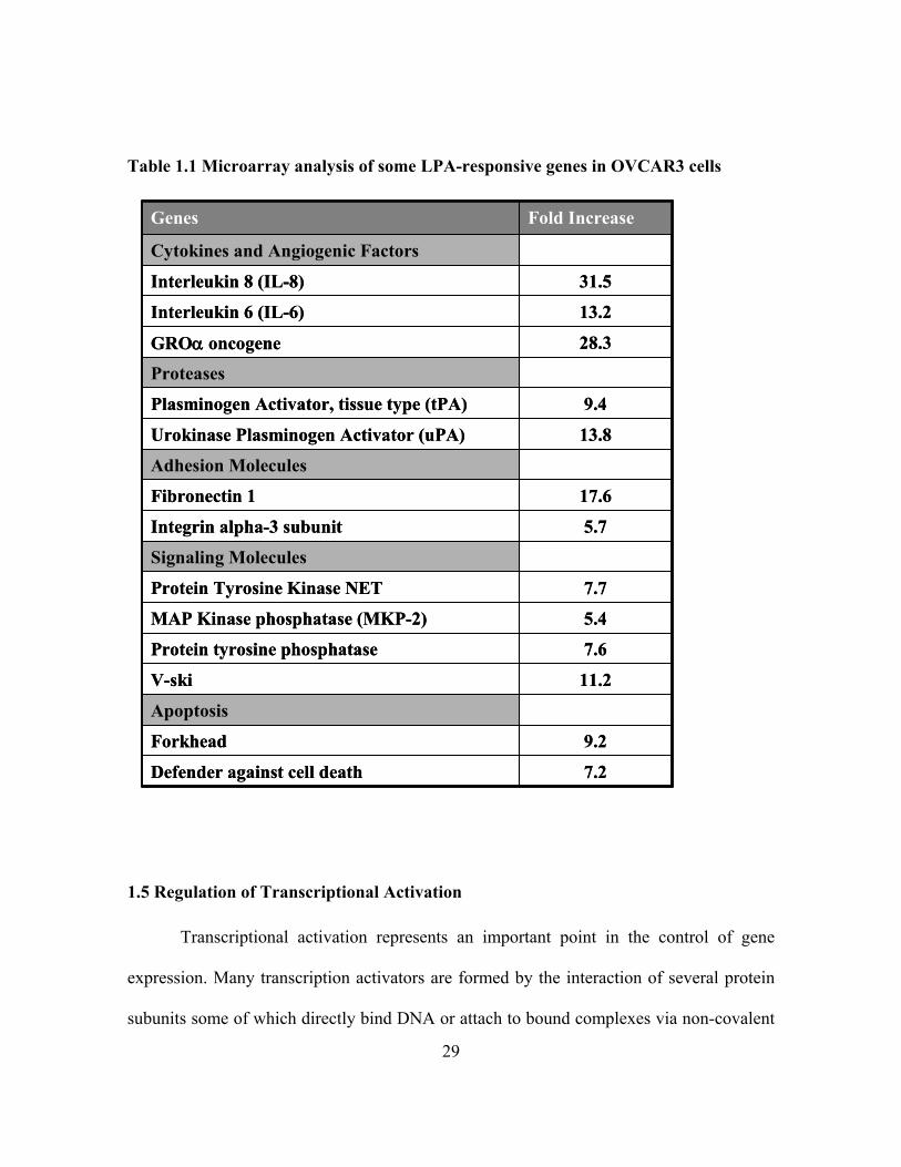
Microarray analysis of LPA-induced gene expression in an ovarian cancer cell line
from our lab showed that LPA stimulated expression of many cancer-related genes (Table
1.1). LPA can therefore modulate cellular responses of malignant cells by inducing
expression of these targets genes including cytokines, proteases, cell adhesion molecules,
proangiogenic factors and anti-apoptotic genes. The array of LPA-target genes continue to
expand ascribing new roles for LPA in more physiological and pathological contexts.
However, the mechanistic details of how LPA regulates the expression of many genes
remain elusive. As described in Chapter 2 of this dissertation, we have focused on LPA-
induced expression of cyclooxygenase 2 (Cox-2) as a model to elucidate the mechanisms
of gene regulation by LPA.
29
Table 1.1 Microarray analysis of some LPA-responsive genes in OVCAR3 cells
7.2Defender against cell death
9.2Forkhead
Apoptosis
11.2V-ski
7.6Protein tyrosine phosphatase
5.4MAP Kinase phosphatase (MKP-2)
7.7Protein Tyrosine Kinase NET
Signaling Molecules
5.7Integrin alpha-3 subunit
17.6Fibronectin 1
Adhesion Molecules
13.8Urokinase Plasminogen Activator (uPA)
9.4Plasminogen Activator, tissue type (tPA)
Proteases
28.3GROα oncogene
13.2Interleukin 6 (IL-6)
31.5Interleukin 8 (IL-8)
Cytokines and Angiogenic Factors
Fold IncreaseGenes
1.5 Regulation of Transcriptional Activation
Transcriptional activation represents an important point in the control of gene
expression. Many transcription activators are formed by the interaction of several protein
subunits some of which directly bind DNA or attach to bound complexes via non-covalent

30
interactions. Emerging evidence suggests that the influence of LPA on gene expression to
a large extent involves activation of transcription factors [67-69]. With 10% of genes in the
human genome coding for transcription factors, this group are the single largest family of
human proteins (approx. 2600 members) [70, 71]. Transcription factors bind to
unexpressed portion of the DNA mainly made up of specific sequences of regulatory
modules. These modules contain distal enhancer elements, core or basal promoter elements
and proximal promoter elements. The TATA element, located 25 base pair (bp) from the
transcription start site and a pyrimidine-rich initiator (Inr) element found at the
transcription start site, represent major modules of the core promoter element [72, 73].
Both elements can function independently or synergistically [73]. A cell responds to
stimuli such as growth factors and hormones by turning off or on signaling cascades that
usually peaks with activation of one or more transcription factors.
Many transcription factors consist of one or more DNA binding domain (DBD)
[74]. They also often possess a trans-activating domain (TAD) and/or a signal sensing
domain (SSD) [75]. The DBD and TAD domains of a single transcription factor can
function independently [74]. Transcription factors positively or negatively modulate the
expression of their target genes. Therefore, a transcription factor can be an activator
(promoting transcription) or a repressor (downregulating or suppressing transcription).
Functionally, a transcription factor can either be constitutively-active (present in the cell all
the time) or conditionally-active (requiring cell-specific or external signal for activation).
The most important tumor suppressor gene is protein p53, a transcription factor
activated following cellular stress [76, 77]. More than 40% of epithelial ovarian
31
carcinomas are known to harbor inactivating mutations in p53 gene [78, 79]. The presence
of inactivating p53 mutation in many cancer types underscores the importance of the anti-
proliferative functions of this transcription factor. In fact, drug resistance in cancer therapy
has been associated with p53 mutations [80-82]. Unlike p53, most of the other
transcription factors known to play important roles in the proliferation and survival of
cancer cells are either overexpressed or highly activated. Several studies have described
the general mechanisms for the activation of common transcription factors including
activator protein 1 (AP-1), signal transducers and activators of transcription (STATs),
specificity protein 1 (Sp-1), CCAAT/enhancer binding proteins (C/EBPs) and nuclear
factor-kappa light chain enhancer of B cells (NF-κB). However, specific information of
how cellular context might modulate the activities of these proteins in many human
malignancies including ovarian cancer is still lacking. Many LPA-target genes harbor
binding sites for a common subset of transcription factors in their promoters, suggesting
common mechanisms for their regulation by LPA (Fig. 1.2). Targeting pathways that
activate these transcription factors remains an attractive option for the treatment of cancer.
Mechanistic details of the activation of these transcription factors by LPA in ovarian
cancer will promote better understanding of ovarian oncogenesis and may lead to
identification of novel targets for treatments of ovarian cancer.

32
1.5.1 Activation of AP-1 family of Transcription Factors
AP-1 proteins belong to the bZIP family, a group of protein that possesses a
bipartite DNA-binding motif consisting of a basic region for DNA contact and a leucine
zipper region for dimerization. AP-1 members include the Jun proteins (c-Jun, Jun B, Jun
D), ATF and Fos proteins (c-Fos, FosB, Fra-1 and Fra-2) [83, 84]. These transcription
factors play a central role in the regulation of gene expression and cell transformation. AP-
1 controls cellular processes such as differentiation, proliferation and apoptosis in response
to a variety of stimuli including growth factors, stress, cytokines and bacterial and viral
infections [84, 85]. AP-1 proteins interact with a TPA (12-O-tetradecanoylphorbol-13-
acetate) responsive element (TRE) TGAC/GTCA on the promoter and enhancer regions of
Fig. 1.2. Representation of transcription factors response elements in the human Cox-2, IL-6 and uPA promoters Upstream location to the start site of transcription is indicated with numbers (not to scale). Black shapes represent binding site for indicated transcription factor.
‐1‐500IL‐6
‐1‐1000Cox‐2
‐1‐1600uPA C/EBP‐β
AP‐1NF‐κB
‐1‐500IL‐6
‐1‐1000Cox‐2
‐1‐1600uPA C/EBP‐β
AP‐1NF‐κB
‐1‐500IL‐6
‐1‐500 ‐1‐500IL‐6
‐1‐1000Cox‐2
‐1‐1000 ‐1‐1000Cox‐2
‐1‐1600uPA
‐1‐1600 ‐1‐1600uPA C/EBP‐β
AP‐1NF‐κB
C/EBP‐βC/EBP‐βAP‐1AP‐1NF‐κBNF‐κB

33
their target genes, and bind DNA after dimerization [86, 87]. Fos proteins though lacking
DNA-binding domain, become transcriptionally active when they form heterodimers with
other AP-1 components. In contrast, Jun proteins can form both homodimers and
heterodimers though Jun-Fos heterodimers are more stable and therefore favored [88]. In
fact, the re-introduction of c-Fos in F9 teratocarcinoma cells was shown to enhance the
transcriptional and transforming properties of c-Jun and JunB [89, 90]. Dimers of AP-1
proteins can stimulate or repress transcription. While c-Jun/c-Fos heterodimers are known
transcriptional activators, Jun B/c-Fos complexes are mostly repressors of transcription
[91-93]. Since c-Jun and c-Fos members vary significantly in their relative abundance in
different cell types, a complex network of transcriptional regulators is formed when these
proteins interact with family members and with additional proteins. AP-1 proteins dimerize
efficiently with other transcription factors such as ATF/CREB family of proteins [94-96].
Nearly all AP-1 components have been implicated in tumor development and
progression; and many of these proteins have also been shown to possess transforming
potentials [97, 98]. The expression pattern of AP-1 proteins in tumors varies depending on
tissue type [99-101]. For example, high expression levels of Fra-1 and Fra-2 are associated
with metastatic cell lines such as mouse mammary adenocarcinoma CSML-100 [102, 103].
However, no detectable expression of c-Fos or Fos B was found in this cell type. In a
closely associated weakly invasive and non-metastatic CSML-10, only c-Fos was detected;
but the expression of c-Jun remained essentially the same in both cell lines [102].
Immunohistochemical studies using well differentiated endometrioid endometrial tumor
samples showed significant correlation of high tumor grade or disease stage with

34
expression of c-Fos [101]. The same studies showed that the overexpression of c-Fos seem
to substitute for the expression and perhaps the role of Fra-1 in non-endometriod tumors
such as breast carcinomas. The role of various AP-1 components in other cancer types is
inconclusive. For instance, reduced cell viability was observed in ovarian cancer cells
overexpressing a dominant-negative form of c-Fos in the presence of non lethal doses of
cisplatin, an anticancer drug [104]. However, another experimental system demonstrated
that c-Fos protein levels in ovarian carcinoma cell lines correlate with response to
paclitaxel therapy in nude mouse xenograft [105].
In many cell types, expression of AP-1 proteins is often temporally modulated in
response to stimuli. As such, the functional activity of AP-1 in a particular cell is not only
dynamic with respect to time but also a function of the differentiation state and
environment of the cell. The DNA binding activity and transcriptional capacity of AP-1
proteins are greatly affected by post-translational modifications particularly
phosphorylation. The c-Jun N-terminal kinases (JNK or stress activated protein kinase
SAPK), members of the MAPK family, are the mediators of phosphorylation of Jun family
members [106] while a variety of proteins have been reported as putative c-Fos kinases
including MEK5, RSK and p38 MAPKs [107-109]. c-Jun is phosphorylated by JNK at
specific serine residues. In particular, phosphorylation of c-Jun at Serine 63 and 73 located
within its transactivation domain increases its transactivation capacity [106]. Studies show
that deacetylation of c-Jun by CBP, a histone deacetylase enhances the transcriptional
activity by several folds [110, 111]. Further, the promoter of c-Jun harbors the binding
sites of many transcription factors including AP-1 itself. Thus, agonist-induced c-Jun

35
transcription is often followed by an increase in the expression of c-Jun. This positive auto-
regulatory loop is a common phenomenon shared by many other AP-1 components [112-
114]. AP-1 target genes include MMPs [115], uPA [116], VEGF [117], CD44 [118], and
Bcl-2 [119]. A subset of these genes responds to LPA stimulation in diverse cell types [12,
62].
1.5.2 Activation of C/EBP
The C/EBP family is made up of six members: C/EBP-α, C/EBP-β, C/EBP-γ,
C/EBP-δ, C/EBP-ε, and C/EBP-ζ [120, 121]. With the exception of C/EBP-ε, and C/EBP-
ζ, this subfamily of transcription factors belong to the exclusive group of liver-enriched
transcription factors having been first discovered in the liver. C/EBPs are also members of
the bZIP family of proteins [121]. C/EBPs form homo- and heterodimers with family
members and with other bZIP family of transcription factors including the AP-1 proteins c-
Jun and c-Fos [122, 123]. C/EBPs recognize a specific palindromic sequence in the major
groove of DNA. It has been proposed that dimerization between two groups of leucine
zipper proteins brings the basic amino acids of DNA binding domain into close proximity
[120, 124, 125]. Hence C/EBPs dimerization is a prerequisite for DNA binding and dimers
readily dissociate into monomers when not bound to DNA. The C/EBP proteins also
contain activation and regulatory domains in the N-termini [125].
C/EBPs play important roles in cell proliferation and differentiation, liver
regeneration, energy metabolism, tumorigenesis and other physiological processes [121,
36
126, 127]. Although tissue expression patterns of C/EBPs often overlap, there exist
significant differences in the functions of each member of the family [128]. C/EBP-α-/-
mice are neonatal lethal due to hypoglycemia and lack of stored liver glycogen,
accentuating the role of C/EBP-α in glucose metabolism and terminal differentiation of
adipogenesis and hematopoiesis [124, 129]. However, C/EBP-β-/- mice are viable with
serious defects in hematopoiesis and immune system [130, 131]. In addition, these mice
showed a defective female reproduction system [132]. The loss of fertility in C/EBP-β-/-
mice underscores the involvement C/EBP-β in ovarian follicular development and corpus
leteum formation, enhancing the effects of lutenizing hormone (LH/hCG) [132-134].
Importantly, C/EBP-β is preferentially expressed in endometrial adenocarcinoma and has
been shown to be overexpressed in ovarian cancer. Its expression level highly correlates
with progression of the disease [135].
In some cell systems, expression of C/EBP-β gene can be induced by inflammatory
cytokines, steroid hormones and growth factors [122, 136]. The effects of these stimuli on
C/EBP-β expression can be simple in some cellular context but complicated in others.
Insulin is a classical modulator of C/EBP-β expression in the liver [127]. In a rat hepatoma
cell line, co-stimulation with insulin resulted in attenuation of C/EBP-β mRNA expression
induced by cytokines and dexamethasone, while on its own, insulin increased C/EBP-β
mRNA [137, 138]. The mechanisms involved in the regulation of C/EBP-β expression in
other tissues types are not fully understood.
37
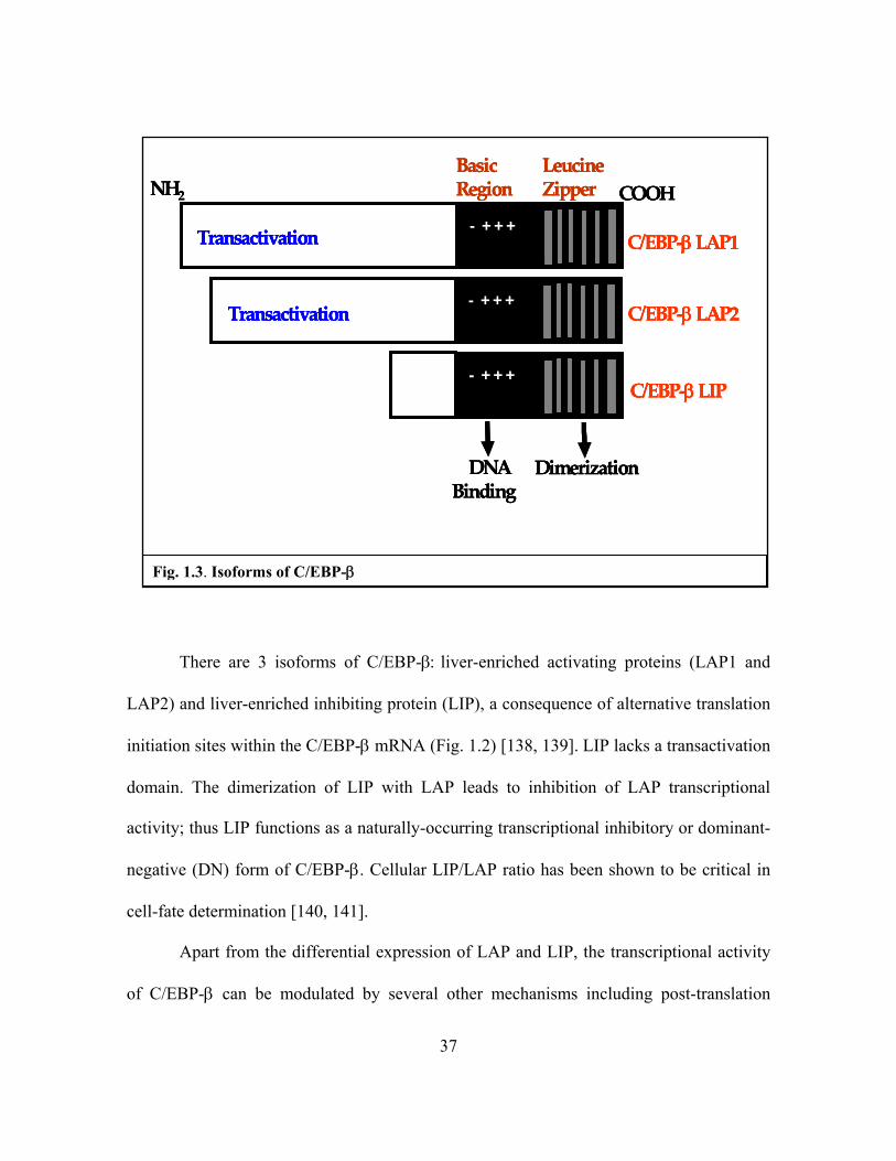
There are 3 isoforms of C/EBP-β: liver-enriched activating proteins (LAP1 and
LAP2) and liver-enriched inhibiting protein (LIP), a consequence of alternative translation
initiation sites within the C/EBP-β mRNA (Fig. 1.2) [138, 139]. LIP lacks a transactivation
domain. The dimerization of LIP with LAP leads to inhibition of LAP transcriptional
activity; thus LIP functions as a naturally-occurring transcriptional inhibitory or dominant-
negative (DN) form of C/EBP-β. Cellular LIP/LAP ratio has been shown to be critical in
cell-fate determination [140, 141].
Apart from the differential expression of LAP and LIP, the transcriptional activity
of C/EBP-β can be modulated by several other mechanisms including post-translation
NH2 COOH- + + +Transactivation C/EBP‐βLAP1
Basic Region
LeucineZipper
DNA Binding
Dimerization
C/EBP‐βLAP2- + + +
Transactivation
C/EBP‐βLIP- + + +
NH2 COOHNH2 COOH- + + +Transactivation C/EBP‐βLAP1- + + +Transactivation - + + +- + + +Transactivation C/EBP‐βLAP1
Basic Region
LeucineZipper
DNA Binding
Dimerization
Basic Region
LeucineZipper
DNA Binding
Dimerization
C/EBP‐βLAP2- + + +
Transactivation C/EBP‐βLAP2- + + +
Transactivation- + + +- + + +
Transactivation
C/EBP‐βLIP- + + +
C/EBP‐βLIP- + + +- + + +
Fig. 1.3. Isoforms of C/EBP-β
38
modifications, nucleo-cytoplasmic shuttling and direct protein-protein interaction between
C/EBP-β and transcription factors of other classes [123, 142]. Phosphorylation of C/EBP-β
at specific serine or threonine residues is an important event that often results in the
increase in its transcriptional activity [121, 125, 126]. However phosphorylation at other
sistes such as Serine 173, 223 and 240 may decrease its DNA binding activity [121,123,
142]. The kinases that mediate the phosphorylation of C/EBP-β include PKA, PKC,
Camodulin Kinase II and MAP kinase [125]. These kinases lie downstream of diverse
signaling cascades including those of GPCRs.
C/EBP-β can interact with other transcription factors including Sp1, AP-1 and NF-
κB to activate transcription. This feature is mediated by the leucine zipper and the DNA
binding domains [123, 143]. Heterodimeriztion between C/EBP and other transcription
factors could enhance transcription activity of individual dimer partners. The crosstalk
could also bring about changes in target specificity. For example, CREB/ATF and C/EBP-
β heterodimer causes C/EBP-β to bind onto palindromic cAMP responsive elements
(CREs) on the DNA rather than the CCAAT C/EBP binding sites resulting in regulation of
different target genes [144]. The activation and involvement of C/EBP-β in LPA-induced
gene regulation is the focus of work described in Chapter 3 of this dissertation.

39
1.5.3 Activation of NF-κB
NF-κB is a ubiquitous transcription factor that plays important roles in many
physiological and pathological processes. It is a central mediator of several inflammatory
responses and immune function. NF-κB is activated by a wide variety of stimuli including
inflammatory cytokines (e.g. tumor necrosis factor TNF) and microbial pathogens (e.g.
lipopolysaccharide LPS) that bind cell surface receptors [145, 146]. Other important
activators of NF-κB are genotoxic stress, DNA damage, UV light, oxidative stress,
chemotherapeutic drugs, phorbol esters, growth factors and physiologic mediators such as
angiotensin II and PAF [145]. Abnormal regulation of NF-κB has been linked to several
disease conditions including inflammatory and autoimmune diseases, septic shock,
improper immune development, viral infection and cancer [147, 148].
In vertebrates, the NF-κB family consists of five Rel protein subunits, so called
because they all share a common N-terminal Rel homology domain (RHD). Rel proteins
may be classified into two groups. The first group consists of RelA (p65), RelB and c-Rel
[149]. These subunits possess within their structures a C-terminal transactivation domain
(TAD) to promote transcription. The second group, p50 and p52 are synthesized from large
precursor molecules p105/p50 (NF-κB1) and p100/p52 (NF-κB2) respectively and lack the
TAD [149]. However, p105 and p100 contain a series of five to seven ankyrin repeats that
blocks a nuclear localization signal within the RHD. Both of these precursor proteins are
processed by cleavage to generate the mature transcription factors, p50 and p52.

40
Rel proteins form homo- and heterodimers by employing a C-terminal Ig-like
domain of about 100 amino acids within the RHD commonly called the dimerization
domain (DimD). Twelve of the fifteen possible dimers are able to bind to a consensus
sequence (5’-GGGACTTTC-3’) on DNA and effectively participate in gene transcription
[150-152]. While some Rel dimers, such as RelA/p50 heterodimers are well known as
transcriptional activators, some others particularly dimers lacking RelA, RelB or c-Rel
such as a p50 and p52 homodimers are generally repressors of κB site transcription [151,
153, 154]. In addition to nuclear localization, dimerization and DNA binding, the RHD
also mediates the interaction with the inhibitors of NF-κB, the IκBs. These proteins,
sometimes regarded in literature as inhibitory subunits of NF-κB, possess a similar ankyrin
repeat domain as in p100 and p105. They however lack the RHD. IκBs include IκBα,
IκBβ, IκBγ (derived from c-terminal domain of p100), ΙκBε IkBζ, Bcl-3, pp40 and avian
fever viral protein p28.2 [155-157]. In unstimulated conditions, NF-κB is complexed with
IκBs through the ankyrin repeat domain resulting in its sequestration in the cytoplasm
(IκBζ is known to retain NF-κB in the nucleus), away from its target genes.
The importance of NF-κB in processes that require rapidly-acting primary
transcription factors (“first responders”) such as inflammation is underscored by the fact
that NF-κB activation does not require new protein synthesis [158]. There are two known
pathways for activation of NF-κB in stimulated cells. In the classical or canonical
pathway, activating signal induces the degradation of IκB proteins. A typical activator of
this pathway is TNFα. Upon ligand-induced TNF receptor activation, multiple signals near

41
the cell membrane converge at the IκB kinase (IKK) complex that consists of IKKα, IKKβ
and IKKγ (or NEMO- “NF-κB essential modulator”). The IKK complex is activated by the
interaction of NEMO with a Lys-63-linked polyubiquitinylated receptor-interacting protein
1, RIP1, a serine-threonine kinase. Activated IKKβ phosphorylates IκBα at two serine
residues, Serine 32 and Serine 36. This tags the NF-κB inhibitor for multiple
ubiquitinylation with subsequent degradation by the 26S proteasome [159]. Previously
concealed NF-κB nuclear localization signal becomes exposed targeting the dimer, usually
p65/p50 to the nucleus for transcription. Interestingly, IκB itself is one of NF-κB target
genes and its expression is upregulated following NF-κB activation.
The alternate or non-canonical pathway of NF-κB activation involves IKKα rather
than IKKβ of the IKK complex. This pathway is based on the processing of p100
following cellular stimulation by cytokines such as lymphotoxin β (LT-β), B cell
activating factor (BAFF), CD40 ligand and viruses including the Epstein-Barr virus (EBV)
[160]. Signal-induced post-translational stabilization of NF-κB inducing kinase (NIK)
causes the protein to interact with a homodimer of IKKα thereby activating the latter. A
key point in this pathway is IKKα phosphorylation of p100, an event that leads to its
polyubitinylation and proteasomal degradation of the C-terminus of the protein. The
remnant portion, p52 continues to interact with RelB, now as a transcriptionally-active
heterodimer that quickly moves into the nucleus. Beside stimulus-specific activation of
either the classical or alternate pathway, a dynamic interaction of different NF-κB dimers

42
with specific gene promoters provides a critical control of the expression of NF-κB target
genes [152].
In addition to IκB-dependent activation the NF-κB, diverse post-translational
modifications serve as alternatives to regulate NF-κB activity. p50, an important partner of
the most-studied NF-κB dimer RelA/p50, is regulated through processing of its precursor,
p105. Similar to p100, stimulus-induced phosphorylation of p105 results in its
polyubiquitinylation and subsequent proteolytic degradation to p50. The DNA binding
activity of p50 has also been shown to be enhanced by phosphorylation of Serine 337
located within its RHD domain [161]. p65 (RelA) can be phosphorylated by several
protein kinases at specific residues and this inducible phosphorylation of p65 is often used
as readout of NF-κB activation. A well-studied phosphorylation site is Serine 536
catalyzed by IKKα/β, IKKε or TBK1 (TANK binding kinase 1) [162-164]. Although they
share sequence homology with IKKα/β, IKKε and TBK1 are not part of the IKK complex.
Phosphorylation of p65 at Serine 536 impairs its interaction with IκBα and increases its
nuclear accumulation. However, whether Serine 536 phosphorylation of p65 is required for
transcription remains controversial [165-167]. Other site-specific phosphorylation of p65
at serine residues includes Serine 435 (by camodulin kinase IV, CaMKIV), Serine 468 (by
IKKβ, IKKε and GSK-3β) [168] and Serine 276 (by catalytic subunit protein kinase A,
PKAc) [169]. While phosphorylation of p65 at these residues are known to increase its
transactivation potential, phosphorylation of threonine residues have been shown to
suppress the activity of NF-κB. p65 phosphorylation of C-terminal Threonine 435 and

43
Threonine 505 is induced by ARF tumor suppressor, p14ARF in a p53-independent
manner [170, 171]. Threonine 505 phosphorylation causes p65 to interact with histone
deactelylase 1 (HDAC1), which greatly inhibits p65 transactivation. The phosphoryation
of p65 is often the prerequisite for other post-translation modifications such as
ubiquitinylation and acetylation that regulate the activity of the protein [172, 173].
Acetylation, like most phosphorylation events often results in enhanced activity of NF-κB
[174].
1.6 Crosstalk between GPCRs and Receptor Tyrosine Kinases (RTKs)
Many transcription factors including those described in the preceding sections are
substrate for molecules downstream of GPCR-induced signaling cascade, particularly
kinases. For example, Gq-dependent activation of IKK with subsequent activation of NF-
κB has been described in many systems [175, 176]; and ATF-2 a member of AP-1 family
is a substrate for p38/MAPK and the JNK/SAPKs, downstream effectors of MAPK [177].
The stimulation of MAPK pathway via pertussis-toxin sensitive Gi represents a major
signaling event downstream of LPA GPCRs. Gi-mediated activation of Ras seems to occur
through a tyrosine kinase (TK)-dependent manner [178, 179]. The intracellular TK linking
Gi signal to Ras activation has not been identified. Some studies suggest receptor tyrosine
kinase (RTK), particularly epidermal growth factor receptor (EGFR), could serve the role
[180, 181]. Indeed, many biological functions of GPCRs are known to depend on EGFR
[181, 182]. Furthermore, emerging evidences suggest that some ligand-induced RTK

44
signaling may also require the cooperation of GPCRs [183]. The current dogma for the
crosstalk between GPCRs and RTKs suggests that GPCR ligands such as LPA activate
cellular responses through transactivation of EGFR or other highly expressed RTK [184].
The so-called transactivation model is not consistent with LPA stimulation of EGFR
phosphorylation and activation or with LPA induction of proteolytic release of EGFR
ligands such as EGF or HB-EGF in certain cellular systems [180, 185, 186]. However,
recent evidence suggests that these two receptor types independently control different
pathways leading to Ras activation in response to LPA. Background EGFR activity is
necessary for basal nucleotide exchange on Ras, whereas the LPA receptor controls an
inducible exchange activity [187, 188]. Thus, activation of Ras by LPA involves two
parallel inputs: one directly from GPCR and the other signal from basal EGFR, a mode of
action differing from the transactivation model [187, 189].
The work described in Chapters 3 and 4 took advantage of LPA-induced activation
of transcription factors as readout to analyze the role of RTKs in LPA regulation of gene
expression. Our results indicate that LPA-induced activation of AP-1 and C/EBP-β
requires an input from EGFR while activation of NF-kB by LPA is independent of EGFR
activity. The differential requirement of EGFR for activation of different transcription
factors are underlied by EGFR-dependent or independent G proteins signaling cascades
involved in activation of these transcription factors.

45
CHAPTER 2
REGULATION OF CYCLOOXYGENASE-2 EXPRESSION BY LPA: A
PARADIGM OF LPA-INDUCED GENE EXPRESSION
Part of the work presented in this chapter has been published in FASEB Journal 22: 2639-2651 (2008).
2.0 Abstract
Cyclooxygenase-2 (Cox-2) is a key enzyme in the biosynthesis of prostaglandin
(PGE) and thus functions as a critical mediator of inflammation. In addition to this well-
established role, Cox-2 is implicated in the pathogenesis of human malignancies including
colon, breast and skin cancers. The role of Cox-2 and the mechanism for its regulation in
ovarian cancer are poorly understood. In the current study, we demonstrated that LPA, a
previously identified lipid mediator of ovarian cancer, induced expression of Cox-2 in
ovarian cancer cell lines. Treatment of cells with LPA resulted in a rapid and robust
accumulation of PGE2 in culture supernatants, indicating that LPA-induced Cox-2
expression leads to PGE2 synthesis and release. We downregulated LPA receptors
expression with siRNA and found that only a subset of LPA receptors participate in LPA-
induced Cox-2 expression. The effect of LPA involves both transcriptional activation and
post-transcriptional enhancement of Cox-2 mRNA stability. The consensus sites for C/EBP

46
in the Cox-2 promoter were essential for transcriptional activation of Cox-2 by LPA. The
NF-κB and AP-1 transcription factors commonly involved in inducible Cox-2 expression
were dispensable. Dominant negative form C/EPB-β inhibited LPA-induced activation of
the Cox-2 promoter and expression. The RNA stabilization protein HuR bound to and
protected Cox-2 mRNA in LPA-stimulated cells, indicating an active role for HuR in
sustaining Cox-2 induction during physiological responses to LPA
2.1 Introduction
LPA is a naturally occurring phospholipid mediator of diverse biological activities
[3, 7, 29, 190]. It is produced by activated platelets during coagulation and thus is a normal
constituent of serum [14]. At least seven G protein-coupled receptors (GPCRs) of LPA
have been identified. The LPA1/Edg2, LPA2/Edg4 and LPA3/Edg7 receptors are members
of the endothelial cell differentiation gene (Edg) family and share 50-57% homology in
their amino acid sequences [36-38]. LPA4/P2Y9/GPR23 and LPA5/P2Y5 of the P2Y family
of receptors are two novel LPA receptors structurally distant from the LPA receptors of the
Edg family, sharing only 20-24% homology with LPA1-3 [39, 40]. More recently, LPA has
also been identified as a possible ligand for two additional orphan receptors GPR87 and
P2Y10 [41, 42]. In addition to these cell surface GPCRs, LPA also been shown to bind and
activate the peroxisome proliferator–activated receptor γ (PPARγ) which plays critical
roles in controlling fat and energy metabolism [55].

47
A number of G-protein-dependent signaling cascades have been identified as
potentially mediating the actions of LPA e.g. stimulation of phospholipases C and D [16,
49], inhibition of adenylate cyclase [49], activation of Ras and the downstream mitogen-
activated protein kinase (MAPK), and tyrosine phosphorylation of focal-adhesion proteins
[50, 52]. Activation of these signaling events downstream of LPA receptors culminates in
cell morphological changes, cell growth, survival and migration [50, 51]. Recently, we and
others described that LPA is also a potent modulator of gene expression, in particular, the
genes involved in the inflammatory processes and carcinogenesis [9, 62, 191-195]. The
effect of LPA on gene expression has been mainly investigated in human ovarian cancer
cells wherein both LPA receptors (LPA2 and LPA3) and LPA levels are found to be
upregulated [4, 62]. A number of inflammatory cytokines, angiogenic factors and
oncoproteins such as interleukin-6 (IL-6) [9, 191], interleukin 8 (IL-8) [9], vascular
endothelial growth factor (VEGF) [193], urokinase plasminogen activator (uPA) [194] and
cyclooxygenase-2 (Cox-2) [195] have been reported to be induced induced by treatment of
ovarian cancer cells with LPA .
Cyclooxygenases are involved in biosynthesis of prostaglandins (PGE) from
arachidonic acid (AA) [196]. Cox-1 is constitutively expressed in most cell types while
Cox-2 is an inducible form, upregulated by pro-inflammatory cytokines, stress and growth
factors [196]. In addition to the well established role in inflammation, Cox-2 has been
implicated in human carcinogenesis, particularly in cancers of the colon, breast and skin
[196-198]. Pharmacological suppression of Cox-2 activity with specific inhibitors reduces
the number and size of adenomas in patients with familial adenomatous polyposis and

48
prevents colon cancer development [196-198]. The role of Cox-2 in the development of
other types of malignancies including ovarian cancer is more controversial. Recent
evidence indicates that a majority of ovarian tumors including serous, endometroid, clear
cell and mucinous carcinomas and borderline tumors display positive Cox-2
immunoreactivity with approximately 70% overall cases showing moderate to high levels
of expression [195]. LPA, a lipid mediator present in ascites of ovarian cancer patients
[62], is a potent stimulus of Cox-2 expression in ovarian cancer cell lines [195]. Because
both expression of LPA receptors and LPA levels are elevated in ovarian cancer [62], the
ability of LPA to induce Cox-2 gene expression may reflect a physiological role for LPA
in regulation of prostaglandins in ovarian tumor cells in vivo. In addition, genetic deletion
of the LPA3 receptor in mice leads to a delayed implantation and defective embryo spacing,
associated with reduced uterine expression of Cox-2 mRNA in the LPA3-deficient female
mice [199], suggesting that LPA is an endogenous regulator of prostaglandin generation in
the uterus crucial to mammalian reproduction.
Despite the prominent role of LPA signaling in regulation of Cox-2 [195, 199,
200], little is known about the LPA receptors, intracellular signaling pathways and
transcription factors involved in the process. The results presented in the current work
demonstrate that LPA-induced expression of Cox-2 involves both transcriptional and
posttranscriptional regulation. The transcriptional activation of Cox-2 by LPA is mediated
primarily by the CCAAT enhancer-binding protein (C/EBP) transcription factor
independently of other transcription factors such as NF-κB and AP-1 commonly involved
in inducible Cox-2 expression. Further, we demonstrated that the transcriptional

49
stimulation is reinforced by posttranscriptional protection of Cox-2 mRNA stability
mediated by the RNA binding protein HuR, leading to sustained induction of Cox-2 in
LPA-treated cells.
2.2 Materials and Methods
Materials 1-Oleoly (18:1) LPA and sphingosine 1 phosphate (S1P) were obtained from
Avanti Polar Lipids, Inc. (Alabaster, AL). Prior to use, these phospholipids were dissolved
in PBS containing 0.5% fatty acid-free bovine serum albumin (BSA). BSA, Fugene 6 and
protease inhibitor cocktail tablets were purchased from Roche (Indianapolis, IN). The
PGE2 EIA kit, NS-398 and AA were purchased from Cayman Co. (Ann Arbor, MI). [3H]-
AA and [32P]-dCTP were purchased from Perkin Elmer (Boston, MA) and Amersham
Biosciences (Piscataway, NJ), respectively. Plasmid DNA was purified using the endo-free
purification kit from Qiagen (Valencia, CA). Luciferase assay reagents were obtained from
Promega (Madison, WI). GW9662, and pharmacological inhibitors of MAPKs were from
Calbiochem (San Diego, CA). Pertussis toxin (PTX) was purchased from List Biological
Laboratories, Inc. (Campbell, CA). All oligonucleotides and primers were synthesized by
Operon Biotechnologies, Inc (Huntsville, AL). Phospho-specific antibodies for
phosphorylated ERK, JNK, CEBP-β and anti-tubulin α/β antibodies were obtained from
Cell Signaling (Danvers, MA). The monoclonal antibodies against Cox-2 and HuR and a
polyclonal antibody against Cox-1 were purchased from Santa Cruz Biotechnology (Santa
Cruz, CA). Insulin, TRIzol and cell culture medium were obtained from Invitrogen Inc.
50
(Carlsbad, CA). Bovine fetal serum was from Biomeda (Foster City, CA). Insulin-like
growth factor I (IGF-I) was from Upstate Biotechnology (Lake Placid, NY). Hepatocyte
growth factor (HGF) was from R & D systems (Minneapolis, MN). Epidermal growth
factor (EGF), AG1478 and anti-β-actin monoclonal antibody were obtained from Sigma-
Aldrich (St. Louis, MO).
Plasmids The C/EBP-β, liver-enriched transcriptional activator protein 1 (LAP1) and
LAP2 expression vectors were kindly provided by Dr. L. Sealy (Vanderbilt University
School of Medicine) [139, 201]. The expression of C/EBP-β from these vectors in
transfected cells was confirmed by immunoblotting. The dominant negative form of
C/EBP-β, LIP (liver-enriched inhibitory protein), [202] was cloned into pcDNA3.1 by RT-
PCR amplification of a 444 bp cDNA fragment of C/EBP-β from Caov-3 cells (see primer
details in Table 2.1). The structure of pcDNA3-LIP was confirmed by automatic
sequencing and immunoblotting analysis of expression of the short, truncated form of
C/EBP-β (21 kD) [201] in transfected cells.
Cell Culture The sources of ovarian cancer cell lines used in the study were described
previously [9, 192]. These cells were cultured in RPMI medium supplemented with 10%
FBS, 100 units/ml penicillin and 100 μg/ml streptomycin. All cell lines were frozen at
early passages and used for less than 10 weeks in continuous culture.
51
Western Blot Cells were lysed in SDS sample buffer or in ice-cold X-100 lysis buffer [1%
Triton X-100, 50 mM HEPES (pH 7.4), 150 mM NaCl, 1.5 mM MgCl2, 1 mM EGTA,
10% glycerol, 100 mM NaF, 10 mM Na PPi, and protease inhibitor cocktail]. Total cellular
proteins were resolved by SDSPAGE, transferred to Immun-Blot membrane
[poly(vinylidene difluoride)] (BIO-RAD, Hercules, CA), and immunoblotted with
antibodies following the protocols of manufacturers. Immunocomplexes were visualized
with an enhanced chemiluminescence detection kit from Amersham (Piscataway, NJ)
using the horseradish peroxidase–conjugated secondary antibodies (Cell Signaling,
Danvers, MA).
Quantitative determination of PGE2 in culture supernatants Ovarian cancer cell lines
were plated in 6-well plates and grown to 60% confluence in complete medium. The cells
were starved for 24 hours before stimulation with LPA or vehicle for the specified periods
of time. The levels of prostaglandins E2 (PGE2) present in culture supernatants were
quantified using the PGE2 EIA kit.
AA release Ovarian cancer cell lines were plated in 6-well plates and grown to 60%
confluence in complete medium. The cells were labeled with 1 μCi [3H] AA/well in 2 ml
of serum-free DMEM for 20 hours. The cells were washed 3 times with DMEM and
incubated with DMEM containing 0.1% fatty acid-free BSA (DMEM+BSA) for 3 hours.
The cells were then refed new DMEM+BSA and incubated with LPA or ATP for the
indicated periods of time. The cells were dissolved in 2 ml of 0.2 N NaOH overnight. The

52
radioactivity in the supernatants and cells was determined by scintillation spectrometry.
The AA release was presented as percentages of the activity present in medium versus the
total labeling in both medium and cells.
Northern blot and mRNA stability assays Total cellular RNA was extracted from cell
lines using the TRIzol reagent following the instruction of the supplier. RNA samples were
electrophoresed on agarose gel containing formaldehyde, stained with ethidium bromide,
and transferred to N+ hybrid nylon. RNA was immobilized with UV cross-linking,
prehybridized and hybridized to 32P-labeled cDNA probes at 65oC overnight in a
hybridization buffer (1% BSA, 0.5 M NaH2PO4, 1 mM EDTA, 7% SDS, 10 μg/ml salmon
sperm DNA). The cDNA of the human Cox-2 and LPA receptors genes were isolated by
RT-PCR amplification from Caov-3 cells. The 32P-deoxy-CTP-labeled DNA probes were
prepared using the High Prime labeling system (Roche). Equal loading of RNA samples
was confirmed by rehybridization to the cDNA of 18S rRNA (ATCC). To determine Cox-
2 mRNA stability, Caov-3 cells were treated with or without 10 μM LPA for 6 hours
before actinomycin D (5 μg/ml) was added to stop new RNA synthesis. Total cellular RNA
was isolated from the cells using TRIzol at 0, 2, 4 and 6 hr after addition of actinomycin D.
Reverse transcription was performed to synthesize single stranded cDNAs using
ThermoScript (Invitrigen). The relative levels of Cox-2 mRNA were quantified by qPCR
using the human Cox-2 specific probe and the TaqMan system from Applied Biosystems
(Foster City, CA).

53
siRNA - The human LPA1-3 and LPA5 receptor SMARTpool siRNAs and the Bcl10
siRNAs and the non-target control siRNA (see Table 2.1) were purchased from Ambion
(Austin, TX). The specific siRNA or non-target control siRNA (2.25 μg) was transfected
into ovarian cancer cell lines (1.25 x10⁶ cells) with Amaxa nucleofector II (Kit T, Program
T32). The transfected cells were cultured in 6-well plates in complete medium. After 48
hours, the cells were starved in serum-free RPMI 1640 and stimulated with LPA for 6 or
12 hours for Cox-2 induction. RNA was isolated from parallel cultures for RT-qPCR
analysis to determine the efficiency of siRNA knockdown.
The reporter vectors and luciferase assays The proximal sequence (–980 to +15) of the
human Cox-2 gene promoter [202] was cloned from the genomic DNA of Caov-3 cells by
PCR amplification and inserted into the pGL2-Basic vector (Promega) and verified by
automatic sequencing. The 7.2 kb Cox-2 promoter fragment was kindly provided by Dr.
TM McIntyre (Cleveland Clinic Foundation) [202] and were cloned into the pGL2-Basic
vector. Ovarian cancer cell lines were seeded in 6-well plates and grown to 30-40 %
confluence before transfection with the luciferase vectors using Fugene 6 (Roche) or
TransIT-TKO (Mirus Bio Corp., Madison, WI) according to the instructions of the
manufacturers. About 48 hours after transfection, the cells were starved for 24-36 hours
before stimulation with LPA or vehicle for 6 hours. Cell extracts were prepared and
assayed for luciferase activity using the luciferase assay kit from Promega. The luciferase
activity was normalized on the basis of the activity of cotransfected β-galactosidase
reporter driven by the cytomegalovirus promoter (pCMVβ-gal).

54
Table 2.1 Plasmids and Primers
Target Gene Primers
Cox-2-1kb-Luc 5’-TTTAGCGTCCCTGCAAATTCTGGC-3’(SENSE)
5’-CGCTCACTGCAAGTCGTATGACAA-3’ (ANTISENSE). Cox-2 cDNA 5’-AGATCATAAGCGAGGGCCAGCTTT-3’ (SENSE)
5’-ACTTTCTGTACTGCGGGTGGAACA-3’ (ANTISENSE).
LPA1 cdna 5’-TGGTGGTCATTGTGGTCATCTGGA-3’(SENSE)
5’-AAGGTGGCGCTCATTTCTTTGTCG-3’ (ANTISENSE). LPA2 cdna 5’-TACAACGAGACCATCGGCTTCTTC-3’(SENSE)
5’-GCAAGAGTACACAGCATTGAC-3’ (ANTISENSE). LPA3 cdna 5’-AATTGCCTCAACATCTCTGCC-3’(SENSE)
5’-TATGTACTGGCTGCCTGTGTCACT-3’ (ANTISENSE). LPA4 cdna 5’-CGCCACCATGGACTACAAG-3’(SENSE)
5’-AAGAGGCTGAAATACCGCCACTGA-3’ (ANTISENSE). LPA5 cdna 5’-CAGAGCAACACGGAGCACAGGT-3’(SENSE)
5’-GATGCAGCTGCCGTACATGTTCATCT-3’ (ANTISENSE). LIP 5’-GGAATCAAGCTTGGCGCACATGGCGGCG-3’ (SENSE)
5’-GCAATACTCGAGCGCTAGCAGTGGCCGGAGG-3’ (ANTISENSE). GAPDH 5’-TGAAGGTCGGAGTCAACGGATTTGGT-3’(SENSE)
5’-CATGTGGGCCATGAGGTCCACCAC-3’ (ANTISENSE). Cox-2 3’ UTR-T7 5’-TCCTAATACGACTCACTATAGGGAAGTCTAATGATCATATTTAT-3’ (SENSE)
5’-GCTATTTAGGTGACACTATAATCATGGAAGATGCATTG-3’ (ANTISENSE). Cox-2 484 cdna fragment 5’-TGTTCCACCCGCAGTA-CAGAAAGT-3’ (SENSE)
5’-GCGGGAAGAACTTGCATTGATGGT-3’ (ANTISENSE). HuR siRNA 5’- GGAUGAGUUACGAAGCCUGtt -3’ (SENSE)
5’- CAGGCUUCGUAACUCAUCCtg -3’ (ANTISENSE).
Bcl10 siRNA1 5’- GGAAAACCCAAAAGGUCUGtt -3’ (SENSE)
5’- CAGACCUUUUGGGUUUUCCtg -3’ (ANTISENSE). Bcl10 siRNA2 5’- GGUCUGGACACCCUUGUUGtt -3’ (SENSE)
5’- CAACAAGGGUGUCCAGACCtt -3’ (ANTISENSE). Bcl10 siRNA3 5’- GCAUACUUCUAGGAUAGCUtt -3’ (SENSE)
5’- AGCUAUCCUAGAAGUAUGCtt -3’ (ANTISENSE). Non-target control siRNA 5’- AGUACUGCUUACGAUACGGtt -3’ (SENSE)
5’- CCGUAUCGUAAGCAGUACUtt -3’ (ANTISENSE).

55
Chromatin Immunoprecipation (ChIP) Assay - Cells were fixed by cross-linking the
chromatin with 1% formaldehyde for 5 min, cells and then were scraped with a rubber
policeman and collected by centrifugation. The cells were lysed gently with a hypotonic
buffer [10 mM Tris-HCl (pH 7.4), 10 mM KCl, 3 mM MgCl2, 0.5% NP-40], and then
sonicated six times, 15 s each with 1-min intervals on ice by using a Sonic Dismembrator
(Fisher Scientific). The average fragment size was 600 bp under these conditions. An equal
amount of chromatin was incubated with at least 5 µg of either C/EBP-β-specific
immunoglobulin G (IgG) or normal mouse IgG (Santa Cruz Biotech) at 4°C overnight. IP
products were collected after incubation with protein G-coated sepharose beads
(Amersham). The beads were washed and protein-DNA complexes were eluted and then
cross-links were reversed by incubating at 65oC in the presence of 0.2M NaCl. After
digestion of proteins with Proteinase K (0.5 μg/ml) at 55oC for 1 hour, DNA was purified
by Qiagen PCR purification columns according to manufacturer’s instruction. Purified
DNA from the input and IP samples were subjected to PCR with Cox-2 promoter specific
primers and analyzed by agarose gel electrophoresis.
Deletion and site-directed mutagenesis - The unique AP-1-like site at around –577 was
deleted from the pGL2-Cox2-1kb-Luc by restriction digestion with Af1III followed by re-
circulation of the plasmid. The consensus sequences of NF-κB and C/EBP transcription
factors present within –980-+15 of the Cox-2 promoter in pGL2-Cox2-1kb-Luc were
mutated into inactive sequences using site-directed mutagenesis kits from Stratagene
(Cedar Creek, TX). The distal NF-κB site GGGGATTCCCTG was changed to
56
CGTCATTCCCTG and the proximal NF-κB site GGGGACTACCCC mutated to
GGTCACTACCCC. The distal C/EBP site GCCTTTCTTAAC was mutated to
GCCCCTATTAAC and the proximal one GGCTTACGCAAT converted to
GACTTACGCTCT. The desired deletion and mutation of these binding sites for AP-1,
NF-κB and C/EBP were confirmed by automatic sequencing before the plasmids were
used for luciferase assays.
Binding of Cox-2 transcripts with HuR in vitro and in vivo To assess the interaction of
HuR with Cox-2 mRNA, the 3’ un-translated region (3’-UTR) of Cox-2 mRNA (375 bp
from nt 1950-2325, NM_000963) was reversely transcribed and PCR amplified from
Caov-3 cells using primers containing the T7 promoter sequences (Table 2.1). The PCR
product was purified and utilized as a template for in vitro transcription. The transcripts
equivalent to the 3’-UTR of Cox-2 mRNA was synthesized and labeled with biotin-11-
CTP by transcription from the T7 promoter using in vitro transcription kit (Promega).
Lysates from control and LPA-treated cells were incubated with the biotinylated Cox-2 3’-
UTR transcripts in 1X binding buffer [10 mM Tris-HCl (pH 8.0), 1 mM EDTA, 250 mM
NaCl, 0.5% Triton X-100] for 30 min at room temperature. The binding complexes were
isolated with paramagnetic streptavidin-conjugated Dynabeads (Invitrogen) and washed
thoroughly with PBS followed by Western blotting analysis of HuR. To assess the
association of HuR with Cox-2 mRNA endogenously, Caov-3 cells treated with or without
LPA were harvested in PBS by scraping from dishes, pelleted and resuspended in
polysome lysis buffer containing 100 mM KCl, 5 mM MgCl2, 10 mM Hepes (pH 7.0),
57
0.5% NP-40, 1 mM DTT, 100 U/ml RNaseOUT (Promega), and the complete protease
inhibitor cocktail (Roche). For immunoprecipitation, 1,500 μg cellular proteins were
diluted with 700 μl NT2 buffer [50 mM Tris (pH 7.4), 150 mM NaCl, and 1 mM MgCl2,
0.05% NP-40, 5% fatty acid-free BSA, 1 mM DTT, 200 units/ml RNaseOUT and 15 mM
EDTA] and incubated for 2 hours with 7.5 μg anti-HuR antibody or an IgG1 isotype
control antibody. The immunocomplex was incubated for 1 hour with protein A Sepharose
beads (GE Biosciences), and washed thoroughly with ice-cold NT2 buffer. After digestion
of proteins present in the beads with Proteinase K (0.5 mg/ml) at 55oC for 20 min, the
bead-free supernatants were extracted with phenol/chloroform and precipitated with 0.3 M
NaAc, 150 μg/ml glycogen and 2.5 volumes of 100% ethanol. The precipitates were
dissolved in 15 μl of nuclease-free water. Potential contamination with genomic DNA was
removed using the DNA-free™ DNase Treatment & Removal kit (Ambion). Reverse
transcription was performed on 5 μl of the samples with the ThermoScript kit (Invitrogen)
followed by PCR amplification of a fragment of 481 bp close to the 3’ UTR of Cox-2
mRNA.
Statistics All numerical results were presented as mean ± SD. The statistical significance
of differences was analyzed using Student's t-test, where P<0.05 was considered
statistically significant.

58
2.3 Results
2.3.1 LPA induces Cox-2 protein expression in ovarian cancer cells
Several studies have shown that LPA induces expression of Cox-2 in various cell
types including renal mesangial cells and colon and ovarian cancer cells [195, 200, 203].
However, the molecular mechanism regulating Cox-2 gene expression in response to LPA
is poorly understood. LPA induced robust and sustained expression of Cox-2 protein in
several ovarian cancer cell lines examined, with the most striking effect seen in Caov-3
and OVCA-429 cells (Fig. 2.1).
Fig. 2.1. LPA induces expression of Cox-2 protein in various ovarian cancer cell lines. The ovarian cancer cell lines were starved and stimulated with 10μM or indicated concentrations of LPA for 6 hr. The cells were lysed in SDS sample buffer and analyzed by immunoblotting for Cox-2. Re-blotting for β-actin was included to show equal loading among samples.
Cox-2
β-actin
- + - + - + - 5 μM 10 μM - + LPA
OVCA429 Dov-13 2780cp OVCA433 Caov-3
Cox-2
β-actin
- + - + - + - 5 μM 10 μM - + LPA
OVCA429 Dov-13 2780cp OVCA433 Caov-3
59
We chose the most responsive Caov-3 and OVCA-429 cells for further
characterization. In both cell lines, the effect of LPA was detectable at sub-micromolar
concentrations and the maximum effect was achieved with 1-10 μM of LPA (Fig. 2.2 A
and B). Upon treatment with LPA, Cox-2 levels increased significantly within 1 hour and
peaked at 12 hours (Fig. 2.2 A and B). LPA did not affect Cox-1 expression at early hour
points. We observed a slight increase in Cox-1 in Caov-3 cells only after prolonged
incubation with LPA for 12-18 hours (Fig. 2.2 A), likely reflecting an indirect effect of
LPA. When Caov-3 cells were cultured in complete medium without starvation, LPA
remained capable of stimulating Cox-2 expression (Fig. 2.2 C). However, the basal
expression of Cox-2 was higher in unstarved cells and the LPA-mediated induction was
weaker compared to that achieved in serum-starved cells (Fig. 2.2 C).
We next compared the effects of LPA, FBS, S1P and the peptide growth factors
EGF, HGF, and IGF-1 on Cox-2 expression in Caov-3 cells (Fig. 2.2 D). FBS was able to
stimulate a prominent increase in Cox-2 protein. LPA is a component of FBS [14, 62, 190]
and may account for the ability of FBS to drive Cox-2 expression. Interestingly, EGF,
HGF and S1P only weakly stimulated Cox-2 expression (Fig. 2.2 D). The poor response to
S1P suggests that induction of Cox-2 is specifically linked to certain GPCRs and is not a
general outcome of GPCR activation.

60
Fig. 2.2. LPA is preferred in the induction of Cox expression in ovarian cancer cell lines. The ovarian cancer cell lines were starved and stimulated with different concentrations of LPA for the indicated periods of time or 6hr. The cells were lysed in SDS sample buffer and analyzed by immunoblotting for Cox-2. Re-blotting for Cox-1, α/β tubulin or β-actin was included to show equal loading among samples. The effect of LPA (10 μM, 6 hr) on Cox-2 expression was also compared with that of S1P (5 μM), EGF (50 ng/ml), FBS (5%), and HGF (20 ng/ml) (C). The effect of LPA (10 μM, 6 hr) was also compared between serum-starved cells and unstarved cells (D).
B
A
C D
Cox-1
Cox-2
Tubulin
0 0.5 1 3 6 12 18
LPA (μM), 6 hrHr, LPA 10 μM
0 0.1 1 5 10 20
Caov-3
Cox-1
Cox-2
Tubulin
0 0.5 1 3 6 12 18
LPA (μM), 6 hrHr, LPA 10 μM
0 0.1 1 5 10 20
Caov-3
OVCA429
LPA (μM), 6 hrHr, LPA 10 μM
0 0.5 1 2 4 6 0 0.1 1 5 10 15
Cox-1
Cox-2
OVCA429
LPA (μM), 6 hrHr, LPA 10 μM
0 0.5 1 2 4 6 0 0.1 1 5 10 15
Cox-1
Cox-2
Con
trol
LPA
Con
trol
LPA
unstarved starved
Con
trol
LPA
Con
trol
LPA
unstarved starved
S1P
Con
trol
LPA
HG
F
EG
F
FBS
S1P
Con
trol
LPA
HG
F
EG
F
FBS
S1P
Con
trol
LPA
HG
F
EG
F
FBS

61
2.3.2 LPA induces PGE2 production and AA release
We examined whether LPA-induced Cox-2 enzyme is functionally active,
contributing to biosynthesis and release of PGE2. As shown in Fig. 2.3, LPA treatment
strongly increased PGE2 levels in culture supernatants in a dose-dependent manner. LPA
treatment led to significant release of AA (Fig. 2.4 A), supplying substrate for production
of PGE2 in LPA-stimulated cells. Further, addition of exogenous AA (10 μM) to
unstimulated Caov-3 cells also resulted in significant PGE2 generation (Fig. 2.4 B),
suggesting the cells could utilize the basal Cox enzyme(s) to synthesize PGE2 when the
substrate becomes available.
Fig. 2.3. LPA induces release of PGE2 in ovarian cancer cell line, Caov-3. Caov-3 cells were treated with the indicated concentrations of LPA for 5, 10 or 15 hr before PGE2 levels in culture supernatants were quantified using the PGE2 EIA kits. The results are mean ± SD of triplicates, representative of three independent experiments. The statistically significant differences were indicated with asterisks.
15 hr
0
2500
5000
7500
10000
12500
0 1 5 10 0 1 5 10 0 1 5 10
5 hr 10 hr
[LPA] (μM)
PGE
2(p
g)
**
*
*
*
*
**
*15 hr
0
2500
5000
7500
10000
12500
0 1 5 10 0 1 5 10 0 1 5 10
5 hr 10 hr
[LPA] (μM)
PGE
2(p
g)
**
*
*
*
*
**
*
62
We also observed a synergism between LPA treatment and exogenous AA in
stimulation of PGE2 production (Fig. 2.4 B). However, these effects of LPA, AA and LPA
plus AA on PGE2 generation were all highly sensitive to the Cox-2 enzyme inhibitor NS-
398 (Fig. 2.4 C). NS-398 did not affect LPA-induced Cox-2 protein expression (Fig. 2.4
D). These results suggest that LPA activates both AA release and Cox-2 expression. The
two processes cooperate to upregulate PGE2 levels in LPA-treated ovarian cancer cells.
2.3.3 The LPA1, LPA2 and LPA5 receptors mediate LPA-induced Cox-2 expression
To identify the LPA receptors responsible for Cox-2 induction, we assessed
expression of LPA receptors in ovarian cancer cell lines by Northern blotting and RT-PCR.
Caov-3 and OVCA-429 expressed mRNAs of the LPA1-3 receptors and the newly
identified LPA5 receptor (Fig. 2.5). The RT-PCR product of LPA4 mRNA was not detected
in Caov-3 cells and therefore was not further assessed for its role in Cox-2 induction. We
utilized small interfering RNA (siRNA) to downregulate expression of each of the
expressed LPA receptors in Caov-3 cells (Fig. 2.6 A). The expression of LPA1-3 and LPA5
mRNAs was decreased by 60-80% as determined by RT-qPCR (Fig. 2.6 B).
Downregulation of LPA1, LPA2 or LPA5 caused a significant suppression of LPA-induced
Cox-2 expression (Fig. 2.6 A). In contrast, downregulation of LPA3 did not affect LPA-
dependent induction of Cox-2 (Fig. 2.6 A).

63
Fig. 2.4. Involvement of arachidonic acid (AA) and Cox-2 enzyme activity in PGE2 production AA release in Caov-3 cells treated with LPA (10 μM) or ATP (100 μM) for the indicated periods of time was quantified as described in Materials and Methods (A). The effect of exogenous AA (1 or 10 μM, 6 hr) on PGE2 production in Caov-3 cells was determined and compared with that of LPA (10 μM, 6 hr) (B). In (C), PGE2 production in Caov-3 cells induced by AA (10 μM, 6 hr), LPA (10 μM, 6 hr) and AA+LPA in the presence of the Cox-2 inhibitor NS398 (50 μM) or vehicle was analyzed and compared. The results are mean ± SD of triplicates, representative of three independent experiments. The statistically significant differences were indicated with asterisks.
B
A
C
vehicleLPAATP
AA
rel
ease
(% o
f tot
al A
A la
belin
g)
00.5
11.5
22.5
3
3.510 min 20 min 30 min 60 min
*
* *
**
*
**
vehicleLPAATP
vehicleLPAATP
AA
rel
ease
(% o
f tot
al A
A la
belin
g)
00.5
11.5
22.5
3
3.510 min 20 min 30 min 60 min
*
* *
**
*
**
2500
5000
7500
10000
cont
rol
AA
1μM LPA
AA
10μ
M
PGE
2 (p
g)
0
*
*
2500
5000
7500
10000
cont
rol
AA
1μM LPA
AA
10μ
M
PGE
2 (p
g)
0
*
*
5000
10000
15000
20000
0control AA+LPALPA
PGE
2(p
g)
vehicleNS-398 (50 μM)
AA
* **
5000
10000
15000
20000
0control AA+LPALPA
PGE
2(p
g)
vehicleNS-398 (50 μM)
AA
* ***
D
Cox-2
Cox-1
0 10 25 62.5 NS-398 (μM)- + - + - + - + LPA
Cox-2
Cox-1
0 10 25 62.5 NS-398 (μM)- + - + - + - + LPA
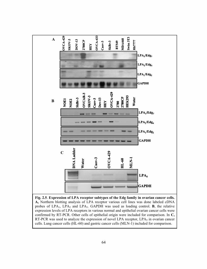
64
Fig. 2.5. Expression of LPA receptor subtypes of the Edg family in ovarian cancer cells. A, Northern blotting analysis of LPA receptor various cell lines was done labeled cDNA probes of LPA1, LPA2 and LPA3. GAPDH was used as loading control. B, the relative expression levels of LPA receptors in various normal and epithelial ovarian cancer cells were confirmed by RT-PCR. Other cells of epithelial origin were included for comparison. In C, RT-PCR was used to analyze the expression of novel LPA receptor, LPA5 in ovarian cancer cells. Lung cancer cells (HL-60) and gastric cancer cells (MLN-1) included for comparison.
B
A
C
NO
E1
NO
E2
SkB
r-3
OV
CA
R-3
SKO
V-3
Cao
v-3
HE
Y
OV
CA
-429
Dov
-13
2780
CP
Wat
er
HE
K29
3
2780
LPA3/Edg7
LPA2/Edg4
LPA1/Edg2
GAPDH
NO
E1
NO
E2
SkB
r-3
OV
CA
R-3
SKO
V-3
Cao
v-3
HE
Y
OV
CA
-429
Dov
-13
2780
CP
Wat
er
HE
K29
3
2780
NO
E1
NO
E2
SkB
r-3
OV
CA
R-3
SKO
V-3
Cao
v-3
HE
Y
OV
CA
-429
Dov
-13
2780
CP
Wat
er
HE
K29
3
2780
LPA3/Edg7
LPA2/Edg4
LPA1/Edg2
GAPDH
DN
A L
adde
r
Wat
er
Cao
v-3
OV
CA
-429
HL
-60
ML
N-1
LPA5
GAPDH
DN
A L
adde
r
Wat
er
Cao
v-3
OV
CA
-429
HL
-60
ML
N-1
LPA5
GAPDH
OV
CA
-429
SKO
V-3
DO
V-1
3
2780
CP
HE
Y
OV
CA
-433
Cao
v-3
SkB
r-3
BT
549
MD
A46
8
Rh7
777
Swis
s 3T
3
LPA3/Edg7
LPA2/Edg4
LPA1/Edg2
GAPDH
OV
CA
-429
SKO
V-3
DO
V-1
3
2780
CP
HE
Y
OV
CA
-433
Cao
v-3
SkB
r-3
BT
549
MD
A46
8
Rh7
777
Swis
s 3T
3
LPA3/Edg7
LPA2/Edg4
LPA1/Edg2
GAPDH
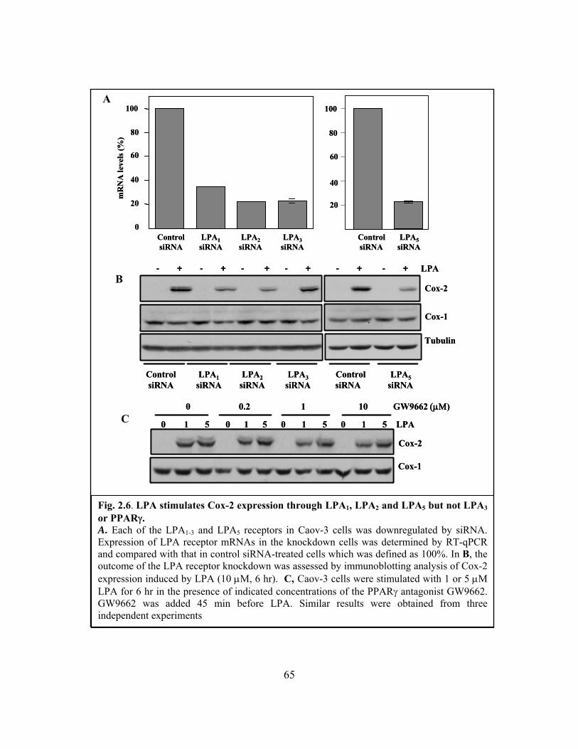
65
Fig. 2.6. LPA stimulates Cox-2 expression through LPA1, LPA2 and LPA5 but not LPA3 or PPARγ. A. Each of the LPA1-3 and LPA5 receptors in Caov-3 cells was downregulated by siRNA. Expression of LPA receptor mRNAs in the knockdown cells was determined by RT-qPCR and compared with that in control siRNA-treated cells which was defined as 100%. In B, the outcome of the LPA receptor knockdown was assessed by immunoblotting analysis of Cox-2 expression induced by LPA (10 μM, 6 hr). C, Caov-3 cells were stimulated with 1 or 5 μM LPA for 6 hr in the presence of indicated concentrations of the PPARγ antagonist GW9662. GW9662 was added 45 min before LPA. Similar results were obtained from three independent experiments
B
A
C
0
20
40
60
80
100m
RN
A le
vels
(%)
20
40
60
80
100
Control LPA1 LPA2 LPA3 Control LPA5siRNA siRNA siRNA siRNA siRNA siRNA
0
20
40
60
80
100m
RN
A le
vels
(%)
20
40
60
80
100
Control LPA1 LPA2 LPA3 Control LPA5siRNA siRNA siRNA siRNA siRNA siRNA
- + - + - + - + - + - + LPA
Cox-2
Tubulin
Cox-1
Control LPA1 LPA2 LPA3 Control LPA5siRNA siRNA siRNA siRNA siRNA siRNA
- + - + - + - + - + - + LPA
Cox-2
Tubulin
Cox-1
Control LPA1 LPA2 LPA3 Control LPA5siRNA siRNA siRNA siRNA siRNA siRNA
Cox-2
Cox-1
0 1 5 0 1 5 0 1 5 0 1 5 LPA
0 0.2 1 10 GW9662 (μΜ)
Cox-2
Cox-1
0 1 5 0 1 5 0 1 5 0 1 5 LPA
0 0.2 1 10 GW9662 (μΜ)
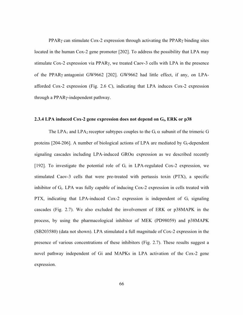
66
PPARγ can stimulate Cox-2 expression through activating the PPARγ binding sites
located in the human Cox-2 gene promoter [202]. To address the possibility that LPA may
stimulate Cox-2 expression via PPARγ, we treated Caov-3 cells with LPA in the presence
of the PPARγ antagonist GW9662 [202]. GW9662 had little effect, if any, on LPA-
afforded Cox-2 expression (Fig. 2.6 C), indicating that LPA induces Cox-2 expression
through a PPARγ-independent pathway.
2.3.4 LPA induced Cox-2 gene expression does not depend on Gi, ERK or p38
The LPA1 and LPA2 receptor subtypes couples to the Gi α subunit of the trimeric G
proteins [204-206]. A number of biological actions of LPA are mediated by Gi-dependent
signaling cascades including LPA-induced GROα expression as we described recently
[192]. To investigate the potential role of Gi in LPA-regulated Cox-2 expression, we
stimulated Caov-3 cells that were pre-treated with pertussis toxin (PTX), a specific
inhibitor of Gi. LPA was fully capable of inducing Cox-2 expression in cells treated with
PTX, indicating that LPA-induced Cox-2 expression is independent of Gi signaling
cascades (Fig. 2.7). We also excluded the involvement of ERK or p38MAPK in the
process, by using the pharmacological inhibitor of MEK (PD98059) and p38MAPK
(SB203580) (data not shown). LPA stimulated a full magnitude of Cox-2 expression in the
presence of various concentrations of these inhibitors (Fig. 2.7). These results suggest a
novel pathway independent of Gi and MAPKs in LPA activation of the Cox-2 gene
expression.

67
2.3.5 The effect of LPA involves both transcriptional activation and post-
transcriptional enhancement of Cox-2 mRNA stability
Cox-2 is one of the immediate early response genes and its induction usually
declines rapidly within 1-4 hours [207]. However, LPA-mediated Cox-2 protein expression
lasted much longer in ovarian cancer cells and was detectable at high levels 12-18 hours
after addition of LPA (Fig. 2.2). Consistent with this, Cox-2 mRNA levels reached the
maximum at 6 hours and remained highly elevated at 12 hours (Fig. 2.8 A). The sustained
induction of Cox-2 mRNA by LPA suggests that Cox-2 mRNA could be stabilized in
LPA-treated cells. To examine this possibility, we compared the decay kinetics of Cox-2
LPA - + - + - + - + - + - + - +
MAPK-p
Cox-2
Cox-1
0 12.5 25 50 0 15 30PTX (ng/ml) PD98059 (μM)
LPA - + - + - + - + - + - + - +
MAPK-p
Cox-2
Cox-1
0 12.5 25 50 0 15 30PTX (ng/ml) PD98059 (μM)
Fig. 2.7. Mechanism of LPA-induced Cox-2 gene expression is Gi and ERK independent A. Caov-3 cells were incubated with the indicated concentration of PTX before stimulation with 10μM LPA. Western blotting was performed to assess Cox-2 protein levels. Cox-1is included as a loading control. In B, the involvement of p38 MAPK was excluded, after western blot analysis from lysates of cells treated with PD98059 showed no inhibition of LPA-induced Cox-2 gene expression.
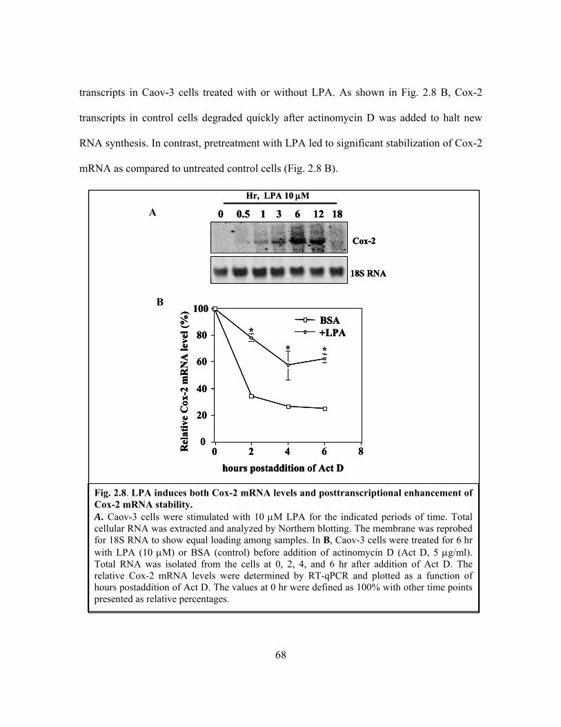
68
transcripts in Caov-3 cells treated with or without LPA. As shown in Fig. 2.8 B, Cox-2
transcripts in control cells degraded quickly after actinomycin D was added to halt new
RNA synthesis. In contrast, pretreatment with LPA led to significant stabilization of Cox-2
mRNA as compared to untreated control cells (Fig. 2.8 B).
Fig. 2.8. LPA induces both Cox-2 mRNA levels and posttranscriptional enhancement of Cox-2 mRNA stability. A. Caov-3 cells were stimulated with 10 μM LPA for the indicated periods of time. Total cellular RNA was extracted and analyzed by Northern blotting. The membrane was reprobed for 18S RNA to show equal loading among samples. In B, Caov-3 cells were treated for 6 hr with LPA (10 μM) or BSA (control) before addition of actinomycin D (Act D, 5 μg/ml). Total RNA was isolated from the cells at 0, 2, 4, and 6 hr after addition of Act D. The relative Cox-2 mRNA levels were determined by RT-qPCR and plotted as a function of hours postaddition of Act D. The values at 0 hr were defined as 100% with other time points presented as relative percentages.
B
A
Cox-2
18S RNA
0 0.5 1 3 6 12 18
Hr, LPA 10 μM
Cox-2
18S RNA
0 0.5 1 3 6 12 18
Cox-2
18S RNA
0 0.5 1 3 6 12 18
Hr, LPA 10 μM
0
20
40
60
80
100
0 2 4 6 8
BSA+LPA
hours postaddition of Act D
Rel
ativ
e C
ox-2
mR
NA
leve
l (%
)
*
* *
0
20
40
60
80
100
0 2 4 6 8
BSA+LPA
hours postaddition of Act D
Rel
ativ
e C
ox-2
mR
NA
leve
l (%
)
0
20
40
60
80
100
0 2 4 6 8
BSA+LPABSA+LPA
hours postaddition of Act D
Rel
ativ
e C
ox-2
mR
NA
leve
l (%
)
*
* *
69
To determine whether LPA induction of Cox-2 mRNA was initiated from
transcriptional activation, we cloned an approximately 1 kb proximal fragment (-980 to
+15) of the human Cox-2 gene promoter and evaluated its ability to drive transcription of
the luciferase reporter in response to LPA. LPA stimulated 5-20 fold increases in luciferase
activity in Caov-3 (Fig. 2.9 A). In addition, we compared the response of this reporter
with that of a luciferase vector containing 7.2 kb 5’ flanking region (pGL2-Cox2-7.2kb-
Luc) [202]. Similar ranges of LPA-stimulated luciferase activity were obtained from each
Fig. 2.9. Effect of LPA on Cox-2 expression involves transcriptional activation of Cox-2 promoter Caov-3 cells transfected with pGL2-Cox2-1kb-Luc containing the –980-+15 fragment of the Cox-2 promoter were stimulated with LPA at the indicated concentrations for 6 hours and assayed for luciferase activity (A) LPA-induced luciferase activity from Caov-3 cells transfected with pGL2-Cox2-1kb-Luc (defined as 100%) was compared with the activity from the cells transfected with pGL2-Cox2-7.2bk-Luc (B). All numeric results were mean ± SD of triplicates, representative of three independent experiments. The statistically significant differences of the data were indicated with asterisks.
B A
0
10
20
30
40
0 0.1 1 10 20 μM
Rel
ativ
e L
ucife
rase
Uni
t
[LPA]
pGL2-Cox2-1kb-Luc
*
*
**
0
10
20
30
40
0 0.1 1 10 20 μM
Rel
ativ
e L
ucife
rase
Uni
t
[LPA]
pGL2-Cox2-1kb-Luc
*
*
**
pGL2-Cox2-1kb-Luc
pGL2-Cox2-7.2kb-Luc
Rel
ativ
e ac
tivity
(%)
100
75
50
25
0
pGL2-Cox2-1kb-Luc
pGL2-Cox2-7.2kb-Luc
pGL2-Cox2-1kb-Luc
pGL2-Cox2-7.2kb-Luc
Rel
ativ
e ac
tivity
(%)
100
75
50
25
0
Rel
ativ
e ac
tivity
(%)
100
75
50
25
0

70
of the two plasmids, suggesting that essential LPA-regulatory elements are located within
the 1 kb sequences of the Cox-2 promoter (Fig. 2.9 B).
2.3.6 LPA induces transcriptional activation of Cox-2 via C/EBP
To identify the transcription factors driving Cox-2 expression, we analyzed the
Cox-2 gene promoter by deletion and mutation of the regulatory cis elements. Within the 1
kb region that responded well to LPA, there are numerous transcription factor binding
sites, including those for AP-1, NF-κB, and C/EBP (Fig. 2.10). Deletion of the unique AP-
1-like site at around -577 did not attenuate the response to LPA as determined by luciferase
assays (Fig. 2.10). In agreement with this, ectopic expression of TAM67, a dominant
negative form of c-Jun [208], did not inhibit LPA-induced Cox-2 (Fig. 2.11 A).
Similarly, mutation of the two NF-κB consensus sites did not interfere with the
responsiveness of the promoter to LPA (Fig. 2.10 A). Recent studies suggest that LPA and
other GPCR agonists stimulate NF-κB activation through the CARMA3/Bcl10/Malt1
signalosome, a process similar to antigen receptor-mediated NF-κB activation in
lymphocytes [209, 210].

71
Fig. 2.10. LPA induces transcriptional activation of Cox-2 through C/EBP independently of AP-1 or NF-κB. The unique AP-1-like, two NF-kB and two C/EBP binding sites were deleted or point mutated as detailed in Materials and Methods. Caov-3 cells transfected with the wild type or mutant constructs were treated with LPA for 6 hours and assayed for luciferase activity. tool.Data shown are representative of three independent experiments.
NF-
kBdo
uble
mut
ant
CE
BP
doub
le m
utan
t
Wild
type
AP-
1 de
letio
n
+15
-980
NF-
κBA
P-1 C/E
BP
NF-
κB
C/E
BP
X+1
5-9
80N
F-κB
AP-
1 C/E
BP
NF-
κB
C/E
BPX
X+1
5-9
80X
NF-
κBA
P-1 C/E
BP
NF-
κB
C/E
BP
+15
NF-
κB
C/E
BP
NF-
κB
C/E
BP
-577
Luc
act
ivity
(arb
itrar
y un
it)
5
10
15
20
25
30
35
0
BSALPA
*
NF-
kBdo
uble
mut
ant
CE
BP
doub
le m
utan
t
Wild
type
AP-
1 de
letio
n
+15
-980
NF-
κBA
P-1 C/E
BP
NF-
κB
C/E
BP
X+1
5-9
80N
F-κB
AP-
1 C/E
BP
NF-
κB
C/E
BPX
X+1
5-9
80X
NF-
κBA
P-1 C/E
BP
NF-
κB
C/E
BP
+15
NF-
κB
C/E
BP
NF-
κB
C/E
BP
-577
NF-
kBdo
uble
mut
ant
CE
BP
doub
le m
utan
t
Wild
type
AP-
1 de
letio
n
+15
-980
NF-
κBA
P-1 C/E
BP
NF-
κB
C/E
BP
X+1
5-9
80N
F-κB
AP-
1 C/E
BP
NF-
κB
C/E
BPX
X+1
5-9
80X
NF-
κBA
P-1 C/E
BP
NF-
κB
C/E
BP
+15
NF-
κB
C/E
BP
NF-
κB
C/E
BP
-577
Luc
act
ivity
(arb
itrar
y un
it)
5
10
15
20
25
30
35
0
BSALPA
*Luc
act
ivity
(arb
itrar
y un
it)
5
10
15
20
25
30
35
0
BSALPA
*

72
To confirm the mutagenesis results, we downregulated the expression of Bcl10
with three individual siRNAs. Each significantly reduced Bcl10 expression. However,
none of these siRNAs was able to prevent LPA-mediated Cox-2 expression (Fig. 2.11 B).
In contrast, the Bcl10 siRNAs markedly inhibited LPA-stimulated IL-8 production (Fig.
2.11 C), a response fully dependent on NF-κB activation as we described previously [9].
The results suggest that in sharp contrast to many other Cox-2 inducers, LPA stimulated
Cox-2 expression independently of NF-κB although LPA potently induced NF-κB DNA-
binding and the transcriptional activity in these cells (Fig. 4.5 of Chapter 4).
We next targeted the two C/EBP consensus motifs present in the 1 kb fragment of
the Cox-2 promoter. Although mutation of the individual site only slightly decreased the
promoter activity, simultaneous mutation of the two C/EBP sites resulted in almost
complete loss of LPA-induced luciferarse activity (Fig. 2.10), suggesting that these
C/EBP-binding sites are essential for the transcriptional activation of Cox-2 by LPA. In
further support of this, co-expression of LIP, a dominant negative, truncated form of
C/EBP-β [139], inhibited LPA-induced luciferase activity by 60% in cells transfected with
pGL-2-Cox2-1kb-Luc (Fig. 2.12 A). LIP also suppressed LPA-induced Cox-2 protein
expression when transiently transfected and expressed in Caov-3 cells (Fig. 2.12 B). ChIP
assay confirmed the recruitment of C/EBP-β to the Cox-2 gene promoter following LPA
treatment (Fig. 2.12 C). Treated or untreated Caov-3 cell lysates were subjected to
immunoprecipitation with anti-C/EBP-β and gel eletrophoretic analysis of precipitated
DNA using Cox-2 promoter specific primers. As shown in Fig. 2.12 C, LPA induced a
rapid localization of C/EBP-β to Cox-2 promoter sequences.
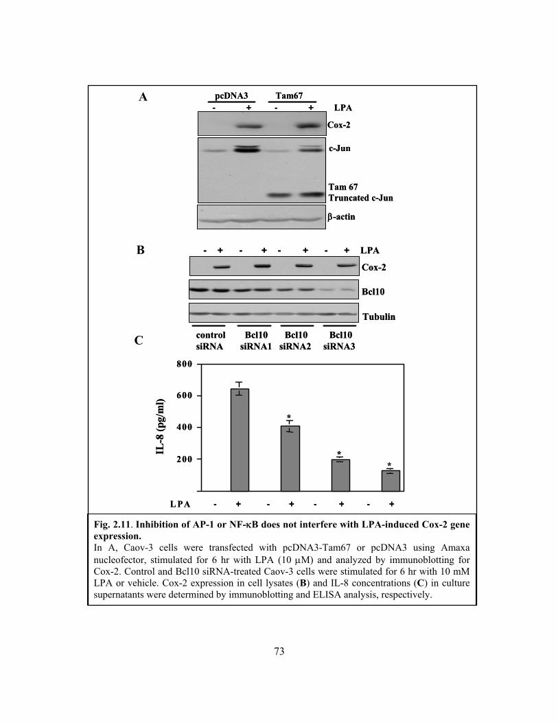
73
Fig. 2.11. Inhibition of AP-1 or NF-κB does not interfere with LPA-induced Cox-2 gene expression. In A, Caov-3 cells were transfected with pcDNA3-Tam67 or pcDNA3 using Amaxa nucleofector, stimulated for 6 hr with LPA (10 μM) and analyzed by immunoblotting for Cox-2. Control and Bcl10 siRNA-treated Caov-3 cells were stimulated for 6 hr with 10 mM LPA or vehicle. Cox-2 expression in cell lysates (B) and IL-8 concentrations (C) in culture supernatants were determined by immunoblotting and ELISA analysis, respectively.
B
A
C
Cox-2
Bcl10
Tubulin
- + - + - + - + LPA
control Bcl10 Bcl10 Bcl10siRNA siRNA1 siRNA2 siRNA3
Cox-2
Bcl10
Tubulin
- + - + - + - + LPA
control Bcl10 Bcl10 Bcl10siRNA siRNA1 siRNA2 siRNA3
L PA - + - + - + - +
200
400
600
800
IL-8
(pg/
ml)
*
**
L PA - + - + - + - +
200
400
600
800
IL-8
(pg/
ml)
*
**
- + - + LPApcDNA3 Tam67
Cox-2
β-actin
c-Jun
Tam 67Truncated c-Jun
- + - + LPApcDNA3 Tam67
Cox-2
β-actin
c-Jun
Tam 67Truncated c-Jun

74
C/EBP-β, a major isoform of the C/EBP family, was previously reported to be
overexpressed in ovarian cancers [135]. To assess the effect of overexpression of C/EBP-
β on activity of the Cox-2 promoter, we transfected Caov-3 cells with pGL2-Cox2-1kb-
Luc along with the C/EBP-β LAP1, LAP2 or a control vector. Expression of the C/EBP-
β LAP1 or LAP2 was not sufficient to induce activation of luciferase activity from pGL2-
Cox2-1kb-Luc (data not shown). The observation suggests that LPA induced Cox-2
expression requires C/EBP activation rather than changes in C/EBP-β protein levels.
2.3.7 The mRNA-binding protein HuR associates with and stabilizes Cox-2 mRNA in
LPA-treated cells
There are several regulatory mechanisms to control mRNA stability under different
physiological and pathophysiological conditions [211, 212]. One of such regulations
involves the RNA-binding protein HuR that associates with AUUUA repeats present in the
3’ UTR of mRNAs encoding cytokines and angiogenic factors [212, 213]. Two major Cox-
2 transcripts (2.8 kb and 4.6 kb) are derived from alternative polyadenylation of the Cox-2
gene [212, 214].
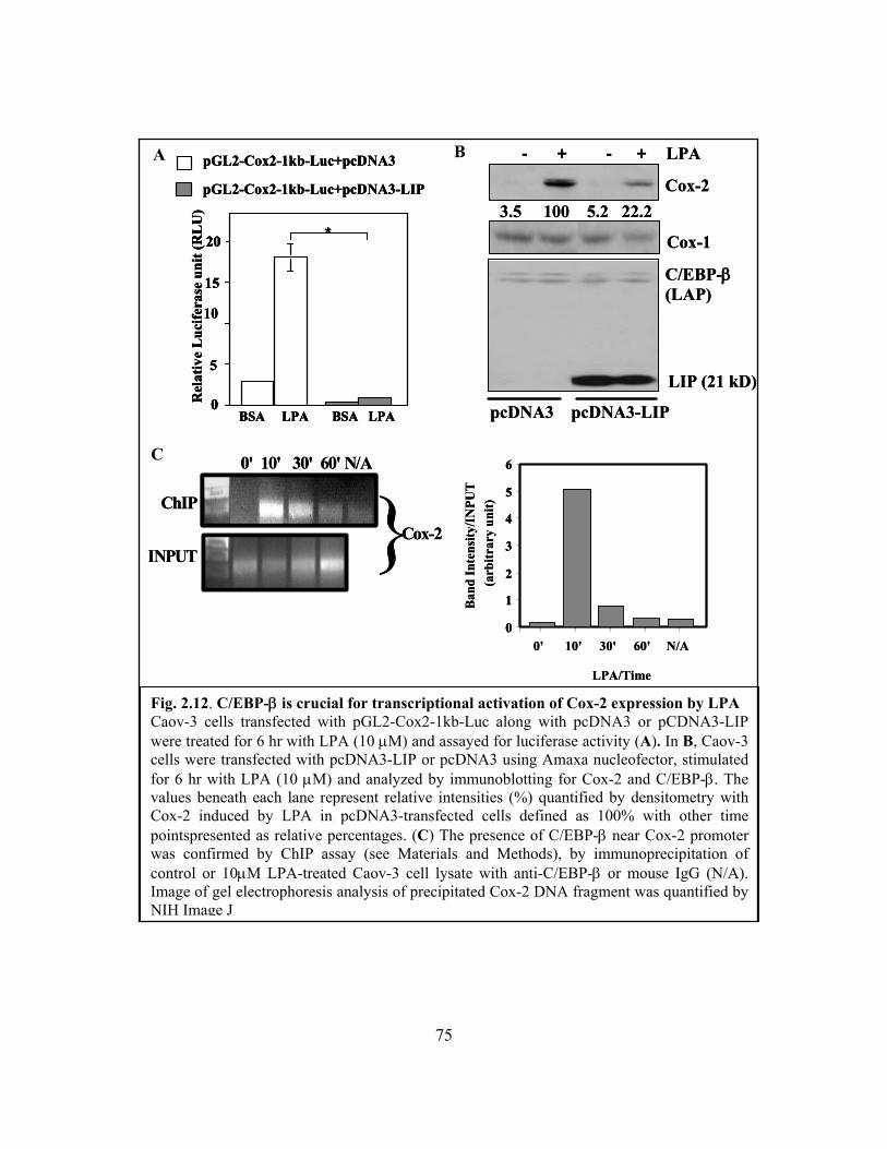
75
Fig. 2.12. C/EBP-β is crucial for transcriptional activation of Cox-2 expression by LPA Caov-3 cells transfected with pGL2-Cox2-1kb-Luc along with pcDNA3 or pCDNA3-LIP were treated for 6 hr with LPA (10 μM) and assayed for luciferase activity (A). In B, Caov-3 cells were transfected with pcDNA3-LIP or pcDNA3 using Amaxa nucleofector, stimulated for 6 hr with LPA (10 μM) and analyzed by immunoblotting for Cox-2 and C/EBP-β. The values beneath each lane represent relative intensities (%) quantified by densitometry with Cox-2 induced by LPA in pcDNA3-transfected cells defined as 100% with other time pointspresented as relative percentages. (C) The presence of C/EBP-β near Cox-2 promoter was confirmed by ChIP assay (see Materials and Methods), by immunoprecipitation of control or 10μM LPA-treated Caov-3 cell lysate with anti-C/EBP-β or mouse IgG (N/A). Image of gel electrophoresis analysis of precipitated Cox-2 DNA fragment was quantified by NIH Image J
C
A B pGL2-Cox2-1kb-Luc+pcDNA3
pGL2-Cox2-1kb-Luc+pcDNA3-LIP
10
Rel
ativ
e L
ucife
rase
unit
(RL
U)
0
5
15
20 *
BSA LPA BSA LPA
pGL2-Cox2-1kb-Luc+pcDNA3
pGL2-Cox2-1kb-Luc+pcDNA3-LIP
pGL2-Cox2-1kb-Luc+pcDNA3
pGL2-Cox2-1kb-Luc+pcDNA3-LIP
10
Rel
ativ
e L
ucife
rase
unit
(RL
U)
0
5
15
20 *
BSA LPA BSA LPA
10
Rel
ativ
e L
ucife
rase
unit
(RL
U)
0
5
15
20 *
BSA LPA BSA LPA0
5
15
20 *
BSA LPA BSA LPA
C/EBP-β(LAP)
LIP (21 kD)
Cox-2
Cox-1
- + - + LPA
pcDNA3 pcDNA3-LIP
3.5 100 5.2 22.2
C/EBP-β(LAP)
LIP (21 kD)
Cox-2
Cox-1
- + - + LPA
pcDNA3 pcDNA3-LIP
3.5 100 5.2 22.2
ChIP
Cox-2INPUT
0' 10' 30' 60' N/A
}ChIP
Cox-2INPUT
0' 10' 30' 60' N/A
}0
1
2
3
4
5
6
0' 10' 30' 60' N/A
LPA/Time
Ban
d In
tens
ity/I
NPU
T(a
rbitr
ary
unit)
0
1
2
3
4
5
6
0
1
2
3
4
5
6
0' 10' 30' 60' N/A
LPA/Time
Ban
d In
tens
ity/I
NPU
T(a
rbitr
ary
unit)
76
In ovarian cancer cells, LPA induced the 4.6 kb transcript which contains a long 3’-
UTR [212, 214]. The 2.8 kb transcript lacking the 3’-UTR was not induced at detectable
levels by LPA (Fig. 2.8 A). Importantly, multiple AUUUA repeats are present within the
3’-UTR of the 4.6 kb Cox-2 transcript [212, 215]. Moreover, HuR is highly expressed in
primary ovarian cancers and ovarian cancer cell lines [216]. These observations prompted
us to examine whether HuR participates in protection of Cox-2 mRNA stability,
contributing to the persistent induction of Cox-2 observed in LPA-stimulated cells.
We first examined whether HuR physically binds to Cox-2 mRNA in vitro. To this
end, the 3’UTR of Cox-2 mRNA was synthesized and labeled with biotin by in vitro
transcription. As demonstrated in Fig. 2.13 A, the Cox-2 3’-UTR was capable of binding
with HuR protein from lysates of Caov-3 cells. Of note, incubation of 90 μg cellular
protein with 1.5 μg Cox-2 3’-UTR brought down amounts of HuR similar to that present in
30 μg cell lysates (Fig. 2.13 A). Therefore, approximately one third of the total HuR
protein formed complex with the exogenous Cox-2 3’-UTR, demonstrating that HuR has a
strong binding affinity for the Cox-2 3’-UTR at least in in vitro binding assays.
To confirm that the HuR-Cox-2 mRNA association occurs within the cell, we
immunoprecipitated HuR from Caov-3 cells treated with LPA or BSA for 6 hours. The
immunocomplexes were then subjected to reverse transcription followed by PCR
amplification of a 481bp fragment of Cox-2 cDNA. As shown in Fig. 2.13B, the fragment
was detected in immunoprecipitates of the cells treated with LPA, but not present in that of
control cells. It was also absent from immunoprecipitates of LPA-treated cells when a
control antibody was used to replace the specific HuR antibody. HuR therefore indeed

77
formed complex with Cox-2 mRNA in LPA-treated cells. The lack of Cox-2 mRNA in the
immunoprecipitates of control cells suggests that the interaction between HuR and Cox-2
mRNA may not be constitutive. It could be regulated through activation of HuR protein or
rely on LPA-dependent increases in Cox-2 mRNA abundance.
To determine the functional significance of HuR binding to Cox-2 mRNA, we
assessed the effect of downregulation of HuR on LPA-induced Cox-2 expression.
Knockdown of HuR expression resulted in a significant decrease in LPA-induced Cox-2 as
shown in Fig. 2.13 C. Stronger suppression of Cox-2 expression was observed in cells
treated with LPA for 12 hours compared to the cells treated with LPA for 6 hours. The
observation suggests that HuR-mediated stabilization of Cox-2 mRNA contributes more
significantly to Cox-2 expression when transcriptional activation becomes less active after
prolonged incubation with LPA.

78
Fig. 2.13. HuR binds to Cox-2 mRNA, contributing to the sustained induction of Cox-2 by LPA. A. In vitro transcribed, biotinylated 3’UTR of the Cox-2 mRNA (1. 5 μg) was incubated with cellular protein (90 μg) from Caov-3 cells treated with BSA or LPA (10 μM, 6 hr). Amounts of HuR pulled down by the Cox-2 3’UTR were analyzed by immunoblotting with an anti-HuR antibody. Cell lysates (30 μg) were included to serve as inputs. B. HuR was immunoprecipitated from Caov-3 cells treated for 6 hr with BSA (control) or LPA (10 μM). The mRNAs present in the HuR immunocomplex were extracted, reverse transcribed and amplified by PCR using Cox-2 specific primers as detailed in Materials and Methods. C. The control and HuR downregulated Caov-3 cells were stimulated for 6 hr (left) or 12 hr (right) with 10 μM LPA. Cox-2, Cox-1 and HuR levels in these cells were analyzed with immunoblotting. The values beneath each lane represent relative intensities (%) quantified by densitometry with Cox-2 induced by LPA in control siRNA-treated cells defined as 100%. Similar results were obtained from two independent assays.
B A
C
BSA LPA
HuR
HuR (input)
- + - + Cox-2 UTR
BSA LPA
HuR
HuR (input)
- + - + Cox-2 UTR BSA
LPABSA
LPA
Cox‐2
HuR
Tubulin
3.7 100 3.1 23.3
control HuR control HuRsiRNA siRNA siRNA siRNA
LPA(6 hours) LPA(12 hours)
1.2 100 1.4 43.2
- + - + - + - + LPA
Cox‐2
HuR
Tubulin
3.7 100 3.1 23.3
control HuR control HuRsiRNA siRNA siRNA siRNA
LPA(6 hours) LPA(12 hours)
1.2 100 1.4 43.2
- + - + - + - + LPA
79
2.4 Discussion
In the present study, we demonstrated that LPA induces robust and sustained
expression of Cox-2 in ovarian cancer cells through both transcriptional and post-
transcriptional mechanisms, mediated by the C/EBP transcription factor and the mRNA
stability protein HuR, respectively. The identification of C/EBP as a primary transcription
factor responsible for LPA-induced Cox-2 expression is somewhat surprising, as C/EBP
has not been previously linked to any biological actions of LPA. Other transcription factors
activated by LPA and commonly involved in inducible Cox-2 expression such as NF-κB
and AP-1 are instead dispensable for LPA-induced Cox-2. Previous studies of GPCR
regulation of Cox-2 in different cell systems have led to diverse mechanisms involving
multiple transcription factors [217, 218]. A predominant and specific role of C/EBP in
transcriptional activation of Cox-2 via GPCR signaling has not been previously described
and likely represents a general mechanism regulating Cox-2 expression by LPA and other
GPCR agonists in different cell types.
Since C/EBP-β expression is elevated in primary ovarian cancers and in ovarian
cancer cell lines [135], the isotype likely plays a major role in mediating transcriptional
activation of Cox-2 in response to LPA. C/EBP-β has been shown to be a key transcription
factor in regulation of Cox-2 expression by aspirin and salicylate [219]. Furthermore,
expression of other isotypes of C/EBP such as C/EBP-α and C/EBP-δ is limited or
undetectable in ovarian cancer cell lines (data not shown). We did not observe activation of
the Cox-2 promoter by means of transfection of exogenous C/EBP-β, suggesting that
80
overexpression of C/EBP-β protein is insufficient to confer the transcriptional activation of
Cox-2. The luciferase reporter analysis established that LPA treatment stimulates C/EBP
transcriptional activity. There are multiple post-translational modifications associated with
C/EBP activation including phosphorylation [220], acetylation [221] and sumoylation
[201, 222]. Treatment with LPA resulted in prominent phosphorylation of C/EBP-β. It is
not known whether LPA-dependent activation of C/EBP is also regulated by acetylation
and sumoylation in addition to phosphorylation.
LPA-induced Cox-2 gene expression is mediated by LPA1, LPA2 and LPA5
receptors independent of LPA3 and LPA4. The LPA1 receptor is the most commonly
expressed subtype present in both normal and cancerous tissues [62]. The LPA2 receptor
subtype is abnormally overexpressed in ovarian cancers and other human malignancies
[61, 62, 64]. It mediates LPA-dependent cytokine production in ovarian and breast cancer
cells [9, 192]. Similarly, Hu et al. described that LPA2 expression correlates with the ability
of LPA to induce VEGF expression in ovarian cancer cells [193]. The importance of LPA2
in modulation of gene expression is further highlighted by the observation that transgenic
expression of LPA2 driven by an ovary-selective promoter led to the production of higher
levels of VEGF and uPA mRNA and proteins in ovaries of transgenic mice [223]. In
addition to the role in modulation of gene expression, LPA2 may also regulate cell motility
[224, 225]. Involvement of LPA5 in LPA-induced Cox-2 expression is an interesting
observation since the biological functions of this new LPA receptor are totally unknown
[40]. Requirement of LPA1, LPA2 and LPA5 in LPA induction of Cox-2 suggests that these
LPA receptors could each contribute to the response to LPA. Alternatively, the effect of

81
LPA may depend on combined functions of these receptors. A recent study suggests that
these LPA receptors can crosstalk to each other through forming heterodimers with
signaling properties likely different from their homodimers [226]. In addition, the LPA3
receptor does not seem to be a mediator of Cox-2 induction in ovarian cancer cells
although this receptor is highly expressed in most ovarian cancer cell lines [62]. The result
is inconsistent with the critical role of LPA3 in the uterine Cox-2 expression as suggested
by studies of LPA3 knockout mice [199], indicating different LPA receptor subtypes are
capable of mediating Cox-2 expression depending on the cellular context.
The proximal region of the 3'-UTR of COX-2 mRNA contains highly conserved
AU-rich elements that have potential to interact with multiple mRNA binding proteins
including β-catenin, TIAR, AUF1, HuR, hnTIA-1 and hnRNP [211, 212, 215]. In the
current study, we focused on the role of HuR, a member of the ELAV (embryonic lethal
abnormal vision) family of mRNA-binding proteins [213]. HuR overexpression is
associated with increased levels of Cox-2 protein in cancers of the colon, stomach, breast
and ovary [135, 212, 227, 228]. However, few studies have provided direct evidence that
HuR plays a causal role in Cox-2 overexpression in malignant cells. Our results indicate
that HuR physically binds to the Cox-2 3’-UTR and protects Cox-2 mRNA stability. This
protein-mRNA association contributes significantly to the sustained induction of Cox-2 by
LPA. HuR thus provides a positive feedback to Cox-2 induction during physiological
responses to LPA and probably other environmental stimuli.

82
CHAPTER 3
MECHANISMS FOR ACTIVATION OF C/EBP-β BY LPA
Part of the work presented in this chapter has been published FASEB J. 22:2639-2651 (2008).
3.0 Abstract
We previously showed that LPA induced the expression of Cox-2, one of the
enzymes that convert arachidonic acid to prostaglandins. Through site-directed
mutagenesis and the ectopic expression of a dominant negative form C/EBP-β, we
identified C/EBP-β as an essential transcription factor in the induction of Cox-2 gene
expression by LPA. Here, we further examined the precise mechanisms underlying LPA-
induced activation of C/EBP-β. LPA induced a rapid and transient phosphorylation of
C/EBP-β in ovarian cancer cells. Prolonged exposure to LPA stimulation caused an
increase in C/EBP-β protein levels. Using electromobility shift assay, we show a sustained
increase in DNA binding activity of C/EBP-β following LPA stimulation. Functionally,
LPA induced multifold increase in C/EBP-β transcriptional activity in ovarian cancer cells
transfected with a reporter construct of consensus C/EBP binding site. Furthermore, we
report a novel mechanism integrating GPCR signals and a permissive activity from a
receptor tyrosine kinase (RTK) in LPA-induced phosphorylation and activation of C/EBP-
83
β. This role of RTK was not consistent with LPA activation of C/EBP-β through
transactivation of RTK, as full activation of RTKs by their own agonists only weakly
stimulated C/EBP-β activities. Interestingly, the Cox-2 promoter activity and gene
expression were also dependent on RTK signaling, further substantiating the regulatory
role of C/EBP-β in LPA-induced Cox-2 gene expression. These results together provide a
picture of signaling cascades involved in LPA activation of C/EBP-β.
3.1 Introduction
C/EBP-β belongs to a widely expressed, highly conserved family of basic
region-leucine zipper (bZIP) class of transcription factors that play essential role in many
physiological and pathological processes such as cellular differentiation and inflammation
[121, 126, 127]. Other members are C/EBP-α, C/EBP-γ, C/EBP-δ, C/EBP-ε, and C/EBP-ζ.
The genes of most C/EBPs are intronless and exhibit a high degree of homology in the
basic and leucine zipper regions [120, 121]. However, each C/EBP isoform may exhibit
distinct functions. For instance, ablation of the C/EBP-α gene in mice led to neonatal
lethality due to hypoglycemia and absence of stored liver glycogen [124, 129]. C/EBP-β-
null mice are, however, viable in spite of serious defects in hematopoiesis and the immune
system [130, 131]. The C/EBP-β gene is expressed as three isoforms as a consequence of
alternative initiation start sites within the C/EBP-β transcript: liver-enriched activating
proteins (LAP1—49kd and LAP2—45 kda) and liver-enriched inhibiting protein (LIP—20
kda) [138, 139, 229]. The LAP isoforms function as activators of transcription while LIP,

84
which lacks the transactivation domain, is a dominant negative transcriptional repressor.
The differential expression of these LAP and LIP is regulated by C/EBP-α [140] and has
been found to be critical in cell fate determination [141].
Several studies have shown that C/EBP-β is important in mammalian reproduction
including the development and differentiation of the mammary glands. C/EBP-β is also
involved in ovarian follicular development and corpus leteum formation by enhancing the
effects of lutenizing hormone (LH/hCG) [132-134]. Moreover, C/EBP-β null mice showed
defective female reproduction [132]. C/EBP-β has been implicated in many cancer types
and its expression highly correlates with aggressive behavior in ovarian cancer cells [135].
C/EBP binding sites are present in the promoters of cancer or metastasis-related genes such
as interleukin 6 (IL-6), cyclooxygenase 2 (cox-2), urokinase plasminogen activator (uPA)
and tumor necrosis factor alpha (TNF-α). In fact, C/EBP-β was identified in pioneer
studies as a protein that binds the IL-1 response element on IL-6 promoter and hence, was
originally named NF-IL6 [138, 230, 231]. We recently described C/EBP-β as an essential
transcription factor for induction of the Cox-2 gene expression by LPA (Chapter 2 , [232]
). The exact mechanism by which LPA induces activation of C/EBP-β has not been
elucidated.
Recent studies showed that many biological functions of GPCRs depend on EGFR.
It is currently believed that ligands for GPCRs such as LPA activate cellular responses
through transactivation of EGFR or other highly expressed RTK through the proteolytic
release of EGFR ligands such as EGF or HB-EGF. The transactivation model is not
85
consistent with LPA stimulation of EGFR phosphorylation and activation. In addition, it is
not known whether the crosstalk between GPCRs and EGFR involves activation of
transcription factors such as C/EBP-β.
Here, we provide evidence that LPA modulates the transcriptional activities of
C/EBP-β in ovarian cancer cells by inducing its phosphorylation and DNA binding. LPA
also induced expression of C/EBP-β at later time points which may contribute to the
sustained induction of Cox-2. The effects of LPA on C/EBP-β phosphorylation, DNA-
binding and transcriptional activation required permissive signals from EGFR or an
alternate RTK. The dependence of LPA actions on RTK also applies to other C/EBP-β
target genes including IL-6 and uPA in addition to Cox-2 underscoring a general role of
RTK in GPCR activation of C/EBP-β and C/EBP-β-target genes.
3.2 Materials and Methods
Materials 1-Oleoly (18:1) LPA and sphingosine 1 phosphate (S1P) were obtained from
Avanti Polar Lipids, Inc. (Alabaster, AL). Prior to use, these phospholipids were dissolved
in PBS containing 0.5% fatty acid-free bovine serum albumin (BSA). BSA, Fugene 6 and
protease inhibitor cocktail tablets were purchased from Roche (Indianapolis, IN). [Plasmid
DNA was purified using the endo-free purification kit from Qiagen (Valencia, CA).
Luciferase assay reagents were obtained from Promega (Madison, WI). All
oligonucleotides and primers were synthesized by Operon Biotechnologies, Inc

86
(Huntsville, AL). QuikChange XL site directed mutageneis kit were purchased from
Stratagene (La Jolla, CA). Anti-phospho CEBPβ and anti-tubulin α/β antibodies were
obtained from Cell Signaling (Danvers, MA). Insulin and cell culture medium were
obtained from Invitrogen Inc. (Carlsbad, CA). Bovine fetal serum was from Biomeda
(Foster City, CA). Insulin-like growth factor I (IGF-I) was from Upstate Biotechnology
(Lake Placid, NY). Hepatocyte growth factor (HGF) and Quantikine IL-6 ELISA kit were
from R & D systems (Minneapolis, MN). Anti-uPA monoclonal antibody #394 was
obtained from American Diagnostica (Stamford, CT). Epidermal growth factor (EGF),
AG1478 and anti-β-actin monoclonal antibody were obtained from Sigma (St. Louis, MO).
Plasmids Construction Transcription factor binding sites were identified using
TFSEARCH. The construction of Cox-2 promoter plasmid, pGL2-Cox-2-1kb-luc, has been
described in Materials and Method section of Chapter 2 of this dissertation. The C/EBP
responsive luciferase vector (pGL2-5xCEBP-TKLuc) was generated by cloning five
repeats of the C/EBP consensus sequence (TTGCGCAATCT) into the Nhe1 and Hind III
sites in front of the herpes simplex virus thymidine kinase (TK) gene promoter (-35-+50)
in the pGL2-TK-Luc vector. The C/EBP-β, liver-enriched transcriptional activator protein
1 (LAP1) and LAP2 expression vectors were kindly provided by Dr. L. Sealy (Vanderbilt
University School of Medicine) [139, 201]. The expression of C/EBP-β from these vectors
in transfected cells was confirmed by immunoblotting. The dominant negative form of
C/EBP-β, LIP (liver-enriched inhibitory protein) [139] was cloned into pcDNA3.1 by RT-
PCR amplification of a 444 bp cDNA fragment of C/EBP-β from Caov-3 cells. The
87
structure of pcDNA3-LIP was confirmed by automatic sequencing and immunoblotting
analysis of expression of the short, truncated form of C/EBPβ (21 kD) [139]in transfected
cells.
Cell Culture The sources of ovarian cancer cell lines used in the study were described
previously [9, 192]. These cells were cultured in RPMI medium supplemented with 10%
FBS, 100 units/ml penicillin and 100 μg/ml streptomycin. All cell lines were frozen at
early passages and used for less than 10 weeks in continuous culture.
Western Blot Cells were lysed in SDS sample buffer or in ice-cold X-100 lysis buffer [1%
Triton X-100, 50 mM HEPES (pH 7.4), 150 mM NaCl, 1.5 mM MgCl2, 1 mM EGTA,
10% glycerol, 100 mM NaF, 10 mM Na PPi, and protease inhibitor cocktail]. Total cellular
proteins were resolved by SDSPAGE, transferred to Immun-Blot membrane
[poly(vinylidene difluoride)] (BIO-RAD, Hercules, CA), and immunoblotted with
antibodies following the protocols of manufacturers. Immunocomplexes were visualized
with an enhanced chemiluminescence detection kit from Amersham (Piscataway, NJ)
using the horseradish peroxidase–conjugated secondary antibodies (Cell Signaling,
Danvers, MA).
Transient transfection and luciferase assays Ovarian cancer cell lines were seeded in 6-
well plates and grown to 30-40 % confluence before transfection with the luciferase
vectors using Fugene 6 (Roche) or TransIT-TKO (Mirus Bio Corp., Madison, WI)
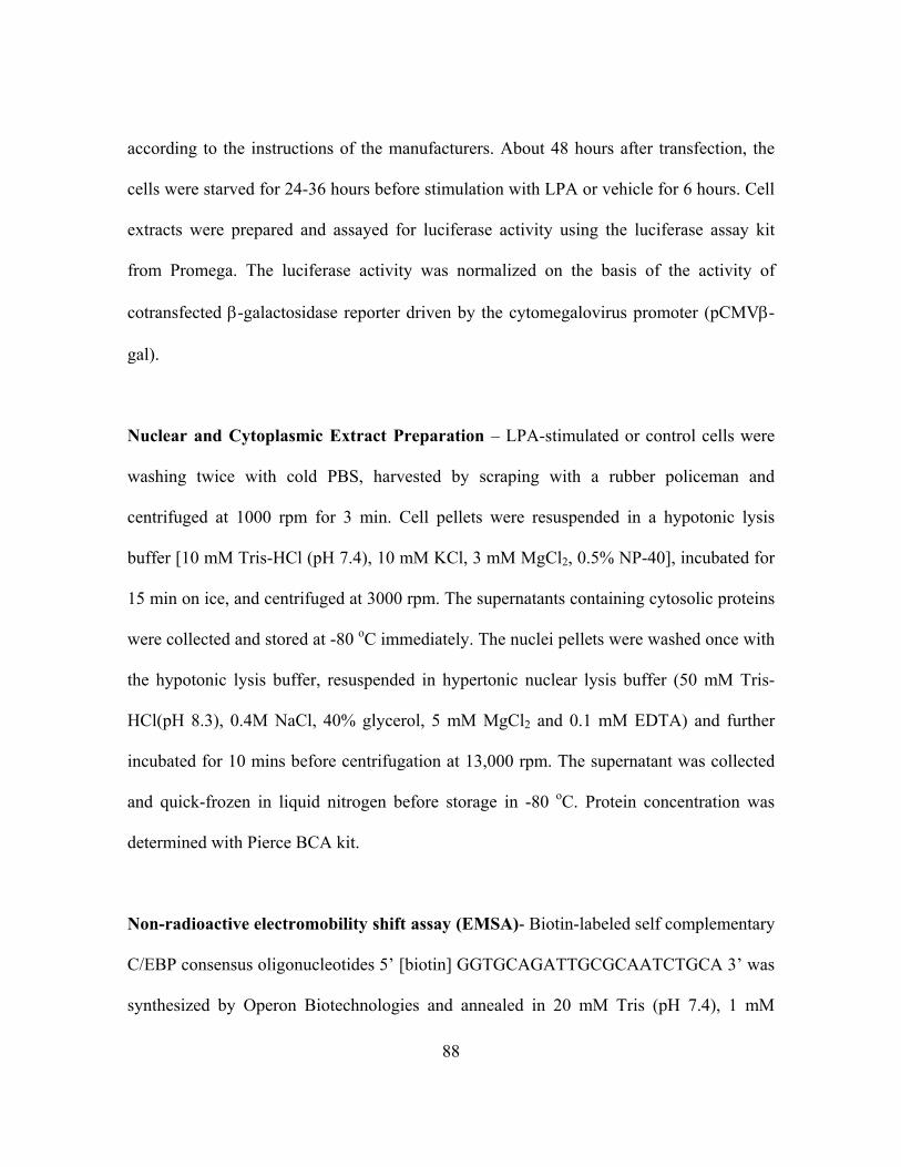
88
according to the instructions of the manufacturers. About 48 hours after transfection, the
cells were starved for 24-36 hours before stimulation with LPA or vehicle for 6 hours. Cell
extracts were prepared and assayed for luciferase activity using the luciferase assay kit
from Promega. The luciferase activity was normalized on the basis of the activity of
cotransfected β-galactosidase reporter driven by the cytomegalovirus promoter (pCMVβ-
gal).
Nuclear and Cytoplasmic Extract Preparation – LPA-stimulated or control cells were
washing twice with cold PBS, harvested by scraping with a rubber policeman and
centrifuged at 1000 rpm for 3 min. Cell pellets were resuspended in a hypotonic lysis
buffer [10 mM Tris-HCl (pH 7.4), 10 mM KCl, 3 mM MgCl2, 0.5% NP-40], incubated for
15 min on ice, and centrifuged at 3000 rpm. The supernatants containing cytosolic proteins
were collected and stored at -80 oC immediately. The nuclei pellets were washed once with
the hypotonic lysis buffer, resuspended in hypertonic nuclear lysis buffer (50 mM Tris-
HCl(pH 8.3), 0.4M NaCl, 40% glycerol, 5 mM MgCl2 and 0.1 mM EDTA) and further
incubated for 10 mins before centrifugation at 13,000 rpm. The supernatant was collected
and quick-frozen in liquid nitrogen before storage in -80 oC. Protein concentration was
determined with Pierce BCA kit.
Non-radioactive electromobility shift assay (EMSA)- Biotin-labeled self complementary
C/EBP consensus oligonucleotides 5’ [biotin] GGTGCAGATTGCGCAATCTGCA 3’ was
synthesized by Operon Biotechnologies and annealed in 20 mM Tris (pH 7.4), 1 mM

89
dithiothreitol, 50 mM NaCl and 10 mM MgCl2. For the gel shift assay, binding reaction
was performed by incubating 4 μg of nuclear protein in gel shift buffer (10 mM HEPES
(pH 7.8), 10% glycerol, 1 mM EDTA, 25 mM MgCl2, 50 mM KCl, 1 µg of poly(dI.dC), 3
µg BSA and protease inhibitors) in a final volume of 20 µL for 10 mins at 25oC. The
biotin-labeled and unlabeled (cold) oligonucleotides were added to the reaction mixture,
incubated for an additional 15 mins at 25oC. In supershift experiments, nuclear extracts
were incubated for 15 min at room temperature with 1 µg of anti-CEBP-β monoclonal
antibody (Santa Cruz Biotech) prior to gel shift reaction. Complexes were separated by
electrophoresis on 5% non-denaturing polyacrylamide (PAGE) gel, transferred to a nylon
membrane (Amersham Hy-bond XL) and crosslinked using UV Stratalinker 2400
(Stratagene). The signals were visualized by using Phototope-Star biotin detection kit
(NEB), according to the manufacturer’s protocol.
ChIP assay- Cells were fixed by cross-linking the chromatin with 1% formaldehyde for 5
min, cells, scraped with a rubber policeman and collected by centrifugation. The cells were
sonicated six times, 15 seconds each with 1-min intervals under ice by using a Sonic
Dismembrator (Fisher Scientific). The average fragment size was 600 bp under these
conditions. An equal amount of chromatin was incubated with at least 5 µg of either
C/EBP-β-specific immunoglobulin G (IgG) or normal mouse IgG (Santa Cruz Biotech) at 4
°C overnight. IP products were collected after incubation with protein G-coated sepharose
beads (Amersham). The beads were washed, protein-DNA cross-links were reversed, and
DNA was purified by Qiagen PCR purification columns according to manufacturer’s

90
instructions. Purified DNA from the input and IP samples were subjected to real-time
quantitative PCR with uPA promoter specific primers.
ELISA Assay- An enzyme-linked immunosorbent assay (ELISA) was used for the
quantification of IL-6. Briefly, culture supernatants of Caov-3 treated without or with LPA
or other ligands were collected and analyzed for IL-6 using the Quantikine IL-6 ELISA kits
(R&D Systems) according the manufacturer’s instruction. Colorimetric reactions were read
on a EL800 microplate reader (Bio-TEK Instruments, Winooski,VT). The concentrations
of IL-6 in samples were calculated by comparing with those of standards provided with the
ELISA kits.
Stable expression of C/EBP-β LIP by retrovirus – The human LIP cDNA was excised
from pcDNA3-LIP using HindIII and EcoRI and cloned into a moloney murine leukemia
retrovirus vector, S-001AB (LZRS-EGFP)( a gift from Dr. J. Chun, the Scripps Research
Institute, La Jolla, CA). The complete insert and flanking regions of the final retrovirus
vector were confirmed by automatic sequencing. The LZRS-EGFP-LIP and control vector
were transfected into the PZ67 packaging cell line with lipofectamine 2000 as previously
described [9]. Virus stocks were harvested 72 hrs post-transfection by centrifugation of the
supernatants at 2100 rpm for 10 min and stored in -80 °C. Skov-3 cells grown in 6-well
dishes were infected with 1.4 ml virus to cells in the presence of 7.5 μg/ml polybrene
(Sigma-Aldrich) and incubated at 37 oC overnight. The infection medium was then
replaced with growth medium. Cells were split into 10 cm dishes after 48 hours. Viral-

91
transduced GFP-positive cell were sorted out by flow cytometry at the VCU flow
cytometry core facilities.
Table 3.1 Plasmids and Primers
Plasmid Wildtype or Mutation primers
pGL2-Cox-1kb-Luc 5’-TTTAGCGTCCCTGCAAATTCTGGC-3’(SENSE)
5’-CGCTCACTGCAAGTCGTATGACAA-3’ (ANTISENSE) pGL2-IL-6-Luc 5’- GAGCTCAAGGATCCTCCTGCAAGAGA -3’(SENSE)
5’- TAGAGCTTCTCTTTCGTTCCCGGTGG -3’ (ANTISENSE) pGL2-IL-6-mut-Luc 5’- GGACGTCACAGTCTACTCTCTTAATAAGGTTTCC -3’(SENSE)
5’- GGAAACCTTATTAAGAGAGTAGACTGTGACGTCC -3’ (ANTISENSE) pGL2-uPA-Luc 5’- CGGATCACAAGGTCAGGAAGATCGAG -3’(SENSE)
5’- TCTCCGACTGTGCTGCGA C 3’ (ANTISENSE) pGL2-uPA-mut-Luc 5’- GAGGCAGTCTTAGGCGGGTTGGGGCCCAGCG -3’(SENSE)
5’- CGCTGGGCCCCAACCCGCCTAAGACTGCCTC -3’ (ANTISENSE pGL2-5xCEBP-TK-
Luc
5’CTAGCATCTAAGTTTGCGCAATCTTTGCGCAATCTTTGCGCAATCTTTGC
GCAATCTTTGCGCAA -3’(SENSE)
5’AGCTTTTGCGCAAAGATTGCGCAAAGATTGCGCAAAGATTGCGCAAAGA
TTGCGCAAACTTAGAT 3’ (ANTISENSE) pcDNA3-LIP 5’-GGAATCAAGCTTGGCGCACATGGCGGCG-3’ (SENSE)
5’-GCAATACTCGAGCGCTAGCAGTGGCCGGAGG-3’ (ANTISENSE).
3.3 Results
3.3.1 LPA induces phosphorylation and protein expression of C/EBP-β
We previously established the necessity for C/EBP binding and transcriptional
activity in LPA-induced gene expression using Cox-2 as a model [232]. A major
biochemical process associated with transcriptional activation of C/EBP-β is
phosphorylation. We examined whether LPA triggers activation of C/EBP-β by

92
modulating the phosphorylation status of the protein. By western blot analysis of total cell
extract from Caov-3 cells, we showed that stimulation with LPA caused a rapid
phosphorylation of C/EBP-β (Fig. 3.1 A). The signal decreased after 30 mins incubation
with LPA. However, levels of phosphorylated C/EBP-β remained substantially elevated
above background hours after exposure to LPA. In some cell models, phosphorylation has
been shown to be prerequisite for nuclear transportation of C/EBP-β [142]. As such, we
assessed the influence of LPA stimulation on nuclear-cytoplasmic partitioning of C/EBP-β.
As shown in Fig. 3.1 B and Fig. 3.1 C, C/EBP-β was exclusively resident in the nucleus in
both untreated and treated Caov-3 and Skov-3 cells. Further, LPA did not induce
cytoplasmic translocation of C/EBP-β, an event that has been shown to result in
suppression of C/EBP-β target genes [233, 234]. However, we observed significant
increase in expression levels of C/EBP-β after prolonged stimulation of ovarian cancer
cells with LPA (Fig. 3.1 B and C). This is consistent with the fact that C/EBP-β promoter
possesses binding sites for C/EBP [235, 236]. Thus, C/EBP-β may be auto-induced in
LPA-treated cells.
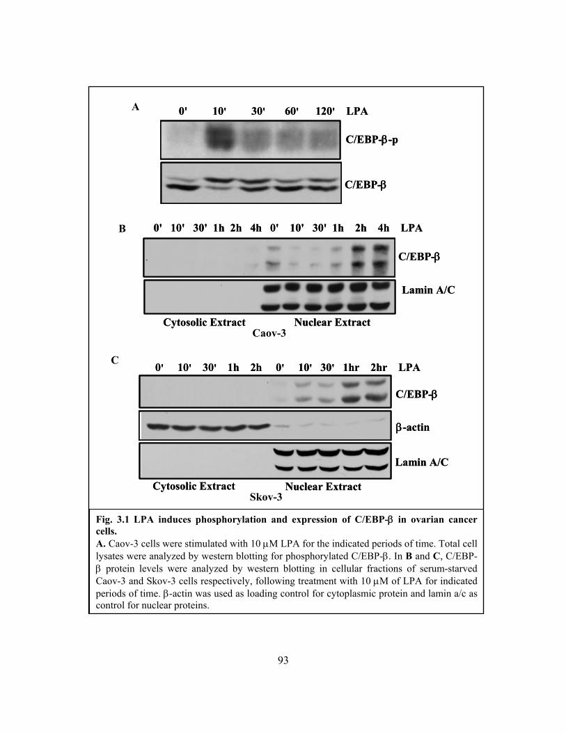
93
Fig. 3.1 LPA induces phosphorylation and expression of C/EBP-β in ovarian cancer cells. A. Caov-3 cells were stimulated with 10 μM LPA for the indicated periods of time. Total cell lysates were analyzed by western blotting for phosphorylated C/EBP-β. In B and C, C/EBP-β protein levels were analyzed by western blotting in cellular fractions of serum-starved Caov-3 and Skov-3 cells respectively, following treatment with 10 μM of LPA for indicated periods of time. β-actin was used as loading control for cytoplasmic protein and lamin a/c as control for nuclear proteins.
B
A
C
0' 10' 30' 60' 120' LPA
C/EBP-β-p
C/EBP-β
0' 10' 30' 60' 120' LPA
C/EBP-β-p
C/EBP-β
0' 10' 30' 1h 2h 4h 0' 10' 30' 1h 2h 4h LPA
Cytosolic Extract Nuclear Extract
Lamin A/C
C/EBP-β
0' 10' 30' 1h 2h 4h 0' 10' 30' 1h 2h 4h LPA
Cytosolic Extract Nuclear Extract
Lamin A/C
C/EBP-β
0' 10' 30' 1h 2h 0' 10' 30' 1hr 2hr LPA
Cytosolic Extract Nuclear Extract
β-actin
Lamin A/C
C/EBP-β
0' 10' 30' 1h 2h 0' 10' 30' 1hr 2hr LPA
Cytosolic Extract Nuclear Extract
β-actin
Lamin A/C
C/EBP-β
Caov-3
Skov-3
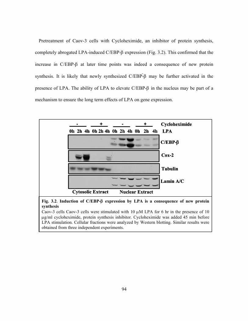
94
Pretreatment of Caov-3 cells with Cycloheximide, an inhibitor of protein synthesis,
completely abrogated LPA-induced C/EBP-β expression (Fig. 3.2). This confirmed that the
increase in C/EBP-β at later time points was indeed a consequence of new protein
synthesis. It is likely that newly synthesized C/EBP- β may be further activated in the
presence of LPA. The ability of LPA to elevate C/EBP-β in the nucleus may be part of a
mechanism to ensure the long term effects of LPA on gene expression.
0h 2h 4h 0h 2h 4h 0h 2h 4h 0h 2h 4h LPA
Cytosolic Extract Nuclear Extract
C/EBP-β
Tubulin
Cox-2
Lamin A/C
Cycloheximide- + - +0h 2h 4h 0h 2h 4h 0h 2h 4h 0h 2h 4h LPA
Cytosolic Extract Nuclear Extract
C/EBP-β
Tubulin
Cox-2
Lamin A/C
Cycloheximide- + - +
Fig. 3.2. Induction of C/EBP-β expression by LPA is a consequence of new protein synthesis Caov-3 cells Caov-3 cells were stimulated with 10 μM LPA for 6 hr in the presence of 10 μg/ml cycloheximide, protein synthesis inhibitor. Cycloheximide was added 45 min before LPA stimulation. Cellular fractions were analyzed by Western blotting. Similar results were obtained from three independent experiments.

95
3.3.2 LPA induces C/EBP-β DNA-binding and transcriptional activities
C/EBP-β contains intramolecular inhibitory elements that hinder its DNA binding
site [237]. Phosphorylation of C/EBP-β has been shown to increase its DNA binding
activity in vitro [220]. To determine whether C/EBP-β phosphorylation induced by LPA
result in enhancement in C/EBP-β DNA binding activity, we performed electromobility
shift assay. The binding of C/EBP to consensus oligonucleotides was increased following
LPA stimulation (Fig 3.3 A) and bound complexes were supershifted in the presence of
anti-C/EBP-β. The binding depended on C/EBP-β phosphorylation as it was blocked by
pretreatment of nuclear extract with potato alkaline phosphatase (PAP) to remove
phosphate groups (Fig 3.3 B). Further, C/EBP-DNA complexes significantly increased at
later time points when more C/EBP-β was present (Fig. 3.3 A and B).
Next, we constructed a C/EBP-responsive luciferase plasmid (pGL2-5xCEBP-TK-
Luc), in which five copies of the C/EBP consensus sequence were linked to the basic TK
promoter. As shown by Fig. 3.3 C, LPA stimulated 5-10 fold increase in luciferase activity
in pGL2-5xCEBP-TK-Luc-transfected ovarian cancer cells but not in the cells transfected
with the backbone vector lacking the C/EBP responsive sites (pGL2-TK-Luc). Taken
together, these results indicate that LPA induces C/EBP-β DNA-binding and
transcriptional activities.

96
Fig. 3.3. LPA activates binding and transcriptional activities of C/EBP-β . A, Nuclear extracts obtained from Caov-3 cells stimulated with 10 μM LPA for the indicated periods of time were subjected to gel shift assay (EMSA) (see Materials and Methods). Supershift experiments were performed on 2h LPA-treated nuclear extracts using 2 and 5 μg anti-C/EBP-β mouse monoclonal antibody. For dephosphorylation of nuclear extracts, 1 unit of potato alkaline phosphatase (PAP) was added for 15 min prior to binding reaction (B). In C, Caov-3 cells were transfected with a luciferase construct of a 5 copies of C/EBP binding sites upstream of a TK promoter. Cells were treated with 10 μM LPA for 6 hours and analyzed for luciferase activity.
B A
C C
ompe
titio
n
0' 10' 30' 1h 2h 4h Com
petit
ion
0' 10' 30' 1h 2h 4h
C/e
bp-β
mA
b
C/e
bp-β
mA
b
ME
S bu
ffer
ME
S +P
AP
Supershift
C/EBP-β
0 2h 4h C/e
bp-β
mA
b
C/e
bp-β
mA
b
ME
S bu
ffer
ME
S +P
AP
Supershift
C/EBP-β
0 2h 4h
0
25
50
75
pG L2-5xC /EBP-T K -Luc
Rel
ativ
e L
ucife
rase
Uni
t (R
LU
) *
BSA LPA0
25
50
75
pG L2-5xC /EBP-T K -Luc
Rel
ativ
e L
ucife
rase
Uni
t (R
LU
) *
BSA LPA
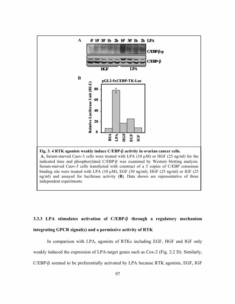
97
3.3.3 LPA stimulates activation of C/EBP-β through a regulatory mechanism
integrating GPCR signal(s) and a permissive activity of RTK
In comparison with LPA, agonists of RTKs including EGF, HGF and IGF only
weakly induced the expression of LPA-target genes such as Cox-2 (Fig. 2.2 D). Similarly,
C/EBP-β seemed to be preferentially activated by LPA because RTK agonists, EGF, IGF
Fig. 3. 4 RTK agonists weakly induce C/EBP-β activity in ovarian cancer cells. A, Serum-starved Caov-3 cells were treated with LPA (10 μM) or HGF (25 ng/ml) for the indicated time and phosphorylated C/EBP-β was examined by Western blotting analysis. Serum-starved Caov-3 cells transfected with construct of a 5 copies of C/EBP consensus binding site were treated with LPA (10 μM), EGF (50 ng/ml), HGF (25 ng/ml) or IGF (25 ng/ml) and assayed for luciferase activity (B). Data shown are representative of three independent experiments.
B
A
C/EBP-β-p
C/EBP−β
0' 10' 30' 1h 2h 10' 30' 1h 2h LPA
HGF LPA
C/EBP-β-p
C/EBP−β
0' 10' 30' 1h 2h 10' 30' 1h 2h LPA
HGF LPA
0
20
40
60
80
LPA
HG
F
EG
F
IGF
BSA
Rel
ativ
e L
ucife
rase
Uni
t (R
LU
) pGL2-5xCEBP-TK-Luc
0
20
40
60
80
LPA
HG
F
EG
F
IGF
BSA
Rel
ativ
e L
ucife
rase
Uni
t (R
LU
) pGL2-5xCEBP-TK-Luc

98
and HGF, were weak stimuli of C/EBP-β phosphorylation (Fig. 3.4 A) and transcriptional
activity (Fig. 3.4 B). However, pretreatment of Caov-3 cells with a specific inhibitor of
EGFR kinase activity, AG1478, resulted in a profound decrease in LPA-induced
phosphorylation of C/EBP-β (Fig. 3.5 A). AG1478 attenuated the LPA-induced increase in
C/EBP-β protein levels (Fig. 3.5 B). In addition, LPA-induced binding of C/EBP to its
consensus oligonucleotide (Fig. 3.5 C) and C/EBP mediated transactivation, as measured
by the C/EBP-responsive luciferase vector were significantly inhibited by AG1478 (Fig.
3.5 D). Therefore, LPA-induced C/EBP-β phosphorylation, DNA binding and functional
activation were invariably sensitive to EGFR inhibition suggesting that EGFR activity,
albeit insufficient on its own to trigger strong C/EBP-β activation, was required for LPA
GPCR signaling to C/EBP-β.
We observed that compared to EGF, LPA only weakly induced activation of
EGFR, as assessed by phosphorylation at Y-1068 (Fig. 3.5 E), ruling out the possibility
that LPA activation of C/EBP-β was through transactivation of EGFR. To further explore
this novel mode of crosstalk between the two receptor types, we examined whether the
requirement of EGFR in the process could be relieved by activation of another RTK, such
as c-Met. Treatment of Caov-3 cells with HGF alone only slightly activated C/EBP-β
phosphorylation (Fig. 3.4 A) and C/EBP transcriptional activity (Fig. 3.4 B). However,
when EGFR was inhibited by AG1478, LPA was fully capable of stimulating C/EBP-β
phosphorylation (Fig. 3.5 A) and transcriptional activity (Fig. 3.5 D) if cells were co-
stimulated with HGF to activate c-Met. Taken together, these results demonstrate that

99
B
A
C
D
C/EBP-β-p
C/EBP-β
Vehicle AG1478 AG1478+HGF
0' 10' 30' 1h 2h 0' 10' 30' 1h 2h 10' 30' 1h 2h LPA
C/EBP-β-p
C/EBP-β
Vehicle AG1478 AG1478+HGF
0' 10' 30' 1h 2h 0' 10' 30' 1h 2h 10' 30' 1h 2h LPA
0h 1h 2h 4h 0h 1h 2h 4h LPA+DMSO +AG1478
C/EBP-β
β-Actin
0h 1h 2h 4h 0h 1h 2h 4h LPA+DMSO +AG1478
C/EBP-β
β-Actin
0 2hr
4hr
0 +A
G14
782h
r +A
G14
784h
r +
AG
1478
0 2hr
4hr
0 +A
G14
782h
r +A
G14
784h
r +
AG
1478
0
25
50
75
100
BSA
LPA
BSA LPA
LPA
+HG
F
Rel
ativ
e L
ucife
rase
Uni
t (R
LU
)
AG1478vehicle
pGL2-5xCEBP-TK-Luc
*
0
25
50
75
100
BSA
LPA
BSA LPA
LPA
+HG
F
Rel
ativ
e L
ucife
rase
Uni
t (R
LU
)
AG1478vehicle
pGL2-5xCEBP-TK-Luc
*
100020003000400050006000700080009000
Ban
d In
tens
ity (a
rbitr
ary
unit)
0h 2h 4h 0h 2h 4hDMSO + LPA LPA +AG1478
100020003000400050006000700080009000
Ban
d In
tens
ity (a
rbitr
ary
unit)
0h 2h 4h 0h 2h 4hDMSO + LPA LPA +AG1478
Con
trol
LPA
S1P
LPA
+ S
1P
PDG
FR
IGF-
1
In
EG
F
5% F
BS
EGFR
EGFR-p
Con
trol
LPA
S1P
LPA
+ S
1P
PDG
FR
IGF-
1
In
EG
F
5% F
BS
EGFR
EGFR-p
E
Fig. 3.5. EGFR inhibitor blocks LPA-induced activation of C/EBP-β Serum-starved Caov-3 cells were treated with LPA (10 μM) and/or AG1478 (1 μM) for the indicated time and phosphorylated (A) or total C/EBP-β (B) was examined by Western blotting analysis. Co-stimulation with HGF (25 ng/ml) in the presence of AG1478 restored LPA-induced phosphorylation of C/EBP-β (A). C, nuclear extracts from LPA-treated or untreated Caov-3 cells were analyzed by gel shift assay using biotin-labeled consensus C/EBP oligonucleotides (see Materials and Methods). Band intensities were quantified with NIH Image J tool. In D, Caov-3 cells transfected with a luciferase construct of a 5 copies of C/EBP binding sites upstream of a TK promoter. Serum-starved transfected cells were then pretreated AG1478, stimulated with LPA and/or HGF for 6 hours and examined for luciferase activity. Cell lysates of Caov-3 cells stimulated indicated agonists for 30’ were analyzed by western blotting for tyrosine 1068 phosphorylation of EGFR (E). Results are representative of three independent experiments.

100
RTK, not necessarily EGFR, provides an obligatory activity that acts in concert with LPA
GPCR signaling to activate C/EBP-β.
3.3.4 RTK-dependent activation of C/EBP-β mediates induction of Cox-2 and other
LPA-target genes
Our observation that LPA induced the expression of Cox-2 in a C/EBP-β-
dependent manner led us to ask whether the requirement of RTK for C/EBP-β activation
also applied to LPA-induced Cox-2 gene expression. Compared to LPA, agonists of RTK,
including EGF, HGF and IGF only weakly induced Cox-2 expression in Caov-3 cells as
shown in Fig. 3.6 A. Similarly, these RTK agonists were weak stimuli of C/EBP-
β transcriptional activity as indicated by luciferase assays with the C/EBP-responsive
construct pGL2-5x-CEBP-TK-luc (Fig. 3.4 B). Thus, C/EBP-β seems to be rate-limiting
regulator of Cox-2, preferentially activated by GPCRs rather than RTKs. In further support
of the role of C/EBP-β in the transcriptional activation of Cox-2, HGF efficiently reversed
the inhibitory effect of AG1478 not only on C/EBP-β activation (Fig. 3.5 A and D) but
also on LPA-induced Cox-2 (Fig. 3.6 B).
In addition to Cox-2, a subset of LPA target genes including IL-6 and uPA bear
binding sites for C/EBP in their promoters (Fig. 1.2 of Chapter 1). IL-6 is a pleiotropic
cytokine and a prominent mediator of inflammation. ELISA analyses of conditioned
medium from Caov-3 cells treated with LPA showed a robust increase in IL-6 production
(Fig. 3.6 C). The ability of LPA to induce the expression of IL-6 in many cell models may

101
explain why high concentrations of IL-6 are present in the ascites of ovarian cancer
patients [238, 239]. In addition, IL-6 levels in ovarian cancer correlate with poor prognosis
[240].
It has been reported that C/EBP-β activates IL-6 promoter in response to IL-1,
bacterial LPS and many other stimuli [241, 242]. To assess the contribution of C/EBP-β to
LPA-induced IL-6 expression, we constructed a luciferase reporter carrying 1.2 kb of
wildtype IL-6 promoter. Disruption C/EBP binding site by point mutation dramactically
decreased the IL-6 promoter activity induced by LPA by 60% loss in promoter response to
LPA as measured by luciferase assay (Fig 3.6 D). Co-transfection of Caov-3 cells with
inhibitory C/EBP-β isoform, LIP, also attenuated the IL-6 promoter activation by LPA
(Fig. 3.6 E). Stable expression of LIP in Skov-3 cells (Fig. 3.6 F, lower right) (see
Materials and Methods) caused a modest reduction of LPA-induced IL-6 production (Fig.
3.6 F), suggesting that stably-transfected Skov-3-LIP cells may have activated some
mechanisms to suppress the effects of enhanced LIP/LAP ratio, such that the impact of
inhibiting transcriptional activities of C/EBP-β on LPA-induced IL-6 gene expression
becomes not as dramatic as in transient experiments. However, AG1478 inhibited LPA-
induced IL-6 promoter response and IL-6 production (Fig. 3.6 G). Similarly, co-
stimulation with HGF restored the ability of LPA-stimulated Caov-3 cells to generate IL-6.

102
Fig. 3.6. RTK-dependent activation of C/EBP-β mediates induction of Cox-2 and IL-6. A. Serum-starved Caov-3 cells transfected with Cox-2 promoter construct were treated with LPA (10 μM), EGF (50 ng/ml), HGF (25 ng/ml) or IGF (25 ng/ml) and assayed for luciferase activity (B) Cox-2 protein levels in Caov-3 cells pretreated AG1478 and stimulated with LPA and/or HGF, were examined by Western blotting analysis. In C, cultured supernatant of Caov-3 cells treated with or without LPA for 18hrs were assayed for IL-6 by ELISA. D, Caov-3 cells transfected with wildtype or C/EBP mutated IL-6 promoter were treated with LPA for 6 hours before luciferase assay.
C
A B
D
0
10
20
30
40
BSA
LPA
HG
F
EG
F
IGF
pGL2-Cox2-1kb-Luc
Rel
ativ
e L
ucife
rase
Uni
t (R
LU
)
0
10
20
30
40
BSA
LPA
HG
F
EG
F
IGF
pGL2-Cox2-1kb-Luc
Rel
ativ
e L
ucife
rase
Uni
t (R
LU
)
Cox-2
Cox-1
AG1478
- + - + - + - + LPA- - + + - - + + HGFDMSO
Cox-2
Cox-1
AG1478
- + - + - + - + LPA- - + + - - + + HGFDMSO
IL-6 Release
0
5000
10000
15000
20000
25000
BSA LPA
IL-6
pg/
mL
IL-6 Release
0
5000
10000
15000
20000
25000
BSA LPA
IL-6
pg/
mL
0
5
10
15
20
25
30
35
40
LPA
Rel
ativ
e L
ucife
rase
Uni
t (R
LU
)
LPABSA BSA
Wildtype IL-6 PromoterC/EBP-mut IL-6 Promoter
0
5
10
15
20
25
30
35
40
LPA
Rel
ativ
e L
ucife
rase
Uni
t (R
LU
)
LPABSA BSA
Wildtype IL-6 PromoterC/EBP-mut IL-6 PromoterWildtype IL-6 PromoterWildtype IL-6 PromoterC/EBP-mut IL-6 PromoterC/EBP-mut IL-6 Promoter
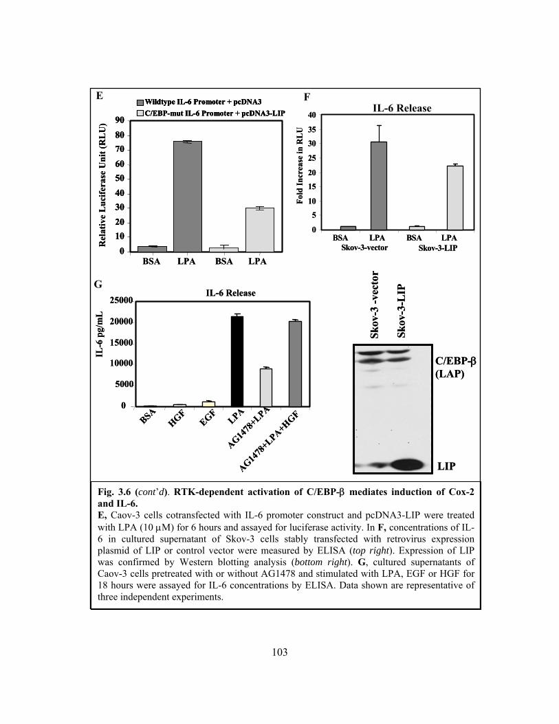
103
Fig. 3.6 (cont’d). RTK-dependent activation of C/EBP-β mediates induction of Cox-2 and IL-6. E, Caov-3 cells cotransfected with IL-6 promoter construct and pcDNA3-LIP were treated with LPA (10 μM) for 6 hours and assayed for luciferase activity. In F, concentrations of IL-6 in cultured supernatant of Skov-3 cells stably transfected with retrovirus expression plasmid of LIP or control vector were measured by ELISA (top right). Expression of LIP was confirmed by Western blotting analysis (bottom right). G, cultured supernatants of Caov-3 cells pretreated with or without AG1478 and stimulated with LPA, EGF or HGF for 18 hours were assayed for IL-6 concentrations by ELISA. Data shown are representative of three independent experiments.
E
G
F
0
5
10
15
20
25
30
35
40
Skov-3-vectorBSA LPA BSA LPA
Fold
Incr
ease
in R
LU
Skov-3-LIP
0
5
10
15
20
25
30
35
40
Skov-3-vectorBSA LPA BSA LPA
Fold
Incr
ease
in R
LU
Skov-3-LIP
IL-6 Release
0
5000
10000
15000
20000
25000
BSAHGF
EGFLPA
AG1478
+LPA
AG1478
+LPA+HGF
IL-6
pg/
mL
IL-6 Release
0
5000
10000
15000
20000
25000
BSAHGF
EGFLPA
AG1478
+LPA
AG1478
+LPA+HGF
IL-6
pg/
mL
0102030405060708090
BSA LPA BSA LPA
Rel
ativ
e L
ucife
rase
Uni
t (R
LU
)Wildtype IL-6 Promoter + pcDNA3C/EBP-mut IL-6 Promoter + pcDNA3-LIP
0102030405060708090
0102030405060708090
BSA LPA BSA LPA
Rel
ativ
e L
ucife
rase
Uni
t (R
LU
)Wildtype IL-6 Promoter + pcDNA3C/EBP-mut IL-6 Promoter + pcDNA3-LIPWildtype IL-6 Promoter + pcDNA3Wildtype IL-6 Promoter + pcDNA3C/EBP-mut IL-6 Promoter + pcDNA3-LIPC/EBP-mut IL-6 Promoter + pcDNA3-LIP IL-6 Release
Skov
-3-L
IP
Skov
-3 -v
ecto
rC/EBP-β(LAP)
LIP
Skov
-3-L
IP
Skov
-3 -v
ecto
rC/EBP-β(LAP)
LIP
104
Finally, consistent with previous studies, LPA induced the activation of uPA, an
important component of tumor cell invasion and metastasis (Fig. 3.7 A). We evaluated the
involvement of C/EBP-β in LPA-induced activation of uPA gene expression. By utilizing
ChIP assay (see Materials and Methods), we found an increased presence of C/EBP-β at
the uPA promoter after 2 hours following LPA stimulation (Fig. 3.7 B). Compared to
wildtype construct, mutation of the proximal C/EBP binding site on uPA promoter
(uPAmut-luc) resulted in a partial yet significant decrease in LPA-induced promoter
activity (Fig. 3.7 C). Co-transfection of LIP in Caov-3 cells also markedly decreased uPA
promoter activation by LPA (Fig. 3.7 D). In agreement with activation of C/EBP-β,
AG1478 completely abrogated LPA-induced uPA activation (Fig. 3.7 E). These results
established that RTK-dependent activation of C/EBP-β reflects a general signaling
mechanism to regulate expression of LPA-target genes Cox-2, IL-6 and uPA.

105
Fig. 3.7. LPA activation of uPA involves C/EBP-β and requires EGFR kinase activity. A, Serum-starved Caov-3 cells lysate were stimulated with 10 μM LPA for 18hrs and analyzed by Western blotting under non-reducing conditions for activated uPA. B, Caov-3 cells were treated with LPA for the indicated period of time and then subjected to ChIP assay (see Materials and Methods). Immunoprecipitation was done using anti-C/EBP-β monoclonal antibody or no antibody (N/A). Purified DNA from inputs (crude chromatin) and immunoprecipitates were analyzed by qPCR using uPA promoter-specific primers. Results of uPA DNA average values in samples were normalized to corresponding input values. PCR and agarose gel electrophoresis were used to analyze DNA of crude inputs (bottom).
BSA LPA
uPA
Tubulin
BSA LPA
uPA
Tubulin
020406080
100120140160180
0' 10' 30' 1h 2h 4h N/ALPA
uPA
cDN
AA
vgva
lue/
INPU
T
020406080
100120140160180
0' 10' 30' 1h 2h 4h N/ALPA
uPA
cDN
AA
vgva
lue/
INPU
T
A
B
0' 10' 30' 1h 2h 4h H20 LPA
INPUT
0' 10' 30' 1h 2h 4h H20 LPA
INPUT

106
Fig. 3.7 (cont’d). LPA activation of uPA involves C/EBP-β and requires EGFR kinase activity. Serum-starved Caov-3 cells transfected with wildtype or C/EBP mutated uPA promoter (C), and in (D) uPA promoter construct and pcDNA3 or pcDNA3-LIP were treated with LPA for 6 hours and analyzed for luciferase activity. Data shown are representative of three independent experiments. E, serum-starved Caov-3 cells pretreated with or without AG1478 and stimulated with LPA were analyzed by Western blotting under non-reducing conditions for activated uPA . Data shown are representative of three independent experiments.
D C
E
0123456789
10
Rel
ativ
e L
ucife
rase
Uni
t (R
LU
)Wildtype uPA PromoterC/EBP-mut uPA Promoter
LPALPABSA BSA0123456789
10
Rel
ativ
e L
ucife
rase
Uni
t (R
LU
)Wildtype uPA PromoterC/EBP-mut uPA Promoter
Wildtype uPA PromoterWildtype uPA PromoterC/EBP-mut uPA PromoterC/EBP-mut uPA Promoter
LPALPABSA BSA
+DMSO +AG1478
uPA
BSA LPA BSA LPA
β-actin
+DMSO +AG1478
uPA
BSA LPA BSA LPA
β-actin
0
1
2
3
4
5
6
BSA LPA BSA LPA
uPAluc Promoter + pcDNA3uPAluc Promoter + LIP
0
1
2
3
4
5
6
BSA LPA BSA LPA0
1
2
3
4
5
6
0
1
2
3
4
5
6
BSA LPA BSA LPA
uPAluc Promoter + pcDNA3uPAluc Promoter + LIPuPAluc Promoter + pcDNA3uPAluc Promoter + pcDNA3
uPAluc Promoter + LIPuPAluc Promoter + LIP
107
3.4 Discussion
In this chapter, we explored the signaling mechanisms by which LPA stimulates
activation of C/EBP-β, a key transcription factor in LPA induction of Cox-2. Our results
indicated that LPA induced phosphorylation of C/EBP-β, which correlated with increase in
C/EBP-β DNA binding activity. By analyzing different cellular fractions, we found that
C/EBP-β was essentially localized to the nuclei in ovarian cancer cells. A very minute
amount was present in the cytoplasmic fraction. LPA did not cause any translocation of
C/EBP-β between cytosol and nucleus. At later hours, there was net increase in total
C/EBP-β protein levels in LPA-treasted cells, probably reflecting auto-induction by
C/EBP-β itself as the C/EBP-β promoter contains C/EBP consensus sequences. The
increase in C/EBP-β protein levels could contribute to sustained effects of LPA on
expression of Cox-2 and other LPA-target genes.
In pursuit of the molecular mechanism linking the LPA receptors to C/EBP-β
activation, we observed that LPA-induced C/EBP activation involves an obligatory activity
from EGFR. Fig. 3.8 is a simplified representation of this obligatory role of EGFR in LPA
GPCRs signaling. Transactivation of EGFR has been proposed as a mechanism to mediate
many biological actions of LPA in numerous studies [181, 243]. In contrast, the possibility
for involvement of a parallel RTK signal in biological responses to LPA or other GPCR
agonists has been rarely studied [189]. In our experiments, EGF and other RTK agonists
only weakly stimulated C/EBP-β activity compared to LPA, allowing us to distinguish the
input of GPCR from that of RTK in these cells.

108
Based on the differential abilities of LPA and EGF to activate C/EBP-β, it is hard
to imagine that transactivation of EGFR could trigger robust activation of C/EBP-β and
Cox-2 expression in LPA-stimulated cells. It is more likely that optimal activation of
C/EBP-β relies on combinatorial signaling components from LPA GPCR(s) and EGFR.
The EGFR signal may feed in at some point downstream of GPCRs. Furthermore, our
Fig. 3.8. Hypothetical model of LPA GPCR-RTK crosstalk in activation of transcription factors and gene expression. Basal activity of EGFR synergizes with signals from LPA GPCR to mediate transcription factor activation. Inhibition of EGFR kinase activity with pharmacological inhibitor AG1478 attenuates LPA-induced activation of gene expression. Activation of another RTK, c-Met by costimulation with HGF in the presence of AG1478 rescues LPA signals to transcription factors.
AG1478
C/EBP-β
EGFR LPA# C-Met
PP
PP
AG1478AG1478
C/EBP-βC/EBP-βC/EBP-β
EGFREGFR LPA#LPA# C-MetC-Met
PP
PP
PP
PP

109
results indicate that activities of other RTKs, not necessarily EGFR, could cooperate with
the LPA GPCR(s) in regulation of C/EBP activity. EGFR is usually recognized to serve
such a role probably because it is more universally expressed and exhibits higher activity
than other RTKs, particularly in cancer cells [244]. The high EGFR activity present in
malignant cells may be necessary for appropriate GPCR signaling.
In addition to Cox-2, we demonstrated that two other LPA-target genes, IL-6 and
uPA are also induced at least partially through activation of C/EBP-β. Thus, the effects of
LPA on IL-6 production and uPA activation were sensitive to inhibition of EGFR. The
comprehensive evaluation of C/EBP-β activation in regulation of gene expression by LPA
described in the present study has further implicated this transcription factor in two
important parts of tumor progression: inflammation and metastasis. Early immediate
response genes such as Cox-2 require prompt transcription. The rapid induction of C/EBP-
β presence to Cox-2 promoter upon LPA stimulation (Fig. 2.12 of Chapter 2) indicate that
C/EBP-β may play critical roles in the initiation of transcription and the recruitment of
transcription factors and co-activators to the Cox-2 promoter [123, 143]; thus its activities
may not be necessary for continued transcription. Unlike Cox-2, activated uPA, a member
of a 3-component metastasis system (others are uPA receptor [uPAR] and plasminogen
activator inhibitors 1 and 2 [PAI-1 and PAI-2]), often accumulates in late response to
stimulation, a result of dependence on the activation of other early response genes [245-
248]. C/EBP-β appeared much later near uPA promoter upon LPA stimulation, clearly
demonstrating that continued activation of C/EBP-β significantly contributes to both
instantaneous and prolong or sustained induction of LPA-target genes.
110
CHAPTER 4
DIFFERENTIAL REQUIREMENT OF RECEPTOR TYROSINE KINASE
ACTIVITY FOR LYSOPHOSPHATIDIC ACID-INDUCED ACTIVATION OF G
PROTEIN SIGNALING CASCADES AND TRANSCRIPTION FACTORS
4.0 Abstract
The role of the epidermal growth factor receptor (EGFR) in provoking biological
actions of G protein-coupled receptors (GPCRs) has been one of the most controversial
research subjects in the field of GPCR signal transduction. We and others have recently
provided evidence for a permissive input from a receptor tyrosine kinase for activation of
GPCR signaling [18, 232, 260]. As described in Chapter 3, LPA-induced activation of
CEBP-β and CEBP responsive genes (Cox-2 and IL-6) requires a RTK activity. In the
current study, we substantiated the crosstalk between the two receptor subtypes to
determine where the RTK input is integrated with GPCR signals to stimulate transcription
factors. In ovarian cancer cell lines, activation of AP-1 components by LPA also depended
upon EGFR, which could be prevented by activation of another receptor tyrosine kinase c-
Met with hepatocyte growth factor (HGF), indicating that LPA-induced activation of AP-1
requires a permissive signal from a receptor tyrosine kinase, not necessarily EGFR. In
contrast, LPA induced activation of another prominent transcription factor NF-κB in an

111
EGFR-independent manner. These differential requirements indicate that the RTK activity
is involved in activation of selective signaling pathways downstream of LPA receptors
rather than activation of LPA receptors themselves. In keeping with this, EGFR was
required for LPA-induced activation of Gi, but not Gq or G12/13 as determined by analyzing
respective effectors of individual classes of G proteins. Further molecular and
pharmacological experiments indicated that Gi was essential for activation of AP-1 by LPA
while NF-κB activation lied downstream of the EGFR-independent Gq pathway.
Consistent with essential roles for Gi and the downstream AP-1 transcription factor in
pleiotropic biological processes, most of cellular responses to LPA such as cytokine
production, cell proliferation, migration and invasion required intact EGFR. These results
reveal a novel dimension of molecular mechanism for the requirement of RTK in
transmission of GPCR signals.
4.1 Introduction
The LPA receptors couple to multiple G proteins, G12/13, Gi, Gq, and probably Gs
[35, 39, 41, 42, 249]. These G proteins link to diverse signaling pathways including
stimulation of phospholipase C and D [16, 49], inhibition of adenylyl cyclase [49], and
activation of Ras and the downstream mitogen-activated protein kinases and
phosphoinositide 3-kinase [50, 52]. Activation of these signaling cascades downstream of
LPA receptors culminates in morphological changes and promotion of cell growth,
survival and motility [50, 51]. Recently, we and others demonstrated that LPA induces
112
activation of transcription factors, upregulating expression of many target genes involved
in cell proliferation, survival and migration/invasion [9, 62, 191-195].
How LPA receptors link to transcriptional activation has become an ideal system to
investigate the molecular mechanisms of LPA signal transduction. Many biological effects
of GPCR have been thought to occur through transactivation of receptor tyrosine kinases,
especially EGFR [181, 182]. In our previous studies, however, the effect of LPA on gene
expression was much more potent than that of EGF itself. LPA indeed weakly
transactivates EGFR as reflected by induction of low levels of phosphorylation of EGFR
(Fig. 3.5 E of Chapter 3) which was in no means comparable to that stimulated by EGF.
Intriguingly, the effects of LPA on gene expression were sensitive to inhibition of EGF,
suggesting requirement of a permissive or parallel input from RTK in transducing LPA
GPCR signals. In further support of this mode of crosstalk between GPCR and RTK, the
dependence on EGFR could be overcome by co-stimulation of c-Met with HGF to provide
an alternate RTK activity. In the current study, we explored the role of EGFR in LPA-
induced activation of the transcription factors AP-1 and NF-κB. Our results indicate that
activation of AP-1 components by LPA was highly sensitive to inhibition of EGFR while
LPA stimulated NF-κB via an EGFR-independent manner, suggesting that EGFR activity
is required for selective signaling cascades rather than proximal activation of LPA
receptors. We further identified EGFR-dependent and independent G protein signaling
cascades involved in activation of these transcription factors. Consistent with involvement
of AP-1 in a multitude of biological processes, RTK activity is needed for LPA-induced
cytokine production, cell proliferation, migration and invasion.
113
4.2 Materials and Methods
Materials Sources of materials were as described in previous chapters. 1-Oleoly (18:1)
LPA was obtained from Avanti Polar Lipids, Inc. (Alabaster, AL). Prior to use, these
phospholipids were dissolved in PBS containing 0.5% fatty acid-free bovine serum
albumin (BSA). BSA, Fugene 6 and protease inhibitor cocktail tablets were purchased
from Roche (Indianapolis, IN). Plasmid DNA was purified using the endo-free purification
kit from Qiagen (Valencia, CA). Luciferase assay reagents were obtained from Promega
(Madison, WI). All oligonucleotides and primers were synthesized by Operon
Biotechnologies, Inc (Huntsville, AL). QuikChange XL site directed mutageneis kit were
purchased from Stratagene (La Jolla, CA). Anti-phospho p65, anti-phospho PKD and anti-
tubulin α/β antibodies were obtained from Cell Signaling (Danvers, MA). Cell culture
medium and lipofectamine 2000 were obtained from Invitrogen Inc. (Carlsbad, CA).
Bovine fetal serum was from Biomeda (Foster City, CA). Enzymes were from New
England biolab (Ipswich, MA) Hepatocyte growth factor (HGF) was obtained from R & D
systems (Minneapolis, MN). Epidermal growth factor (EGF), AG1478, U73122, GF-
109203X and anti-β-actin monoclonal antibody were obtained from Sigma-Aldrich (St.
Louis, MO). All other antibodies were obtained from Santa Cruz Biotechnology (Santa
Cruz, CA).

114
Cell Culture The sources of ovarian cancer cell lines used in the study were described
previously [9, 192]. These cells were cultured in RPMI medium supplemented with 10%
FBS, 100 units/ml penicillin and 100 μg/ml streptomycin. All cell lines were frozen at
early passages and used for less than 10 weeks in continuous culture.
Nuclear Extract Preparation– LPA-stimulated or control cells were washing twice with
cold PBS, harvested by scraping with a rubber policeman and centrifuged at 1000 rpm for
3 min. Cell pellets were resuspended in a hypotonic lysis buffer [10 mM Tris-HCl (pH
7.4), 10 mM KCl, 3 mM MgCl2, 0.5% NP-40], incubated for 15 min on ice, and
centrifuged at 3000 rpm. The nuclei pellet was washed once with the hypotonic lysis
buffer, resuspended in hypertonic nuclear lysis buffer (50 mM Tris-HCl (pH 8.3), 0.4M
NaCl, 40% glycerol, 5 mM MgCl2 and 0.1 mM EDTA) and further incubated for 10 mins
before centrifugation at 13,000 rpm. The supernatant was collected and quick-frozen in
liquid nitrogen before storage at -80oC. Protein concentration was determined with Pierce
BCA kit.
Western Blot Cells were lysed in SDS sample buffer or in ice-cold X-100 lysis buffer [1%
Triton X-100, 50 mM HEPES (pH 7.4), 150 mM NaCl, 1.5 mM MgCl2, 1 mM EGTA,
10% glycerol, 100 mM NaF, 10 mM Na PPi, and protease inhibitor cocktail]. Total cellular
proteins were resolved by SDSPAGE, transferred to Immun-Blot membrane
[poly(vinylidene difluoride)] (BIO-RAD, Hercules, CA), and immunoblotted with
antibodies following the protocols of manufacturers. Immunocomplexes were visualized

115
with an enhanced chemiluminescence detection kit from Amersham (Piscataway, NJ)
using the horseradish peroxidase–conjugated secondary antibodies (Cell Signaling,
Danvers, MA).
Plasmids Constructs A truncated EGFR cDNA (amino acids residues 1-677) lacking the
cytosolic domain was cloned into pcDNA3 vector (Invitrogen, Carlsbad, CA) using
primers: EGFR-Fwd 5’ CATAAGCTTGGAGCAGCGATGCGACCCTCC 3’ and DN-
EGFR-rev 5’CATCTCGAGGCGCTTCCGAACGATGTGG3’. The AP-1 responsive
luciferase vector pGL2-3xAP1-Luc was made by cloning three repeats of AP-1 consensus
binding sequence (TGATGACTCAG) in front of the minimum TK promoter and the
luciferase gene as we described previously [9]. The NF-κB responsive luciferase vector
pGL2-3xNF-kB-Luc was obtained from Stratagene (La Jolla, CA). The vector contains
three repeats of the NF-κB consensus sequence (GGGGACTTTCC) cloned into the pGL2-
basic vector in front of a minimum TK promoter. The Gq dominant-negative mutant vector
pLZRS-IRES-Gq(G208A) was kindly provided by Dr. E. Roos [250].
Transient transfection and luciferase assays Ovarian cancer cell lines were seeded in 6-
well plates and grown to 30-40 % confluence before transfection with the luciferase
vectors using Fugene 6 (Roche) or TransIT-TKO (Mirus Bio Corp., Madison, WI)
according to the instructions of the manufacturers. About 48 hours after transfection, the
cells were starved for 24-36 hours before stimulation with LPA or vehicle for 6 hours. Cell
extracts were prepared and assayed for luciferase activity using the luciferase assay kit
116
from Promega (Madison, WI). The luciferase activity was normalized on the basis of the
activity of cotransfected β-galactosidase reporter driven by the cytomegalovirus promoter
(pCMVβ-gal). Retroviral stock from EGFR-DN stable cells was a kind gift from Dr. Paul
Dent, VCU Medical Centre.
Electromobility shift assay (EMSA)- AP-1 and NF-κB consensus oligonucleotides AP-1
sense 5’GGCGCTTGATGACTCAGCCGGAA 3’; AP-1 antisense
5’GGTTCCGGCTGAGTCATCAAGCG 3’; NF-κB sense
5’ATGTTGAGGGGACTTTCCCAGGCGG 3’ and NF-κB antisense 5’
GCCTGGGAAAGTCCCCTCAACTGG 3’ were synthesized by Operon Biotechnologies
and annealed in 20 mM Tris (pH 7.4), 1 mM dithiothreitol, 50 mM NaCl and 10 mM
MgCl2. Oligonucleotides were labeled at 3’ end with [α-32P]dCTP using klenow enzyme.
For the gel shift assay, binding reaction was performed by incubating 4 μg of nuclear
protein in gel shift buffer (10 mM HEPES (pH 7.8), 10% glycerol, 1 mM EDTA, 25 mM
MgCl2, 50 mM KCl, 1 µg of poly(dI.dC), 3 µg BSA and protease inhibitors) in a final
volume of 20 µL for 10 mins at 25oC. Specificity of binding with each 32P-random labeled
probe was determined with 50-fold excess of unlabeled oligonucleotides. In supershift
experiments, nuclear extracts were incubated for 15 min at room temperature with 1 µg of
anti-c-Jun, anti-p65 or normal mouse IgG (Santa Cruz Biotech) prior to gel shift reaction.
Complexes were separated by electrophoresis on 5% non-denaturing polyacrylamide gel

117
(PAGE). Gels were dried under vacuum and subjected to autoradiography using a
Phosphoimager.
Cell Growth Assay- The growth of Caov-3 and Skov-3 were assayed by seeding cells in
6-well dishes at 3 to 5 x 10⁴ cells per well in complete growth medium (RPMI
supplemented with 10% FBS, 100 units/ml penicillin and 100 μg/ml streptomycin). After
48-72 h of culture, culture medium was replaced with serum-free medium with further
incubation for 18 h. Cells were stimulated with or without 1 μM LPA and/or 1 μM
AG1478 in fresh serum-free medium. Cell counts were performed on triplicate trypsinized
cultures before and post stimulation using a cell counter (Model ZI; Coulter Electronics,
Hialeah, FL).
Migration Assay- The migration of Skov-3 cells was assayed using transwell chambers
(pore size 8 µM) (BD Biosciences, Bedford, MA). The inserts were precoated with
collagen. LPA (10 μM or 1 μM) or vehicle was added to the lower chamber. Serum-
starved cells (1 x 10⁵) were loaded to the upper chamber with or without AG1478 at a final
concentration of 1 μM. Non-migrated cells were removed from the top filter surface with a
cotton swab. Migrated cells attached to the underside of the transwells were washed with
PBS, stained with crystal violet and counted under a microscope.
The invasion of SKOV-3 cells was measured using Transwells coated with growth
factor–reduced Matrigel Basement Membrane Matrix (pore size 8 µM; BD Biosciences;

118
cat. no. 354483). The procedure for invasion was similar to that of the migration assay
described above except that cells were allowed to invade for 20-24 h at 37°C.
Scratch (Wound Closure) Assay- Confluent monolayers of Caov-3 were serum starved
for 18 hr. Scratches were made using sterile 1 μl pipette tips. Displaced cell debris was
washed off with serum-free media before stimulation with 5 μM LPA or BSA (vehicle)
with or without 1 μM AG1478. Images of wounded areas were captured at 0 h and 16 h
after addition of LPA.
Rho and Ras Activation Assays- Activation of Rho and Ras were analyzed by glutathione
S-transferase (GST) pulldown assays [251]. The cells were grown in 10-cm dishes to
subconfluence, starved overnight, and stimulated with LPA or vehicle for the indicated
periods of time. The cells were lysed in Magnesium-containing lysis buffer (MLB: 25 mM
HEPES, pH 7.5, 150 mM NaCl, 1% NP40, 10% glycerol, 10 mM MgCl2, 1 mM EDTA, 1
mM sodium orthovanadate, 10 µg/ml leupeptin, 10 µg/ml aprotinin). Clarified lysates were
incubated for 45-60 min at 4°C with GST-Rhotekin-RBD (Rho binding domain of
Rhotekin, residues 7-89; [252] ) or GST-Raf-RBD (Ras binding domain of Raf, residues 1-
149[253]) produced in Escherichia coli and immobilized on glutathione-coupled Sepharose
beads. Beads were washed in MLB three times, eluted with SDS sample buffer, and
analyzed by Western blotting using monoclonal anti-Rac antibody (BD Biosciences; Cat.
No. 610650) or rabbit anti-Ras antibody (Cell signaling, Denvers, MA).
119
Densitometry and Statistics- Intensities of western blot bands were quantified using the
NIH Image J software. All numerical data were presented as mean ± SD. The statistical
significance of differences was analyzed using Student's t test where P < 0.05 was
considered statistically significant.
4.3 Results
4.3.1 Activation AP-1 Proteins by LPA
LPA is a master inter-cellular regulator of gene expression in mammalian cells.
Although post-transcriptional regulation may be involved in the reinforcement of the effect
LPA on gene expression, the initial input is driven by transcriptional activation. We have
previously shown that the effects of LPA on gene expression are mediated by a number of
prominent transcription factors including AP-1, NF-κB, C/EBP and Sp-1 [9, 232, 254].
Thus activation of transcription factors offers an ideal readout to study functions of LPA
receptors, their downstream signaling networks and their crosstalk with RTKs. In ovarian
and other cancer cell lines expressing LPA receptors, treatment with LPA led to induction
of various AP-1 proteins.
As demonstrated in Fig. 4.1 A, LPA induced c-Jun, Fos, Jun B and Fra-1
expression in a time-dependent manner in Caov-3 cells. Induction of c-Jun and Fos
expression occurred immediately and peaked at 1 hr after exposure to LPA. Jun B and Fra-
1 were induced at later hours and highest levels were seen at 4-6 hr of LPA treatment (Fig.
4.1). The sequential induction of these AP-1 components could lead to sustained increases
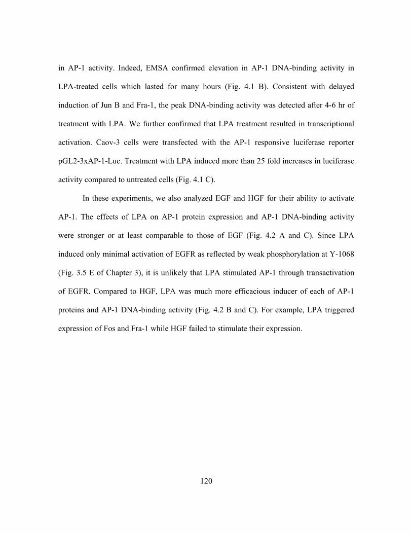
120
in AP-1 activity. Indeed, EMSA confirmed elevation in AP-1 DNA-binding activity in
LPA-treated cells which lasted for many hours (Fig. 4.1 B). Consistent with delayed
induction of Jun B and Fra-1, the peak DNA-binding activity was detected after 4-6 hr of
treatment with LPA. We further confirmed that LPA treatment resulted in transcriptional
activation. Caov-3 cells were transfected with the AP-1 responsive luciferase reporter
pGL2-3xAP-1-Luc. Treatment with LPA induced more than 25 fold increases in luciferase
activity compared to untreated cells (Fig. 4.1 C).
In these experiments, we also analyzed EGF and HGF for their ability to activate
AP-1. The effects of LPA on AP-1 protein expression and AP-1 DNA-binding activity
were stronger or at least comparable to those of EGF (Fig. 4.2 A and C). Since LPA
induced only minimal activation of EGFR as reflected by weak phosphorylation at Y-1068
(Fig. 3.5 E of Chapter 3), it is unlikely that LPA stimulated AP-1 through transactivation
of EGFR. Compared to HGF, LPA was much more efficacious inducer of each of AP-1
proteins and AP-1 DNA-binding activity (Fig. 4.2 B and C). For example, LPA triggered
expression of Fos and Fra-1 while HGF failed to stimulate their expression.
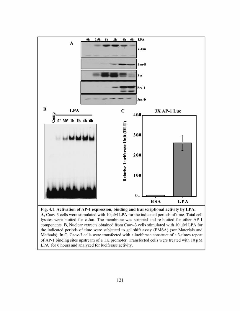
121
Fig. 4.1. Activation of AP-1 expression, binding and transcriptional activity by LPA. A, Caov-3 cells were stimulated with 10 μM LPA for the indicated periods of time. Total cell lysates were blotted for c-Jun. The membrane was stripped and re-blotted for other AP-1 components. B, Nuclear extracts obtained from Caov-3 cells stimulated with 10 μM LPA for the indicated periods of time were subjected to gel shift assay (EMSA) (see Materials and Methods). In C, Caov-3 cells were transfected with a luciferase construct of a 3-times repeat of AP-1 binding sites upstream of a TK promoter. Transfected cells were treated with 10 μM LPA for 6 hours and analyzed for luciferase activity.
c-Jun
0h 0.5h 1h 2h 4h 6h LPA
Jun-B
Fos
Fra-1
Jun-D
c-Jun
0h 0.5h 1h 2h 4h 6h LPA
Jun-B
Fos
Fra-1
Jun-D
Com
p LPA
0’ 30’ 1h 2h 4h 6h Com
p LPA
0’ 30’ 1h 2h 4h 6h Com
p LPA
0’ 30’ 1h 2h 4h 6h
0
100
200
300
400
B SA L P A
Rel
ativ
e L
ucife
rase
Uni
t (R
LU
)
0
100
200
300
400
B SA L P A
Rel
ativ
e L
ucife
rase
Uni
t (R
LU
)
A
B C 3X AP-1 Luc
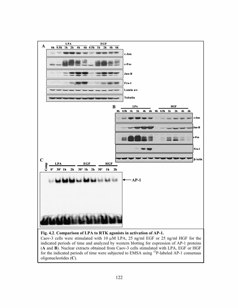
122
Fig. 4.2. Comparison of LPA to RTK agonists in activation of AP-1. Caov-3 cells were stimulated with 10 μM LPA, 25 ng/ml EGF or 25 ng/ml HGF for the indicated periods of time and analyzed by western blotting for expression of AP-1 proteins (A and B). Nuclear extracts obtained from Caov-3 cells stimulated with LPA, EGF or HGF for the indicated periods of time were subjected to EMSA using 32P-labeled AP-1 consensus oligonucleotides (C).
B
A LPALPA EGFEGF
Tubulin
Lamin a/c
Fra-1
Jun-B
c-Fos
c-Jun
6h4h2h1h0.5h6h4h2h1h0.5h0hLPALPA EGFEGF
Tubulin
Lamin a/c
Fra-1
Jun-B
c-Fos
c-Jun
6h4h2h1h0.5h6h4h2h1h0.5h0hC
omp LPA EGF HGF
0’ 30’ 1h 2h 30’ 1h 2h 30’ 1h 2hCom
p LPA EGF HGF
0’ 30’ 1h 2h 30’ 1h 2h 30’ 1h 2h
C
AP-1
LPALPA HGFHGF
0.5h
β-Actin
Fra-1
c-Fos
Jun-B
c-Jun
6h4h2h1h0h6h4h2h1h0.5h0h
LPALPA HGFHGF
0.5h
β-Actin
Fra-1
c-Fos
Jun-B
c-Jun
6h4h2h1h0h6h4h2h1h0.5h0h

123
4.3.2 Requirement of EGFR or an alternate RTK for LPA-induced activation of AP-1
Having observed the dependence of LPA-induced C/EBP-β activation on RTK
activity, we asked if this obligatory role was a general requirement for activation of
transcription factors by LPA. Pretreatment of ovarian cancer cells with AG1478, a specific
pharmacological inhibitor of EGFR kinase activity, abrogated LPA-induced expression of
JunB and Fra-1 (Fig. 4.3 A). The expression of c-Jun and c-Fos was dramatically inhibited
by the presence of AG1478. Similarly, LPA-induced AP-1 DNA binding and
transcriptional activities were drastically suppressed by AG1478 (Fig. 4.3 B and C)
To determine whether EGFR is specifically needed for GPCR signaling to AP-1,
we co-stimulated Caov-3 cells with LPA and HGF in the presence of AG1478. The
stimulatory effects of LPA on expression of AP-1 proteins and AP-1 DNA-binding activity
were fully recovered by co-treatment of the cells with HGF to activate the c-Met, another
receptor tyrosine kinase (Fig. 4.3 A and B). The role of HGF was not due to activation of
AP-1 by HGF itself as the effects of HGF on AP-1 protein expression and DNA-binding
activity were marginal compared to those of LPA (Fig. 4.2 B and C). The restoration of
AP-1 activity by HGF also demonstrated that AG1478 did not block LPA-induced AP-1
activation through nonspecific or toxic effects. These results indicate that activity of a
RTK, not necessarily EGFR, provides an activity indispensable for transmission of GPCR
signals to AP-1 although such an activity itself is insufficient for full activation of AP-1.

124
Fig. 4.3. Requirement of RTK for LPA-induced activation of AP-1. Serum-starved Caov-3 cells were treated with LPA (10 μM), HGF (25 ng/ml) and/or AG1478 (1 μM) for the indicated time. Expression of AP-1 proteins was examined by Western blotting analysis (A). Nuclear extracts from LPA-treated or untreated Caov-3 cells were analyzed by EMSA using 32P-labeled consensus AP-1 oligonucleotides (B). In C, Caov-3 cells transfected with a luciferase construct of a 3 copies of AP-1 consensus binding sites upstream of a TK promoter. Serum-starved transfected cells were then pretreated with AG1478, stimulated with LPA for 6 hours and examined for luciferase activity. Results are representative of three independent experiments.
GSK-1
Fra-1
c- Fos
Jun-D
Jun-B
c-Jun
LPA/hour0 0.5 1 2 4 6 0 0.5 1 2 4 6 0 0.5 1 2 4 6
DMSO AG1478 AG1478 + HGF
GSK-1
Fra-1
c- Fos
Jun-D
Jun-B
c-Jun
LPA/hour0 0.5 1 2 4 6 0 0.5 1 2 4 6 0 0.5 1 2 4 6
DMSO AG1478 AG1478 + HGF
0 0.5 1 2 4 0 0.5 1 2 4 0.5 1 2 4 LPA/hour
DMSO AG1478 AG1478 + HGF
0 0.5 1 2 4 0 0.5 1 2 4 0.5 1 2 4 LPA/hour
DMSO AG1478 AG1478 + HGF
3X AP1-luc
05
1015
20253035
Fold
Incr
ease
BSA LPA BSA LPADMSO AG1478
3X AP1-luc
05
1015
20253035
05
1015
20253035
Fold
Incr
ease
BSA LPA BSA LPADMSO AG1478
A
B
C

125
Fig. 4.4. Effects of overexpression of dominant negative EGFR on LPA-induced AP-1 protein expression. A, Caov-3 cells were infected for 48hours with adenoviral stock from adeno-EGFR-DN or control adenovirus overexpressing cells then stimulated with 10 μM LPA for the indicated periods of time. Total cell lysates were analyzed for expression of AP-1 proteins by Western blotting. Similarly, in B, AP-1 proteins expression were analyzed by western blotting after LPA stimulation of Caov-3 cells transfected with pcDNA3-EGFRDN or control vector. C, schematic representation of truncated EGFR-DN and its inhibitory function in stimulated condition. Only wildtype homodimers possess kinase activity, represented by phosphorylated tyrosine residues (red circles).
A
B
PP
PP
stimuli
PP
PP
PP
PP
PP
PP
stimuli
C
Jun-B
c-Fos
β-actin
Fra-1
EGFR
0' 15' 30' 1h 2h 0' 15' 30' 1h 2h LPAControl Adenovirus EGFR-DN Adenovirus
c-Jun
Jun-B
c-Fos
β-actin
Fra-1
EGFR
0' 15' 30' 1h 2h 0' 15' 30' 1h 2h LPAControl Adenovirus EGFR-DN Adenovirus
c-Jun
Jun-B
c-Jun
c-Fos
Fra-1
β-actin
Vector EGFR-DN- + - + LPA
EGFR
EGFR(truncated)
Jun-B
c-Jun
c-Fos
Fra-1
β-actin
Vector EGFR-DN- + - + LPA
EGFR
EGFR(truncated)
126
In further support of these results derived from pharmacological inhibition of
EGFR, overexpression of a dominant negative, truncated form of EGFR (EGFR-DN) [255]
in Caov-3 cells by both adenoviral transduction and direct expression plasmid suppressed
LPA-induced expression of each of AP-1 proteins (Fig. 4.4). These pharmacological and
molecular approaches together establish an obligatory role for EGFR or an alternate RTK
in activation of the AP-1 transcription factor by LPA.
4.3.3 EGFR-independent activation of NF-κB by LPA
The role of EGFR in LPA activation of CEBP-β (Chapter 3) and AP-1 raises the
possibility that a basal RTK activity might be ubiquitously required for GPCR actions.
This could be due to the requirement of RTK activity for activation of GPCR itself. If the
RTK input is involved in activation of the specific intracellular G protein signaling
processes instead of GPCR activation on the membrane, some LPA signaling pathways
may not rely on RTK activity. To distinguish these possibilities, we examined LPA-
induced activation of another transcription factor, NF-κB, a transcription factor critical for
activation of many LPA target genes involved in inflammation and cancer progression. As
shown in Fig. 4.5 A-C, in Caov-3 and Skov-3 cells treated with AG1478 to block EGFR
function, LPA induced NF-κB p65 phosphorylation, IκB phosphorylation and IκB
degradation at levels comparable to those in control cells untreated with AG1478.

127
Fig. 4.5. EGFR-independent activation of NF-κB by LPA. Total cell lysates of serum-starved Caov-3 (A and C) or Skov-3 cells (B) stimulated with 10 μM LPA and/or 1 μM AG1478 for the indicated periods of time were analyzed by Western blotting for phosphorylated NF-κB p65. The membrane was reprobed with anti-phospho-IκBα (B) or anti-IκBα (C). IKKα/β, β-actin or Tubulin were used to show equal loading. c-Jun or c-Fos expression was used to confirm efficacy of batch of AG1478 used in experiment.
B
A
c‐Fos
IKKα/β
AG1478
0’ 10’ 30’ 1h 2h 4hr 0’ 10’ 30’ 1h 2h 4h
DMSO
LPA
p65‐p
c‐Fos
IKKα/β
AG1478
0’ 10’ 30’ 1h 2h 4hr 0’ 10’ 30’ 1h 2h 4h
DMSO
LPA
AG1478
0’ 10’ 30’ 1h 2h 4hr 0’ 10’ 30’ 1h 2h 4h
DMSO
LPA
p65‐p
p65-p
0’ 10’ 30’ 1h 2h 0’ 10’ 30’ 1h 2h L PA /T im eD M SO A G 1478
c-Jun
β -A ctin
IκB α− p
p65-p
0’ 10’ 30’ 1h 2h 0’ 10’ 30’ 1h 2h L PA /T im eD M SO A G 1478
c-Jun
β -A ctin
IκB α− p
Caov-3
Skov-3
IκBα
AG1478
0’ 10’ 30’ 1h 2h 4hr 0’ 10’ 30’ 1h 2h 4h
DMSO
LPA
Tubulin
IκBα
AG1478
0’ 10’ 30’ 1h 2h 4hr 0’ 10’ 30’ 1h 2h 4h
DMSO
LPA
AG1478
0’ 10’ 30’ 1h 2h 4hr 0’ 10’ 30’ 1h 2h 4h
DMSO
LPA
Tubulin
C
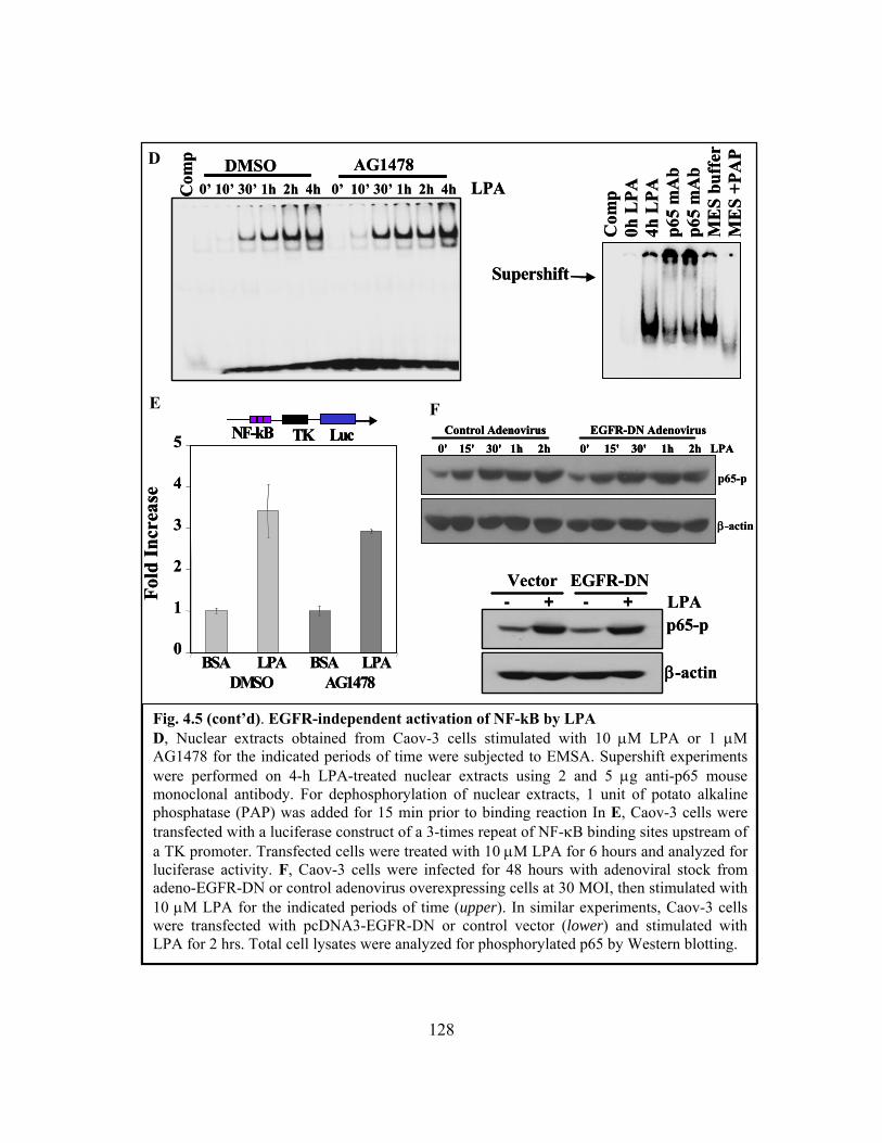
128
Fig. 4.5 (cont’d). EGFR-independent activation of NF-kB by LPA D, Nuclear extracts obtained from Caov-3 cells stimulated with 10 μM LPA or 1 μM AG1478 for the indicated periods of time were subjected to EMSA. Supershift experiments were performed on 4-h LPA-treated nuclear extracts using 2 and 5 μg anti-p65 mouse monoclonal antibody. For dephosphorylation of nuclear extracts, 1 unit of potato alkaline phosphatase (PAP) was added for 15 min prior to binding reaction In E, Caov-3 cells were transfected with a luciferase construct of a 3-times repeat of NF-κB binding sites upstream of a TK promoter. Transfected cells were treated with 10 μM LPA for 6 hours and analyzed for luciferase activity. F, Caov-3 cells were infected for 48 hours with adenoviral stock from adeno-EGFR-DN or control adenovirus overexpressing cells at 30 MOI, then stimulated with 10 μM LPA for the indicated periods of time (upper). In similar experiments, Caov-3 cells were transfected with pcDNA3-EGFR-DN or control vector (lower) and stimulated with LPA for 2 hrs. Total cell lysates were analyzed for phosphorylated p65 by Western blotting.
0’ 10’ 30’ 1h 2h 4h 0’ 10’ 30’ 1h 2h 4h LPADMSO AG1478
Com
p0’ 10’ 30’ 1h 2h 4h 0’ 10’ 30’ 1h 2h 4h LPA
DMSO AG1478C
omp
Com
p0h
LPA
4h L
PAp6
5 m
Ab
p65
mA
bM
ES
buff
erM
ES
+PA
P
Supershift
Com
p0h
LPA
4h L
PAp6
5 m
Ab
p65
mA
bM
ES
buff
erM
ES
+PA
P
Supershift
D
E F NF-kB TK Luc
0
1
2
3
4
5
BSA LPA BSA LPADMSO AG1478
Fold
Incr
ease
NF-kB TK LucNF-kB TK Luc
0
1
2
3
4
5
BSA LPA BSA LPADMSO AG1478
Fold
Incr
ease
p65-p
β-actin
0' 15' 30' 1h 2h 0' 15' 30' 1h 2h LPAControl Adenovirus EGFR-DN Adenovirus
p65-p
β-actin
0' 15' 30' 1h 2h 0' 15' 30' 1h 2h LPAControl Adenovirus EGFR-DN Adenovirus
0' 15' 30' 1h 2h 0' 15' 30' 1h 2h LPAControl Adenovirus EGFR-DN Adenovirus
p65-p
β-actin
Vector EGFR-DN- + - + LPA
p65-p
β-actin
Vector EGFR-DN- + - + LPA

129
Similarly, LPA-stimulated NF-κB DNA-binding activity was not compromised by
AG1478 as measured by EMSA (Fig. 4.5 D); nor was LPA-driven NF-κB transcriptional
activity significantly ablated by incubation of cells with AG1478 as analyzed by the NF-
κB responsive luciferase reporter assay (Fig. 4.5 E). Further, overexpression of dominant
negative EGFR (EGFR-DN) did not interfere with LPA-induced p65 phosphorylation (Fig.
4.5 F) despite strong inhibitory effect of EGFR-DN on AP-1 protein expression (Fig. 4.4).
Therefore, in sharp contrast to AP-1 activation, LPA-elicited NF-κB activity occurs via an
EGFR-independent route. The results also indicate that the crosstalk with RTK is required
only for a selective subset of biochemical events but not overall activities of LPA
receptors.
4.3.4 G protein cascades mediating LPA-induced AP-1 and NF-κB activation
To identify the mechanism for the differential requirements of RTK in transmitting
GPCR signals to AP-1 and NF-κB, we examined G protein signaling cascades responsible
for activating AP-1 and NF-κB. The classical Edg LPA receptors expressed in cancer cell
lines couple to Gi, Gq and G12/13 [36-42]. Inhibition of Gi with pertussis toxin (PTX)
strongly decreased LPA-induced AP-1 proteins c-Jun and c-Fos as shown in Fig. 4.6 A,
indicating that Gi signaling links to AP-1 activation by LPA. However, Gi was disposable
for NF-κB activation as PTX did not interfere with NF-κB p65 phosphorylation induced
by LPA.
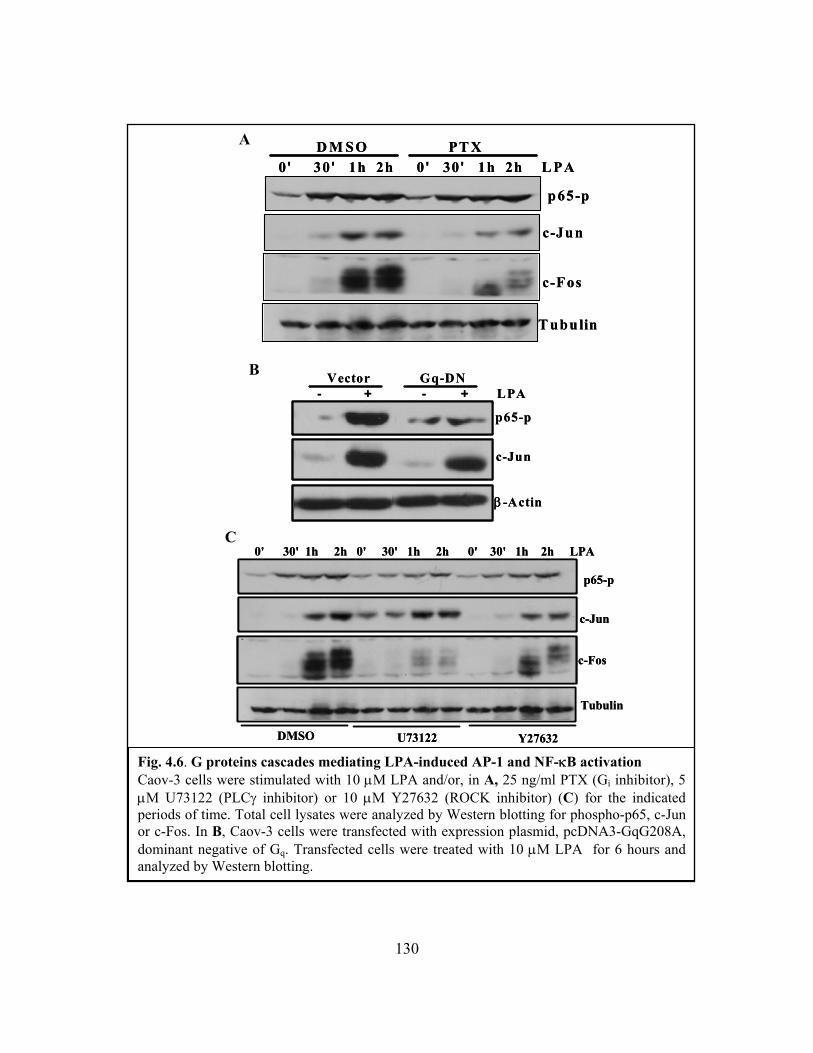
130
Fig. 4.6. G proteins cascades mediating LPA-induced AP-1 and NF-κB activation Caov-3 cells were stimulated with 10 μM LPA and/or, in A, 25 ng/ml PTX (Gi inhibitor), 5 μM U73122 (PLCγ inhibitor) or 10 μM Y27632 (ROCK inhibitor) (C) for the indicated periods of time. Total cell lysates were analyzed by Western blotting for phospho-p65, c-Jun or c-Fos. In B, Caov-3 cells were transfected with expression plasmid, pcDNA3-GqG208A, dominant negative of Gq. Transfected cells were treated with 10 μM LPA for 6 hours and analyzed by Western blotting.
p65-p
c-Jun
c-Fos
D M SO PT X 0 ' 30 ' 1h 2h 0 ' 30 ' 1h 2h L PA
T ubulin
p65-p
c-Jun
c-Fos
D M SO PT X 0 ' 30 ' 1h 2h 0 ' 30 ' 1h 2h L PA
T ubulin
Vector Gq-DN
p65-p
c-Jun
β -Actin
- + - + LPAVector Gq-DN
p65-p
c-Jun
β -Actin
- + - + LPA
C
A
B
DMSO U73122 Y27632
0' 30' 1h 2h 0' 30' 1h 2h 0' 30' 1h 2h LPA
c-Jun
p65-p
c-Fos
Tubulin
DMSO U73122 Y27632
0' 30' 1h 2h 0' 30' 1h 2h 0' 30' 1h 2h LPA
c-Jun
p65-p
c-Fos
Tubulin
131
To assess the contributions of Gq signaling cascade to AP-1 and NF-κB activation,
we took advantage of a dominant negative form of Gq (G208A) that has been shown to
specifically block Gq–mediated pathways in different cell systems [250, 256, 257]. The
GqG208A was transfected into Caov-3 cells using Amaxa nuocleofector Kit T that yield
high transfection efficiency in ovarian cancer cell lines as we described previously
(Chapter 2, Materials and Methods). Expression of the GqG208A mutant almost
completely prevented LPA-induced p65 phosphorylation but not c-Jun expression (Fig. 4.6
B). Induction of Fos by LPA was decreased by overeexpression of GqG208A. Due to the
lack of commercially available Gq pharmacological inhibitors, we made use of U73122, an
antagonist of PLC that lies downstream of Gq [16, 49]. U73122 inhibited LPA-induced
NF-κB p65 phosphorylation and c-Fos induction (Fig. 4.6 C). Only slight reduction in
LPA-induced c-Jun expression was observed in U73122-treated cells. These data establish
that the Gq-mediated signaling is critical for NF-κB activation in response to LPA.
Activation of Gq may also contribute to LPA induction of certain AP-1 proteins such as c-
Fos.
We also examined the role of G12/13 in LPA-mediated activation of AP-1 and NF-
κB through inhibition of the G12/13 effector ROCK. ROCK has been reported to participate
in LPA-induced c-Jun expression in NIH 3T3 cells [258]. We examined the effects of a
highly selective ROCK inhibitor Y-27632 on LPA-induced AP-1 and NF-κB activation.
The compound did not affect LPA-induced p65 phosphorylation but compromised c-Jun
and c-Fos induction (Fig. 4.6 C). Based on these results, each of G protein modules (Gi,

132
Gq, and G12/13) seems to contribute to AP-1 activity while only the Gq pathway couples to
NF-κB activation in LPA-stimulated cells.
4.3.5 Differential requirement of EGFR for activation of G signaling cascades
We next explored whether EGFR is differentially required for activation of
intracellular G protein signaling modules. Since it is practically difficult to analyze
activation of G proteins, we examined the downstream effectors of each class of G proteins
coupled to LPA receptors. Ras activation is a well-defined Gi-dependent signal, Rho lies
downstream of G12/13 and activation of Gq could be monitored by analyzing the PKC-PKD
pathway. As demonstrated in Fig. 4.7 A, LPA-induced Ras activation was completely
blocked by AG1478 as measured by GST pulldown assay for the GTP-bound Ras (GTP-
Ras). In agreement with EGFR-dependent activation of Ras by LPA, Erk activation was
also highly sensitive to AG1478 (Fig. 4.7 B), suggesting that LPA-induced activation of Gi
relies on a permissive signal from EGFR. However, the GST pulldown analysis of Rho
activation demonstrated that EGFR was not involved in activation of G12/13 as the
downstream Rho was fully activated by LPA in the presence of AG1478 (Fig. 4.7 C). PKD
is a well characterized substrate of various isoforms of PKC [259]. LPA stimulated a rapid
and sustained PKD phosphorylation at Serine 916 (Fig. 4.8A). Blockade of PKC with GF-
109203X specific inhibitor of classical PKCs, or inhibition of PLCγ with U73122 (Fig. 4.8
C) prevented LPA-induced phosphorylation of PKD, confirming that the PLC-PKC
cascade lies upstream of PKD.
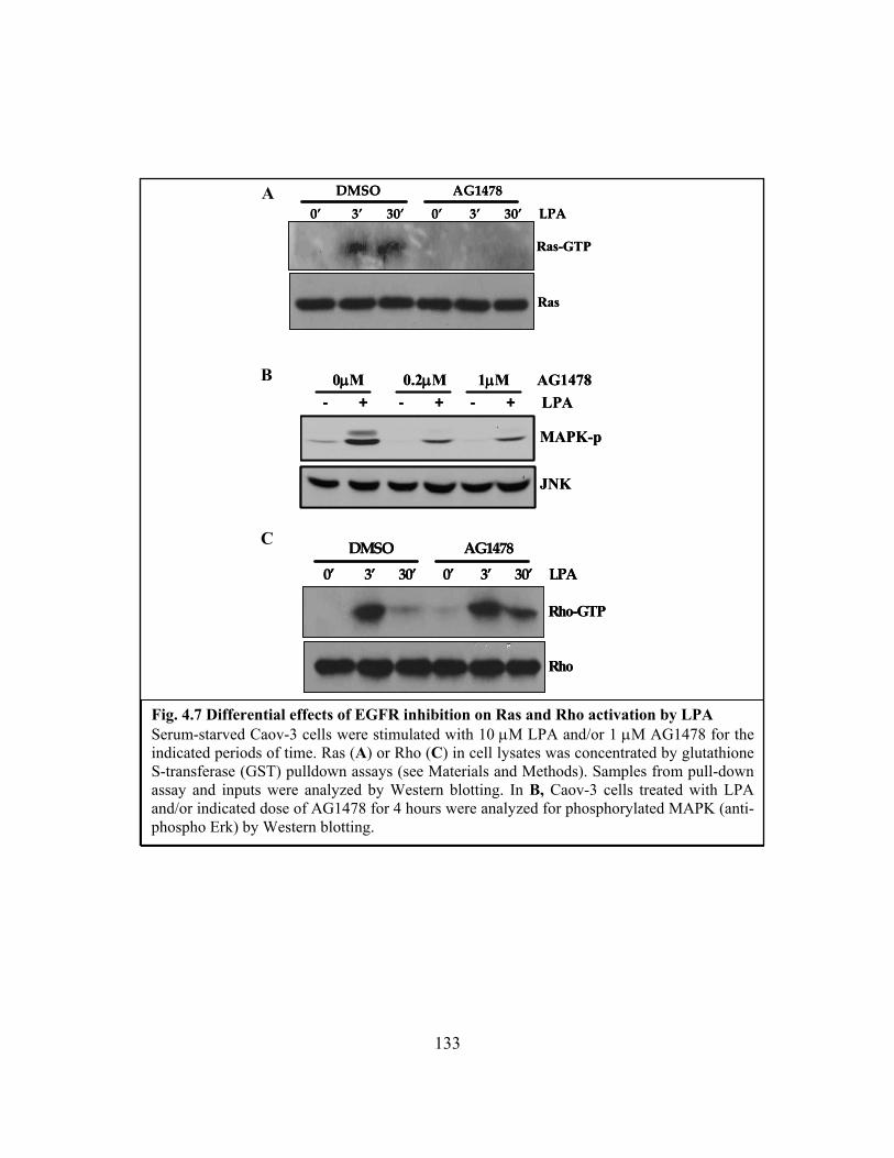
133
Fig. 4.7 Differential effects of EGFR inhibition on Ras and Rho activation by LPA Serum-starved Caov-3 cells were stimulated with 10 μM LPA and/or 1 μM AG1478 for the indicated periods of time. Ras (A) or Rho (C) in cell lysates was concentrated by glutathione S-transferase (GST) pulldown assays (see Materials and Methods). Samples from pull-down assay and inputs were analyzed by Western blotting. In B, Caov-3 cells treated with LPA and/or indicated dose of AG1478 for 4 hours were analyzed for phosphorylated MAPK (anti-phospho Erk) by Western blotting.
C
A 0’ 3’ 30’ 0’ 3’ 30’ LPA
DMSO AG1478
Ras-GTP
Ras
0’ 3’ 30’ 0’ 3’ 30’ LPA
DMSO AG1478
Ras-GTP
Ras
B
0’ 3’ 30’ 0’ 3’ 30’ LPA DMSO AG1478
Rho-GTP
Rho
0’ 3’ 30’ 0’ 3’ 30’ LPA DMSO AG1478
Rho-GTP
Rho
- + - + - + LPA0μM 0.2μM 1μM AG1478
MAPK-p
JNK
- + - + - + LPA0μM 0.2μM 1μM AG1478
MAPK-p
JNK
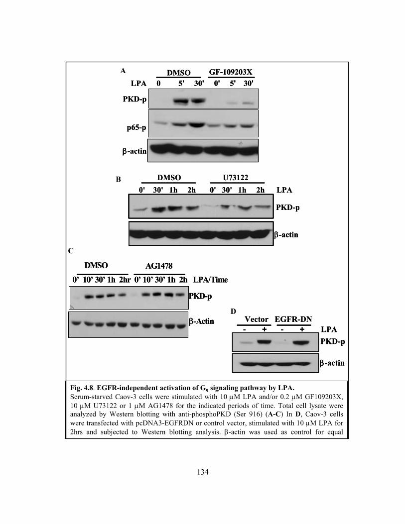
134
Fig. 4.8. EGFR-independent activation of Gq signaling pathway by LPA. Serum-starved Caov-3 cells were stimulated with 10 μM LPA and/or 0.2 μM GF109203X, 10 μM U73122 or 1 μM AG1478 for the indicated periods of time. Total cell lysate were analyzed by Western blotting with anti-phosphoPKD (Ser 916) (A-C) In D, Caov-3 cells were transfected with pcDNA3-EGFRDN or control vector, stimulated with 10 μM LPA for 2hrs and subjected to Western blotting analysis. β-actin was used as control for equal
PKD-p
0’ 10’ 30’ 1h 2hr 0’ 10’ 30’ 1h 2h LPA/Time
DMSO AG1478
β-Actin
PKD-p
0’ 10’ 30’ 1h 2hr 0’ 10’ 30’ 1h 2h LPA/Time
DMSO AG1478
β-Actin
β-actin
Vector EGFR-DN- + - + LPA
PKD-p
β-actin
Vector EGFR-DN- + - + LPA
PKD-p
D
C
A
0' 30' 1h 2h 0' 30' 1h 2h LPADMSO U73122
β-actin
PKD-p
0' 30' 1h 2h 0' 30' 1h 2h LPADMSO U73122
β-actin
PKD-p
PKD-p
p65-p
LPA 0 5' 30' 0' 5' 30'DMSO GF-109203X
β-actin
PKD-p
p65-p
LPA 0 5' 30' 0' 5' 30'DMSO GF-109203X
β-actin
B
135
However, inhibition of EGFR function with AG1478 or EGFR-DN did not affect
PKD phosphorylation induced by LPA (Fig. 4.8 C and D). Therefore, activation of the Gq-
PLC-PKC-PKD pathway does not require EGFR activity. These results reveal EGFR-
dependent Gi and EGFR-independent Gq and G12/13 signaling cascades downstream of LPA
receptors. Since these G protein pathways are linked to activation of specific transcription
factors as specified above, these results provide a molecular basis for the differential
requirements of EGFR in LPA-stimulated activation of the AP-1 and NF-κB transcription
factors.
4.3.6 Essential roles of EGFR in multiple biological responses to LPA
If Gi and the downstream AP-1 depend on EGFR for activation, we expected that
many cellular processes mediated by Gi or AP-1 are sensitive to EGFR inhibition. To
further test this speculation, we first examined the effect of EGFR inhibition on LPA-
induced IL-8 production, a functional outcome of synergistic actions of NF-κB and AP-1
as we described previously [9]. As demonstrated in Fig. 4.9 A, the prominent effect of
LPA on IL-8 production was suppressed by inhibition of EGFR with AG1478, consistent
with the necessity of EGFR for LPA-induced AP-1 activation. Further analysis of other
cellular responses to LPA demonstrated that inhibition of EGFR suppressed LPA-afforded
cell growth (Fig. 4.9 B). LPA-mediated migration, invasion and wound closure in ovarian
cancer cell lines were also attenuated in the presence of AG1478 (Fig. 4.9 C-E), in support

136
of essential roles of EGFR-dependent Gi and its downstream signaling pathways in
promotion of a broad range of cellular responses to LPA [18, 260].
Fig. 4. 9. Role of EGFR in LPA-mediated biological responses. A, Cultured supernatants of serum-starved Caov-3 cells pretreated with or without 1 μM AG1478 and stimulated with 10 μM LPA for 18hrs were assayed for IL-6 concentrations by ELISA. In B, triplicate samples of Caov-3 or Skov-3 cells were serum starved for 12hrs, stimulated with 5 μM LPA and/or 1 μM AG1478 for the indicated period of time. Cells were trypsinized and quantified in a cell counter.
0
100
200
300
400
Day 0 Day 1-24hrs
Day 2-48hrs
Day 3-72hrs
Cel
l Num
ber
DMSO-LPAAG1478-LPA
0
100
200
300
400
Day 0 Day 1-24hrs
Day 2-48hrs
Day 3-72hrs
0
100
200
300
400
Day 0 Day 1-24hrs
Day 2-48hrs
Day 3-72hrs
0
100
200
300
400
Day 0 Day 1-24hrs
Day 2-48hrs
Day 3-72hrs
Cel
l Num
ber
DMSO-LPAAG1478-LPADMSO-LPAAG1478-LPA
0
200
400
600
800
Day 0 Day 1-24hrs
Day 2-48hrs
Day 3-72hrs
DMSO-LPAAG1478-LPA
0
200
400
600
800
Day 0 Day 1-24hrs
Day 2-48hrs
Day 3-72hrs
0
200
400
600
800
Day 0 Day 1-24hrs
Day 2-48hrs
Day 3-72hrs
Day 0 Day 1-24hrs
Day 2-48hrs
Day 3-72hrs
DMSO-LPAAG1478-LPADMSO-LPAAG1478-LPA
A
B
Caov-3 Skov-3
-500
50100150200250300350
BSA
LPA+DMSO
LPA+0.2μM
AG
LPA+1μM
AG
IL-8
pg/
ml
-500
50100150200250300350
-500
50100150200250300350
BSA
LPA+DMSO
LPA+0.2μM
AG
LPA+1μM
AG
IL-8
pg/
ml
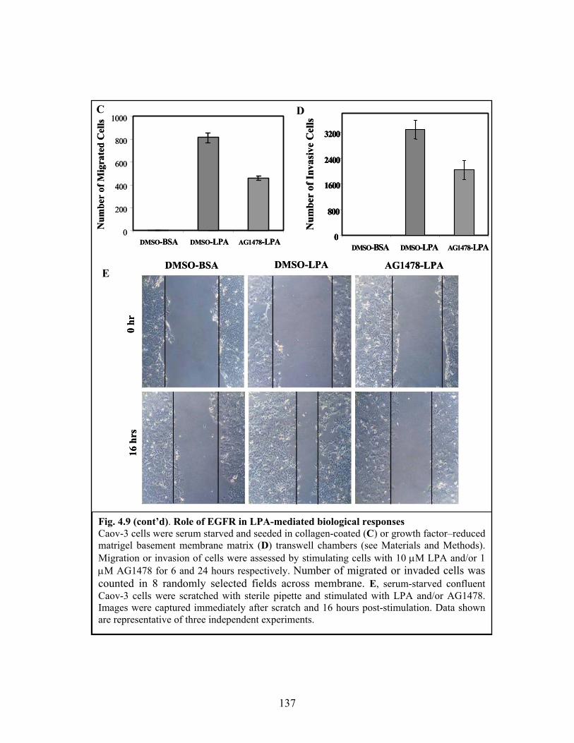
137
Fig. 4.9 (cont’d). Role of EGFR in LPA-mediated biological responses Caov-3 cells were serum starved and seeded in collagen-coated (C) or growth factor–reduced matrigel basement membrane matrix (D) transwell chambers (see Materials and Methods). Migration or invasion of cells were assessed by stimulating cells with 10 μM LPA and/or 1 μM AG1478 for 6 and 24 hours respectively. Number of migrated or invaded cells was counted in 8 randomly selected fields across membrane. E, serum-starved confluent Caov-3 cells were scratched with sterile pipette and stimulated with LPA and/or AG1478. Images were captured immediately after scratch and 16 hours post-stimulation. Data shown are representative of three independent experiments.
0
800
1600
2400
3200
DMSO-BSA DMSO-LPA AG1478-LPA
Num
ber
of In
vasi
ve C
ells
0
800
1600
2400
3200
0
800
1600
2400
3200
DMSO-BSA DMSO-LPA AG1478-LPA
Num
ber
of In
vasi
ve C
ells
0
200
400
600
800
1000
DMSO-BSA DMSO-LPA AG1478-LPA
Num
ber
of M
igra
ted
Cel
ls
0
200
400
600
800
1000
DMSO-BSA DMSO-LPA AG1478-LPA
Num
ber
of M
igra
ted
Cel
lsC D
E DMSO-BSA DMSO-LPA AG1478-LPA
16 h
rs0
hr
DMSO-BSA DMSO-LPA AG1478-LPA
16 h
rs0
hr

138
4.4 Discussion
We have previously shown that LPA induces expression of multiple cancer-
associated genes via activation of transcription factors [9, 232, 254]. In Chapter 2 and 3,
we used LPA-induced Cox-2 expression as a model system to identify transcription factors
and signaling networks involved in activation of Cox-2 expression by LPA. Through these
studies, we demonstrated that CEBP-β is a key transcription factor responsible for
initiating transcription of Cox-2. The activation of CEBP-β by LPA results from
integration of signals from LPA receptor(s) and a permissive activity from RTK. To
substantiate this crosstalk between GPCR and EGFR in transmission of GPCR signals, we
have devoted the study in this Chapter to determining whether the EGFR signal is
generally required for activation of other transcription factors by LPA. The results
presented here indicate that LPA-induced activation of AP-1 relies on such an activity
from EGFR in analogy to LPA-mediated C/EBP-β activation while LPA stimulates NF-κB
in an EGFR-independent manner. The differential requirements of EGFR for AP-1 and
NF-κB activation suggest that the EGFR signal is involved in activation of specific
intracellular signaling cascades downstream of LPA receptors rather than proximal
activation of LPA receptors on the membrane. Furthermore, we identified the intracellular
G signaling cascades that interact with EGFR. Our results revealed that the Gi-mediated
pathway relies on an EGFR input for activation while Gq and G12/13 signals are refractory
to inhibition of EGFR. AP-1 activation by LPA heavily depends on the Gi pathway and
139
accordingly is EGFR dependent. On the other hand, activation of NF-κB by LPA is
mediated through an EGFR-independent Gq signaling process.
The crosstalk between RTK and GPCR in cellular functions has been a subject of
extensive research in the area of signal transduction [18, 260]. Both “transactivation” and
“permissive signal” models have been proposed to explain the functional dependence of
GPCR signals on RTK [260]. In our studies using ovarian cancer cell lines, we did not
observe strong transactivation of EGFR by LPA as reflected by only weak induction of
EGFR phosphorylation (Fig. 3.5 E of Chapter 3). In addition, the effects of LPA on
activation of transcription factors and the downstream gene expression were generally
speaking more profound than EGF itself. Therefore, it is hard to imagine that LPA induces
these biochemical and biological events through transactivation of EGFR. In contrast, our
results are in concert with a permissive role of EGFR or an alternate RTK in activation of
GPCR signaling. Elucidation of EGFR-dependent and EGFR-independent G protein
signaling cascades and their downstream biochemical events allow us to conclude that only
selective GPCR signaling pathways are regulated by EGFR.
It remains to be determined how EGFR is integrated with GPCR signaling to
activate Gi and events downstream of Gi. As shown in this Chapter, again, the role of
EGFR could be substituted for by activation of another RTK such as c-Met. The
observation indicates that a RTK activity, not necessarily EGFR, is involved in linking
GPCR to Gi activation. It has been well documented that EGFR is overexpressed or
activated through mutation in many types of human cancers including ovarian cancer [244,
260]. It is conceivable that the elevated EGFR activity in cancer cells likely functions as a
140
default RTK to interact with GPCR signals. The basal, unstimulated activities of other
RTKs may not be sufficiently high to provide such a permissive signal. However, when
activated by their specific ligands, these other RTKs such as c-Met may function as
alternate RTKs to interact with GPCR signals. Targeting EGFR has been an attractive
approach to cancer intervention. Based on our results presented in this chapter, EGFR is
essential for many biological processes evoked by GPCRs including cytokine production,
cell proliferation, and cell migration and invasion. Many GPCR agonists are important
mediators of these biological processes in cancer cells. Thus, inhibition of EGFR may
yield therapeutic benefits from interference with GPCR signaling in addition to
disconnection of EGFR from its own direct downstream effectors.
141
CHAPTER 5
GENERAL DISCUSSION
LPA is present in the blood at sub-micromolar concentrations [19, 20]. It is one of
growth factors found in ascitic fluids of ovarian cancer patients [4, 6, 261]. Elevated
plasma levels of LPA are found at both early and advanced stages of ovarian cancer,
offering LPA as a potential biomarker for early detection of the malignancy [4, 5, 262].
LPA receptors are also overexpressed in primary ovarian cancer and ovarian cancer cell
lines [35, 59, 263, 264]. LPA stimulates proliferation, survival and motility of ovarian and
other types of cancer cells [3, 7, 29]. LPA presence in tumor microenvironment enhances
aggressiveness of cancer cells at least partially through modulating expression of diverse
target genes ranging from inflammatory cytokines to proteases.
Dysregulated gene expression is a hallmark of tumorigenesis. The overall aim of
my project was to delineate the mechanistic details of LPA-induced gene expression in
ovarian cancer cells wherein LPA has been implicated as an activating factor.
Identification of major components of LPA signal transduction leading to gene expression
may offer novel therapeutic targets for treatment of ovarian cancer and other human
malignancies. The current work demonstrated that (i) LPA upregulates gene expression at
both transcriptional and post-transcriptional levels; (ii) LPA triggers transcriptional

142
activation through activating major transcription factors such as AP-1, NF-κB and C/EBP;
(iii) LPA activates transcription factors via RTK-dependent and independent pathways.
As described in Chapter 2, we analyzed the molecular mechanisms by which LPA
stimulated expression of the Cox-2 gene in ovarian cancer cells. Cox-2 is an important
inducible enzyme in the formation of prostanoids including prostaglandins, prostacyclins
and thromboxanes. Targeting Cox-2 with non-steroidal anti-inflammatory drugs (NSAIDs)
provides relief from symptoms of pain and inflammation [207]. Since selective inhibition
of Cox-2 also reduces the risk of colon cancer [265, 266], it is generally believed that Cox-
2 contributes to the pathogenesis of human cancer including ovarian cancer [267].
Therefore, it was of interest to study how the Cox-2 gene is regulated in cancer cells.
Compared to many peptide growth factors, LPA was more potent inducer of Cox-2
expression in ovarian cancer cell lines, an effect mediated by multiple LPA receptors
including LPA1, LPA2 and LPA5. In addition to the striking potency, another feature we
observed was the prolonged duration of Cox-2 induction by LPA. LPA-induced Cox-2
expression remained highly elevated 16 hours after exposure to LPA, in sharp contrast to
the transient induction of Cox-2, as an early response gene, by other agonists. Analysis of
Cox-2 mRNA stability revealed that it was significantly enhanced in LPA-treated cells. We
further identified HuR as an mRNA stability factor that bound to Cox-2 transcripts at 3’-
UTR and protected them from degradation. Knockdown of HuR expression with siRNA
attenuated the prolonged stimulation of Cox-2 by LPA, confirming an active role of HuR
in sustaining Cox-2 expression in LPA-treated cells. At present, it is not known whether

143
LPA activates HuR through biochemical modifications or whether the association of HuR
and Cox-2 mRNA is solely dependent on increased transcript levels of the latter.
An important observation made in Chapter 2 was the crucial role of C/EBP-β in
transcriptional initiation of Cox-2 gene expression in response to LPA. In contrast to other
studies implication of AP-1 and/or NF-κB in inducible Cox-2 expression, we observed
minimal contributions of these factors to LPA-induced Cox-2 expression. In fact,
expression of a dominant negative form of c-Jun (Tam67) modestly potentiated LPA-
induced Cox-2 expression. The finding of the essential and unique role of C/EBP-β in
driving Cox-2 expression by LPA was somewhat surprising to us as C/EBP-β had not been
implicated in any biological effects of LPA. However, our results from multiple
approaches including promoter analysis, ChIP assay, and expression of dominant negative
of C/EBP-β, LIP all pointed to critical involvement of C/EBP-β in activation of the Cox-2
gene promoter in LPA-stimulated cells.
In Chapter 3, we extended to investigate the molecular mechanism for activation of
C/EBP-β by LPA and the general role of this transcription factor in LPA-mediated gene
expression. We focused on the C/EBP-β isoform because it was abundantly expressed in
ovarian cancer and correlated with aggressiveness of the disease [135]. We showed that
LPA induced a rapid phosphorylation of C/EBP-β and its DNA-binding and transcriptional
activities. The C/EBP-β phosphorylation is one of the major biochemical events associated
with its activation [121, 125, 126]. Since the C/EBP-β phosphorylation correlated well
with DNA-binding and transcriptional activities in LPA treated cells, it is likely that LPA-

144
induced phosphorylation represent a key step leading to functional activation of the
transcription factor although the impacts of LPA on other biochemical changes associated
with C/EBP-β activation, such as ubiquitination, sumoylation and acetylation, are yet to be
assessed and therefore should be a focus of future studies.
Some mammalian cells carry C/EBP-β in the cytosol and nucleus and activation of
C/EBP-β commensurate with an increased nuclear translocation of the protein [142, 268].
In ovarian cancer cells, however, C/EBP-β was exclusively seen in the nuclei and LPA
treatment did not change such a distribution pattern. Thus, nuclear translocation is not a
mechanism to activate transcriptional capacity of C/EBP-β. In addition, the kinase that
activates nuclear C/EBP-β upon LPA stimulation has not been identified. Considering the
role of C/EBP-β in the induction of Cox-2 expression by LPA, it is unlikely that nuclear
C/EBP-β kinase would be of MAPK lineage given that inhibition of MAPK cascade did
not block LPA-induced Cox-2 expression. The detail of that unique LPA GPCR-activated
pathway that bypasses MAPK signaling components may be critical not only for the
identification of nuclear C/EBP-β kinase but perhaps for the discovery of more upstream
targets of Cox-2 activity. Moreover, the export of C/EBP-β out of the nucleus by nuclear
exportins has been shown to result in repression of certain genes. Thus, temporal
manipulation of C/EBP-β levels via nuclear export may be an attractive therapeutic option.
Overexpression of LAP was not sufficient to increase C/EBP-β transcriptional
activity strongly suggesting that LPA-mediated CEBP-β activation is due to biochemical
modification of C/EBP-β rather than in an increase in overall CEBP-β protein levels. In
145
extended stimulation conditions, activation and expression of C/EBP-β-target genes may
continue to benefit from continual presence of LPA, such as is found in the ascites of
ovarian cancer patients. By extending our study to other LPA-target genes, we
demonstrated that, in addition to Cox-2, C/EBP-β was also involved in LPA induction of
inflammatory cytokine IL-6 and metastatic factor uPA, underscoring a general role of
CEBP-β in modulation of multiple LPA target genes.
A highlight of the work described in Chapter 3 is the finding of the dependence of
C/EBP-β activation on crosstalk between LPA GPCRs and EGFR. The effects of LPA on
CEBP-β phosphorylation, DNA-binding and transcriptional activities and the ultimate
induction of Cox-2 were all sensitivity to inhibition of EGFR kinase activity with AG1478.
LPA-induced IL-6 and uPA gene expression was also impaired by inhibition of EGFR,
further substantiating C/EBP-β as rate-limiting factor in LPA-induced Cox-2, IL-6 and
uPA expression. It is interesting to note that the dependence on EGFR could be overcome
by activation of another RTK c-Met. In recovery experiments, costimulation with HGF
efficiently revoked the suppression imposed by EGFR inhibition. This observation
suggests that a receptor tyrosine kinase activity, not specifically EGFR, is required for
LPA-induced activation of C/EBP-β and expression of the downstream targets. The reason
that EGFR plays such a default role is most likely due to the fact that the basal EGFR
activity is more prominent than other RTKs, particularly in malignant cells where EGFR is
commonly overexpressed and activated through mutations [244, 260]. Another interesting
question is whether the basal, unstimulated activity of EGFR in ovarian cancer cells is
146
sufficient to provide such as permissive signal for GPCR activation of CEBP-β.
Alternatively, EGFR could be “transactivated” during LPA stimulation. However, we
observed only weak activation of EGFR by LPA compared to the marked effect of EGF
itself in ovarian cancer cell lines. It is difficult to fit our observations into the
“transactivation” model given the fact that full activation of EGFR by EGF only triggers
weak activation of C/EBP-β and Cox-2 expression. Therefore our results are more
compatible with the hypothesis that a basal EGFR activity is necessary for transmission of
GPCR signaling to C/EBP-β activation.
To further characterize the crosstalk between GPCR and RTK in activation of
transcription factors, we next asked whether the RTK signal is a general requirement for
activation of other transcription factors by LPA. As described in Chapter 4, we examined
LPA-induced activation of AP-1 and NF-κB in ovarian cancer cell lines. Interestingly, AP-
1 activation in response to LPA indeed relied on EGFR activity analogous to LPA-induced
C/EBP-β activation. However, LPA stimulated NF-κB activation independently of EGFR
activity. Hence, the intact EGFR is differentially required for activation of these prominent
transcription factors. NF-κB activation by LPA offers an excellent readout of EGFR-
independent signals downstream of LPA receptors. It however leaves with us a quest to
identify more EGFR-independent transcription factors and genes. Such molecules may be
involved in unknown mechanisms through which ovarian tumor cells exhibit resistance to
EGFR therapy [269]. A microarray analysis of LPA-induced transcriptome in cells
pretreated with EGFR inhibitors would be an excellent methodology through which such
informative profile may be obtained.

147
Molecular and pharmacological approaches demonstrated that activation of AP-1
and NF-κB were mediated by different G protein signaling cascades downstream of LPA
receptors. The EGFR-independent Gq pathway was a mediator of NF-κB while the EGFR-
dependent Gi signaling cascade was the predominant and most critical player in LPA-
induced activation of AP-1. These results presented in Chapter 4 provide in-depth insights
Fig. 5.1. Hypothetical model of activation of G proteins and transcription factors by LPA. Obligatory inputs from basal EGFR activity may feed into signals downstream of LPA receptors at diverse points, particular before and/or after activation of Gi. Activation of AP-1 by LPA is mediated by activation of multiple G proteins. However, NF-κB is activated downstream of Gq signaling cascade.
AP-1
LPA#EGFR
PP
PP
Gq
G12/13
Gi
NF-κBAP-1AP-1AP-1
LPA#LPA#EGFREGFR
PP
PP
PP
PP
Gq
G12/13
Gi
Gq
G12/13
Gi
NF-κB
148
into the role of EGFR in activation of intracellular G proteins and their downstream
molecules.
The hypothetical model depicted by Fig. 5.1 simplifies the current status of this
project as well as future prospects. It will be of particular interest to elucidate how EGFR
activity is involved in activation of Gi in cooperation with GPCR. It is also possible that
EGFR is required for activation of a Gi effector that is located downstream of Gi but
upstream of Ras. Another direction is to find the link between Gi and C/EBP-β. Based
upon the observation that both Gi and C/EBP-β activation rely on EGFR activity, we
predict that phosphorylation of C/EBP-β lies downstream of Gi. We have already assessed
a number of Gi-mediated signals including the MAPK pathways. We have unfortunately
not confirmed the connection yet.
The Gi signaling is essential for many biological responses to LPA and other GPCR
ligands. We observed significant but incomplete inhibition of LPA-induced cell
proliferation, migration and invasion by EGFR inhibitors. These results suggest that
EGFR-independent, Gq or G12/13-medaited signals could be operational partially mediating
these responses. Thus, simultaneous targeting EGFR and LPA receptors seems to be an
appealing approach to inhibit proliferation, invasion and metastasis of ovarian cancer cells.
This could be evaluated both in vitro and in vivo.

149
References
150
REFERENCES
1. American Cancer Society: Cancer Facts and Figures. American Cancer Society. 2008. [http://www.cancer.org/]
2. Mills GB, May C, Hill M, Campbell S, Shaw P, Marks A: Ascitic fluid from human ovarian cancer patients contains growth factors necessary for intraperitoneal growth of human ovarian adenocarcinoma cells. J Clin Invest 1990, 86:851-855.
3. Moolenaar WH, Jalink K, van Corven EJ: Lysophosphatidic acid: a bioactive phospholipid with growth factor-like properties. Rev Physiol Biochem Pharmacol 1992, 119:47-65.
4. Xu Y, Shen Z, Wiper DW, Wu M, Morton RE, Elson P, Kennedy AW, Belinson J, Markman M, Casey G: Lysophosphatidic acid as a potential biomarker for ovarian and other gynecologic cancers. JAMA 1998, 280:719-723.
5. Xu Y, Gaudette DC, Boynton JD, Frankel A, Fang XJ, Sharma A, Hurteau J, Casey G, Goodbody A, Mellors A: Characterization of an ovarian cancer activating factor in ascites from ovarian cancer patients. Clin Cancer Res 1995, 1:1223-1232.
6. Westermann AM, Havik E, Postma FR, Beijnen JH, Dalesio O, Moolenaar WH, Rodenhuis S: Malignant effusions contain lysophosphatidic acid (LPA)-like activity. Ann Oncol 1998, 9:437-442.
7. Fang X, Gaudette D, Furui T, Mao M, Estrella V, Eder A, Pustilnik T, Sasagawa T, Lapushin R, Yu S, Jaffe RB, Wiener JR, Erickson JR, Mills GB: Lysophospholipid growth factors in the initiation, progression, metastases, and management of ovarian cancer. Ann N Y Acad Sci 2000, 905:188-208.
8. Xu Y, Fang XJ, Casey G, Mills GB: Lysophospholipids activate ovarian and breast cancer cells. Biochem J 1995, 309:933-940.
9. Fang X, Yu S, Bast RC, Liu S, Xu HJ, Hu SX, LaPushin R, Claret FX, Aggarwal BB, Lu Y, Mills GB: Mechanisms for lysophosphatidic acid-induced cytokine production in ovarian cancer cells. J Biol Chem 2004, 279:9653-9661.
10. Xu Y, Casey G, Mills GB: Effect of lysophospholipids on signaling in the human Jurkat T cell line. J Cell Physiol 1995, 163:441-450.

151
11. Baudhuin LM, Cristina KL, Lu J, Xu Y: Akt activation induced by lysophosphatidic acid and sphingosine-1-phosphate requires both mitogen-activated protein kinase kinase and p38 mitogen-activated protein kinase and is cell-line specific. Mol Pharmacol 2002, 62:660-671.
12. Estrella VC, Eder AM, Liu S, Pustilnik TB, Tabassam FH, Claret FX, Gallick GE, Mills GB, Wiener JR: Lysophosphatidic acid induction of urokinase plasminogen activator secretion requires activation of the p38MAPK pathway. Int J Oncol 2007, 31:441-449.
13. Aoki J: Mechanisms of lysophosphatidic acid production. Semin Cell Dev Biol 2004, 15:477-489.
14. Eichholtz T, Jalink K, Fahrenfort I, Moolenaar WH: The bioactive phospholipid lysophosphatidic acid is released from activated platelets. Biochem J 1993, 291:677-680.
15. Siess W, Zangl KJ, Essler M, Bauer M, Brandl R, Corrinth C, Bittman R, Tigyi G, Aepfelbacher M: Lysophosphatidic acid mediates the rapid activation of platelets and endothelial cells by mildly oxidized low density lipoprotein and accumulates in human atherosclerotic lesions. Proc Natl Acad Sci U S A 1999, 96:6931-6936.
16. van der Bend RL, de Widt J, van Corven EJ, Moolenaar WH, van Blitterswijk WJ: The biologically active phospholipid, lysophosphatidic acid, induces phosphatidylcholine breakdown in fibroblasts via activation of phospholipase D. Comparison with the response to endothelin. Biochem J 1992, 285:235-240.
17. Valet P, Pages C, Jeanneton O, Daviaud D, Barbe P, Record M, Saulnier-Blache JS, Lafontan M: Alpha2-adrenergic receptor-mediated release of lysophosphatidic acid by adipocytes. A paracrine signal for preadipocyte growth. J Clin Invest 1998, 101:1431-1438.
18. Bektas M, Payne SG, Liu H, Goparaju S, Milstien S, Spiegel S: A novel acylglycerol kinase that produces lysophosphatidic acid modulates cross talk with EGFR in prostate cancer cells. J Cell Biol 2005, 169:801-811.
19. Aoki J, Inoue A, Okudaira S: Two pathways for lysophosphatidic acid production. Biochim Biophys Acta 2008, 1781:513-518.
20. Sugiura T, Nakane S, Kishimoto S, Waku K, Yoshioka Y, Tokumura A: Lysophosphatidic acid, a growth factor-like lipid, in the saliva. J Lipid Res 2002, 43:2049-2055.

152
21. Aoki J, Taira A, Takanezawa Y, Kishi Y, Hama K, Kishimoto T, Mizuno K, Saku K, Taguchi R, Arai H: Serum lysophosphatidic acid is produced through diverse phospholipase pathways. J Biol Chem 2002, 277:48737-48744.
22. Umezu-Goto M, Kishi Y, Taira A, Hama K, Dohmae N, Takio K, Yamori T, Mills GB, Inoue K, Aoki J, Arai H: Autotaxin has lysophospholipase D activity leading to tumor cell growth and motility by lysophosphatidic acid production. J Cell Biol 2002, 158:227-233.
23. Jansen S, Stefan C, Creemers JW, Waelkens E, Van Eynde A, Stalmans W, Bollen M: Proteolytic maturation and activation of autotaxin (NPP2), a secreted metastasis-enhancing lysophospholipase D. J Cell Sci 2005, 118:3081-3089.
24. Sekas G, Patton GM, Lincoln EC, Robins SJ: Origin of plasma lysophosphatidylcholine: evidence for direct hepatic secretion in the rat. J Lab Clin Med 1985, 105:190-194.
25. Hatch GM, Oskin A, Vance DE: Involvement of the lysosome in the catabolism of intracellular lysophosphatidylcholine and evidence for distinct pools of lysophosphatidylcholine. J Lipid Res 1993, 34:1873-1881.
26. Baisted DJ, Robinson BS, Vance DE: Albumin stimulates the release of lysophosphatidylcholine from cultured rat hepatocytes. Biochem J 1988, 253:693-701.
27. Xie Y, Gibbs TC, Mukhin YV, Meier KE: Role for 18:1 lysophosphatidic acid as an autocrine mediator in prostate cancer cells. J Biol Chem 2002, 277:32516-32526.
28. Eder AM, Sasagawa T, Mao M, Aoki J, Mills GB: Constitutive and lysophosphatidic acid (LPA)-induced LPA production: role of phospholipase D and phospholipase A2. Clin Cancer Res 2000, 6:2482-2491.
29. Ren J, Xiao YJ, Singh LS, Zhao X, Zhao Z, Feng L, Rose TM, Prestwich GD, Xu Y: Lysophosphatidic acid is constitutively produced by human peritoneal mesothelial cells and enhances adhesion, migration, and invasion of ovarian cancer cells. Cancer Res 2006, 66(6):3006-3014.
30. Pilquil C, Singh I, Zhang Q, Ling Z, Buri K, Stromberg LM, Dewald J, Brindley DN: Lipid phosphate phosphatase-1 dephosphorylates exogenous lysophosphatidate and thereby attenuates its effects on cell signalling. Prostaglandins 2001, 64:83-92.
31. Xu J, Zhang QX, Pilquil C, Berthiaume LG, Waggoner DW, Brindley DN: Lipid phosphate phosphatase-1 in the regulation of lysophosphatidate signaling. Ann N Y Acad Sci 2000, 905:81-90.
153
32. Tomsig JL, Snyder AH, Berdyshev EV, Skobeleva A, Mataya C, Natarajan V, Brindley DN, Lynch KR: Lipid phosphate phosphohydrolase type 1 (LPP1) degrades extracellular lysophosphatidic acid in vivo. Biochem J 2009, 419:611-8.
33. Eberhardt C, Gray PW, Tjoelker LW: Human lysophosphatidic acid acyltransferase. cDNA cloning, expression, and localization to chromosome 9q34.3. J Biol Chem 1997, 272:20299-20305.
34. Leung DW: The structure and functions of human lysophosphatidic acid acyltransferases. Front Biosci 2001, 6:D944-53.
35. Wang P, Wu X, Chen W, Liu J, Wang X: The lysophosphatidic acid (LPA) receptors their expression and significance in epithelial ovarian neoplasms. Gynecol Oncol 2007, 104:714-720.
36. Hecht JH, Weiner JA, Post SR, Chun J: Ventricular zone gene-1 (vzg-1) encodes a lysophosphatidic acid receptor expressed in neurogenic regions of the developing cerebral cortex. J Cell Biol 1996, 135:1071-1083.
37. An S, Dickens MA, Bleu T, Hallmark OG, Goetzl EJ: Molecular cloning of the human Edg2 protein and its identification as a functional cellular receptor for lysophosphatidic acid. Biochem Biophys Res Commun 1997, 231:619-622.
38. Bandoh K, Aoki J, Hosono H, Kobayashi S, Kobayashi T, Murakami-Murofushi K, Tsujimoto M, Arai H, Inoue K: Molecular cloning and characterization of a novel human G-protein-coupled receptor, EDG7, for lysophosphatidic acid. J Biol Chem 1999, 274:27776-27785.
39. Noguchi K, Ishii S, Shimizu T: Identification of p2y9/GPR23 as a novel G protein-coupled receptor for lysophosphatidic acid, structurally distant from the Edg family. J Biol Chem 2003, 278:25600-25606.
40. Lee CW, Rivera R, Gardell S, Dubin AE, Chun J: GPR92 as a new G12/13- and Gq-coupled lysophosphatidic acid receptor that increases cAMP, LPA5. J Biol Chem 2006, 281:23589-23597.
41. Tabata K, Baba K, Shiraishi A, Ito M, Fujita N: The orphan GPCR GPR87 was deorphanized and shown to be a lysophosphatidic acid receptor. Biochem Biophys Res Commun 2007, 363:861-866.
42. Murakami M, Shiraishi A, Tabata K, Fujita N: Identification of the orphan GPCR, P2Y(10) receptor as the sphingosine-1-phosphate and lysophosphatidic acid receptor. Biochem Biophys Res Commun 2008, 371:707-712.

154
43. Gros R, Benovic JL, Tan CM, Feldman RD: G-protein-coupled receptor kinase activity is increased in hypertension. J Clin Invest 1997, 99:2087-2093.
44. Kowluru A, Kowluru RA, Yamazaki A: Functional alterations of G-proteins in diabetic rat retina: a possible explanation for the early visual abnormalities in diabetes mellitus. Diabetologia 1992, 35:624-631.
45. Vendelin J, Pulkkinen V, Rehn M, Pirskanen A, Raisanen-Sokolowski A, Laitinen A, Laitinen LA, Kere J, Laitinen T: Characterization of GPRA, a novel G protein-coupled receptor related to asthma. Am J Respir Cell Mol Biol 2005, 33:262-270.
46. Li S, Huang S, Peng SB: Overexpression of G protein-coupled receptors in cancer cells: involvement in tumor progression. Int J Oncol 2005, 27:1329-1339.
47. Drews J: Drug discovery: a historical perspective. Science 2000, 287:1960-1964.
48. Heilker R, Wolff M, Tautermann CS, Bieler M: G-protein-coupled receptor-focused drug discovery using a target class platform approach. Drug Discov Today 2009, 14:231-40 .
49. van Corven EJ, Groenink A, Jalink K, Eichholtz T, Moolenaar WH: Lysophosphatidate-induced cell proliferation: identification and dissection of signaling pathways mediated by G proteins. Cell 1989, 59:45-54.
50. Moolenaar WH, Kranenburg O, Postma FR, Zondag GC: Lysophosphatidic acid: G-protein signalling and cellular responses. Curr Opin Cell Biol 1997, 9:168-173.
51. Mills GB, Moolenaar WH: The emerging role of lysophosphatidic acid in cancer. Nat Rev Cancer 2003, 3:582-591.
52. Howe LR, Marshall CJ: Lysophosphatidic acid stimulates mitogen-activated protein kinase activation via a G-protein-coupled pathway requiring p21ras and p74raf-1. J Biol Chem 1993, 268:20717-20720.
53. Hilal-Dandan R, Means CK, Gustafsson AB, Morissette MR, Adams JW, Brunton LL, Heller Brown J: Lysophosphatidic acid induces hypertrophy of neonatal cardiac myocytes via activation of Gi and Rho. J Mol Cell Cardiol 2004, 36:481-493.
54. Murph MM, Scaccia LA, Volpicelli LA, Radhakrishna H: Agonist-induced endocytosis of lysophosphatidic acid-coupled LPA1/EDG-2 receptors via a dynamin2- and Rab5-dependent pathway. J Cell Sci 2003, 116:1969-1980.
155
55. McIntyre TM, Pontsler AV, Silva AR, St Hilaire A, Xu Y, Hinshaw JC, Zimmerman GA, Hama K, Aoki J, Arai H, Prestwich GD: Identification of an intracellular receptor for lysophosphatidic acid (LPA): LPA is a transcellular PPARgamma agonist. Proc Natl Acad Sci USA 2003, 100:131-136.
56. Zhang C, Baker DL, Yasuda S, Makarova N, Balazs L, Johnson LR, Marathe GK, McIntyre TM, Xu Y, Prestwich GD, Byun HS, Bittman R, Tigyi G: Lysophosphatidic acid induces neointima formation through PPARgamma activation. J Exp Med 2004, 199:763-774.
57. Barroso I, Gurnell M, Crowley VE, Agostini M, Schwabe JW, Soos MA, Maslen GL, Williams TD, Lewis H, Schafer AJ, Chatterjee VK, O'Rahilly S: Dominant negative mutations in human PPARgamma associated with severe insulin resistance, diabetes mellitus and hypertension. Nature 1999, 402:880-883.
58. Chinetti G, Fruchart JC, Staels B: Peroxisome proliferator-activated receptors (PPARs): nuclear receptors at the crossroads between lipid metabolism and inflammation. Inflamm Res 2000, 49:497-505.
59. Goetzl EJ, Dolezalova H, Kong Y, Hu YL, Jaffe RB, Kalli KR, Conover CA: Distinctive expression and functions of the type 4 endothelial differentiation gene-encoded G protein-coupled receptor for lysophosphatidic acid in ovarian cancer. Cancer Res 1999, 59:5370-5375.
60. Goetzl EJ, Graeler M, Huang MC, Shankar G: Lysophospholipid growth factors and their G protein-coupled receptors in immunity, coronary artery disease, and cancer. ScientificWorldJournal 2002, 2:324-338.
61. Schulte KM, Beyer A, Kohrer K, Oberhauser S, Roher HD: Lysophosphatidic acid, a novel lipid growth factor for human thyroid cells: over-expression of the high-affinity receptor edg4 in differentiated thyroid cancer. Int J Cancer 2001, 92:249-256.
62. Fang X, Schummer M, Mao M, Yu S, Tabassam FH, Swaby R, Hasegawa Y, Tanyi JL, LaPushin R, Eder A, Jaffe R, Erickson J, Mills GB: Lysophosphatidic acid is a bioactive mediator in ovarian cancer. Biochim Biophys Acta 2002, 1582:257-264.
63. Nakamoto T, Yasuda K, Yasuhara M, Yoshimura T, Kinoshita T, Nakajima T, Okada H, Ikuta A, Kanzaki H: Expression of the endothelial cell differentiation gene 7 (EDG-7), a lysophosphatidic acid receptor, in ovarian tumor. J Obstet Gynaecol Res 2005, 31:344-351.
64. Shida D, Watanabe T, Aoki J, Hama K, Kitayama J, Sonoda H, Kishi Y, Yamaguchi H, Sasaki S, Sako A, Konishi T, Arai H, Nagawa H: Aberrant expression of

156
lysophosphatidic acid (LPA) receptors in human colorectal cancer. Lab Invest 2004, 84:1352-1362.
65. Yang M, Zhong WW, Srivastava N, Slavin A, Yang J, Hoey T, An S: G protein-coupled lysophosphatidic acid receptors stimulate proliferation of colon cancer cells through the {beta}-catenin pathway. Proc Natl Acad Sci U S A 2005, 102:6027-6032.
66. Yu S, Murph MM, Lu Y, Liu S, Hall HS, Liu J, Stephens C, Fang X, Mills GB: Lysophosphatidic acid receptors determine tumorigenicity and aggressiveness of ovarian cancer cells. J Natl Cancer Inst 2008, 100:1630-1642.
67. Hernandez M, Barrero MJ, Crespo MS, Nieto ML: Lysophosphatidic acid inhibits Ca2+ signaling in response to epidermal growth factor receptor stimulation in human astrocytoma cells by a mechanism involving phospholipase C(gamma) and a G(alphai) protein. J Neurochem 2000, 75:1575-1582.
68. Ediger TL, Schulte NA, Murphy TJ, Toews ML: Transcription factor activation and mitogenic synergism in airway smooth muscle cells. Eur Respir J 2003, 21:759-769.
69. Saatian B, Zhao Y, He D, Georas SN, Watkins T, Spannhake EW, Natarajan V: Transcriptional regulation of lysophosphatidic acid-induced interleukin-8 expression and secretion by p38 MAPK and JNK in human bronchial epithelial cells. Biochem J 2006, 393:657-668.
70. Messina DN, Glasscock J, Gish W, Lovett M: An ORFeome-based analysis of human transcription factor genes and the construction of a microarray to interrogate their expression. Genome Res 2004, 14:2041-2047.
71. Kim SY, Kim Y: Genome-wide prediction of transcriptional regulatory elements of human promoters using gene expression and promoter analysis data. BMC Bioinformatics 2006, 7:330.
72. Blake MC, Jambou RC, Swick AG, Kahn JW, Azizkhan JC: Transcriptional initiation is controlled by upstream GC-box interactions in a TATAA-less promoter. Mol Cell Biol 1990, 10:6632-6641.
73. Swaminathan S, Malhotra P, Manohar CF, Dhar R, Thimmapaya B: Activation of a dual adenovirus promoter containing nonconsensus TATA motifs in Schizosaccharomyces pombe: role of TATA sequences in the efficiency of transcription. Nucleic Acids Res 1993, 21:2737-2746.
74. Latchman DS: Transcription factors: an overview. Int J Biochem Cell Biol 1997, 29:1305-1312.

157
75. Warnmark A, Treuter E, Wright AP, Gustafsson JA: Activation functions 1 and 2 of nuclear receptors: molecular strategies for transcriptional activation. Mol Endocrinol 2003, 17:1901-1909.
76. Cheah PL, Looi LM: P53: an Overview of Over Two Decades of Study. Malays J Pathol 2001, 23:9-16.
77. Seemann S, Maurici D, Olivier M, de Fromentel CC, Hainaut P: The tumor suppressor gene TP53: implications for cancer management and therapy. Crit Rev Clin Lab Sci 2004, 41:551-583.
78. Hall J, Paul J, Brown R: Critical evaluation of p53 as a prognostic marker in ovarian cancer. Expert Rev Mol Med 2004, 6:1-20.
79. Feki A, Irminger-Finger I: Mutational spectrum of p53 mutations in primary breast and ovarian tumors. Crit Rev Oncol Hematol 2004, 52:103-116.
80. Petty R, Evans A, Duncan I, Kurbacher C, Cree I: Drug resistance in ovarian cancer - the role of p53. Pathol Oncol Res 1998, 4:97-102.
81. Fuster JJ, Sanz-Gonzalez SM, Moll UM, Andres V: Classic and novel roles of p53: prospects for anticancer therapy. Trends Mol Med 2007, 13:192-199.
82. Borst P, Jonkers J, Rottenberg S: What makes tumors multidrug resistant? Cell Cycle 2007, 6:2782-2787.
83. Karin M, Liu Z, Zandi E: AP-1 function and regulation. Curr Opin Cell Biol 1997, 9:240-246.
84. Hess J, Angel P, Schorpp-Kistner M: AP-1 subunits: quarrel and harmony among siblings. J Cell Sci 2004, 117(Pt 25):5965-5973.
85. Ransone LJ, Verma IM: Nuclear proto-oncogenes fos and jun. Annu Rev Cell Biol 1990, 6:539-557.
86. Szabo E, Preis LH, Brown PH, Birrer MJ: The role of jun and fos gene family members in 12-O-tetradecanoylphorbol-13-acetate induced hemopoietic differentiation. Cell Growth Differ 1991, 2:475-482.
87. Herschman HR: Primary response genes induced by growth factors and tumor promoters. Annu Rev Biochem 1991, 60:281-319.

158
88. O'Shea EK, Rutkowski R, Stafford WF,3rd, Kim PS: Preferential heterodimer formation by isolated leucine zippers from fos and jun. Science 1989, 245:646-648.
89. Muller R, Wagner EF: Differentiation of F9 teratocarcinoma stem cells after transfer of c-fos proto-oncogenes. Nature 1984, 311:438-442.
90. Mason I, Murphy D, Hogan BL: Expression of c-fos in parietal endoderm, amnion and differentiating F9 teratocarcinoma cells. Differentiation 1985, 30:76-81.
91. Deng T, Karin M: JunB differs from c-Jun in its DNA-binding and dimerization domains, and represses c-Jun by formation of inactive heterodimers. Genes Dev 1993, 7:479-490.
92. Abate C, Curran T: Encounters with Fos and Jun on the road to AP-1. Semin Cancer Biol 1990, 1:19-26.
93. Chinenov Y, Kerppola TK: Close encounters of many kinds: Fos-Jun interactions that mediate transcription regulatory specificity. Oncogene 2001, 20:2438-2452.
94. Abdel-Hafiz HA, Chen CY, Marcell T, Kroll DJ, Hoeffler JP: Structural determinants outside of the leucine zipper influence the interactions of CREB and ATF-2: interaction of CREB with ATF-2 blocks E1a-ATF-2 complex formation. Oncogene 1993, 8:1161-1174.
95. Chatton B, Bocco JL, Goetz J, Gaire M, Lutz Y, Kedinger C: Jun and Fos heterodimerize with ATFa, a member of the ATF/CREB family and modulate its transcriptional activity. Oncogene 1994, 9:375-385.
96. Manna PR, Stocco DM: Crosstalk of CREB and Fos/Jun on a single cis-element: transcriptional repression of the steroidogenic acute regulatory protein gene. J Mol Endocrinol 2007, 39:261-277.
97. Vogt PK, Bos TJ: The oncogene jun and nuclear signalling. Trends Biochem Sci 1989, 14:172-175.
98. Kouzarides T, Ziff E: Behind the Fos and Jun leucine zipper. Cancer Cells 1989, 1:71-76.
99. Belguise K, Kersual N, Galtier F, Chalbos D: FRA-1 expression level regulates proliferation and invasiveness of breast cancer cells. Oncogene 2005, 24:1434-1444.
100. Zoumpourlis V, Papassava P, Linardopoulos S, Gillespie D, Balmain A, Pintzas A: High levels of phosphorylated c-Jun, Fra-1, Fra-2 and ATF-2 proteins correlate with
159
malignant phenotypes in the multistage mouse skin carcinogenesis model. Oncogene 2000, 19:4011-4021.
101. Milde-Langosch K, Roder H, Andritzky B, Aslan B, Hemminger G, Brinkmann A, Bamberger CM, Loning T, Bamberger AM: The role of the AP-1 transcription factors c-Fos, FosB, Fra-1 and Fra-2 in the invasion process of mammary carcinomas. Breast Cancer Res Treat 2004, 86:139-152.
102. Kustikova O, Kramerov D, Grigorian M, Berezin V, Bock E, Lukanidin E, Tulchinsky E: Fra-1 induces morphological transformation and increases in vitro invasiveness and motility of epithelioid adenocarcinoma cells. Mol Cell Biol 1998, 18:7095-7105.
103. Tulchinsky E: Fos family members: regulation, structure and role in oncogenic transformation. Histol Histopathol 2000, 15:921-928.
104. Bonovich M, Olive M, Reed E, O'Connell B, Vinson C: Adenoviral delivery of A-FOS, an AP-1 dominant negative, selectively inhibits drug resistance in two human cancer cell lines. Cancer Gene Ther 2002, 9:62-70.
105. Bani MR, Nicoletti MI, Alkharouf NW, Ghilardi C, Petersen D, Erba E, Sausville EA, Liu ET, Giavazzi R: Gene expression correlating with response to paclitaxel in ovarian carcinoma xenografts. Mol Cancer Ther 2004, 3:111-121.
106. Vlahopoulos S, Zoumpourlis VC: JNK: a key modulator of intracellular signaling. Biochemistry (Mosc) 2004, 69:844-854.
107. Taylor LK, Marshak DR, Landreth GE: Identification of a nerve growth factor- and epidermal growth factor-regulated protein kinase that phosphorylates the protooncogene product c-Fos. Proc Natl Acad Sci U S A 1993, 90:368-372.
108. Terasawa K, Okazaki K, Nishida E: Regulation of c-Fos and Fra-1 by the MEK5-ERK5 pathway. Genes Cells 2003, 8:263-273.
109. Murphy LO, Smith S, Chen RH, Fingar DC, Blenis J: Molecular interpretation of ERK signal duration by immediate early gene products. Nat Cell Biol 2002, 4:556-564.
110. Yamaguchi K, Lantowski A, Dannenberg AJ, Subbaramaiah K: Histone deacetylase inhibitors suppress the induction of c-Jun and its target genes including COX-2. J Biol Chem 2005, 280:32569-32577.
111. Rahmsdorf HJ: Jun: transcription factor and oncoprotein. J Mol Med 1996, 74:725-747.

160
112. Suzuki T, Murakami M, Onai N, Fukuda E, Hashimoto Y, Sonobe MH, Kameda T, Ichinose M, Miki K, Iba H: Analysis of AP-1 function in cellular transformation pathways. J Virol 1994, 68:3527-3535.
113. Sonobe MH, Yoshida T, Murakami M, Kameda T, Iba H: fra-2 promoter can respond to serum-stimulation through AP-1 complexes. Oncogene 1995, 10:689-696.
114. Murakami M, Ui M, Iba H: Fra-2-positive autoregulatory loop triggered by mitogen-activated protein kinase (MAPK) and Fra-2 phosphorylation sites by MAPK. Cell Growth Differ 1999, 10:333-342.
115. Xie S, Nie R, Wang J, Li F, Yuan W: Transcription factor decoys for activator protein-1 (AP-1) inhibit oxidative stress-induced proliferation and matrix metalloproteinases in rat cardiac fibroblasts. Transl Res 2009, 153:17-23.
116. Parra M, Lluis F, Miralles F, Caelles C, Munoz-Canoves P: The cJun N-terminal kinase (JNK) signaling pathway mediates induction of urokinase-type plasminogen activator (uPA) by the alkylating agent MNNG. Blood 2000, 96:1415-1424.
117. Textor B, Sator-Schmitt M, Richter KH, Angel P, Schorpp-Kistner M: c-Jun and JunB are essential for hypoglycemia-mediated VEGF induction. Ann N Y Acad Sci 2006, 1091:310-318.
118. Lamb RF, Hennigan RF, Turnbull K, Katsanakis KD, MacKenzie ED, Birnie GD, Ozanne BW: AP-1-mediated invasion requires increased expression of the hyaluronan receptor CD44. Mol Cell Biol 1997, 17:963-976.
119. Wang X, Zhang Y, Xiao G, Gao X, Liu X: c-Fos enhances the survival of thymocytes during positive selection by upregulating Bcl-2. Cell Res 2009, 19:340-347.
120. Ramji DP, Foka P: CCAAT/enhancer-binding proteins: structure, function and regulation. Biochem J 2002, 365:561-575.
121. Takiguchi M: The C/EBP family of transcription factors in the liver and other organs. Int J Exp Pathol 1998, 79:369-391.
122. Nerlov C: The C/EBP family of transcription factors: a paradigm for interaction between gene expression and proliferation control. Trends Cell Biol 2007, 17:318-324.
123. Chumakov AM, Silla A, Williamson EA, Koeffler HP: Modulation of DNA binding properties of CCAAT/enhancer binding protein epsilon by heterodimer formation and interactions with NFkappaB pathway. Blood 2007, 109:4209-4219.

161
124. Roesler WJ: The role of C/EBP in nutrient and hormonal regulation of gene expression. Annu Rev Nutr 2001, 21:141-165.
125. Schrem H, Klempnauer J, Borlak J: Liver-enriched transcription factors in liver function and development. Part II: the C/EBPs and D site-binding protein in cell cycle control, carcinogenesis, circadian gene regulation, liver regeneration, apoptosis, and liver-specific gene regulation. Pharmacol Rev 2004, 56:291-330.
126. Lekstrom-Himes J, Xanthopoulos KG: Biological role of the CCAAT/enhancer-binding protein family of transcription factors. J Biol Chem 1998, 273:28545-28548.
127. Diehl AM: Roles of CCAAT/enhancer-binding proteins in regulation of liver regenerative growth. J Biol Chem 1998, 273:30843-30846.
128. Bamberger AM, Makrigiannakis A, Schroder M, Bamberger CM, Relakis C, Gellersen B, Milde-Langosch K, Loning T: Expression pattern of the CCAAT/enhancer-binding proteins C/EBP-alpha, C/EBP-beta and C/EBP-delta in the human placenta. Virchows Arch 2004, 444:149-152.
129. Wang ND, Finegold MJ, Bradley A, Ou CN, Abdelsayed SV, Wilde MD, Taylor LR, Wilson DR, Darlington GJ: Impaired energy homeostasis in C/EBP alpha knockout mice. Science 1995, 269:1108-1112.
130. Screpanti I, Romani L, Musiani P, Modesti A, Fattori E, Lazzaro D, Sellitto C, Scarpa S, Bellavia D, Lattanzio G: Lymphoproliferative disorder and imbalanced T-helper response in C/EBP beta-deficient mice. EMBO J 1995, 14:1932-1941.
131. Tanaka T, Akira S, Yoshida K, Umemoto M, Yoneda Y, Shirafuji N, Fujiwara H, Suematsu S, Yoshida N, Kishimoto T: Targeted disruption of the NF-IL6 gene discloses its essential role in bacteria killing and tumor cytotoxicity by macrophages. Cell 1995, 80:353-361.
132. Sterneck E, Tessarollo L, Johnson PF: An essential role for C/EBPbeta in female reproduction. Genes Dev 1997, 11:2153-2162.
133. Pall M, Hellberg P, Brannstrom M, Mikuni M, Peterson CM, Sundfeldt K, Norden B, Hedin L, Enerback S: The transcription factor C/EBP-beta and its role in ovarian function; evidence for direct involvement in the ovulatory process. EMBO J 1997, 16:5273-5279.
134. Nalbant D, Williams SC, Stocco DM, Khan SA: Luteinizing hormone-dependent gene regulation in Leydig cells may be mediated by CCAAT/enhancer-binding protein-beta. Endocrinology 1998, 139:272-279.

162
135. Sundfeldt K, Ivarsson K, Carlsson M, Enerback S, Janson PO, Brannstrom M, Hedin L: The expression of CCAAT/enhancer binding protein (C/EBP) in the human ovary in vivo: specific increase in C/EBPbeta during epithelial tumour progression. Br J Cancer 1999, 79:1240-1248.
136. Tengku-Muhammad TS, Hughes TR, Ranki H, Cryer A, Ramji DP: Differential regulation of macrophage CCAAT-enhancer binding protein isoforms by lipopolysaccharide and cytokines. Cytokine 2000, 12:1430-1436.
137. Campos SP, Baumann H: Insulin is a prominent modulator of the cytokine-stimulated expression of acute-phase plasma protein genes. Mol Cell Biol 1992, 12:1789-1797.
138. Baumann H, Morella KK, Campos SP, Cao Z, Jahreis GP: Role of CAAT-enhancer binding protein isoforms in the cytokine regulation of acute-phase plasma protein genes. J Biol Chem 1992, 267:19744-19751.
139. Descombes P, Schibler U: A liver-enriched transcriptional activator protein, LAP, and a transcriptional inhibitory protein, LIP, are translated from the same mRNA. Cell 1991, 67:569-579.
140. Welm AL, Timchenko NA, Darlington GJ: C/EBPalpha regulates generation of C/EBPbeta isoforms through activation of specific proteolytic cleavage. Mol Cell Biol 1999, 19:1695-1704.
141. Li Y, Bevilacqua E, Chiribau CB, Majumder M, Wang C, Croniger CM, Snider MD, Johnson PF, Hatzoglou M: Differential control of the CCAAT/enhancer-binding protein beta (C/EBPbeta) products liver-enriched transcriptional activating protein (LAP) and liver-enriched transcriptional inhibitory protein (LIP) and the regulation of gene expression during the response to endoplasmic reticulum stress. J Biol Chem 2008, 283:22443-22456.
142. Ghosh AK, Bhattacharyya S, Mori Y, Varga J: Inhibition of collagen gene expression by interferon-gamma: novel role of the CCAAT/enhancer binding protein beta (C/EBPbeta). J Cell Physiol 2006, 207:251-260.
143. Lee YH, Williams SC, Baer M, Sterneck E, Gonzalez FJ, Johnson PF: The ability of C/EBP beta but not C/EBP alpha to synergize with an Sp1 protein is specified by the leucine zipper and activation domain. Mol Cell Biol 1997, 17:2038-2047.
144. Vallejo M, Ron D, Miller CP, Habener JF: C/ATF, a member of the activating transcription factor family of DNA-binding proteins, dimerizes with

163
CAAT/enhancer-binding proteins and directs their binding to cAMP response elements. Proc Natl Acad Sci U S A 1993, 90:4679-4683.
145. Gilmore TD: Introduction to NF-kappaB: players, pathways, perspectives. Oncogene 2006, 25:6680-6684.
146. Hoffmann A, Natoli G, Ghosh G: Transcriptional regulation via the NF-kappaB signaling module. Oncogene 2006, 25:6706-6716.
147. Haefner B: NF-kappa B: arresting a major culprit in cancer. Drug Discov Today 2002, 7:653-663.
148. Calzado MA, Bacher S, Schmitz ML: NF-kappaB inhibitors for the treatment of inflammatory diseases and cancer. Curr Med Chem 2007, 14:367-376.
149. Nabel GJ, Verma IM: Proposed NF-kappa B/I kappa B family nomenclature. Genes Dev 1993, 7:2063.
150. Saccani S, Pantano S, Natoli G: Modulation of NF-kappaB activity by exchange of dimers. Mol Cell 2003, 11(6):1563-1574.
151. Marienfeld R, May MJ, Berberich I, Serfling E, Ghosh S, Neumann M: RelB forms transcriptionally inactive complexes with RelA/p65. J Biol Chem 2003, 278:19852-19860.
152. Perkins ND: Achieving transcriptional specificity with NF-kappa B. Int J Biochem Cell Biol 1997, 29(12):1433-1448.
153. Schmitz ML, Baeuerle PA: The p65 subunit is responsible for the strong transcription activating potential of NF-kappa B. EMBO J 1991, 10:3805-3817.
154. Sengchanthalangsy LL, Datta S, Huang DB, Anderson E, Braswell EH, Ghosh G: Characterization of the dimer interface of transcription factor NFkappaB p50 homodimer. J Mol Biol 1999, 289:1029-1040.
155. Wulczyn FG, Naumann M, Scheidereit C: Candidate proto-oncogene bcl-3 encodes a subunit-specific inhibitor of transcription factor NF-kappa B. Nature 1992, 358:597-599.
156. Nolan GP, Baltimore D: The inhibitory ankyrin and activator Rel proteins. Curr Opin Genet Dev 1992, 2:211-220.
164
157. Chytil M, Verdine GL: The Rel family of eukaryotic transcription factors. Curr Opin Struct Biol 1996, 6:91-100.
158. D'Acquisto F, May MJ, Ghosh S: Inhibition of nuclear factor kappa B (NF-B): an emerging theme in anti-inflammatory therapies. Mol Interv 2002, 2:22-35.
159. Magnani M, Crinelli R, Bianchi M, Antonelli A: The ubiquitin-dependent proteolytic system and other potential targets for the modulation of nuclear factor-kB (NF-kB). Curr Drug Targets 2000, 1:387-399.
160. Lin L, DeMartino GN, Greene WC: Cotranslational biogenesis of NF-kappaB p50 by the 26S proteasome. Cell 1998, 92:819-828.
161. Guan H, Hou S, Ricciardi RP: DNA binding of repressor nuclear factor-kappaB p50/p50 depends on phosphorylation of Ser337 by the protein kinase A catalytic subunit. J Biol Chem 2005, 280:9957-9962.
162. Sakurai H, Chiba H, Miyoshi H, Sugita T, Toriumi W: IkappaB kinases phosphorylate NF-kappaB p65 subunit on serine 536 in the transactivation domain. J Biol Chem 1999, 274:30353-30356.
163. Wang D, Westerheide SD, Hanson JL, Baldwin AS,Jr: Tumor necrosis factor alpha-induced phosphorylation of RelA/p65 on Ser529 is controlled by casein kinase II. J Biol Chem 2000, 275:32592-32597.
164. Buss H, Dorrie A, Schmitz ML, Hoffmann E, Resch K, Kracht M: Constitutive and interleukin-1-inducible phosphorylation of p65 NF-{kappa}B at serine 536 is mediated by multiple protein kinases including I{kappa}B kinase (IKK)-{alpha}, IKK{beta}, IKK{epsilon}, TRAF family member-associated (TANK)-binding kinase 1 (TBK1), and an unknown kinase and couples p65 to TATA-binding protein-associated factor II31-mediated interleukin-8 transcription. J Biol Chem 2004, 279:55633-55643.
165. Mattioli I, Sebald A, Bucher C, Charles RP, Nakano H, Doi T, Kracht M, Schmitz ML: Transient and selective NF-kappa B p65 serine 536 phosphorylation induced by T cell costimulation is mediated by I kappa B kinase beta and controls the kinetics of p65 nuclear import. J Immunol 2004, 172:6336-6344.
166. Sasaki CY, Barberi TJ, Ghosh P, Longo DL: Phosphorylation of RelA/p65 on serine 536 defines an I{kappa}B{alpha}-independent NF-{kappa}B pathway. J Biol Chem 2005, 280:34538-34547.
165
167. Hu J, Nakano H, Sakurai H, Colburn NH: Insufficient p65 phosphorylation at S536 specifically contributes to the lack of NF-kappaB activation and transformation in resistant JB6 cells. Carcinogenesis 2004, 25:1991-2003.
168. Schwabe RF, Sakurai H: IKKbeta phosphorylates p65 at S468 in transactivaton domain 2. FASEB J 2005, 19:1758-1760.
169. Zhong H, SuYang H, Erdjument-Bromage H, Tempst P, Ghosh S: The transcriptional activity of NF-kappaB is regulated by the IkappaB-associated PKAc subunit through a cyclic AMP-independent mechanism. Cell 1997, 89:413-424.
170. Yeh PY, Yeh KH, Chuang SE, Song YC, Cheng AL: Suppression of MEK/ERK signaling pathway enhances cisplatin-induced NF-kappaB activation by protein phosphatase 4-mediated NF-kappaB p65 Thr dephosphorylation. J Biol Chem 2004, 279:26143-26148.
171. Campbell KJ, Witty JM, Rocha S, Perkins ND: Cisplatin mimics ARF tumor suppressor regulation of RelA (p65) nuclear factor-kappaB transactivation. Cancer Res 2006, 66:929-935.
172. Karin M, Ben-Neriah Y: Phosphorylation meets ubiquitination: the control of NF-[kappa]B activity. Annu Rev Immunol 2000, 18:621-663.
173. Chen LF, Mu Y, Greene WC: Acetylation of RelA at discrete sites regulates distinct nuclear functions of NF-kappaB. EMBO J 2002, 21:6539-6548.
174. Chen LF, Williams SA, Mu Y, Nakano H, Duerr JM, Buckbinder L, Greene WC: NF-kappaB RelA phosphorylation regulates RelA acetylation. Mol Cell Biol 2005, 25:7966-7975.
175. Prosch S, Heine AK, Volk HD, Kruger DH: CCAAT/enhancer-binding proteins alpha and beta negatively influence the capacity of tumor necrosis factor alpha to up-regulate the human cytomegalovirus IE1/2 enhancer/promoter by nuclear factor kappaB during monocyte differentiation. J Biol Chem 2001, 276:40712-40720.
176. Zwergal A, Quirling M, Saugel B, Huth KC, Sydlik C, Poli V, Neumeier D, Ziegler-Heitbrock HW, Brand K: C/EBP beta blocks p65 phosphorylation and thereby NF-kappa B-mediated transcription in TNF-tolerant cells. J Immunol 2006, 177:665-672.
177. Whitmarsh AJ, Davis RJ: Transcription factor AP-1 regulation by mitogen-activated protein kinase signal transduction pathways. J Mol Med 1996, 74:589-607.

166
178. Hawes BE, van Biesen T, Koch WJ, Luttrell LM, Lefkowitz RJ: Distinct pathways of Gi- and Gq-mediated mitogen-activated protein kinase activation. J Biol Chem 1995, 270:17148-17153.
179. Della Rocca GJ, van Biesen T, Daaka Y, Luttrell DK, Luttrell LM, Lefkowitz RJ: Ras-dependent mitogen-activated protein kinase activation by G protein-coupled receptors. Convergence of Gi- and Gq-mediated pathways on calcium/calmodulin, Pyk2, and Src kinase. J Biol Chem 1997, 272:19125-19132.
180. Daub H, Weiss FU, Wallasch C, Ullrich A: Role of transactivation of the EGF receptor in signalling by G-protein-coupled receptors. Nature 1996, 379:557-560.
181. Daub H, Wallasch C, Lankenau A, Herrlich A, Ullrich A: Signal characteristics of G protein-transactivated EGF receptor. EMBO J 1997, 16:7032-7044.
182. Voisin L, Foisy S, Giasson E, Lambert C, Moreau P, Meloche S: EGF receptor transactivation is obligatory for protein synthesis stimulation by G protein-coupled receptors. Am J Physiol Cell Physiol 2002, 283:C446-55.
183. Delcourt N, Bockaert J, Marin P: GPCR-jacking: from a new route in RTK signalling to a new concept in GPCR activation. Trends Pharmacol Sci 2007, 28(12):602-607.
184. Gschwind A, Prenzel N, Ullrich A: Lysophosphatidic acid-induced squamous cell carcinoma cell proliferation and motility involves epidermal growth factor receptor signal transactivation. Cancer Res 2002, 62:6329-6336.
185. Vacca F, Bagnato A, Catt KJ, Tecce R: Transactivation of the epidermal growth factor receptor in endothelin-1-induced mitogenic signaling in human ovarian carcinoma cells. Cancer Res 2000, 60:5310-5317.
186. Prenzel N, Zwick E, Daub H, Leserer M, Abraham R, Wallasch C, Ullrich A: EGF receptor transactivation by G-protein-coupled receptors requires metalloproteinase cleavage of proHB-EGF. Nature 1999, 402:884-888.
187. Downward J: Role of receptor tyrosine kinases in G-protein-coupled receptor regulation of Ras: transactivation or parallel pathways? Biochem J 2003, 376:e9-10.
188. Rubio I, Rennert K, Wittig U, Beer K, Durst M, Stang SL, Stone J, Wetzker R: Ras activation in response to phorbol ester proceeds independently of the EGFR via an unconventional nucleotide-exchange factor system in COS-7 cells. Biochem J 2006, 398:243-256.

167
189. Rubio I, Rennert K, Wittig U, Wetzker R: Ras activation in response to lysophosphatidic acid requires a permissive input from the epidermal growth factor receptor. Biochem J 2003, 376:571-576.
190. Moolenaar WH: LPA: a novel lipid mediator with diverse biological actions. Trends Cell Biol 1994, 4:213-219.
191. Sivashanmugam P, Tang L, Daaka Y: Interleukin 6 mediates the lysophosphatidic acid-regulated cross-talk between stromal and epithelial prostate cancer cells. J Biol Chem 2004, 279:21154-21159.
192. Lee Z, Swaby RF, Liang Y, Yu S, Liu S, Lu KH, Bast RC,Jr, Mills GB, Fang X: Lysophosphatidic acid is a major regulator of growth-regulated oncogene alpha in ovarian cancer. Cancer Res 2006, 66:2740-2748.
193. Hu YL, Tee MK, Goetzl EJ, Auersperg N, Mills GB, Ferrara N, Jaffe RB: Lysophosphatidic acid induction of vascular endothelial growth factor expression in human ovarian cancer cells. J Natl Cancer Inst 2001, 93:762-768.
194. Pustilnik TB, Estrella V, Wiener JR, Mao M, Eder A, Watt MA, Bast RC,Jr, Mills GB: Lysophosphatidic acid induces urokinase secretion by ovarian cancer cells. Clin Cancer Res 1999, 5:3704-3710.
195. Symowicz J, Adley BP, Woo MM, Auersperg N, Hudson LG, Stack MS: Cyclooxygenase-2 functions as a downstream mediator of lysophosphatidic acid to promote aggressive behavior in ovarian carcinoma cells. Cancer Res 2005, 65:2234-2242.
196. Tjiu JW, Liao YH, Lin SJ, Huang YL, Tsai WL, Chu CY, Kuo ML, Jee SH: Cyclooxygenase-2 Overexpression in Human Basal Cell Carcinoma Cell Line Increases Antiapoptosis, Angiogenesis, and Tumorigenesis. J Invest Dermatol 2006, 126:1143-51
197. Torrance CJ, Jackson PE, Montgomery E, Kinzler KW, Vogelstein B, Wissner A, Nunes M, Frost P, Discafani CM: Combinatorial chemoprevention of intestinal neoplasia. Nat Med 2000, 6:1024-1028.
198. Oshima M, Dinchuk JE, Kargman SL, Oshima H, Hancock B, Kwong E, Trzaskos JM, Evans JF, Taketo MM: Suppression of intestinal polyposis in Apc delta716 knockout mice by inhibition of cyclooxygenase 2 (COX-2). Cell 1996, 87:803-809.
168
199. Ye X, Hama K, Contos JJ, Anliker B, Inoue A, Skinner MK, Suzuki H, Amano T, Kennedy G, Arai H, Aoki J, Chun J: LPA3-mediated lysophosphatidic acid signalling in embryo implantation and spacing. Nature 2005, 435:104-108.
200. Shida D, Kitayama J, Yamaguchi H, Yamashita H, Mori K, Watanabe T, Nagawa H: Lysophosphatidic acid transactivates both c-Met and epidermal growth factor receptor, and induces cyclooxygenase-2 expression in human colon cancer LoVo cells. World J Gastroenterol 2005, 11:5638-5643.
201. Eaton EM, Sealy L: Modification of CCAAT/enhancer-binding protein-beta by the small ubiquitin-like modifier (SUMO) family members, SUMO-2 and SUMO-3. J Biol Chem 2003, 278:33416-33421.
202. Pontsler AV, St Hilaire A, Marathe GK, Zimmerman GA, McIntyre TM: Cyclooxygenase-2 is induced in monocytes by peroxisome proliferator activated receptor gamma and oxidized alkyl phospholipids from oxidized low density lipoprotein. J Biol Chem 2002, 277:13029-13036.
203. Hahn A, Barth H, Kress M, Mertens PR, Goppelt-Struebe M: Role of Rac and Cdc42 in lysophosphatidic acid-mediated cyclo-oxygenase-2 gene expression. Biochem J 2002, 362:33-40.
204. Chuprun JK, Raymond JR, Blackshear PJ: The heterotrimeric G protein G alpha i2 mediates lysophosphatidic acid-stimulated induction of the c-fos gene in mouse fibroblasts. J Biol Chem 1997, 272:773-781.
205. Grey A, Banovic T, Naot D, Hill B, Callon K, Reid I, Cornish J: Lysophosphatidic acid is an osteoblast mitogen whose proliferative actions involve G(i) proteins and protein kinase C, but not P42/44 mitogen-activated protein kinases. Endocrinology 2001, 142:1098-1106.
206. Pietruck F, Busch S, Virchow S, Brockmeyer N, Siffert W: Signalling properties of lysophosphatidic acid in primary human skin fibroblasts: role of pertussis toxin-sensitive GTP-binding proteins. Naunyn Schmiedebergs Arch Pharmacol 1997, 355:1-7.
207. Smith WL, DeWitt DL, Garavito RM: Cyclooxygenases: structural, cellular, and molecular biology. Annu Rev Biochem 2000, 69:145-182.
208. Brown PH, Alani R, Preis LH, Szabo E, Birrer MJ: Suppression of oncogene-induced transformation by a deletion mutant of c-jun. Oncogene 1993, 8:877-886.
169
209. Wang D, You Y, Lin PC, Xue L, Morris SW, Zeng H, Wen R, Lin X: Bcl10 plays a critical role in NF-kappaB activation induced by G protein-coupled receptors. Proc Natl Acad Sci U S A 2007, 104:145-150.
210. McAllister-Lucas LM, Ruland J, Siu K, Jin X, Gu S, Kim DS, Kuffa P, Kohrt D, Mak TW, Nunez G, Lucas PC: CARMA3/Bcl10/MALT1-dependent NF-kappaB activation mediates angiotensin II-responsive inflammatory signaling in nonimmune cells. Proc Natl Acad Sci U S A 2007, 104:139-144.
211. Cok SJ, Acton SJ, Morrison AR: The proximal region of the 3'-untranslated region of cyclooxygenase-2 is recognized by a multimeric protein complex containing HuR, TIA-1, TIAR, and the heterogeneous nuclear ribonucleoprotein U. J Biol Chem 2003, 278:36157-36162.
212. Erkinheimo TL, Lassus H, Sivula A, Sengupta S, Furneaux H, Hla T, Haglund C, Butzow R, Ristimaki A: Cytoplasmic HuR expression correlates with poor outcome and with cyclooxygenase 2 expression in serous ovarian carcinoma. Cancer Res 2003, 63:7591-7594.
213. Fan XC, Steitz JA: Overexpression of HuR, a nuclear-cytoplasmic shuttling protein, increases the in vivo stability of ARE-containing mRNAs. EMBO J 1998, 17(12):3448-3460.
214. Ristimaki A, Narko K, Hla T: Down-regulation of cytokine-induced cyclo-oxygenase-2 transcript isoforms by dexamethasone: evidence for post-transcriptional regulation. Biochem J 1996, 318:325-331.
215. Dean JL, Sully G, Clark AR, Saklatvala J: The involvement of AU-rich element-binding proteins in p38 mitogen-activated protein kinase pathway-mediated mRNA stabilisation. Cell Signal 2004, 16:1113-1121.
216. Denkert C, Weichert W, Pest S, Koch I, Licht D, Kobel M, Reles A, Sehouli J, Dietel M, Hauptmann S: Overexpression of the embryonic-lethal abnormal vision-like protein HuR in ovarian carcinoma is a prognostic factor and is associated with increased cyclooxygenase 2 expression. Cancer Res 2004, 64:189-195.
217. Ki SH, Choi MJ, Lee CH, Kim SG: Galpha12 specifically regulates COX-2 induction by sphingosine 1-phosphate. Role for JNK-dependent ubiquitination and degradation of IkappaBalpha. J Biol Chem 2007, 282:1938-1947.
218. Nie M, Pang L, Inoue H, Knox AJ: Transcriptional regulation of cyclooxygenase 2 by bradykinin and interleukin-1beta in human airway smooth muscle cells:
170
involvement of different promoter elements, transcription factors, and histone h4 acetylation. Mol Cell Biol 2003, 23:9233-9244.
219. Wu KK, Liou JY, Cieslik K: Transcriptional Control of COX-2 via C/EBPbeta. Arterioscler Thromb Vasc Biol 2005, 25:679-685.
220. Trautwein C, Caelles C, van der Geer P, Hunter T, Karin M, Chojkier M: Transactivation by NF-IL6/LAP is enhanced by phosphorylation of its activation domain. Nature 1993, 364:544-547.
221. Cesena TI, Cardinaux JR, Kwok R, Schwartz J: CCAAT/enhancer-binding protein (C/EBP) beta is acetylated at multiple lysines: acetylation of C/EBPbeta at lysine 39 modulates its ability to activate transcription. J Biol Chem 2007, 282:956-967.
222. Kim J, Cantwell CA, Johnson PF, Pfarr CM, Williams SC: Transcriptional activity of CCAAT/enhancer-binding proteins is controlled by a conserved inhibitory domain that is a target for sumoylation. J Biol Chem 2002, 277:38037-38044.
223. Huang MC, Lee HY, Yeh CC, Kong Y, Zaloudek CJ, Goetzl EJ: Induction of protein growth factor systems in the ovaries of transgenic mice overexpressing human type 2 lysophosphatidic acid G protein-coupled receptor (LPA2). Oncogene 2004, 23:122-129.
224. Xu J, Lai YJ, Lin WC, Lin FT: TRIP6 enhances lysophosphatidic acid-induced cell migration by interacting with the lysophosphatidic acid 2 receptor. J Biol Chem 2004, 279:10459-10468.
225. Park SY, Schinkmann KA, Avraham S: RAFTK/Pyk2 mediates LPA-induced PC12 cell migration. Cell Signal 2006, 18:1063-1071.
226. Zaslavsky A, Singh LS, Tan H, Ding H, Liang Z, Xu Y: Homo- and hetero-dimerization of LPA/S1P receptors, OGR1 and GPR4. Biochim Biophys Acta 2006, 1761:1200-1212.
227. Denkert C, Koch I, von Keyserlingk N, Noske A, Niesporek S, Dietel M, Weichert W: Expression of the ELAV-like protein HuR in human colon cancer: association with tumor stage and cyclooxygenase-2. Mod Pathol 2006, 19:1261-1269.
228. Denkert C, Weichert W, Winzer KJ, Muller BM, Noske A, Niesporek S, Kristiansen G, Guski H, Dietel M, Hauptmann S: Expression of the ELAV-like protein HuR is associated with higher tumor grade and increased cyclooxygenase-2 expression in human breast carcinoma. Clin Cancer Res 2004, 10:5580-5586.
171
229. Ossipow V, Descombes P, Schibler U: CCAAT/enhancer-binding protein mRNA is translated into multiple proteins with different transcription activation potentials. Proc Natl Acad Sci U S A 1993, 90:8219-8223.
230. Akira S, Isshiki H, Sugita T, Tanabe O, Kinoshita S, Nishio Y, Nakajima T, Hirano T, Kishimoto T: A nuclear factor for IL-6 expression (NF-IL6) is a member of a C/EBP family. EMBO J 1990, 9:1897-1906.
231. Akira S, Isshiki H, Nakajima T, Kinoshita S, Nishio Y, Natsuka S, Kishimoto T: Regulation of expression of the interleukin 6 gene: structure and function of the transcription factor NF-IL6. Ciba Found Symp 1992, 167:47-62.
232. Oyesanya RA, Lee ZP, Wu J, Chen J, Song Y, Mukherjee A, Dent P, Kordula T, Zhou H, Fang X: Transcriptional and post-transcriptional mechanisms for lysophosphatidic acid-induced cyclooxygenase-2 expression in ovarian cancer cells. FASEB J 2008, 22:2639-2651.
233. Buck M, Zhang L, Halasz NA, Hunter T, Chojkier M: Nuclear export of phosphorylated C/EBPbeta mediates the inhibition of albumin expression by TNF-alpha. EMBO J 2001, 20:6712-6723.
234. Sprott KM, Chumley MJ, Hanson JM, Dobrowsky RT: Decreased activity and enhanced nuclear export of CCAAT-enhancer-binding protein beta during inhibition of adipogenesis by ceramide. Biochem J 2002, 365:181-191.
235. Niehof M, Manns MP, Trautwein C: CREB controls LAP/C/EBP beta transcription. Mol Cell Biol 1997, 17:3600-3613.
236. Niehof M, Kubicka S, Zender L, Manns MP, Trautwein C: Autoregulation enables different pathways to control CCAAT/enhancer binding protein beta (C/EBP beta) transcription. J Mol Biol 2001, 309:855-868.
237. Kowenz-Leutz E, Twamley G, Ansieau S, Leutz A: Novel mechanism of C/EBP beta (NF-M) transcriptional control: activation through derepression. Genes Dev 1994, 8:2781-2791.
238. Tempfer C, Zeisler H, Sliutz G, Haeusler G, Hanzal E, Kainz C: Serum evaluation of interleukin 6 in ovarian cancer patients. Gynecol Oncol 1997, 66:27-30.
239. Plante M, Rubin SC, Wong GY, Federici MG, Finstad CL, Gastl GA: Interleukin-6 level in serum and ascites as a prognostic factor in patients with epithelial ovarian cancer. Cancer 1994, 73:1882-1888.
172
240. van der Zee AG, de Cuyper EM, Limburg PC, de Bruijn HW, Hollema H, Bijzet J, Krans M, de Vries EG: Higher levels of interleukin-6 in cystic fluids from patients with malignant versus benign ovarian tumors correlate with decreased hemoglobin levels and increased platelet counts. Cancer 1995, 75:1004-1009.
241. Hungness ES, Luo GJ, Pritts TA, Sun X, Robb BW, Hershko D, Hasselgren PO: Transcription factors C/EBP-beta and -delta regulate IL-6 production in IL-1beta-stimulated human enterocytes. J Cell Physiol 2002, 192:64-70.
242. Spooner CJ, Sebastian T, Shuman JD, Durairaj S, Guo X, Johnson PF, Schwartz RC: C/EBPbeta serine 64, a phosphoacceptor site, has a critical role in LPS-induced IL-6 and MCP-1 transcription. Cytokine 2007, 37:119-127.
243. Wu J, Cunnick JM: Trans-regulation of epidermal growth factor receptor by lysophosphatidic acid and G protein-coupled receptors. Biochim Biophys Acta 2002, 1582:100-106.
244. Maihle NJ, Baron AT, Barrette BA, Boardman CH, Christensen TA, Cora EM, Faupel-Badger JM, Greenwood T, Juneja SC, Lafky JM, Lee H, Reiter JL, Podratz KC: EGF/ErbB receptor family in ovarian cancer. Cancer Treat Res 2002, 107:247-258.
245. Gualandris A, Presta M: Transcriptional and posttranscriptional regulation of urokinase-type plasminogen activator expression in endothelial cells by basic fibroblast growth factor. J Cell Physiol 1995, 162:400-409.
246. Rotem N, Axelrod JH, Miskin R: Induction of urokinase-type plasminogen activator by UV light in human fetal fibroblasts is mediated through a UV-induced secreted protein. Mol Cell Biol 1987, 7:622-631.
247. Hayakawa Y, Tazawa S, Ishikawa T, Niiya K, Sakuragawa N: Transcriptional regulation of tissue- and urokinase-type plasminogen activator genes by thrombin in human fetal lung fibroblasts. Thromb Haemost 1995, 74:704-710.
248. Song CY, Kim BC, Hong HK, Kim BK, Kim YS, Lee HS: Biphasic regulation of plasminogen activator/inhibitor by LDL in mesangial cells. Am J Physiol Renal Physiol 2002, 283:F423-30.
249. Lee CW, Rivera R, Gardell S, Dubin AE, Chun J: GPR92 as a new G12/13- and Gq-coupled lysophosphatidic acid receptor that increases cAMP, LPA5. J Biol Chem 2006, 281(33):23589-23597.
250. Soede RD, Wijnands YM, Kamp M, van der Valk MA, Roos E: Gi and Gq/11 proteins are involved in dissemination of myeloid leukemia cells to the liver and
173
spleen, whereas bone marrow colonization involves Gq/11 but not Gi. Blood 2000, 96:691-698.
251. Bernard A, Kazlauskas A: Phosphospecific antibodies reveal temporal regulation of platelet-derived growth factor beta receptor signaling. Exp Cell Res 1999, 253:704-712.
252. Ren XD, Schwartz MA: Determination of GTP loading on Rho. Methods Enzymol 2000, 325:264-272.
253. Dhillon AS, Meikle S, Yazici Z, Eulitz M, Kolch W: Regulation of Raf-1 activation and signalling by dephosphorylation. EMBO J 2002, 21:64-71.
254. Song Y, Wu J, Oyesanya RA, Lee Z, Mukherjee A, Fang X: Sp-1 and c-Myc mediate lysophosphatidic acid-induced expression of vascular endothelial growth factor in ovarian cancer cells via a hypoxia-inducible factor-1-independent mechanism. Clin Cancer Res 2009, 15:492-501.
255. Kashles O, Yarden Y, Fischer R, Ullrich A, Schlessinger J: A dominant negative mutation suppresses the function of normal epidermal growth factor receptors by heterodimerization. Mol Cell Biol 1991, 11:1454-1463.
256. Bueno C, Lemke CD, Criado G, Baroja ML, Ferguson SS, Rahman AK, Tsoukas CD, McCormick JK, Madrenas J: Bacterial superantigens bypass Lck-dependent T cell receptor signaling by activating a Galpha11-dependent, PLC-beta-mediated pathway. Immunity 2006, 25:67-78.
257. Soede RD, Zeelenberg IS, Wijnands YM, Kamp M, Roos E: Stromal cell-derived factor-1-induced LFA-1 activation during in vivo migration of T cell hybridoma cells requires Gq/11, RhoA, and myosin, as well as Gi and Cdc42. J Immunol 2001, 166:4293-4301.
258. Marinissen MJ, Chiariello M, Tanos T, Bernard O, Narumiya S, Gutkind JS: The small GTP-binding protein RhoA regulates c-jun by a ROCK-JNK signaling axis. Mol Cell 2004, 14:29-41.
259. Wang QJ: PKD at the crossroads of DAG and PKC signaling. Trends Pharmacol Sci 2006, 27:317-323.
260. Fischer OM, Hart S, Gschwind A, Ullrich A: EGFR signal transactivation in cancer cells. Biochem Soc Trans 2003, 31:1203-1208.
174
261. Mills GB, May C, Hill M, Campbell S, Shaw P, Marks A: Ascitic fluid from human ovarian cancer patients contains growth factors necessary for intraperitoneal growth of human ovarian adenocarcinoma cells. J Clin Invest 1990, 86:851-855.
262. Westermann AM, Beijnen JH, Moolenaar WH, Rodenhuis S: Growth factors in human ovarian cancer. Cancer Treat Rev 1997, 23:113-131.
263. Contos JJ, Ishii I, Chun J: Lysophosphatidic acid receptors. Mol Pharmacol 2000, 58:1188-1196.
264. An S: Molecular identification and characterization of G protein-coupled receptors for lysophosphatidic acid and sphingosine 1-phosphate. Ann N Y Acad Sci 2000, 905:25-33.
265. Ricchi P, Pignata S, Iaffaioli RV, Daniele B: Cyclo-oxygenase inhibition in colorectal adenomas and cancer. J Clin Gastroenterol 2003, 37:281-287.
266. Grosch S, Tegeder I, Niederberger E, Brautigam L, Geisslinger G: COX-2 independent induction of cell cycle arrest and apoptosis in colon cancer cells by the selective COX-2 inhibitor celecoxib. FASEB J 2001, 15:2742-2744.
267. Altinoz MA, Korkmaz R: NF-kappaB, macrophage migration inhibitory factor and cyclooxygenase-inhibitions as likely mechanisms behind the acetaminophen- and NSAID-prevention of the ovarian cancer. Neoplasma 2004, 51:239-247.
268. Bradley MN, Zhou L, Smale ST: C/EBPbeta regulation in lipopolysaccharide-stimulated macrophages. Mol Cell Biol 2003, 23:4841-4858.
269. Scambia G, Benedetti-Panici P, Ferrandina G, Distefano M, Salerno G, Romanini ME, Fagotti A, Mancuso S: Epidermal growth factor, oestrogen and progesterone receptor expression in primary ovarian cancer: correlation with clinical outcome and response to chemotherapy. Br J Cancer 1995, 72:361-366.

175
VITA
REGINA ADENIKE OYESANYA 2324 JOHNSON PLACE, RICHMOND, VA 23223
PHONE: (804) 814-8215 · EMAIL: [email protected], [email protected]; PERSONAL INFORMATION Date of Birth: November 28th 1978
Place of Birth: Lagos
Citizenship: Nigeria
Sex: Female
EDUCATION
Ph.D. Virginia Commonwealth University, Richmond, VA, April 2009.
Biochemistry and Molecular Biology
B.Sc. University of Agriculture, Ogun State, Nigeria, February, 2002.
Environmental Management & Toxicology

176
RESEARCH AND TEACHING EXPERIENCE
• 2005-Present Graduate Research Assistant, Virginia Commonwealth University
o Advisor: Xianjun Fang, PhD
Dissertation: Molecular mechanisms for regulation of gene expression
by lysophosphatidic acid (LPA) in ovarian carcinoma cells
• 2004-2005 Research Assistant, Virginia Commonwealth University, Richmond
o Advisor: Ching-Kang Chen, PhD
• 2004-2005 Research Assistant, Virginia Commonwealth University
o Advisor: Matthew Beckman, PhD
• 2003-2004 Graduate Teaching Assistant, Georgetown University, Washington, DC
o Advisor: Faye Rubinson, PhD
PUBLICATIONS
• Oyesanya RA, Fang X Differential Requirement of Basal Tyrosine Kinase Activity in
LPA-Induced Transcription factors Activation In Preparation
• Oyesanya RA, Lee ZP, Wu J, Chen J, Song Y, Mukherjee A, Dent P, Kordula T, Zhou H,
Fang X Transcriptional and post-transcriptional mechanisms for lysophosphatidic acid-
induced cyclooxygenase-2 expression by in ovarian carcinoma cells, FASEB J 2008
Aug;22(8):2639-51
• Song Y, Wu J, Oyesanya RA, Lee ZP, Mukherjee A, , Fang X Lysophosphatidic acid
induces multiple isoforms of vascular endothelial growth factor in ovarian cancer cells
through c-Myc and Sp-1 transcription factors. Clin Cancer Res. 2009 Jan 15;15(2):492-
501.
• Song Y, Wilkins P, Hu W, Murthy KS, Chen J, Lee Z, Oyesanya R, Wu J, Barbour SE,
Fang X Inhibition of calcium-independent phospholipase A2 suppresses proliferation and
tumorigenicity of ovarian carcinoma cells Biochem J. 2007 406(3):427-36
177
AWARDS & HONORS
• Prestigious Herbert Evans Award for Excellence in Biochemistry 2009 Recipient
• Phi Kappa Phi Honor Society Graduate School Scholarship & Membership
May, 2008 · 2008 Recipient
• CC Clayton Award for Academic & Research Excellence May, 2007 · 2006 Recipient
• Excellence in Biochemistry Award May, 2007 · Forbes Day Seminar Presentation
• NIH Travel award, 2006 Southeastern Regional Lipid Conference November, 2006 · Short Seminar Presentation
• Award for Outstanding Presentation, Integrated Cellular and Molecular Signaling
(ICAMS) Research Retreat October, 2006 · Poster Presentation
CONFERENCES AND SYMPOSIA ATTENDED
• Daniel Watts Day Symposium October, 2008 · Poster Presentation
• Forbes Day Seminar May, 2007 · Forbes Day Seminar Presentation
• 2006 Southeastern Regional Lipid Conference November, 2006 · Short Seminar Presentation
• Daniel Watts Day Symposium October, 2006 · Poster Presentation
• Integrated Cellular and Molecular Signaling (ICAMS) Research Retreat October, 2006 · Poster Presentation
• VCU Graduate Student Research Symposium April, 2006 & 2007 · Poster Presentation
• Southeastern Regional Lipid Conference November, 2005 · Poster Presentation