Molecular Mechanism of Taurocholate Transport by the Bile ...
13
1521-0111/92/4/401–413$25.00 https://doi.org/10.1124/mol.117.108688 MOLECULAR PHARMACOLOGY Mol Pharmacol 92:401–413, October 2017 Copyright ª 2017 by The American Society for Pharmacology and Experimental Therapeutics Molecular Mechanism of Taurocholate Transport by the Bile Salt Export Pump, an ABC Transporter Associated with Intrahepatic Cholestasis s Muhammad Imran Sohail, Diethart Schmid, Katrin Wlcek, Matthias Spork, Gergely Szakács, Michael Trauner, Thomas Stockner, and Peter Chiba Institute of Medical Chemistry, Center for Pathobiochemistry and Genetics (M.I.S., M.S., P.C.), Institute of Physiology, Center for Physiology and Pharmacology (D.S.), Institute of Cancer Research (G.S.), Hans Popper Laboratory of Molecular Hepatology, Division of Gastroenterology and Hepatology, Department of Internal Medicine III (M.T.), and Institute of Pharmacology, Center for Physiology and Pharmacology (T.S.), Medical University of Vienna, Vienna, Austria; Department of Zoology, Government College University Lahore, Lahore, Pakistan (M.I.S.); and Department of Pharmaceutical Chemistry, Faculty of Life Sciences, University of Vienna, Vienna, Austria (K.W.) Received March 2, 2017; accepted July 31, 2017 ABSTRACT The bile salt export pump (BSEP/ABCB11) transports bile salts from hepatocytes into bile canaliculi. Its malfunction is associ- ated with severe liver disease. One reason for functional impairment of BSEP is systemic administration of drugs, which as a side effect inhibit the transporter. Therefore, drug candi- dates are routinely screened for potential interaction with this transporter. Hence, understanding the functional biology of BSEP is of key importance. In this study, we engineered the transporter to dissect interdomain communication paths. We introduced mutations in noncanonical and in conserved residues of either of the two nucleotide binding domains and determined the effect on BSEP basal and substrate-stimulated ATPase activity as well as on taurocholate transport. Replacement of the noncanonical methionine residue M584 (Walker B sequence of nucleotide binding site 1) by glutamate imparted hydrolysis competency to this site. Importantly, this mutation was able to sustain 15% of wild-type transport activity, when the catalytic glutamate of the canonical nucleotide binding site 2 was mutated to glutamine. Kinetic modeling of experimental results for the ensuing M584E/E1244Q mutant suggests that a transfer of hydrolytic capacity from the canonical to the noncanonical nucleotide binding site results in loss of active and adoption of facilitative characteristics. This facilitative transport is ATP- gated. To the best of our knowledge, this result is unprecedented in ATP-binding cassette proteins with one noncanonical nucle- otide binding site. Our study promotes an understanding of the domain interplay in BSEP as a basis for exploration of drug interactions with this transporter. Introduction ATP-binding cassette (ABC) proteins constitute one of the largest protein families found in all kingdoms of life. In humans, 48 genes encode for ABC proteins. Most of them are transporters and expressed in tissue-specific manner to serve translocation of diverse cargo across cellular membranes. Malfunction of 22 ABC proteins has been reported to be linked to at least 27 disease etiologies (Chiba et al., 2014). A functional ABC transporter is composed of a minimum of four domains, as follows: two transmembrane domains (TMDs) and two nucleotide binding domains (NBDs). The former confer substrate specificity and provide the translocation paths for ligands, whereas the latter are involved in binding and hydrolyzing ATP, thus providing energy for the active transport of cargo molecules. NBDs contain conserved motifs, including the Walker A and B motifs, ABC signature sequence, and A-, D-, H-, and Q-loops, which are unique to the protein family (Walker et al., 1982; Hyde et al., 1990; Higgins, 1992). These conserved motifs form composite nucleotide binding sites (NBSs). These sites involve the Walker A and B motifs, Q- and H-loop of one, and the signature motif and D-loop of the other NBD (Jin et al., 2012). The bile salt export pump (BSEP/ABCB11) functions at the canicular membrane of hepatocytes and is responsible for the concentrative transport of bile salts from the hepatocyte cytoplasm into the bile canaliculus. Malfunction of this trans- porter leads to different disease entities, as follows: 1) pro- gressive familial intrahepatic cholestasis type 2; 2) benign recurrent intrahepatic cholestasis; and 3) cholestasis of This work was supported by grants from the Austrian Science Fund within the scope of SFB35 (to G.S., M.T., T.S., and P.C). M.I.S. is indebted to the Higher Education Commission Pakistan for providing him with a PhD scholarship to conduct this study. The corresponding author on behalf of all authors declares that no competing interests exist. https://doi.org/10.1124/mol.117.108688. s This article has supplemental material available at molpharm.aspetjournals. org. ABBREVIATIONS: ABC, ATP-binding cassette; AMP-PNP, 59-adenylyl-imidodiphosphate; BSEP, bile salt export pump; DMEM, Dulbecco’s modified Eagle’s medium; HEK, human embryonic kidney; NBD, nucleotide binding domain; NBS, nucleotide binding site; PIC, protease inhibitor cocktail; TMD, transmembrane binding domain. 401 http://molpharm.aspetjournals.org/content/suppl/2017/08/07/mol.117.108688.DC1 Supplemental material to this article can be found at: at ASPET Journals on February 18, 2022 molpharm.aspetjournals.org Downloaded from
Transcript of Molecular Mechanism of Taurocholate Transport by the Bile ...
1521-0111/92/4/401–413$25.00 https://doi.org/10.1124/mol.117.108688MOLECULAR PHARMACOLOGY Mol Pharmacol 92:401–413, October 2017Copyright ª 2017 by The American Society for Pharmacology and Experimental Therapeutics
Molecular Mechanism of Taurocholate Transport by the BileSalt Export Pump, an ABC Transporter Associated withIntrahepatic Cholestasis s
Muhammad Imran Sohail, Diethart Schmid, Katrin Wlcek, Matthias Spork, Gergely Szakács,Michael Trauner, Thomas Stockner, and Peter ChibaInstitute of Medical Chemistry, Center for Pathobiochemistry and Genetics (M.I.S., M.S., P.C.), Institute of Physiology, Center forPhysiology and Pharmacology (D.S.), Institute of Cancer Research (G.S.), Hans Popper Laboratory of Molecular Hepatology,Division of Gastroenterology and Hepatology, Department of Internal Medicine III (M.T.), and Institute of Pharmacology, Centerfor Physiology and Pharmacology (T.S.), Medical University of Vienna, Vienna, Austria; Department of Zoology, GovernmentCollege University Lahore, Lahore, Pakistan (M.I.S.); and Department of Pharmaceutical Chemistry, Faculty of Life Sciences,University of Vienna, Vienna, Austria (K.W.)
Received March 2, 2017; accepted July 31, 2017
ABSTRACTThe bile salt export pump (BSEP/ABCB11) transports bile saltsfrom hepatocytes into bile canaliculi. Its malfunction is associ-ated with severe liver disease. One reason for functionalimpairment of BSEP is systemic administration of drugs, whichas a side effect inhibit the transporter. Therefore, drug candi-dates are routinely screened for potential interaction with thistransporter. Hence, understanding the functional biology ofBSEP is of key importance. In this study, we engineered thetransporter to dissect interdomain communication paths. Weintroducedmutations in noncanonical and in conserved residuesof either of the two nucleotide binding domains and determinedthe effect on BSEP basal and substrate-stimulated ATPaseactivity as well as on taurocholate transport. Replacement of thenoncanonical methionine residue M584 (Walker B sequence of
nucleotide binding site 1) by glutamate imparted hydrolysiscompetency to this site. Importantly, this mutation was able tosustain 15% of wild-type transport activity, when the catalyticglutamate of the canonical nucleotide binding site 2wasmutatedto glutamine. Kinetic modeling of experimental results for theensuing M584E/E1244Q mutant suggests that a transfer ofhydrolytic capacity from the canonical to the noncanonicalnucleotide binding site results in loss of active and adoption offacilitative characteristics. This facilitative transport is ATP-gated. To the best of our knowledge, this result is unprecedentedin ATP-binding cassette proteins with one noncanonical nucle-otide binding site. Our study promotes an understanding of thedomain interplay in BSEP as a basis for exploration of druginteractions with this transporter.
IntroductionATP-binding cassette (ABC) proteins constitute one of the
largest protein families found in all kingdoms of life. In humans,48 genes encode for ABCproteins.Most of themare transportersand expressed in tissue-specificmanner to serve translocation ofdiverse cargo across cellularmembranes.Malfunction of 22ABCproteins has been reported to be linked to at least 27 diseaseetiologies (Chiba et al., 2014). A functional ABC transporter iscomposed of a minimum of four domains, as follows: twotransmembrane domains (TMDs) and two nucleotide binding
domains (NBDs). The former confer substrate specificity andprovide the translocation paths for ligands, whereas the latterare involved in binding and hydrolyzing ATP, thus providingenergy for the active transport of cargo molecules. NBDs containconserved motifs, including the Walker A and B motifs, ABCsignature sequence, and A-, D-, H-, and Q-loops, which areunique to the protein family (Walker et al., 1982; Hyde et al.,1990; Higgins, 1992). These conserved motifs form compositenucleotide binding sites (NBSs). These sites involve the WalkerA andBmotifs, Q- andH-loop of one, and the signaturemotif andD-loop of the other NBD (Jin et al., 2012).The bile salt export pump (BSEP/ABCB11) functions at the
canicular membrane of hepatocytes and is responsible for theconcentrative transport of bile salts from the hepatocytecytoplasm into the bile canaliculus. Malfunction of this trans-porter leads to different disease entities, as follows: 1) pro-gressive familial intrahepatic cholestasis type 2; 2) benignrecurrent intrahepatic cholestasis; and 3) cholestasis of
This work was supported by grants from the Austrian Science Fund withinthe scope of SFB35 (to G.S., M.T., T.S., and P.C). M.I.S. is indebted to theHigher Education Commission Pakistan for providing him with a PhDscholarship to conduct this study.
The corresponding author on behalf of all authors declares that nocompeting interests exist.
https://doi.org/10.1124/mol.117.108688.s This article has supplemental material available at molpharm.aspetjournals.
org.
ABBREVIATIONS: ABC, ATP-binding cassette; AMP-PNP, 59-adenylyl-imidodiphosphate; BSEP, bile salt export pump; DMEM, Dulbecco’smodified Eagle’s medium; HEK, human embryonic kidney; NBD, nucleotide binding domain; NBS, nucleotide binding site; PIC, protease inhibitorcocktail; TMD, transmembrane binding domain.
401
http://molpharm.aspetjournals.org/content/suppl/2017/08/07/mol.117.108688.DC1Supplemental material to this article can be found at:
at ASPE
T Journals on February 18, 2022
molpharm
.aspetjournals.orgD
ownloaded from
pregnancy (Stieger et al., 2007; Kubitz et al., 2012). Further-more, several systemically administered drugs, among themantidiabetics, antibiotics, endothelin antagonists, and tyro-sine kinase inhibitors, compromise BSEP function and thushave the undesired side effect of causing intrahepatic chole-stasis (Morgan et al., 2010; Dawson et al., 2012; Pedersenet al., 2013; Ritschel et al., 2014; Montanari et al., 2016).BSEP is one of four full-length transporters of the ABCB
subfamily. Available crystal structures of type I exportersindicate twofold (C2) rotational symmetry (Dawson andLocher, 2006; Aller et al., 2009; Shintre et al., 2013). Inhomodimeric half transporters such as ABCB10, binding ofATP at the NBD interface occurs in two rotationally symmet-ric and otherwise identical NBSs. Similarly, protein architec-ture necessitates two rotationally symmetric substrate-binding modes at the TMD interface. In full transporters(commonly appreciated to have arisen from half transportersby gene duplication) (Chen et al., 1986), the two halves havediverged in the course of evolution, but nevertheless haveretained similarity. Our group previously demonstrated thatthe paradigmatic ABCB transporter P-gp (ABCB1) can bindsubstrates such as rhodamine 123, verapamil, vinblastine,and propafenones in dual pseudosymmetric mode (Parveenet al., 2011; Dönmez Cakil et al., 2014; Spork et al., 2017). AsP-gp has two canonical NBSs, the possibility exists thatsubstrate-binding modes and NBSs function in dedicatedmanner. In contrast to P-gp, BSEP has only one hydrolysis-competent NBS (NBS2), whichmost likely drives taurocholatetransport from a single binding site.We followed the strategy to generate mutants, in which,
based on conservation within the ABCB subfamily, noncanon-ical residues were replaced by canonical ones. We subsequentlyexplored the effect of thesemutations on taurocholate transportand ATPase activity. Our data show that selected residues inthe noncanonicalNBS1of BSEPare crucial for function, as bothtransport and ATPase activity decreased, when these residueswere replaced.Wealsomutated conserved lysines of theWalkerA motifs and conserved aspartates of the D-loops in both NBS1andNBS2. Furthermore, we exchanged the catalytic glutamatebetween NBS2 and NBS1. Most interestingly, our experimentsshowed that the ensuing mutant M584E/E1244Q retained theability to transport taurocholate. However, active transportcharacteristics were lost in this mutant, and, with the help of akinetic model, we were able to demonstrate that it functions asan ATP-gated facilitator. These experiments allowed us to shedlight on the transport mechanism and develop a model forinterdomain communication.
Materials and MethodsMaterials. [3H]Taurocholate was purchased from American Ra-
diolabeled Chemicals (St. Louis,MO), and Filter Countwas purchasedfrom PerkinElmer (Waltham, MA). Sodium taurocholate, Mg(NO3)2,DNase I, ATP, cell culture medium, and supplements were boughtfrom Sigma-Aldrich (St. Louis, MO). The protease inhibitor cocktail(PIC) was from Roche (Mannheim, Germany). Chemicals used forbuffer preparation, including sucrose, KCl, KNO3, HEPES, and Tris,were purchased from Roth (Karlsruhe, Germany).
Generation of BSEP Mutants. The following mutations wereintroduced into humanBSEP in the entry vector pENTR4 (Invitrogen,Carlsbad, CA) by using the listed forward and reverse primers(Table 1) (Microsynth, Wolfurt, Austria). The BSEP gene was sub-sequently transferred from pENTR4 to the proprietary pCEP4d
destination vector by using the gateway cloning technology, asdescribed (Hartley et al., 2000). Authenticity of all mutants wasconfirmed by sequencing of the entire insert in the destination vector.Also, the destination vector was sequenced in its entirety prior totransfer of the insert.
Expression of BSEP in Human Embryonic Kidney 293 Cells.Human embryonic kidney (HEK) 293 cells were transiently transfectedwith TurboFect (Fisher Scientific, Vienna, Austria), according to themanufacturer’s recommendations. Briefly, HEK293 cells were seededinto T25 flasks in Dulbecco’s modified Eagle’s medium (DMEM)containing 10% fetal bovine serum and incubated at 37°C in anatmosphere containing 5%CO2. After 24 hours, amixture of 5 mg BSEPcontaining pCEP4d plasmid and 10 ml TurboFect in 500 ml DMEMwasadded to each flask for transfection. Cells were subsequently incubatedfor another 48 hours, before they were transferred to a T75 flask inselection medium (DMEM, 10% fetal bovine serum, and 85 mg/mlhygromycin B). After 3–5 days, cells were transferred to T175 flask andused for experiments after another 3–5 days.
Membrane Vesicle Preparation. Plasma membrane vesicleswere isolated according to Hirano et al. (2005) and Müller et al.(1994). Eight to 10 days after transfection, cells were harvested,resuspended in 1 ml ice-cold phosphate-buffered saline containingPIC, and centrifuged at 500g for 6 minutes at 4°C. The cell pellet waswashed with ice-cold phosphate-buffered saline containing PIC, andagain centrifuged at 500g for 10minutes at 4°C. The pellet was diluted40-fold with hypotonic buffer (1 mM Tris/HCl, 0.1 mM EDTA, pH 7.4)containing PIC and 100 U DNase I (dissolved in 1 ml 20 mM Tris,50 mM NaCl) and stirred gently (∼150g) for 1 hour at 4°C. Sampleswere centrifuged at 141,000g (30 minutes, 4°C), and the pellet wasresuspended in 4 ml ice-cold isotonic TS buffer (10 mM Tris/HCl,250 mM sucrose, pH 7.4) without PIC. The resuspended pellet washomogenized by 30 strokes with a Dounce B homogenizer (glass/glass,tight pestle). The homogenate was layered over a 38% (w/vol) sucrosesolution in 5 mM Tris/HEPES (pH 7.4), and tubes were centrifuged inan SW28 rotor (L60 ultracentrifuge) at 141,000g for 2 hours at 4°C.The plasmamembrane fractionwas collected at the gradient interface,diluted to 9 ml with TS buffer, and centrifuged in a fixed angled TLA100.3 rotor at 100,000g for 30 minutes at 4°C. The pellet wasresuspended in 400 ml resuspension buffer (50 mM sucrose, 100 mMKNO3, 10 mM Tris/HEPES, pH 7.4), and vesicles were formed bypassing the solution through a 26-gauge needle 30 times. Themembrane vesicles were frozen in liquid nitrogen and stored at280°C until use. The protein concentration was determined usingthe Bradford assay with bovine serum albumin as the standard.
Western Blot Analysis. Membrane proteins (2.5 mg/lane) wereseparated in a 7.5% sodium dodecyl sulfate polyacrylamide gel andtransferred onto a 0.45-mm nitrocellulose membrane (GE HealthcareLife Sciences, Buckinghamshire, UK). The nitrocellulose membranewas incubated overnight at 4°C with a mouse monoclonal anti-BSEPantibody [1:1000 dilution, BSEP (F-6); SantaCruzBiotechnology,Dallas,TX] and a mouse monoclonal anti-Na1/K1 ATPase antibody (1:1000dilution, M7-PB-E9; Santa Cruz Biotechnology). Then the nitrocellulosemembranewas further incubated for 1hour at roomtemperaturewith ananti-mouse secondary antibody, conjugated to horseradish peroxidase (1:500; GE Healthcare Life Sciences). The protein–antibody complex wasdetected by using SuperSignal West Pico Chemiluminescence Substrate(Thermo Fischer Scientific, Vienna, Austria), according to the manufac-turer’s recommendations.
Vesicle Transport Studies. ATP-dependent uptake of taurocho-late into isolated plasmamembrane vesicles wasmeasured using a rapidfiltration technique, as described previously (Gerloff et al., 1998). Vesicleswere diluted with resuspension buffer (50 mM sucrose, 100 mM KNO3,10 mM Tris/HEPES, pH 7.4) to obtain a concentration of 2.5 mg/ml. Atotal of 10ml vesicles was prewarmed in a 37°Cwater bath for 30 seconds.Thereafter, taurocholate uptake was initiated by the addition of 40 mluptake buffer [50 mM sucrose, 100 mM KNO3, 12.5 mM Mg(NO3)2,10 mM Tris/HEPES, pH 7.4] containing 3 mCi/ml [3H]taurocholate andunlabeled taurocholate to yield a final taurocholate concentration of
402 Sohail et al.
at ASPE
T Journals on February 18, 2022
molpharm
.aspetjournals.orgD
ownloaded from
2.5mM. It has to be born inmind that substrates of BSEP are detergents.Therefore, taurocholate concentrationwas carefully selected based on thefollowing rationales: 1)Not exceedingphysiologic concentrations; the levelof conjugated bile acids in human plasma has been determined by liquidchromatography mass spectrometry to be in the low micromolar range(Bathena et al., 2013). At these concentrations, we thus did not expectdamage to occur in our assay system. 2) The concentration of bile acids inwild-type vesicles was found to be about fourfold higher than in theincubation medium and thus would not exceed 10 mM. 3) In the originalpublication byGerloff et al. (1998), whichwe followed in our experiments,concentrations of taurocholate of up to 15mMwere usedwithout evidencefor vesicle damage. 4)Wedonot see a time-dependent deflection of uptakecurves over a time period of 80minutes. This indicates that after reachinga steady state of uptake, an influence of time on vesicle stability is notobserved for at least 1 hour at 37°C. This indicates that vesicle stability isnot compromised at the taurocholate concentrations used in our assaysystem.
The reaction was monitored over time intervals that assuredlinearity of the reaction at a taurocholate concentration of 2.5 mM.Transport was stopped by adding 3 ml ice-cold stop solution (50 mMsucrose, 100 mM KCl, 10 mM Tris/HCl, pH 7.4) into the incubationtube and immediately filtering the mixture through a 0.45-mmnitrocellulose acetate filter (Sartorius, Göttingen, Germany), which
had been pretreated by passing 3 ml of a 1 mM taurocholate solutionthrough the filter. The tubewas rinsedwith an additional 3ml ice-coldstop solution, which was then also filtered. Filters were washed twicewith 3 ml ice-cold stop solution and immersed and dissolved in liquidscintillation fluid (Filter Count; PerkinElmer). Vesicle-associatedradioactivity was measured in a Packard CA2000 liquid scintillationcounter (Packard International, Conroe, TX). To control for non-specifically bound radioactivity, transport studies were performed inthe presence and absence of 5 mM ATP. To monitor taurocholatetransport in the presence of ADP and 59-adenylyl-imidodiphosphate(AMP-PNP), ATP was replaced with these nucleotides in the uptakebuffer at identical concentration.
Time Dependence of Taurocholate Transport. For time-dependent transport of taurocholate, membrane vesicles (25 mg) wereincubated in uptake buffer at 37°C for 5, 10, 20, 40, and 80 minutes.Samples were processed, as described above. Curves were fitted to thedata points according to the following equation:
y5a×ð12 e2ktÞ
where a is the difference in accumulation between the zero and infinitetime point, e is the Euler number, k is the first order rate constant, andt is the time in minutes.
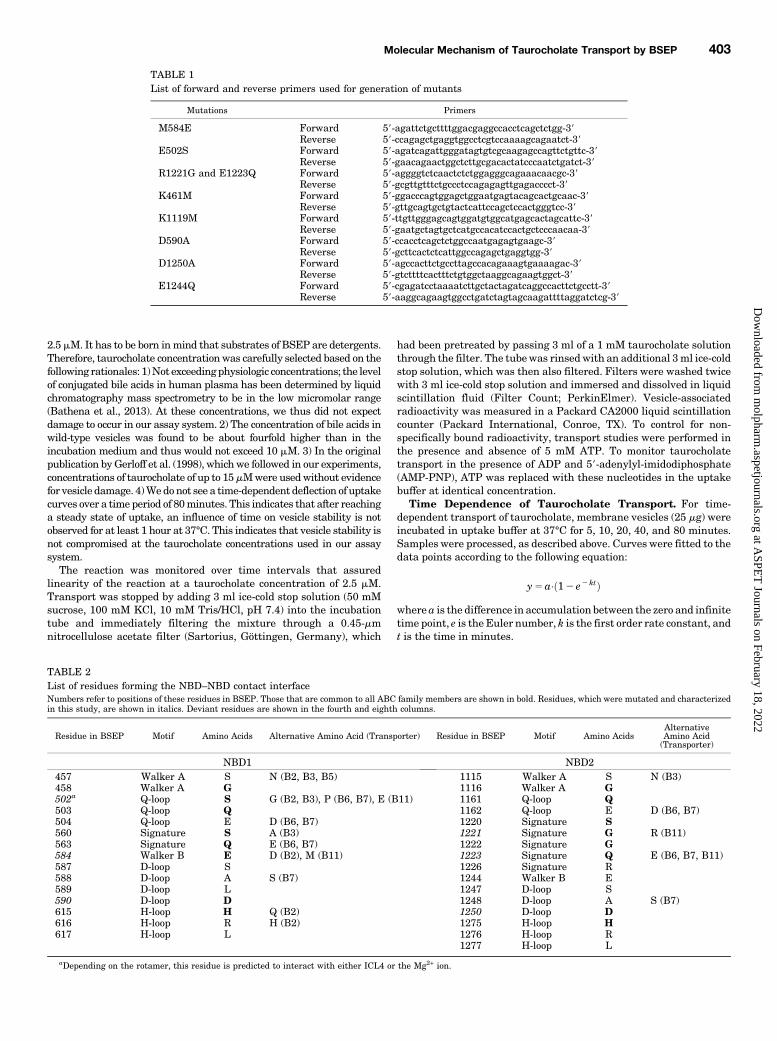
TABLE 1List of forward and reverse primers used for generation of mutants
Mutations Primers
M584E Forward 59-agattctgcttttggacgaggccacctcagctctgg-39Reverse 59-ccagagctgaggtggcctcgtccaaaagcagaatct-39
E502S Forward 59-agatcagattgggatagtgtcgcaagagccagttctgttc-39Reverse 59-gaacagaactggctcttgcgacactatcccaatctgatct-39
R1221G and E1223Q Forward 59-aggggtctcaactctctggagggcagaaacaacgc-39Reverse 59-gcgttgtttctgccctccagagagttgagacccct-39
K461M Forward 59-ggacccagtggagctggaatgagtacagcactgcaac-39Reverse 59-gttgcagtgctgtactcattccagctccactgggtcc-39
K1119M Forward 59-ttgttgggagcagtggatgtggcatgagcactagcattc-39Reverse 59-gaatgctagtgctcatgccacatccactgctcccaacaa-39
D590A Forward 59-ccacctcagctctggccaatgagagtgaagc-39Reverse 59-gcttcactctcattggccagagctgaggtgg-39
D1250A Forward 59-agccacttctgccttagccacagaaagtgaaaagac-39Reverse 59-gtcttttcactttctgtggctaaggcagaagtggct-39
E1244Q Forward 59-cgagatcctaaaatcttgctactagatcaggccacttctgcctt-39Reverse 59-aaggcagaagtggcctgatctagtagcaagattttaggatctcg-39
TABLE 2List of residues forming the NBD–NBD contact interfaceNumbers refer to positions of these residues in BSEP. Those that are common to all ABC family members are shown in bold. Residues, which were mutated and characterizedin this study, are shown in italics. Deviant residues are shown in the fourth and eighth columns.
Residue in BSEP Motif Amino Acids Alternative Amino Acid (Transporter) Residue in BSEP Motif Amino AcidsAlternativeAmino Acid(Transporter)
NBD1 NBD2457 Walker A S N (B2, B3, B5) 1115 Walker A S N (B3)458 Walker A G 1116 Walker A G502a Q-loop S G (B2, B3), P (B6, B7), E (B11) 1161 Q-loop Q503 Q-loop Q 1162 Q-loop E D (B6, B7)504 Q-loop E D (B6, B7) 1220 Signature S560 Signature S A (B3) 1221 Signature G R (B11)563 Signature Q E (B6, B7) 1222 Signature G584 Walker B E D (B2), M (B11) 1223 Signature Q E (B6, B7, B11)587 D-loop S 1226 Signature R588 D-loop A S (B7) 1244 Walker B E589 D-loop L 1247 D-loop S590 D-loop D 1248 D-loop A S (B7)615 H-loop H Q (B2) 1250 D-loop D616 H-loop R H (B2) 1275 H-loop H617 H-loop L 1276 H-loop R
1277 H-loop L
aDepending on the rotamer, this residue is predicted to interact with either ICL4 or the Mg2+ ion.
Molecular Mechanism of Taurocholate Transport by BSEP 403
at ASPE
T Journals on February 18, 2022
molpharm
.aspetjournals.orgD
ownloaded from

Osmolarity Plot. The osmolarity plot was prepared according toWlcek et al. (2014). Briefly, the membrane vesicles (25 mg) wereincubated in uptake buffer [50 mM sucrose, 100 mM KNO3, 12.5 mMMg(NO3)2, 10 mM Tris/HEPES, pH 7.4] at 37°C for 10 minutes.Outside osmolarity was increased by adding sucrose [50 mM (isotonic)to 800 mM]. Taurocholate transport (picomoles per milligram proteinper minute) was plotted against the inverse of the osmolarity. Non-specific binding of taurocholate to vesicles was calculated by extrap-olating the curve to the intercept with the y-axis.
ATPase Assay. The ATPase activity was determined by measuringthe release of inorganic phosphate using a calorimetricmethod describedbyChifflet et al. (1988) andSchmid et al. (1999). Briefly, 25mgmembranevesicles was added to the ATPase assay buffer (300mM sucrose, 4.8 mMMgCl2, 0.2 mM EGTA, 10 mM NaN3, 0.17 mM ouabain, and 20 mMTris/HCl, pH 7.4). Ouabain, EGTA, and NaN3 were added in the assaymixture to block Na1/K1, Ca21, and mitochondrial ATPases, respec-tively. Orthovanadate (0.56 mM) was used to block ABC transporter–associated ATPase activity. BSEP-associated ATPase activity wascalculated as the difference of total and vanadate-insensitive ATPaseactivity. The latter had to be corrected for vanadate-sensitive ATPaseactivity in mock-transfected cells. For ATPase stimulation, taurocholatewasusedat a concentration of 50mM.Theassaymixturewasprewarmedto 37°C for 5 minutes, and the reaction was started by addition of ATP(4mM). After 10, 20, 30, and 40minutes, a 50ml aliquot of the incubationmixturewas added to 50ml 12%SDS in assay buffer (in 96-wellmicrotiterplate) to stop the reaction. For color development, 100 ml freshlyprepared6%ascorbic acid in 1MHCl and1%ammoniumheptamolybdatewas added, andplateswere incubatedat room temperature for 5minutes.Subsequently, 150 ml 2% (w/v) trisodium citrate and 2% (w/v) sodium(meta)arsenite in 2% (v/v) acetic acid were added, and the mixture wasincubated at room temperature for 20 minutes to stabilize and enhancethe color. The optical density was measured at 630 nm. Na2HPO4 (stock2 mM) was used to prepare the standard curve.
Homology Modeling of BSEP. The sequences of ABCB1, BSEP,and Sav1866 were aligned andmanually adjusted, according to Stockneret al., (2009).Models of BSEPwere createdwithMODELER version 9.15(Sali and Blundell, 1993) using the crystal structure of Sav1866 fromStaphylococcus aureus as the template (PDB ID: 2ONJ) (Dawson andLocher, 2007). Mutations of residues in NBS1 were introduced withMODELER. The extracellular loop 1 was modeled to continue a-helicityof transmembrane helices 1 and 2, based on secondary structurepredictions. The cocrystallized nucleotide AMP-PNP was replaced byATP; Na1 ions in the crystal structure were replaced by Mg12 ions.
Kinetic Modeling. Taurocholate uptake and ATPase activity ofBSEP-containing plasma membrane vesicles were simulated accord-ing to a previously published kinetic model of the ABCB1 transportcycle (Al-Shawi et al., 2003) with modifications. The geometry of thevesicleswasassumed tobe sphericalwithadiameter of 2mm(V54.18 fl).Loss of taurocholate from vesicles to the incubation medium was takeninto account in themodel. Permeability of taurocholate was estimated onbasis of the physicochemical property “polar surface area,” as described(Schmid et al., 2015). Time-dependent changes in state occupancies, aswell as substrate efflux and phosphate generation, were evaluated bynumerical integration of the resulting system of differential equationsusing the Systems Biology Toolbox (Schmidt and Jirstrand, 2006) andMATLAB 2012a (Mathworks, Natick, MA).
Statistical Analysis. The GraphPad prism software (GraphPadSoftware, La Jolla, CA) was used for data analysis. Statisticallysignificant differences between basal and taurocholate-stimulatedATPase activity were determined by two-way analysis of variance,followed by Bonferroni post hoc analysis. For comparison of taurocho-late transport, one-way analysis of variance, followed by Tukey’s posthoc analysis, was used. AP value,0.05 was considered to be statisticallysignificant. A comparison of the goodness of fit for a linear and nonlinearfit was performed using the extra sum-of-squares F test. To be able toperform an extra sum-of-squares F test, we alternatively fitted datapoints not only with the kinetic model, but also with a quadratic function,according to the equation y5 ax2 1 bx 1 c.
Fig. 1. Homology model of BSEP based on the crystal structure ofSav1866. NBD1 and NBD2 are shown in ribbon representation in cyanand blue, respectively. Residues that are in direct contact at the interfaceof the NBDs are rendered in surface representation (Walker A motif, red;Q-loop, pink; signature sequence, green; Walker B, orange; D-loop,magenta; H-loop, yellow). The two ATP molecules are shown inlicorice rendering. Mg2+ ions are depicted as gray spheres: (A) Sideview of the NBD1 interface with a view angle that runs perpendicularto the plane of the interface. (B) Side view of the NBD2 interface[rotated by 180° relative to (A)]. (C) Top view with a view angle thatruns along the NBD interface.
404 Sohail et al.
at ASPE
T Journals on February 18, 2022
molpharm
.aspetjournals.orgD
ownloaded from
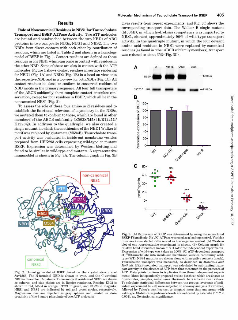
ResultsRole ofNoncanonicalResidues inNBS1 forTaurocholate
Transport and BSEP ATPase Activity. Two ATPmoleculesare bound and sandwiched between the two NBDs of ABCproteins in two composite NBSs, NBS1 and NBS2. The twoNBDs form direct contacts with each other by contribution ofresidues, which are listed in Table 2 and shown in a homologymodel of BSEP in Fig. 1. Contact residues are defined as thoseresidues in oneNBD, which can come in contact with residues inthe other NBD. Some of these are also in contact with the ATPmolecules. Figure 1 shows contact residues in surface renderingfor NBD1 (Fig. 1A) and NBD2 (Fig. 1B) in a head-on view ontothe respectiveNBDand in a top view for bothNBDs (Fig. 1C). Allcontact residues lie close, or conform to conserved residues ofNBD motifs in the primary sequence. All four full transportersof the ABCB subfamily show complete contact–interface con-servation, except for four residues in BSEP, which all lie in thenoncanonical NBS1 (Fig. 2).To assess the role of these four amino acid residues and to
establish the functional relevance of asymmetry in the NBSs,we mutated them to conform to those, which are found in othermembers of the ABCB subfamily (E502S/M584E/R1221G/E1223Q). In addition to the quadruple, we also created asinglemutant, in which themethionine of the NBD1Walker Bmotif was replaced by glutamate (M584E). Taurocholate trans-port activity was evaluated in inside-out membrane vesiclesprepared from HEK293 cells expressing wild-type or mutantBSEP. Expression was determined by Western blotting andfound to be similar in wild-type and mutants. A representativeimmunoblot is shown in Fig. 3A. The column graph in Fig. 3B
gives results from repeat experiments, and Fig. 3C shows thecorresponding transport data. The Walker B single mutant(M584E), in which hydrolysis competency was imparted toNBS1, showed approximately 90% of wild-type transportactivity. In the quadruple mutant, in which the four deviantamino acid residues in NBS1 were replaced by canonicalresidues (as found in otherABCBsubfamilymembers), transportwas reduced to about 35% (Fig. 3C).
Fig. 2. Homology model of BSEP based on the crystal structure ofSav1866. The N-terminal NBD is shown in cyan, and the C-terminalNBD in blue color. C-a atoms of noncanonical residues of NBS1 are shownas spheres, and side chains are in licorice rendering. Residue E502 isshown in red, M584 in orange, R1221 in green, and E1223 in magenta.NBS1 and NBS2 are indicated by red and green circles, respectively.Magnesium ions are depicted as gray spheres and located in closeproximity of the b and g phosphate of two ATP molecules.
Fig. 3. (A) Expression of BSEP was determined by using the monoclonalBSEP-F6 antibody. Na+/K+ ATPase was used as a loading control. Vesiclesfrom mock-transfected cells served as the negative control. (A) Westernblot of one representative experiment is shown. (B) Column graph forrelative band intensities (mean6 S.D.) of three independent experiments.Expression of wild-type was taken as 100%. (C) ATP-dependent transportof [3H]taurocholate into inside-out membrane vesicles containing wild-type (WT); NBS1 mutants are shown along with negative controls (mock).Taurocholate transport was measured, as described in Materials andMethods. BSEP-mediated transport was calculated by subtracting trans-port activity in the absence of ATP from that measured in the presence ofATP. Data points conform to triplicates from three independent experi-ments (three independently prepared vesicle batches), which are shown asfilled circles, triangles, and squares. Horizontal bars indicatemean values.To calculate statistical differences between the groups, averages of indi-vidual experiment (n = 3) were subjected to one-way analysis of variance,followed by Tukey’s post hoc test to compare more than one group withwild-type. Statistical significance levels are indicated by asterisks (***P,0.001). ns, No statistical significance.
Molecular Mechanism of Taurocholate Transport by BSEP 405
at ASPE
T Journals on February 18, 2022
molpharm
.aspetjournals.orgD
ownloaded from

AsATP binding and hydrolysis is a prerequisite for transportfunction, we also measured basal and taurocholate-stimulatedATPase activity of the mutants. Two negative controls wereused in these experiments: 1) vesicles from mock-transfectedcells and 2) cells transfected with the E1244Q mutant. Thismutant lacks a catalytic glutamate and thus is unable to eitherhydrolyze ATP or transport taurocholate (see below). Vesiclepreparations from these negative controls showed vanadate-sensitive ATPase activity of about 3 nmol Pi/mg per minute.This ATPase activity is not BSEP associated as it is notstimulated by taurocholate (Fig. 4).Basal BSEP ATPase activity of the M584E mutant was
comparable to that of wild-type protein. ATPase and transportactivity in the quadruple mutant was decreased to about 35%.ATPase stimulation by substrate was retained in both thesingle and the quadruple mutant. This indicates that direc-tional communication between the taurocholate binding site inthe TMDs and the hydrolysis-competent NBS2 is retaineddespite the introduction of these mutations in NBS1.
Exploring the Role of Canonical Residues in NBS1and NBS2. Despite the presence of individual noncanonicalresidues, most of the residues in NBS1 are conserved withinthe ABCB subfamily. These include the lysine residue in theWalker A motif and the aspartate residue in the D-loop.Hence, we explored the effect of mutation of these residues ontaurocholate transport and ATPase activity in NBS1. As acontrol, we also introduced these mutations in NBS2.Role of Walker A Lysine Residues. Walker A lysines
were shown to form contacts with the b and g phosphate ofATP (Hung et al., 1998). We replaced these lysines bymethionines (K461M and K1119M), as previously describedfor ABCB1 (Lapinski et al., 2001; Bársony et al., 2016).Surface expression of these mutants was found to be compa-rable to that of wild-type protein (Fig. 5, A and B). Taurocho-late transport was found to be abolished in both mutants (Fig.5C), whereas a basal BSEP-associated ATPase activity wasstill found to be associated with the mutants. The K1119Mmutant lost its ability to be stimulated by taurocholate (Fig. 4).
Fig. 4. ATPase activity wasmeasured in inside-out plasmamembrane vesicles prepared fromHEK293 cells transfected with wild-type ormutant BSEP.Generally, orthovanadate is used to determine BSEP-associated ATPase activity. Mock-transfected cells and cells transfected with the ATPase-deficientE1244Q mutant were used as negative controls (gray and light blue bars, respectively). The black dotted line shows the proportion of ATPase activity,which is not BSEP associated. It is due to endogenous vanadate-inhibited ATPases, which are present in the plasma membrane of HEK293 cells. ForATPase stimulation, taurocholate (TC) was used at a final concentration of 50 mM. This exceeds the concentration required for half-maximal stimulationin wild-type protein by four- to fivefold (Supplemental Fig. 1). Stimulation here was taken as a qualitative parameter for interdomain communication.Fold stimulation thus is not intended to provide quantitative information, as curves for concentration dependencies might be shifted in mutants relativeto wild-type. The number of experiments was three for all mutants (quadruplicate determinations from independent incubation mixtures). In individualexperiments, these mutants were put together in different combinations, whereby wild-type was always included as a positive control. Thus, nwas 3 forall mutants, but 13 for wild-type. Mean values6 S.D. are shown. Statistically significant differences between basal and taurocholate-stimulated ATPaseactivity were determined by two-way analysis of variance, followed by Bonferroni post hoc analysis. Statistical significance levels are indicated byasterisks (*P , 0.05; ***P , 0.001). ns, no statistical significance.
406 Sohail et al.
at ASPE
T Journals on February 18, 2022
molpharm
.aspetjournals.orgD
ownloaded from
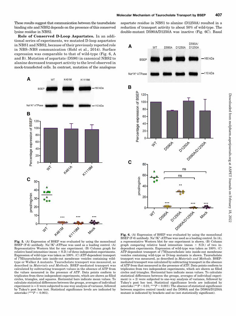
These results suggest that communication between the taurocholatebinding site andNBS2 depends on the presence of this conservedlysine residue in NBS2.Role of Conserved D-Loop Aspartates. In an addi-
tional series of experiments, we mutated D-loop aspartatesin NBS1 andNBS2, because of their previously reported rolein NBS–NBS communication (Hohl et al., 2014). Surfaceexpression was comparable to that of wild-type (Fig. 6, Aand B). Mutation of aspartate (D590) in canonical NBS2 toalanine decreased transport activity to the level observed inmock-transfected cells. In contrast, mutation of the analogous
aspartate residue in NBS1 to alanine (D1250A) resulted in areduction of transport activity to about 50% of wild-type. Thedouble-mutant D590A/D1250A was inactive (Fig. 6C). Basal
Fig. 5. (A) Expression of BSEP was evaluated by using the monoclonalBSEP (F-6) antibody. Na+/K+-ATPase was used as a loading control. (A)Representative Western blot for one experiment. (B) Column graph forrelative band intensities (mean6 S.D.) of three independent experiments.Expression of wild-type was taken as 100%. (C) ATP-dependent transportof [3H]taurocholate into inside-out membrane vesicles containing wild-type or Walker A mutants. Taurocholate transport was measured, asdescribed in Materials and Methods. BSEP-mediated transport wascalculated by subtracting transport values in the absence of ATP fromthe values measured in the presence of ATP. Data points conform totriplicates from three independent experiments, which are shown as filledcircles, triangles, and squares. Horizontal bars indicate mean values. Tocalculate statistical differences between the groups, averages of individualexperiment (n = 3) were subjected to one-way analysis of variance, followedby Tukey’s post hoc test. Statistical significance levels are indicated byasterisks (***P , 0.001).
Fig. 6. (A) Expression of BSEP was evaluated by using the monoclonalBSEP (F-6) antibody. Na+/K+-ATPase was used as a loading control. In (A),a representative Western blot for one experiment is shown. (B) Columngraph comparing relative band intensities (mean 6 S.D.) of two in-dependent experiments. Expression of wild-type was taken as 100%. (C)ATP-dependent transport of [3H]taurocholate into inside-out membranevesicles containing wild-type or D-loop mutants is shown. Taurocholatetransport was measured, as described in Materials and Methods. BSEP-mediated transport was calculated by subtracting transport in the absenceof ATP from that measured in the presence of ATP. Data points conform totriplicates from two independent experiments, which are shown as filledcircles and triangles. Horizontal bars indicate mean values. To calculatestatistical differences between the groups, averages of individual experi-ment (n = 2) were subjected to one-way analysis of variance, followed byTukey’s post hoc test. Statistical significance levels are indicated byasterisks (**P, 0.01; ***P, 0.001). The absence of statistical significancebetween negative control (mock) and the D590A and the D590A/D1250Amutant is indicated by brackets and ns (not statistically significant).
Molecular Mechanism of Taurocholate Transport by BSEP 407
at ASPE
T Journals on February 18, 2022
molpharm
.aspetjournals.orgD
ownloaded from
ATPase activity in the single and the double mutant wascomparable to that of wild-type protein. NBS1 mutant D1250Awas stimulated by taurocholate. However, this stimulation wasabsent in the D590A (NBS2) and in the double-mutant D590A/D1250A (Fig. 4).Exchanging the Catalytic Glutamate between NBS2
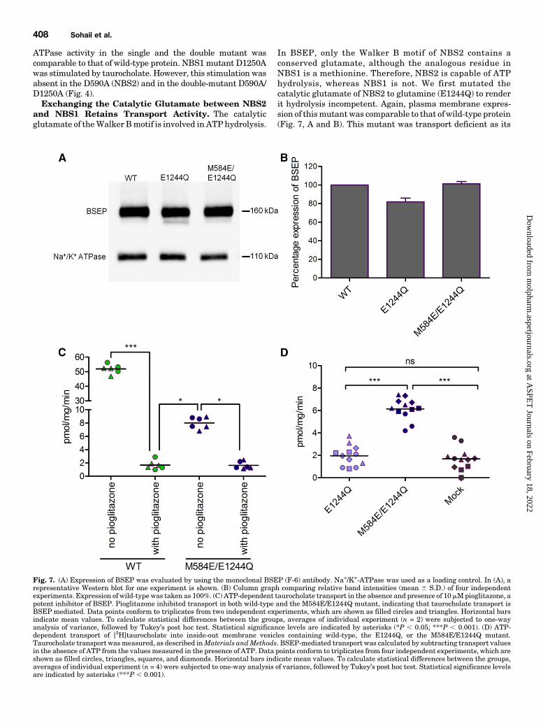
and NBS1 Retains Transport Activity. The catalyticglutamate of theWalker Bmotif is involved in ATP hydrolysis.
In BSEP, only the Walker B motif of NBS2 contains aconserved glutamate, although the analogous residue inNBS1 is a methionine. Therefore, NBS2 is capable of ATPhydrolysis, whereas NBS1 is not. We first mutated thecatalytic glutamate of NBS2 to glutamine (E1244Q) to renderit hydrolysis incompetent. Again, plasma membrane expres-sion of thismutantwas comparable to that of wild-type protein(Fig. 7, A and B). This mutant was transport deficient as its
Fig. 7. (A) Expression of BSEP was evaluated by using the monoclonal BSEP (F-6) antibody. Na+/K+-ATPase was used as a loading control. In (A), arepresentative Western blot for one experiment is shown. (B) Column graph comparing relative band intensities (mean 6 S.D.) of four independentexperiments. Expression of wild-type was taken as 100%. (C) ATP-dependent taurocholate transport in the absence and presence of 10mMpioglitazone, apotent inhibitor of BSEP. Pioglitazone inhibited transport in both wild-type and the M584E/E1244Q mutant, indicating that taurocholate transport isBSEP mediated. Data points conform to triplicates from two independent experiments, which are shown as filled circles and triangles. Horizontal barsindicate mean values. To calculate statistical differences between the groups, averages of individual experiment (n = 2) were subjected to one-wayanalysis of variance, followed by Tukey’s post hoc test. Statistical significance levels are indicated by asterisks (*P , 0.05; ***P , 0.001). (D) ATP-dependent transport of [3H]taurocholate into inside-out membrane vesicles containing wild-type, the E1244Q, or the M584E/E1244Q mutant.Taurocholate transport wasmeasured, as described inMaterials andMethods. BSEP-mediated transport was calculated by subtracting transport valuesin the absence of ATP from the values measured in the presence of ATP. Data points conform to triplicates from four independent experiments, which areshown as filled circles, triangles, squares, and diamonds. Horizontal bars indicate mean values. To calculate statistical differences between the groups,averages of individual experiment (n = 4) were subjected to one-way analysis of variance, followed by Tukey’s post hoc test. Statistical significance levelsare indicated by asterisks (***P , 0.001).
408 Sohail et al.
at ASPE
T Journals on February 18, 2022
molpharm
.aspetjournals.orgD
ownloaded from

transport activity was not significantly different from thatfound in mock-transfected cells. Also, ATPase activity wascomparable to that found in mock-transfected cells (Fig. 4).Thus, any BSEP-associated ATPase activity was absent.We next replaced the noncanonical methionine residue in
NBS1 with glutamate in the background of the E1244Qmutant.The catalytic glutamatewas thus exchanged betweenNBS2 andNBS1 (M584E/E1244Q) to reinstate hydrolysis competency inthe protein. The mutant again showed a surface expression thatwas comparable to that of wild-type protein (Fig. 7, A and B).Most interestingly, the double mutant consistently showedapproximately 15% residual transport activity (Fig. 7C), whichwas demonstrated to be BSEP mediated by using the high-affinity inhibitor pioglitazone [published IC50 values of 0.4 mM(Morgan et al., 2010) and 0.3 mM (Dawson et al., 2012)]. At aconcentration of 10 mM pioglitazone, transport was completelyabolished in themutant and in wild-type (Fig. 7C). This residualtransport activity by the M584E/E1244Q mutant was signifi-cantly higher than that of the catalytically inactive mutantE244Q and mock (Fig. 7D). To further characterize this mutant,wemonitored taurocholate transport in the absence of nucleotideand in presence of ATP, ADP, and AMP-PNP. Similar to wild-type, the mutant showed strict ATP dependence of transportactivity (Fig. 8).We further determined basal ATPase activity of the mutant
and the ability of taurocholate to stimulate it (Fig. 4). Basalactivity was comparable to wild-type, indicating that theM584E/E1244Q mutant was capable of hydrolyzing ATP. AsNBS2 in this mutant is hydrolysis incompetent, this activitymust be mediated by NBS1. However, stimulation of ATPaseactivity by taurocholate was not observed. Hence, the taur-ocholate binding site cannot signal the presence of substrate toNBS1. Because the mutant is capable of taurocholate trans-port, this lack of substrate stimulation is not considered to bebrought about by a loss of the ability to bind taurocholate.To further explore themolecularmechanism of taurocholate
transport by theM584E/E1244Qmutant, we compared steadystate levels of uptake in the mutant and wild-type protein.
Uptake of taurocholate was monitored over 80 minutes at aconcentration of 2.5 mM (first order) (Fig. 9). Steady statelevels of uptake were more than four times lower in themutant (approximately 170 pmol/mg) as compared with wild-type (approximately 700 pmol/mg), and transport velocity wasreduced (half-life of accumulation 5.1 minutes in wild-typeand 14.6 minutes in the mutant). Despite similar basal ATPhydrolysis rates, steady state levels of taurocholate accumu-lation were thus reduced in the mutant.Taurocholate is an amphipathic small molecule, which
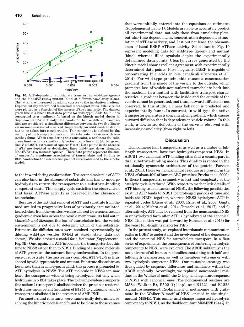
readily distributes into the environment of biomembranes. Todiscriminate between biomembrane partitioning and accumu-lation in the vesicle lumen, transport was monitored in thepresence of an increasing concentration of sucrose in the uptakebuffer. This collapses vesicles (Supplemental Fig. 2). Transportrate is expected to be linearly related to the inverse of theosmolarity, where the intercept with the ordinate conforms tovesicle-associated radioactivity at zero inside volume. Underthe assumption that binding of taurocholate to the transporterdoes not significantly contribute to this value, the intercept ofwild-type and mutant should be identical.Figure 10 shows that the mutant indeed demonstrates
linear behavior. In contrast, data points for wild-type trans-porter seem to deviate from linearity.When fitted linearly, theintercept value is much higher than for the mutant. A similardeviation of data points from linear fits can be observed in anexperimental setup that is comparable to ours (Wlcek et al.,2014). To resolve the discrepancy of nonconforming y-interceptsfor wild-type protein and mutant, we simulated the processof taurocholate accumulation in vesicles by using a kineticBSEP model (Supplemental Fig. 3A). Briefly, this model isbased on the following assumptions: one molecule of ATPis bound to the transporter in NBS1 in the inward-facingconformation. Substrate binding induces binding of a secondmolecule of ATP in NBS2. This brings the transporter into anoutward-facing conformation. Subsequent hydrolysis of ATPin NBS2 leads to substrate release. In two additional steps,phosphate and ADP are released. This resets the transporter
Fig. 8. The transport of taurocholate (2.5 mM) was monitored in thepresence of 5 mM ATP, ADP, or AMP-PNP. Data points conform totriplicates from two independent experiments from two separately pre-pared vesicle batches. The horizontal bar indicates the mean value of datapoints.
Fig. 9. Time dependence of taurocholate uptake in inside-out membranevesicles containing wild-type BSEP (green) or the M584E/E1244Qmutant(blue). Data points conform to triplicates from two independent experi-ments from two separately prepared vesicle batches.
Molecular Mechanism of Taurocholate Transport by BSEP 409
at ASPE
T Journals on February 18, 2022
molpharm
.aspetjournals.orgD
ownloaded from
to the inward-facing conformation. The secondmolecule of ATPcan also bind in the absence of substrate and has to undergohydrolysis to return the transporter to a substrate-bindingcompetent state. This empty cycle satisfies the observationthat basal ATPase activity is observed in the absence oftaurocholate.Because of the fact that removal of ATP and substrate from the
medium led to progressive loss of previously accumulatedtaurocholate fromthevesicles,wealso allowed for a concentrationgradient–driven loss across the vesicle membrane. As laid out inMaterials and Methods, this loss of taurocholate into the vesicleenvironment is not due to detergent action of the substrate.Estimates for diffusion rates were obtained experimentally bydiluting wild-type vesicles 60-fold at steady state (data notshown). We also devised a model for a facilitator (SupplementalFig. 3B).Onceagain, oneATP is bound to the transporter, but thistime to NBS2 rather than to NBS1. Binding of a secondmoleculeof ATP generates the outward-facing conformation. In the pres-ence of substrate, the quaternary complex ATP2-Te-S is thusshared bywild-type protein andmutant. Substrate dissociates atlower rate than inwild-type, inwhich this process is promoted byATP hydrolysis in NBS2. The ATP molecule in NBS2 can nowleave the transporter without being hydrolyzed, but only whenhydrolysis in NBS1 takes place. The following evidence supportsthisnotion: 1) transport is abolishedwhen theprotein is renderedhydrolysis incompetent (mutation of E1244 to glutamine) and 2)transport is abolished in the presence of AMP-PNP.Parameters and constants were numerically determined by
solving the kinetic models and found to be close to those values
that were initially entered into the equations as estimates(Supplemental Table 1). Models are able to accurately predictall experimental data, not only those from osmolarity plots,but also time dependencies, concentration-dependent stimu-lation of ATPase activity, and, last but not least, the phenom-enon of basal BSEP ATPase activity. Solid lines in Fig. 10represent modeling data for wild-type (green) and mutant(blue), whereas filled symbols depict the experimentallydetermined data points. Clearly, curves generated by thekinetic model show excellent agreement with experimentallydetermined data points. Physiologically, BSEP is capable ofconcentrating bile acids in bile canaliculi (Cuperus et al.,2014). For wild-type protein, this causes a concentrationgradient from the inside of the vesicle to the outside, whichpromotes loss of vesicle-accumulated taurocholate back intothe medium. In a mutant with facilitative transport charac-teristics, a gradient between the inside and the outside of thevesicle cannot be generated, and thus, outward diffusion is notobserved. In this study, a linear behavior is predicted andexperimentally found. In contrast, in wild-type the activetransporter generates a concentration gradient, which causesoutward diffusion that is dependent on vesicle volume. In thiswork, a downward deflection of the curve is observed withincreasing osmolarity (from right to left).
DiscussionHomodimeric half transporters, as well as a number of full-
length transporters, have two hydrolysis-competent NBSs. InABCB1 two canonical ATP binding sites find a counterpart indual substrate-binding modes. This duality is rooted in therotationally symmetric architecture of the protein (Parveenet al., 2011). However, noncanonical residues are present in theNBS1 of about 40% of humanABCproteins (Procko et al., 2009).In these transporters, symmetry is lost and complexity of thecatalytic cycle is reduced. With respect to mechanistic details ofATP binding to a noncanonical NBS1, the following possibilitiesexist: 1) ATP binds to NBS1 for prolonged periods of time andholds the NBDs together, whereas NBS2 hydrolyzes ATP inrepeated cycles (Basso et al., 2003; Ernst et al., 2008; Guptaet al., 2014; Mishra et al., 2014; Timachi et al., 2017), and 2)alternatively, ATPmay be released from the noncanonical NBSin unhydrolyzed form after ATP is hydrolyzed at the canonicalNBS. This notion was put forward by Furman et al. (2013) forthe yeast full-length transporter Pdr5.In the present study,we explored interdomain communication
paths in BSEP to understand the involvement of the degenerateand the canonical NBS for taurocholate transport. In a firstseries of experiments, the consequences of conferring hydrolysiscompetency to NBS1 were explored. The ABCB subfamily is themost diverse of all human subfamilies, containing both half- andfull-length transporters, as well as members with one or withtwo hydrolysis-competent NBSs. Our mutation strategy wasthus guided by sequence differences and similarity within theABCB subfamily. Accordingly, we replaced noncanonical resi-dues in the Walker B motif, the Q-loop, and signature sequenceof NBS1 with canonical ones. The noncanonical residues areM584 (Walker B), E502 (Q-loop), and R1221 and E1223(signature sequence). Replacement of methionine with gluta-mate in the Walker B motif of NBS1 ensued in the single-mutant M584E. This amino acid change imparted hydrolysiscompetency to NBS1, as the double-mutantM584E/E1244Q, in
Fig. 10. ATP-dependent taurocholate transport in wild-type (green)and the M584E/E1244Q mutant (blue) at different osmolarity (Osm).The latter was increased by adding sucrose to the incubation medium.Experimentally determined taurocholate transport rates (filled circles)were plotted as a function of the inverse of the osmolarity. The dashedgreen line is a linear fit of data points for wild-type BSEP. Solid linescorrespond to a nonlinear fit based on the kinetic model shown inSupplemental Fig. 3. If only data points for the five different osmolar-ities are considered, a significant difference between the two fits (linearversus nonlinear) is not observed. Importantly, an additional constrainthas to be taken into consideration. This constraint is defined by theinability of the transporter to accumulate substrate in vesicles with zeroinside volume. When considering this constraint, a nonlinear fit (solidgreen line) performs significantly better than a linear fit (dotted greenline, P = 0.0001, extra sum of squares F test). Data points in the absenceof ATP are depicted as dot-dashed lines (wild-type: down triangles;M584E/E1244Q mutant: squares). These data points represent the sumof unspecific membrane association of taurocholate and binding toBSEP and define the intersection point of curves obtained by the kineticmodel.
410 Sohail et al.
at ASPE
T Journals on February 18, 2022
molpharm
.aspetjournals.orgD
ownloaded from
which mutation of the catalytic glutamate E1244 rendersNBS2 hydrolysis incompetent, shows ATPase activity that iscomparable to wild-type protein. Thus, the ability of NBS1 tohydrolyze ATP (in the single M584E) does not result in asignificant loss of transport activity. Hydrolysis in turn wouldlead to dissociation of residues in the two nucleotide bindingdomains, which jointly form NBS1, and thus, a platformfunction of NBS1 for repeated cycles of hydrolysis in NBS2 isunlikely. The importance of the presence of the three addi-tional noncanonical residues in NBS1 of BSEP presentlyremains elusive.In a next series of experiments, we explored the role of
selected residues in conserved motifs, namely the Walker Alysines and D-loop aspartates in both NBSs for ATPaseactivity and taurocholate transport. The Walker A mutantscompletely lost transport activity, irrespective of the NBS inwhich theyweremutated. These results are in agreementwithearlier data on human TAP1/2, in which mutation of con-served lysine residues in the Walker A motifs similarlyabolished transport activity (Azzaria et al., 1989; Lapinskiet al., 2001). Basal ATPase activity was retained in thesemutants, indicating that hydrolytic capacity alone is notsufficient to sustain the transport process.The D-loops have been indicted to be involved in NBD–NBD
communication. The eponymous aspartate is conserved inboth NBSs of BSEP. In either of the D-loop mutants, basalATPase activity was comparable to that of wild-type. Thus, adirect involvement of these residues in ATP hydrolysis seemsunlikely. Although the D1250A mutant in NBS1 retained its
ability to be stimulated by taurocholate, this property was lostby mutating residue D590 in NBS2. In the latter taurocholatetransport was abolished, whereas for analogous mutation inNBS1 (D1250A) transport activity was decreased to 50%.Similar results were reported for two other ABC transporterswith one noncanonical NBS, TM287/288 (Hohl et al., 2014)and Pdr5 (Furman et al., 2013). Taken together, we interpretour data to indicate that the taurocholate binding site is ableto communicate the presence of substrate to NBS2, but not toNBS1. For this communication, the Walker A lysine and theD-loop aspartate of NBS2 are required.In a final set of experiments, we replaced the catalytic
glutamate of NBS2 by glutamine. In the ensuing E1244Qmutant, we then introduced a glutamate residue into theWalker Bmotif of NBS1. This double mutant (M584E/E1244Q)consistently showed 15% of wild-type transport activity. Timedependencies showed that steady state levels of uptake were75%–80% reduced in the mutant (Fig. 9), suggesting that thismutant may have lost active and adopted facilitative charac-teristics. We used a kinetic model to support this notion. Thismodel comprised a term for loss of taurocholate from the vesiclelumen, which we found to occur after washout of taurocholateand ATP from the incubation medium (data not shown).Although this loss did not in any way compromise interpreta-tion of transport data, it was taken advantage of, to corroboratethat transport in themutantwas facilitative. The rationale wasas follows: loss of taurocholate from the vesicle would only beobserved when concentrative transport into the vesicle occurs.This would lead to nonlinear curve characteristics. In contrast,
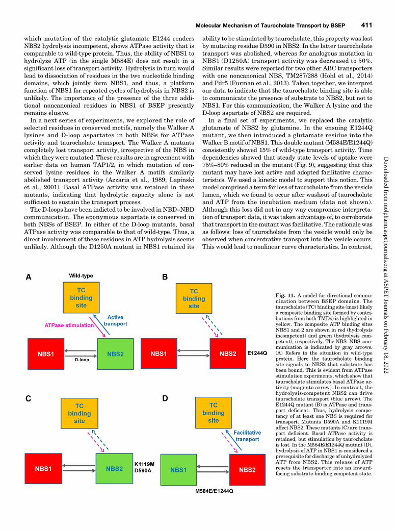
Fig. 11. A model for directional commu-nication between BSEP domains. Thetaurocholate (TC) binding site (most likelya composite binding site formed by contri-butions from both TMDs) is highlighted inyellow. The composite ATP binding sitesNBS1 and 2 are shown in red (hydrolysisincompetent) and green (hydrolysis com-petent), respectively. The NBS–NBS com-munication is indicated by gray arrows.(A) Refers to the situation in wild-typeprotein. Here the taurocholate bindingsite signals to NBS2 that substrate hasbeen bound. This is evident from ATPasestimulation experiments, which show thattaurocholate stimulates basal ATPase ac-tivity (magenta arrow). In contrast, thehydrolysis-competent NBS2 can drivetaurocholate transport (blue arrow). TheE1244Q mutant (B) is ATPase and trans-port deficient. Thus, hydrolysis compe-tency of at least one NBS is required fortransport. Mutants D590A and K1119Maffect NBS2. These mutants (C) are trans-port deficient. Basal ATPase activity isretained, but stimulation by taurocholateis lost. In the M584E/E1244Q mutant (D),hydrolysis of ATP in NBS1 is considered aprerequisite for discharge of unhydrolyzedATP from NBS2. This release of ATPresets the transporter into an inward-facing substrate-binding competent state.
Molecular Mechanism of Taurocholate Transport by BSEP 411
at ASPE
T Journals on February 18, 2022
molpharm
.aspetjournals.orgD
ownloaded from

curves would be expected to be linear for facilitators. This isindeed observed experimentally (Fig. 10). Because of its strictATP dependence, the M584E/E1244Q mutant has adoptedproperties of an ATP-gated facilitator. To the best of ourknowledge, this has not been shown for any other ABC trans-porter with one noncanonical NBS. An earlier report by RobertTampé’s group engineered TAP1/2 to act as a facilitator(Grossmann et al., 2014). However, in this case, the change inproperties was brought about by mutating the aspartate in theD-loop of TAP1; the transporter differs from the BSEP mutantin that ADP and AMP-PNP can replace ATP in function.In Fig. 11, we propose a model for the interdomain commu-
nication of BSEP based on data from this study. Transport is ameasure of the ability of NBS2 (canonical) to communicateconformational changes to the taurocholate binding site. Incontrast, stimulation of basal ATPase activity by taurocholatereflects the ability of the taurocholate binding site to signal toNBS2 that substrate has been bound. Both these interdomaincommunication paths are present inwild-type protein (Fig. 11A),but lost in the E1244Q (Fig. 11B), the K1119M, and the D590Amutant (Fig. 11C). In the M584E/E1244Q mutant (Fig. 11D),taurocholate is transported in facilitative manner. In this study,NBS2 is not able to hydrolyze ATP. However, we propose thatATP is able to dissociate from its binding site in unhydrolyzedform, when ATP is hydrolyzed in NBS1. This ensures that theprotein can complete its catalytic cycle to return to a substrate-binding competent state. Because of the inability of NBS2 tohydrolyze ATP, substrate affinity would remain higher for thefacilitator in the outward-facing conformation and thus result inthe loss of active transport characteristics.With respect to a reduction of the number of hydrolysis-
competent NBSs from two to one in about half of human ABCtransporters (including BSEP and the entire ABCC subfamily),the following can be stated: 1) development of full transporters,from homodimeric half transporters by gene duplication, repre-sented a prerequisite for evolution of these asymmetric proteins(Chen et al., 1986;Ueda et al., 1989; Kerr, 2002). A second path toABC proteins with only one noncanonical NBSwas the evolutionof heterodimeric half transporters. This is infrequently observedand only seen for TAP1/2 and ABCG5/G8. 2) A selective advan-tage may have arisen from the ensuing change in substratespecificity, be it in either broadening it in full transporters withtwo hydrolysis-competent NBSs or narrowing it in those thatonly contain one.Anyproof of coevolution of residues in theTMDsand causally related changes in NBS1 (evolutionary coupling)(Hopf et al., 2017) should be able to provide evidence for thisnotion. 3) In BSEP a single hydrolysis-competent NBSmay haveresulted in restricting substrate specificity to negatively chargedbile salts, while excluding compounds of alternative chemistry.Although entirely conjectural at present, the large number ofsmall-molecule drugs, which are capable of direct interactionwith (and inhibition of) BSEP,may be a reflection of the presenceof an evolutionarily retained former substrate binding site, whichhas lost coupling to a canonical NBS.
ConclusionWe engineered BSEP to shed light on the molecular
mechanism of taurocholate transport. This allows us to pre-sent a model for taurocholate transport with respect to itscoupling to the energy-generating step. Our data are expectedto form a basis for exploration of the taurocholate binding site
and an understanding of the interaction between the trans-porter and drugs. It is also expected to promote an under-standing of the functional biology of other members of thehuman ABC protein family that harbor one noncanonicalNBS.
Acknowledgments
The authors are grateful to Bruno Stieger for providing us withplasmids containing the BSEP construct and for continuing supportand discussion; Johannes Noe for in-depth discussion of plasmidpropagation in Escherichia coli; Enrico Klotzsch for critically readingthe manuscript and the Institute of Medical Statistics of the MedicalUniversity of Vienna (FlorianFrommlet) for expert statistical support.
Authorship Contributions
Participated in research design: Sohail, Chiba.Conducted experiments: Sohail, Wlcek.Contributed new reagents or analytic tools: Schmid.Performed data analysis: Sohail, Schmid, Spork, Stockner, Chiba.Wrote or contributed to the writing of the manuscript: Sohail,
Schmid, Szakács, Trauner, Chiba.
References
Aller SG, Yu J, Ward A, Weng Y, Chittaboina S, Zhuo R, Harrell PM, Trinh YT,Zhang Q, Urbatsch IL, et al. (2009) Structure of P-glycoprotein reveals a molecularbasis for poly-specific drug binding. Science 323:1718–1722.
Al-Shawi MK, Polar MK, Omote H, and Figler RA (2003) Transition state analysis ofthe coupling of drug transport to ATP hydrolysis by P-glycoprotein. J Biol Chem278:52629–52640.
Azzaria M, Schurr E, and Gros P (1989) Discrete mutations introduced in thepredicted nucleotide-binding sites of the mdr1 gene abolish its ability to confermultidrug resistance. Mol Cell Biol 9:5289–5297.
Bársony O, Szalóki G, Türk D, Tarapcsák S, Gutay-Tóth Z, Bacsó Z, Holb IJ,Székvölgyi L, Szabó G, Csanády L, et al. (2016) A single active catalytic site issufficient to promote transport in P-glycoprotein. Sci Rep 6:24810.
Basso C, Vergani P, Nairn AC, and Gadsby DC (2003) Prolonged nonhydrolytic in-teraction of nucleotide with CFTR’s NH2-terminal nucleotide binding domain andits role in channel gating. J Gen Physiol 122:333–348.
Bathena SP, Mukherjee S, Olivera M, and Alnouti Y (2013) The profile of bile acidsand their sulfate metabolites in human urine and serum. J Chromatogr B AnalytTechnol Biomed Life Sci 942–943:53–62.
Chen CJ, Chin JE, Ueda K, Clark DP, Pastan I, Gottesman MM, and Roninson IB(1986) Internal duplication and homology with bacterial transport proteins in themdr1 (P-glycoprotein) gene from multidrug-resistant human cells. Cell 47:381–389.
Chiba P, Freissmuth M, and Stockner T (2014) Defining the blanks–pharmacochaperoningof SLC6 transporters and ABC transporters. Pharmacol Res 83:63–73.
Chifflet S, Torriglia A, Chiesa R, and Tolosa S (1988) A method for the determinationof inorganic phosphate in the presence of labile organic phosphate and high con-centrations of protein: application to lens ATPases. Anal Biochem 168:1–4.
Cuperus FJ, Claudel T, Gautherot J, Halilbasic E, and Trauner M (2014) The role ofcanalicular ABC transporters in cholestasis. Drug Metab Dispos 42:546–560.
Dawson RJ and Locher KP (2006) Structure of a bacterial multidrug ABC trans-porter. Nature 443:180–185.
Dawson RJ and Locher KP (2007) Structure of the multidrug ABC transporterSav1866 from Staphylococcus aureus in complex with AMP-PNP. FEBS Lett 581:935–938.
Dawson S, Stahl S, Paul N, Barber J, and Kenna JG (2012) In vitro inhibition of thebile salt export pump correlates with risk of cholestatic drug-induced liver injury inhumans. Drug Metab Dispos 40:130–138.
Dönmez Cakil Y, Khunweeraphong N, Parveen Z, Schmid D, Artaker M, Ecker GF,Sitte HH, Pusch O, Stockner T, and Chiba P (2014) Pore-exposed tyrosine residuesof P-glycoprotein are important hydrogen-bonding partners for drugs. Mol Phar-macol 85:420–428.
Ernst R, Kueppers P, Klein CM, Schwarzmueller T, Kuchler K, and Schmitt L (2008)A mutation of the H-loop selectively affects rhodamine transport by the yeastmultidrug ABC transporter Pdr5. Proc Natl Acad Sci USA 105:5069–5074.
Furman C, Mehla J, Ananthaswamy N, Arya N, Kulesh B, Kovach I, Ambudkar SV,and Golin J (2013) The deviant ATP-binding site of the multidrug efflux pump Pdr5plays an active role in the transport cycle. J Biol Chem 288:30420–30431.
Gerloff T, Stieger B, Hagenbuch B, Madon J, Landmann L, Roth J, Hofmann AF,and Meier PJ (1998) The sister of P-glycoprotein represents the canalicular bilesalt export pump of mammalian liver. J Biol Chem 273:10046–10050.
Grossmann N, Vakkasoglu AS, Hulpke S, Abele R, Gaudet R, and Tampé R (2014)Mechanistic determinants of the directionality and energetics of active export by aheterodimeric ABC transporter. Nat Commun 5:5419.
Gupta RP, Kueppers P, Hanekop N, and Schmitt L (2014) Generating symmetry inthe asymmetric ATP-binding cassette (ABC) transporter Pdr5 from Saccharomycescerevisiae. J Biol Chem 289:15272–15279.
Hartley JL, Temple GF, and Brasch MA (2000) DNA cloning using in vitro site-specific recombination. Genome Res 10:1788–1795.
Higgins CF (1992) ABC transporters: from microorganisms to man. Annu Rev CellBiol 8:67–113.
412 Sohail et al.
at ASPE
T Journals on February 18, 2022
molpharm
.aspetjournals.orgD
ownloaded from
Hirano M, Maeda K, Hayashi H, Kusuhara H, and Sugiyama Y (2005) Bile salt exportpump (BSEP/ABCB11) can transport a nonbile acid substrate, pravastatin. JPharmacol Exp Ther 314:876–882.
Hohl M, Hürlimann LM, Böhm S, Schöppe J, Grütter MG, Bordignon E, and SeegerMA (2014) Structural basis for allosteric cross-talk between the asymmetric nu-cleotide binding sites of a heterodimeric ABC exporter. Proc Natl Acad Sci USA111:11025–11030.
Hopf TA, Ingraham JB, Poelwijk FJ, Schärfe CP, Springer M, Sander C, and MarksDS (2017) Mutation effects predicted from sequence co-variation. Nat Biotechnol35:128–135.
Hung LW, Wang IX, Nikaido K, Liu PQ, Ames GF, and Kim SH (1998) Crystalstructure of the ATP-binding subunit of an ABC transporter. Nature 396:703–707.
Hyde SC, Emsley P, Hartshorn MJ, Mimmack MM, Gileadi U, Pearce SR, GallagherMP, Gill DR, Hubbard RE, and Higgins CF (1990) Structural model of ATP-bindingproteins associated with cystic fibrosis, multidrug resistance and bacterial trans-port. Nature 346:362–365.
Jin MS, Oldham ML, Zhang Q, and Chen J (2012) Crystal structure of the multidrugtransporter P-glycoprotein from Caenorhabditis elegans. Nature 490:566–569.
Kerr ID (2002) Structure and association of ATP-binding cassette transporternucleotide-binding domains. Biochim Biophys Acta 1561:47–64.
Kubitz R, Dröge C, Stindt J, Weissenberger K, and Häussinger D (2012) The bile saltexport pump (BSEP) in health and disease.Clin Res Hepatol Gastroenterol 36:536–553.
Lapinski PE, Neubig RR, and Raghavan M (2001) Walker A lysine mutations ofTAP1 and TAP2 interfere with peptide translocation but not peptide binding. JBiol Chem 276:7526–7533.
Mishra S, Verhalen B, Stein RA, Wen PC, Tajkhorshid E, and Mchaourab HS (2014)Conformational dynamics of the nucleotide binding domains and the power strokeof a heterodimeric ABC transporter. eLife 3:e02740.
Montanari F, Pinto M, Khunweeraphong N, Wlcek K, Sohail MI, Noeske T, Boyer S,Chiba P, Stieger B, Kuchler K, et al. (2016) Flagging drugs that inhibit the bile saltexport pump. Mol Pharm 13:163–171.
Morgan RE, Trauner M, van Staden CJ, Lee PH, Ramachandran B, Eschenberg M,Afshari CA, Qualls CW, Jr, Lightfoot-Dunn R, and Hamadeh HK (2010) In-terference with bile salt export pump function is a susceptibility factor for humanliver injury in drug development. Toxicol Sci 118:485–500.
Müller M, Meijer C, Zaman GJ, Borst P, Scheper RJ, Mulder NH, de Vries EG,and Jansen PL (1994) Overexpression of the gene encoding the multidrugresistance-associated protein results in increased ATP-dependent glutathioneS-conjugate transport. Proc Natl Acad Sci USA 91:13033–13037.
Parveen Z, Stockner T, Bentele C, Pferschy S, Kraupp M, Freissmuth M, Ecker GF,and Chiba P (2011) Molecular dissection of dual pseudosymmetric solute trans-location pathways in human P-glycoprotein. Mol Pharmacol 79:443–452.
Pedersen JM, Matsson P, Bergström CA, Hoogstraate J, Norén A, LeCluyse EL,and Artursson P (2013) Early identification of clinically relevant drug interactionswith the human bile salt export pump (BSEP/ABCB11). Toxicol Sci 136:328–343.
Procko E, O’Mara ML, Bennett WF, Tieleman DP, and Gaudet R (2009) The mech-anism of ABC transporters: general lessons from structural and functional studiesof an antigenic peptide transporter. FASEB J 23:1287–1302.
Ritschel T, Hermans SM, Schreurs M, van den Heuvel JJ, Koenderink JB, GreupinkR, and Russel FG (2014) In silico identification and in vitro validation of potentialcholestatic compounds through 3D ligand-based pharmacophore modeling of BSEPinhibitors. Chem Res Toxicol 27:873–881.
Sali A and Blundell TL (1993) Comparative protein modelling by satisfaction ofspatial restraints. J Mol Biol 234:779–815.
Schmid D, Ecker G, Kopp S, Hitzler M, and Chiba P (1999) Structure-activity re-lationship studies of propafenone analogs based on P-glycoprotein ATPase activitymeasurements. Biochem Pharmacol 58:1447–1456.
Schmid D, Koenig X, Bulusu S, Schicker K, Freissmuth M, Sitte HH, and Sandtner W(2015) The conservative view: is it necessary to implant a stent into the dopaminetransporter? Br J Pharmacol 172:4775–4778.
Schmidt H and Jirstrand M (2006) Systems biology toolbox for MATLAB: acomputational platform for research in systems biology. Bioinformatics 22:514–515.
Shintre CA, Pike AC, Li Q, Kim JI, Barr AJ, Goubin S, Shrestha L, Yang J, BerridgeG, Ross J, et al. (2013) Structures of ABCB10, a human ATP-binding cassettetransporter in apo- and nucleotide-bound states. Proc Natl Acad Sci USA 110:9710–9715.
Spork M, Sohail MI, Schmid D, Ecker GF, Freissmuth M, Chiba P, and Stockner T(2017) Folding correction of ABC-transporter ABCB1 by pharmacological chaper-ones: a mechanistic concept. Pharmacol Res Perspect 5:e00325.
Stieger B, Meier Y, and Meier PJ (2007) The bile salt export pump. Pflugers Arch453:611–620.
Stockner T, de Vries SJ, Bonvin AM, Ecker GF, and Chiba P (2009) Data-drivenhomology modelling of P-glycoprotein in the ATP-bound state indicates flexibilityof the transmembrane domains. FEBS J 276:964–972.
Timachi MH, Hutter CA, Hohl M, Assafa T, Böhm S, Mittal A, Seeger MA,and Bordignon E (2017) Exploring conformational equilibria of a heterodimericABC transporter. eLife 6.
Ueda K, Yamano Y, Kioka N, Kakehi Y, Yoshida O, Gottesman MM, Pastan I,and Komano T (1989) Detection of multidrug resistance (MDR1) gene RNA ex-pression in human tumors by a sensitive ribonuclease protection assay. Jpn JCancer Res 80:1127–1132.
Walker JE, Saraste M, Runswick MJ, and Gay NJ (1982) Distantly related se-quences in the alpha- and beta-subunits of ATP synthase, myosin, kinases andother ATP-requiring enzymes and a common nucleotide binding fold. EMBO J 1:945–951.
Wlcek K, Hofstetter L, and Stieger B (2014) Transport of estradiol-17b-glucuronide,estrone-3-sulfate and taurocholate across the endoplasmic reticulum membrane:evidence for different transport systems. Biochem Pharmacol 88:106–118.
Address correspondence to: Peter Chiba, Institute of Medical Chemistry,Center for Pathobiochemistry and Genetics, Medical University of Vienna,Waehringerstrasse 10, A-1090 Vienna, Austria. E-mail: [email protected]
Molecular Mechanism of Taurocholate Transport by BSEP 413
at ASPE
T Journals on February 18, 2022
molpharm
.aspetjournals.orgD
ownloaded from