Molecular implications of adenosine in obesity
57
Accepted Manuscript Molecular implications of adenosine in obesity Fabián Pardo, Roberto Villalobos-Labra, Delia I. Chiarello, Rocío Salsoso, Fernando Toledo, Jaime Gutierrez, Andrea Leiva, Luis Sobrevia PII: S0098-2997(16)30085-1 DOI: 10.1016/j.mam.2017.01.003 Reference: JMAM 682 To appear in: Molecular Aspects of Medicine Received Date: 1 October 2016 Revised Date: 30 December 2016 Accepted Date: 13 January 2017 Please cite this article as: Pardo, F., Villalobos-Labra, R., Chiarello, D.I., Salsoso, R., Toledo, F., Gutierrez, J., Leiva, A., Sobrevia, L., Molecular implications of adenosine in obesity, Molecular Aspects of Medicine (2017), doi: 10.1016/j.mam.2017.01.003. This is a PDF file of an unedited manuscript that has been accepted for publication. As a service to our customers we are providing this early version of the manuscript. The manuscript will undergo copyediting, typesetting, and review of the resulting proof before it is published in its final form. Please note that during the production process errors may be discovered which could affect the content, and all legal disclaimers that apply to the journal pertain.
Transcript of Molecular implications of adenosine in obesity
Accepted Manuscript
Molecular implications of adenosine in obesity
Fabián Pardo, Roberto Villalobos-Labra, Delia I. Chiarello, Rocío Salsoso, FernandoToledo, Jaime Gutierrez, Andrea Leiva, Luis Sobrevia
PII: S0098-2997(16)30085-1
DOI: 10.1016/j.mam.2017.01.003
Reference: JMAM 682
To appear in: Molecular Aspects of Medicine
Received Date: 1 October 2016
Revised Date: 30 December 2016
Accepted Date: 13 January 2017
Please cite this article as: Pardo, F., Villalobos-Labra, R., Chiarello, D.I., Salsoso, R., Toledo, F.,Gutierrez, J., Leiva, A., Sobrevia, L., Molecular implications of adenosine in obesity, Molecular Aspectsof Medicine (2017), doi: 10.1016/j.mam.2017.01.003.
This is a PDF file of an unedited manuscript that has been accepted for publication. As a service toour customers we are providing this early version of the manuscript. The manuscript will undergocopyediting, typesetting, and review of the resulting proof before it is published in its final form. Pleasenote that during the production process errors may be discovered which could affect the content, and alllegal disclaimers that apply to the journal pertain.
MANUSCRIP
T
ACCEPTED
ACCEPTED MANUSCRIPT
1
Molecular implications of adenosine in obesity
Fabián Pardo1,2*, Roberto Villalobos-Labra2, Delia I Chiarello2, Rocío Salsoso2,3,
Fernando Toledo 2,4, Jaime Gutierrez2,5, Andrea Leiva2, Luis Sobrevia2,3,6*.
1Metabolic Diseases Research Laboratory, Center of Research, Development and Innovation in Health - Aconcagua Valley, San Felipe Campus, School of Medicine, Faculty
of Medicine, Universidad de Valparaiso, 2172972 San Felipe, Chile. 2Cellular and Molecular Physiology Laboratory, Division of Obstetrics and Gynaecology, School of
Medicine, Faculty of Medicine, Pontificia Universidad Católica de Chile, Santiago 8330024, Chile. 3Department of Physiology, Faculty of Pharmacy, Universidad de Sevilla, Seville E-41012, Spain. 4
Department of Basic Sciences, Faculty of Sciences, Universidad del Bío-Bío, Chillán 3780000, Chile. 5Cellular Signaling Differentiation and Regeneration
Laboratory, Health Sciences Faculty, Universidad San Sebastian, Santiago, Chile, 6University of Queensland Centre for Clinical Research, Faculty of Medicine and Biomedical Sciences, University of Queensland, Herston, QLD 4029, Queensland,
Australia.
*Correspondence: Dr Fabián Pardo Metabolic Diseases Research Laboratory Center of Research, Development and Innovation in Health
Aconcagua Valley San Felipe Campus, School of Medicine, Faculty of Medicine Universidad de Valparaiso San Felipe 2172972, Chile Telephone: +56-34-2431254 E-mails: [email protected]
Professor Luis Sobrevia Cellular and Molecular Physiology Laboratory (CMPL) Division of Obstetrics and Gynaecology School of Medicine, Faculty of Medicine Pontificia Universidad Católica de Chile P.O. Box 114-D, Santiago 8330024, Chile. Telephone: +562-23548117 Fax: +562-26321924 E-mail: [email protected]
MANUSCRIP
T
ACCEPTED
ACCEPTED MANUSCRIPT
2
VITAE
Fabian Pardo is a medical technologist with a PhD in sciences with mention in
molecular and cellular biology at the Universidad Austral de Chile, doing his PhD thesis at
the Universitat de Barcelona (Spain). His postdoctoral training is related to fetoplacental
vascular function in obesity and gestational diabetes. He is funded as an independent young
researcher from the Universidad de Valparaiso (Chile) focused in obesity and gestational
weight gain in the fetoplacental vascular endothelial function.
Roberto Villalobos-Labra is a biochemist and PhD student at the Pontificia
Universidad Católica de Chile. His research focus regards with pre-gestational maternal
obesity and the consequences in the fetoplacental endothelial function.
Delia I Chiarello is Bioanalist from Los Andes University (Venezuela) and holds a
PhD in Biochemistry at the Venezuelan Institute for Scientific Research (IVIC). She is
postdoctorant at the Pontificia Universidad Católica de Chile, her research focus in the
effect of magnesium salts on placental microvascular vessels function in late-onset and
early onset preeclampsia. Additionally, she is involved in studying the role of exosome on
the fetoplacental microvascular endothelial function in preeclampsia.
Rocío Salsoso is a pharmacist from de Universida de Sevila (Spain) and currently,
is finish her PhD studies at the Pontificia Universidad Católica de Chile and the
Universidad de Sevilla (Spain). Her research focus regards with the effect of adenosine
receptor and insulin in the fetoplacental vascular endothelial function in preeclampsia.
MANUSCRIP
T
ACCEPTED
ACCEPTED MANUSCRIPT
3
Fernando Toledo is a mathematician and professor from Universidad del Bio-Bio
(Chile). His expertise is in statistics analysis and modelling of physiological phenomena.
Jaime Gutierrez is a biochemist and holds a PhD in molecular and cellular biology
from the Pontificia Universidad Católica de Chile. He has postdoctoral training in
extracellular matrix remodeling and stem cells biology. He is an independent research at
Universidad San Sebastian (Chile) and his research is focused in mechanism involved in
the differentiation and invasion of trophoblast cells in preeclampsia.
Andrea Leiva is biochemists and holds a PhD in Medical Sciences from the
Pontificia Universidad Católica de Chile and postdoctoral training in the area of vascular
physiology and dyslipidaemia. She is an independent researcher focused in the vascular
effects of maternal hypercholesterolaemia in the placental function.
Luis Sobrevia is a BSc in biological sciences holding a MSc in Physiological
Sciences from the Universidad de Concepción (Chile) and a PhD in Physiology and
Medical Sciences and postdoctoral training in vascular physiology from the King’s College
London from University of London (UK). His research line regards with human vascular
endothelial dysfunction in diseases of pregnancy involving cell signalling through
adenosine receptors and insulin receptors, and the role of membrane transport systems in
this phenomenon.
MANUSCRIP
T
ACCEPTED
ACCEPTED MANUSCRIPT
4
ABSTRACT
Adenosine has broad activities in organisms due to the existence of multiple
receptors, the differential adenosine concentrations necessary to activate these receptors
and the presence of proteins able to synthetize, degrade or transport this nucleoside. All
adenosine receptors have been reported to be involved in glucose homeostasis,
inflammation, adipogenesis, insulin resistance, and thermogenesis, indicating that
adenosine could participate in the process of obesity. Since adenosine seems to be
associated with several effects, it is plausible that adenosine participates in the initiation
and development of obesity or may function to prevent it. Thus, the purpose of this review
was to explore the involvement of adenosine in adipogenesis, insulin resistance and
thermogenesis, with the aim of understanding how adenosine could be used to avoid, treat
or improve the metabolic state of obesity. Treatment with specific agonists and/or
antagonists of adenosine receptors could reverse the obesity state, since adenosine receptors
normalizes several mechanisms involved in obesity, such as lipolysis, insulin sensitivity
and thermogenesis. Furthermore, obesity is a preventable state, and the specific activation
of adenosine receptors could aid in the prevention of obesity. Nevertheless, for the
treatment of obesity and its consequences, more studies and therapeutic strategies in
addition to adenosine are necessary.
Key words: adenosine; obesity; adenosine receptor; adipogenesis; insulin resistance;
thermogenesis.
MANUSCRIP
T
ACCEPTED
ACCEPTED MANUSCRIPT
5
Contents
1. Introduction
2. Obesity
2.1 Adipogenesis
2.1.1 Role of adenosine in obesity-related adipogenesis
2.2 Role of adenosine in obesity-associated insulin resistance
2.2.1. Adipose tissue
2.2.2. Skeletal muscle
2.2.3. Liver
2.3 Association of adenosine with thermogenesis during obesity
2.3.1 Thermogenesis in adipose tissue
2.3.2 Role of adenosine in thermogenesis during obesity
3. Adenosine as a therapeutic target
4. Concluding remarks
References
MANUSCRIP
T
ACCEPTED
ACCEPTED MANUSCRIPT
6
Abbreviations ADORA2B, adenosine A2B receptor gene; ATP, adenosine triphosphate; BAT, brown adipose tissue; BWA1433, 1,3-dipropyl-8-(p-acrylic) phenyl xanthine; C/EBPα, CCAAT/enhancer-binding protein alpha; C/EBPβ, CCAAT/enhancer-binding protein beta; C/EBPδ, CCAAT/enhancer-binding protein delta;cAMP, cyclic adenosine 3’,5’-monophosphate; CX3CL1, chemokine (C-X3-C motif) ligand 1; FAS, fatty acid synthase; FFA, free fatty acids; FGF21, fibroblast growth factor 21; GLUT4, glucose transporter type 4; HFD, high-fat diet; HMEC-1, human vascular endothelial cell line 1; IL-10, interleukin-10; IL-1β, interleukin-1 beta; IL-4, interleukin-4; IL-6, interleukin-6; IR, insulin receptor; IRS2, insulin receptor substrate 2; KLF4, Kruppel-like factor 4; KLFs, Kruppel like factors; LPL, lipoprotein lipase; MCP-1, monocyte chemoattractant protein-1; mRNA, messenger ribonucleic acid;Myf5-, myogenic factor 5 negative; Myf5, myogenic factor 5; Myf5+, myogenic factor 5 positive; Myh11, myosin heavy chain 11; NST, non-shivering thermogenesis; Pax7+ paired box 7 positive; PET–CT, positron emission tomography combined with computed tomography; PGC1α, peroxisome proliferator-activated receptor γ co-activator 1 α; PKA, protein kinase A; PPARγ, peroxisome proliferator-activated receptor gamma; PTP1, protein tyrosine phosphatase 1; SAT, subcutaneous adipose tissue; Sca-1+, stem cell antigen-1 positive; SNS, sympathetic nervous system; ST, shivering thermogenesis; TNFα, tumor necrosis factor-α; UCP1, uncoupling protein 1; VAT, visceral adipose tissue; Vegfa, vascular endothelial growth factor A gene; WAT, white adipose tissue.

MANUSCRIP
T
ACCEPTED
ACCEPTED MANUSCRIPT
7
1. Introduction
The nucleoside adenosine is an endogenous purine formed by and adenine and D-
ribose bound by a β-N9-glycosidic bond that is produced by the degradation of ATP, ADP
and AMP. Produced in almost all mammalian cells, the extracellular adenosine
concentration is highly regulated, and depend of ATP, ADP and AMP levels, CD73 and
adenosine deaminase (ADA) enzymatic activity and the nucleoside uptake transport
capacity of the cell (Fernandez et al., 2013; Zabielska et al., 2015). The broad actions of
adenosine are largely due to the existence of multiple receptors. However, the receptor
expression, the adenosine concentration required for receptor activation, and the presence
of proteins able to synthetize, degrade or transport this nucleoside are also important factors
that regulate the actions of adenosine. Hence, it is possible to observe a dichotomous effect
of adenosine in several tissues, where it can participate in a physiological and
pathophysiological manner (Fredholm, 2014, 2010). The effects of adenosine are mediated
by the A1, A2A, A2B and A3 receptors, which are G protein-coupled receptors that exhibit
different expression patterns depending on the tissue and disease state (Koupenova &
Ravid, 2013). Regardless of their expression pattern, these adenosine receptors have been
demonstrated to be involved in glucose homeostasis, inflammation, adipogenesis and
insulin resistance (Crist et al, 2001; Csoka et al, 2014; Eisenstein et al, 2014). Thus, it is
expected that adenosine could participate in obesity.
Obesity is defined as the over-storage of lipids in adipose tissue that occurs when
there is an imbalance between the energy intake and energy used (Shoelson et al, 2007).
This phenomenon is associated with metabolic syndrome, which is characterized by
multiple systemic complications including hypertension, dyslipidemia, diabetes mellitus
and insulin resistance (Fernández-Sánchez et al, 2011; Ouchi et al, 2011). Since adenosine
MANUSCRIP
T
ACCEPTED
ACCEPTED MANUSCRIPT
8
seems to be associated with many different effects, it is possible that it not only participates
in the obesity stage, but is also involved in the initiation of obesity, and it may have anti-
obesity activities as well. However, the role of this nucleoside in obesity is not well studied.
During obesity, many metabolic alterations occur that can damage several organs, such as
vascular, adipose, skeletal muscle or liver tissue, resulting in the dysfunction of these
tissues (Pardo et al, 2015). Thus, we aim to explore the involvement of adenosine in this
phenomenon before obesity occurs (i.e., adipogenesis) to avoid it and during obesity (i.e.,
insulin resistance) to treat it as well as to understand its potential as therapeutic target to
improve the metabolic state (i.e., thermogenesis).
2. Obesity
Adipose tissue is considered a ‘master regulator’ of systemic energy homeostasis
that is involved in the regulation of key metabolic organs, such as the liver, pancreas,
kidney or skeletal muscle (Kusminski et al., 2016), and its dysfunction is associated with
the disrupted metabolic homeostasis and insulin resistance seen in obesity. Because of this,
approaches to treat the dysfunctional adipose tissue are arising as novel therapeutic
strategies.
There are three kinds of adipose tissue recognized in organisms: white adipose
tissue (WAT), brown adipose tissue (BAT), and the recently described beige or “brown-in-
white” adipose tissue (Lidell et al., 2013; Wu et al., 2012). WAT is the major site of
adipose depot and its main role is the storage of energy by adipocytes in the form of lipid
droplets (Moseti et al, 2016). In a healthy state, this fat is released into the blood stream as
free fatty acids (FFA), which are used as an energy source by several organs (Siersbaek et
al, 2010). During fasting and exercise, lipolysis occurs, leading to the release of FFA and
MANUSCRIP
T
ACCEPTED
ACCEPTED MANUSCRIPT
9
glycerol into the blood stream. Meanwhile, in the postprandial state, adipocytes begin starts
to store high levels of lipids and glucose in the form of triglycerides as an energy resource.
Additionally, elevated amounts of insulin in the postprandial state increase glucose uptake
and the inhibition of lipolysis, contributing to the storage of glucose as triacylglycerol
(Summers et al, 1999).
Despite its participation in glucose uptake, WAT is involved in the regulation of
systemic insulin-induced glucose uptake sensitization through its function as an endocrine
organ, secreting adiponectin, leptin or pro-inflammatory cytokines, such as tumour necrosis
factor-α (TNFα), interleukin-6 (IL-6), or IL-1β, which are inducer of insulin resistance
(McArdle et al., 2013). The hyperplasia (increased adipocyte number) and hypertrophy
(increased adipocyte size) of this organ have been tightly related to obesity-associated
metabolic alterations (McArdle et al., 2013). In this regard, adipogenesis plays an important
role, and its dysregulation is considered to be one of the key events occurring in the first
steps of obesity, promoting large adipocyte formation and excess fat storage, which induce
the release of pro-inflammatory cytokines and the dysregulation of adipokine secretion
(Ouchi et al, 2011). Thus, a novel pharmacological intervention could allow for the
prevention of the increase adipose tissue by inhibiting adipogenesis, avoiding the
hypertrophy of adipose tissue in obesity. In this matter, it has been shown that adenosine,
through the activation of adenosine receptors, could play an important role in the
modulation of these processes in obesity, regulating lipolysis, insulin sensitivity in key
metabolic organs such as adipose, liver or skeletal muscle, and even adipogenesis.
2.1 Adipogenesis
MANUSCRIP
T
ACCEPTED
ACCEPTED MANUSCRIPT
10
The process responsible for the increase in WAT formation is adipogenesis. This
process includes several molecular events that induce changes in cell morphology and
secretion molecules, generating a mature adipocyte containing lipid droplets (Moseti et al,
2016). Several studies have shown that the main nuclear factor regulators of adipogenesis
are peroxisome proliferator-activated receptor γ (PPARγ) and CCAAT/enhancer binding
protein α (C/EBPα) (Gross et al, 2016; Lefterova et al, 2014; Rosen et al, 2000). Moreover,
Rosen et al (2002) have shown that PPARγ is capable of promoting adipogenesis in
cultured mammalian cells lacking C/EBPα, but C/EBPα was unable to promote
adipogenesis in an immortalized line of fibroblasts lacking PPARγ. Nevertheless, C/EBPα-
deficient cells produce dysfunctional adipocytes with a low capacity to store lipid droplets
(Wu et al, 1999), indicating that both nuclear factors are necessary for proper adipocyte
function. During differentiation, the gene expression pattern in the cell continues to change,
making it possible to classify early, intermediate and late markers, along with increased
triglyceride accumulation (Gregoire et al, 1998). In response to high levels of glucose and
fatty acids, the preadipocyte increases C/EBPβ and C/EBPδ expression in the early state
(Siersbæk et al, 2014). This results in an increase in PPARγ and C/EBPα expression,
leading to the intermediate state (Wu et al, 1999). Finally, when the preadipocyte is
transformed into an adipocyte at the late state, it expresses specific markers, such as
glucose transporter 4 (GLUT4), lipoprotein lipase (LPL) and fatty acid synthase (FAS)
(Moseti et al, 2016). In the early state, one of the markers is the family of Kruppel-like
factors (KLFs), of which isoform 4 (KLF4) has been characterized as an early marker of
adipogenesis initiation (Birsoy et al, 2008) and whose expression seems to be crucial in this
process (Birsoy et al, 2008). Interestingly, a recent study showed that KLF4 is essential in
MANUSCRIP
T
ACCEPTED
ACCEPTED MANUSCRIPT
11
the adipogenesis inhibition mediated by the A2B receptor activation (Eisenstein et al.,
2014).
2.1.1 Role of adenosine in obesity-related adipogenesis
Adipose tissue as an energy depository, in a positive energy balance, it is able to
store energy as triglycerides, while in a negative energy balance (caused by exercise or
fasting), it is responsible for degrading fat into FFA to be used as energy in the peripheral
organs (Frühbeck et al, 2014; Rodriguez et al, 2015). However, as part of their endocrine
function, adipocytes secrete adipokines capable of increasing insulin sensitivity (i.e.,
adiponectin) or insulin resistance (i.e., TNFα or resistin) (Blüher et al, 2014).
Obesity presents an increase in fat mass related to hypertrophy and hyperplasia due
to a high rate of adipogenesis (Hausman et al, 2001). Thus, this effect will produce an
excess storage of fat and, in accordance with the function of adipose tissue as an endocrine
organ, alterations in adipokines secretion, resulting in adipose tissue dysfunction (Rosen et
al, 2000). These alterations included the secretion of monocyte chemoattractant protein-1
(MCP-1) and TNFα, promoting a whole-body inflammatory state that is maintained over
time, which is referred to as a chronic inflammation (Guilherme et al, 2008).
It has been shown that adenosine is able to promote adipogenesis via activation of
A1 receptor, but also to inhibit adipogenesis mediated by the activation of the A2B receptor
in the preadipocyte (Gharibi et al, 2012). It has been reported that the A2B receptor is highly
expressed in human primary preadipocytes in culture, and its activation reduces the
transformation from preadipocytes to adipocytes (Eisenstein et al, 2014). This phenomenon
seems to be mediated by KLF4, since the knock down of expression in stromal vascular
cells from mouse adipose tissue inhibits this effect, indicating the existence of a novel A2B
MANUSCRIP
T
ACCEPTED
ACCEPTED MANUSCRIPT
12
receptor-KLF4 inhibition axis. However, it has been shown that an elevated adenosine
concentration is necessary to activate this receptor (Fredholm et al, 2001; Patel et al, 2003);
thus, it is likely that this receptor is not active until the concentration of adenosine is a high
as it is during obesity, when the increased adipogenesis has already occurred. Supporting
this hypothesis, adipose tissue from obese patients exhibits an increased expression of the
ADORA2B gene, which encodes the A2B receptor (Johnston-Cox et al, 2012), that is highly
correlated with KLF4 expression (Eisenstein et al, 2014). However, further investigation is
necessary to evaluate if this could be a mechanism to ameliorate the increase in adipocytes
during obesity or whether it is possible to revert this process.
2.2 Role of adenosine in obesity-associated insulin resistance
Substantial evidence shows that obesity is closely related with a state of insulin
resistance (McArdle et al., 2013), which is considered to be a key step in the development
of diabetes and metabolic syndrome (Ginsberg, 2000). Insulin resistance is characterized by
low sensitivity and/or responsiveness to insulin by target organs and tissues (Hardy et al.,
2012; Savage et al., 2005), resulting in alterations in glucose uptake or metabolism. It has
been reported that the insulin-dependent glucose uptake in a postprandial state is mainly
dependent on GLUT4-mediated transport in skeletal muscle, which accounts for
approximately 60-70% of the insulin-dependent glucose uptake in the body, and adipose
tissue, which accounts for approximately 10% (Wilcox, 2005). However, though glucose
uptake is not insulin-dependent in liver, this organ is responsible for approximately 30% of
the insulin-mediated glucose disposal in the body, which occurs through insulin-regulated
metabolic processes in the liver, such as glycogenesis, the inhibition of gluconeogenesis,
MANUSCRIP
T
ACCEPTED
ACCEPTED MANUSCRIPT
13
and releasing of glucose (Wilcox, 2005). Thus, it is typically thought that in an insulin-
resistant state, insulin actions on these tissues are altered.
Several studies have demonstrated the existence of a relationship between adenosine
and insulin. The activation of the A1 receptor has been shown to be related to insulin-
dependent glucose uptake in muscle, since reduced endogenous adenosine concentrations
or the blockage of the A1 receptor decrease insulin-mediated glucose uptake (Thong et al,
2007). Meanwhile, in endothelial cells treated with an A2A receptor antagonist, the
increased L-arginine transport induced by insulin is blocked (Guzmán-Gutierrez et al,
2016). Nevertheless, the effect of adenosine on insulin responsiveness is not quite clear,
and its actions in the vascular bed have been recently well discussed (see review by Silva et
al, 2016). Human studies on the use of non-selective antagonists of adenosine receptors
(e.g., aminophylline or pentoxifylline) have shown that adenosine is involved in glucose
homeostasis and insulin metabolism (Arias et al., 2001; Corssmit et al., 1994, 1996).
Studies in animals support this finding and link adenosine with obesity-associated insulin
resistance (Table 1).
In rats with insulin resistance induced by obesity, the inhibition of adenosine
receptors by the systemic administration of general antagonist (i.e., 8-phenyltheophylline
and BWA1433) increased insulin sensitivity in muscle and liver (Challis et al., 1984; Crist
et al., 2001, 1998), but reduced the insulin response in adipose tissue (Crist et al., 1998).
Conversely, in lean mice the general activation of the adenosine receptors generates
glucose intolerance (Figler et al., 2011). Thus, adenosine could be involved in alterations of
insulin resistance and glucose homeostasis in obesity, generating differential effects on
adipose tissue, skeletal muscle and liver likely due to the involvement of different
adenosine receptors isoforms (Figure 1).
MANUSCRIP
T
ACCEPTED
ACCEPTED MANUSCRIPT
14
2.2.1. Adipose tissue
As discussed above, WAT is involved in the pathophysiology of insulin resistance,
participating in systemic metabolic control. WAT is divided in different depositions zones,
the main ones being the abdominal subcutaneous adipose tissue (SAT) and visceral adipose
tissue (VAT). SAT is located in the lower parts of the body and is primarily involved in
storage capacity, being considered important to the accumulation of triglycerides in periods
of excess energy intake and to their release in periods of fasting, starvation or exercise
(Bjørndal et al., 2011). On the other hand, VAT surrounds inner organs and is divided
according to its localization in omental, mesenteric, retroperitoneal, gonadal and pericardial
(Bjørndal et al., 2011). Since VAT has less insulin sensitivity than SAT, it does not respond
to the anti-lipolytic insulin effect, increasing lipolysis and FFA plasma levels (Berg and
Scherer 2005; Rodríguez et al. 2007). Moreover, VAT shows a higher secretion of pro-
inflammatory and proatherogenic factors, such as TNFα, IL-6, C reactive protein,
angiotensinogen, plasminogen activator inhibitor-1 and vascular endothelial growth factor
(VEGF), among others (Berg and Scherer 2005). Thus, VAT has been shown to be tightly
related to the development of insulin resistance and metabolic syndrome (Bjørndal et al.,
2011; Hardy et al., 2012).
WAT plays a central role in the control of systemic insulin resistance by regulating
insulin sensitivity in other insulin target tissues, including skeletal muscle and liver, the two
major organs involved in the control of glucose homeostasis (Hardy et al., 2012; Wilcox,
2005). Mechanisms linking WAT and obesity-associated insulin resistance include the
secretion of adipokines (i.e., adiponectin and leptin) and proinflammatory cytokines (i.e.,
TNFα, IL-6 or IL-1). Adiponectin is an insulin sensitizer whose secretion by VAT is
MANUSCRIP
T
ACCEPTED
ACCEPTED MANUSCRIPT
15
reduces in obesity, while leptin is also a sensitizer of insulin whose levels are increased in
obesity but with reduced effects (Yadav et al., 2013). Meanwhile, TNFα, IL-6 or IL-1 have
deleterious effects on insulin actions and are increased in obesity (Hardy et al., 2012;
McArdle et al., 2013). On the other hand, insulin resistance itself and the expansion of
VAT increases the release of FFA into the blood stream, and this increase generates the
accumulation of triglycerides in other tissues such as skeletal muscle and liver, a
phenomenon known as lipotoxicity, promoting insulin resistance and cardiovascular risk
(Guilherme et al., 2008; Kahn and Flier, 2000; McArdle et al., 2013). Thus, the
understanding of mechanisms regulating VAT in obesity seems to be relevant in elucidating
the control of systemic insulin resistance.
It has been shown that adipocytes from WAT express the A1, A3, A2A and A2B
receptors (in order of abundance) (Gnad et al., 2014). Most studies in animal models of
obesity have shown that the specific activation of the A1 receptor reduces obesity-
associated systemic insulin resistance (Dhalla et al., 2007; Schoelch et al., 2004), an effect
that, according to specific KO or overexpression in obese mice, is ascribed mainly to WAT
(Dong et al., 2001; Johansson et al., 2007).
On the other hand, the treatment of obese rats with BWA1433, an antagonist that
mainly inhibits the A1 and A2B receptors (LaNoue et al., 2000), showed that even though
the systemic glucose homeostasis was improved (Xu et al., 1998), the insulin-induced
glucose uptake in adipose tissue was impaired (Crist et al., 1998), an effect likely mediated
by the inhibition of the A1 receptor, which is the adenosine receptor most highly expressed
in WAT, as described above. It has been reported that in lean rats, adipocytes release
adenosine spontaneously and, by both autocrine and paracrine processes, increase insulin
signaling through A1 receptor activation (Takasuga et al., 1999). Along with this effect, a
MANUSCRIP
T
ACCEPTED
ACCEPTED MANUSCRIPT
16
higher adenosine concentration was found in adipose tissue from obese subjects (Kaartinen
et al., 1991), which could be related to the increased activation of A1 receptor signaling
observed in adipose tissue from patients with obesity and in obese rats (Berkich et al.,
1995; Kaartinen et al., 1991). Additionally, A1 receptor expression is reduced in SAT from
obese patients (Kaartinen et al., 1991), an effect that could be due to desensitization in
response to the higher extracellular adenosine concentrations. However, although the
increase in this signaling pathway in obesity is likely due to increased local adenosine
concentrations, as mentioned above, an altered insulin response in adipose tissue is still
present (Crist et al., 1998; Dong et al, 2001), and this local increase of adenosine could be
used to avoid the worsening of insulin resistance. Thus, the primary evidence shows that
the increase of adenosine in obesity seems to help to reduce the deleterious effects of
insulin resistance in obesity on adipose tissue through the activation of A1 receptor.
Adenosine also participates in the regulation of adipocyte lipolysis exerting a potent
anti-lipolytic effect through its interaction with the A1 receptor (Frühbeck et al. 2014). This
activation reduced the synthesis of FFA in the adipocyte (Johansson et al, 2007). FFA is
used as an important energy source during fasting, and its formation is inhibited by insulin
(Dhalla et al, 2007). A1 receptor activation blocks lipolysis via the activation of an
inhibitory G protein (Gi) and the subsequent inhibition of adenylate cyclase (Clifford et al.,
1998; Liang et al., 2002; Schoelch et al., 2004). Indeed, A1 receptor antagonism or the
inhibition of Gi protein-coupled adenosine receptors (such us leptin or TNFα) can lead to
an increased in lipolysis (Botion et al. 2001; G Frühbeck, Gómez-Ambrosi, and Salvador
2001; Yang et al. 2009). Therefore, it is though that the increase of A1 receptor activity in
obesity, probably induced by an increased adenosine concentration, alters the hormonal
control of lipolysis, promoting the reduction in lipolysis and the higher gain weight in
MANUSCRIP
T
ACCEPTED
ACCEPTED MANUSCRIPT
17
obese rats (Berkich et al., 1995; Schoelch et al., 2004; Vannucci et al., 1989) and thus,
contributing to the lipolysis inhibition induced by insulin. Furthermore, this phenomenon
could explain why transgenic mice overexpressing the A1 receptor in adipose tissue and fed
a high-fat diet (HFD) do not develop obesity-associated insulin resistance despite the
presence of high body weight (Dong et al., 2001). Indeed, it has been determined that A1
receptor expression in the adipose tissue of patients with obesity is inversely related to the
ability to lose weight (Barakat et al., 2006; Johnson et al., 2001). Additionally, along with
reduced lipolysis, the increased activity of the A1 receptor also increases glucose uptake by
adipocytes, leading to increased triglyceride content in adipose tissue and increasing the
likelihood of obesity (Leto and Saltiel, 2012). Thus, through A1 receptor activation,
adenosine seems to play an important role in the excess weight gain seen in obesity, but its
activation could improve systemic glucose homeostasis in obese patients (Figure 2).
Furthermore, some researchers have proposed lipolysis inhibitors, such as A1 receptor
antagonists, as a therapeutic target (Dalla et al, 2009).
A2A receptor activity has also been shown to be involved in lipolysis. According to
Gnad et al (2014), the specific activation of the A2A receptor increases lipolysis in
adipocytes; moreover, the systemic activation of this receptor by a specific agonist in obese
mice reduces WAT deposits, resulting in weight loss. Nevertheless, the predominance of
lipolysis inhibition in obesity despite the increase in adenosine concentration might be due
to the high expression of the A1 receptor and the inability to reach adenosine concentration
necessary to activate the A2A receptor. This idea is supported by findings showing that
increasing the adenosine concentration to over 1000 µmol/l in primary cultures of human
white adipocytes or the overexpression of the A2A receptor in white adipocytes results in an
increase in lipolysis (Gnad et al., 2014).
MANUSCRIP
T
ACCEPTED
ACCEPTED MANUSCRIPT
18
In addition, adenosine could be involved in the secretion of adiponectin and leptin,
which are key molecules in systemic insulin sensitization. It has been reported that the
activation of the A1 receptor by a specific agonist, or even by endogenous adenosine,
increases plasma leptin levels in rats (Rice et al., 2000). These results were complemented
by in vitro studies showing that rat epididymal fat tissue increases leptin secretion in
response to A1 receptor activation (Rice et al., 2000). Additionally, the activity of the A1
receptor was shown to increase the release of adiponectin from isolated rat adipocytes in
response to insulin (Szkudelski et al., 2011). Thus, alterations in A1 receptor expression in
WAT due to obesity might be involved in alterations in endocrinal function, such as leptin
and adiponectin release.
Obesity causes the increased activation and enhanced recruitment of macrophages in
the adipose tissue, generated by a pro-inflammatory environment that also promotes
systemic insulin resistance (Chawla et al., 2011; Patsouris et al., 2008). About anti-
inflammatory effect of adenosine, A2B receptor activation in monocytes and macrophages is
involved in the alternative activation of macrophages, which results in the secretion of anti-
inflammatory cytokines (i.e., IL-10, IL-4) (Csoka et al., 2012; Koscsó et al., 2013).
Furthermore, Csoka et al (2014) and collaborators found that A2B receptor -/- mice
exhibited an increase in activated macrophages and inflammatory markers in adipose tissue.
Moreover, A2B receptor-specific activation blocks the fatty acid-stimulated activation of
macrophages in vitro (Csóka et al., 2014). Macrophage activation switches the synthesis of
anti-inflammatory cytokines for pro-inflammatory cytokines such as TNFα or IL-6, which
are known to promote systemic insulin resistance. Supporting this idea, Johnston-Cox et al
(2012) found that the expression of A2B receptor mRNA correlates positively with the
mRNA expression of insulin receptor substrate 2 (IRS2), a key signaling protein in the
MANUSCRIP
T
ACCEPTED
ACCEPTED MANUSCRIPT
19
insulin response, in adipose tissue from obese patients, showing the probable involvement
of the A2B receptor in insulin sensitization. However, the obesity-induced alterations in
glucose metabolism were not further impaired in A2B receptor -/- mice (Csóka et al., 2014),
indicating that A2B receptor signaling might be involved in that mechanism, and proposing
it as a potential target to improve insulin sensitivity in obesity. Even more, the restitution of
the expression of A2B receptor in macrophages is enough to protect from inflammation and
insulin resistance in a HFD–induced obesity model in A2B receptor -/- mice (Johnston-Cox
et al, 2014).
2.2.2. Skeletal muscle
As mentioned above, adenosine receptor activation impairs insulin action in skeletal
muscle. In contrast to adipose tissue, the probable mechanisms involved in the effects of
adenosine in skeletal muscle are less clear. According to Crist et al. (2001), in the skeletal
muscle of rats with HFD-induced obesity there is a reduction in insulin receptor (IR)
phosphorylation in response to insulin, probably due to an increase in the phosphatase
activity of protein tyrosine phosphatase 1 (PTP1). Interestingly, the general antagonism of
adenosine receptors reduced PTP1 activity and increased insulin-stimulated IR
phosphorylation, indicating a potential role of the adenosine receptors in the impairment of
the skeletal muscle response to insulin (Crist et al., 2001). Currently, it is unclear which
adenosine receptors are the primary mediators of this action. A1 receptor -/- mice did not
exhibit changes in the HFD-induced reduction of glucose uptake in skeletal muscle
(Johansson et al., 2007), showing that this receptor does not seem to participate in the
alterations of skeletal muscle glucose homeostasis induced by obesity. Conversely, studies
have shown that the A2A and A2B receptors are highly expressed in the skeletal muscle of
MANUSCRIP
T
ACCEPTED
ACCEPTED MANUSCRIPT
20
humans, mice and rats (Crist et al., 1998; Johansson et al., 2007; Lynge and Hellsten,
2000), thus it would expect that the general antagonism of adenosine receptor effect on
insulin receptor activation could be mediated by one of these receptors.
It has been reported that A2A receptor -/- mice did not exhibit changes in the skeletal
muscle response to insulin at basal conditions, showing that this isoform is not involved
(Figler et al., 2011). In addition, the absence of the A2B receptor resulted in the avoidance
of the skeletal muscle insulin resistance induced by the injection of a general adenosine
receptor agonist (Figler et al., 2011). Moreover, in an insulin-resistant diabetic mouse
model, the specific inhibition of the A2B receptor increased the glucose uptake in skeletal
muscle induced by insulin (Figler et al., 2011). Thus, upon the general activation of
adenosine receptors, the A2B receptor could be responsible for the induction of insulin
resistance in skeletal muscle in obese animals. However, whether endogenous adenosine is
involved in the activation of this receptor in the skeletal muscle of obese patients is
unknown.
2.2.3. Liver
Though the liver is a central organ in the control of glucose homeostasis and is
therefore important in the systemic insulin resistance phenomenon (Wilcox, 2005), there
are few studies regarding the potential role of adenosine receptors in obesity-induced
insulin resistance in the liver. Similar to skeletal muscle, the general inhibition of adenosine
receptors improves the HFD-induced impairment response to insulin in the rat’ liver (Crist
et al., 1998). Studies suggest that the A2B receptor is potentially involved in this effect;
however, the data are controversial. One study showed that obese mice exhibit an increase
in A2B receptor expression in the liver. Additionally, the specific activation of the A2B
MANUSCRIP
T
ACCEPTED
ACCEPTED MANUSCRIPT
21
receptor in mice increased liver IRS2 expression and restored the obesity-associated altered
glucose homeostasis (Johnston-Cox et al., 2012).
In accordance with these findings, A2B receptor -/- mice showed impaired glucose
homeostasis, reduced IRS2 expression and increased TNFα expression in the liver
(Johnston-Cox et al., 2012). However, it has been reported that a specific antagonist of the
A2B receptor reduced hepatic glucose production in mice and its activation reduced glucose
tolerance in the whole animal (Figler et al., 2011; Tilg et al., 2011). Additionally, the same
study showed that single nucleotide polymorphisms in the ADORA2B gene are highly
correlated with plasma levels of IL-6 and C-reactive protein in diabetic patients (Figler et
al, 2011), which have been associated with the development of insulin resistance (Blüher et
al., 2005; Tangvarasittichai et al., 2016). Thus, studies in animal models show that the A2B
receptor could be involved in the hepatic regulation of glucose homeostasis; however, its
role in patients with obesity is still unknown.
The A3 receptor also has a potential role in the liver involvement in glucose
homeostasis. It has been reported that inosine, an endogenous nucleotide derived from the
deamination of adenosine (Barankiewicz & Cohen, 1985), through A3 receptor activation,
can stimulate gluconeogenesis, glycogenolysis and the release of glucose in isolated rat
hepatocytes (Guinzberg et al., 2006). Furthermore, the same group showed that the
activation of this receptor by both, adenosine and inosine, is the responsible for the hepatic-
induced hyperglycemia in response to ischemia-reperfusion stress in rats (Cortes et al.,
2009). Thus, due to the increase in adenosine concentrations in obesity, A3 receptor
activation could be involved in obesity-associated alterations in glucose homeostasis;
however, whether this adenosine receptor is involved in obesity is still unknown.
MANUSCRIP
T
ACCEPTED
ACCEPTED MANUSCRIPT
22
2.3 Association of adenosine with thermogenesis during obesity
The imbalance between energy intake and energy expenditure observed in obesity
gave rise the hypothesis that excess calories could be converted into heat through adaptive
thermogenesis, a therapeutic approach to obesity. Thermogenesis involves the loss of
energy as heat (energy expenditure) by coordinated mechanisms, such as shivering and
non-shivering in response to low environmental temperature or diet, in order to maintain
the core temperature (Daanen and Van Marken Lichtenbelt, 2016; Celi et al., 2015).
Shivering thermogenesis (ST) refers to the increased heat production caused by rhythmic
skeletal muscle contractions while non-shivering thermogenesis (NST) involves heat
production by chemical reactions in the mitochondria of brown adipocytes, through the
activation of uncoupling protein-1 (UCP1) activation, stimulated by the sympathetic
nervous system (SNS) or by non-adrenergic pathways, such as natriuretic peptides, FGF21,
bile acids or irisin, among others, which induce heat production through uncoupling
respiration from ATP synthesis (Blondin et al., 2015; Villarroya and Vidal-Puig, 2013).
2.3.1 Thermogenesis in adipose tissue
In newborns, BAT is located in the cervical, interscapular, axillary, perirenal and
paraaortic areas (Park et al, 2016). In adults, the detection of the glucose uptake, detected
by positron emission tomography combined with computed tomography (PET–CT), has
demonstrated the presence of active BAT depots located in the supraclavicular, neck,
paravertebral and perirenal regions (Peng et al, 2015; Izzi-Engbeaya et al, 2014). These
BAT depots in adults are composed of two types of cells, classical (brown) and brown
adipocyte-like (beige) cells. Beige adipocytes are brown-like white adipocytes with a
characteristic genetic expression pattern that during exercise, cold exposure or stimulation
MANUSCRIP
T
ACCEPTED
ACCEPTED MANUSCRIPT
23
with several molecules (i.e., irisin, FGF21, follistatin) induced an increase in UCP1 protein
levels, and show thermogenic properties, in a process termed “fat browning” (Thuzar and
Ho, 2016; Rodriguez et al, 2015). Beige depots are located mainly in the inguinal and
epididymal WAT (Dempersmier and Sul, 2015; Park et al., 2014).
It is noteworthy that the different varieties of adipose tissue come from different
precursors. Classical brown adipocytes originate from the myogenic lineage of Myf5-
positive, stem cell antigen-1-positive and paired box 7-positives cells (Myf5+/Sca-
1+/Pax7+) of the dermomyotome, a common progenitor of skeletal muscle and dermis,
while all white adipocytes originate from a Myf5-negative (Myf5-) precursor
(Dempersmier et al, 2015). Meanwhile, beige adipocytes come from a heterogeneous
population of cells (Mulya and Kirwan, 2016). Depending on different metabolic factors,
beige adipocytes can be derived from Myf5- lineage precursor cells and from Myf5+ cells
or smooth muscle-like precursors positive for myosin heavy chain 11 (Myh11) (Long et al.,
2014; Sanchez-Gurmaches and Guertin, 2014). However, it has been reported that
adrenergic stimulation increases the amount of beige adipocyte cells in WAT from adult
humans and rats (Frontini et al, 2013; Himms-Hagen et al, 2000). Since these cells do not
present pre-existing proliferation precursors (Frontini et al, 2013) and have reduced
proliferative indexes (Himms-Hagen et al, 2000), these findings support the idea that the
increase of beige adipocytes is mainly due to white into brown transdifferentiation,
phenomenon known as ‘browning’ (Cinti S, 2016).
BAT is characterized as a highly vascularized tissue, innervated by the SNS and is
highly oxidative due to the presence of multilocular fat cells with abundant mitochondria
aimed to produce heat (adaptative thermogenesis) through the utilization of glucose and
lipids (Lizcano and Vargas, 2016). The activation of both brown and beige fat involves
MANUSCRIP
T
ACCEPTED
ACCEPTED MANUSCRIPT
24
input from the SNS through β-adrenergic receptors, which trigger PPARγ coactivator 1α
(PGC1α) and a cascade of intracellular changes via cyclic AMP (cAMP). This results in the
increased lipolysis of intracellular triglycerides, raising FFA levels that are used by
uncoupling protein 1 (UCP1) located in the inner mitochondrial membrane, acting as a
H+/fatty acid symporter (Peng et al., 2015; Park et al., 2014). Through this, mechanism the
uncoupling of mitochondrial respiration is initiated, separating oxidative phosphorylation
from ATP synthesis and resulting in a futile cycle that generates heat instead of ATP (non-
shivering thermogenesis). To compensate for the lack of ATP adrenergic stimulation
enhances BAT glucose uptake and glycolysis (Albert et al., 2016; Labbé et al., 2016). Thus,
the ability of beige adipocytes to increase energy expenditure in order to produce heat by
the stimulation of the SNS or the administration of β-adrenergic receptor agonists, such as
chronic cold exposure, exercise or diet, is a promising target to evaluate in obesity.
2.3.2 Role of adenosine in thermogenesis during obesity
It has been reported that thermogenic tissue is inversely related to body mass index
and body fat percentage (Granneman, 2015; Peng et al., 2015; Vosselman et al., 2015).
Nevertheless, there are multiple environmental and metabolic factors involved in BAT
activation and the browning of WAT. Browning implies morphologic and molecular
changes, such as the size reduction, mitochondriogenesis and multilocularization of lipid
droplets (Giordano et al., 2016; van der Lans et al., 2013). Thus, triggering this
thermogenic profile in WAT could be an excellent approach to reducing the obesity-
associated deleterious effects. It is thought that adenosine, through the A2A receptor,
increases lipolysis and NST in brown and white human adipocytes; moreover, it has been
reported that the concentration of adenosine required to achieve the half-maximal activation
MANUSCRIP
T
ACCEPTED
ACCEPTED MANUSCRIPT
25
of lipolysis in primary human brown and white adipocytes is 3 nmol/l and 1170 nmol/l,
respectively (Gnad et al, 2014). Nevertheless, it has been also observed that adenosine
inhibits lipolysis in WAT through A1 receptor activation (Johansson et al, 2008). On the
one hand, this opposite effect could be explained by the differential expression of the
adenosine receptors in the adipocytes, since the A1 receptor is highly expressed in white
adipocytes while the expression of the A2A receptor is significantly higher in BAT than in
WAT (Gnad et al, 2014). On the other hand, this phenomenon could be explained by
different local concentrations of adenosine in each tissue.
The hypertrophy, hyperplasia, and inflammation of WAT in obesity are factors
involved in the reduction of thermogenic adipose tissue (Shimizu et al, 2014; Polyak et al,
2016). In this context, there is a report showing that HFD-induced BAT inflammation in
mice is accompanied by the increased expression of lipogenic enzymes, which results in the
‘whitening’ of BAT (i.e., BAT to WAT). This process is characterized by an increased
expression of lipogenic enzymes and morphologicals alterations such as the coalescence of
lipid droplets and elevated mitochondrial ROS production (Polyák et al, 2016; Shimizu et
al, 2014). As mentioned before, obesity is associated with a chronic inflammatory state. It
has been reported that one of the pro-inflammatory cytokines increased during obesity is
the chemokine fractalkine (CX3CL1), which is a chemoattractant synthesized by
adipocytes and mediates macrophage adhesion to WAT and BAT during the development
of obesity through the activation of its specific receptor, CX3CR1, which is highly
expressed on monocytes and T cells in humans (Polyák et al., 2016, 2014; Jung et al.,
2000). This whitening of BAT during obesity could increase the lipogenic profile.
Interestingly, it has been observed that in neuronal and microglial cells, fractalkine is
capable of releasing adenosine as a neuroprotective response (Lauro, 2015) only in the
MANUSCRIP
T
ACCEPTED
ACCEPTED MANUSCRIPT
26
presence of a functional A1 receptor (Lauro et al., 2010, 2008). Thus, it is possible that, as
occurs in neuronal and microglial cells, the increased fractalkine-CX3CR1 binding during
obesity could be associated with the increased extracellular adenosine concentrations and
A1 receptor activation also seen in obesity.
Due to the high vascularization of BAT, the endothelial dysfunction observed in
obesity could enhance this whitening of BAT (Elias et al, 2012). In fact, it has been
reported that the deletion of the Vegfa gene in the adipose tissue of non-obese mice resulted
in BAT whitening (Shimizu et al, 2014); therefore, the improvement of vascular function in
BAT could help restore thermogenic function during obesity. Adenosine is known to play
an important role in vascular function; for example, it has been reported that the treatment
of a melanoma mouse model with an A2B receptor agonist enhanced Vegfa expression and
vessel density in tumors (Sorrentino et al, 2015). Moreover, another study showed that the
A2B receptor increased the production of VEGF-A via cAMP-PKA-CREB in human
vascular endothelial cell line 1 (HMEC-1) (Du et al., 2015). Thus, adenosine may be
playing a role in the modulation of BAT during obesity. However, more studies are
necessary.
In contrast to BAT, which constitutively expresses high levels of thermogenic genes
such as UCP1, beige adipocytes require constant stimulation for the induction of
thermogenic genes (Wu et al., 2013; Petrovic et al., 2010). In fact, a recent study
demonstrated the reversible thermogenic profile of beige adipocytes (Park, et al 2014).
Thus, the plasticity of adipose tissue to change its metabolic profile and increase energy
expenditure in a stimulus-dependent manner is currently one of the main approaches to
reverse the obesity and is the focus of numerous studies (Contreras et al., 2016; Ramseyer
and Granneman, 2016; Liu et al., 2015.; Lo and Sun, 2013).
MANUSCRIP
T
ACCEPTED
ACCEPTED MANUSCRIPT
27
Finally, recent studies in animals have indicated the involvement of the A2A receptor
in the browning of WAT. It has been reported that cold- or β-adrenergic agonist-induced
activation of sympathetic signaling, which is associated with browning, is attenuated by the
ablation of the A2A receptor (Dempersmier and Sul, 2015). Moreover, the direct injection of
lentiviral vectors expressing the A2A receptor into WAT depots induced the formation of
beige adipocytes in an HFD-induced obese mouse model (Gnad et al, 2014), and its specific
activation in BAT and WAT increased the expression of thermogenic markers, including a
7-fold increase of UCP1 in WAT, reflecting the browning of WAT (Gnad et al, 2014).
Additionally, there is a report showing that A2B receptor activation increases the levels of
C/EBPβ, IRF4, and PPARγ in adipose tissue (Csóka et al 2014), which are nuclear factors
associated with the formation of BAT in transgenic mice overexpressing PPARγ in adipose
tissue (Zhou et al, 2014). Thus, it seems that adenosine could be involved in the activation
of adaptative thermogenesis in BAT and in the induction of beige adipocyte formation,
indicating that the adenosine or adenosine receptor-mediated browning of WAT is a
potential probable novel target in the treatment of obesity.
3. Adenosine as a therapeutic target
Because of all the effects attributed to adenosine, the therapeutic response could
vary according to obesity state, tissue and adenosine receptor involvement. As described
above, the effect of adenosine depends on the type and number of receptors expressed,
modulating the potency of the effect and on the expression of enzymes that synthesize,
degrade or transport adenosine, which results in specific microdomain adenosine
concentrations that activate the receptors in both autocrine and paracrine manners
(Fredholm et al, 2014).
MANUSCRIP
T
ACCEPTED
ACCEPTED MANUSCRIPT
28
Few studies on obesity have determined the concentration of adenosine in plasma or
adipose tissue (Table 2). Neither a proposal for the metabolic consequences of an elevated
adenosine concentration in obese patients is at present proposed. When the concentration of
adenosine during obesity has been determined by different methodologies and in various
organs, an approximate 1.5-fold increase compare to the normal state has been observed,
which seems to show that the adenosine concentration rises systemically during obesity.
Though the adenosine concentration observed in normal plasma could activate the A1, A2A
and A3 receptors, this concentration is not able to activate A2B (Table 3) (Fredholm, 2014;
Funaya et al, 1997). Nevertheless, Patel et al (2003) described the activation of the A2B
receptor with a 2 µmol/l concentration of adenosine mediated by pH variations. Even
though the adenosine concentration may increase by 5-fold in pathological conditions
(Fredholm, 2014), specific alterations in the synthesis, degradation or transport of
adenosine in the tissue could increase or reduce the local adenosine concentration and
activate the adenosine receptors specifically expressed in that tissue.
In summary (Figure 3), first, the activation of the A2B receptor could be necessary to
avoid obesity, since its activation results in the inhibition of adipogenesis and improves
insulin resistance. Second, treatment with the adenosine receptor antagonist BWA1433
improved glucose tolerance, increasing glucose uptake in muscle and liver and reducing it
in adipose tissue, but also inhibited on adiponectin secretion, promoting insulin resistance
(Crist et al, 1998; Xu et al, 1998). These results highlight the differential adenosine receptor
pattern among tissues, however, whether this effect is produced by the inactivation of the
A1 or A2B receptors is still unclear. In WAT, the expression of the A2A receptor is lower
than that of the A1 receptor, and it is therefore expected that the main adenosine effects
would be the improvement of insulin response in WAT and the inhibition of lipolysis,
MANUSCRIP
T
ACCEPTED
ACCEPTED MANUSCRIPT
29
promoting weight gain. Third, the activation of the A2A receptor is involved in the
browning process, resulting in increased lipolysis and leading to a reduction in adipocyte
size but also to the release of FFA into the blood, which could cause other side effects.
Nevertheless, the treatment with a specific tissue-directed A2A receptor agonist could be a
pharmacological approach to reverse the obesity-associated adipose tissue increases and
comorbidities, along with increased exercise and diet regulations to reduce the levels of
FFA in blood.
4. Concluding remarks
The pharmacological modulation of the adenosine receptors could be beneficial in
the obesity treatment. Since adenosine is involved in the adipogenesis, insulin sensitivity
and thermogenesis activation, an adequate strategy could allow to reduce obesity
prevalence. Adenosine treatment as a unique therapy will be difficult to implement due to
the diverse systemic effects of this nucleoside. Thus, it is necessary to dissect the tissue-
specific effects to prevent any secondary consequences. In conclusion, in order to revert
obesity and its comorbidities, the modulation of adenosine receptors seems to be an
attractive approach.
MANUSCRIP
T
ACCEPTED
ACCEPTED MANUSCRIPT
30
Acknowledgments
Authors thank Mrs Amparo Pacheco from CMPL, Pontificia Universidad Católica
de Chile (PUC), for excellent technical and secretarial assistance. This work was supported
by Fondo Nacional de Desarrollo Científico y Tecnológico [FONDECYT 11150083,
1150377, 1150344, 3160194], Chile. RV-L and RS hold Comisión Nacional de
Investigación en Ciencia y Tecnología (CONICYT) Chile–Ph.D. fellowships. RS holds a
Faculty of Medicine, PUC–Ph.D. fellowship. Founding sources had no role in the study
design, in the collection, analysis and interpretation of data, in the writing of the report, and
in the decision to submit the article for publication.
Conflict of interest
The authors confirm that there are no conflicts of interest.
MANUSCRIP
T
ACCEPTED
ACCEPTED MANUSCRIPT
31
References
Albert, V., Svensson, K., Shimobayashi, M., Colombi, M., Muñoz, S., Jimenez, V.,
Handschin, C., Bosch, F., Hall, M.N., 2016. mTORC2 sustains thermogenesis via Akt-
induced glucose uptake and glycolysis in brown adipose tissue. EMBO Mol. Med. 8,
232–46. doi:10.15252/emmm.201505610
Arias, A.M., Bisschop, P.H., Ackermans, M.T., Nijpels, G., Endert, E., Romijn, J.A.,
Sauerwein, H.P., 2001. Aminophylline stimulates insulin secretion in patients with
type 2 diabetes mellitus. Metab. Exp. 50, 1030–1035. doi:10.1053/meta.2001.25800
Barakat, H., Davis, J., Lang, D., Mustafa, S.J., McConnaughey, M.M., 2006. Differences in
the expression of the adenosine A1 receptor in adipose tissue of obese black and white
women. J. Clin. Endocrinol. Metab. 91, 1882–1886. doi:10.1210/jc.2005-2109
Barankiewicz, J., Cohen, A., 1985. Purine nucleotide metabolism in resident and activated
rat macrophages in vitro. Eur. J. Immunol. 15, 627–631. doi:10.1038/nrm2391
Berg, A.H., Scherer, P.E., 2005. Adipose Tissue, Inflammation, and Cardiovascular
Disease. Circ. Res. 96, 939–949. doi:10.1161/01.RES.0000163635.62927.34
Berkich, D.A., Luthin, D.R., Woodard, R.L., Vannucci, S.J., Linden, J., LaNoue, K.F.,
1995. Evidence for regulated coupling of A1 adenosine receptors by phosphorylation
in Zucker rats. Am. J. Physiol. 268, E693–E704.
Birsoy, K., Chen, Z., Friedman, J., 2008. Transcriptional regulation of adipogenesis by
KLF4. Cell Metab. 7, 339–347. doi:10.1016/j.cmet.2008.02.001
Bjørndal, B., Burri, L., Staalesen, V., Skorve, J., Berge, R.K., 2011. Different adipose
depots: their role in the development of metabolic syndrome and mitochondrial
response to hypolipidemic agents. J. Obes. 2011, 490650. doi:10.1155/2011/490650
Blondin, D.P., Labbé, S.M., Phoenix, S., Guérin, B., Turcotte, É.E., Richard, D.,
MANUSCRIP
T
ACCEPTED
ACCEPTED MANUSCRIPT
32
Carpentier, A.C., Haman, F., 2015. Contributions of white and brown adipose tissues
and skeletal muscles to acute cold-induced metabolic responses in healthy men. J.
Physiol. 593, 701–14. doi:10.1113/jphysiol.2014.283598
Blüher, M., Fasshauer, M., Tönjes, A., Kratzsch, J., Schön, M.R., Paschke, R., 2005.
Association of interleukin-6, C-reactive protein, interleukin-10 and adiponectin plasma
concentrations with measures of obesity, insulin sensitivity and glucose metabolism.
Exp. Clin. Endocrinol. Diabetes 113, 534–537. doi:10.1055/s-2005-872851
Blüher, M., 2014. Adipokines - removing road blocks to obesity and diabetes therapy. Mol.
Metab. 3, 230–240. doi:10.1016/j.molmet.2014.01.005
Botion, L.M., Brasier, A.R., Tian, B., Udupi, V., Green, A., 2001. Inhibition of proteasome
activity blocks the ability of TNFα to down-regulate Gi proteins and stimulate
lipolysis. Endocrinology 142, 5069–5075. doi:10.1210/en.142.12.5069
Celi, F.S., Le, T.N., Ni, B., 2015. Physiology and relevance of human adaptive
thermogenesis response. Trends Endocrinol. Metab. doi:10.1016/j.tem.2015.03.003
Challis, R.A., Budohoski, L., McManus, B., Newsholme, E.A., 1984. Effects of an
adenosine-receptor antagonist on insulin-resistance in soleus muscle from obese
Zucker rats. Biochem. J. 221, 915–917. doi:10.1042/bj2210915
Chawla, A., Nguyen, K.D., Goh, Y.P.S., 2011. Macrophage-mediated inflammation in
metabolic disease. Nat. Rev. Immunol. 11, 738–749. doi:10.1038/nri3071
Cinti, S., 2016. UCP1 protein: The molecular hub of adipose organ plasticity. Biochimie.
doi:10.1016/j.biochi.2016.09.008
Clifford, G.M., McCormick, D.K.T., Londos, C., Vernon, R.G., Yeaman, S.J., 1998.
Dephosphorylation of perilipin by protein phosphatases present in rat adipocytes.
FEBS Lett. 435, 125–129. doi:10.1016/S0014-5793(98)01052-7
MANUSCRIP
T
ACCEPTED
ACCEPTED MANUSCRIPT
33
Contreras, C., Nogueiras, R., Diéguez, C., Medina-Gómez, G., López, M., 2016.
Hypothalamus and thermogenesis: Heating the BAT, browning the WAT. Mol. Cell.
Endocrinol. doi:10.1016/j.mce.2016.08.002
Corssmit, E.P., Romijn, J.A., Endert, E., Sauerwein, H.P., 1994. Pentoxifylline inhibits
basal glucose production in humans. J. Appl. Physiol. 77, 2767–2772.
Corssmit, E.P.M., Romijn, J.A., Endert, E., Sauerwein, H.P., 1996. Modulation of glucose
production by indomethacin and pentoxifylline in healthy humans. Metabolism. 45,
1458–1465. doi:10.1016/S0026-0495(96)90173-0
Cortés, D., Guinzberg, R., Villalobos-Molina, R., Piña, E., 2009. Evidence that endogenous
inosine and adenosine-mediated hyperglycaemia during ischaemia-reperfusion through
A3 adenosine receptors. Auton. Autacoid Pharmacol. 29, 157–164.
doi:10.1111/j.1474-8665.2009.00443.x
Crist, G.H., Xu, B., Berkich, D.A., LaNoue, K.F., 2001. Effects of adenosine receptor
antagonism on protein tyrosine phosphatase in rat skeletal muscle. Int. J. Biochem.
Cell Biol. 33, 817–830. doi:10.1016/S1357-2725(01)00051-6
Crist, G.H., Xu, B., LaNoue, L.A., Lang, C.H., Lanoue, K.F., Lang, C.H., 1998. Tissue-
specific effects of in vivo adenosine receptor blockade on glucose uptake in Zucker
rats. Faseb J. 12, 1301–1308.
Csóka, B., Koscsó, B., Töro, G., Kókai, E., Virág, L., Németh, Z.H., Pacher, P., Bai, P.,
Haskó, G., 2014. A2B Adenosine receptors prevent insulin resistance by inhibiting
adipose tissue inflammation via maintaining alternative macrophage activation.
Diabetes 63, 850–866. doi:10.2337/db13-0573
Csoka, B., Selmeczy, Z., Koscso, B., Nemeth, Z.H., Pacher, P., Murray, P.J., Kepka-
Lenhart, D., Morris, S.M., Gause, W.C., Leibovich, S.J., Hasko, G., 2012. Adenosine
MANUSCRIP
T
ACCEPTED
ACCEPTED MANUSCRIPT
34
promotes alternative macrophage activation via A2A and A2B receptors. FASEB J.
26, 376–386. doi:10.1096/fj.11-190934
Daanen, H.A.M., Van Marken Lichtenbelt, W.D., 2016. Human whole body cold
adaptation. Temperature 3, 104–118. doi:10.1080/23328940.2015.1135688
Dempersmier, J., Sul, H.S., 2015. Shades of brown: a model for thermogenic fat. Front.
Endocrinol. (Lausanne). 6, 71. doi:10.3389/fendo.2015.00071
Dhalla, A.K., Chisholm, J.W, Reaven, G.M, Belardinelli, L., 2009. Adenosine Receptors in
Health and Disease, in: Wilson, C.N., Mustafa, S.J., (Eds.), A1 Adenosine receptor:
role in diabetes and obesity. Springer-Verlag; Berlin, New York, pp. 271-298. doi
10.1007/978-3-540-89615-9
Dhalla, A.K., Wong, M.Y., Voshol, P.J., Belardinelli, L., Reaven, G.M., 2007. A1
adenosine receptor partial agonist lowers plasma FFA and improves insulin resistance
induced by high-fat diet in rodents. Am. J. Physiol. Endocrinol. Metab. 292, E1358–
E1363. doi:10.1152/ajpendo.00573.2006
Dong, Q., Ginsberg, H.N., Erlanger, B.F., 2001. Overexpression of the A 1 adenosine
receptor in adipose tissue protects mice from obesity-related insulin resistance.
Diabetes, Obes. Metab. 3, 360–366. doi:10.1046/j.1463-1326.2001.00158.x
Du, X., Ou, X., Song, T., Zhang, W., Cong, F., Zhang, S., Xiong, Y., 2015. Adenosine A2B
receptor stimulates angiogenesis by inducing VEGF and eNOS in human
microvascular endothelial cells. Exp. Biol. Med. (Maywood). 240, 1472–9.
doi:10.1177/1535370215584939
Eisenstein, A., Carroll, S.H., Johnston-Cox, H., Farb, M., Gokce, N., Ravid, K., 2014. An
adenosine receptor-Krüppel-like factor 4 protein axis inhibits adipogenesis. J. Biol.
Chem. 289, 21071–21081. doi:10.1074/jbc.M114.566406
MANUSCRIP
T
ACCEPTED
ACCEPTED MANUSCRIPT
35
Escudero, A., Carreno, B., Retamal, N., Celis, C., Castro, L., Aguayo, C., Acurio, J.,
Escudero, C., 2012. Elevated concentrations of plasma adenosine in obese children.
Biofactors 38, 422–428. doi:10.1002/biof.1039
Fernandez-Sanchez, A., Madrigal-Santillan, E., Bautista, M., Esquivel-Soto, J., Morales-
Gonzalez, A., Esquivel-Chirino, C., Durante-Montiel, I., Sanchez-Rivera, G., Valadez-
Vega, C., Morales-Gonzalez, J.A., 2011. Inflammation, oxidative stress, and obesity.
Int. J. Mol. Sci. 12, 3117–3132. doi:10.3390/ijms12053117
Fernández, P., Perez-Aso, M., Smith, G., Wilder, T., Trzaska, S., Chiriboga, L., Franks, A.,
Robson, S.C., Cronstein, B.N., Chan, E.S.L., 2013. Extracellular generation of
adenosine by the ectonucleotidases CD39 and CD73 promotes dermal fibrosis. Am. J.
Pathol. 183, 1740–1746. doi:10.1016/j.ajpath.2013.08.024
Figler, R.A., Wang, G., Srinivasan, S., Jung, D.Y., Zhang, Z., Pankow, J.S., Ravid, K.,
Fredholm, B., Hedrick, C.C., Rich, S.S., Kim, J.K., LaNoue, K.F., Linden, J., 2011.
Links between Insulin resistance, adenosine A2B receptors, and inflammatory markers
in mice and humans. Diabetes 60, 669–679. doi:10.2337/db10-1070
Fredholm, B.B., 2014. Adenosine--a physiological or pathophysiological agent? J. Mol.
Med. (Berl). 92, 201–206. doi:10.1007/s00109-013-1101-6
Fredholm, B.B., 2010. Adenosine receptors as drug targets. Exp. Cell Res.
doi:10.1016/j.yexcr.2010.02.004
Frontini, A., Vitali, A., Perugini, J., Murano, I., Romiti, C., Ricquier, D., Guerrieri, M.,
Cinti, S., 2013. White-to-brown transdifferentiation of omental adipocytes in patients
affected by pheochromocytoma. Biochim. Biophys. Acta 1831, 950–9.
doi:10.1016/j.bbalip.2013.02.005
Frühbeck, G., Gómez-Ambrosi, J., Salvador, J., 2001. Leptin-induced lipolysis opposes the
MANUSCRIP
T
ACCEPTED
ACCEPTED MANUSCRIPT
36
tonic inhibition of endogenous adenosine in white adipocytes. FASEB J. 15, 333–340.
doi:10.1096/fj.00-0249com
Frühbeck, G., Méndez-Giménez, L., Fernández-Formoso, J.-A., Fernández, S., Rodríguez,
A., 2014. Regulation of adipocyte lipolysis. Nutr. Res. Rev. 27, 63–93.
doi:10.1017/S095442241400002X
Funaya, H., Kitakaze, M., Node, K., Minamino, T., Komamura, K., Hori, M., 1997. Plasma
adenosine levels increase in patients with chronic heart failure. Circulation 95, 1363–
1365. doi:10.1161/01.CIR.95.6.1363
Gharibi, B., Abraham, A.A., Ham, J., Evans, B.A.J., 2012. Contrasting effects of A1 and
A2b adenosine receptors on adipogenesis. Int. J. Obes. (Lond). 36, 397–406.
doi:10.1038/ijo.2011.129
Ginsberg, H.N., 2000. Insulin resistance and cardiovascular disease. J. Clin. Invest. 106,
453–458. doi:10.1172/JCI10762
Giordano, A., Frontini, A., Cinti, S., 2016. Convertible visceral fat as a therapeutic target to
curb obesity. Nat. Rev. Drug Discov. 15, 405–24. doi:10.1038/nrd.2016.31
Gnad, T., Scheibler, S., von Kügelgen, I., Scheele, C., Kilić, A., Glöde, A., Hoffmann, L.S.,
Reverte-Salisa, L., Horn, P., Mutlu, S., El-Tayeb, A., Kranz, M., Deuther-Conrad, W.,
Brust, P., Lidell, M.E., Betz, M.J., Enerbäck, S., Schrader, J., Yegutkin, G.G., Müller,
C.E., Pfeifer, A., 2014. Adenosine activates brown adipose tissue and recruits beige
adipocytes via A2A receptors. Nature 516, 395–399. doi:10.1038/nature13816
Granneman, J.G., 2015. Renaissance of brown adipose tissue research: integrating the old
and new. Int. J. Obes. Suppl. 5, S7–S10. doi:10.1038/ijosup.2015.3
Gregoire, F.M., Smas, C.M., Sul, H.S., 1998. Understanding adipocyte differentiation.
Physiol. Rev. 78, 783–809.
MANUSCRIP
T
ACCEPTED
ACCEPTED MANUSCRIPT
37
Gross, B., Pawlak, M., Lefebvre, P., Staels, B., 2016. PPARs in obesity-induced T2DM,
dyslipidaemia and NAFLD. Nat. Rev. Endocrinol. doi:10.1038/nrendo.2016.135
Guilherme, A., Virbasius, J. V., Puri, V., Czech, M.P., 2008. Adipocyte dysfunctions
linking obesity to insulin resistance and type 2 diabetes. Nat. Rev. Mol. Cell Biol. 9,
367–377. doi:10.1038/nrm2391
Guinzberg, R., Cortés, D., Díaz-Cruz, A., Riveros-Rosas, H., Villalobos-Molina, R., Piña,
E., 2006. Inosine released after hypoxia activates hepatic glucose liberation through
A3 adenosine receptors. Am. J. Physiol. Endocrinol. Metab. 290, E940–E951.
doi:10.1152/ajpendo.00173.2005
Guzmán-Gutiérrez, E., Armella, A., Toledo, F., Pardo, F., Leiva, A., Sobrevia, L., 2016.
Insulin requires A1 adenosine receptors expression to reverse gestational diabetes-
increased L-arginine transport in human umbilical vein endothelium. Purinergic
Signal. 12, 175–190. doi:10.1007/s11302-015-9491-2
Hardy, O.T., Czech, M.P., Corvera, S., 2012. What causes the insulin resistance underlying
obesity? Curr. Opin. Endocrinol. Diabetes. Obes. 19, 81–87.
doi:10.1097/MED.0b013e3283514e13
Hausman, D.B., DiGirolamo, M., Bartness, T.J., Hausman, G.J., Martin, R.J., 2001. The
biology of white adipocyte proliferation. Obes. Rev. 2, 239–254. doi:10.1046/j.1467-
789X.2001.00042.x
Himms-Hagen, J., Melnyk, A., Zingaretti, M.C., Ceresi, E., Barbatelli, G., Cinti, S., 2000.
Multilocular fat cells in WAT of CL-316243-treated rats derive directly from white
adipocytes. Am. J. Physiol. Cell Physiol. 279, C670–C681. doi:10.1292/jvms.61.403
Izzi-Engbeaya, C., Salem, V., Atkar, R.S., Dhillo, W.S., 2014. Insights into Brown Adipose
Tissue Physiology as Revealed by Imaging Studies. Adipocyte 4, 1–12.
MANUSCRIP
T
ACCEPTED
ACCEPTED MANUSCRIPT
38
doi:10.4161/21623945.2014.965609
Johansson, S.M., Lindgren, E., Yang, J.-N., Herling, A.W., Fredholm, B.B., 2008.
Adenosine A1 receptors regulate lipolysis and lipogenesis in mouse adipose tissue-
interactions with insulin. Eur. J. Pharmacol. 597, 92–101.
doi:10.1016/j.ejphar.2008.08.022
Johansson, S.M., Salehi, A., Sandström, M.E., Westerblad, H., Lundquist, I., Carlsson,
P.O., Fredholm, B.B., Katz, A., 2007. A1 receptor deficiency causes increased insulin
and glucagon secretion in mice. Biochem. Pharmacol. 74, 1628–1635.
doi:10.1016/j.bcp.2007.08.006
Johnson, J. a, Fried, S.K., Pi-Sunyer, F.X., Albu, J.B., 2001. Impaired insulin action in
subcutaneous adipocytes from women with visceral obesity. Am. J. Physiol.
Endocrinol. Metab. 280, E40–E49.
Johnston-Cox, H., Eisenstein, A.S., Koupenova, M., Carroll, S., Ravid, K., 2014. The
macrophage A2b adenosine receptor regulates tissue insulin sensitivity. PLoS One 9,
e98775. doi:10.1371/journal.pone.0098775
Johnston-Cox, H., Koupenova, M., Yang, D., Corkey, B., Gokce, N., Farb, M.G.,
LeBrasseur, N., Ravid, K., 2012. The A2b adenosine receptor modulates glucose
homeostasis and obesity. PLoS One 7, e40584. doi:10.1371/journal.pone.0040584
Jung, S., Aliberti, J., Graemmel, P., Sunshine, M.J., Kreutzberg, G.W., Sher, A., Littman,
D.R., 2000. Analysis of fractalkine receptor CX(3)CR1 function by targeted deletion
and green fluorescent protein reporter gene insertion. Mol. Cell. Biol. 20, 4106–14.
Kaartinen, J.M., Hreniuk, S.P., Martin, L.F., Ranta, S., LaNoue, K.F., Ohisalo, J.J., 1991.
Attenuated adenosine-sensitivity and decreased adenosine-receptor number in
adipocyte plasma membranes in human obesity. Biochem. J. 279, 17–22.
MANUSCRIP
T
ACCEPTED
ACCEPTED MANUSCRIPT
39
Kahn, B., Flier, J., 2000. Obesity and insulin resistance. J. Clin. Invest. 106, 473–481.
doi:10.1172/JCI10842
Koscsó, B., Csóka, B., Kókai, E., Németh, Z.H., Pacher, P., Virág, L., Leibovich, S.J.,
Haskó, G., 2013. Adenosine augments IL-10-induced STAT3 signaling in M2c
macrophages. J. Leukoc. Biol. 94, 1309–1315. doi:10.1189/jlb.0113043
Koupenova, M., Ravid, K., 2013. Adenosine, adenosine receptors and their role in glucose
homeostasis and lipid metabolism. J. Cell. Physiol. doi:10.1002/jcp.24352
Labbe, S.M., Caron, A., Chechi, K., Laplante, M., Lecomte, R., Richard, D., 2016.
Metabolic activity of brown, “beige,” and white adipose tissues in response to chronic
adrenergic stimulation in male mice. Am. J. Physiol. Endocrinol. Metab. 311, E260–8.
doi:10.1152/ajpendo.00545.2015
LaNoue, K.F., Crist, G.H., Linden, J.M., 2000. U.S. Patent No. 6,060,481. Washington,
DC: U.S. Patent and Trademark Office.
Lauro, C., 2015. Fractalkine: multiple strategies to counteract glutamate receptors
activation leading to neuroprotection. Neural Regen. Res. 10, 1214–5.
doi:10.4103/1673-5374.162697
Lauro, C., Cipriani, R., Catalano, M., Trettel, F., Chece, G., Brusadin, V., Antonilli, L., van
Rooijen, N., Eusebi, F., Fredholm, B.B., Limatola, C., 2010. Adenosine A1 receptors
and microglial cells mediate CX3CL1-induced protection of hippocampal neurons
against Glu-induced death. Neuropsychopharmacology 35, 1550–9.
doi:10.1038/npp.2010.26
Lauro, C., Di Angelantonio, S., Cipriani, R., Sobrero, F., Antonilli, L., Brusadin, V.,
Ragozzino, D., Limatola, C., 2008. Activity of adenosine receptors type 1 Is required
for CX3CL1-mediated neuroprotection and neuromodulation in hippocampal neurons.
MANUSCRIP
T
ACCEPTED
ACCEPTED MANUSCRIPT
40
J. Immunol. 180, 7590–7596. doi:10.4049/jimmunol.180.11.7590
Lefterova, M.I., Haakonsson, A.K., Lazar, M.A., Mandrup, S., 2014. PPARγ and the global
map of adipogenesis and beyond. Trends Endocrinol. Metab. 25, 293–302.
doi:10.1016/j.tem.2014.04.001
Leto, D., Saltiel, A.R., 2012. Regulation of glucose transport by insulin: traffic control of
GLUT4. Nat. Rev. Mol. Cell Biol. 13, 383–396. doi:10.1038/nrm3351
Liang, H.-X., Belardinelli, L., Ozeck, M.J., Shryock, J.C., 2002. Tonic activity of the rat
adipocyte A1-adenosine receptor. Br. J. Pharmacol. 135, 1457–1466.
doi:10.1038/sj.bjp.0704586
Lidell, M.E., Betz, M.J., Leinhard, O.D., Heglind, M., Elander, L., Slawik, M., Mussack,
T., Nilsson, D., Romu, T., Nuutila, P., Virtanen, K.A., Beuschlein, F., Persson, A.,
Borga, M., Enerbäck, S., 2013. Evidence for two types of brown adipose tissue in
humans. Nat. Med. 19, 631–634. doi:10.1038/nm.3017
Liu, P.-S., Lin, Y.-W., Burton, F.H., Wei, L.-N., 2015. M1-M2 balancing act in white
adipose tissue browning – a new role for RIP140. Adipocyte 4, 146–148.
doi:10.4161/21623945.2014.981428
Lizcano, F., Vargas, D., 2016. Biology of Beige Adipocyte and Possible Therapy for Type
2 Diabetes and Obesity. Int. J. Endocrinol. 2016, 9542061. doi:10.1155/2016/9542061
Lo, K.A., Sun, L., 2013. Turning WAT into BAT: a review on regulators controlling the
browning of white adipocytes. Biosci. Rep. 33, 711–719. doi:10.1042/BSR20130046
Long, J.Z., Svensson, K.J., Tsai, L., Zeng, X., Roh, H.C., Kong, X., Rao, R.R., Lou, J.,
Lokurkar, I., Baur, W., Castellot, J.J., Rosen, E.D., Spiegelman, B.M., 2014. A smooth
muscle-like origin for beige adipocytes. Cell Metab. 19, 810–20.
doi:10.1016/j.cmet.2014.03.025
MANUSCRIP
T
ACCEPTED
ACCEPTED MANUSCRIPT
41
Lynge, Hellsten, 2000. Distribution of adenosine A1, A(2A) and A(2B) receptors in human
skeletal muscle. Acta Physiol. Scand. 169, 283–290. doi:10.1046/j.1365-
201X.2000.00742.x
McArdle, M.A., Finucane, O.M., Connaughton, R.M., McMorrow, A.M., Roche, H.M.,
2013. Mechanisms of obesity-induced inflammation and insulin resistance: Insights
into the emerging role of nutritional strategies. Front. Endocrinol. (Lausanne).
doi:10.3389/fendo.2013.00052
Moseti, D., Regassa, A., Kim, W.K., 2016. Molecular regulation of adipogenesis and
potential anti-adipogenic bioactive molecules. Int. J. Mol. Sci.
doi:10.3390/ijms17010124
Mulya, A., Kirwan, J.P., 2016. Brown and Beige Adipose Tissue: Therapy for Obesity and
Its Comorbidities? Endocrinol. Metab. Clin. North Am. 45, 605–621.
doi:10.1016/j.ecl.2016.04.010
Ouchi, N., Parker, J.L., Lugus, J.J., Walsh, K., 2011. Adipokines in inflammation and
metabolic disease. Nat. Rev. Immunol. 11, 85–97. doi:10.1038/nri2921
Pardo, F., Silva, L., Sáez, T., Salsoso, R., Gutiérrez, J., Sanhueza, C., Leiva, A., Sobrevia,
L., 2015. Human supraphysiological gestational weight gain and fetoplacental vascular
dysfunction. Int. J. Obes. 39, 1264–1273. doi:10.1038/ijo.2015.57
Park, A., Kim, W.K., Bae, K.-H., 2014. Distinction of white, beige and brown adipocytes
derived from mesenchymal stem cells. World J. Stem Cells 6, 33–42.
doi:10.4252/wjsc.v6.i1.33
Patel, H., Porter, R.H.P., Palmer, A.M., Croucher, M.J., 2003. Comparison of human
recombinant adenosine A2B receptor function assessed by Fluo-3-AM fluorometry
and microphysiometry. Br. J. Pharmacol. 138, 671–677. doi:10.1038/sj.bjp.0705091
MANUSCRIP
T
ACCEPTED
ACCEPTED MANUSCRIPT
42
Patsouris, D., Li, P.P., Thapar, D., Chapman, J., Olefsky, J.M., Neels, J.G., 2008. Ablation
of CD11c-Positive Cells Normalizes Insulin Sensitivity in Obese Insulin Resistant
Animals. Cell Metab. 8, 301–309. doi:10.1016/j.cmet.2008.08.015
Peng, X.R., Gennemark, P., O’Mahony, G., Bartesaghi, S., 2015. Unlock the thermogenic
potential of adipose tissue: Pharmacological modulation and implications for treatment
of diabetes and obesity. Front. Endocrinol. doi:10.3389/fendo.2015.00174
Petrovic, N., Walden, T.B., Shabalina, I.G., Timmons, J.A., Cannon, B., Nedergaard, J.,
2010. Chronic peroxisome proliferator-activated receptor γ (PPARγ) activation of
epididymally derived white adipocyte cultures reveals a population of thermogenically
competent, UCP1-containing adipocytes molecularly distinct from classic brown
adipocytes. J. Biol. Chem. 285, 7153–7164. doi:10.1074/jbc.M109.053942
Polyák, A., Ferenczi, S., Dénes, A., Winkler, Z., Kriszt, R., Pintér-Kübler, B., Kovács, K.J.,
2014. The fractalkine/Cx3CR1 system is implicated in the development of metabolic
visceral adipose tissue inflammation in obesity. Brain. Behav. Immun. 38, 25–35.
doi:10.1016/j.bbi.2014.01.010
Polyák, Á., Winkler, Z., Kuti, D., Ferenczi, S., Kovács, K.J., 2016. Brown adipose tissue in
obesity: Fractalkine-receptor dependent immune cell recruitment affects metabolic-
related gene expression. Biochim. Biophys. Acta 1861, 1614–1622.
doi:10.1016/j.bbalip.2016.07.002
Ramseyer, V.D., Granneman, J.G., 2016. Adrenergic regulation of cellular plasticity in
brown, beige/brite and white adipose tissues. Adipocyte 5, 119–129.
doi:10.1080/21623945.2016.1145846
Rice, A.M., Fain, J.N., Rivkees, S.A., 2000. A1 Adenosine Receptor Activation Increases
Adipocyte Leptin Secretion. Endocrinology 141, 1442–5.
MANUSCRIP
T
ACCEPTED
ACCEPTED MANUSCRIPT
43
doi:10.1210/endo.141.4.7423
Rodríguez, A., Catalán, V., Gómez-Ambrosi, J., Frühbeck, G., 2007. Visceral and
subcutaneous adiposity: are both potential therapeutic targets for tackling the
metabolic syndrome?. Curr. Pharm. Des. 13, 2169–2175.
doi:10.2174/138161207781039599
Rodríguez, A., Ezquerro, S., Méndez-Giménez, L., Becerril, S., Frühbeck, G., 2015.
Revisiting the adipocyte: a model for integration of cytokine signaling in the
regulation of energy metabolism. Am. J. Physiol. Endocrinol. Metab. 309, E691–
E714. doi:10.1152/ajpendo.00297.2015
Rosen, E.D., Hsu, C.H., Wang, X., Sakai, S., Freeman, M.W., Gonzalez, F.J., Spiegelman,
B.M., 2002. C/EBPα induces adipogenesis through PPARγ: A unified pathway. Genes
Dev. 16, 22–26. doi:10.1101/gad.948702
Rosen, E.D., Spiegelman, B.M., 2000. Molecular Regulation of Adipogenesis. Annu. Rev.
Cell Dev. Biol. 16, 145–171. doi:10.1146/annurev.cellbio.16.1.145
Sanchez-Gurmaches, J., Guertin, D.A., 2014. Adipocytes arise from multiple lineages that
are heterogeneously and dynamically distributed. Nat. Commun. 5, 4099.
doi:10.1038/ncomms5099
Savage, D.B., Petersen, K.F., Shulman, G.I., 2005. Mechanisms of insulin resistance in
humans and possible links with inflammation. Hypertension 45, 828–833.
doi:10.1161/01.HYP.0000163475.04421.e4
Schoelch, C., Kuhlmann, J., Gossel, M., Mueller, G., Neumann-haefelin, C., Belz, U.,
Kalisch, J., Biemer-daub, G., Kramer, W., Juretschke, H., Herling, A.W., 2004.
Characterization of adenosine-A1 receptor-mediated antilipolysis in rats by tissue
microdialysis, 1H-spectroscopy, and glucose clamp studies. Diabetes 53, 1920–1926.
MANUSCRIP
T
ACCEPTED
ACCEPTED MANUSCRIPT
44
doi:10.2337/diabetes.53.7.1920
Shimizu, I., Aprahamian, T., Kikuchi, R., Shimizu, A., Papanicolaou, K.N., MacLauchlan,
S., Maruyama, S., Walsh, K., 2014. Vascular rarefaction mediates whitening of brown
fat in obesity. J. Clin. Invest. 124, 2099–2112. doi:10.1172/JCI71643
Shoelson, S.E., Herrero, L., Naaz, A., 2007. Obesity, Inflammation, and Insulin Resistance.
Gastroenterology 132, 2169–2180. doi:10.1053/j.gastro.2007.03.059
Siersbæk, R., Nielsen, R., Mandrup, S., 2010. PPARγ in adipocyte differentiation and
metabolism - Novel insights from genome-wide studies. FEBS Lett. 584, 3242–3249.
doi:10.1016/j.febslet.2010.06.010
Siersbæk, R., Rabiee, A., Nielsen, R., Sidoli, S., Traynor, S., Loft, A., Poulsen, L.,
Rogowska-Wrzesinska, A., Jensen, O.N., Mandrup, S., 2014. Transcription factor
cooperativity in early adipogenic hotspots and super-enhancers. Cell Rep. 7, 1443–
1455. doi:10.1016/j.celrep.2014.04.042
Silva, L., Subiabre, M., Araos, J., Sáez, T., Salsoso, R., Pardo, F., Leiva, A., San Martín,
R., Toledo, F., Sobrevia, L., 2016. Insulin/adenosine axis linked signalling. Mol.
Aspects Med. doi:10.1016/j.mam.2016.11.002
Sorrentino, C., Miele, L., Porta, A., Pinto, A., Morello, S., 2015. Myeloid-derived
suppressor cells contribute to A2B adenosine receptor-induced VEGF production and
angiogenesis in a mouse melanoma model. Oncotarget 6, 27478–27489.
doi:10.18632/oncotarget.4393
Summers, L.K.M., Fielding, B.A., Herd, S.L., Ilic, V., Clark, M.L., Quinlan, P.T., Frayn,
K.N., 1999. Use of structured triacylglycerols containing predominantly stearic and
oleic acids to probe early events in metabolic processing of dietary fat. J. Lipid Res.
40, 1890–1898.
MANUSCRIP
T
ACCEPTED
ACCEPTED MANUSCRIPT
45
Szkudelski, T., Nogowski, L., Szkudelska, K., 2011. Short-term regulation of adiponectin
secretion in rat adipocytes. Physiol. Res. 60, 521–530.
Takasuga, S., Katada, T., Ui, M., Hazeki, O., 1999. Enhancement by adenosine of insulin-
induced activation of phosphoinositide 3-kinase and protein kinase B in rat adipocytes.
J. Biol. Chem. 274, 19545–19550. doi:10.1074/jbc.274.28.19545
Tangvarasittichai, S., Pongthaisong, S., Tangvarasittichai, O., 2016. Tumor Necrosis
Factor-A, Interleukin-6, C-Reactive Protein Levels and Insulin Resistance Associated
with Type 2 Diabetes in Abdominal Obesity Women. Indian J. Clin. Biochem. 31, 68–
74. doi:10.1007/s12291-015-0514-0
Thong, F.S.L., Lally, J.S. V, Dyck, D.J., Greer, F., Bonen, A., Graham, T.E., 2007.
Activation of the A1 adenosine receptor increases insulin-stimulated glucose transport
in isolated rat soleus muscle. Appl. Physiol. Nutr. Metab. 32, 701–710.
doi:10.1139/H07-039
Thuzar, M., Ho, K.K.Y., 2016. Mechanisms in endocrinology: Brown adipose tissue in
humans: Regulation and metabolic significance. Eur. J. Endocrinol. doi:10.1530/EJE-
15-1217
Tilg, H., Moschen, A.R., Kaneider, N.C., 2011. Pathways of liver injury in alcoholic liver
disease. J. Hepatol. 55, 1159–1161. doi:10.1016/j.jhep.2011.05.015
Van Der Lans, A.A.J.J., Hoeks, J., Brans, B., Vijgen, G.H.E.J., Visser, M.G.W.,
Vosselman, M.J., Hansen, J., Jörgensen, J.A., Wu, J., Mottaghy, F.M., Schrauwen, P.,
Van Marken Lichtenbelt, W.D., 2013. Cold acclimation recruits human brown fat and
increases nonshivering thermogenesis. J. Clin. Invest. 123, 3395–3403.
doi:10.1172/JCI68993
Vannucci, S.J., Klim, C.M., Martin, L.F., LaNoue, K.F., 1989. A1-adenosine receptor-
MANUSCRIP
T
ACCEPTED
ACCEPTED MANUSCRIPT
46
mediated inhibition of adipocyte adenylate cyclase and lipolysis in Zucker rats. Am. J.
Physiol. 257, E871–E878.
Villarroya, F., Vidal-Puig, A., 2013. Beyond the sympathetic tone: The new brown fat
activators. Cell Metab. 17, 638–643. doi:10.1016/j.cmet.2013.02.020
Vosselman, M.J., Hoeks, J., Brans, B., Pallubinsky, H., Nascimento, E.B.M., van der Lans,
A.A.J.J., Broeders, E.P.M., Mottaghy, F.M., Schrauwen, P., van Marken Lichtenbelt,
W.D., 2015. Low brown adipose tissue activity in endurance-trained compared with
lean sedentary men. Int. J. Obes. (Lond). 39, 1696–702. doi:10.1038/ijo.2015.130
Wilcox, G., 2005. Insulin and insulin resistance. Clin. Biochem. Rev. 26, 19–39.
doi:10.1016/S0025-7125(03)00128-7
Wu, J., Boström, P., Sparks, L.M., Ye, L., Choi, J.H., Giang, A.H., Khandekar, M.,
Virtanen, K.A., Nuutila, P., Schaart, G., Huang, K., Tu, H., Van Marken
Lichtenbelt, W.D., Hoeks, J., Enerbäck, S., Schrauwen, P., Spiegelman, B.M., 2012.
Beige adipocytes are a distinct type of thermogenic fat cell in mouse and human.
Cell 150, 366–376. doi:10.1016/j.cell.2012.05.016
Wu, J., Cohen, P., Spiegelman, B.M., 2013. Adaptive thermogenesis in adipocytes: Is beige
the new brown? Genes Dev. doi:10.1101/gad.211649.112
Wu, Z., Rosen, E.D., Brun, R., Hauser, S., Adelmant, G., Troy, A.E., McKeon, C.,
Darlington, G.J., Spiegelman, B.M., 1999. Cross-Regulation of C/EBPα and PPARγ
Controls the Transcriptional Pathway of Adipogenesis and Insulin Sensitivity. Mol.
Cell 3, 151–158. doi:10.1016/S1097-2765(00)80306-8
Xu, B., Berkich, D. a, Crist, G.H., LaNoue, K.F., 1998. A1 adenosine receptor antagonism
improves glucose tolerance in Zucker rats. Am. J. Physiol. 274, E271–E279.
Yadav, A., Kataria, M.A., Saini, V., Yadav, A., 2013. Role of leptin and adiponectin in
MANUSCRIP
T
ACCEPTED
ACCEPTED MANUSCRIPT
47
insulin resistance. Clin. Chim. Acta 417, 80–84. doi:10.1016/j.cca.2012.12.007
Yang, J.-N., Björklund, O., Lindström-Törnqvist, K., Lindgren, E., Eriksson, T.M.,
Kahlström, J., Chen, J.-F., Schwarzschild, M.A., Tobler, I., Fredholm, B.B., 2009.
Mice heterozygous for both A1 and A(2A) adenosine receptor genes show similarities
to mice given long-term caffeine. J. Appl. Physiol. 106, 631–639.
doi:10.1152/japplphysiol.90971.2008
Zabielska, M.A., Borkowski, T., Slominska, E.M., Smolenski, R.T., 2015. Inhibition of
AMP deaminase as therapeutic target in cardiovascular pathology. Pharmacol. Rep.
67, 682–688. doi:10.1016/j.pharep.2015.04.007
Zhou, Y., Yang, J., Huang, J., Li, T., Xu, D., Zuo, B., Hou, L., Wu, W., Zhang, L., Xia, X.,
Ma, Z., Ren, Z., Xiong, Y., 2014. The formation of brown adipose tissue induced by
transgenic over-expression of PPARγ2. Biochem. Biophys. Res. Commun. 446, 959–
964. doi:10.1016/j.bbrc.2014.03.033
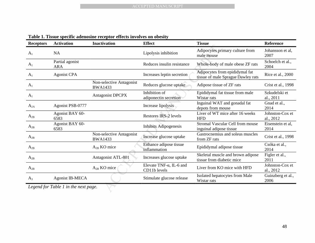
MANUSCRIP
T
ACCEPTED
ACCEPTED MANUSCRIPT
48
Table 1. Tissue specific adenosine receptor effects involves on obesity Receptors Activation Inactivation Effect Tissue Reference
A1 NA Lipolysis inhibition Adipocytes primary culture from male mouse
Johansson et al, 2007
A1 Partial agonist ARA
Reduces insulin resistance Whole-body of male obese ZF rats Schoelch et al., 2004
A1 Agonist CPA Increases leptin secretion Adipocytes from epididymal fat tissue of male Sprague Dawley rats
Rice et al., 2000
A1 Non-selective Antagonist BWA1433
Reduces glucose uptake Adipose tissue of ZF rats Crist et al., 1998
A1 Antagonist DPCPX Inhibition of adiponectin secretion
Epididymal fat tissue from male Wistar rats
Szkudelski et al., 2011
A2A Agonist PSB-0777 Increase lipolysis Inguinal WAT and gonadal fat depots from mouse
Gnad et al., 2014
A2B Agonist BAY 60-6583
Restores IRS-2 levels Liver of WT mice after 16 weeks HFD
Johnston-Cox et al., 2012
A2B Agonist BAY 60-6583
Inhibits Adipogenesis Stromal Vascular Cell from mouse inguinal adipose tissue
Eisenstein et al, 2014
A2B Non-selective Antagonist BWA1433
Increase glucose uptake Gastrocnemius and soleus muscles from ZF rats
Crist et al., 1998
A2B A2B KO mice Enhance adipose tissue inflammation
Epididymal adipose tissue Csóka et al., 2014
A2B Antagonist ATL-801 Increases glucose uptake Skeletal muscle and brown adipose tissue from diabetic mice
Figler et al., 2011
A2B A2B KO mice Elevate TNF-α, IL-6 and CD11b levels
Liver from KO mice with HFD Johnston-Cox et al., 2012
A3 Agonist IB-MECA Stimulate glucose release Isolated hepatocytes from Male Wistar rats
Guinzberg et al., 2006
Legend for Table 1 in the next page.
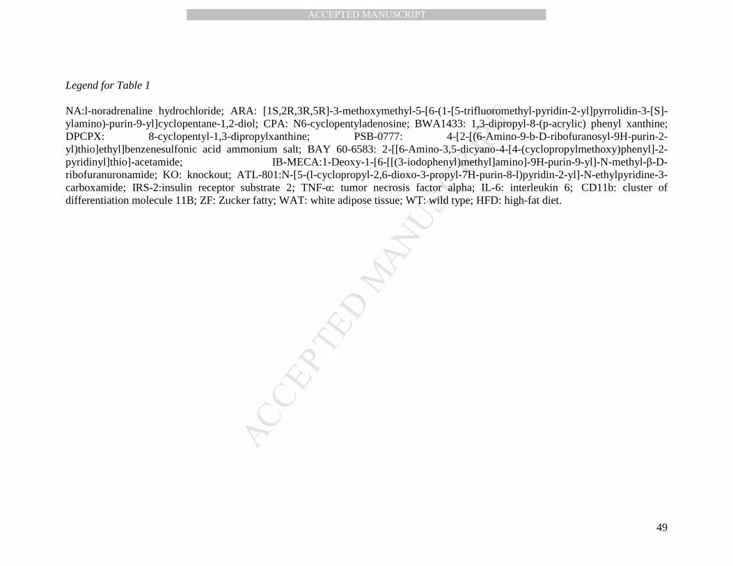
MANUSCRIP
T
ACCEPTED
ACCEPTED MANUSCRIPT
49
Legend for Table 1 NA:l-noradrenaline hydrochloride; ARA: [1S,2R,3R,5R]-3-methoxymethyl-5-[6-(1-[5-trifluoromethyl-pyridin-2-yl]pyrrolidin-3-[S]-ylamino)-purin-9-yl]cyclopentane-1,2-diol; CPA: N6-cyclopentyladenosine; BWA1433: 1,3-dipropyl-8-(p-acrylic) phenyl xanthine; DPCPX: 8-cyclopentyl-1,3-dipropylxanthine; PSB-0777: 4-[2-[(6-Amino-9-b-D-ribofuranosyl-9H-purin-2-yl)thio]ethyl]benzenesulfonic acid ammonium salt; BAY 60-6583: 2-[[6-Amino-3,5-dicyano-4-[4-(cyclopropylmethoxy)phenyl]-2-pyridinyl]thio]-acetamide; IB-MECA:1-Deoxy-1-[6-[[(3-iodophenyl)methyl]amino]-9H-purin-9-yl]-N-methyl-β-D-ribofuranuronamide; KO: knockout; ATL-801:N-[5-(l-cyclopropyl-2,6-dioxo-3-propyl-7H-purin-8-l)pyridin-2-yl]-N-ethylpyridine-3-carboxamide; IRS-2:insulin receptor substrate 2; TNF-α: tumor necrosis factor alpha; IL-6: interleukin 6; CD11b: cluster of differentiation molecule 11B; ZF: Zucker fatty; WAT: white adipose tissue; WT: wild type; HFD: high-fat diet.
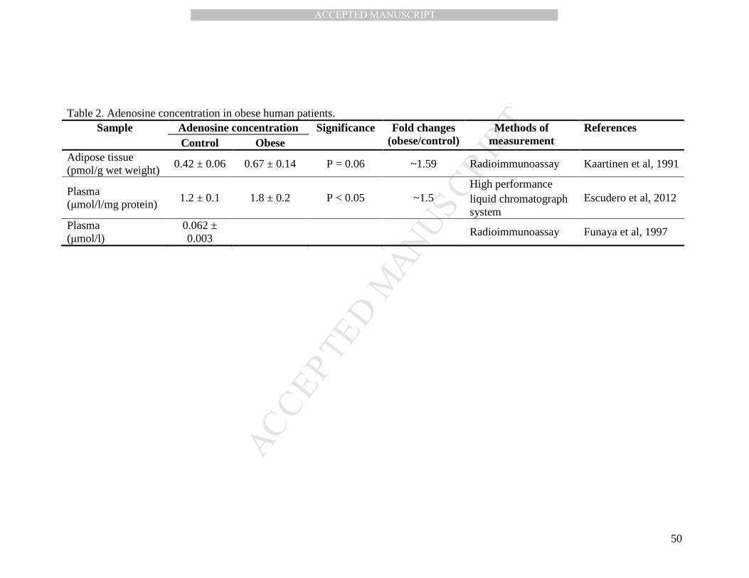
MANUSCRIP
T
ACCEPTED
ACCEPTED MANUSCRIPT
50
Table 2. Adenosine concentration in obese human patients.
Sample Adenosine concentration Significance Fold changes (obese/control)
Methods of measurement
References Control Obese
Adipose tissue (pmol/g wet weight)
0.42 ± 0.06 0.67 ± 0.14 P = 0.06 ~1.59 Radioimmunoassay Kaartinen et al, 1991
Plasma (µmol/l/mg protein)
1.2 ± 0.1 1.8 ± 0.2 P < 0.05 ~1.5 High performance liquid chromatograph system
Escudero et al, 2012
Plasma (µmol/l)
0.062 ± 0.003
Radioimmunoassay Funaya et al, 1997
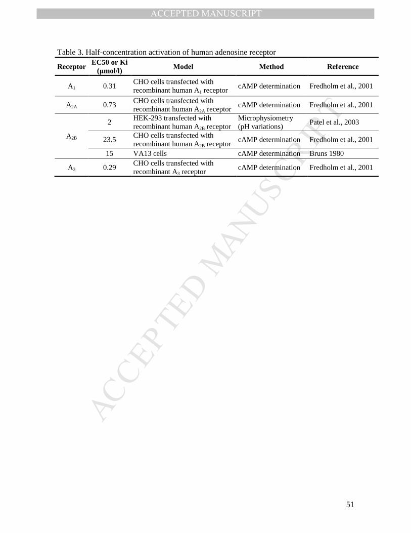
MANUSCRIP
T
ACCEPTED
ACCEPTED MANUSCRIPT
51
Table 3. Half-concentration activation of human adenosine receptor
Receptor EC50 or Ki (µmol/l) Model Method Reference
A1 0.31 CHO cells transfected with recombinant human A1 receptor
cAMP determination Fredholm et al., 2001
A2A 0.73 CHO cells transfected with recombinant human A2A receptor
cAMP determination Fredholm et al., 2001
A2B
2 HEK-293 transfected with recombinant human A2B receptor
Microphysiometry (pH variations)
Patel et al., 2003
23.5 CHO cells transfected with recombinant human A2B receptor
cAMP determination Fredholm et al., 2001
15 VA13 cells cAMP determination Bruns 1980
A3 0.29 CHO cells transfected with recombinant A3 receptor
cAMP determination Fredholm et al., 2001
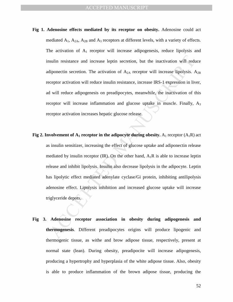
MANUSCRIP
T
ACCEPTED
ACCEPTED MANUSCRIPT
52
Fig 1. Adenosine effects mediated by its receptor on obesity. Adenosine could act
mediated A1, A2A, A2B and A3 receptors at different levels, with a variety of effects.
The activation of A1 receptor will increase adipogenesis, reduce lipolysis and
insulin resistance and increase leptin secretion, but the inactivation will reduce
adiponectin secretion. The activation of A2A receptor will increase lipolysis. A2B
receptor activation will reduce insulin resistance, increase IRS-1 expression in liver,
ad will reduce adipogenesis on preadipocytes, meanwhile, the inactivation of this
receptor will increase inflammation and glucose uptake in muscle. Finally, A3
receptor activation increases hepatic glucose release.
Fig 2. Involvement of A1 receptor in the adipocyte during obesity. A1 receptor (A1R) act
as insulin sensitizer, increasing the effect of glucose uptake and adiponectin release
mediated by insulin receptor (IR). On the other hand, A1R is able to increase leptin
release and inhibit lipolysis. Insulin also decrease lipolysis in the adipocyte. Leptin
has lipolytic effect mediated adenylate cyclase/Gi protein, inhibiting antilipolysis
adenosine effect. Lipolysis inhibition and increased glucose uptake will increase
triglyceride depots.
Fig 3. Adenosine receptor association in obesity during adipogenesis and
thermogenesis. Different preadipocytes origins will produce lipogenic and
thermogenic tissue, as withe and brow adipose tissue, respectively, present at
normal state (lean). During obesity, preadipocite will increase adipogenesis,
producing a hypertrophy and hyperplasia of the white adipose tissue. Also, obesity
is able to produce inflammation of the brown adipose tissue, producing the
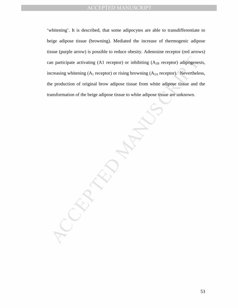
MANUSCRIP
T
ACCEPTED
ACCEPTED MANUSCRIPT
53
‘whitening’. It is described, that some adipocytes are able to transdifferentiate to
beige adipose tissue (browning). Mediated the increase of thermogenic adipose
tissue (purple arrow) is possible to reduce obesity. Adenosine receptor (red arrows)
can participate activating (A1 receptor) or inhibiting (A2B receptor) adipogenesis,
increasing whitening (A1 receptor) or rising browning (A2A receptor). Nevertheless,
the production of original brow adipose tissue from white adipose tissue and the
transformation of the beige adipose tissue to white adipose tissue are unknown.
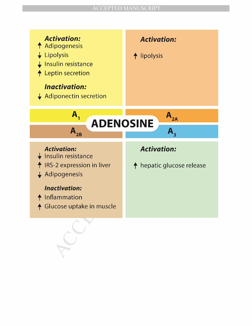
MANUSCRIP
T
ACCEPTED
ACCEPTED MANUSCRIPT
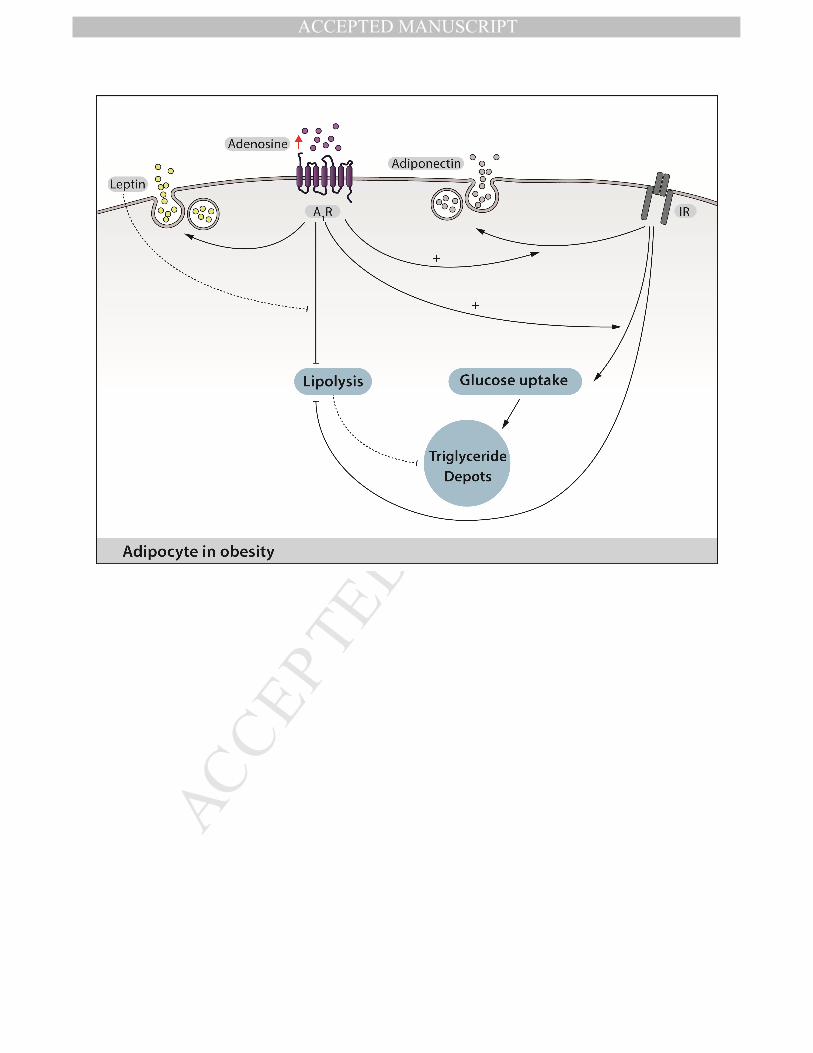
MANUSCRIP
T
ACCEPTED
ACCEPTED MANUSCRIPT
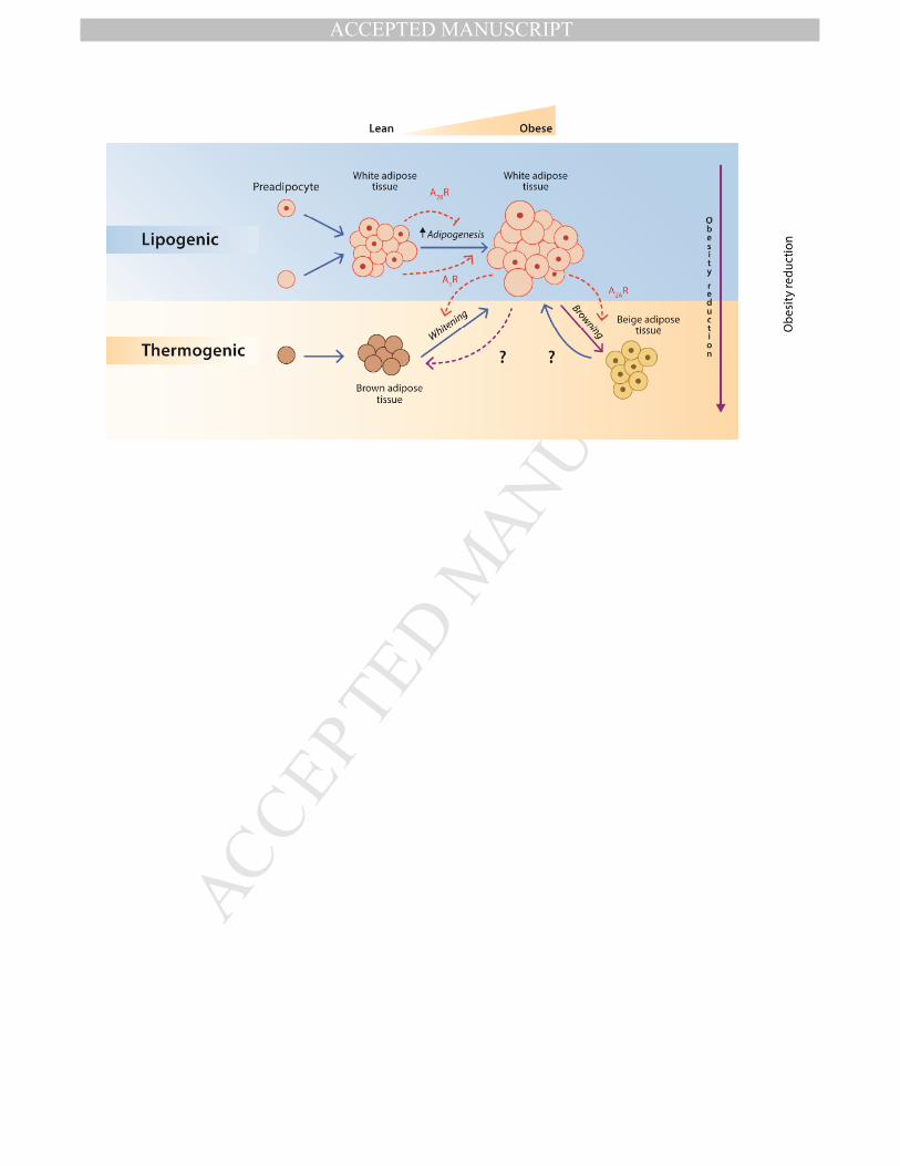
MANUSCRIP
T
ACCEPTED
ACCEPTED MANUSCRIPT