Molecular dynamics simulation study of valyl-tRNA synthetase...
10
Molecular dynamics simulation study of valyl-tRNA synthetase with its pre- and post-transfer editing substrates Nagakumar Bharatham 1 , Kavitha Bharatham 2 , Yuno Lee, Keun Woo Lee ⁎ Division of Applied Life Science (BK21 Program), Environmental Biotechnology National Core Research Center (EB-NCRC), Plant Molecular Biology and Biotechnology Research Center (PMBBRC), Gyeongsang National University, Jinju 660-701, Republic of Korea abstract article info Article history: Received 25 January 2009 Received in revised form 22 March 2009 Accepted 22 March 2009 Available online 28 March 2009 Keywords: Valyl-tRNA synthetase Molecular dynamics simulation Pre-transfer editing Post-transfer editing GOLD molecular docking Mutational studies The main role of aminoacyl-tRNA synthetases (aaRSs) is to transfer the cognate amino acids to the 3′-end of their tRNA by strictly discriminating from non-cognate amino acids. Some aaRSs accomplish this via proofreading and editing mechanisms, among which valyl-tRNA synthetase (ValRS) hydrolyses the non- cognate amino acid, threonine. In ValRS, existence of pre-transfer editing process is still unclear, although crystal structure of editing site with pre-transfer substrate analog (Thr-AMS) was released. In the case of isoleucyl-tRNA synthetase (IleRS), editing mechanism is well studied and mutational analyses revealed the existence of post- and pre-transfer editing mechanisms. Our aim is to investigate the possibility of pre- transfer editing process by performing molecular dynamics (MD) simulation studies. Simulations were carried out for ValRS with pre-transfer substrates (Thr-AMP/Val-AMP) and post-transfer substrates (Thr- A76/Val-A76) to understand their binding pattern. Two important point mutation studies were performed to observe their effect on editing process. This study also intends to compare and contrast the pre-transfer editing with post-transfer editing of ValRS. Interestingly, the MD simulation results revealed that non- cognate substrates (Thr-AMP/Thr-A76) bind more strongly than the cognate substrates (Val-AMP/Val-A76) in both pre- and post-transfer editing respectively. The editing site mutations (Lys270Ala and Asp279Ala) severely affected the binding ability of pre-transfer substrate (Thr-AMP) by different ways. Even though pre- and post-transfer substrates bind to the same site, specific differences were observed which has led us to believe the existence of the pre-transfer editing process in ValRS. © 2009 Elsevier B.V. All rights reserved. 1. Introduction The fidelity of protein synthesis is guaranteed by the specific recognition of amino acids and tRNAs by aminoacyl-tRNA synthe- tases (aaRSs) [1–3]. The aaRSs catalyze the specific attachment of their cognate amino acid to the 3′ end of their cognate tRNA. The aminoacylation of tRNA proceeds in two steps: first, the synthesis of an aminoacyl-adenylate, as an active intermediate, from the amino acid and adenosine tri-phosphate (ATP), and second the transfer of the aminoacyl moiety from the adenylate to the 3′-terminal adenosine of tRNA [4]. However, the affinity difference is not large enough for the enzyme to discriminate strictly between similar and non-cognate amino acids from the cognate one [5]. Some aaRSs must discriminate their cognate amino acid from non-cognate ones. For example, isoleucyl-tRNA synthetase (IleRS) should discriminate the cognate isoleucine from the non-cognate valine, which differs by only one methylene group [6–8]. Valyl-tRNA synthetase (ValRS) should discriminate valine from threonine, which has a quite similar shape and size as valine with a hydroxyl group in its side chain instead of a methyl group [9]. Other aaRSs, such as the leucyl-, threonyl- and alanyl-tRNA synthetases also have editing activities [10–12]. To maintain high translational fidelity, these aaRSs catalyze proofreading (editing) reactions in which the mis-products are hydrolyzed. This editing reaction is carried out in the domain named as editing (or CP1) domain which is located more than 30 Å away from the aminoacylation site [13–22]. The editing reaction of ValRS comprises of two pathways depicted as follows [19]: Biophysical Chemistry 143 (2009) 34–43 ⁎ Corresponding author. E-mail address: [email protected] (K. Woo Lee). 1 Current address: Division of Structural and Computational Biology, School of Biological Sciences, Nanyang Technological University,637551, Singapore. 2 Current address: Biomolecular Modeling and Design Division, Bioinformatics Institute, 30 Biopolis Street, #07-01 Matrix,138671, Singapore. 0301-4622/$ – see front matter © 2009 Elsevier B.V. All rights reserved. doi:10.1016/j.bpc.2009.03.009 Contents lists available at ScienceDirect Biophysical Chemistry journal homepage: http://www.elsevier.com/locate/biophyschem
Transcript of Molecular dynamics simulation study of valyl-tRNA synthetase...

Biophysical Chemistry 143 (2009) 34–43
Contents lists available at ScienceDirect
Biophysical Chemistry
j ourna l homepage: ht tp : / /www.e lsev ie r.com/ locate /b iophyschem
Molecular dynamics simulation study of valyl-tRNA synthetase with its pre- andpost-transfer editing substrates
Nagakumar Bharatham 1, Kavitha Bharatham 2, Yuno Lee, Keun Woo Lee ⁎Division of Applied Life Science (BK21 Program), Environmental Biotechnology National Core Research Center (EB-NCRC),Plant Molecular Biology and Biotechnology Research Center (PMBBRC), Gyeongsang National University, Jinju 660-701, Republic of Korea
⁎ Corresponding author.E-mail address: [email protected] (K. Woo Lee).
1 Current address: Division of Structural and ComBiological Sciences, Nanyang Technological University, 6
2 Current address: Biomolecular Modeling and DeInstitute, 30 Biopolis Street, #07-01 Matrix, 138671, Sing
0301-4622/$ – see front matter © 2009 Elsevier B.V. Adoi:10.1016/j.bpc.2009.03.009
a b s t r a c t
a r t i c l e i n f oArticle history:Received 25 January 2009Received in revised form 22 March 2009Accepted 22 March 2009Available online 28 March 2009
Keywords:Valyl-tRNA synthetaseMolecular dynamics simulationPre-transfer editingPost-transfer editingGOLD molecular dockingMutational studies
The main role of aminoacyl-tRNA synthetases (aaRSs) is to transfer the cognate amino acids to the 3′-end oftheir tRNA by strictly discriminating from non-cognate amino acids. Some aaRSs accomplish this viaproofreading and editing mechanisms, among which valyl-tRNA synthetase (ValRS) hydrolyses the non-cognate amino acid, threonine. In ValRS, existence of pre-transfer editing process is still unclear, althoughcrystal structure of editing site with pre-transfer substrate analog (Thr-AMS) was released. In the case ofisoleucyl-tRNA synthetase (IleRS), editing mechanism is well studied and mutational analyses revealed theexistence of post- and pre-transfer editing mechanisms. Our aim is to investigate the possibility of pre-transfer editing process by performing molecular dynamics (MD) simulation studies. Simulations werecarried out for ValRS with pre-transfer substrates (Thr-AMP/Val-AMP) and post-transfer substrates (Thr-A76/Val-A76) to understand their binding pattern. Two important point mutation studies were performed toobserve their effect on editing process. This study also intends to compare and contrast the pre-transferediting with post-transfer editing of ValRS. Interestingly, the MD simulation results revealed that non-cognate substrates (Thr-AMP/Thr-A76) bind more strongly than the cognate substrates (Val-AMP/Val-A76)in both pre- and post-transfer editing respectively. The editing site mutations (Lys270Ala and Asp279Ala)severely affected the binding ability of pre-transfer substrate (Thr-AMP) by different ways. Even though pre-and post-transfer substrates bind to the same site, specific differences were observed which has led us tobelieve the existence of the pre-transfer editing process in ValRS.
© 2009 Elsevier B.V. All rights reserved.
1. Introduction
The fidelity of protein synthesis is guaranteed by the specificrecognition of amino acids and tRNAs by aminoacyl-tRNA synthe-tases (aaRSs) [1–3]. The aaRSs catalyze the specific attachment oftheir cognate amino acid to the 3′ end of their cognate tRNA. Theaminoacylation of tRNA proceeds in two steps: first, the synthesis ofan aminoacyl-adenylate, as an active intermediate, from the aminoacid and adenosine tri-phosphate (ATP), and second the transfer ofthe aminoacyl moiety from the adenylate to the 3′-terminaladenosine of tRNA [4]. However, the affinity difference is not largeenough for the enzyme to discriminate strictly between similar andnon-cognate amino acids from the cognate one [5]. Some aaRSsmust discriminate their cognate amino acid from non-cognate ones.
putational Biology, School of37551, Singapore.sign Division, Bioinformaticsapore.
ll rights reserved.
For example, isoleucyl-tRNA synthetase (IleRS) should discriminatethe cognate isoleucine from the non-cognate valine, which differs byonly one methylene group [6–8]. Valyl-tRNA synthetase (ValRS)should discriminate valine from threonine, which has a quite similarshape and size as valine with a hydroxyl group in its side chaininstead of a methyl group [9]. Other aaRSs, such as the leucyl-,threonyl- and alanyl-tRNA synthetases also have editing activities[10–12].
To maintain high translational fidelity, these aaRSs catalyzeproofreading (editing) reactions in which the mis-products arehydrolyzed. This editing reaction is carried out in the domainnamed as editing (or CP1) domain which is located more than 30 Åaway from the aminoacylation site [13–22].
The editing reaction of ValRS comprises of two pathways depictedas follows [19]:
35N. Bharatham et al. / Biophysical Chemistry 143 (2009) 34–43
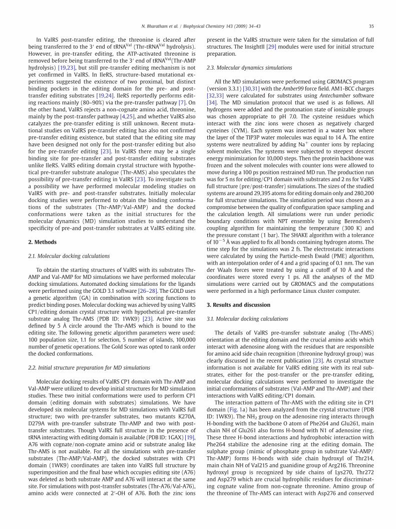
In ValRS post-transfer editing, the threonine is cleared afterbeing transferred to the 3′ end of tRNAVal (Thr-tRNAVal hydrolysis).However, in pre-transfer editing, the ATP-activated threonine isremoved before being transferred to the 3′ end of tRNAVal(Thr-AMPhydrolysis) [19,23], but still pre-transfer editing mechanism is notyet confirmed in ValRS. In IleRS, structure-based mutational ex-periments suggested the existence of two proximal, but distinctbinding pockets in the editing domain for the pre- and post-transfer editing substrates [19,24]. IleRS reportedly performs edit-ing reactions mainly (80–90%) via the pre-transfer pathway [7]. Onthe other hand, ValRS rejects a non-cognate amino acid, threonine,mainly by the post-transfer pathway [4,25], and whether ValRS alsocatalyzes the pre-transfer editing is still unknown. Recent muta-tional studies on ValRS pre-transfer editing has also not confirmedpre-transfer editing existence, but stated that the editing site mayhave been designed not only for the post-transfer editing but alsofor the pre-transfer editing [23]. In ValRS there may be a singlebinding site for pre-transfer and post-transfer editing substratesunlike IleRS. ValRS editing domain crystal structure with hypothe-tical pre-transfer substrate analogue (Thr-AMS) also speculates thepossibility of pre-transfer editing in ValRS [23]. To investigate sucha possibility we have performed molecular modeling studies onValRS with pre- and post-transfer substrates. Initially moleculardocking studies were performed to obtain the binding conforma-tions of the substrates (Thr-AMP/Val-AMP) and the dockedconformations were taken as the initial structures for themolecular dynamics (MD) simulation studies to understand thespecificity of pre-and post-transfer substrates at ValRS editing site.
2. Methods
2.1. Molecular docking calculations
To obtain the starting structures of ValRS with its substrates Thr-AMP and Val-AMP for MD simulations we have performed moleculardocking simulations. Automated docking simulations for the ligandswere performed using the GOLD 3.1 software [26–28]. The GOLD usesa genetic algorithm (GA) in combination with scoring functions topredict binding poses. Molecular docking was achieved by using ValRSCP1/editing domain crystal structure with hypothetical pre-transfersubstrate analog Thr-AMS (PDB ID: 1WK9) [23]. Active site wasdefined by 5 Å circle around the Thr-AMS which is bound to theediting site. The following genetic algorithm parameters were used:100 population size, 1.1 for selection, 5 number of islands, 100,000number of genetic operations. The Gold Score was opted to rank orderthe docked conformations.
2.2. Initial structure preparation for MD simulations
Molecular docking results of ValRS CP1 domain with Thr-AMP andVal-AMP were utilized to develop initial structures for MD simulationstudies. These two initial conformations were used to perform CP1domain (editing domain with substrates) simulations. We havedeveloped six molecular systems for MD simulations with ValRS fullstructure; two with pre-transfer substrates, two mutants K270A,D279A with pre-transfer substrate Thr-AMP and two with post-transfer substrates. Though ValRS full structure in the presence oftRNA interacting with editing domain is available (PDB ID: 1GAX) [19],A76 with cognate/non-cognate amino acid or substrate analog likeThr-AMS is not available. For all the simulations with pre-transfersubstrates (Thr-AMP/Val-AMP), the docked substrates with CP1domain (1WK9) coordinates are taken into ValRS full structure bysuperimposition and the final base which occupies editing site (A76)was deleted as both substrate AMP and A76 will interact at the samesite. For simulations with post-transfer substrates (Thr-A76/Val-A76),amino acids were connected at 2′-OH of A76. Both the zinc ions
present in the ValRS structure were taken for the simulation of fullstructures. The InsightII [29] modules were used for initial structurepreparation.
2.3. Molecular dynamics simulations
All the MD simulations were performed using GROMACS program(version 3.3.1) [30,31] with the Amber99 force field. AM1-BCC charges[32,33] were calculated for substrates using Antechamber software[34]. The MD simulation protocol that we used is as follows. Allhydrogens were added and the protonation state of ionizable groupswas chosen appropriate to pH 7.0. The cysteine residues whichinteract with the zinc ions were chosen as negatively chargedcysteines (CYM). Each system was inserted in a water box wherethe layer of the TIP3P water molecules was equal to 14 Å. The entiresystems were neutralized by adding Na+ counter ions by replacingsolvent molecules. The systems were subjected to steepest descentenergy minimization for 10,000 steps. Then the protein backbone wasfrozen and the solvent molecules with counter ions were allowed tomove during a 100 ps position restrained MD run. The production runwas for 5 ns for editing/CP1 domainwith substrates and 2 ns for ValRSfull structure (pre/post-transfer) simulations. The sizes of the studiedsystems are around 29,395 atoms for editing domain only and 280,200for full structure simulations. The simulation period was chosen as acompromise between the quality of configuration space sampling andthe calculation length. All simulations were run under periodicboundary conditions with NPT ensemble by using Berendsen'scoupling algorithm for maintaining the temperature (300 K) andthe pressure constant (1 bar). The SHAKE algorithm with a toleranceof 10−5 Å was applied to fix all bonds containing hydrogen atoms. Thetime step for the simulations was 2 fs. The electrostatic interactionswere calculated by using the Particle-mesh Ewald (PME) algorithm,with an interpolation order of 4 and a grid spacing of 0.1 nm. The vander Waals forces were treated by using a cutoff of 10 Å and thecoordinates were stored every 1 ps. All the analyses of the MDsimulations were carried out by GROMACS and the computationswere performed in a high performance Linux cluster computer.
3. Results and discussion
3.1. Molecular docking calculations
The details of ValRS pre-transfer substrate analog (Thr-AMS)orientation at the editing domain and the crucial amino acids whichinteract with adenosine along with the residues that are responsiblefor amino acid side chain recognition (threonine hydroxyl group) wasclearly discussed in the recent publication [23]. As crystal structureinformation is not available for ValRS editing site with its real sub-strates, either for the post-transfer or the pre-transfer editing,molecular docking calculations were performed to investigate theinitial conformations of substrates (Val-AMP and Thr-AMP) and theirinteractions with ValRS editing/CP1 domain.
The interaction pattern of Thr-AMS with the editing site in CP1domain (Fig. 1a) has been analyzed from the crystal structure (PDBID: 1WK9). The NH2 group on the adenosine ring interacts throughH-bonding with the backbone O atom of Phe264 and Glu261, mainchain NH of Glu261 also forms H-bond with N1 of adenosine ring.These three H-bond interactions and hydrophobic interaction withPhe264 stabilize the adenosine ring at the editing domain. Thesulphate group (mimic of phosphate group in substrate Val-AMP/Thr-AMP) forms H-bonds with side chain hydroxyl of Thr214,main chain NH of Val215 and guanidine group of Arg216. Threoninehydroxyl group is recognized by side chains of Lys270, Thr272and Asp279 which are crucial hydrophilic residues for discriminat-ing cognate valine from non-cognate threonine. Amino group ofthe threonine of Thr-AMS can interact with Asp276 and conserved

Table 1Information of molecular dynamics simulations.
Simulationno.
Type of simulation Type ofsubstrate
Size of the system(totalno. of atoms)
Simulationtime (ps)
1 Pre-transfer (editingdomain alone)
Thr-AMP ~29,400 5000
2 Pre-transfer (editingdomain alone)
Val-AMP ~29,400 5000
3 Pre-transfer (ValRS fullstructure)
Thr-AMP ~280,200 2000
4 Pre-transfer (ValRS fullstructure)
Val-AMP ~280,200 2000
5 Pre-transfer (ValRS fullstructure with Lys270Alamutation)
Thr-AMP ~280,200 2000
6 Pre-transfer (ValRS fullstructure with Asp279Alamutation)
Thr-AMP ~280,200 2000
7 Post-transfer (ValRS fullstructure)
Thr-A76 ~280,200 2000
8 Post-transfer (ValRS fullstructure)
Val-A76 ~280,200 2000
Fig. 1. Crystal structure of ValRS editing domain with Thr-AMS (a) and the docked conformation of Thr-AMP (b) and Val-AMP (c). The AdnBP residues were shown around theThr-AMP (d). The ValRS residues interacting with the substrates are shown in stick model.
36 N. Bharatham et al. / Biophysical Chemistry 143 (2009) 34–43
Asp279 side chains. For convenience, hereafter we mentioned“adenosine binding pocket” represented by residues Thr214,Val215, Arg216, Glu261, and Phe264 that interact with adenosinering and phosphate/sulphate as “AdnBP”; and “amino acid bindingpocket” representing Lys270, Thr272, Asp276, and Asp279 residuesthat interact with threonine hydroxyl group and amino group as“AABP”.
After molecular docking simulations, the docked conformations ofthe substrates, Thr-AMP and Val-AMPwere selected by comparing theresultant conformations with the crystal structure and by consideringoverall docking score, Gold Score. The final conformations are similarto the Thr-AMS conformation in the crystal structure. The dockedconformation of Thr-AMP at ValRS CP1 domain demonstrates all thecrucial interactions with the AdnBP and AABP similar to the Thr-AMS(Fig. 1b). The Thr-AMP forms H-bonds with the hydrophilic aminoacids present in the AABP (Fig.1d). However, the Val-AMP cannot formany interactions with the AABP although it binds in a similar mannerwith theAdnBP (Fig.1c) due to lack of hydrophilic characteristics of theside chain. These docked conformations were used for MD simulationstudies for pre- and post-transfer editing reactions.
3.2. Pre-transfer editing in CP1 domain structure
Two 5 ns MD simulations were performed to understand thebinding pattern of the substrates (cognate/non-cognate amino acids)
to ValRS editing site. The details of the MD simulation parameters andthe size of the system are listed in Table 1. The MD simulations ofCP1 domain (195–337) with its pre-transfer substrates, Val-AMP and

37N. Bharatham et al. / Biophysical Chemistry 143 (2009) 34–43
Thr-AMP were performed as valine is the cognate amino acid and thethreonine is the non-cognate one. The backbone root mean squaredeviation (RMSD) of the CP1 domain in the presence of Thr-AMP/Val-AMP with respect to the initial structure was analyzed (Fig. 2a). Theaverage RMSDs for Thr-AMP and Val-AMP from 1001 ps to finalsnapshot are 0.127 nm and 0.133 nm, respectively. Both the figures aresmall enough to ensure that the two systems were very stable duringthe simulation time. Hereafter the term ‘average’ refers to theevaluation through 1001 ps to the final snapshot. The trajectorystability for total 5 ns simulation was monitored and confirmed to bestable by inspecting backbone RMSD plots for both the systems.
The binding ability of pre-transfer substrates with the editing sitewas analyzed by counting the number of intermolecular H-bonds(Fig. 2b) as well as by comparing two different energy functions(Fig. 2c and d). Number of H-bonds between ValRS editing site and thesubstrates were measured and the results showed that Thr-AMPformed high number of H-bonds which were maintained stably. Onthe other hand its cognate substrate (Val-AMP) formed less number ofH-bonds initially and they decreased gradually (Fig. 2b). The averagenumber of the intermolecular H-bonds for Thr-AMP and Val-AMP are9.81 and 5.03, respectively. To elucidate in-depth binding ability ofsubstrates with CP1 domain, the short range (SR) columbic and vander Waals (vdw) interaction energies were calculated and displayed(Fig. 2c and d). The SR columbic interaction energy between Thr-AMPand ValRS CP1 domain is comparatively less than Val-AMP as Thr-AMPforms stable hydrophilic interactions than Val-AMP. The columbicinteraction between Thr-AMP and CP1 domain increased slightly ataround 1 ns and thereafter stabilized throughout the 5 ns simulation
Fig. 2. The RMSD (a), the number of intermolecular H-bonds (b), short range columbic interawith Thr-AMP and Val-AMP for 5 ns MD simulation.
with an average of −462.85 kJ/mol. In the case of Val-AMP, sig-nificant increment of columbic interaction energy with an averageof −269.12 kJ/mol was observed from the starting of the simulation(Fig. 2c) but the vdw interaction was relatively stable (Fig. 2d). Weassume that the ValRS editing site is mainly comprised of hydrophilicresidues which are specifically designed for its non-cognate aminoacid, threonine. Therefore, it will be more meaningful to study thecolumbic energy rather than vdw energy between ValRS CP1 domainand its substrates. Conformational changes were observed at GTG loop(Gly265, Thr266 and Gly267), in the presence of Val-AMP whichaffects the interaction between Phe264 CO and NH2 of adenosine ring.On the contrary, the carbonyl group of Phe264 is forming stableinteraction with adenosine ring of Thr-AMP. Though these analysesclearly show the binding ability difference between the two substratesand ValRS CP1 domain, concluding the results may not be accurate atthis point as we have considered only CP1 domain. The conforma-tional changes and binding ability differences may be attributed asartifacts because CP1 domain alone was considered. Therefore, theseresults have encouraged us to perform MD simulations of ValRS fullstructure with tRNA for further verification.
3.3. Pre-transfer editing using tRNA and ValRS full structure
Six MD simulations were performed with tRNA and ValRS fullstructures, among which two (2 ns each) were performed initially inthe presence of pre-transfer editing substrates (Thr-AMP/Val-AMP).Due to the massive size of total ValRS with tRNA system (~280,000atoms) the length of simulation was chosen for 2 ns when compared
ction energy (c), and short range Leonard–Jones potentials (d) of ValRS editing domain

Table 2Occupancy rate of hydrogen bond interactions with editing domain and columbicinteraction energy.
Pre-transfer Post-transfer
Type of interaction Thr-AMP
Val-AMP Lys270Ala(Thr-AMP)
Asp279Ala(Thr-AMP)
Thr-A76
Val-A76
Phe264 main chainCO – NH of adenosine
0.998 0.461 0 0.237 0.944 0.075
38 N. Bharatham et al. / Biophysical Chemistry 143 (2009) 34–43
to the small domain simulation which was performed for 5 ns. Thebackbone RMSD of ValRS full structure with each substrate wasevaluated and it was observed that the fluctuation is comparativelyhigh in the presence of Thr-AMPwhen compared to the Val-AMPwithan average of 0.277 and 0.187 nm respectively (Fig. 3a). Though theoverall protein fluctuation is high, it is clearly evident that Thr-AMPinteracts strongly with editing domain of ValRS than Val-AMP basedon H-bond count and columbic interaction energy (Fig. 3b and c). The
Fig. 3. The RMSD (a), the number of intermolecular H-bonds (b), and short rangecolumbic interaction energy (c) of ValRS full structure with Thr-AMP and Val-AMP for2 ns MD simulation.
Glu261main chainCO – NH of adenosine
0.919 0.826 0.992 0.407 0.902 0.724
Thr214 side chainOH – PO4 group
1.0 0.487 1.0 1.0 NAa NAa
Tyr337 side chainOH – PO4 group
NAa NAa NAa NAa 0.977 0.338
Lys270 side chainNH3+ – OH of Thr
0.829 0 0 0 0.826 0
Columbic interactionenergyb
−562.5 −369.23 −439.87 −429.12 NAa NAa
Occupancy calculated from 1001 ps to end of the simulation.Highly deviatedwith reference toThr-AMP/Thr-A76 highlightedwith bold representation.
a Not applicable for particular simulation.b Average columbic interaction energy between substrate andprotein, values represented
in KJ/mol.
average number of H-bonds is 9.60 and 6.17 and the average columbicinteraction energy is −562.5 kJ/mol and −369.23 kJ/mol for Thr-AMP and Val-AMP with ValRS respectively (Table 2). The columbicinteraction energy increased constantly between ValRS CP1 domainand its own substrate (cognate), Val-AMP which is not favorable fortight binding.
The binding ability of the substrates and the conformationalchanges during the MD simulation were analyzed. Not much changewas observed in the binding conformation of Thr-AMP at editingdomain as it was well accustomed and formed stable interactions withAdnBP and AABP (Fig. 4a and 4b). The interactions of Thr-AMP withAdnBP residues were analyzed by superimposing initial and finalconformations (Fig. 4a). The two H-bonds formed by Thr214 side
Fig. 4. The initial (green) and final (cyan) frames of ValRS full structure 2 ns MDsimulation with Thr-AMP at AdnBP (a) and AABP (b). The superimposed final frames ofVal-AMP (blue) on Thr-AMP (cyan) at AdnBP (c) and AABP (d). The protein is shown asribbons; the crucial residues were shown in sticks with Thr-AMP as sticks and Val-AMPas ball and stick for clarity. (For interpretation of the references to colour in this figurelegend, the reader is referred to the web version of this article.)

Fig. 6. The superimposed final frames of WT and Lys270Ala 2 ns MD simulation withThr-AMP at AdnBP (a) and AABP (b). The superimposed final frames of WT and
39N. Bharatham et al. / Biophysical Chemistry 143 (2009) 34–43
chain OH group and Val215 main chain NH with PO4 group of the Thr-AMP are stable throughout the simulation. The Phe264 present nearGTG loop which is responsible for forming AdnBP is also well adjustedwith Thr-AMP adenosine ring. The Phe264 CO group, Glu261 mainchain carbonyl group also maintains the key H-bond interactions withNH2 of adenosine ring. The Glu261 NH group also contributes an H-bondwith N1 of adenosine ring. To determine the strength of the eachH-bond, occupancy (defined as the number of snapshots having aparticular H-bond from the total simulation snapshots) is calculated.The occupancy of five possible H-bond interactions between Thr-AMPand adenosine ring was 4.82 (Table 2). In a similar manner, Thr-AMPinteracts strongly with AABP (Lys270, Thr272, Asp276 and Asp279).The residues Thr219 and Asp223 are also represented in figures asthey form key interactions with other AABP residues like Lys270 andThr272 to maintain the arrangement of the pocket. The side chains ofLys270, Asp279 form stable interactions with hydroxyl group of Thr-AMP. At the same time Asp279 and Asp276 side chain carboxyl groupsform H-bond with α-NH3
+ of Thr-AMP (Fig. 4b). According to theprevious study, Thr272 side chain is speculated to form another H-bond with OH group of Thr-AMP [23]. In the present study Thr272interacts less often with OH of Thr-AMP but maintains strong H-bondinteraction with Asp223 residue. This result coincides with theexperimental mutational studies where mutation of Thr272 has lesseffect on editing activity than Lys270 and Asp279.
Binding ability of Val-AMP with ValRS full structure was analyzedand it was observed that significant conformational changes were
Fig. 5. The number of intermolecular H-bonds (a) short range columbic interactionenergy (b) of Wild type ValRS full structure, Lys270Ala mutant and Asp279Ala mutantwith Thr-AMP for 2 ns MD simulation.
Asp279Ala 2 ns MD simulation with Thr-AMP at AdnBP (a) and AABP (b).The protein isshown as ribbons, the crucial residues were shown in sticks with Thr-AMP as sticks.
observed both in the AdnBP and AABP (Fig. 4c and d) unlike Thr-AMP.Although much fluctuation was not observed at GTG loop, crucial H-bond network was diminished between the NH2 of adenosine ring andGTG loop amino acid residues like Phe264 and Glu261. The H-bondinteractions at the AdnBP were effected as analyzed by measuringtheir occupancy (Table 2). The residues, Thr214 and Val215 whichinteract with PO4 group of Thr-AMP through H-bonding are not able tomaintain steady interactions with Val-AMP. Both the residues moveaway from the PO4 group which causes loss of interaction (occupancywas 0.487 and 0.792 respectively, but in the case Thr-AMP it was 1.0and 0.935). These results clearly showed that the binding ability ofVal-AMP is fragile than the Thr-AMP at the AdnBP. Two residuesAsp276, Asp279 formed H-bond interactions with α-NH3+ group ofVal-AMP at AABP. These results are also comparable with the CP1domain MD simulation results (Fig. 2) and therefore gave confidencethat 2 ns simulation of ValRS full structure with tRNA is sufficient toobserve the important changes at the CP1 domain and the bindingability of substrates. Based on these results it can be established thatnon-cognate substrate (Thr-AMP) binds strongly at editing site andundergo hydrolysis, but Val-AMP interacts feebly with editing site anddepart along with tRNA.
3.4. Mutational studies
Two most crucial residues present in the AABP (Lys270 andAsp279) were mutated to alanine experimentally and their resultsdisclosed that editing site is not able to recognize the threonine sidechain thereby finally losing its editing ability [23]. In order to observethese mutational effects on pre-transfer editing substrate (Thr-AMP),we have considered two point mutated systems (Lys270Ala,Asp279Ala) and performed 2 ns MD simulations each.
3.5. Lys270 mutation
The effect of Lys270Ala mutation on Thr-AMP binding abilitywas analyzed by intermolecular H-bond count and columbic inter-action energy (Fig. 5a and b) resulting in an average of 8.16 (9.60

Fig. 8. The superimposed structures of initial (pale yellow), final (yellow) along withintermediate structures like 20 ps 60 ps and 100 ps snapshots to observe the ori-entational change of Thr-AMP in Asp279Ala mutation MD simulation. (For inter-pretation of the references to colour in this figure legend, the reader is referred to theweb version of this article.)
40 N. Bharatham et al. / Biophysical Chemistry 143 (2009) 34–43
for wild type) and −439.87 kJ/mol (−562.5 kJ/mol for wild type)respectively. These two analyses together reveal that Lys270mutation has a negative effect on the binding ability of Thr-AMPwith the editing domain of ValRS. Fig. 6a illustrates the bindingpattern of Thr-AMP with AdnBP of Lys270Ala ValRS mutant. The GTGloop which is crucial for adenosine ring binding is affected in such away that the interaction between carbonyl group of Phe264 and NH2
group of adenosine ring is completely vanished. The occupancy ofPhe264 CO and adenosine NH2 H-bonding was zero in stark contrastwith wild type which has 0.998. Although the AABP residue, Lys270was mutated not much effect was observed on amino acid side chainposition of Thr-AMP. It formed all other interactions with Asp276and Asp279 residues reasonably.
The main roles of Lys270 side chain is attributed to the re-cognition of Thr-AMP side chain along with maintaining keyinteraction network with residues Thr219 and Asp223 which mayfurther help in the maintenance of AABP size. Effect of Lys270Alawas clearly observed on the secondary structural features. Themutation has directly affected the GTG loop, as it leads into thebeta sheet containing Lys270. Due to this effect the Phe264carbonyl group has turned away and lost the H-bond interactionwith the adenosine NH2 group. Mutation has been affectedindirectly by losing the interaction with Asp223 and thereby lo-sing secondary structural features from 228–257 residues (Fig. 7b).In the case of wild type and Asp279Ala mutant, this region is stableand maintains all the secondary structural features throughout the2 ns simulations (Fig. 7a and c) unlike Lys270 mutant. The Lys270in wild type forms key interactions with Thr219 but upon mutationto alanine the Arg216 side chain has moved away from AABP thuslosing crucial hydrophobic interaction with methyl group of Thr-AMP. The Lys270Ala mutant has been affected directly and in-directly thereby leading to the loss of recognition of its cognateamino acid.
Fig. 7. The final frames of Wild type (a) Lys270Ala (b) Asp279Ala (c) full structure2 ns MD simulation with certain residues shown in sticks and the substrate shown inlines. The region having observable secondary structure changes has been high-lighted by pale colours in respective structures. The superimposed final framestructures of Wild type (cyan), Lys270Ala (magenta), and Asp279Ala (yellow) tohighlight the dislocation of Thr-AMP from the AABP caused by Asp279 mutation. (Forinterpretation of the references to colour in this figure legend, the reader is referredto the web version of this article.)
3.6. Asp279 mutation
Asp279 present at the editing site is well conserved among all thethree enzymes (ValRS, LeuRS and IleRS) inmost of the organisms. In thecase of T. thermophilus LeuRS (Asp347), IleRS (Asp328) H-bonds wereformed with the α-NH3+ which seem to play a vital role in the editingreaction. Mutation of the Asp328Ala in T. thermophilus IleRS is defectivein the total editing activity [18,35]. In a similar manner Asp279Alamutation in ValRS also demonstrated severe effect on total editingprocess (pre-transfer, if exists and post-transfer editing) [23]. In the caseof ValRS, Asp279 forms H-bond with α-NH3+ and also recognizes theside chain hydroxyl group of threonine by forming another H-bond.
The average number of H-bonds and the average columbicinteraction energy were 8.63 (9.60 for wild type) and −429.12 kJ/mol(−562.5 kJ/mol for wild type) between Thr-AMP and Asp279 mutatedValRS. It is evident that the number of H-bondswas only 6 to 7 until 1 nsthereafter increasing and stabilizing around 8 to 9 between Thr-AMPand mutant ValRS (Fig. 5a). The columbic interaction energy alsorevealed a similar trend of results. The binding pattern of Thr-AMPwithAdnBPof Asp279mutant is illustrated in Fig. 6c. Themutational effect onGTG loopwas similar to that of Lys270mutation and also towild type inthe presence of Val-AMP substrate. The crucial H-bond betweencarbonyl group of Phe264 and NH2 of Thr-AMP adenosine ring formedoccasionally with an occupancy of 0.237 (0.998 for wild type). Anotherimportant H-bond between the carbonyl group of Glu261 and NH2 ofThr-AMPadenosine ringwas also affected severelywith anoccupancyof0.407 (0.919 for wild type). The mutational effect on Thr-AMP bindingand threonine side chain recognitionwas clearly observed in the case ofAsp279Ala mutation (Fig. 6c and d). The threonine side chain of Thr-AMPwas completely dislocated fromtheAABPwhich is not desirable forediting reaction (Fig. 6d). This dislocation is especially observed inAsp279 mutation case, while in the Lys270 case we have not observedmuch change in threonine side chain (OH group) position and is verysimilarly oriented as in wild type simulation (Fig. 7d). The super-imposition of a few selectedMDsimulation frames (0, 20, 60, and 100 pssimulation time) until 100 ps revealed the dislocation process ofthreonine side chain from the AABP and after 100 ps not many changeswere observed in threonine side chain position (Fig. 8). This can be

Fig. 9. Post-transfer MD simulation results. The superimposed initial (peach) andfinal (orange) snapshots of ValRS full structure with Thr-A76 at AdnBP (a) AABP (b).The superimposed final frames of Val-A76 (cement) and Thr-AMP (orange) snap-shots of ValRS full structure at AdnBP (c) AABP (d). The protein is shown as ribbons,the crucial residues were shown in sticks with Thr-A76 as sticks and Val-A76 as balland stick for clarity. (For interpretation of the references to colour in this figurelegend, the reader is referred to the web version of this article.)
41N. Bharatham et al. / Biophysical Chemistry 143 (2009) 34–43
further witnessed by superimposing simulation conformations (Fig. S1)at 0, 800, 1600 and 2000 ps.
Since the threonine side chain dislocated from the AABP, the crucialH-bond interaction between Lys270 side chain and OH group of Thr-AMPwas completelymissing (occupancywas zero). On the other handthe hydrophobic interaction between Arg216 side chain and threonineside chainwas also affected. As amino acid side chain dislocated, mainchain carbonyl group of Lys270 formed H-bond with hydroxyl group(amino acid side chain) of Thr-AMP. The Arg216 side chain movedinside the cavity due to a conformational change in the substratebinding and formed one to two H-bonds with PO4 of Thr-AMP.Although Thr-AMP preserved the binding ability by forming H-bondswith these residues, the editing ability of ValRS have been lost becauseThr-AMP dislocated from the AABP. The other H-bond interactionbetweenα-NH3+groupof Thr-AMPand carboxyl groupof Asp276wasmaintainedwell throughout the simulation. This reveals that although
Fig. 10. The superimposed final frames of post-transfer (orange) and pre-transfer(cyan) snapshots of ValRS full structure at AdnBP (a) AABP (b). (For interpretation ofthe references to colour in this figure legend, the reader is referred to the web versionof this article.)
Asp276 can formH-bondwithα-NH3+ group, it is unable to resist thedislocation of amino acid side chain from the AABP. This demonstratesthat Asp276 has not much importance in the editing process (pre-transfer) though it forms direct interaction with Thr-AMP. As wealready know that Asp328Ala mutation in IleRS severely affects theediting process as it interacts with α-NH3+ group. Most probably asimilar type of side chain dislocation may occur from the AABP.
3.7. Post-transfer editing using tRNA and ValRS full structure
Two different 2 ns simulations were carried out to demon-strate the binding properties of post-transfer substrates (Thr-A76/Val-A76). The simulation stability was analyzed by backboneRMSD and total energy analyses. The average RMSD was 0.211and 0.239 nm for Thr-A76 and Val-A76 respectively stabilizingsoon after 500 ps (Fig. S2a). The total energy analyses also revealedthat both the simulations were well stabilized without anydeviations. The binding ability of post-transfer substrates wasanalyzed by the number of intermolecular H-bonds, whichresulted in an average of 7.54 and 4.41 for Thr-A76 and Val-A76with ValRS respectively (Fig. S2b). From the H-bond count analysesit is evident that Thr-A76 interacts with CP1 domain tightlycompared with Val-A76 indicating that in both pre- and post-transfer process, the non-cognate substrates bind strongly.
Fig. 11. The backbone structures of the extreme frames obtained from ED analysesresults for 2 ns MD simulation of ValRS full structure with Thr-A76 (a) and Thr-AMP(b). The C-alpha distance measured between the two extreme frames of ED analyses(c) for post- and pre-transfer simulations.

42 N. Bharatham et al. / Biophysical Chemistry 143 (2009) 34–43
The binding pattern of Thr-A76 with AdnBP of ValRS CP1 domainrevealed that the GTG loop adjusted well in the presence of Thr-A76and all the three crucial H-bond interactions were firmly maintainedwith adenosine ring of Thr-A76 (Fig. 9a) with an occupancy of ~0.9.The 5′-PO4 group of Thr-A76 is forming firm H-bond interaction withTyr337 hydroxyl group instead of Thr214 and Val215 (pre-transfer)resulting in occupancy of 0.977. There is no important role of Tyr337in pre-transfer substrate binding but this residue specifically inter-acts with the 5′-PO4 group of post-transfer editing substrates. Theamino acid side chain hydroxyl of Thr-A76 interacts stronglywith Lys270 and Asp279 similar to that of pre-transfer (Fig. 9b). Theα-NH3+ group of Thr-A76 interacts firmly with Asp279 side chainbut the H-bond between Asp276 and α-NH3+ group of Thr-A76 hasbeen lost (occupancy is 0.222). This interaction loss was wellcorrelated with a previous report and this interaction is specific forpre-transfer editing [23].
The binding pattern of Val-A76 with AdnBP and AABP of ValRS CP1domain was analyzed by superimposing onto Thr-A76 for comparison(Fig. 9c and d). The GTG loop is affected in the presence of Val-A76 andthe interaction between the carbonyl group of Phe264 and NH2 ofadenosine ring is affected thoroughly with an occupancy of 0.075(0.944 for Thr-A76). In a similar manner the two H-bonds formedbetween Glu261 and adenosine ring are lost gradually. Anotherspecific and important H-bond between Tyr337 hydroxyl group and5′-PO4 group of Val-A76 is also highly affected which is evident withan occupancy of 0.338 (0.977 for Val-A76).
3.8. Comparison of pre- and post-transfer editing
The results clearly demonstrate that both pre-transfer and post-transfer substrates (Fig. 10) can bind to the editing site with fewdifferences (5′-PO4 group can interact with Thr214, Val215 in the caseof pre-transfer whilewith Tyr337 in post-transfer). The binding abilitydifferences between cognate and non-cognate ones in both the editingprocess revealed that non-cognate substrates bind tightly with editingsite to undergo hydrolysis. The essential dynamics analysis wascarried out to understand the correlated domain motion differencesbetween pre- and post-transfer systems. High fluctuation wasobserved in the case of pre-transfer especially at C-terminal domainand editing domain compared to post-transfer editing (Fig. 11a and b).For the comprehensible view Cα deviation graph was drawn for pre-and post-transfer simulations (Fig. 11c). The C-terminal domainwhichis interacting with D-loop of tRNA shows high fluctuation, suggestingthat it may play an important role in translocation of tRNA in thepresence of Thr-AMP (pre-transfer non-cognate substrate). Thisdomain movement has caused high fluctuation in RMSD (Fig. 3a).Such C-terminal domain movement was not observed in the presenceof Val-AMP (pre-transfer cognate substrate). Domain movementcould be observed by superimposing pre- and post-transfer simula-tions final frames with Thr-AMP/Thr-A76 (Fig. S3). The mutationalstudies with pre-transfer substrate (Thr-AMP) also demonstratedreduced binding ability than wild type revealing that these mutationshave such an effect that ValRS treats the non-cognate substrate as itsown cognate substrate thereby loosing its ability to distinguish in theediting process. All these explanations disclose that there may be asingle binding site for post- and pre-transfer editing and still have thepossibility of the existence of pre-transfer editing in ValRS.
4. Conclusions
The possibility of pre-transfer editing process was investigated bycomparing and contrasting the pre- and post-transfer substratesbinding orientations at ValRS CP1 domain by performing moleculardynamics simulations. The GOLD docked conformations of Thr-AMPand Val-AMP at the ValRS CP1 domain were ideal as initial structuresfor MD simulation studies. In total six simulations with pre-transfer
editing substrates (Thr-AMP/Val-AMP) and two simulations withpost-transfer substrates (Thr-A76/Val-A76) were carried out. Thesimulations of pre-transfer substrates with ValRS CP1 domain (5 ns)and ValRS full structure (2 ns) has concomitantly revealed that thenon-cognate substrate (Thr-AMP) binds strongly to the editing sitethan its cognate substrate. This was evident from the number ofintermolecular H-bond and SR columbic interaction energy analyses.The strong binding is attributed to both the AdnBP and AABP of ValRSediting site.
The two simulations of the editing site mutants (Lys270Ala andAsp279Ala) were performed to probe their effect on pre-transferediting. Both the mutants affected the pre-transfer editing processseverely but the mechanism in which they affected was different. Inthe case of Lys270Ala mutant, the secondary structural features of theediting domain was affected as Lys270 interacts with Thr219 andAsp223 which together maintain the AABP. In the case of Asp279Alamutant, the amino acid side chain of Thr-AMPwas dislocated from theAABP of the editing domain. In both the cases the GTG loop fluctuatedhighly such that the interaction between Phe264 and adenosine ringwere lost. The MD simulation studies of ValRS with post-transfersubstrates also revealed that non-cognate substrate can bind stronglythan cognate substrate. This indicates that in both pre- and post-transfer editing process strong binding is required for the hydrolysis ofthe non-cognate substrate. The binding site of both the pre- and thepost-transfer substrates is the same except for the 5′-PO4 group. Asignificant difference in the binding pattern of pre- and post-transfersubstrates is that, 5′-PO4 group interacts with Thr214 and Val215while it interacts with Tyr337 respectively. It was mentioned else-where that Asp276 interacts with pre-transfer substrates alone. Ourresults also substantiate the same, but as both Asp276 and Asp279interact with the amino group, the Asp276 mutant may not be strongenough to dislocate the amino acid side chain as observed in the caseof Asp279 mutant. For distinguishing the pre- and post-transferediting process the mutation of Thr214, Val215 or Tyr337 would bemore helpful than earlier mutations like Arg216Ala, Phe264Ala,Lys270Ala, Thr272Ala, and Asp279Ala which play a crucial role inboth editing mechanisms. Our results revealed that both pre-transferand post-transfer substrates can bind to the same site with specificdifferences, thereby supporting the possibility of pre-transfer editingprocess.
Acknowledgements
Nagakumar Bharatham, Kavitha Bharatham, and Yuno Lee wererecipients of fellowships from the BK21 Programs and this work wassupported by the grant from the MOST/KOSEF for the EnvironmentalBiotechnology National Core Research Center (grant #:R15-2003-012-02001-0).
Appendix A. Supplementary data
Supplementary data associated with this article can be found, inthe online version, at doi:10.1016/j.bpc.2009.03.009.
References
[1] R. Giege, M. Sissler, C. Florentz, Universal rules and idiosyncratic features in tRNAidentity, Nucleic Acids Res. 26 (1998) 5017–5035.
[2] W. Freist, H. Sternbach, F. Cramer, Isoleucyl-tRNA synthetase from baker's yeastand from Escherichia coliMRE 600. Discrimination of 20 amino acids in aminoacyla-tion of tRNA(Ile)-C-C-A, Eur. J. Biochem. 173 (1988) 27–34.
[3] S.A. Martinis, P. Plateau, J. Cavarelli, C. Florentz, Aminoacyl-tRNA synthetases: anew image for a classical family, Biochimie 81 (1999) 683–700.
[4] A.R. Fersht, M.M. Kaethner, Enzyme hyperspecificity. Rejection of threonine bythe valyl-tRNA synthetase by misacylation and hydrolytic editing, Biochemistry15 (1976) 3342–3346.
[5] L. Pauling, In Festschrift Arthur Stroll, Birkha¨user-Verlag, Basel, 1957, pp. 597–602.

43N. Bharatham et al. / Biophysical Chemistry 143 (2009) 34–43
[6] A.N. Baldwin, P. Berg, Transfer ribonucleic acid-induced hydrolysis of valyladeny-late bound to isoleucyl ribonucleic acid synthetase, J. Biol. Chem. 241 (1966)839–845.
[7] A.R. Fersht, Editing mechanisms in protein synthesis. Rejection of valine by theisoleucyl-tRNA synthetase, Biochemistry 16 (1977) 1025–1030.
[8] W. Freist, I. Pardowitz, F. Cramer, Isoleucyl-tRNA synthetase from bakers' yeast:multistep proofreading in discrimination between isoleucine and valine withmodulated accuracy, a scheme for molecular recognition by energy dissipation,Biochemistry 24 (1985) 7014–7023.
[9] A.R. Fersht, C. Dingwall, Establishing the misacylation/deacylation of the tRNApathway for the editing mechanism of prokaryotic and eukaryotic valyl-tRNAsynthetases, Biochemistry 18 (1979) 1238–1245.
[10] S. Englisch, U. Englisch, F. von der Haar, F. Cramer, The proofreading of hydroxyanalogues of leucine and isoleucine by leucyl-tRNA synthetases from E. coli andyeast, Nucleic Acids Res. 14 (1986) 7529–7539.
[11] W. Freist, H. Sternbach, F. Cramer, Threonyl-tRNA synthetase from yeast.Discrimination of 19 amino acids in aminoacylation of tRNA(Thr)-C-C-A andtRNA(Thr)-C-C-A(2′NH2), Eur. J. Biochem. 220 (1994) 745–752.
[12] W.C. Tsui, A.R. Fersht, Probing the principles of amino acid selection using the alanyl-tRNA synthetase from Escherichia coli, Nucleic Acids Res. 9 (1981) 4627–4637.
[13] E. Schmidt, P. Schimmel, Mutational isolation of a sieve for editing in a transferRNA synthetase, Science 264 (1994) 265–267.
[14] R. Fukunaga, S. Yokoyama, Crystal structure of leucyl-tRNA synthetase from thearchaeon Pyrococcus horikoshii reveals a novel editing domain orientation, J. Mol.Biol. 346 (2005) 57–71.
[15] L. Lin, S.P. Hale, P. Schimmel, Aminoacylation error correction, Nature 384 (1996)33–34.
[16] L. Lin, P. Schimmel, Mutational analysis suggests the same design for editingactivities of two tRNA synthetases, Biochemistry 35 (1996) 5596–5601.
[17] J.F. Chen, N.N. Guo, T. Li, E.D. Wang, Y.L. Wang, CP1 domain in Escherichia colileucyl-tRNA synthetase is crucial for its editing function, Biochemistry 39 (2000)6726–6731.
[18] R. Fukunaga, S. Fukai, R. Ishitani, O. Nureki, S. Yokoyama, Crystal structures of theCP1 domain from Thermus thermophilus isoleucyl-tRNA synthetase and itscomplex with L-valine, J. Biol Chem. 279 (2004) 8396–8402.
[19] S. Fukai, O. Nureki, S. Sekine, A. Shimada, J. Tao, D.G. Vassylyev, S. Yokoyama,Structural basis for double-sieve discrimination of L-valine from L-isoleucine and L-threonine by the complex of tRNA(Val) and valyl-tRNA synthetase, Cell 103 (2000)793–803.
[20] O. Nureki, D.G. Vassylyev, M. Tateno, A. Shimada, T. Nakama, S. Fukai, M. Konno, T.L.Hendrickson, P. Schimmel, S. Yokoyama, Enzyme structure with two catalytic sitesfor double-sieve selection of substrate, Science 280 (1998) 578–582.
[21] S. Cusack, A. Yaremchuk, M. Tukalo, The 2 Aο crystal structure of leucyl-tRNAsynthetase and its complex with a leucyl-adenylate analogue, EMBO J. 19 (2000)2351–2361.
[22] L.F. Silvian, J. Wang, T.A. Steitz, Insights into editing from an ile-tRNA synthetasestructure with tRNAile and mupirocin, Science 285 (1999) 1074–1077.
[23] R. Fukunaga, S. Yokoyama, Structural basis for non-cognate amino acid dis-crimination by the valyl-tRNA synthetase editing domain, J Biol Chem. 280 (2005)29937–29945.
[24] T.L. Hendrickson, T.K. Nomanbhoy, V. de Crecy-Lagard, S. Fukai, O. Nureki, S.Yokoyama, P. Schimmel, Mutational separation of two pathways for editing by aclass I tRNA synthetase, Mol. Cell 9 (2002) 353–362.
[25] H. Jakubowski, A.R. Fersht, Alternative pathways for editing non-cognate aminoacids by aminoacyl-tRNA synthetases, Nucleic Acids Res. 9 (1981) 3105–3117.
[26] G. Jones, P. Willett, R.C. Glen, A.R. Leach, R. Taylor, Development and validation ofa genetic algorithm for flexible docking, J. Mol. Biol. 267 (1997) 727–748.
[27] N. Bharatham, K. Bharatham, K.W. Lee, Pharmacophore identification and virtualscreening for methionyl-tRNA synthetase inhibitors, J. Mol. Graph. Model. 25 (2007)813–823.
[28] K. Bharatham, N. Bharatham, K.H. Park, K.W. Lee, Binding mode analyses andpharmacophore model development for sulfonamide chalcone derivatives, a newclass of α-glucosidase inhibitors, J. Mol. Graph. Model. 26 (2008) 1202–1212.
[29] InsightII, Version 2005.3 L, Accelrys Inc., San Diego, 2005, www.accelrys.com.[30] H.J.C. Berendsen, D. van der Spoel, R. van Drunen, GROMACS: a message-passing
parallelmoleculardynamics implementation,Comp.Phys. Commun. 91 (1995)43–56.[31] D. van der Spoel, E. Lindahl, B. Hess, A.R. van Buuren, E. Apol, P.J. Meulenhoff, D.P.
Tieleman, A.L.T.M. Sijbers, K.A. Feenstra, R. van Drunen, H.J.C. Berendsen, 2005.Gromacs User Manual version 3.3, www.gromacs.org.
[32] A. Jakalian, B.L. Bush, D.B. Jack, C.I. Bayly, Fast, efficient generation of high-qualityatomic charges. AM1-BCC model: I. Method, J. Comput. Chem. 21 (2000) 132–146.
[33] A. Jakalian, D.B. Jack, C.I. Bayly, Fast, efficient generation of high-quality atomiccharges. AM1-BCC model: II. Parameterization and validation, J. Comput. Chem. 23(2002) 1623–1641.
[34] J. Wang, W. Wang, P.A. Kollman, D.A. Case, Automatic atom type and bond typeperception in molecular mechanical calculations, J. Mol Graph. Model. 25 (2006)247–260.
[35] A.C. Bishop, T.K. Nomanbhoy, P. Schimmel, Blocking site-to-site translocation of amisactivated amino acid by mutation of a class I tRNA synthetase, Proc. Natl. Acad.Sci. 99 (2002) 585–590.