Molecular detection and characterization of sustainable ...
13
Available online at http://www.ifgdg.org Int. J. Biol. Chem. Sci. 11(5): 2058-2070, October 2017 ISSN 1997-342X (Online), ISSN 1991-8631 (Print) © 2017 International Formulae Group. All rights reserved. 3076-IJBCS DOI : https://dx.doi.org/10.4314/ijbcs.v11i5.10 Original Paper http://ajol.info/index.php/ijbcs http://indexmedicus.afro.who.int Molecular detection and characterization of sustainable intracellular contaminants in commercially used cell cultures for pre-clinical studies Charles NTUNGWEN FOKUNANG 1,4* , Estella Achick TEMBE-FOKUNANG 1 , Salwa BARKWAN 2 , Joseph FOKAM 1 , Luc GWUM 1 , Frederick Aghem KECHIA 6 , Bethelemy NGAMENI 3 , Donatien GATSING 5 and Paul TOMKINS 3 1 Department of Toxicology and Pharmacology, Faculty of Medicine and Biomedical Sciences, University of Yaoundé 1, Cameroon. 3 Centre for Biopolymer and Bio-Molecular Research, Athlone College of Technology, Republic of Ireland. 2 Department of Pharmacognosy and Medicinal Chemistry, Faculty of Medicine and Biomedical Sciences, University of Yaoundé 1, Cameroon. 4 Department of Biological Sciences, Faculty of Science, University of Bamenda, Cameroon. 5 Département de Biochimie, Faculté des Sciences, Université de Dschang, Cameroun. 6 Department of Biomedical Sciences, Faculty of Health Sciences, University of Bamenda, Cameroon. * Corresponding author; E-mail: [email protected]; Tel: (+237) 670902446, +237694218670 ABSTRACT Microbial contamination in cell and tissue culture is a constant problem, which can compromise development and applications of cell lines. An immediate consequence of cell culture contamination is loss of researcher time, money and effort spent developing cultures and setting up experiments. There are adverse effects detected on cultures suffering from undetected biological contamination. This hidden contamination can potentially achieve high densities altering the growth and characteristics of the cultures. The objective of this study was to assess the molecular detection and characterization of sustainable intracellular contaminants in commercially used cell cultures use for regulatory pre-clinical studies for developing new chemical entities. This study was prompted by a series of observations by multiple researchers that cell lines were harbouring visible black particulate contaminants, capable of intracellular mobility with secondary impacts on cell adherence and lyses. This was initially limited to human liver cells, HepG2, C3A and CaCO 2 cell lines, but recently similar evidence has been found in a series of other cell lines as well. Giemsa staining did show many spore-like entities in the cytoplasm mainly of 0.5-1 µ diameter, rounded and transparent in colour. Electron microscope examination of C3A infected cell line revealed the presence of numerous intracellular bacteria located in vacuoles or free in the host cytoplasm. In addition, the interaction of this bacterium with epithelial cells was associated with the elongation of micro-villar extension that extruded from the host cell membranes and engulfed the bacteria. This internalization mechanism strongly resembles Salmonella- or Shigella-induced macro-pinocytosis. The strain was characterized using the 16S RNA sequence that amplified the gene from many genera. The closest phylogenetic relative was |HQ877772.1| Escherichia sp. A94 with 88% 16S ribosomal RNA gene sequence similarity. It was proposed that unidentified strain be assigned as type strain of species of the bacteria origin based on the 16S rRNA gene sequence search in Ribosomal Database project, small subunit rRNA and large subunit rRNA database together with the phylogenetic tree analysis. © 2017 International Formulae Group. All rights reserved. Keywords: Intracellular contaminants, cell cultures, bacteria culture, pre-clinical studies.
Transcript of Molecular detection and characterization of sustainable ...

Available online at http://www.ifgdg.org
Int. J. Biol. Chem. Sci. 11(5): 2058-2070, October 2017
ISSN 1997-342X (Online), ISSN 1991-8631 (Print)
© 2017 International Formulae Group. All rights reserved. 3076-IJBCS
DOI : https://dx.doi.org/10.4314/ijbcs.v11i5.10
Original Paper http://ajol.info/index.php/ijbcs http://indexmedicus.afro.who.int
Molecular detection and characterization of sustainable intracellular
contaminants in commercially used cell cultures for pre-clinical studies
Charles NTUNGWEN FOKUNANG1,4*, Estella Achick TEMBE-FOKUNANG1,
Salwa BARKWAN2, Joseph FOKAM1, Luc GWUM1, Frederick Aghem KECHIA6,
Bethelemy NGAMENI3, Donatien GATSING5 and Paul TOMKINS3
1Department of Toxicology and Pharmacology, Faculty of Medicine and Biomedical Sciences,
University of Yaoundé 1, Cameroon. 3Centre for Biopolymer and Bio-Molecular Research, Athlone College of Technology, Republic of Ireland. 2Department of Pharmacognosy and Medicinal Chemistry, Faculty of Medicine and Biomedical Sciences,
University of Yaoundé 1, Cameroon.
4Department of Biological Sciences, Faculty of Science, University of Bamenda, Cameroon. 5Département de Biochimie, Faculté des Sciences, Université de Dschang, Cameroun.
6Department of Biomedical Sciences, Faculty of Health Sciences, University of Bamenda, Cameroon. *Corresponding author; E-mail: [email protected]; Tel: (+237) 670902446, +237694218670
ABSTRACT
Microbial contamination in cell and tissue culture is a constant problem, which can compromise
development and applications of cell lines. An immediate consequence of cell culture contamination is loss of
researcher time, money and effort spent developing cultures and setting up experiments. There are adverse
effects detected on cultures suffering from undetected biological contamination. This hidden contamination can
potentially achieve high densities altering the growth and characteristics of the cultures. The objective of this
study was to assess the molecular detection and characterization of sustainable intracellular contaminants in
commercially used cell cultures use for regulatory pre-clinical studies for developing new chemical entities.
This study was prompted by a series of observations by multiple researchers that cell lines were harbouring
visible black particulate contaminants, capable of intracellular mobility with secondary impacts on cell
adherence and lyses. This was initially limited to human liver cells, HepG2, C3A and CaCO2 cell lines, but
recently similar evidence has been found in a series of other cell lines as well. Giemsa staining did show many
spore-like entities in the cytoplasm mainly of 0.5-1 µ diameter, rounded and transparent in colour. Electron
microscope examination of C3A infected cell line revealed the presence of numerous intracellular bacteria
located in vacuoles or free in the host cytoplasm. In addition, the interaction of this bacterium with epithelial
cells was associated with the elongation of micro-villar extension that extruded from the host cell membranes
and engulfed the bacteria. This internalization mechanism strongly resembles Salmonella- or Shigella-induced
macro-pinocytosis. The strain was characterized using the 16S RNA sequence that amplified the gene from
many genera. The closest phylogenetic relative was |HQ877772.1| Escherichia sp. A94 with 88% 16S
ribosomal RNA gene sequence similarity. It was proposed that unidentified strain be assigned as type strain of
species of the bacteria origin based on the 16S rRNA gene sequence search in Ribosomal Database project,
small subunit rRNA and large subunit rRNA database together with the phylogenetic tree analysis.
© 2017 International Formulae Group. All rights reserved.
Keywords: Intracellular contaminants, cell cultures, bacteria culture, pre-clinical studies.
C. N. FOKUNANG et al. / Int. J. Biol. Chem. Sci. 11(5): 2058-2070, 2017
2059
INTRODUCTION Microbial contamination in cell and
tissue culture is a constant problem, which can
compromise development and applications of
cell lines. An immediate consequence of cell
culture contamination is loss of researchers’
time, money and effort spent developing
cultures and setting up experiments (Kim,
2007; Fogh et al., 2015) Apart from
mycoplasma, microbial contamination of cell
cultures is usually very detectable and largely
concerns media and can be addressed by
sanitation procedures. Mammalian cells can
host microbes in addition to mycoplasma, in a
form that preserves cell survival but seriously
damages their expressive behaviour and
function is a major concern (Zenk and Hansel,
2009; Everoad et al., 2012). There are adverse
effects on cultures suffering from undetected
biological contamination. This hidden
contamination can potentially achieve high
densities altering the growth and
characteristics of the cultures (Croxton et al.,
2012; Usta et al., 2014).
Tissue culture or cell culture are terms
used interchangeably for the process where
cells are grown and maintained in a controlled
environment such as a laboratory, outside
their natural and original source (Gupta et al.,
2014). Cell culture is a vital technique in
many branches of biological research, from
cancer research to vaccine development and
therefore a cell culture contaminant can be
defined as some element in the culture system
that is undesirable because of its possible
adverse effects on either the system or its use
(Ajibola et al., 2017). ‘Contamination cannot
be totally eliminated, but it can be managed to
reduce both its frequency of occurrence and
the seriousness of its consequences’ (Ryan,
2005; Borman et al., 2013).
The presence of microbial
contamination- bacterial, fungi, mycoplasma,
or protozoa- in cell culture seriously
compromises virtually all research or
production work involving culture technology
(Behrendorff et al., 2013). Although many
contamination events are overt and readily
apparent, others are insidious and more
difficult to detect (Ikonomi, 2006; Ecology
2011). There are many types of contaminants,
but all result in cell death and poor culture
which cost the laboratory both time and
money.
Improvements have been made in
conventional culture methodologies, where
there have been numerous studies analyzing
the use of nucleic acid test for the detection of
positive bacterial contamination (Marlowe,
2003; Thomas et al., 2013). Molecular
biological methods for detection of nucleic
acids have shown to be greater sensitivity than
immunological and staining methods
(Everoad et al., 2012; Mohammad et al.,
2015). Molecular detection methods allows
for detection of non-cultural bacteria (Lleo
and Canepari, 2005). PCR assays provide fast,
dependable and cost-effective methods for
quality assessment, ultimately resulting in
faster product release and product
optimization (Jimenez, 2001; Evaroad et al.,
2012). Sequence based molecular technique
have been used to characterize bacteria, using
16S rRNA sequence as a common tool for
detection of contamination in groundwater
and industrial water systems (Grahn et al.,
2003; Hay, 2010). Studies by Jamenez et al.,
(2007), used the highly conserved bacterial
ribosomal DNA sequence in PCR-based assay
to determine sterility of pharmaceutical
samples, The group also reported that
conserved eubacterial sequences has been
used in clinical and industrial samples for
PCR analysis. The study by Nocker (2008),
described nucleic acids amplification as a
significant improvement in technology for
microbial research laboratories and microbial
diagnostic industries, due to its utility to be
automated.
Scientists must maintain careful aseptic
technique and adhere to proper clean and
sterile procedures while manipulating cells in
order to keep them alive and healthy.
Research take multiple steps, including

C. N. FOKUNANG et al. / Int. J. Biol. Chem. Sci. 11(5): 2058-2070, 2017
2060
aseptic culture handling, filter sterilization,
purchasing of liquid media and solutions, and
inclusion , and inclusion of antibiotics in the
growth media, to keep the cultures free of
unwanted organism (Degelling et al., 2012;
Blazkova et al., 2015). An additional
complication arise when researchers use lines
that are not commercially available and are
often irreplaceable, difficult to obtain, or need
re-derivation from primary cells (Jennifer et
al., 2010; Jerome et al., 2014). In case of non-
commercially available lines, all attempts are
usually taken to clean up the culture by
selectively killing the contamination without
harming the cell line. Often the culprit of
culture contamination is mycoplasma,
intracellular and bacteria that can be almost
undetectable in culture. Approximately 20%
of 460 human cell lines surveyed contained
mycoplasma (Uphoff and Horn, 2011; Kly,
2013). Numerous commercial kits are
available to identify mycoplasma
contamination (Garner and Charkraborti,
2000; Fogh et al., 2015) and antibiotic
solutions are commercially available to rid
cultures of them. Unknown bacterial
contamination can be transient, and
contamination is common with poor aseptic
technique and can be devastating in a research
setting (Croxton et al., 2012). In the
laboratory contaminants can come from
different sources and in some cases from the
laboratory coats of researchers, medical
personal poor hygiene work conditions, and
non-labeling of materials on work surfaces
(Zenk and Hansel, 2009; Awodiran et al.,
2014).
While many undesirable organisms
may steal nutrients from cells lines in cultures,
they may also prey on the cells themselves.
Predatory bacterial have been shown to feed
on other bacteria (Klausegger et al., 2010;
Kly, 2013), particularly in a limited nutrient
environment (Nocker, 2008; Vimlesh et al.,
2009). Experimental results may also be
altered due to unwanted activation of cells.
Different cellular functions, including those
triggered by Tool-like receptors, can be
activated by variety of bacterial components
(Testro, 2009; Zenk and Hansel, 2009;
Blazkova et al., 2015).
The highly biologically reactive
molecules have major influences in vivo on
humoral and cellular systems. Studies of
endotoxins using in vitro systems have shown
that they may affect the growth or
performance of cultures and are a significant
source of experimental variability (Case
Gould, 1984; Ryan, 2005; Weids, 2007).
Furthermore, since the use of cell culture
produced therapeutics, such as hybridomas
and vaccines, are compromised by high
endotoxin levels, efforts must be made to keep
endotoxin levels in culture systems as low as
possible. Biological contamination can be
subdivided into two groups based on the
difficulty of detecting them in culture. The
first concern those that are easy to detect such
as bacteria, molds and yeast and secondly
those as that are more difficult to detect, and
as a result potentially more series culture
problems, such as viruses, protozoa,
mycoplasmas and other cell lines (Fogh ,1971;
Incorporated, 2002).
The objective of this study was to
assess the molecular detection and
characterization of sustainable intracellular
contaminants in commercially used cell
cultures use for regulatory pre-clinical studies
for developing new chemical entities.
MATERIALS AND METHODS This was a prospective analytic cross
sectional study, conducted at the centre for
Bio-molecular and Biopolymer laboratory, at
the Athlone Institute of Technology., Republic
of Ireland
In vitro culture C3A cells were cultured in Dulbecco’s
modified Essential medium (DMEM) with the
addition of 10% bovine serum (FBS).
Cells Lysate preparation The medium for contaminated cells
were removed and the cells washed with PBS
buffer. Trypsin was used to detach cells from
the flask, and cells were collected in universal

C. N. FOKUNANG et al. / Int. J. Biol. Chem. Sci. 11(5): 2058-2070, 2017
2061
tube for centrifugation. The cells were plated
and were exposed to the freeze and thaw
process using liquid nitrogen at 60 ºC. The
DNA isolation Kit from Sigma Aldrich was
used for the isolation of DNA.
DNA Extraction
The cell pellets were obtained by
centrifugation and the concentrated samples
were suspended in a 500 µl of TE buffer( 10
mM Tris-HCL, 1 mM EDTA, pH 7.5) .The
efficiency of the DNA extraction and
purification protocol was tested by including
in each series of experiments and before DNA
extraction, a control consisting of distilled de-
ionized water sample was added. The DNA
concentration was determined by Picodrop
(Testro, 2009).
DNA amplification DNA was quantified using Picodrop,
59 ng of DNA was added to a 50 µl of PCR
reaction using Bioline Kit. The primers of 16 s
gene listed in Table 1.
The reaction were run using the
following cycling parapeters:95 ºC for 4 min,
30 cycles of 30 second at 95 ºC, 30 second at
55 ºC and 45sec at 72 ºC , with final
elongation step of 10 min at 72 ºC before a 4
ºC hold. (RoboCycler® Gradient 96,
Stratagene: Cambridge). A 10 µl of the
reaction mixture were visualized on 1% TEA
agarose with ethidium bromide at 8 V/cm and
the reaction product was visualized under Gel
doc/UV trans-illuminator (Syngene). The
PCR product was purified by Qiagen gel
extraction kit using the following protocol
described below. The DNA fragment was
excised from the agarose gel with a clean
sharp scalpel. Then the gel slice was weighed
in an eppendorf. A 3 volumes of buffer QG
was then added to 1 volume of gel (100 mg ~
100 µl). The mixture was then incubated at
50◦C for 10 minutes. The gel was dissolved by
vortexing the tube every 2-3 mins during the
incubation until the mixture colour was
uniformly yellow. A 1 gel volume of Iso-
propanol was then added to the sample and
mixed. A QIAquick spin column was then
placed in a 2 ml collection tube provided. The
sample was applied to the QIAquick column
followed by centrifugation for one minute to
bind the DNA to the column. The flow-
through was discarded and the QIAquick
column was placed back in the collection
tube. This was followed by adding 0.75 ml of
buffer PE to QIAquick column and
centrifuged for 1 minute to wash. The flow-
through was again discarded and the
QIAquick column centrifuged for an
additional 1 minute at 10,000 × g. The
QIAquick column was then placed into a
clean 1.5 ml eppendorf. A 50 μl of buffer EB
(10mM Tris- Cl, pH 8.5) was added to the
centre of the QIAquick membrane and the
column centrifuged for 1 min to elute the
DNA.
After quantitation of the PCR product,
with Picodrop, DNA (all samples amount
were around 60 ng) and was submitted for
sequencing at the Bioscience Ltd, St. James
Hospital, Dublin, Ireland, along with 100
pmoles primers. For the two sequences, one
originated from the first primer (universal
general), and the other one from the second
Primers (U16S-staph). The two sequences
were aligned using NCBI’s BLAST.
Sequences of around 450 base pairs were
obtained for each of the two samples.
The rRNA based analysis is a central
method in microbiology used not only to
explore microbial diversity but also to identify
new strains. A Sigma Aldrich kit for genome
extraction (Gen Elute Bacterial Genomic
DNA Kit) was used to ensure the isolation of
the microbe genome. To process the freeze
and thaw the liquid nitrogen was used and a
bath at 60 °C was used to lyse the spore like
microbe. The PCR produced different bands
compared to the single band of positive
control of Staphylococcus aureus genomeand
the different bands wer compared with the
single band of positive control of
Staphylococcus aureus.
C. N. FOKUNANG et al. / Int. J. Biol. Chem. Sci. 11(5): 2058-2070, 2017
2062
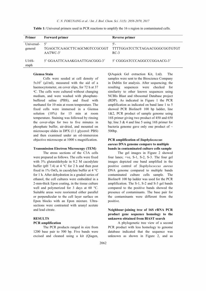
Table 1: Universal primers used in PCR reactions to amplify the 16 s region in contaminated cells.
Primer Forward primer Reverse primer
Universal-
general
5’
TGAGCTCAAGCTTCAGCMGTCCGCGGT
AATWC-3'
5'-
TTTTGGATCCTCTAGAACGGGCGGTGTGT
RC-3
U16S-
staph
5’ GGAATTCAAAKGAATTGACGGG-3’
5’ CGGGATCCCAGGCCCGGAACG-3’
Giemsa Stain
Cells were seeded at cell density of
5x10³ (µl/ml), measured with the aid of a
haemocytometer, on cover slips, for 72 h at 37
ᵒC. The cells were cultured without changing
medium, and were washed with phosphate-
buffered saline (PBS), and fixed with
methanol for 10 min at room temperature. The
fixed cells were immersed in a Giemsa
solution (10%) for 15 min at room
temperature. Staining was followed by rinsing
the cover-slips for two to five minutes in
phosphate buffer, air-dried, and mounted on
microscope slides in DPX (1:1 glycerol: PBS)
and then examined under an oil-immersion
objective microscope at 1000 x magnification.
Transmission Electron Microscopy (TEM)
The cross sections of the C3A cells
were prepared as follows. The cells were fixed
with 3% glutaraldehyde in 0.2 M cacodylate
buffer (pH 7.4) at 4 °C for 2 h and then post
fixed in 1% OsO4 in cacodylate buffer at 4 °C
for 1 h. After dehydration in a graded series of
ethanol, the cell cultures were embedded in a
2-mm-thick Epon coating, in the tissue culture
well and polymerized for 3 days at 60 °C.
Suitable areas were reoriented either parallel
or perpendicular to the cell layer surface on
Epon blocks with an Epon mixture. Ultra-
sections were contrasted with uranyl acetate
and lead citrate.
RESULTS
PCR amplification The PCR products ranged in size from
1200 base pair to 500 bp. Five bands were
excised and cleaned using a kit (Qiagen,
QiAquick Gel extraction Kit, Ltd). The
samples were sent to the Bioscience Company
in Dublin for analysis. After sequencing; the
resulting sequences were checked for
similarity to other known sequences using
NCBIs Blast and ribosomal Database project
(RDP). As indicated in Figure 1 the PCR
amplification as indicated on band lane 1 to 5
showed PCR Bioline® 100 bp ladder, line
1&2, PCR product of sample genome using
16S primer giving two product of 450 and 650
bp; line 3 & 4 and line 5 using 16S primer for
bacteria genome gave only one product of ~
500bp.
PCR amplification of Staphylococcus
aureus DNA genome compare to multiple
bands in contaminated culture cells sample
The gel images in Figure 2 showed
four lanes; +ve, S-1, S-2, S-3. The four gel
images depicted one band amplified in the
positive control of Staphylococcus aureus
DNA genome compared to multiple bands
contaminated culture cells sample. The
Bioline® 100 bp ladder was used for the PCR
amplification. The S-1, S-2 and S-3 gel bands
compared to the positive bands showed the
presence of contaminants. The base pair for
the contaminants were different from the
positive.
Neighbour-joining tree of 16S rRNA PCR
product gene sequence homology to the
unknown obtained from BlAST search A phylogenetic tree view of a second
PCR product with less homology to genome
database indicated that the sequence was
unknown as shown in Figure 2, and as

C. N. FOKUNANG et al. / Int. J. Biol. Chem. Sci. 11(5): 2058-2070, 2017
2063
indicated on the phylogenetic tree analysis.
Phylogenetic tree base on the PCR product
sequences 16S RNA primers Blastn were
searched from NCBI databases. The unknown
sequence in this work is highlighted in yellow
and the homology Bacteria name are indicated
from the NCBI blast search.
A more detailed phylogenetic output
indicating the clade for the different
homology of bacterial sequences are shown in
Figure 3. The unknown bacteria sequence is
highlighted in yellow and needed further
advance blast search for possible
identification for bio-informatics analysis.
We characterized the strain using 16S
RNA sequence that amplified the gene from
many genera. The closest phylogenetic
relative was |HQ877772.1|. Escherichia sp.
A94 with 88% 16S ribosomal RNA gene
sequence similarity. It was proposed that the
unidentified strain be assigned as a typed
strain of species of the bacteria origin based
on the 16S rRNA gene sequence search in
Ribosomal Database project, small subunit
rRNA and large subunit rRNA database,
together with the phylogenetic tree analysis.
In the different main clades there was the
Enterobacter, Salmonella, Escherichia,
Enterobactaceae main families.
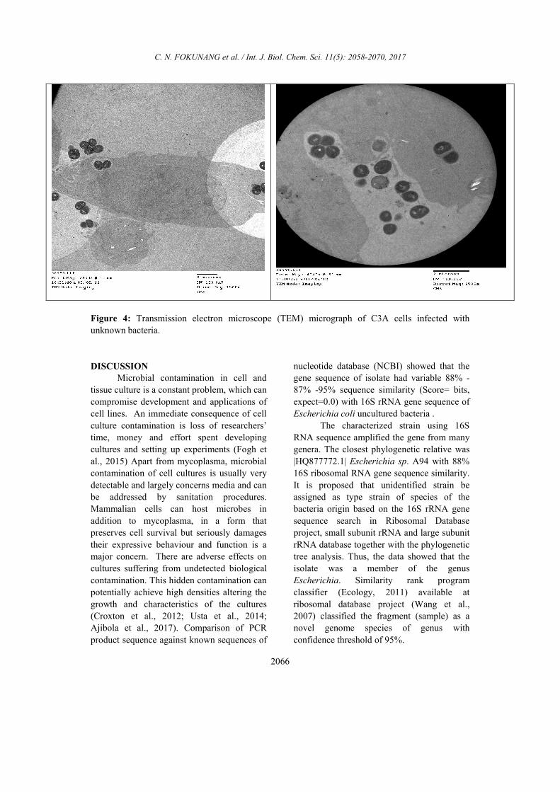
Transmission electron microscope (TEM)
micrograph of C3A cells infected with
unknown bacteria
The cross section of the cells
monolayer as shown in Figure 4 had
numerous intracellular bacteria. Micrograph
showed membrane ruffling upon contact with
bacteria. The bacteria were engulfed by
elongated microvilli from infected epithelial
cells. High magnification showed partially
lysed vacuole membrane containing bacteria,
indicating the ability of bacteria to escape
from the endocytic vacuoles. The
photomicrograph in Figure 4 also clearly
showed the presence of numerous bacteria
inside the cells. Most of the bacteria observed
were enclosed by endocytic vacuoles through
Giemsa stain observation. In addition, some
bacteria were free in the cytoplasm, perhaps
as a result of an escape from endocytic
vacuoles by bacterium-induced lyses of the
vacuole membrane.
Figure 1: PCR L1 AND L2 Bioline® 100 bp ladder, Line 1&2 PCR product of sample genome
using 16S primer giving two product of 450 and 650 bp; line 3 & 4 and 5 using 16S primer for
bacteria genome giving only one product of ~ 500bp.

C. N. FOKUNANG et al. / Int. J. Biol. Chem. Sci. 11(5): 2058-2070, 2017
2064
Figure 2: A phylogenetic tree containing all available homology bacterial sequence was constructed
from a multiple sequence alignment with the neighbour-joining method using Blast nucleotide
search. Scale bar 0.006 changes per site.

C. N. FOKUNANG et al. / Int. J. Biol. Chem. Sci. 11(5): 2058-2070, 2017
2065
Figure 3: A more detailed phylogenetic output indicating the clade for the different homology of
bacterial sequences.
C. N. FOKUNANG et al. / Int. J. Biol. Chem. Sci. 11(5): 2058-2070, 2017
2066
Figure 4: Transmission electron microscope (TEM) micrograph of C3A cells infected with
unknown bacteria.
DISCUSSION Microbial contamination in cell and
tissue culture is a constant problem, which can
compromise development and applications of
cell lines. An immediate consequence of cell
culture contamination is loss of researchers’
time, money and effort spent developing
cultures and setting up experiments (Fogh et
al., 2015) Apart from mycoplasma, microbial
contamination of cell cultures is usually very
detectable and largely concerns media and can
be addressed by sanitation procedures.
Mammalian cells can host microbes in
addition to mycoplasma, in a form that
preserves cell survival but seriously damages
their expressive behaviour and function is a
major concern. There are adverse effects on
cultures suffering from undetected biological
contamination. This hidden contamination can
potentially achieve high densities altering the
growth and characteristics of the cultures
(Croxton et al., 2012; Usta et al., 2014;
Ajibola et al., 2017). Comparison of PCR
product sequence against known sequences of
nucleotide database (NCBI) showed that the
gene sequence of isolate had variable 88% -
87% -95% sequence similarity (Score= bits,
expect=0.0) with 16S rRNA gene sequence of
Escherichia coli uncultured bacteria .
The characterized strain using 16S
RNA sequence amplified the gene from many
genera. The closest phylogenetic relative was
|HQ877772.1| Escherichia sp. A94 with 88%
16S ribosomal RNA gene sequence similarity.
It is proposed that unidentified strain be
assigned as type strain of species of the
bacteria origin based on the 16S rRNA gene
sequence search in Ribosomal Database
project, small subunit rRNA and large subunit
rRNA database together with the phylogenetic
tree analysis. Thus, the data showed that the
isolate was a member of the genus
Escherichia. Similarity rank program
classifier (Ecology, 2011) available at
ribosomal database project (Wang et al.,
2007) classified the fragment (sample) as a
novel genome species of genus with
confidence threshold of 95%.

C. N. FOKUNANG et al. / Int. J. Biol. Chem. Sci. 11(5): 2058-2070, 2017
2067
Most sequence shared 100 identities
with Homo sapiens isolate Li110 control
region, partial sequence; mitochondrial 100%,
while the rest of the 2 sequences showed
highest homology to sequences identity to
(88% gb|HQ877772.1| such as the Escherichia
sp, A94 16S ribosomal RNA gene, partial
sequence, 88% dbj|AB609044.1| Escherichia
coli gene for 16S rRNA, partial sequence,
strain: SI-7, gb|HQ759847.1|, the uncultured
organism clone ELU0045-T454-S-
NIPCRAMgANb_000132 small subunit
ribosomal RNA gene, gb|HQ796635.1|.Lastly,
the uncultured organism clone ELU0139-
T413-S-NIPCRAMgANa_000446 small
subunit ribosomal RNA gene 94%. The
translation of the nucleotide sequence was
more likely to be more accurate than just
blastn search, because the protein sequences
were more evolutionary and more conserved
than nucleotides sequences.
The nucleotide sequences translated
into protein using blastx database, showed that
the result of the search contained same results
from the Blastn search. The highest homology
was two hypothetical protein
HMPREF9553_00243 for [Escherichia coli
MS 200-1], with an 87% homology. The rest
of the sequences gave two different strains of
Escherichia coli (MS-198-1, 182-1, 187-1), as
well as gave a high similarities to conserved
hypothetical protein [Escherichia coli UTI89],
Escherichia coli APEC O1 with 84%
identities (Jerome Boudeau, 1999; Vimlesh
Yadav, 2009). The translation of the
nucleotide sequence is more likely to be more
accurate than just blastn search, because the
protein sequences are more evolutionary and
more conserved than nucleotides sequences
(Wieds, 2007). This study evaluates the
surface changes and effects on in vitro cell
attachment and spreading brought about on
prepared commercially pure titanium by
multiple exposures to common sterilization
methods. Information of contamination of
cells is very relevant in pre-clinical in vitro
cell cultures as a regulatory compliance for
drug discovery high throughput screening
(Eteme et al., 2015).
Mycoplasma contamination in
mammalian cell cultures is often overlooked
yet is a serious issue which can induce a
myriad of cellular changes leading to false
interpretation of experimental results (Ajibola
et al., 2017). A simple and sensitive assay was
used in this study to monitor mycoplasma
contamination (mycosensor) based on
degradation of the Gaussia luciferase reporter
in the conditioned medium of cells. This assay
proved to be more sensitive as compared to a
commercially available bioluminescent assay
in detecting mycoplasma contamination in
seven different cell lines. The Gaussia
luciferase mycosensor assay provides an easy
tool to monitor mammalian cell contaminants
in a high-throughput fashion (Awodiran et al.,
2014). The electron microscopic examination
of contaminated C3A cells was used to
identify the presence of mico-organism TEM
performed on cells monolayer. Bacterial was
observed to adhere closely to C3A cells. The
adhered bacteria strikingly induced the
elongation of microvilli from the cell surface.
At the site of close contact between the
bacteria and the epithelial cell, the elongated
microvilli surrounded the adherent bacteria
Cell culture is one of the most common
methods used to recapitulate a human disease
environment in a laboratory setting. Cell
culture techniques are used to grow and
maintain cells of various types including those
derived from primary tissues, such as stem
cells and cancer tumours (Kim 2007; Zenk
and Hansel, 2009). However, a major
confounding factor with cell culture is the use
of serum and animal (xeno) products in the
media. The addition of animal products
introduces batch and lot variations that lead to
experimental variability, confounds studies
with therapeutic outcomes for cultured cells,
and represents a major cost associated with
cell culture. Here we report a commercially
available serum-free, albumin-free, and xeno
free (XF) media (Neuro-Pure(TM)) that is
more cost-effective than other commercial
media. Neuro-Pure was used to maintain and
differentiate various cells of neuronal
lineages, fibroblasts, as well as specific cancer

C. N. FOKUNANG et al. / Int. J. Biol. Chem. Sci. 11(5): 2058-2070, 2017
2068
cell lines; without the use of contaminants
such serum, albumin, and animal products.
Neuro-Pure allows for a controlled and
reproducible cell culture environment that is
applicable to translational medicine and
general tissue culture.
Conclusion Microbial contamination in cell and
tissue culture is a constant problem, which can
compromise development and applications of
cell lines. This study showed a molecular
approach in detecting the cell contaminant
bacterial and gene sequence alignments. The
closest phylogenetic relative was
|HQ877772.1| Escherichia sp. A94 with 88%
16S ribosomal RNA gene sequence similarity.
It was proposed that unidentified strain be
assigned as type strain of species of the
bacteria origin based on the 16S rRNA gene
sequence search in Ribosomal Database
project, small subunit rRNA and large subunit
rRNA database together with the phylogenetic
tree analysis. Bacterial species were shown to
have at least one copy of the 16S rRNA gene
containing highly conserved regions together
with hyper variable regions. This study has
also shown the important use of 16S rRNA
gene sequence to characterize the bacterial
isolate from different cell lines. The Blastx
database did confirm the nucleotide Blastn
search result by suggesting that the unknown
sequence belong to the bacteria group
indicated very high homology with
Escherichia coli.
The electron microscopic examination
of contaminated C3A cells was used to
identify the presence of micro-organism TEM
performed on cells monolayer. Bacterial was
observed to adhere closely to C3A cells. The
adhered bacteria strikingly induced the
elongation of microvilli from the cell surface.
At the site of close contact between the
bacteria and the epithelial cell, the elongated
microvilli surrounded the adherent bacteria. In
addition, dense area of staining, possibly
related to an accumulation of cytoskeleton
components were observed beneath the sites
of intimate contact.
COMPETING INTERESTS The authors declare that they have no
competing interests.
AUTHORS CONTRIBUTIONS CFN, ETF, SB contributed in the
conception of the protocol, laboratory analysis
and statistics, JF, GL, FAK, BN and DG,
participated in manuscript writing and data
mining. PT the principal investigator and
project sponsor. All the authors participated in
the review of the manuscript.
ACKNOWLEDGEMENTS
The authors thank the Centre for
Biopolymer and Bio-molecular Research,
Athlone Institute of Technology, Republic of
Ireland for the funding of this work, and travel
grants to visiting lecturer.
REFERENCES
Akanbi II AA, Kareem T, Adedoja A,
Nyamngee A, Muhammed MBU,
Abdulkareem K, Atata RF. 2017.
Bacterial contamination of medical
doctors’ white coats as contributing
factor to hospital acquired infections. Int.
J. Biol. Chem. Sci., 11(1): 185-194.
Awodiran MO, Majolagbe FA, Komolafe OO,
Adewumi AA, Oyebola OO, Oyewole
RO. 2014. Cytogenetic study and serum
protein characterization of Clarias
gariepinus (Burchell, 1822) and
Heterobranchus bidorsalis in South
Western Nigeria. Int. J. Biol. Chem. Sci.,
8(6): 2371-2386.
Behrendorff JB, Vickers CE,
Chrysanthopoulos P, Nielsen LK. 2013.
2,2-Diphenyl-1-picryhydrazyl as a
screening tool for recombinant mono-
terpene biosynthesis. Microb. Cell.
Fact., 23: 12:76.
Blazkova HKK, Moudry P, Frisan T, Hodny
Z, Bartek J. 2009. Bacterial intoxication
Evokes cellular senescence with
C. N. FOKUNANG et al. / Int. J. Biol. Chem. Sci. 11(5): 2058-2070, 2017
2069
persistent DNA damage and cytokine
signaling. J Cell. Mol Med., 6(5): 16-27.
Borman AM, Palmer M, Johnson EM. 2013.
Rapid methods for the extraction and
archiving of molecular grade fungal
genomic DNA. Methods Mol Biol., 968:
55-62.
Case Gould MJ. 1984. Endotoxin in
Vertebrate Cell Culture: Its
Measurement and significance in uses
and standardization of vertebrate Cell
lines. Tissue Culture, Association,
Gaithersburg, MD., 31(3): 125-136.
Croxton AN, Wikfors GH, Schulterbrandt-
Gragg RD. 2012. Immunomodulation in
eastern oysters, Crassostrea virginica,
exposed to a PAH-contaminated,
microphytobenthic diatom. Aquat
Toxicol., 15: 118-119.
Degeling MH, Maguire CA, Bovenberg MS,
Tannous BA. 2012. Sensitive assay for
mycoplasma detection in mammalian
cell culture. Anal Chem., 84(9): 4227-
4232.
Ecology CFM. 2011. The Ribosomal
Database Project (RDP). M. S.
University. Michigan, Michigan State
University Board of Trustees, 76 p.
Enow-Tanjong P, Teyim P, Kamga HL, Neba
ES, Nkuo-Akenji T. 2016. Sero-
prevalence of HIV and hepatitis viruses
and their correlation with CD4 T-cell
lymphocyte counts in pregnant women
in the Buea Health District of Cameroon.
Int. J. Biol. Chem. Sci., 10(1): 219-231.
Eteme LF, Fokunang CN, Tchuenguem FF,
Nolna D, Boula A, Ndze NV. 2015.
Epidémiologie moléculaire du rotavirus
du groupe A associé aux gastroentérites
chez les enfants de moins de 5 ans dans
la ville de Yaoundé Cameroun. Int. J.
Biol. Chem. Sci., 9(5): 2561-2573. DOI :
http://dx.doi.org/10.4314/ijbcs.v9i5.25.
Everoad RC, Yoshida S, Tsuboi Y, Date Y,
Kikuchi J, Moriya S. 2012.
Concentration of metabolites from low-
density planktonic communities for
environmental metabolomics using
nuclear magnetic resonance
spectroscopy. J Vis Exp., 7(62): e3163.
Fogh J, Holmgren NB, Ludovici PP. 1971. A
Review of Cell Culture Contaminations.
In Vitro, 7(1): 26-41.
Garner CM, Chakraborti PR. 2000.
Mycoplasmas detection in cell cultures: a
comparison of four methods. Br. J.
Biomed. Sci., 57: 295-301.
Grahn N, Olofsson M, Ellnebo-Svedlund K,
Monstein HJ, Jonasson J. 2003.
Identification of mixed bacterial DNA
contamination in broad-range PCR
amplification of 16S rDNA V1 and V3
variable regions by pyrosequencing of
cloned amplicons. Fed. Euro. Microbiol.
Soc. Microbio. Letts., 219(2): 87-91.
Gupta PA, Ge X, Kostov Y, Rao G .2014. A
completely noninvasive method of
dissolved oxygen monitoring in
disposable small-scale cell culture
vessels based on diffusion through
permeable vessel walls. Biotechnol Prog,
30(1):172-177.
Hay RJ. 1991. Operator-induced
contamination in cell culture systems.
Dev. Biol. Stand., 75(4): 193-204.
Ikonomi RJ. 2006. Cell biology: Laboratory
Handbook, 1(3): 49-53. Incorporated C.
2002. Understanding and Managing Cell
Culture Contamination. T. Bulletin.
USA.
Jennifer P, Sue GMB, Jinnefer IF 2010. Got
black swimming dots in your cells
cultures? Identification of
Achromobacter as a novel cell culture
contamination. Biologicals, 32(2): 273-
277.
Jerome Boudeau AL, Masseret E, Bernard JA,
Darfeuille-Michaud A. 1999.
Expand+Infection and Immunityiai.

C. N. FOKUNANG et al. / Int. J. Biol. Chem. Sci. 11(5): 2058-2070, 2017
2070
asm.orgInfect. Immun., 67(3): 94499-
944509.
Jimenez L, Ignar R, D'Alello R, Grech P.
2007. Use of PCR analysis for sterility
testing in pharmaceutical environments.
J. Rap.Methods. Automat. Microbiol.,
8(85): 11-20.
Kim H. 2007. Ecological variables affecting
predatory success in Myxococcus
xanthus. Microb Ecol, 53: 571.
Klausegger A, Hell M, Berger A, Zinober K,
Baier S, Jones S, Koffler B.1999. "Gram
type-specific broad-range PCR
amplification of rapid detection of 62
pathogenic bacteria. J. Clin. Microbiol.,
37(5): 464-466.
Lleo MM, Bonato B, Tafi MC, Signoretto C,
Pruzzo C, Canepari P. 2005. Molecular
vs culture methods for detection of
bacterial faecal indicators in
groundwater for human use. Lett. App.
Microbiol., 40(1): 289-294.
Mohammad-Qureshi SS, Haddad R, Palmer
KS, Richardson JP, Gomez E, Pavitt GD.
2007. Purification of FLAG-tagged
eukaryotic initiation factor 2B
complexes, subcomplexes, and
fragments from Saccharomyces
cerevisiae. Methods Enzymol., 43(1): 1-
13.
Nocker AC. 2008. Novel approaches toward
preferential detection of viable cells
using nucleic acid amplification
techniques. Fed. Euro. Microbiol. Soc.
Microbiol. Lett., 291(3): 137-142.
Testro AG. 2009. Toll-like receptors and their
role in gastrointestinal disease. J
Gastroenterol Hepatol., 24(6): 943-954.
Thomas PC, Strotman LN, Theberge AB,
Berthier E, O'Connell R, Loeb JM, Berry
SM, Beebe D. 2013. Nucleic acid sample
preparation using spontaneous biphasic
plug flow. J. Anal Chem., 85(18): 8641-
8646.
Uphoff CC, Horn GD. 2001. Prevention of
mycoplasma contamination in leukemia-
lymphoma cell lines. Human Cell., 14:
244-247.
Usta SN, Scharer CD, Xu J, Frey TK, Nash
RJ.2014. Chemically defined serum-free
and xeno-free media for multiple cell
lineages. Ann Transl Med., 2(10): 97-
103.
Vimlesh YSP, Shipra IP, Srivastava PC,
Praveen CV, Verma VG, Gupta VB,
Anil KR. 2009. Identification of
Comamonas species using 16S rRNA
gene sequence. Bioinformation., 3(9):
381–383.
Wang Q, Garrity GM, Tiedje JM, Cole JR.
2007. Naïve Bayesian Classifier for
Rapid Assignment of rRNA Sequences
into the New Bacterial Taxonomy. Appl
Environ Microbiol., 73(16): 5261–5267.
Zenk SF, Hensel M. 2009. Role of Salmonella
enterica lipopolysaccharide in activation
of dendritic cell functions and bacterial
containment. J immunol. 183(4): 2697-
2707.
.