Molecular Cloning and Characterization of Chick Sialoprotein ...
Molecular cloning, characterization and regulationof two different NADH-glutamate synthase cDNAsin bean nodules
LOURDES BLANCO1, PALLAVOLU M. REDDY1, SONIA SILVENTE1, BRUNA BUCCIARELLI3,SANGHAMITRA KHANDUAL1, XOCHITL ALVARADO-AFFANTRANGER2, FEDERICO SÁNCHEZ2,SUSAN MILLER3, CARROLL VANCE3 & MIGUEL LARA-FLORES1
1Centro de Ciencias Genómicas and 2Instituto de Biotecnología, Universidad Nacional Autónoma de México, AvUniversidad, C.P. 62210, Cuernavaca, Morelos, México and 3Agronomy and Plant Genetics, University of Minnesota andUnited States Department of Agriculture/Agriculture Research Service, Plant Science Research Unit, 1991 Upper BufordCircle, St Paul, MN 55108, USA
ABSTRACT
NADH-dependent glutamate synthase (NADH-GOGAT)is a key enzyme in primary ammonia assimilation inPhaseolus vulgaris nodules. Two different types of cDNAclones of PvNADH-GOGAT were isolated from thenodule cDNA libraries. The full-length cDNA clones ofPvNADH-GOGAT-I (7.4 kb) and PvNADH-GOGAT-II(7.0 kb), which displayed an 83% homology betweenthem, were isolated using cDNA library screening, ‘cDNAlibrary walking’ and RT-PCR amplification. Southernanalysis employing specific 5� cDNA probes derived fromPvNADH-GOGAT-I and PvNADH-GOGAT-II indicatedthe existence of a single copy of each gene in the beangenome. Both these proteins contain ~100 amino acidsequences theoretically addressing each isoenzyme to dif-ferent subcellular compartments. RT-PCR analysis indi-cated that PvNADH-GOGAT-II expression is higher thanPvNADH-GOGAT-I during nodule development. Expres-sion analysis by RT-PCR also revealed that both of thesegenes are differentially regulated by sucrose. On the otherhand, the expression of PvNADH-GOGAT-I, but notPvNADH-GOGAT-II, was inhibited with nitrogen com-pounds. In situ hybridization and promoter expressionanalyses demonstrated that the NADH-GOGAT-I and -IIgenes are differentially expressed in bean root and noduletissues. In silico analyses of the NADH-GOGAT promotersrevealed the presence of potential cis elements in them thatcould mediate differential tissue-specific, and sugar andamino acid responsive expression of these genes.
Key-words: Phaseolus vulgaris; carbon and nitrogencompounds; gene expression; promoter analysis;PvNADH-GOGAT.
INTRODUCTION
Glutamine synthetase (GS) and glutamate synthase(GOGAT) form the main route of ammonia assimilation inhigher plants (GS/GOGAT cycle) (Miflin & Lea 1976;Lancien, Gadal & Hodges 2000; Suzuki & Knaff 2005). GScatalyses the ATP-dependent amidation of glutamateresulting in the production of glutamine. GOGAT, on theother hand, catalyses the reductive transfer of the amidegroup of glutamine to the a-keto position of 2-oxoglutarate,forming two molecules of glutamate (Lea, Robinson &Stewart 1990). Ammonia in higher plants is derived bothfrom the primary nitrogen sources (ammonia, nitrate andfor legumes, dinitrogen) and from a number of internalnitrogen-cycling pathways such as photorespiration, aminoacid metabolism and phenylpropanoid metabolism (Vance1997). GOGAT is therefore essential not only for primarynitrogen assimilation but also to maintain the general nitro-gen economy of the plant.
In higher plants, GOGAT occurs as two distinct isoforms:Fd-dependent GOGAT (Fd-GOGAT; EC 1.2.7.1) andNADH-dependent GOGAT (NADH-GOGAT; EC1.4.1.14), and both isoforms differ from one another inmolecular mass, subunit composition, enzyme kinetics,reductant specificity and metabolic function (Temple,Vance& Gantt 1998; Lea & Miflin 2003). These two molecularspecies of GOGAT exist in both green and non-greentissues. The Fd-GOGAT has been shown to be the mostabundant isoform in green tissues and is located in thechloroplast (Wallsgrove, Lea & Miflin 1979). This mono-meric enzyme is an Fe-S protein with an Mr about 150 kDa.In leaves, Fd-GOGAT catalyses the assimilation of NH4
+
derived from both the light-dependent reduction of NO3-
and the NH4+ generated during photorespiration, while the
root isoform has been implicated in the assimilation ofNH4
+ derived from soil NO3- (Sakakibara et al. 1991;
Redinbaugh & Campbell 1993; Suzuki & Knaff 2005). Com-plimentary DNA of Fd-GOGAT has been isolated from anumber of species. Two Fd-GOGAT genes (GLU1 andGLU2) were characterized in Arabidopsis thaliana; GLU1
Correspondence: M. Lara-Flores. Fax: +52 777 317 5581; e-mail:[email protected]
Plant, Cell and Environment (2008) 31, 454–472 doi: 10.1111/j.1365-3040.2008.01774.x
© 2008 The AuthorsJournal compilation © 2008 Blackwell Publishing Ltd454

plays a major role in the assimilation of ammonium derivedfrom photorespiration, whereas the GLU2 gene may play amajor role in the primary nitrogen assimilation in roots(Oliveira et al. 1997; Suzuki & Rothstein 1997; Coschiganoet al. 1998).
NADH-GOGAT is found primarily in the non-photosynthetic tissues of plants, such as roots and legumeroot nodules, where it is located in the plastids. In Medi-cago sativa, it has been immunolocalized to the amylo-plasts of infected cells in the nodules (Trepp et al. 1999a).Pattern of expression was found to shift during rootnodule development and expression is tightly linked toeffective nodules (Trepp et al. 1999b). It has been purifiedfrom legume root nodules as well as rice (Oryza sativa)cell cultures (Chen & Cullimore 1988; Anderson et al.1989; Hayakawa et al. 1992). As Fd-GOGAT, NADH-GOGAT is an Fe-S flavoprotein (Curti et al. 1995; Suzuki& Knaff 2005). NADH-GOGAT exists as a monomer withan Mr of approximately 225–230 kDa (Yamaya et al. 1992;Gregerson et al. 1993; Vance et al. 1995). In nodules, it isthe major form of GOGAT, and where it combines withcytosolic GS to assimilate NH4
+ produced by the nitrogen-fixing bacteroids. NADH-GOGAT activity has been foundto increase markedly during nodule development (Greger-son et al. 1993; Vance et al. 1994), and is three times higherthan the Fd-GOGAT activity. It has been proposedthat NADH-GOGAT catalyses the rate-limiting step inammonia assimilation in the root nodules (Trepp et al.1999a,b).
In non-legumes such as rice, NADH-GOGAT has beencharacterized with studies of gene organization, expressionof mRNA, protein localization and protein structure(Yamaya et al. 1992, 1995; Goto et al. 1998). The expressionof the NADH-GOGAT gene in rice is regulated in a celltype-specific, age-specific and nitrogen-responsive manner(Hayakawa et al. 1994, 1999). It has been located in roots,leaves, etiolated leaves, vascular bundles of unexpandedleaf tissues and the developing grains, and was shown tofunction in the primary assimilation of NH4
+ derived fromsoil NO3
- and the re-assimilation of NH4+ released during
amino acid catabolism and/or during seed germination.Full-length cDNA and genomic clones of NADH-
GOGAT have been isolated from M. sativa and O. sativa,and was found that NADH-GOGAT is encoded by a singlegene (Gregerson et al. 1993; Vance et al. 1995; Goto et al.1998). The only report that described the existence of twoisoforms of NADH-GOGAT in plants is in bean noduleswhere two isoenzymes (I and II) were identified (Chen &Cullimore 1988). The isoenzyme NADH-GOGAT-II islocalized in the central tissue of the nodule, while the isoen-zyme NADH-GOGAT-I is found to be present both inthe cortex and central tissue of the nodules, as well as inthe roots (Chen & Cullimore 1989). NADH-GOGAT-IIaccounted for the majority of the activity in the rootnodules. Specific activity of NADH-GOGAT in roots isfound to be about 27 times lower than that in the nodules,and consisted almost entirely of NADH-GOGAT-I. Bothisoenzymes were partially purified, and were shown to have
similar Mrs of about 200 kDa with marginal differences inkinetic characteristics (Chen & Cullimore 1988).
In this paper, we report for the first time the isolation,characterization, localization and regulation of expressionof two distinct types of cDNAs encoding NADH-GOGATfrom bean (Phaseolus vulgaris cv. Negro Jamapa) nodules.The data presented here indicate that the two NADH-GOGAT isoforms described by Chen & Cullimore (1988)in the P. vulgaris nodules are encoded by two distinct genes.The expression analysis suggests different roles for thesetwo NADH-GOGAT isoforms in the assimilation of am-monia in symbiotically nitrogen-fixing bean plants.
MATERIALS AND METHODS
Plant growth, treatments and tissue sampling
P. vulgaris cv. Negro Jamapa seeds (ProNaSe, Guanajuato,México) were surface sterilized in 20% sodium hypochloritesolution for 10 min, washed thoroughly with sterile distilledwater and germinated on a moist sterile filter paper. Three-day-old seedlings were transferred to the pots containingsterile vermiculite (five plants per pot) and then inoculatedwith Rhizobium tropici CIAT899, Rhizobium etli CE3 orR. etli CE3 NifA-. The uninoculated plants served as thecontrol. The plants were grown in a naturally lighted green-house as described by Ortega et al. (1992). For thePvNADH-GOGAT expression analysis, the nodules wereharvested at various time points during 12–21 d after inocu-lation (DAI). In addition, roots, stems and fully expandedleaves of 13-day-old nodulated plants, and cotyledonaryleaves from 3-day-old plants, and flowers and pods frommature plants were also collected for analysis. In the studiespertaining to assessing the effects of carbon and nitrogentreatments on the PvNADH-GOGAT expression, theplants were watered for 4 d, starting on the 17th DAI, withthe Hoagland nutrient solution containing either sucrose(0.5, 1 and 2%), asparagine, glutamine, xanthine, allantoin(10 mM) or allopurinol (2 mM), and then the nodules wereharvested for analysis.The nodules were also collected fromthe control plants that were treated with the Hoaglandnutrient solution devoid of any carbon and nitrogen com-pounds. All tissues were immediately frozen in liquid nitro-gen and stored at -70 °C until use.
Isolation of RNA
Poly(A)+ RNA for constructing the cDNA library was iso-lated from the nodules following the standard method(Sambrook, Fritsch & Maniatis 1989). Total and poly(A)+
nodule RNA for isolating full-length bean NADH-GOGATs by RT-PCR were extracted using RNeasy PlantKit and Oligotex mRNA Mini Kit (Qiagen, Valencia, CA,USA), respectively. The total RNA from the plant tissuesfor the expression analysis by RT-PCR was isolated byemploying the RNA Tripure Isolation Reagent (RocheDiagnostics Corporation, Indianapolis, IN, USA).
NADH-GOGAT isoforms in Phaseolus vulgaris 455
© 2008 The AuthorsJournal compilation © 2008 Blackwell Publishing Ltd, Plant, Cell and Environment, 31, 454–472

Isolation of PvNADH-GOGAT cDNAs
Two lZAP cDNA libraries (constructed using lZAP-cDNA synthesis Kit; Stratagene, La Jolla, CA, USA) of P.vulgaris derived from poly(A)+ RNA of 15-day-old noduleswere screened for isolating PvNADH-GOGAT clones. ThecDNA probes were labelled with 32P using random primerlabeling kit (Megaprime DNA labelling system; AmershamPharmacia Biotech, Little Chalfont, Bucks, UK), andemployed to screen the cDNA libraries. The library screen-ing was done on nylon membrane (Amersham Life Sci-ences, Little Chalfont, UK) replicas by standard procedures(Sambrook et al. 1989). Hybridization was carried out over-night in 5X Denhardt’s solution (1X Denhardt’s solution is0.02% Ficoll, 0.02% polyvinylpyrrolidone, 0.02% BSA), 6XSSPE (1X SSPE is 0.15 M NaCl, 10 mM sodium phosphateand 1 mM ethylenediaminetetraacetic acid, pH 7.4), 1%sodium dodecyl sulphate (SDS) and 0.1 mg mL–1 denaturedsalmon sperm DNA at 55 °C. Membrane filters werewashed once at room temperature for 5 min in 2X SSC (1XSSC is 0.15 M NaCl and 0.015 M sodium citrate, pH 7.0) and0.1% SDS, and then twice at 50 °C for 20 min each in 0.2XSSC and 0.1% SDS. Positive lZAP clones were plaquepurified and cDNA inserts were obtained in the BluescriptSK- vector by in vivo excision according to the manufactur-er’s instructions (Stratagene).
Library screening was done in two steps. Initial libraryscreening was done using a 1.6 kb cDNA probe derivedfrom the alfalfa NADH-GOGAT (pGOGAT7.2; Greger-son et al. 1993). As a consequence, we isolated two clonescontaining cDNA inserts of 2.5 kb (PvNADH-GOGAT-I)and 3 kb (PvNADH-GOGAT-II). Subsequently, utilizingthese partial PvNADH-GOGAT-I and PvNADH-GOGAT-II cDNAs as probes, longer cDNA inserts wereobtained by screening a second nodule cDNA library,which was prepared using size-fractionated (3–9 kb)nodule cDNA. From the second screening, two longerclones of 5 kb each corresponding to PvNADH-GOGAT-Iand -II were isolated and sequenced. In order to expandthe 5′ region of the cDNA inserts, size-fractionated nodulecDNA library was used as a template for ‘cDNA walking’,and three sequential rounds of PCR (profile A; Supple-mental Table S1) using Expand Long Template PCRSystem (Roche Diagnostics Corporation) were performedusing pBluescript T3 primer in combination with gene spe-cific 5′ region primers designed for each of the PvNADH-GOGAT cDNA insert/fragment (gene-specific reverseprimers for the amplification of 5′ regions; SupplementalTable S2). The gene specific 5′ primers were derived fromthe sequence analysis of the cDNA inserts in the clones aswell as the amplified fragments obtained from each roundof PCR amplification.
Finally, RT-PCR was performed in order to obtain thefull-length clones for each of the PvNADH-GOGATs. Forthis purpose, fresh nodule cDNA was synthesized from thetotal RNA isolated from 15-day-old nodules using Power-Script reverse transcriptase (Clontech, Palo Alto, CA,USA), primed with oligo (dT)12–18 (Invitrogen, Carlsbad,
CA, USA), and employed for the amplification of thefull-length cDNAs using gene-specific 5′ and 3′ primers (foramplifying full-length clones; Supplemental Table S2)designed based on the assembled sequences of the clonesobtained through both library screening and ‘cDNA librarywalking’ (see above). In the PCR step of the reaction, theExpand Long Template PCR System (Roche DiagnosticsCorporation) was used for the amplification of the full-length 7.4 kb PvNADH-GOGAT-I and 7.05 kb PvNADH-GOGAT-II (profile B; Supplemental Table S1). Bothfragments were cloned in pGEM-T easy vector (PromegaCorp., Madison, WI, USA) and transformed into XL10GOLD Escherichia coli cells (Stratagene) and sequenced.
Sequence analysis
DNA sequencing of all isolated clones was done atMedigenomix (Munich, Germany), and the sequences ofthe full-length clones were corroborated with thosesequences previously obtained from the partial clones. Tomanage the sequence data, the DNA strider 1.3 program(Marck 1988) was used. Similarity searches of databaseswere performed according to Altschul et al. (1990) with theNCBI BLAST server (http://www.ncbi.nih.gov/BLAST/).Transit peptide was identified and analysed with Plant-Ploc(Plant Protein Subcellular Location Prediction; http://202.120.37.186/bioinf/plant/; Chou 2005; Shen & Chou 2006;Chou & Shen 2007) and WoLF PSORT (Prediction ofProtein Sorting Signals and Localization Sites in AminoAcid Sequences; http://wolfpsort.org/; Horton et al. 2006,2007) programmes. The protein molecular masses and pIwere estimated by using the pI/MW program at Uniprot(EMBL-EBI; http://au.expasy.org/tools/pi_tool.html).
RT-PCR analysis
The tissue samples were ground in liquid nitrogen, and thetotal RNA was extracted with Tripure (Roche DiagnosticsCorporation) according to the instructions of the manufac-turer. For RT-PCR studies, 5 mg of total RNA was convertedinto first-strand cDNA by using the PowerScript pre-amplification system (Clontech) and oligo (dT)12–18 (Invit-rogen). One microlitre of cDNA sample was amplified(profile C; Supplemental Table S1) in a 50 mL PCR mixturecontaining DNA polymerase buffer (Roche DiagnosticsCorporation), 2.5 mM MgCl2, 200 mM of each dNTPs,0.1 mM of each gene-specific primers (RT-PCR primers;Supplemental Table S2) and 1 unit of Taq DNA polymerase(Roche Diagnostics Corporation).
Plant DNA extraction and DNA gel blot analysis
Genomic DNA was isolated from the leaves of 5-day-oldseedlings using the DNAzol procedure (Gibco BRL LifeTechnologies, Inc., Grand Island, NY, USA). For the gel blotanalyses, genomic DNA was digested with PstI, XbaI, KpnIand EcoRV restriction enzymes, separated on a 0.8%agarose gel (15 mg DNA per lane) and transferred to nylon
456 L. Blanco et al.
© 2008 The AuthorsJournal compilation © 2008 Blackwell Publishing Ltd, Plant, Cell and Environment, 31, 454–472

membranes (Hybond-N+, Amersham Life Sciences).Hybridization was performed using, as labelled probes, a611 bp and a 461 bp fragments specific for PvNADH-GOGAT-I and PvNADH-GOGAT-II, respectively. Bothfragments contain the 5′ untranslated region (UTR) andtransit peptide-coding regions. The probes were radiolabelled with 32P by random priming with rediprime IIsystem (Amersham Life Sciences). Hybridization wascarried out overnight at 65 °C in 300 mM phosphate buffer(pH 7.2) and 7% SDS.The hybridized filters were washed at65 °C with 2X SSC (15 min), 0.2X SSC (15 min) and 0.1XSSC (10 min) plus 0.1% (w/v) SDS, and exposed to KodakX Omat films.
Isolation of the NADH-GOGAT promoters
The 5′ flanking genomic sequences of PvNADH-GOGAT-Iand PvNADH-GOGAT-II were isolated by long-distancePCR using bean DNA and GenomeWalker kit (Clontech)according to the manufacturer’s instructions. Gene specificprimers employed for genome walking of the 5′ flank-ing sequences of PvNADH-GOGAT-I and PvNADH-GOGAT-II are given in the Supplemental Table S3. Long-distance PCR was performed using Advantage 2Polymerase Mix with the following cycling profiles. PrimaryPCR amplification was carried out with five initial cycles of28 s at 94 °C and 4 min at 72 °C, followed by 20 cycles of 28 sat 94 °C and 4 min at 67 °C, and a single final cycle of 7 minat 67 °C. Secondary PCR was carried out with an initialcycle of 1 min at 94 °C, followed by 30 cycles of 30 s at 94 °C,1 min at 64 °C, and 2 min at 72 °C, and a single final cycle of7 min at 72 °C. Isolated 5′ flanking regions were sequencedat Medigenomics, and the transcription start sites weredetermined using the Neural Network Promoter Predic-tion Program (Reese 2001; http://www.bdgp.org/seq_tools/promoter.html) in conjunction with the untranslated5′ sequences of PvNADH-GOGAT-I and PvNADH-GOGAT-II cDNAs. The promoter sequences were alsoexplored for identifying cis-acting regulatory elementsin them using PLACE (Higo et al. 1999; http://www.dna.affrc.go.jp/PLACE/) as well as PlantCARE data-bases (Lescot et al. 2002; http://bioinformatics.psb.ugent.be/webtools/plantcare/html/).
Development of plant transformation vectorsand agrobacterial strains carrying the binaryvectors, and inoculum preparation
The binary vectors pBI-PrPvNADH-GOGAT-I-EGFPGUS and pBI-PrPvNADH-GOGAT-II-EGFPGUScontaining the P. vulgaris NADH-GOGAT promoterstranscriptionally coupled to the egfp-gusA coding fusionsequence and the 3′ nos terminator sequence were con-structed as follows. Five prime flanking sequences com-prising of 1501 bp PvNADH-GOGAT-I and 1366 bpPvNADH-GOGAT-II, upstream of their respective codingsequences were PCR amplified as described above,
digested with SalI and XmaI (restriction sites wereembedded in the forward and reverse primers used forpromoter amplification; see Supplemental Table S3) andcloned upstream of the egfp-gusA coding sequence ina similarly digested pBI101-EGFPGUS binary vectoryielding pBI-PrPvNADH-GOGAT-I-EGFPGUS andpBI-PrPvNADH-GOGAT-II-EGFPGUS. The pBI101-EGFPGUS vector was constructed by replacing gusAcoding sequence in pBI101.1 (Clontech) with egfpgusAchimeric reporter gene derived from pHGWFS7 (Karimi,Inze & Depicker 2002).
The binary vectors used for the bean transformation,pBI101-EGFPGUS (promoterless EGFPGUS, controlvector), pBI-PrPvNADH-GOGAT-I-EGFPGUS or pBI-PrPvNADH-GOGAT-II-EGFPGUS, were transformedinto Agrobacterium rhizogenes K599 by electroporation(Nagel et al. 1990). A. rhizogenes strains harbouring thetransformation vectors were grown at 30 °C in Luria-Bertani (LB) plates supplemented with 50 mg mL-1 kana-mycin. The bacterial cells from overnight-grown culturefrom a single plate were harvested and resuspended in5 mL sterile distilled water, and used for infecting thebean seedlings for the induction of hairy root formation(see below).
Hairy root transformation andgrowth conditions
Bean seeds harvested from the greenhouse-grown plantswere surface sterilized and germinated in pots containingsterile vermiculite in the plant growth room (maintained ata 14 h light/10 h dark cycle and a temperature of 25 °C) asdescribed earlier (Silvente et al. 2008), and irrigated withthe Summerfield nutrient solution (Summerfield, Huxley &Minchin 1977) supplemented with 5 mM KNO3. The hairyroot transformation of the bean plants was performedaccording to Estrada-Navarrete et al. (2006) as modified bySilvente et al. (2008) using A. rhizogenes strains carrying thebinary vectors pBI101-EGFPGUS (control vector), pBI-PrPvNADH-GOGAT-I-EGFPGUS or pBI-PrPvNADH-GOGAT-II-EGFPGUS. A. rhizogenes-transformed com-posite bean plants were grown in pots and inoculated with1–2 mL R. tropici CIAT899 culture (rhizobial inoculum wasprepared by growing CIAT899 strain to a density of 106 cellsmL-1 in peptone-yeast extract (PY) medium supplementedwith nalidixic acid, 20 mg mL-1), irrigated with the Summer-field nutrient solution (Summerfield et al. 1977) having ahighly reduced level (0.385 mM) of KNO3 and incubated inthe plant growth chamber maintained at a 14 h light/10 hdark cycle and a temperature of 25 °C. After 4–5 d of accli-matization, the transformed plants were transferred to thenaturally lit greenhouse maintained at 25 °C. The plants(five replicates per each treatment) were harvested 3–4weeks after rhizobial inoculation, and the roots and noduleswere analysed for GUS and GFP activity. GUS and GFPanalysis revealed no conspicuous plant to plant variationsin the pattern or level of expression of the reportergenes.
NADH-GOGAT isoforms in Phaseolus vulgaris 457
© 2008 The AuthorsJournal compilation © 2008 Blackwell Publishing Ltd, Plant, Cell and Environment, 31, 454–472

Histochemical localization of GUS activity andother microscopic methods for thevisualization of GFP
GUS staining of the transgenic roots was performed at30 °C for 12–24 h essentially as described by Reddy et al.(1998). After staining, the roots were rinsed in 100 mMphosphate buffer and fixed for more than 4 h in a solutioncontaining 3.7% formaldehyde, 5% acetic acid and 50%ethanol, examined as whole specimens and photographedunder a stereomicroscope (Carl Zeiss Stemi 2000-C; CarlZeiss Jena GmbH, Jena, Germany).
For the GFP analysis, the roots and nodules were pro-cessed essentially according to Estrada-Navarrete et al.(2006). Briefly, the tissues were cross-linked with 100 mMm-maleimidobenzoyl-N-hydroxysuccinimide-ester, fixedwith 4% paraformaldehyde, passed through dehydrationethanol-methacrylate series, and finally embedded bypolymerization in butyl-methacrylate/methyl-methacrylate(4:1, vol/vol; Sigma Aldrich, St Louis, MO, USA) mixturewith 0.375% (wt/vol) benzoin ethyl ether (Fluka Chemika,Buchs, Switzerland) in gelatin capsules (Electron Micros-copy Sciences, Fort Washington, PA, USA). The embeddedtissue was sectioned (3 mm thickness) using ultramicro-tome (Leica, Bensheim, Germany), processed to removemethacrylate, mounted with citifluor (Ted Pella, Inc.,Redding, CA, USA) and observed for GFP fluorescenceusing a Zeiss LSM 510 Meta confocal microscopeequipped with Axiovert 200M. GFP excitation wasobtained at 488 nm using an argon laser and an HFT UV488/543/633-nm dual dichroic excitation mirror, and thefluorescence was detected with an LP 505-nm emissionfilter. The images were processed using Adobe PhotoshopElements 2.0 software (Adobe Systems Incorporated,Mountain View, CA, USA).
In situ hybridization
All RNA probes were generated from linearized pBlue-script plasmids containing either the 602 bp PvNADH-GOGAT-I fragment, the 563 bp PvNADH-GOGAT-IIfragment or the 600 bp Pv-leghemoglobin fragment. BothPvNADH-GOGAT-I (between 6695 and 7297 bp) and II(between 6419 and 6982 bp) fragments (probes) werederived from the non-homologous 3′ UTR and C terminalcoding regions of the full-length cDNA clones.The plasmidsharbouring PvNADH-GOGAT-I, -II or Pv-leghemoglobinfragments were linearized with the appropriate restrictionenzymes and transcribed in the sense and antisense direc-tions using the T3 or T7 RNA polymerase and radiolabelled with 35S UTP nucleotide. The hydrolysis of thetranscribed probes and the subsequent in situ hybridizationprotocols have been described by Trepp et al. (1999b).
The sequence data from this paper have been deposited atthe Genbank under the accession numbers PvNADH-GOGAT-I cDNA,AF314925; PvNADH-GOGAT-II cDNA,AF314924; PvNADH-GOGAT-I promoter, AB195997; andPvNADH-GOGAT-II promoter, AB195998.
RESULTS
Isolation and characterization of two beancDNAs encoding NADH-GOGAT
In order to isolate NADH-GOGAT homologs from thebean (P. vulgaris), we first screened a 15-day-old rootnodule cDNA library by hybridization with a 1.6 kb cDNAprobe derived from M. sativa NADH-GOGAT. Conse-quently, two clones containing bean cDNA inserts of 2.5 kb(PvNADH-GOGAT-I) and 3 kb (PvNADH-GOGAT-II)were isolated. The sequencing of both cDNA clonesrevealed an open reading frame (ORF) encoding a partialpolypeptide similar to MsNADH-GOGAT, with different3′ UTR sequences between them. The detection of twocDNAs of PvNADH-GOGAT with different 3′ UTRsequences suggested the presence of two PvNADH-GOGAT genes in the bean. RNA gel blot analysis utilizingspecific PvNADH-GOGAT clones as probes revealed 7 kbmRNAs as expected for proteins with a molecular masshigher than 200 kD. In order to obtain longer PvNADH-GOGAT cDNA clones, 2.5 and 3 kb fragments ofPvNADH-GOGAT-I and PvNADH-GOGAT-II, respec-tively, were used as hybridization probes to screen a secondbean nodule cDNA library, which was prepared with size-fractionated (3–9 kb) nodule cDNA. From the secondscreening, two longer clones of 5 kb each correspondingto PvNADH-GOGAT-I and PvNADH-GOGAT-II wereisolated. Full-length PvNADH-GOGAT-I (7406 bp) andPvNADH-GOGAT-II (7050 bp) cDNA clones were subse-quently obtained by ‘cDNA library walking’ with nestedamplification by PCR using the bean root nodule library astemplate (see material and methods). Sequence analysis ofdifferent sub-clones, as well as of RT-PCR derived full-length cDNA clones, confirmed the existence of two differ-ent PvNADH-GOGAT genes in bean.
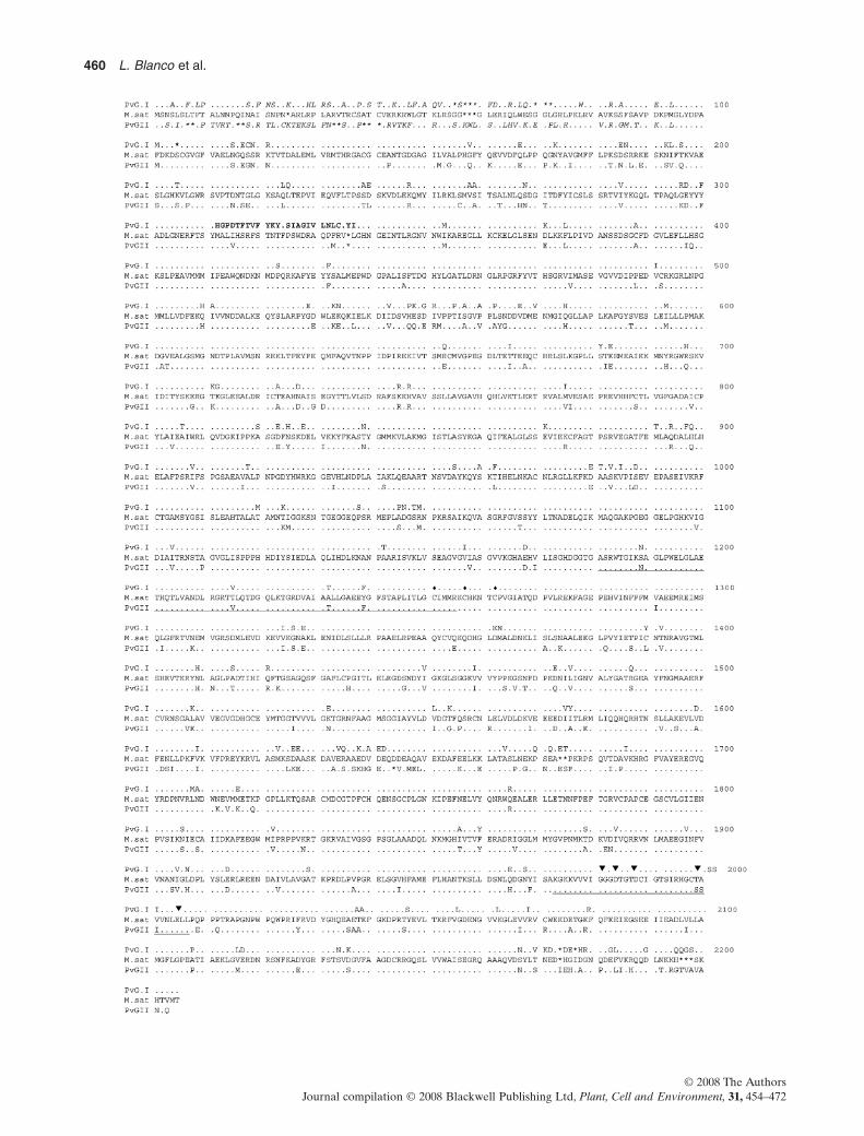
The sequence of the PvNADH-GOGAT-I cDNA(7406 bp) showed that it contains a 435 bp 5′ UTR withseveral translation stop codons in all three reading framesbefore the first ATG, and revealed an ORF 6591 bp long,starting at residue 436. A 367 bp long 3′ UTR and a 13 bppolyA+ were also defined. PvNADH-GOGAT-I ORF codesfor a 2196 amino acid polypeptide with a predicted molecu-lar mass of 241 kD and a theoretical pI of 6.1 (Fig. 1).PvNADH-GOGAT-II clone is 7050 bp long with a 5′ UTRof 189 bp with several translational stop codons in all threereading frames before the first ATG codon start site.The PvNADH-GOGAT-II clone showed a single ORF of6582 bp from 190 to 6771 bp, a 3′ UTR 264 bp long and15 bp polyA+. Putatively, the PvNADH-GOGAT-II codesfor a 2192 amino acid polypeptide with a predicted molecu-lar mass of 241 kD and a pI of 6.18 (Fig. 1). Completecoding regions of both clones showed 81% identity at thenucleotide level and 86% at the amino acid level. It isimportant to note that the theoretical pI predicted for thePvNADH-GOGAT deduced proteins of the present studyclosely correlated with the pI 6.069 and 6.159, respectively,reported for PvNADH-GOGAT-I and -II earlier (Chen &Cullimore 1988).
458 L. Blanco et al.
© 2008 The AuthorsJournal compilation © 2008 Blackwell Publishing Ltd, Plant, Cell and Environment, 31, 454–472

A BLAST search of the protein databases with thededuced amino acid sequences of PvNADH-GOGAT-Iand PvNADH-GOGAT-II showed an identity of 85 and83%, respectively, with the MsNADH-GOGAT (Fig. 1).Additionally, PvNADH-GOGAT-I and -II polypeptidesshowed an identity of 78% with A. thaliana and 74% withO. sativa NADH-GOGATs. PvNADH-GOGAT-I and -II,at amino acid level, showed an identity ranging from 39to 46% with Fd-GOGAT from A. thaliana (GLU1 andGLU2), Spinacia oleracea, Zea mays, O. sativa andGlycine max.
Functional domain analysis
Deduced amino acid sequences indicated that each of thePvNADH-GOGAT I and PvNADH-GOGAT-II contain apre-sequence of 98 and 96 amino acids, respectively, resem-bling that of MsNADH-GOGAT (Gregerson et al. 1993).The analysis using Plant-Ploc and WoLF PSORT pro-grammes suggested a plastid targeting sequence for bothPvNADH-GOGAT-I and PvNADH-GOGAT-II. It isextraordinary that these pre-sequences are rich in Ser andThr residues, but reduced in Asp, Glu and Tyr content,which is characteristic of the nuclear-encoded plastid pre-cursor protein (Karlin-Neumann & Tobin 1986; von Heijne,Steppuhn & Herrmann 1989).
In addition, the examination of the predicted polypep-tide sequences of the bean NADH-GOGAT isoformsrevealed three main domains that are putatively respon-sible for various enzymatic functions as delineated inNADH-GOGATs of alfalfa and other plants: The firstdomain is concerned with carrying out the glutamineamido transferase reaction (Figs 1 & 2). Interestingly,within this domain, PvNADH-GOGAT-I exhibited a26-amino acid sequence, which is different from bothPvNADH-GOGAT-II and MsNADH-GOGAT (desig-nated in bold letters in Fig. 1 and black square in Fig. 2).The second domain (underlined in Fig. 1) is involved inthe binding of FMN, and located in close proximity to threecysteine residues (indicated by diamonds in Fig. 1) puta-tively implicated as a binding site for [3Fe-4S] cluster.FMN and [3Fe-4S] carry out the electron transport tothe intermediate 2-iminoglutarate to form glutamate from2-oxoglutarate. The third domain is an NADH-bindingdomain (double underlined in Fig. 1) and contains five con-served residues (identified by arrowheads in Fig. 1) that areimportant for the binding of the cofactor (Gregerson et al.1993).
Genomic organization of NADH-GOGATsin the bean
In order to confirm if the two PvNADH-GOGAT clonesrepresent two different genes and to assess whether theremay be other homologs of these two NADH-GOGATs inthe bean, genomic DNA Southern blot analysis was per-formed. As is shown in Fig. 3, a single hybridizing fragment
was detected in each of the genomic DNA digestions tested.When a 611 bp probe containing 435 bp of the 5′ UTR and176 bp of the N-terminal coding region was used for thespecific detection of PvNADH-GOGAT-I, a single hybrid-ization fragment was detected in each of the DNA diges-tions tested (Fig. 3a). On the other hand, when a specificprobe for the detection of PvNADH-GOGAT-II (461 bplong fragment containing 189 bp of the 5′ UTR and 272 bpof the N-terminal coding region) was used, KpnI andEcoRV digestions generated each a single hybridizing frag-ment of the same size (Fig. 3b). PstI and XbaI digestionsalso generated each a unique hybridizing fragment ofalmost the same size but larger than those fragments gen-erated by KpnI and EcoRV digestions (Fig. 3b). Theseresults indicated that PvNADH-GOGAT-I and PvNADH-GOGAT-II represent two different NADH-GOGAT genesand that there is only a single copy of each gene in the P.vulgaris genome.
Expression of NADH-GOGATs in bean tissues
To determine whether the two P. vulgaris NADH-GOGATsare differentially expressed in various plant tissues,RT-PCR analysis was performed using RNA isolatedfrom roots, stem, young cotyledonary leaves, mature leaves,flowers, pods and nodules. As is shown in Fig. 4, transcriptsof both PvNADH-GOGATs were detected in all tissuesanalysed. In most tissues, excepting mature leaves,PvNADH-GOGAT-II expression was found to be higherthan that of PvNADH-GOGAT-I (Fig. 4a,b). RT-PCRanalysis utilizing mRNA isolated from the nodules har-vested from the plants 12–21 d after rhizobial inoculationshowed high and nearly constant levels of expression ofPvNADH-GOGAT-II during all stages of nodule develop-ment as compared to PvNADH-GOGAT-I (Fig. 4a,b). Incontrast, PvNADH-GOGAT-I showed a peak in expres-sion only at day 18 following inoculation and a declinethereafter. These results indicated that both NADH-GOGAT genes are differentially regulated in various beantissues including the nodules.
Relationship between PvNADH-GOGATexpression and nitrogen fixation
As already mentioned, NADH-GOGAT plays an impor-tant role in the ammonia assimilation pathway. In orderto delineate the extent of the participation of individualPvNADH-GOGATs in the assimilation of the ammoniaderived from the symbiotically nitrogen-fixing rhizobialcells, we analysed the expression of PvNADH-GOGAT-Iand -II in fix+ and fix- nodules induced respectively bythe CE3 wild type and the NifA- mutant strains of R. etli.RT-PCR analysis demonstrated that PvNADH-GOGAT-Iexpression was significantly lowered in non-fixing nodules(Fig. 4a,b). In contrast, the level of PvNADH-GOGAT-IIexpression remained unaffected in fix- nodules, until at least18 dpi, as compared to fix+ nodules. As these fix- nodules
NADH-GOGAT isoforms in Phaseolus vulgaris 459
© 2008 The AuthorsJournal compilation © 2008 Blackwell Publishing Ltd, Plant, Cell and Environment, 31, 454–472
460 L. Blanco et al.
© 2008 The AuthorsJournal compilation © 2008 Blackwell Publishing Ltd, Plant, Cell and Environment, 31, 454–472

have a normal morphology and contain bacteroides in theinfected cells, this result suggests that the expression ofPvNADH-GOGAT-I, but not of PvNADH-GOGAT-II, isaffected by the nitrogen status of the nodule and not is dueby a developmental deficiency.
In an analogous study in P. vulgaris, working with fix+ andfix- Rhizobium strains, Chen, Bennett & Cullimore (1990)
observed changes in the activity of NADH-GOGAT-Iisoenzyme in the nodules in response to nitrogen fixation,while the induction of NADH-GOGAT-II isoenzymeremained unaltered. Based on these findings, it was con-cluded that unlike NADH-GOGAT-I, the induction of theNADH-GOGAT-II isoenzyme occurs independently ofnitrogen fixation, but that nitrogen fixation was clearlyrequired to increase and maintain the magnitude of theinduction in P. vulgaris.
Regulation of PvNADH-GOGAT expression bycarbon and nitrogen compounds
As the nitrogen status of the nodule affected the PvNADH-GOGAT-I but not PvNADH-GOGAT-II expression, wedecided to study the effect of different nitrogen compoundsas well as sucrose on the expression of these two NADH-GOGATs in nodules. RT-PCR analysis revealed that thetranscript levels of PvNADH-GOGAT-I increased in thenodules derived from the plants treated with 0.5% sucrose,but no further enhancement was noticed when supple-mented with higher sucrose concentrations (Fig. 5a,b). Incontrast, the PvNADH-GOGAT-II expression increasedconcomitantly with the increase in sucrose concentration.The quantitative analysis of the sugars revealed that indeedthe levels of metabolizable sugars are enhanced in thenodules of the plants supplemented with exogenoussucrose, fructose or glucose (Silvente et al. 2008). Thesefindings, together with the above results, indicate that bothPvNADH-GOGATs are positively regulated by carbonreserves, thus, implicating carbon availability as a limitingfactor in the GS/GOGAT ammonia assimilation pathway innodules. The presence of different nitrogenous compoundsin the nutrient solution did not affect the expression ofPvNADH-GOGAT-II in the nodules (Fig. 5a,b). However,PvNADH-GOGAT-I expression declined in the presenceof amides (asparagine and glutamine), and most noticeablywith ureides (xanthine and allantoine) and with allopurinol,
Figure 1. Amino acid sequence alignment of bean PvNADH-dependent glutamate synthase (NADH-GOGAT)-I andPvNADH-GOGAT-II with alfalfa NADH-GOGAT. Residues numbering begins with the first amino acid in the proteins. Identicalresidues are indicated by dots. Asterisks denote the gaps introduced to maximize sequence similarity. Residues presented in italicsrepresent targeting leader sequences. Residues represented in bold letters denote dissimilar/distinct amino acid sequence in the amidotransferase domain of PvNADH-GOGAT-I. FMN- and NADH-binding domains are single and double underlined, respectively.Diamonds denote three cysteine residues involved in binding a [3Fe-4S] cluster. Five conserved residues that are important for NADHbinding are indicated by arrowheads.
Figure 2. Diagramatic representation of the predicted PvNADH-dependent glutamate synthase (NADH-GOGAT)-I andPvNADH-GOGAT-II polypeptides showing different functional domains. Black box indicates 26 amino acid sequence that isdifferent between these two polypeptides. Amino acid numbering starts with the initiation methionine in each polypeptide.
(a) (b)
Figure 3. DNA gel blot analyses of PvNADH-dependentglutamate synthase (NADH-GOGAT)-I and PvNADH-GOGAT-II in bean genome. Bean DNA (15 mg per lane) wasdigested with PstI, XbaI, KpnI or EcoRV, electophoresedthrough a 0.8% agarose gel, denatured, transferred to a nylonmembrane and probed with (a) PvNADH-GOGAT-I (611 bp)and (b) PvNADH-GOGAT-II (461 bp) specific probes.
NADH-GOGAT isoforms in Phaseolus vulgaris 461
© 2008 The AuthorsJournal compilation © 2008 Blackwell Publishing Ltd, Plant, Cell and Environment, 31, 454–472

which is an inhibitor of ureide synthesis. It is intriguing thatboth the ureides and the ureide inhibitor, like allopurinol,down regulate the expression of PvNADH-GOGAT-I.The inhibition of PvNADH-GOGAT-I by allopurinolprobably suggests that the metabolites upstream of xan-thine production (such as purine pathway intermediates)also negatively affect the PvNADH-GOGAT-I expression.
The above results indicate in one hand that both nodulePvNADH-GOGATs are positively regulated by carbonavailability and in the other, that nodule PvNADH-GOGAT-I is negatively regulated by metabolic intermedi-ates of the ureide biosynthetic pathway.
Isolation and characterization ofPvNADH-GOGAT-I and PvNADH-GOGAT-IIpromoters
To study how PvNADH-GOGAT-I and PvNADH-GOGAT-II genes are expressed in the roots and nodules,we isolated the promoter regions of these genes from thebean genomic DNA, employing the genome walking strat-egy, and utilized them to study their activities during rootand nodule development in bean hairy root system (seematerials and methods). The sequencing revealed that thecloned PvNADH-GOGAT-I and PvNADH-GOGAT-IIpromoters were 1501 and 1366 bp long, respectively. Thedetermination of the transcriptional start sites in the pro-moters, employing the Neural Network Promoter Predic-tion Program (see materials and methods), enabled theprecise delineation of a 436 bp long 5′ untranslated region
(5′ UTR) in PvNADH-GOGAT-I and a 192 bp long 5′ UTRin PvNADH-GOGAT-II cDNA transcripts (Supplemen-tary Fig. S1). A TATA box (TATATAT) was recognized inthe promoter of PvNADH-GOGAT-I at positions -460 to-466 bp upstream of the translational initiation site (+1),whereas in the PvNADH-GOGAT-II promoter, the TATAbox (TATAAA) was found to be at positions -215 to-220 bp. Putative CAAT box motifs were located at -930 to-933 bp and -254 to -257 bp, respectively, in the PvNADH-GOGAT-I and PvNADH-GOGAT-II promoters. Thus, theproximal regions of PvNADH-GOGAT-I and PvNADH-GOGAT-II promoters consist of the elements shown to beessential for the initiation of transcription in the promotersof other plants.
Tissue-specific expression patterns ofPvNADH-GOGAT-I and PvNADH-GOGAT-IIgenes during root and nodule development
To elucidate the patterns of expression of PvNADH-GOGAT-I and PvNADH-GOGAT-II genes during root andnodule development, we have utilized PvNADH-GOGATpromoters fused to the egfp-gusA chimeric reporter gene(Fig. 6 top panel) and produced compound bean plantswith transgenic hairy roots through A. rhizogenes-mediatedtransformation (see materials and methods).The hairy rootsof the plants transformed with the control vector (promot-erless egfp-gusA vector) served as the control. The trans-genic hairy roots from the symbiotically grown controlplants did not exhibit any GUS activity or GFP fluorescence
Figure 4. (a) RT-PCR analyses of beanPvNADH-dependent glutamate synthase(NADH-GOGAT)-I and PvNADH-GOGAT-II mRNA levels in variousorgans, and in developing nodulesformed by Rhizobium etli CE3 wild typeand NifA-mutant strains. Bean aquaporinexpression is used as an internalreference. (b) Histogram depicting theratios of the PvNADH-GOGAT-I andPvNADH-GOGAT-II band intensities(of the corresponding PCR productsrepresented in ‘a’) normalized against therespective aquaporin bands. R, root; S,stem; Ly, young cotyledonary leaf; L,mature leaf; F, flower; P0.5, pod of 0.5 cmlong; P1.5, pod of 1.5 cm long; dpi, dayspost inoculation.
(a)
(b)
462 L. Blanco et al.
© 2008 The AuthorsJournal compilation © 2008 Blackwell Publishing Ltd, Plant, Cell and Environment, 31, 454–472

in them.On the other hand, the transgenic roots of the plantstransformed with the vectors carrying PvNADH-GOGAT-Iand PvNADH-GOGAT-II promoter:egfp-gusA fusions dis-played GUS/GFP expression in roots and nodules, thoughthe intensity of expression varied depending on the pro-moter and developmental stage (Fig. 6). Both promotersexhibited an intense GUS expression in the meristematiccells located at the root tip (Fig. 6a,j). In the roots expressingthe PvNADH-GOGAT-I promoter, intense GUS stainingwas observed in both young and mature portions of theroot (Fig. 6a–f) as compared to the roots carrying thePvNADH-GOGAT-II promoter, which showed strongGUS expression mostly in the young portions of the roots(Fig. 6j) but not in the mature zones (Fig. 6k–m).In addition,the vascular tissues in the mature portions of the rootsexhibited comparatively greater GUS expression withPrPvNADH-GOGAT-I than with PrPvNADH-GOGAT-II(compare Fig. 6b–d with k–m).The visualization of the GFPexpression, however, revealed that the induction with boththe promoters was relatively more pronounced in root vas-cular tissues vis-a-vis cortical cells (Fig. 6g,p). During theinitiation of lateral root formation, the GUS expression withPvNADH-GOGAT-II promoter was relatively weaker inthe meristematic cells situated in the lateral root primordia,as compared to the strong expression shown by thePvNADH-GOGAT-I promoter (compare Fig. 6b with k,l).
On the other hand, the histochemical staining of the rootstransformed with both GOGAT promoters revealed rela-tively a strong GUS activity in the nodule primordia(Fig. 6c,d,m) as well as in the mature nodules (Fig. 6e,f,n,o).A detailed examination with confocal microscopy, asrevealed by EGFP expression,established that the activity ofboth promoters was mainly restricted to the infected corticalcells in the central zone of the nodules, although moreintensely with the PvNADH-GOGAT-II promoter than thePvNADH-GOGAT-I promoter (Fig. 6h,i,q,r). In contrast tothe infected cortical cells, the uninfected cells virtuallyshowed no EGFP expression. The outer cortical cells of thenodules, on the other hand, exhibited a low EGFP inductionwith both the promoters;here again in the outer cortical cellsthe expression with PrPvNADH-GOGAT-I being lessintense than PrPvNADH-GOGAT-II.
In order to further verify the sites of expression of bothPvNADH-GOGAT genes, and to precisely identify thelocations of accumulation of PvNADH-GOGAT-I andPvNADH-GOGAT-II mRNA transcripts in the nodules, weperformed in situ hybridization studies. For this, longitudi-nal sections of paraffin-embedded P. vulgaris mature rootnodule tissues were hybridized with sense or antisense 35SUTP-labelled RNA probes (Fig. 7) for either PvNADH-GOGAT-I or -II. Results showed that both PvNADH-GOGAT-I and -II are expressed within the central tissue ofthe bean root nodule (Fig. 7a,d). A higher magnification ofthis region demonstrated that PvNADH-GOGAT-I and -IImRNAs are mostly localized within the infected cells(Fig. 7j,k).As expected, the nodule sections probed with thePv-leghemoglobin transcript, used as a positive control,showed a high expression in the central tissue of the nodule(Fig. 7g), specifically, the infected cells. Thus, the in situhybridization results have provided additional confirmationshowing the infected cells as the major sites of expression ofboth NADH-GOGAT genes, as evidenced with the expres-sion of respective promoters in the bean nodules (seeabove; Fig. 6).
In silico identification of the potential cis-actingelements that regulate tissue-specific, andsugar and amino acid responsive expressionof the NADH-GOGAT promoters
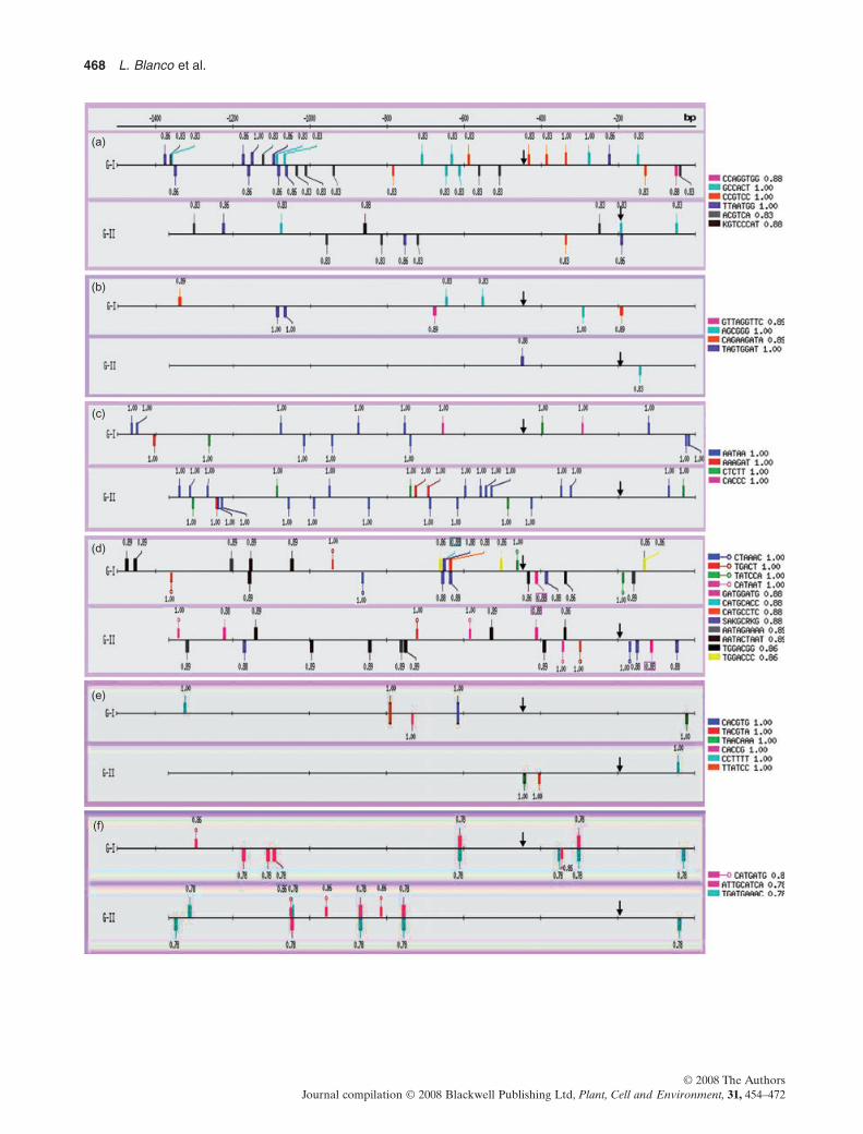
Since PvNADH-GOGAT-I and PvNADH-GOGAT-IIexhibited differential expression in root tissues (Fig. 6), weperformed in silico analysis to assess whether any differencescould be deciphered in these promoters with regard to theputative cis-elements that were shown to mediate tissue-specific expression in other plants. PLACE and PlantCAREdatabase searches revealed the presence of a large array ofputative cis-acting elements in both PvNADH-GOGAT-I(~95 motifs) and PvNADH-GOGAT-II (~70 motifs) pro-moters that potentially participate in the regulation of geneexpression in response to various cellular and environmentalcues. In both PvNADH-GOGAT promoters, we found cis-acting elements that were shown to mediate tissue-specificexpression in various other plants (Fig. 8a–c). Even though
(a)
(b)
Figure 5. (a) RT-PCR analyses of bean PvNADH-dependentglutamate synthase (NADH-GOGAT)-I and PvNADH-GOGAT-II mRNA levels in the nodules of the bean plantstreated with different carbon and nitrogen compounds. Beanaquaporin expression is used as an internal reference. Nodulesnursed with (left panel) 0.5, 1.0 or 2.0% sucrose, and (rightpanel) 10 mM asparagine (Asn), glutamine (Gln), xanthine(Xan), allantoin (Alla) or 2 mM allopurinol (Allop). C, untreatednodules served as control. (b) Histogram depicting the ratios ofthe PvNADH-GOGAT-I and PvNADH-GOGAT-II bandintensities (of the corresponding PCR products represented in‘a’) normalized against the respective aquaporin bands.
NADH-GOGAT isoforms in Phaseolus vulgaris 463
© 2008 The AuthorsJournal compilation © 2008 Blackwell Publishing Ltd, Plant, Cell and Environment, 31, 454–472

(a)
(g) (h) (i)
(p) (q) (r)
(b) (c)
(j) (k) (l) (m) (n) (o)
(d) (e) (f)
464 L. Blanco et al.
© 2008 The AuthorsJournal compilation © 2008 Blackwell Publishing Ltd, Plant, Cell and Environment, 31, 454–472

both promoters harboured meristematic and vascular tissue-specific regulatory motifs, the PvNADH-GOGAT-I pro-moter contained more of them, both in terms of number anddiversity, than PrPvNADH-GOGAT-II (Fig. 8a,b). In con-trast, PvNADH-GOGAT-II contained a higher concentra-tion of nodule-specific cis-elements in its promoter (Fig. 6c).In both promoters, we have also located several sugarresponsive plant cis-acting elements including those respon-sible for sucrose-inducible expression (Fig. 8d). cis-Elements that regulate sugar repression were also detectedin both promoters, though more of such motifs were found inPrPvNADH-GOGAT-I than in PrPvNADH-GOGAT-II(Fig. 8e). In addition, both promoters harboured consensussequences of cis-elements that resembled amino acidresponsive motifs encountered in mammalian asparaginesynthetase and CHOP (homolog of CAAAT/enhancer-binding protein) gene promoters (Guerrini et al. 1993;Bruhat et al. 2000, 2002; Barbosa-Tessmann et al. 2000;Fig. 8f). Consensus sequences of the conserved cis-elementslocated in both NADH-GOGAT promoters that potentiallymediate tissue-specific, and sugar and amino acid responsiveexpression,together with relevant references,are included inFig. 8 and its legend. In addition to the above mentionedcis-elements, both the promoters also exhibited stretches ofAT-rich regions and numerous perfect direct and invertrepeats of varying lengths up to 14 bp (not shown). Futurework can only decipher the functional significance of theserepeat motifs.
DISCUSSION
Legume plants can obtain their nitrogen from atmosphericN2 through symbiotic association with rhizobia. Ammonia,which is fixed by rhizobia, is assimilated in the plant via thejoint action of GS and GOGAT, which convert glutamineand 2-oxoglutarate into glutamate. These two reactions arenow generally agreed to be the primary route of nitrogenassimilation in plants, and the products glutamine andglutamate, donate their amido and amino groups for thesynthesis of most of the nitrogen-containing compounds ofthe cell. One interesting point is that, in non-nodulated beanplants, the assimilated ammonia is mobilized in the form ofamides, but when bean plants are symbiotically fixing nitro-gen, ureides are the main nitrogen-containing compoundsinvolved in the transport of the nitrogenase-derived
ammonia to the rest of the plant (Atkins 1982). To comple-ment our understanding of the ammonia assimilationprocess in bean nodules, we isolated and characterized twoNADH-GOGAT cDNAs, studied their expression in differ-ent plant tissues and assessed their regulation by carbonand nitrogen compounds in the root nodules. This researchextends our understanding of legume root nodule nitrogenmetabolism and NADH-GOGAT by: (1) demonstratingthat NADH-GOGAT in bean is encoded by two separategenes having distinct patterns of expression; (2) the expres-sion of the PvNADH-GOGAT-1 is reduced by productsof nitrogen fixation; (3) in the 5′-upstream region of bothPvNADH-GOGAT genes selected cis-elements are con-served between the two; (4) the 5′-upstream region of bothGOGAT genes drives reporter gene expression to infectedcells of root nodules;and (5) effective nitrogen fixation is notrequired for the expression of either PvNADH-GOGATgenes, suggesting that the regulation of the NADH-GOGATgenes in bean nodules may be under developmental control.
Two distinct clones of 7406 bp (PvNADH-GOGAT-I)and 7050 bp (PvNADH-GOGAT-II) cDNAs capable ofencoding a 241 kD protein each, were isolated and charac-terized. Both clones presented, at the amino acid level,an identity ranging from 74 to 85% with other NADH-GOGAT enzymes and a similarity lower than 46% withFd-GOGAT enzymes already characterized from differentplants. The analysis of the deduced amino acid sequence ofeach of the PvNADH-GOGATs showed an amido trans-ferase domain between amino acids 96 and 454 (Figs 1 & 2).In addition, three regions involved in cofactor binding arealso detected; namely, the sequences similar to those impli-cated in FMN binding (between the residues 1180 and1253) (Fig. 1, underlined), which are adjacent to three con-served cysteine residues (1251, 1257 and 1262) (Fig. 1, dia-monds) that are involved in binding the [3Fe-4S] cluster(Knaff et al. 1991; Sakakibara et al. 1991; Vanoni & Curti2005), and a third domain containing five highly conservedamino acids (Fig. 1, arrowheads), which are critical forNADH binding (Scruton, Berry & Perham 1990; Suzuki &Knaff 2005). These regions in PvNADH-GOGATs are verysimilar and almost in the same positions as that describedin MsNADH-GOGAT (Gregerson et al. 1993). Further, theamino acid sequence also exhibited a region (residues 1–98) encoding a transit peptide that predicts a plastidlocalization for both PvNADH-GOGATs.These data are in
Figure 6. Top panel: basic structure of T-DNA in the binary vectors pBI-PrPvNADH-GOGAT-I-EGFPGUS andpBI-PrPvNADH-GOGAT-II-EGFPGUS used in Agrobacterium rhizogenes K599-mediated hairy root transformation of Phaseolusvulgaris. (a–r) Patterns of PvNADH-GOGAT-I (a–i) and PvNADH-GOGAT-II (j–r) promoter activity as visualized by GUS and EGFPexpression in roots and nodules of P. vulgaris. Bright field stereo-micrographs showing histochemical staining of PvNADH-GOGAT-I andPvNADH-GOGAT-II promoter-mediated GUS expression in roots (a–d and j–m), lateral root primordia (b and k, l) and noduleprimordia (c, d and m), and fully matured nodules (e, f and n, o). Laser confocal micrographs showing PvNADH-GOGAT-I (g–i) andPvNADH-GOGAT-II (p–r) promoter-mediated GFP expression in 3-mm-thick root (g and p) and nodule (h, i and q, r) sections. Barlength equals 50 mm (g, i, p, r), 100 mm (h), 200 mm (a, j, q), 250 mm (b, c, k–m), 300 mm (e, n), 350 mm (f, o), 400 mm (d). BR, right border;BL, left border; PrNos, nopaline synthase promoter; NPTII, kanamycin resistance gene; T, nos terminator; PrPvN-G-I / II,PvNADH-GOGAT-I or II promoter; EGFPGUS, egfp-gusA fusion gene; S, SalI; X, XbaI; Sc, SacI; E, EcoRI; vb, vascular bundle; lrp,lateral root primordium; np, nodule primordium; dn, developing nodule; mn, mature nodule; c, cortex; ct, central tissue; oc, outer cortex; in,infected cell; un, uninfected cell.
NADH-GOGAT isoforms in Phaseolus vulgaris 465
© 2008 The AuthorsJournal compilation © 2008 Blackwell Publishing Ltd, Plant, Cell and Environment, 31, 454–472

(a) (b) (c)
(d) (e) (f)
(g) (h)
(j) (k)
(i)
Figure 7. Localization of PvNADH-dependent glutamate synthase (NADH-GOGAT)-I and II transcripts in Phaseolus vulgaris rootnodules by in situ hybridization. Longitudinal sections of paraffin embedded P. vulgaris mature root nodules probed with sense andantisense 35S UTP-labelled RNA probes. The signal specific to the radio-labelled probe is seen as white silver grains when tissue is viewedunder dark field optics and as dark particles when viewed under bright field. (a and b) Nodule sections probed with PvNADH-GOGAT-Iradio-labelled antisense (a) and sense (b) riboprobe. (d and e) Nodule sections probed with PvNADH-GOGAT-II radio-labelledantisense (d) and sense (e) riboprobe. (g and h) Nodule sections probed with Pv-leghemoglobin radio-labelled antisense (g) and sense (h)riboprobe. Panels (c, f and i) are bright field views of (a, d and g) to show nodule anatomy. (j and k) High magnification of portions of thepanels (a) and (d), respectively, that show signal (dark grains) located predominately in the infected cells of the central tissue.Magnification for (a–i) = 40¥, (j and k) = 400¥. Bar length equals 0.5 mm (a–i), 0.05 mm (j and k). ct, central tissue; oc, outer cortex; in,infected cell; un, uninfected cell.
466 L. Blanco et al.
© 2008 The AuthorsJournal compilation © 2008 Blackwell Publishing Ltd, Plant, Cell and Environment, 31, 454–472

concordance with the previous results, which indicated thatin bean nodules, the two NADH-GOGAT isoenzymes arelocalized in the plastids (Chen & Cullimore 1989). In con-trast to the above, however, the amino acid analysis alsorevealed a region that distinguishes PvNADH-GOGATsfrom each other. A sequence comprising of 26 amino acidsin the amido transferase domain of PvNADH-GOGAT-I isfound to be completely different from the correspondingsequence in PvNADH-GOGAT-II (Fig. 2, designated inbold letters). This dissimilarity in the amino acid composi-tion at the amido transferase domain between bothPvNADH-GOGATs could explain the different affinitiesfor L-glutamine between the two NADH-GOGAT isoen-zymes reported by Chen & Cullimore (1988).
The data presented here demonstrate that contrary towhat has been reported for other plants, in P. vulgaris,NADH-GOGAT is encoded by two different genes, andstrongly suggest that these genes encode the two NADH-GOGAT isoenzymes reported by Chen & Cullimore(1988). The fact that the PvNADH-GOGAT-II, as com-pared to PvNADH-GOGAT-I, is strongly expressed innodules (present study; Figs 4a,b & 6f,i,o,r) and the NADH-GOGAT-II isoenzyme, in contrast to NADH-GOGAT-Iisoform, accounted for most of the nodule GOGAT activity(Chen & Cullimore 1988), will allow us to suggest thatthe PvNADH-GOGAT-II gene most likely encodes theNADH-GOGAT-II isoenzyme, while the PvNADH-GOGAT-I gene codes for the NADH-GOGAT-I isoen-zyme.This conclusion gains further support from the findingthat the expression of NADH-GOGAT-I, but not NADH-GOGAT-II, is affected by nitrogen fixation in the nodules(Fig. 4a,b), as was similarly reported for NADH-GOGAT-Iand II isoenzyme activities in response to nitrogen fixation(Chen et al. 1990).
The interaction between cis-regulatory elements presentin a promoter region and transacting factors is an indispens-able requirement for regulating gene expression. Specificcis-regulatory elements in the promoter are probablyshared by the genes that are responsive to similar cellularcues/factors. In silico analysis against the plant regulatoryelement databases revealed the presence of cis sequencespotentially responsible for organ-/tissue-specific, and sugarresponsive expression in the promoters of both PvNADH-GOGAT-I and -II (Fig. 8). The analysis showed that eventhough both promoters contained meristematic and vascu-lar tissue-specific regulatory motifs, PvNADH-GOGAT-Ipromoter harboured more of such cis-elements, both interms of number and diversity, than PrPvNADH-GOGAT-II (Fig. 8a,b). In contrast, PvNADH-GOGAT-IIcontained higher concentration of nodule-specific cis-elements in its promoter (Fig. 8c). In consonance with theseobservations, we found a relatively higher activity ofPvNADH-GOGAT-I promoter in the meristematic tissues(particularly lateral root primordia), as well as in the vas-cular traces (Fig. 6a–d), while PvNADH-GOGAT-II pro-moter exhibited greater activity in the nodules (Fig. 6q,r).In both promoters, we have also located several sugarresponsive plant cis-acting elements (Fig. 8d,e) which
perhaps participate in the sucrose-mediated regulation ofthe expression of both these NADH-GOGAT genes as well(Fig. 5a). RT-PCR analyses showed that the PvNADH-GOGAT-I expression, but not that of PvNADH-GOGAT-II, is regulated by glutamine and asparagine (Fig. 5b).Searches of plant regulatory element databases did notpresent any records of cis-elements that mediate amino acidresponsive expression of plant promoters. On the otherhand, the examination of the promoter sequences ofboth PvNADH-GOGAT genes against animal genome se-quences revealed the presence of consensus sequencesresembling amino acid responsive cis-elements encoun-tered in mammalian asparagine synthetase and CHOP genepromoters (Fig. 8f). In the mammalian asparagine syn-thetase gene, the palindromic sequence CATGATG local-ized in the proximal portion of the promoter region wasfound to be necessary for the regulation by amino acidavailability (Guerrini et al. 1993). In our studies, thePvNADH-GOGAT-I promoter was also found to carry thepalindromic sequence CATGATG in its proximal region.Future work dealing with deletion and mutational analysesof the NADH-GOGAT promoters can only clarify theprecise significance and function of the above describedpotential cis-acting elements in the regulation of tissue-specific, and sugar and amino acid responsive expression ofthese genes in bean roots.
As compared to PvNADH-GOGAT-II, relatively lowexpression of PvNADH-GOGAT-I in nodules (Figs 4 & 6i)and its down regulation in the presence of amides andureides (Fig. 5b) strongly suggest that this NADH-GOGATisoform may not be playing a main role in the ammoniaassimilation process in bean plant, which is a ureide trans-porter (Atkins 1982). However, a moderate enhancementin the expression of PvNADH-GOGAT-I in nodules at day18 (Fig. 4) probably implies that this isoform may be per-forming a supportive role in ammonia assimilation duringthe active phase of nitrogen fixation. In contrast, high levelsof expression of PvNADH-GOGAT-II in nodules (Fig. 4),particularly in the nitrogen-fixing infected cells (Figs 6r &7d), and the maintenance of elevated levels of expressioneven in the presence of excessive concentrations of amidesand ureides (Fig. 5b) point to the possibility that PvNADH-GOGAT-II may be the main isoform that mediatesammonia assimilation. The differential susceptibility forexpression of these two NADH-GOGATs to various nitro-gen compounds could be an adaptative mechanism allowingthe N2-fixing bean plants to mobilize ammonia,derived fromnitrogenase activity,efficiently in the form of ureides (Atkins1982) instead of amides.
ACKNOWLEDGMENTS
We are grateful to Araceli Sánchez for the technical supportat the greenhouse, and Paul Gaytan and Eugenio López-Bustos for the synthesis of the primers. This work was sup-ported in part by Consejo Nacional de Ciencia y Tecnología(CONACyT, Mexico) grant G31751-B and DirecciónGeneral del Personal Académico (DGAPA-UNAM) grant
NADH-GOGAT isoforms in Phaseolus vulgaris 467
© 2008 The AuthorsJournal compilation © 2008 Blackwell Publishing Ltd, Plant, Cell and Environment, 31, 454–472
(a)
(b)
(c)
(d)
(e)
(f)
468 L. Blanco et al.
© 2008 The AuthorsJournal compilation © 2008 Blackwell Publishing Ltd, Plant, Cell and Environment, 31, 454–472

IN215407 to M. L-F., and Mexico-USDA/Foreign Agricul-ture Service Grant MX 161 to C.V.
REFERENCES
Altschul S.F., Gish W., Miller W., Myers E.W. & Lipman D.J. (1990)Basic local alignment search. Journal of Molecular Biology 215,403–410.
Anderson M.P., Vance C.P., Heichel G.H. & Miller S. (1989) Puri-fication and characterization of NADH-glutamate synthase fromalfalfa root nodules. Plant Physiology 90, 351–358.
Atkins C.P. (1982) De novo purine synthesis in nitrogen-fixingnodules of cowpea (Vigna unguiculata L. Walp.) and soybean(Glycine max L.Merr). Plant Physiology 70, 55–60.
Barbosa-Tessmann I.P., Chen C., Zhong C., Siu F., Schuster S.M.,Nick H.S. & Kilberg M.S. (2000) Activation of the human aspar-agine synthetase gene by the amino acid response and the endo-plasmic reticulum stress response pathways occurs by commongenomic elements. Journal of Biological Chemistry 275, 26976–26985.
Bruhat A., Jousse C., Carraro V., Reimold A.M., Ferrara M. &Fafournoux P. (2000) Amino acids control mammalian genetranscription: activating transcription factor 2 is essential foramino acid responsiveness of the CHOP promoter. Molecularand Cellular Biology 20, 7192–7204.
Bruhat A., Averous J., Carraro V., Zhong C., Reimold A.M.,Kilberg M.S. & Fafournoux P. (2002) Differences in the molecu-lar mechanisms involved in the transcriptional activation of theCHOP and asparagine synthetase genes in response to aminoacid deprivation or activation of the unfolded protein response.Journal of Biological Chemistry 277, 48107–48114.
Chen F.L. & Cullimore J.V. (1988) Two isozymes of NADH-dependent glutamate synthase in root nodules of Phaseolusvulgaris L. Plant Physiology 88, 1411–1417.
Chen F.L. & Cullimore J.V. (1989) Location of two isoenzymesof NADH-dependent glutamate synthase in root nodules ofPhaseolus vulgaris L. Planta 179, 441–447.
Chen F.L., Bennett M.J. & Cullimore J.V. (1990) Effect of thenitrogen supply on the activities of isoenzymes of NADH-dependent glutamate synthase and glutamine synthetase in rootnodules of Phaseolus vulgaris L. Journal of Experimental Botany41, 1215–1221.
Chou K.C. (2005) Using amphiphilic pseudo amino acid composi-tion to predict enzyme subfamily classes. Bioinformatics 21,10–19.
Chou K.C. & Shen H.B. (2007) Large-scale plant protein subcellu-lar location prediction. Journal of Cellular Biochemistry 100,665–678.
Coschigano K.T., Melo-Oliveira R., Lim J. & Coruzzi G.M. (1998)Arabidopsis gls mutants and distinct Fd-GOGAT genes: impli-cations for photorespiration and primary nitrogen assimilation.Plant Cell 10, 741–752.
Curti B., Vanoni M.A., Verzotti E. & Zanetti G. (1995) Glutamatesynthase: a complex iron-sulfur flavoprotein. BiochemicalSociety Transactions 24, 95–99.
Elliott K.A. & Shirsat A.H. (1998) Promoter regions of the extAextension gene from Brassica napus control activation inresponse to wounding and tensile stress. Plant Molecular Biology37, 675–687.
Estrada-Navarrete G., Alvarado-Affantranger X., Olivares J.-E.,et al. (2006) Agrobacterium rhizogenes transformation of thePhaseolus spp.: a tool for functional genomics. Molecular Plant-Microbe Interactions 19, 1385–1393.
Fehlberg V., Vieweg M.F., Dohmann E.M.N., Hohnjec N., PühlerA., Perlick A.M. & Küster H. (2005) The promoter of the leghae-moglobin gene VfLb29: functional analysis and identification ofmodules necessary for its activation in the infected cells of rootnodules and in the arbuscule-containing cells of mycorrhizalroots. Journal of Experimental Botany 56, 799–806.
Geisler M., Kleczkowski L.A. & Karpinski S. (2006) A universalalgorithm for genome-wide in silico identification of biologicallysignificant gene promoter putative cis-regulatory-elements;identification of new elements for reactive oxygen species andsucrose signaling in Arabidopsis. The Plant Journal 45, 384–398.
Goto S., Akagawa T., Kojima S., Hayakawa T. & Yamaya T. (1998)Organization and structure of NADH-dependent glutamate syn-thase gene from rice plants. Biochimica et Biophysica Acta 1387,298–308.
Gregerson R.G., Miller S.S., Twary S.N., Gantt J.S. & Vance C.P.(1993) Molecular characterization of NADH-glutamate syn-thase from alfalfa nodules. Plant Cell 5, 215–226.
Grierson C., Du J.S., Zabala M.T., Beggs K., Smith C., HoldsworthM. & Bevan M. (1994) Separate cis sequences and trans factorsdirect metabolic and developmental regulation of a potato tuberstorage protein gene. The Plant Journal 5, 815–826.
Guerrini L., Gong S.S., Mangasarian K. & Basilico C. (1993) Cis-and trans-acting elements involved in amino acid regulation ofasparagine synthetase gene expression. Molecular Cell Biology13, 3202–3212.
Hayakawa T., Yamaya T., Kamachi K. & Ojima K. (1992)Purification, characterization, and immunological properties of
Figure 8. Diagram depicting the occurrence and location of various putative motifs within PvNADH-dependent glutamate synthase(NADH-GOGAT)-I and -II promoters that mediate organ-/tissue-specific, and sugar and amino acid responsive expression. Consensussequences for (a) meristem-specific expression: CCAGGTGG (Kosugi, Suzuka & Ohashi 1995), GCCACT, CCGTCC (Meshi, Taoka& Iwabuchi 2000), TTAATGG (Kamiya et al. 2003), ACGTCA (Terada et al. 1995) and KGTCCCAT (Kosugi et al. 1995; Kosugi &Ohashi 1997; Klinedinst et al. 2000); (b) vascular expression: GTTAGGTTC (Patzlaff et al. 2003), AGCGGG (Lacombe et al. 2000),CAGAAGATA (Yin et al. 1997) and TAGTGGAT (Elliott & Shirsat 1998; Nontachaiyapoom et al. 2007); (c) nodule-specific expression:AATAA, AAAGAT, CTCTT and CACCC (Sandal, Bojsen & Marcker 1987; Stougaard et al. 1990; Szczyglowski et al. 1994; Vieweg et al.2004; Fehlberg et al. 2005); (d) sugar-inducible expression: CTAAAC (Zourelidou et al. 2002), TGACT (Sun et al. 2003), TATCCA (Lu,Lim & Yu 1998; Lu et al. 2002), CATAAT (Li et al. 2006), GATGGATG, CATGCACC, CATGCCTC, SAKGCRKG (Geisler, Kleczkowski& Karpinski 2006), AATAGAAAA (Sun et al. 2003), AATACTAAT (Grierson et al. 1994), TGGACGG and TGGACCC (Maeo et al.2001; Morikami et al. 2005); (e) sugar repression: CACGTG (Menkens, Schindler & Cashmore 1995; Urwin & Jenkins 1997), TACGTA(Toyofuku, Umemura & Yamaguchi 1998), CACCG (Niu, Helentjaris & Bate 2002), TAACAAA, CCTTTT (Morita et al. 1998) andTTATCC (Tatematsu et al. 2005); (f) amino acid responsive expression: CATGATG (Guerrini et al. 1993), ATTGCATCA (Bruhat et al.2000) and TGATGAAAC (Barbosa-Tessmann et al. 2000; Bruhat et al. 2002). Numbers following the motif sequences depict the degreeof conservation of the cis-elements (1.00 depicts the perfect match). Actual cis-element sequences, together with the highest degree ofconservation of a given motif, are represented on the right side of the panels.
NADH-GOGAT isoforms in Phaseolus vulgaris 469
© 2008 The AuthorsJournal compilation © 2008 Blackwell Publishing Ltd, Plant, Cell and Environment, 31, 454–472

NADH-dependent glutamate synthase from rice cell cultures.Plant Physiology 98, 1317–1322.
Hayakawa T., Nakamura T., Hattori F., Mae T., Ojima K. &Yamaya T. (1994) Cellular localization of NADH-dependentglutamate-synthase protein in vascular bundles of unexpandedleaf blades and young grains of rice plants. Planta 193, 455–460.
Hayakawa T., Hopkins L., Peat L.J., Yamaya T. & Tobin A.K.(1999) Quantitative intercellular localization of NADH-dependent glutamate synthase protein in different types of rootcells in rice plants. Plant Physiology 119, 409–416.
von Heijne G., Steppuhn J. & Herrmann R.G. (1989) Domainstructure of mitochondrial and chloroplast targeting peptides.European Journal of Biochemistry 180, 535–545.
Higo K., Ugawa Y., Iwamoto M. & Korenaga T. (1999) Plant cis-acting regulatory DNA elements (PLACE) database. NucleicAcids Research 27, 297–300.
Horton P., Park K.-J., Obayashi T. & Nakai K. (2006) Protein sub-cellular localization prediction with WoLF PSORT. Proceedingsof the 4th Annual Asia Pacific Bioinformatics ConferenceAPBC06, Taipei, Taiwan. pp. 39–48.
Horton P., Park K.-J., Obayashi T., Fujita N., Harada H., Adams-Collier C.J. & Nakai K. (2007) WoLF PSORT: protein localiza-tion predictor. Nucleic Acids Research 35, W585–W587.
Kamiya N., Nagasaki H., Morikami A., Sato Y. & Matsuoka M.(2003) Isolation and characterization of a rice WUSCHEL-typehomoebox gene that is specifically expressed in the central cellsof a quiescent center in the root apical meristem. The PlantJournal 35, 429–441.
Karimi M., Inze D. & Depicker A. (2002) Gateway vectors forAgrobacterium-mediated plant transformation. Trends in PlantScience 7, 193–195.
Karlin-Neumann G.A. & Tobin E.M. (1986) Transit peptide ofnuclear encoded chloroplast proteins share a common acidframework. EMBO Journal 5, 9–13.
Klinedinst S., Pascuzzi P., Redman J., Desai M. & Arias J. (2000) Axenobiotic-stress-activated transcription factor and its cognatetarget genes are preferentially expressed in root tip meristems.Plant Molecular Biology 42, 679–688.
Knaff D.B., Willie A., Long J.E., Kriauciunas A., Durham B. &Millett F. (1991) Reaction of cytochrome C2 with photosyntheticreaction centers from Rhodopseudomonas viridis. Biochemistry30, 1303–1310.
Kosugi S. & Ohashi Y. (1997) PCF1 and PCF2 specifically bind tocis elements in the rice proliferating cell nuclear antigen gene.Plant Cell 9, 1607–1619.
Kosugi S., Suzuka I. & Ohashi Y. (1995) Two of three promoterelements identified in a rice gene for proliferating cell nuclearantigen are essential for meristematic tissue-specific expression.The Plant Journal 7, 877–886.
Lacombe E., Van Doorsselaere J., Boerjan W., Boudet A.M. &Grima-Pettenati J. (2000) Characterization of cis-elementsrequired for vascular expression of the cinnamoyl CoA reduc-tase gene and for protein-DNA complex formation. The PlantJournal 23, 663–676.
Lancien M., Gadal P. & Hodges M. (2000) Enzyme redundancy andthe importance of 2-oxoglutarate in higher plant ammoniumassimilation. Plant Physiology 123, 817–824.
Lea P.J. & Miflin B.J. (2003) Glutamate synthase and the synthesisof glutamate in plants. Plant Physiology and Biochemistry 41,555–564.
Lea P.J., Robinson S.A. & Stewart G.R. (1990) The enzymology andmetabolism of gutamine, glutamate and asparagines. In The Bio-chemistry of Plants (ed. B.J. Miflin), pp. 121–159. AcademicPress, San Diego, CA, USA.
Lescot M., Déhais P., Thijs G., Marchal K., Moreau Y., Van de PeerY., Rouzé P. & Rombauts S. (2002) PlantCARE, a database of
plant cis-acting regulatory elements and a portal to tools for insilico analysis of promoter sequences. Nucleic Acids Research 30,325–327.
Li Y., Lee K.K., Walsh S., Smith C., Hadingham S., Sorefan K.,Cawley G. & Bevan M.W. (2006) Establishing glucose- andABA-regulated transcription networks in Arabidopsis bymicroarray analysis and promoter classification using a Rel-evance Vector Machine. Genome Research 16, 414–427.
Lu C.A., Lim E.K. & Yu S.M. (1998) Sugar response sequence inthe promoter of a rice alpha-amylase gene serves as a transcrip-tional enhancer. Journal of Biological Chemistry 273, 10120–10131.
Lu C.A., Ho T.H.D., Ho S.L. & Yu S.M. (2002) Three novel MYBproteins with one DNA binding repeat mediate sugar andhormone regulation of alpha-amylase gene expression. PlantCell 14, 1963–1980.
Maeo K., Tomiya T., Hayashi K., Akaike M., Morikami A., IshiguroS. & Nakamura K. (2001) Sugar-responsible elements in thepromoter of a gene for b-amylase of sweet potato. Plant Molecu-lar Biology 46, 627–637.
Marck C. (1988) ‘DNA strider’: a ‘C’ program for the fast analysisof DNA and protein sequences on the Apple Macintosh familyof computers. Nucleic Acids Research 16, 1829–1836.
Menkens A.E., Schindler U. & Cashmore A.R. (1995) The G-box:a ubiquitous regulatory DNA element in plants bound by theGBF family of bzip proteins. Trends in Biochemistry 20, 506–510.
Meshi T., Taoka K. & Iwabuchi M. (2000) Regulation of histonegene expression during the cell cycle. Plant Molecular Biology43, 645–659.
Miflin B.J. & Lea P.J. (1976) The pathway of ammonia assimilationin plants. Phytochemistry 15, 873–885.
Morikami A., Matsunaga R., Tanaka Y., Suzuki S., Mano S. &Nakamura K. (2005) Two cis-acting regulatory elements areinvolved in the sucrose-inducible expression of the sporamingene promoter from sweet potato in transgenic tobacco. Molecu-lar Genetics and Genomics 272, 690–699.
Morita A., Umemura T., Kuroyanagi M., Futsuhara Y., Perata P. &Yamaguchi J. (1998) Functional dissection of a sugar-repressedalpha-amylase gene (Ramy1A) promoter in rice embryos. FEBSLetters 423, 81–85.
Nagel R., Elliot A., Masel A., Birch R.G. & Manners J.M. (1990)Electroporation of binary Ti plasmid vector into Agrobacteriumtumefaciens and Agrobacterium rhizogenes. FEMS MicrobiologyLetters 67, 325–328.
Niu X., Helentjaris T. & Bate N.J. (2002) Maize ABI4 binds cou-pling element1 in abscisic acid and sugar response genes. PlantCell 14, 2565–2575.
Nontachaiyapoom S., Scott P.T., Men A.E., Kinkema M., SchenkP.M. & Gresshoff P.M. (2007) Promoters of orthologous Glycinemax and Lotus japonicus nodulation autoregulation genes inter-changeably drive phloem-specific expression in transgenicplants. Molecular Plant–Microbe Interactions 20, 769–780.
Oliveira I.C., Lam H.M., Coschigano K., Melo-Oliveira R. &Coruzzi G. (1997) Molecular-genetic dissection of ammoniumassimilation in Arabidopsis thaliana. Plant Physiology and Bio-chemistry 35, 185–198.
Ortega J.L., Sánchez F., Soberón M. & Lara M. (1992) Regulationof nodule glutamine synthetase by CO2 levels in bean (Phaseo-lus vulgaris L). Plant Physiology 98, 584–587.
Patzlaff A., Newman L.J., Dubos C., Whetten R.W., Smith C.,McInnis S., Bevan M.W., Sederoff R.R. & Campbell M.M. (2003)Characterisation of Pt MYB1, an R2R3-MYB from pine xylem.Plant Molecular Biology 53, 597–608.
Reddy P.M., Ladha J.K., Ramos M.S., Maillet F., Hernandez R.J.,Torrizo L.B., Oliva N.P., Datta S.K. & Datta K. (1998) Rhizobiallipochitoligosaccharide nodulation factors activate expression of
470 L. Blanco et al.
© 2008 The AuthorsJournal compilation © 2008 Blackwell Publishing Ltd, Plant, Cell and Environment, 31, 454–472

the legume early nodulin gene ENOD12 in rice. The PlantJournal 14, 693–702.
Redinbaugh M.G. & Campbell W.H. (1993) Glutamine synthetaseand ferredoxin-dependent glutamate synthase expression in themaize (Zea mays) root primary response to nitrate. Plant Physi-ology 101, 1249–1255.
Reese M.G. (2001) Application of a time-delay neural network topromoter annotation in the Drosophila melanogaster genome.Computational Chemistry 26, 51–56.
Sakakibara H., Watanabe M., Hase T. & Sugiyama T. (1991)Molecular cloning and characterization of complementary DNAencoding ferredoxin-dependent glutamate synthase in maizeleaf. Journal of Biological Chemistry 266, 2028–2035.
Sambrook J., Fritsch E.F. & Maniatis T. (1989) Molecular Cloning:A Laboratory Manual, 2nd edn, Cold Spring Harbor LaboratoryPress, New York, NY, USA.
Sandal N.N., Bojsen K. & Marcker K.A. (1987) A small family ofnodule-specific gene from soybean. Nucleic Acids Research 15,1507–1519.
Scruton N.S., Berry A.B. & Perham R.N. (1990) Redesign of thecoenzyme specificity of a dehydrogenase by protein engineering.Nature 343, 38–43.
Shen H.B. & Chou K.C. (2006) Ensemble classifier for proteinfolding pattern recognition. Bioinformatics 22, 1717–1722.
Silvente S., Reddy P.M., Khandual S., Blanco L., Alvarado-Affantranger X., Sanchez F. & Lara-Flores M. (2008) Evidencefor sugar signaling in the regulation of asparagine synthetasegene expressed in Phaseolus vulgaris roots and nodules. Journalof Experimental Botany (communicated).
Stougaard J., Jorgensen J.E., Christensen T., Kuhle A. & MarckerK.A. (1990) Independence and nodule specifity of cis actingregulatory elements in the soybean leghemoglobin ibc3 and N23gene promoters. Molecular and General Genetics 220, 353–360.
Summerfield R.J., Huxley P.A. & Minchin F.R. (1977) Plant hus-bandry and management techniques for growing grain legumesunder simulated tropical conditions in controlled environments.Experimental Agriculture 13, 81–92.
Sun C., Palmqvist S., Olsson H., Borén M., Ahlandsberg S. &Jansson C. (2003) A novel WRKY transcription factor,SUSIBA2, participates in sugar signaling in barley by binding tothe sugar-responsive elements of the iso1 promoter. Plant Cell15, 2076–2093.
Suzuki A. & Knaff D.B. (2005) Glutamate synthase: structural,mechanistic and regulatory properties, and role in the amino acidmetabolism. Photosynthetic Research 83, 191–217.
Suzuki A. & Rothstein S. (1997) Structure and regulation offerredoxin-dependent glutamate synthase from Arabidopsisthaliana: cloning of cDNA, expression in different tissues of wild-type and gltS mutant strains, and light induction. EuropeanJournal of Biochemistry 243, 708–718.
Szczyglowski K., Szabados L., Fujimoto S.Y., Silver D. & de BruijnF.J. (1994) Site-specific mutagenesis of the nodule-infected cellexpression (NICE) element and the AT-rich element ATRE-BS2asterisk of the Sesbania rostrata leghemoglobin glb3 promoter.Plant Cell 6, 317–332.
Tatematsu K., Ward S., Leyser O., Kamiya Y. & Nambara E. (2005)Identification of cis-elements that regulate gene expressionduring initiation of axillary bud outgrowth in Arabidopsis. PlantPhysiology 138, 757–766.
Temple S.J., Vance C.P. & Gantt S.J. (1998) Glutamate synthaseand nitrogen assimilation. Trends in Plant Science 3, 51–56.
Terada R., Nakayama T., Iwabuchi M. & Shimamoto K. (1995) Atype I element composed of the hexamer (ACGTCA) andoctamer (CGCGGATC) motifs plays a role(s) in meristematicexpression of a wheat histone H3 gene in transgenic rice plants.Plant Molecular Biology 27, 17–26.
Toyofuku K., Umemura T. & Yamaguchi J. (1998) Promoter ele-ments required for sugar-repression of the RAmy3D gene foralpha-amylase in rice. FEBS Letters 428, 275–280.
Trepp G.B., Plank D.W., Gantt J.S. & Vance C.P. (1999a) NADH-glutamate synthase in alfalfa root nodules. Immunocytochemicallocalization. Plant Physiology 119, 829–837.
Trepp G.B., van de Mortel M., Yoshoika H., Miller S.S., Samac D.A.,Gantt J.S. & Vance C.P. (1999b) NADH-glutamate synthase inalfalfa roots. Genetic regulation and cellular expression. PlantPhysiology 119, 817–828.
Urwin N.A.R. & Jenkins G.I. (1997) A sucrose repression elementin the Phaseolus vulgaris rbcS2 gene promoter resembles ele-ments responsible for sugar stimulation of plant and mammaliangenes. Plant Molecular Biology 3, 929–942.
Vance C.P. (1997) The molecular biology of N metabolism. In PlantMetabolism (eds D.T. Dennis, D.H. Turpin, D.D. Lefebre & D.B.Layzell), pp. 449–477. Longman Scientific, London, UK.
Vance C.P., Gregerson R.G., Robinson D.L., Miller S.S. & Gantt J.S.(1994) Primary assimilation of nitrogen in alfalfa nodules:molecular features of the enzyme involved. Plant Science 101,51–64.
Vance C.P., Miller S.S., Gregerson R.G., Samac D.A., RobinsonD.L. & Gantt J.S. (1995) Alfalfa NADH-dependent glutamatesynthase: structure of gene and importance in symbiotic N2 fixa-tion. Plant Journal 8, 345–358.
Vanoni M.A. & Curti B. (2005) Structure-function studies on theiron-sulfur flavoenzyme glutamate synthase: an unexpectedlycomplex self-regulated enzyme. Archives of Biochemistry andBiophysics 433, 193–211.
Vieweg M.F., Frühling M., Quandt H.J., Heim U., Bäumlein H.,Pühler A., Küster H. & Perlick A.M. (2004) The promoter of theVicia faba L. leghemoglobin gene VfLb29 is specifically activatedin the infected cells of root nodules and in the arbuscule-containing cells of mycorrhizal roots from different legume andnon-legume plants. Molecular Plant–Microbe Interactions 17,62–69.
Wallsgrove R.M., Lea P.J. & Miflin B.J. (1979) Distribution ofenzymes of nitrogen metabolism within pea leaf cells. PlantPhysiology 63, 232–236.
Yamaya T., Hayakawa T., Tanasawa K., Kamachi K., Mae T. &Ojima K. (1992) Tissue distribution of glutamate synthase andglutamine synthetase in rice leaves. Occurrence of NADH-dependent glutamate synthase protein and activity in the unex-panded non-green leaf blades. Plant Physiology 100, 917–925.
Yamaya T., Tanno H., Hirose N., Watanabe S. & Hayakawa T.(1995) A supply of nitrogen causes increase in the level ofNADH-dependent glutamate synthase protein and the activityof the enzyme in roots of rice seedlings. Plant and Cell Physiol-ogy 36, 1197–1204.
Yin Y., Zhu Q., Da S., Lam C. & Beachy R.N. (1997) RF2a, a bZIPtranscriptional activator of the phloem-specific rice tungro bacil-liform virus promoter, functions in vascular development.EMBO Journal 16, 5247–5259.
Zourelidou M., de Torres-Zabala M., Smith C. & Bevan M.W.(2002) Storekeeper defines a new class of plant-specific DNA-binding proteins and is a putative regulador of patatin expres-sion. The Plant Journal 30, 489–497.
Received 25 August 2007; received in revised form 13 December2007; accepted for publication 19 December 2007
SUPPLEMENTARY MATERIAL
The following supplementary material is available for thisarticle:
NADH-GOGAT isoforms in Phaseolus vulgaris 471
© 2008 The AuthorsJournal compilation © 2008 Blackwell Publishing Ltd, Plant, Cell and Environment, 31, 454–472
Figure S1. Sequences of the Phaseolus vulgaris NADH-dependent glutamate synthase (NADH-GOGAT)-I andNADH-GOGAT-II 5′ flanking regions. The upstream1501 bp and 1366 bp flanking regions of the NADH-GOGAT-I and NADH-GOGAT-II genes, respectively, fromthe putative translation start codons are shown. TATAbox is highlighted in red, and CAAT boxes in green,and predicted transcription start site is shaded in blue.Uppercase letters denoted in black correspond to the1065 bp and 1174 bp regions of the putative NADH-GOGAT-I and NADH-GOGAT-II promoters, whereas theletters indicated in blue correspond to the 436 bp and192 bp 5′ untranslated regions of the respective cDNAs.Thefirst nucleotide upstream of the NADH-GOGAT-I andNADH-GOGAT-II ORFs is labelled as -1.Table S1. Profiles of PCR cycles employed for isolating full-length PvNADH-dependent glutamate synthase (NADH-GOGAT)-I and -II cDNAs, and RT-PCR
Table S2. Primers used for obtaining the 5′ regionsof PvNADH-dependent glutamate synthase (NADH-GOGAT)-I and -II full-length cDNAs, and RT-PCRanalysis.Table S3. PCR primers used for genome walking of the 5′flanking region and amplification of the full-length promot-ers of PvNADH-dependent glutamate synthase (NADH-GOGAT)-I and -II genes.
This material is available as part of the online articlefrom http://www.blackwell-synergy.com/doi/abs/10.1111/j.1365-3040.2008.01774.x(This link will take you to the article abstract)
Please note: Blackwell Publishing is not responsible for thecontent or functionality of any supplementary materialssupplied by the authors. Any queries (other than missingmaterial) should be directed to the corresponding authorfor the article.
472 L. Blanco et al.
© 2008 The AuthorsJournal compilation © 2008 Blackwell Publishing Ltd, Plant, Cell and Environment, 31, 454–472