Molecular cloning and characterization of tiger shrimp ...
16
Molecular cloning and characterization of tiger shrimp (Penaeus monodon) transglutaminase Chih-Cheng Huang a , Kallaya Sritunyalucksana b , Kenneth So ¨derha ¨ll c , Yen-Ling Song a,d, * a Institute of Zoology, National Taiwan University, Taipei 106, Taiwan, ROC b Center of Excellence in Shrimp Molecular Biology and Biotechnology, Faculty of Science, Mahidol University, Bangkok 10250, Thailand c Department of Comparative Physiology, Evolutionary Biology Centre, Uppsala University, Sweden d Department of Life Science, National Taiwan University, Taipei 106, Taiwan, ROC Received 16 March 2003; revised 20 August 2003; accepted 27 August 2003 Abstract Transglutaminases (TG) are important for blood coagulation and post-translation remodeling of proteins. Using a plaque screening assay, we isolated cDNA encoding a novel TG from a shrimp (Penaeus monodon) hemocyte cDNA library. The TG cDNA consists of 2988 bp with an open reading frame of 2271 bp. The deduced protein has 757 amino acid residues, a calculated molecular mass of 84,713 Da and an isoelectric point of 5.56. Neither a typical hydrophobic leader sequence nor a transmembrane domain could be identified from the deduced sequence. Thus, shrimp TG may be a typical cytoplasmic protein. The sequence of shrimp TG was similar to crayfish, other invertebrate and vertebrate TG sequences. Enzyme activity was detected in all organs tested. This is consistent with the widespread, low-level expression of TG mRNA. However, high levels of TG expression were detected in hematopoietic tissue. TG signals were stronger in mitotic cells, indicating that cell proliferation and TG synthesis are associated. Preliminary data showed that recombinant TG existed the enzyme activity but lacked coagulation activity. q 2003 Elsevier Ltd. All rights reserved. Keywords: Transglutaminase; Coagulation; Penaeus monodon; Shrimp; In situ hybridization; Hematopoietic tissue; Hemocyte; Cell proliferation 1. Introduction Transglutaminases (TG) (EC 2.3.2.13) are known primarily for their roles in blood coagulation and post- translational protein remodeling. Enzymes in this family use a modified double-displacement mechan- ism to execute a calcium-dependent acyl transfer reaction between the g-carboxamide group of a peptide-bound glutamine residue and the 1-amino group of a peptide-bound lysine or the primary amino group of a polyamine. When a protein-bound lysine residue acts as an acyl acceptor, intermolecular or intramolecular 1-(g-glutamyl) lysine bonds form, resulting in protein polymerization [1–4]. 0145-305X/$ - see front matter q 2003 Elsevier Ltd. All rights reserved. doi:10.1016/j.dci.2003.08.005 Developmental and Comparative Immunology 28 (2004) 279–294 www.elsevier.com/locate/devcompimm * Corresponding author. Tel.: þ 886-223-630-231x3355; fax: þ 886-223-660-243. E-mail address: [email protected] (Y.-L. Song).
Transcript of Molecular cloning and characterization of tiger shrimp ...

Molecular cloning and characterization of tiger shrimp
(Penaeus monodon) transglutaminase
Chih-Cheng Huanga, Kallaya Sritunyalucksanab, Kenneth Soderhallc,Yen-Ling Songa,d,*
aInstitute of Zoology, National Taiwan University, Taipei 106, Taiwan, ROCbCenter of Excellence in Shrimp Molecular Biology and Biotechnology, Faculty of Science, Mahidol University, Bangkok 10250, Thailand
cDepartment of Comparative Physiology, Evolutionary Biology Centre, Uppsala University, SwedendDepartment of Life Science, National Taiwan University, Taipei 106, Taiwan, ROC
Received 16 March 2003; revised 20 August 2003; accepted 27 August 2003
Abstract
Transglutaminases (TG) are important for blood coagulation and post-translation remodeling of proteins. Using a plaque
screening assay, we isolated cDNA encoding a novel TG from a shrimp (Penaeus monodon) hemocyte cDNA library. The TG
cDNA consists of 2988 bp with an open reading frame of 2271 bp. The deduced protein has 757 amino acid residues, a
calculated molecular mass of 84,713 Da and an isoelectric point of 5.56. Neither a typical hydrophobic leader sequence nor a
transmembrane domain could be identified from the deduced sequence. Thus, shrimp TG may be a typical cytoplasmic protein.
The sequence of shrimp TG was similar to crayfish, other invertebrate and vertebrate TG sequences. Enzyme activity was
detected in all organs tested. This is consistent with the widespread, low-level expression of TG mRNA. However, high levels
of TG expression were detected in hematopoietic tissue. TG signals were stronger in mitotic cells, indicating that cell
proliferation and TG synthesis are associated. Preliminary data showed that recombinant TG existed the enzyme activity but
lacked coagulation activity.
q 2003 Elsevier Ltd. All rights reserved.
Keywords: Transglutaminase; Coagulation; Penaeus monodon; Shrimp; In situ hybridization; Hematopoietic tissue; Hemocyte; Cell
proliferation
1. Introduction
Transglutaminases (TG) (EC 2.3.2.13) are known
primarily for their roles in blood coagulation and post-
translational protein remodeling. Enzymes in this
family use a modified double-displacement mechan-
ism to execute a calcium-dependent acyl transfer
reaction between the g-carboxamide group of a
peptide-bound glutamine residue and the 1-amino
group of a peptide-bound lysine or the primary amino
group of a polyamine. When a protein-bound lysine
residue acts as an acyl acceptor, intermolecular or
intramolecular 1-(g-glutamyl) lysine bonds form,
resulting in protein polymerization [1–4].
0145-305X/$ - see front matter q 2003 Elsevier Ltd. All rights reserved.
doi:10.1016/j.dci.2003.08.005
Developmental and Comparative Immunology 28 (2004) 279–294
www.elsevier.com/locate/devcompimm
* Corresponding author. Tel.: þ886-223-630-231x3355; fax:
þ886-223-660-243.
E-mail address: [email protected] (Y.-L. Song).

In vertebrates, eight different TG gene products
have been characterized based on primary structure
[5]. In addition to this gene level diversity, TG
undergo a number of post-translational modifications,
including phosphorylation, fatty acylation, and pro-
teolytic cleavage, that regulate their enzymatic
activity and subcellular localization [6–10]. TG are
widely distributed in tissues and body fluids, and
specialize in cross-linking particular proteins or tissue
structures for a variety of biological processes. Blood
coagulation factor XIIIa catalyzes the cross-linking of
fibrin monomer during blood coagulation [11]. Both
TG1 (a membrane-bound form of TGK in keratino-
cytes) and TG3 (TGE occurring as a proenzyme in the
epidermis) help maintain toughened skin epidermis
and hair follicle epithelia by cross-linking structural
proteins [12–16]. TG2 (tissue TG/TGC distributed
mainly in the cytosol of many cells and tissues) has
been implicated in apoptosis, and the formation and
stabilization of the extracellular matrix. In certain
signal transduction pathways, TG2 acts as the GTP-
binding protein, Gh. It profoundly affects cells by
regulating the biological activity of signaling mol-
ecules, including transforming growth factor-b,
interleukin-2 and midkine, and by modulating cell-
matrix interactions [17–21]. TG4 (TGP found in the
prostate) is essential for semen coagulation [22].
Human TG X, Y and Z have been discovered recently
and their function is still unknown [5,10].
In invertebrates, only one TG gene product has
been identified from each species. In vertebrates and
invertebrates, blood clotting reduces the blood loss
following injury. A novel TG gene, whose sequence is
homologous with that of factor XIIIa, was isolated
from crayfish (Pacifastacus leniusculus) [23]. Cray-
fish TG plays a role in blood clotting. The presence of
endogenous TG causes rapid assembly of a specific
plasma clotting protein [24]. In contrast, the TG
cloned from horseshoe crab (Limulus polyphemus)
hemocytes does not recognize coagulogen or coagulin
as the substrate [25,26]. However, horseshoe crabs use
the TG cross-linking reaction during the final stage of
coagulation [27]. Annulin is homologous to the TG
expressed at limb segment boundaries in grasshopper
(Schistocerca americana) embryos. The pattern of
annulin expression within the limb and the embryo is
associated with areas undergoing morphogenetic
rearrangements, movements, or rapid cell division. It
may stabilize cells under mechanical stress or
participate in morphogenesis in some other way
[28]. A novel TG (nuclear TG, nTG) that occurs
exclusively in the nucleus of embryonic starfish
(Asterina pectinifra) cells was described recently
[29]. Because nTG mRNA first appears during the
early blastula stage and increases thereafter, this
enzyme may be important for nuclear remodeling
during early starfish embryogenesis. A TG homologue
from the Drosophila genome, gene product CG7356,
has been identified [30]. Its function is still unknown.
In this study, we isolated from tiger shrimp
(Penaeus monodon) a cDNA that encodes a novel
TG. Based on the preservation of residues critical for
enzyme function and domain folding, and the
extensive, overall similarity of shrimp TG to the
other members of the TG family with catalytic
activity, we postulate that the characterized cDNA
encodes an active TG. The specific function of shrimp
TG was not identified in this study. However, we
found high levels of TG expression in different
tissues. Thus, shrimp TG may be involved in other
biological processes.
2. Materials and methods
2.1. Construction of a cDNA library and screening
for transglutaminase
Live shrimp (P. monodon) were purchased from
the local market. Shrimp hemolymph was collected
using an anticoagulant, shrimp salt solution (SSS)
[31], and centrifuged for 5 min at 500g and 4 8C. The
hemocyte pellet was re-suspended in Trizol (Gibco
BRL). The total RNA was extracted and further
purified using an mRNA isolation kit (Pharmacia). An
oligo-dT primed cDNA library was constructed using
a UNI-ZAP XR cDNA synthesis kit (Stratagene). The
cDNA was inserted into the Eco RI–Xho I site of l
phages and subjected to in vivo packaging, as
described in the manufacturer’s instructions.
A 392 bp-fragment, which was derived from
crayfish TG (AAK69025) and contained His and
Asp of the catalytic triad, was 32P-labelled using the
Megaprimee DNA labeling system (Amersham). The
labeled fragments were separated from unincorpo-
rated 32P-labelled nucleotides (NICKe column,
C.-C. Huang et al. / Developmental and Comparative Immunology 28 (2004) 279–294280
Pharmacia Biotech) and then used for the initial
screening of 5,00,000 plaques from the hemocyte
cDNA library. Hybridizations were carried out over-
night at 63 8C in a hybridization buffer containing
5 £ Denhardt’s solution, 5 £ SSC (0.75 M NaCl,
0.075 M Na-citrate, pH 7.0), 0.5% SDS and 1 mg/ml
salmon sperm DNA. Membranes were washed at
63 8C with 2 £ SSC containing 0.1% SDS and
autoradiographed. Secondary screening was per-
formed to purify positive plaques. Phagemids,
obtained by in vivo excision using EXASSIST Helper
phage with SOLR strain (Stratagene), were used for
sequencing.
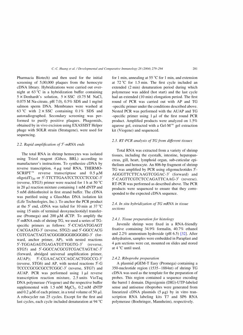
2.2. Rapid amplification of 5 0-mRNA ends
The total RNA in shrimp hemocytes was isolated
using Trizol reagent (Gibco, BRL) according to
manufacturer’s instructions. To synthesize cDNA by
reverse transcription, 4 mg total RNA, THERMO-
SCRIPTe reverse transcriptase and 0.5 mM
oligo(dT)20 or 50-TTCTTGAATCCTCCCTCCGC-30
(reverse, STG3) primer were reacted for 1 h at 50 8C
in 20 ml reaction mixture containing 1 mM dNTP and
5 mM dithiothreitol in first strand buffer. The cDNA
was purified using a GlassMax DNA isolation kit
(Life Technologies, Inc.). To anchor the PCR product
at the 50 end, cDNA was tailed for 10 min at 37 8C
using 15 units of terminal deoxynucleotidyl transfer-
ase (Promega) and 200 mM dCTP. To amplify the
50-mRNA ends of shrimp TG, we used a series of TG-
specific primers as follows: 50-CCAGATGGAGT
CACGAATG-30 (reverse, STG2) and 50-GGCCACG
CGTCGACTAGTACGGGIIGGGIIGGGIIG-30 (for-
ward, anchor primer, AP), with nested reactions
50-TGGAGAGTGAGATGTTGGTG-30 (reverse,
STG5) and 50-GGCCACGCGTCGACTAGTAC-30
(forward, abridged universal amplification primer,
AUAP); 50-CGAACACCCAGCACTGGCCG-30
(reverse, STG6) and AP, with nested reactions 50-G
TCCCCGCGCGCCTCGGC-30 (reverse, STG7) and
AUAP. PCR was performed using 1 ml reverse
transcription reaction mixture, 2.5 units VioTaq
DNA polymerase (Viogene) and the respective buffer
supplemented with 1.5 mM MgCl2, 0.2 mM dNTP
and 0.2 mM of each primer, in a total volume of 50 ml.
A robocycler ran 25 cycles. Except for the first and
last cycles, each cycle included denaturation at 94 8C
for 1 min, annealing at 55 8C for 1 min, and extension
at 72 8C for 1.5 min. The first cycle included an
extended (2 min) denaturation period during which
polymerase was added (hot start) and the last cycle
had an extended (10 min) elongation period. The first
round of PCR was carried out with AP and TG
-specific primer under the conditions described above.
Nested PCR was performed with the AUAP and TG
-specific primer using 1 ml of the first round PCR
product. Amplified products were analyzed on 1.5%
agarose gel, extracted with a Gel-Me gel extraction
kit (Viogene) and sequenced.
2.3. RT-PCR analysis of TG from different tissues
Total RNA was extracted from a variety of shrimp
tissues, including the eyestalk, intestine, hepatopan-
creas, gill, heart, lymphoid organ, sub-cuticular epi-
thelium and hemocyte. An 806-bp fragment of shrimp
TG was amplified by PCR using oligonucleotides 50-
AGGGTTCTTCAAGTCGGAC-30 (forward) and
50-CAGTTCGTCTCCAGATTCAAG-30 (reverse).
RT-PCR was performed as described above. The PCR
products were sequenced to ensure that they corre-
sponded to the expected cDNA sequence.
2.4. In situ hybridization of TG mRNA in tissue
sections
2.4.1. Tissue preparation for histology
Juvenile shrimp were fixed in a RNA-friendly
fixative containing 34.9% formalin, 40.7% ethanol
and 2.2% ammonium hydroxide (pH 6.5) [32]. After
dehydration, samples were embedded in Paraplast and
4 mm sections were cut, mounted on slides and stored
at 4 8C until used.
2.4.2. Riboprobe preparation
A plasmid pGEM-T Easy (Promega) containing a
350-nucleotide region (1535–1884nt) of shrimp TG
cDNA was used as the template for the preparation of
probes. This region contained a sequence encoding
the barrel 1 domain. Digoxigenin (DIG)-UTP-labeled
sense and antisense riboprobes were generated from
linearized cDNA plasmids (5 mg) by in vitro tran-
scription RNA labeling kits T7 and SP6 RNA
polymerase (Boehringer, Mannheim), respectively.
C.-C. Huang et al. / Developmental and Comparative Immunology 28 (2004) 279–294 281

2.4.3. In situ hybridisation
Tissue sections were deparaffinized in xylene and
hydrated through a graded ethanol series. After
proteinase K digestion (20 mg/ml, 30 min at 37 8C),
the sections were acetylated for 10 min at room
temperature (0.25% acetic acid, 0.1 M triethanola-
mine, pH 8.0), dehydrated in ethanol and air-dried.
Sections were incubated at 42 8C in a hybridization
buffer of 1 £ SSC, 50% formamide (Amresco),
1 £ Denhardt’s solution, 5% dextran sulfate
(Sigma), 1 mg/ml yeast tRNA (Sigma), and
10 ng/slide of probe. After hybridization, the sections
were washed in 1 £ SSC for 30 min and in 0.1 £ SSC
for 15 min at 50 8C. Then the sections were digested
with 20 mg/ml RNase (Sigma) for 30 min. Hybridiz-
ation signals were detected with an enzyme-linked
immunoassay as described in the manufacturer’s
manual (Nucleic Acid Detection Kit: Boehinger,
Germany). The enzyme reaction was stopped with
100% methanol for 10 min. Sections were counter-
stained with Bismarck Brown Y (Sigma), dehydrated
and made transparent with a graded ethanol and
xylene series, and then mounted in GEL/MOUNTe
(Biømeda corp).
2.5. TG activity in different tissues
2.5.1. Preparation of sample
Hemolymph was collected into an anticoagulant
(10 mM Tris–HCl, 250 mM sucrose, 100 mM sodium
citrate, pH 7.6) and centrifuged for 10 min at 500g and
4 8C to separate the hemocytes and plasma. Shrimp
tissues, including the eyestalk, intestine, hepatopan-
creas, gill, heart, lymphoid organ and sub-cuticular
epithelium, were manually homogenized. Liquid
nitrogen was added continually to maintain a
sufficiently low temperature. The hemocyte pellet
and tissue homogenate were re-suspended in a Tris–
EDTA buffer (50 mM Tris–HCl and 1 mM EDTA,
pH 7.4), centrifuged to eliminate debris, and stored at
220 8C. Plasma was mixed with Tris–EDTA buffer
and stored at 220 8C. Standard enzyme, a tissue-type
TG from guinea pig liver (Sigma), was dissolved in
Tris–EDTA buffer to an initial activity of 0.25 U/mg.
2.5.2. Assay procedure
TG activity was assayed as described by Song et al.
[33]. Casein (200 mg/well), coated on the microtitre
plates overnight, and biotin-labeled casein (0.8 mg/
well) were used as substrates in a TG-dependent
cross-linking reaction. For the assay, each sample was
serially diluted two-fold with 50 mM Tris–HCl,
1 mM EDTA, pH 7.4, and supplemented with CaCl2or EDTA to achieve a final concentration of 10 mM in
each well. Streptavidin-labeled alkaline phosphatase
(Calbiochem), followed by p-nitrophenyl phosphate,
were added to visualize immobilized biotin. Absor-
bance at 405 nm was measured. Using guinea pig liver
TGase, a standard curve was established for enzyme
activity (unit/mg) and OD 405 nm. The protein
concentration in each sample was determined by the
Bradford method [34] using bovine serum albumin
(Bio-Rad Protein assay Kit II) as the standard.
2.6. Analysis of nucleotide and amino acid sequences
The nucleotide and deduced amino acid sequences
of shrimp TG cDNA were analyzed using a GCG
(Genetic Computer Group, Inc., Madison, Wisconsin)
software package. The sequences of different species
were compared with the NCBI BLAST search
program and NCBI Blast search with Entrez. The
amino acid sequences of all cloned TG were aligned
with the DAMBE (Data Analysis in Molecular
Biology and Evolution, version 4.0.75) software
package [35]. A multiple sequence alignment was
created with Clustal W. Phylogenetic and evolution-
ary molecular analyses were conducted using MEGA
version 2.1 [36]. The phylogenetic tree was con-
structed using the neighbor-joining method [37].
2.7. Expression of recombinant TG (rTG)
The Stu I-Bgl II fragment, which contained the
entire coding region of shrimp TG, was inserted
between the Stu I and Bgl II sites of baculovirus
transfer vector pABhRpX to obtain a pABhRpX-
TG transfer plasmid. Sf21 insect cells were grown
at 26 8C in TNM-FH insect medium (GIBCO)
containing 8% heat-inactived fetal bovine serum
(HyClone) in a monolayer flask. The pABhRpX-TG
plasmid was transfected with a BaculoGold linear-
ized Baculovirus DNA (BD) into Sf21 cells with
Lipofectin (GIBCO). After plaque purification, a
single clone of each recombinant virus
was amplified and used for protein expression.
C.-C. Huang et al. / Developmental and Comparative Immunology 28 (2004) 279–294282

Harvested cells was re-suspended in Tris–EDTA
buffer (50 mM Tris–HCl and 1 mM EDTA, pH
7.4), sonicated, centrifuged to eliminate debris and
stored at 220 8C until used in the TG activity and
coagulation assay.
2.8. Coagulation assay
2.8.1. Preparation of sample
Hemolymph was collected into an anticoagulant
(10 mM Tris – HCl, 250 mM sucrose, 100 mM
sodium citrate, pH 7.6) and centrifuged for 10 min
at 500g and 4 8C to separate the hemocytes and
plasma. Plasma was dialyzed against Tris–EDTA
buffer and stored at 220 8C. The relative activity of
TG was adjusted so it was the same in sample
solutions from shrimp tissues and Sf21 cells trans-
fected with rTG. The sample solutions then were used
in coagulation assays.
2.8.2. Assay procedure
To assess the role of shrimp TG in coagulation,
100 ml plasma, 10 ml 200 mM CaCl2 solution and
50 ml of each sample solution were added to a round-
bottom 96-well microdilution plate (Nunc. Den-
mark). The solutions were mixed thoroughly and
incubated at room temperature. Coagulation was
evaluated by eye.
3. Results
3.1. Localization of TG activity
Tissue homogenate was extracted from several
shrimp organs to determine the distribution of TG
protein. TG activity was detected in the eyestalk,
intestine, hepatopancreas, gill, heart, lymphoid
organ, sub-cuticular epithelium and hemocytes, but
no activity was detected in the plasma. In all organs
tested, TG activity was blocked when 10 mM EDTA
was added instead of 10 mM CaCl2. TG activity was
greatest in the hepatopancreas, then the heart,
hemocytes and other organs (Table 1). TG activity
was detected in Sf21 cells transfected with rTG. No
activity was detected in Sf21 cells not transfected
with rTG.
3.2. Determination of the cDNA and amino acid
sequences of shrimp TG
Shrimp TG cDNA is comprised of 2988 bp, with an
open reading frame of 2271 bp (Fig. 1). The probable
initiation codon is in the sequence CAAAATGC. It
exhibits limited homology with the consensus
sequence identified by Kozak [38] that acts as a
signal for efficient transcription in eukaryotes. How-
ever, the critical purine in position -3 is conserved.
The polyadenylation signal (AATAAA) was located
626 bp downstream of the termination codon (TGA).
The deduced protein is comprised of 757 amino acid
residues, has a calculated molecular mass of
84,713 Da and an isoelectric point of 5.56. It does
not contain a typical signal sequence or a transmem-
brane domain. Four potential N-glycosylation sites
and an integrin-binding motif (RGD) were identified
(Fig. 1).
3.3. Sequence comparison of shrimp TGase
with other TG
Shrimp TG had the highest identity/similarity
(55/71%) to crayfish TG (Fig. 2). Shrimp TG
exhibited significant identity/similarity with other
invertebrate TG and members of the vertebrate TG
gene family, especially human factor XIIIa (32/50%).
Detailed comparison of the sequences by individual
Table 1
TG activity in the hemocytes, other tissues of Penaeus monodon and
TG transfected cells
Tissue/cellsa TG activity (Unit/mg)b
Hepatopancreas 27.920
Heart 1.589
Hemocyte 1.111
Lymphoid organ 0.575
Intestine 0.322
Sub-cuticular epithelium 0.224
Eyestalk 0.118
Gill 0.042
Plasma Undetectable
Sf21 Undetectable
TG transfected Sf21 0.195
a All the tissues or cells were prepared in sample buffer
containing 50 mM Tris–HCl and 1 mM EDTA, pH7.4.b There was no detectable activity in the negative control, which
was supplemented with 10 mM EDTA instead of 10 mM CaCl2.
C.-C. Huang et al. / Developmental and Comparative Immunology 28 (2004) 279–294 283

Fig. 1. Nucleotide and deduced amino acid sequences of tiger shrimp TG. The number of nucleotides is shown in the right column of numbers;
the number of amino acids is shown in the left column. The integrin-binding motif (RGD) and polyadenylation signal (ATTAAA) are
underlined. The catalytic triad is shown with bold font and italics. Amino acid residues potentially involved with calcium binding are indicated
with an asterisk ‘*’. Four putative glycosylation sites are indicated with dots.
C.-C. Huang et al. / Developmental and Comparative Immunology 28 (2004) 279–294284
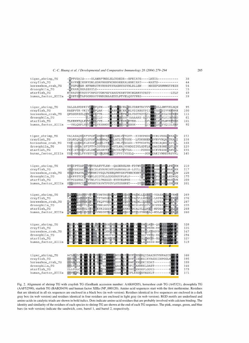
Fig. 2. Alignment of shrimp TG with crayfish TG (GenBank accession number: AAK69205), horseshoe crab TG (A45321), drosophila TG
(AAF52590), starfish TG (BAB20439) and human factor XIIIa (NP_000120). Amino acid sequences start with the first methionine. Residues
that are identical in all six sequences are enclosed in a black box (in web version). Residues identical in five sequences are enclosed in a dark
gray box (in web version) and residues identical in four residues are enclosed in light gray (in web version). RGD motifs are underlined and
amino acids in catalytic triads are shown in bold italics. Dots indicate amino acid residues that are probably involved with calcium binding. The
identity and similarity of the residues of each species to shrimp TG are shown at the end of each TG sequence. The pink, orange, green, and blue
bars (in web version) indicate the sandwich, core, barrel 1, and barrel 2, respectively.
C.-C. Huang et al. / Developmental and Comparative Immunology 28 (2004) 279–294 285

Fig. 2 (continued )
C.-C. Huang et al. / Developmental and Comparative Immunology 28 (2004) 279–294286

TG domains revealed that the highest identity/
similarity occurred between catalytic core domains,
especially those of shrimp and crayfish (66/81%)
(Table 2). In the aligned TG, all amino acid residues
involved in the catalytic triad are perfectly conserved
(shrimp: Cys324, His391, Asp414; Fig. 2). The main
human factor XIIIa Ca2þ -binding site involves the
main chain oxygen atom of Ala-457, and the side
chains from residues Asn-436, Asp-438, Glu-485 and
Glu-490 [39]. Except for Thr-477, the other four
Ca2þþ -binding residues in shrimp TG (Asn454,
Asp456, Glu513, Glu518) are conserved in all aligned
proteins (Fig. 2). The sequence surrounding His352,
Ser350-Ala-His-Asp353, was conserved. These data,
along with crystallography data for factor XIIIa [40],
suggest that His352 interacts with Glu452. In contrast,
residues for the putative GTP-binding region [41] in
TG2 were not observed in shrimp TG.
To analyze the evolutionary relationship between
shrimp TG and TG from other species, we calculated
the amino acid similarity between aligned sequences.
TG from different species are clearly related. All the
algorithms indicated shrimp TG was most closely
related to crayfish TG, then horseshoe crab TG,
starfish TG, and insect TG. Among vertebrate TG,
factor XIIIa and TG1 were most closely related to
invertebrate TG (Fig. 3).
Shrimp TG contains the motif RGD (Arg-Gly-
Asp), which is present in most ligands binding to a
family of membrane receptors called integrins [42].
An RGD motif is present in the same location in
crayfish TG [23], fruit fly TG [30] and human
keratinocyte TG [43,44].
3.4. Distribution of TG mRNA in shrimp tissues
Based on RT-PCR analysis, low TG expression
was detected in the intestine, hepatopancreas, gill,
heart, lymphoid organ, sub-cuticular epithelium and
hemocytes, but not the eyestalk (Fig. 4). In situ
hybridization failed to detect signals in most tissues
tested. However, strong TG signals were detected in
young hemocytes in hematopoietic tissue (Fig. 5a).
Binucleated cells and cells with more condensed
cytoplasm always yielded stronger TG signals
(Fig. 5b).
3.5. Coagulation assay
Only hemocyte lysate supernatant showed coagu-
lation activity. No activity was detected in shrimp
eyestalk, intestine, hepatopancreas, gill, heart, lym-
phoid organ or sub-cuticular epithelium cells. No
coagulation activity was detected in Sf21 insect cells
transfected with rTG or in untransfected Sf21 cells.
TG from guinea pig liver exhibited no coagulation
activity.
4. Discussion
We sequenced the full-length cDNA of a novel
shrimp TG. By comparing shrimp TG to previously
characterized TG, we found that the structural
requirements for TG activity and Ca2þ binding are
conserved. The TG catalytic mechanism was deter-
mined using biochemical data for several TG [2,3]
Table 2
Similarity of shrimp TG protein domains to those of other species. Domain structure is based on X-ray crystallography of the structure of human
factor XIIIa-subunit dimer [43,46]. The corresponding domains of other species were inferred from the sequence alignment shown in Fig. 2
Numbers (%) indicate sequence identity and similarity (in brackets)
Origin of TG Protein domainsa
b-Sandwich Catalytic core b-Barrel 1 b-Barrel 2
Crayfish 40 (60) 66 (81) 51 (69) 46 (66)
Horseshoe crab 29 (44) 55 (70) 41 (58) 25 (49)
Starfish 26 (38) 46 (63) 31 (56) 34 (44)
Drosophila 22 (32) 43 (60) 30 (48) 28 (49)
Human factor XIIIa 23 (33) 44 (62) 25 (48) 29 (46)
a The four individual domains are shown in Fig. 2.
C.-C. Huang et al. / Developmental and Comparative Immunology 28 (2004) 279–294 287

and x-ray crystallography data on the structure of the
factor XIIIa-subunit dimer [45]. The reaction center is
formed by a core domain. In the active site, Cys forms
hydrogen bonds to His and Asp residues to form a
catalytic triad similar to the Cys-His-Asn triad found
in the papain family of cysteine proteases [46]. The
catalytic triad residues are conserved in shrimp TG. In
addition, the core domain is highly conserved,
exhibiting 43–66% sequence identity with other
TG. Nearly every tryptophan in the catalytic core
domain is conserved, including Trp289 in shrimp TG.
The corresponding residue Trp241 in TG2 is highly
conserved in the TG family and is critical for
catalysis, possibly stabilizing the transition states.
No enzyme activity was detected when Trp241 was
replaced with Ala or Gln [47].
The transfer reaction catalyzed by TG depends on a
conformational change induced by binding divalent
Fig. 3. Phylogenetic tree for shrimp TG and 23 other members of the TG family based on ClustalW alignment of amino acid sequences.
Bootstrap values are given in percent and the scale bar indicates 0.1 substitution per site. The GenBank Accession numbers are: crayfish TG
(AAK69205), horseshoe crab TG (A45321), starfish TG (BAB20439), drosophila TG (AAF52590), grasshopper annulin (P52183), human
factor XIIIa (NP_000120), human TG1 (NP_000350), human TG4 (AAH07003), ascidian TG (CAA71263), human TG X (AAF23981), human
TG Z (NP_443187), human TG3 (XP_009572), red sea bream TG (P52181), salmon TG (JC5133), carp TG (AAL02240), zebra fish TG
(AAF61256), chicken TG2 (AAB58463), rat TG2 (NP_062259), guinea pig TG2 (P08587), human TG2 (XP_028806), cow TG2 (P51176).
C.-C. Huang et al. / Developmental and Comparative Immunology 28 (2004) 279–294288

cations, especially Ca2þ [1]. Shrimp TG lacks a
typical Ca2þ-binding motif, such as the EF-hand
structure that comprises the calcium binding site of
calmodulin and many other proteins. The affinity of
shrimp TG Ca2þ-binding sites is low and excess
EDTA easily blocked TG catalysis. The structure of
these sites probably differs from that of high affinity
sites in typical Ca2þ-binding proteins. In TG, regions
rich in negatively charged residues are potential Ca2þ-
binding sites [48]. Except Thr-477, all the amino acids
thought to be involved in calcium binding are
conserved in shrimp TG.
A typical hydrophobic leader sequence and a
transmembrane domain could not be identified in the
deduced sequence of shrimp TG. Thus, shrimp TG
may be a typical cytoplasmic protein. Glycosylation
of a protein lacking a leader sequence cannot occur
because, during synthesis, the cytoplasmic protein
would not be translocated into lumen of the
endoplasmic reticulum (ER) and Golgi apparatus
where glycosylation takes place. All characterized TG
lack a hydrophobic leader sequence and none are
known to undergo glycosylation [1,5,10,22,26,28,29,
43,44,49–51]. Expressing the factor XIIIa-subunit as
a fusion protein with the preprodomain of a secretory
protein in yeast yielded glycosylated variants of factor
XIII that were largely retained and degraded in the ER
[52]. Targeting the factor XIIIa-subunit to the
conventional secretory pathway apparently causes
aberrant, post-translational modifications. Although
TG are not conventionally secreted proteins and the
mechanism for the release of TG from cells remains
unclear, the presence of TG in extracellular space is
well documented. Factor XIII circulates in blood
plasma, TG4 is a component of semen and TG2 is
expressed on the surface of many cells, including
fibroblasts, macrophages, hepatocytes and endothelial
cells [53–56]. In invertebrates, blood clotting reduces
blood and body fluid loss following injury. In crayfish,
an endogenous TG is involved in the rapid assembly
of a specific, plasma clotting protein [24]. The
mechanism by which TG induces clotting of the
clotting protein has been studied in detail in crayfish.
Crayfish TG cross-links the clotting protein via its free
glutamine and lysine residues. This TG-mediated
polymerization occurs very rapidly [57]. In addition,
crayfish TG can use alpha-2-macroglobulin as a
substrate. However, if the alpha-2-macroglobulin
thiolester region is destroyed by methylamine treat-
ment, TG fails to recognize it as a substrate [58].
Recently, a shrimp (P. monodon) hemolymph clotting
protein was characterized [59]. The specific role of
shrimp TG in coagulation of the clotting protein has
yet to be studied.
In this study, RT-PCR analysis of TG mRNA using
TG-specific primers showed that expression is wide-
spread, except in the eyestalk. However, the lack of a
detectable band in the eyestalk does not mean TG
Fig. 4. RT-PCR analysis of TG expression. Total RNA was extracted from the eyestalk, intestine, hepatopancreas, gill, heart, lymphoid organ,
subcuticular epithelium and hemocytes of Penaeus monodon. Primers specific for Penaeus monodon TG were designed to amplify an 806-bp
fragment. RT was omitted from the control reaction to ensure that the amplified band is derived from RNA. The PCR bands were sequenced to
ensure that they corresponded to the expected cDNA. M, marker; 18, first round PCR; 28, second round PCR.
C.-C. Huang et al. / Developmental and Comparative Immunology 28 (2004) 279–294 289

mRNA was absent. PCR inhibitors are present in the
eyestalk and always cause the amplification reaction
to fail [60]. It usually took two rounds of PCR to
visualize the amplified fragment, indicating TG gene
expression was low in all organs tested or expression
was restricted to a limited number of cells. In situ
hybridization, failed to detect signals in these same
organs, confirming that TG expression was very low.
We could not determine the specific cell types that
synthesize TG in these organs. Both RT-PCR and in
situ hybridization indicated that the level of TG
mRNA was very low in circulating hemocytes.
However, young hemocytes in hematopoietic tissue
gave strong signals. Furthermore, binucleated cells
and cells with more condensed cytoplasm, i.e. cells
undergoing mitosis, yielded strong signals. This
indicates cell proliferation and TG synthesis may be
associated. Synthesis of shrimp TG may be associated
with the cell cycle. Synthesis and storage of TG in
young hemocytes facilitates the instant release of TG
protein and blood clotting following injury. Enzyme
activity was detected in all organs tested. TG activity
was greatest in the hepatopancreas, then the heart,
hemocytes and other organs (Table 1). However, only
Fig. 5. In situ hybridization of shrimp TG mRNA in hematopoietic tissue. (a) In situ hybridization with an antisense shrimp TG riboprobe. The
sections were slightly counterstained with Bismarck Brown Y. Positive signals were observed in hemocytes. No positive signals were observed
when using a sense TG riboprobe (data not shown) ( £ 200). (b) Binucleated cells and cells with more condensed cytoplasm (arrow heads)
always contained stronger signals for TG ( £ 1000).
C.-C. Huang et al. / Developmental and Comparative Immunology 28 (2004) 279–294290

hemocyte lysate supernatant showed coagulation
activity. No activity was detected in other tissues.
These results imply that shrimp may contain more
than one type of TG. It is very difficult to distinguish
different types of TG with a TG activity assay. In
tissues, TG activity was not directly correlated to
mRNA level. Previous research has documented the
expression pattern of TG by measuring mRNA and
protein levels. TG2 transcript is most abundant in the
lungs, then the heart, kidneys, red blood cells, liver,
spleen, and testes [6,61–63]. However, TG2 protein is
most abundant in the liver, then the spleen, heart,
kidney and lung [64]. These results imply that TG
expression may be regulated at the transcriptional and
translational levels, or by the protein turnover rate. In
vertebrates, the liver and blood cells are important
sites for TG expression. For example, platelets,
peripheral blood monocytes and the liver may be the
primary sites for plasma factor XIII synthesis [65,66].
In shrimp, the hepatopancreas and hemocytes could
also be important sites for TG expression. In crayfish
and horseshoe crabs, the patterns of TG expression at
the mRNA and protein levels are different [23,25].
Each invertebrate TG could correspond to a specific
type of TG found in vertebrates. Further research is
needed to clarify the function and regulation of
invertebrate TG.
TG activity was detected in Sf21 cells transfected
with rTG. However, the same cells showed no
coagulation activity. It is possible that the shrimp
TG isolated in this study is not involved in
coagulation. Another possibility is that clot formation
in shrimp involves cross-linking of plasma clotting
protein with components in hemocytes. In horseshoe
crabs, coagulin formed coagulin gel during the early
stages of coagulation [67]. During the final stage, TG
cross-linked proxins, a substrate of TG, with coagulin
to form a stable reticulate structure [27]. Coagulin
lacks glutamine residues that function as amine
acceptors, but it has several lysine residues that
function as amine donors for protein cross-linking
with proxins. Thus, TG does not cross-link coagulo-
gen or coagulin, but it does cross-link proxins with
coagulin [27]. Although TG acts solely on peptide-
bonded glutamine residues, the specificity of different
TG for different substrates, such as glutaminyl, varies
[2]. Moreover, a specific TG may recognize a protein
as substrate, but will have a different affinity and
specificity for different glutamine residues. For
example, factor XIIIa has more stringent structural
requirements for glutaminyl substrates than does TG2
[68,69]. Thus, different substrates and assay tech-
niques may yield different results due to differences in
sensitivity.
Based on amino acid similarity, vertebrate factor
XIIIa and TG1 were the vertebrate TG most closely
related to shrimp and other invertebrate TG. This is
consistent with the finding that invertebrate TG is
involved in blood clotting [24].
Based on the preservation of residues critical for
enzyme function and domain folding, and the
extensive, overall similarity of shrimp TG to the
other members of the TG family with catalytic
activity, we postulate that the cDNA characterized
in this study encodes shrimp TG. Further research
is needed to clarify the function and regulation of
shrimp TG.
Acknowledgements
We thank Prof. Yu-Chan Chao, Institute of
Molecular Biology, Academia Sinica, for kindly
providing the baculovirus transfer vector pABhRpX.
This work is financially supported by grant (NSC92-
2313-B-002-079) from the National Science
Council, Republic of China and the Swedish
Research Council.
References
[1] Folk JE, Finlayson JS. The 1-(b-glutamyl) lysine cross-link
and the catalytic role of transglutaminase. Adv Protein**
Chem 1977;31:1–133.
[2] Folk JF. Transglutaminase. Annu Rev Biochem 1980;49:
517–31.
[3] Lorand L, Conrad SM. Transglutaminases. Mol Cell Biochem
1984;58:9–35.
[4] Greenberg CS, Brickbichler PJ, Rice RH. Transglutaminases:
multifunction cross-linking enzymes that stabilize tissue.
FASEB J. 1991;5:3071–7.
[5] Grenard P, Bates MK, Aeschlimann D. Evolution of
transglutaminase genes: identification of a transglutaminase
gene cluster on human chromosome 15q15. J Biol Chem 2001;
276:33066–78.
[6] Aeschlimann D, Paulsson M. Transglutaminase: protein cross-
linking enzymes in tissues and body fluid. Thromb Haemo-
stasis 1994;71(4):402–15.
C.-C. Huang et al. / Developmental and Comparative Immunology 28 (2004) 279–294 291

[7] Kim SY, Chung S-I, Steinert PM. Highly active soluble
processed forms of the transglutaminase 1 enzyme in
epidermal keratinocytes. J Biol Chem 1995;270:18026–35.
[8] Esposito C, Pucci P, Amoresano A, Marino G, Cozzolino A,
Porta P. Transglutaminase from rat coagulating gland
secretion. J Biol Chem 1996;271:27416–23.
[9] Steinert PM, Kim S-Y, Chung S-I, Marekov LN. The
transglutaminase 1 enzyme is variably acylated by myristate
and palmitate during differentiation in epidermal keratino-
cytes. J Biol Chem 1996;271:26242–50.
[10] Aeschlimann D, Koeller MK, Alen-Hoffmann BL, Mosher
DF. Isolation of a cDNA encoding a novel member of the
transglutaminase gene family from human keratinocytes.
J Biol Chem 1998;273:3452–60.
[11] Ichinose A, Bottenus RE, Davie EW. Structure of transglu-
taminase. J Biol Chem 1990;265:13411–4.
[12] Martinet N, Kim HC, Girard JE, Nigra TP, Strong DH, Chung
S-I, Folk JE. Epidermal and hair follicle transglutaminase.
Partial characterization of soluble enzymes in newborn mouse
skins. J Biol Chem 1988;263:4236–41.
[13] Rice RH, Rong X, Chakravarty R. Proteolytic release of
keratinocyte transglutaminase. Biochem J 1990;265:351–7.
[14] Candi E, Melino G, Mei G, Tarcsa E, Chung SI, Marekov LN,
Steinert PM. Biochemical, structural, and transglutaminase
substrate properties of human lorcrin, the major epidermal
cornified cell envelope protein. J Biol Chem 1995;270:
26382–90.
[15] Tarcas E, Marekov LN, Andreoli J, Isler WW, Candi E, Chung
SI, Steinert PM. The fate of trichohyalin. Sequential post-
translational modifications by peptidyl-arginine deiminase and
transglutaminases. J Biol Chem 1997;272:27893–901.
[16] Steinert PM, Kartasova T, Marekov LN. Biochemical
evidence that small proline-rich proteins and trichohyalin
function in epithelia by modulation of the biochemical
properties of their cornified cell envelopes. J Biol Chem
1998;273:11758–69.
[17] Achyuthan KE, Greenberg CS. Identification of a guanosine
triphosphate-binding site on guinea pig liver. J Biol Chem
1987;262:1901–6.
[18] Kojima S, Inui T, Muramatsu H, Suzuki Y, Kadomatsu K,
Yoshizawa M, Hirose S, Sakakibara S, Muramatsu T.
Dimerization of midkine by tissue transglutaminase and its
functional implication. J Biol Chem 1997;272:9410–6.
[19] Nunes I, Gleizes PE, Metz CN, Rifkin DB. Latent transforming
growth factor-beta binding protein domains involved in acti-
vation and transglutaminase-dependent cross-linking of latent
transforming growth factor-beta. J Cell Biol 1997;136:1151–63.
[20] Melino G, Piacentini M. Tissue transglutaminase in cell death:
a downstream or a multifunctional upstream effector? FEBS
Lett 1998;430:59–63.
[21] Aeschlimann D, Thomazy V. Protein crosslinking in assembly
and remodeling of extracellular matrixes: the role of
transglutaminases. Connect Tissue Res 2000;41:1–27.
[22] Ho KC, Quarmby VE, French FS, Wilson EM. Molecular
cloning of rat prostate transglutaminase complementary DNA.
The major androgen-regulated protein DP1 of rat dorsal
prostate and coagulating gland. J Biol Chem 1992;267:
12660–7.
[23] Wang R, Liang Z, Hall M, Soderhall K. A transglutaminase
involved in the coagulation system of the freshwater crayfish.
Pacifastacus leniusculus. Tissue localization and cDNA
cloning. Fish Shellfish Immunol 2001;11:623–37.
[24] Hall M, Wang R, Antwerpen RV, Sottrup-Jensen L, Soderhall
K. The crayfish plasma clotting protein: a vitellogenin-related
protein responsible for clot formation in crustacean blood.
Proc Natl Acad Sci 1997;96:1965–70.
[25] Tokunaga F, Yamada M, Miyata T, Ding Y-L, Hiranaga-
Kawabata M, Muta T, Iwanaga S, Ichinose A, Davie EW.
Limulus hemocyte transglutaminase. Its purification and
characterization, and identification of the intracellular sub-
strates. J Biol Chem 1993;268:252–61.
[26] Tokunaga F, Muta T, Iwanaga S, Ichinose A, Davie EW,
Kuma K, Miyata T. Limulus hemocyte transglutaminase
CDNA cloning, amino acid sequence, and tissue localization.
J Biol Chem 1993;268:262–8.
[27] Osaki T, Okino N, Tokunaga F, Iwanaga S, Kawabata S.
Proline-rich cell surface antigens of horseshoe crab hemocytes
are substrates for protein cross-linking with a clotting protein
coagulin. J Biol Chem 2002;277:40084–90.
[28] Singer MA, Hortsch M, Goodman CS, Bentley D. Annulin, a
protein expressed at limb segment boundaries in the grass-
hopper embryo, is homologous to protein cross-linking
transglutaminase. Dev Biol 1992;154:143–59.
[29] Sugino H, Terakawa Y, Yamasaki A, Nakamura K, Higuchi Y,
Matsubara J, Kuniyoshi H, Ikegami S. Molecular character-
ization of a novel nuclear transglutaminase that is expressed
during starfish embryogenesis. Eur J Biochem 2002;269:
1957–67.
[30] Adams MD, Celniker SE, et al. The genome sequence of
Drosophila melanogaster. Science 2000;287:2185–95.
[31] Vargas-Albores F, Guzman-Murillo MA, Ochoa JL. An
anticoagulant solution for haemolymph collection and pro-
phenoloxidase studies of penaeid shrimp (Penaeus califor-
niensis). Comp Biochem Physiol 1993;106A:299–303.
[32] Hasson KW, Hasson J, Aubert H, Redman RM, Lightner DV.
A new RNA-friendly fixative for the preservation of penaeid
shrimp samples for virological detection using cDNA genomic
probes. J Virol Meth 1997;66:227–36.
[33] Song YL, Yu CI, Lien TW, Huang CC, Lin MN. Haemolymph
parameters of Pacific white shrimp (Litopenaeus vannamei)
infected with Taura syndrome virus. Fish shellfish Immunol
2003;14:317–331.
[34] Bradford MM. A rapid and sensitive method for the quanti-
tation of microgram quantities of proteins utilizing the principle
of protein-dye binding. Anal Biochem 1976;72:248–54.
[35] Xia X, Xie Z. DAMBE: Data analysis in molecular biology
and evolution. J Hered 2001;92:371–3.
[36] Kumar S, Tamura K, Jakobsen IB, Nei M. MEGA2: molecular
evolutionary genetics analysis software. Bioinformatics 2001;
17:1244–5.
[37] Saitou N, Nei M. The neighbor-joining method: a new method
for reconstructing phylogenetic trees. Mol Biol Evol 1987;4:
406–25.
C.-C. Huang et al. / Developmental and Comparative Immunology 28 (2004) 279–294292

[38] Kozak M. Point mutations define a sequence flanking the AUG
initiation codon that modulates translation by eukaryotic
ribosomes. Cell 1986;44:283–92.
[39] Fox BA, Yee VC, Pedersen LC, Le Trong I, Bishop PD,
Stenkamp RE, Teller DC. Identification of the calcium
binding site and a novel ytterbium site in blood coagulation
factor XIII by X-ray crystallography. J Biol Chem 1999;274:
4917–23.
[40] Iismaa SE, Chung L, Wu MJ, Teller DC, Yee VC, Graham
RM. The core domain of the tissue transglutaminase
Gh hydrolyzes GTP and ATP. Biochemistry 1997;36:
11655–64.
[41] Iismaa SE, Wu MJ, Nanda N, Church WB, Graham RM. GTP
binding and signaling by Gh/transglutaminase II involves
distinct residues in a unique GTP-binding pocket. J Biol Chem
2000;275:18259–65.
[42] Ruoslahti E. RGD and other recognition sequences for
integrins. Annu Rev Cell Dev Biol 1996;12:697–715.
[43] Phillips MA, Stewart BE, Qin Q, Chakravarty R, Floyd EE,
Jetten AM, Rice RH. Primary structure of keratinocyte
transglutaminase. Proc Natl Acad Sci 1990;87:9333–7.
[44] Kim HC, Idler WW, Kim IG, Han JH, Chung SI, Steinert PM.
The complete amino acid sequence of the human transluta-
minase K enzyme deduced from the nucleic acid sequences of
cDNA clones. J Biol Chem 1991;266:536–9.
[45] Yee VC, Pedersen LC, Le Trong I, Bishop PD, Stenkamp RE,
Teller DC. Three-dimensional structure of a transglutaminase:
human blood coagulation factor XIIIa. Proc Natl Acad Sci
1994;91:7296–300.
[46] Pedersen LC, Yee VC, Bishop PD, LeTrong I, Teller RC,
Stenkamp RE. Transglutaminase factor XIII uses protemase-
like catalytic triad to crosslink macromolecules. Protein Sci
1994;3:1131–5.
[47] Prasanna Murthy SN, Iismaa S, Begg G, Freymann DM,
Graham RM, Lorand L. Conserved tryptophan in the core
domain of transglutaminase is essential for catalytic activity.
Proc Natl Acad Sci 2002;99:2738–42.
[48] Yee VC, Le Trong I, Bishop PD, Pedersen LC, Stenkamp RE,
Teller DC. Structure and function studies of factor XIIIa by
X-ray crystallography. Semin Thromb Hemost 1996;22:
377–84.
[49] Grundmann U, Amann E, Zettlmeissl G, Kupper HA.
Characterization of cDNA coding for human factor XIIIa.
Proc Natl Acad Sci 1986;83:8024–8.
[50] Ichinose A, Hendrickson LE, Fujikawa K, Davie EW. Amino
acid sequence of the a-subunit of human factor XIII.
Biochemistry 1986;25:6900–6.
[51] Takahashi N, Takahashi Y, Putnam FW. Primary structure of
blood coagulation factor XIIIa (fibrinoligase, transglutami-
nase) from human placenta. Proc Natl Acad Sci 1986;83:
8019–23.
[52] Tharaud C, Ribet A-M, Costes C, Gaillardin C. Secretion of
human blood coagulation factor XIIIa by the yeast Yarrowia
lipolytica. Gene 1994;121:111–9.
[53] Aeschlimann D, Koeller M, Paulsson M. Transglutaminase-
catalyzed matrix crosslinking in differentiating cartilage:
Identification of osteonectin as a major glutaminyl substrate.
J Cell Biol 1995;129:881–92.
[54] Barsigian C, Stern AM, Martinez J. Tissue (type II)
transglutaminase covalently incorporates itself, fibrinogen,
or fibronectin into high molecular weight complexes on the
extracellular surface of isolated hepatocytes. J Biol Chem
1991;266:22501–9.
[55] Martinez J, Chalupowiez DG, Roush RK, Sheth A.
Barsigian. Transglutaminase-mediated processing of fibro-
nectin by endothelial cell monolayers. Biochemistry 1994;
33:2538–45.
[56] Verderio E, Nicholas B, Gross S, Griffin M. Regulated
expression of tissue transglutaminase in Swiss 3T3 fibroblasts:
Effect on the processing of fibronectin, cell attachment, and
cell death. Exp Cell Res 1998;239:119–38.
[57] Kopacek P, Hall M, Soderhall K. Characterisation of a
clotting protein isolated from the plasma of the freshwater
crayfish Pacifastcaus leniusculus. Eur J Biochem 1993;213:
591–7.
[58] Hall M, Soderhall K. Crayfish alpha-2-macroglobulin as a
substrate for transglutaminases. Comp Biochem Physiol 1994;
108B:65–72.
[59] Yeh M-S, Huang C-J, Leu J-H, Lee Y-C, Tsai I-H. Molecular
cloning and characterization of a hemolymph clottable protein
from tiger shrimp (Penaeus monodon). Eur J Biochem 1999;
266:624–33.
[60] Lo CF, Ho CH, Chen CH, Liu KF, Chiu YL, Yeh PY, Prng SE,
Hsu HC, Liu HC, Chang CF, Su MS, Wang CH, Kou GH.
Detection and tissue tropism of white spot syndrome
baculovirus (WSBV) in captured brooders of Penaeus
monodon with a special emphasis on reproductive organs.
Dis Aquat Org 1997;30:53–72.
[61] Gentile V, Saydak M, Chiocca EA, Akande O, Birckbichler
PJ, Lee KN, Stein JP, Davies PJA. Isolation and charateriza-
tion of cDNA clones to mouse macrophage and human
endothelial cell tissue transglutaminase. J Biol Chem 1991;
266:478–83.
[62] Nakanishi K, Nara K, Hagiwara H, Aoyama Y, Ueno H,
Hirose S. Cloning and sequence analysis of cDNA clones for
bovine aortic-endothelial-cell transglutaminase. Eur J Bio-
chem 1991;202:15–21.
[63] Weraarchakul-Boonmark N, Jeong JM, Murthy SNP, Engel
JD, Lorand L. Cloning and expression of chicken
erythrocyte transglutaminase. Proc Natl Acad Sci 1992;89:
9804–8.
[64] Clarke DD, Mycek MJ, Neidle A, Waelsch H. The
incorporation of amines into protein. Arch Biochem Biophys
1959;79:338–54.
[65] Weisberg LJ, Shiu DT, Conkling PR, Shuman MA. Identifi-
cation of normal human peripheral blood monocytes and liver
as sites of synthesis of coagulation factor XIII a-chain. Blood
1987;70:579–82.
[66] Poon M-C, Russell JA, Low S, Sinclair GD, Jones AR, Blahey
W, Ruether BA, Hoar DI. Hemopoietic origin of factor XIII
a-subunits in platelets, monocytes and plasma. Evidence from
bone marrow transplantation studies. J Clin Invest 1989;84:
787–92.
C.-C. Huang et al. / Developmental and Comparative Immunology 28 (2004) 279–294 293
[67] Kawasaki H, Nose T, Muta T, Iwanaga S, Shimohigashi Y,
Kawabata S-I. Head-to-tail polymerization of coagulin, a
clottable protein of the horseshoe crab. J Biol Chem 2000;275:
35297–301.
[68] Gorman JJ, Folk JE. Structure features of glutamine substrates
for transglutaminase: Specificities of human plasma factor
XIIIa and the guinea pig liver enzyme toward synthetic
peptides. J Biol Chem 1981;256:2712–5.
[69] Gorman JJ, Folk JE. Structural features of glutamine
substrates for transglutaminase: role of extended interactions
in the specificity of human plasma factor XIIIa and the guinea
pig liver enzyme. J Biol Chem 1984;259:9007–10.
C.-C. Huang et al. / Developmental and Comparative Immunology 28 (2004) 279–294294