Molecular characterization of the ... - Genes & Development
13
Molecular characterization of the zerkniillt region of the Antennapedia gene complex in Drosophila Christine Rushlow, Helen Doyle, Timothy Hoey, and Michael Levine Department of Biological Sciences, Fairchild Center, Columbia University, New York, New York 10027 USA zerkniillt (zen) is unique among the 18 known homeo box genes in Drosophila since it is required for the differentiation of the dorsal-ventral pattern, and does not appear to be involved in the process of segmentation. Here we show that the zen region of the Antennapedia complex (ANT-C) consists of two closely linked homeo box genes, designated zl and z2. The zl and z2 transcription units show essentially identical patterns of expression during early development, which are consistent with the timing and sites of zen + gene activity. The putative proteins encoded by zl and z2 are highly divergent and are related only by virtue of homeo box homology. We have used P-element-mediated germ line transformation to show that zl alone can provide zen + gene function, suggesting that the z2 gene might be dispensable. The occurrence of closely linked homeo box genes that display similar patterns of expression is not unique to the zen locus. Such gene duplications might provide important clues to the evolution of the homeo box gene family in Drosophila and other organisms. [Key Words: Drosophila; zerki~ullt (zen); homeo box genes] Received August 7, 1987; revised version accepted October 14, 1987. Many of the genes that specify positional information in Drosophila are evolutionarily related and share a similar 180-bp protein coding sequence called the homeo box (McGinnis et al. 1984; Scott and Weiner 1984). There are at least 30 homeo box genes in Drosophila, of which 18 have been cloned and characterized. Most of these genes (16 of 18) control the morphogenesis of anterior-poste- rior pattern elements, and mutations in these genes typi- cally alter the normal segmentation pattern of the em o bryo (for review, see Levine and Harding 1987). In con- trast, two of the genes appear to be involved in the differentiation of the dorsal-ventral pattern and derive from the zerkiiullt (zen)region of the Antennapedia complex (ANT-C)(Doyle et al. 1986; Rushlow et al., 1987). zen is unique among the known homeo box genes in Drosophila in that zen mutants do not directly affect the process of segmentation. Advanced-stage zen mu- tants show a twisting along the germ band and a failure of head involution (Wakimoto et al. 1984). Despite these abnormalities there is a correct number of body seg- ments, and the cuticular pattern elements associated with each segment appear more or less normal. The complex phenotype seen for advanced-stage zen- em- bryos can be explained on the basis of a transformation in cell fate of the dorsal-most ectoderm toward a more ventral pathway of development (Rushlow et al., in prep.). During gastrulation, the dorsal-most ectoderm nor- mally differentiates into an embryonic membrane called the amnioserosa, which plays a key role in the process of germ band elongation (Poulson 1950; Campos-Ortega and Hartenstein 1985). The amnioserosa becomes pro- fusely folded where the posterior tip of the germ band contacts the procephalon and is driven into the procto- deal cavity by the expanding germ band. In zen- em- bryos the dorsal ectoderm fails to differentiate into the amnioserosa. This absence of the amnioserosa might prevent the full expansion of the germ band, thereby causing the characteristic twisting and folding of the germ band seen in advanced-stage mutant embryos. The absence of the amnioserosa in zen mutants does not ap- pear to be the result of cell death, but instead appears to involve the "ventralization" of the dorsal-most ecto- derm. Thus, the cells that normally give rise to the am- nioserosa differentiate into a cell type that is character- istic of ectodermal cells found in more ventral regions of wild-type embryos. It is possible that zen is a selector gene that specifies a dorsal vs. ventral pathway of mor- phogenesis. There is a close correspondence between the sites where products encoded by a homeo box gene called $60 accumulate in wild-type embryos and the regions that are disrupted in zen mutants (Doyle et al. 1986; Rushlow et al., 1987). The $60 gene was isolated on the basis of homeo box cross-homology and was shown to map within the zen region of the ANT-C. Here we report a detailed molecular characterization of zen and show that there are two closely linked homeo box genes, $60 (hereafter called zl) and z2, which show essentially identical patterns of expression. The molecular organi- zation of the two transcription units is similar, and they represent the smallest known homeo box genes. Despite 1268 GENES & DEVELOPMENT 1:1268-1279 © 1987 by Cold Spring Harbor Laboratory ISSN 0890-9369/87 $1.00 Cold Spring Harbor Laboratory Press on November 22, 2021 - Published by genesdev.cshlp.org Downloaded from
Transcript of Molecular characterization of the ... - Genes & Development

Molecular characterization of the zerkniillt region of the Antennapedia gene complex in Drosophila Christine Rushlow, Helen Doyle, T imothy Hoey, and Michael Levine
Department of Biological Sciences, Fairchild Center, Columbia University, New York, New York 10027 USA
z e r k n i i l l t (zen) is unique among the 18 known homeo box genes in D r o s o p h i l a since it is required for the differentiation of the dorsal-ventral pattern, and does not appear to be involved in the process of segmentation. Here we show that the z e n region of the Antennapedia complex (ANT-C) consists of two closely linked homeo box genes, designated zl and z2. The zl and z2 transcription units show essentially identical patterns of expression during early development, which are consistent with the timing and sites of z e n + gene activity. The putative proteins encoded by zl and z2 are highly divergent and are related only by virtue of homeo box homology. We have used P-element-mediated germ line transformation to show that zl alone can provide z e n +
gene function, suggesting that the z2 gene might be dispensable. The occurrence of closely linked homeo box genes that display similar patterns of expression is not unique to the z e n locus. Such gene duplications might provide important clues to the evolution of the homeo box gene family in D r o s o p h i l a and other organisms.
[Key Words: Drosophila; zerki~ullt (zen); homeo box genes]
Received August 7, 1987; revised version accepted October 14, 1987.
Many of the genes that specify positional information in Drosophi la are evolutionarily related and share a similar 180-bp protein coding sequence called the homeo box (McGinnis et al. 1984; Scott and Weiner 1984). There are at least 30 homeo box genes in Drosophila, of which 18 have been cloned and characterized. Most of these genes (16 of 18) control the morphogenesis of anterior-poste- rior pattern elements, and mutations in these genes typi- cally alter the normal segmentation pattern of the em o bryo (for review, see Levine and Harding 1987). In con- trast, two of the genes appear to be involved in the differentiation of the dorsal-ventral pattern and derive from the zerk i iu l l t ( z e n ) r e g i o n of the Antennapedia complex (ANT-C)(Doyle et al. 1986; Rushlow et al., 1987). zen is unique among the known homeo box genes in Drosophi la in that zen mutants do not directly affect the process of segmentation. Advanced-stage zen mu- tants show a twisting along the germ band and a failure of head involution (Wakimoto et al. 1984). Despite these abnormalities there is a correct number of body seg- ments, and the cuticular pattern elements associated with each segment appear more or less normal. The complex phenotype seen for advanced-stage z e n - em- bryos can be explained on the basis of a transformation in cell fate of the dorsal-most ectoderm toward a more ventral pathway of development (Rushlow et al., in prep.).
During gastrulation, the dorsal-most ectoderm nor- mally differentiates into an embryonic membrane called the amnioserosa, which plays a key role in the process of germ band elongation (Poulson 1950; Campos-Ortega
and Hartenstein 1985). The amnioserosa becomes pro- fusely folded where the posterior tip of the germ band contacts the procephalon and is driven into the procto- deal cavity by the expanding germ band. In z e n - em- bryos the dorsal ectoderm fails to differentiate into the amnioserosa. This absence of the amnioserosa might prevent the full expansion of the germ band, thereby causing the characteristic twisting and folding of the germ band seen in advanced-stage mutant embryos. The absence of the amnioserosa in zen mutants does not ap- pear to be the result of cell death, but instead appears to involve the "ventralization" of the dorsal-most ecto- derm. Thus, the cells that normally give rise to the am- nioserosa differentiate into a cell type that is character- istic of ectodermal cells found in more ventral regions of wild-type embryos. It is possible that zen is a selector gene that specifies a dorsal vs. ventral pathway of mor- phogenesis.
There is a close correspondence between the sites where products encoded by a homeo box gene called $60 accumulate in wild-type embryos and the regions that are disrupted in zen mutants (Doyle et al. 1986; Rushlow et al., 1987). The $60 gene was isolated on the basis of homeo box cross-homology and was shown to map within the zen region of the ANT-C. Here we report a detailed molecular characterization of zen and show that there are two closely linked homeo box genes, $60 (hereafter called zl) and z2, which show essentially identical patterns of expression. The molecular organi- zation of the two transcription units is similar, and they represent the smallest known homeo box genes. Despite
1268 GENES & DEVELOPMENT 1:1268-1279 © 1987 by Cold Spring Harbor Laboratory ISSN 0890-9369/87 $1.00
Cold Spring Harbor Laboratory Press on November 22, 2021 - Published by genesdev.cshlp.orgDownloaded from
these similarities, nucleotide sequence analyses show that the putative proteins encoded by these genes are highly divergent and are related only by homeo box ho- mology. Several lines of evidence suggest that zl might correspond to zen . We show that zl alone can provide z en + gene function, since z e n - embryos are rescued by a z l /P- t ransposon that completely lacks z2 sequences. The occurrence of two closely linked homeo box genes that display similar patterns of expression is discussed in the context of other homeo box gene duplications in Drosoph i la .
Results
The z e n region of the ANT-C was isolated in a previous screen of a D r o s o p h i l a DNA library for clones that cross-hybridize with the homeo box sequence of the ho- meot ic gene Sex c o m b s r e d u c e d (Scr ) (Doy le et al. 1986). One of the clones that was obtained in this screen (called
z e n region of ANT-C
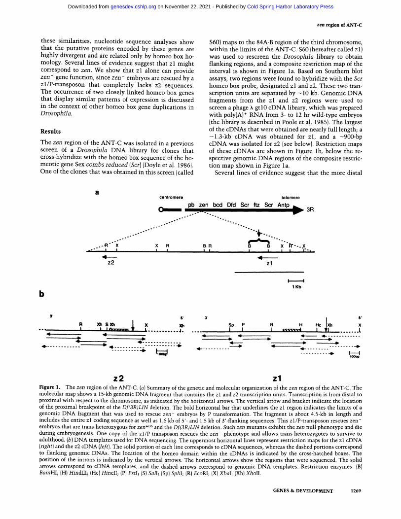
$60) maps to the 84A-B region of the third chromosome, within the l imits of the ANT-C. $60 (hereafter called zl) was used to rescreen the D r o s o p h i l a library to obtain flanking regions, and a composite restriction map of the interval is shown in Figure la. Based on Southern blot assays, two regions were found to hybridize with the Scr homeo box probe, designated zl and z2. These two tran- scription units are separated by ---10 kb. Genomic DNA fragments from the zl and z2 regions were used to screen a phage ~ gt l 0 cDNA library, which was prepared with poly(A) + RNA from 3- to 12 hr wild-type embryos (the library is described in Poole et al. 1985). The largest of the cDNAs that were obtained are nearly full length; a -~l.3-kb cDNA was obtained for zl, and a -~900-bp cDNA was isolated for z2 (see below). Restriction maps of these cDNAs are shown in Figure l b, below the re- spective genomic DNA regions of the composite restric- tion map shown in Figure l a.
Several lines of evidence suggest that the more distal
b
° °
° °
°
. . . I
z2
contromere tolomoro
pb zen bcd Dfd Scr ftz Scr Antp h ,~ ~ i . . - - - - - i ~ 3R
. - ° °
° •
o ° t o
o o ,,,. " " " ' ,,, ,,,, ,. ° ,,,.
. ° . , , . o ° q ' " Q " " ° ~ " ° ° . ,
A L ° o ° °
X R B R B e X" ~ ' - . ~ X I I I I I I I I n ' - ° ,
zl
t - - - - . - - -4 1 Kb
o o - -
A
,4--
XhS' 3' ~ " 5' R /)01 S X h 1• X Sp P B F( Hc X
n ~ ' . . . . . . . . . . . J . . . . . . I 1 .__ I . . . . ~ 4 I . . . . . . . L :~ ~ ~ , ~ ~ ~ . . . . . . . . . . . . . .
" ' - ' , 9 ) -4~ ~ "" ~ 1 - . . . . . . . . " . . . . . . . . " ~ 4~ ~ ~ . . . . . . . . . .
. . . . . . . . . . --~ F - - - - I 10obp
z2 z l Figure 1. The zen region of the ANT-C. (a) Summary of the genetic and molecular organization of the zen region of the ANT-C. The molecular map shows a 15-kb genomic DNA fragment that contains the zl and z2 transcription units. Transcription is from distal to proximal with respect to the chromosome, as indicated by the horizontal arrows. The vertical arrow and bracket indicate the location of the proximal breakpoint of the Df(3R)LIN deletion. The bold horizontal bar that underlines the zl region indicates the limits of a genomic DNA fragment that was used to rescue zen- embryos by P transformation. The fragment is about 4.5-kb in length and includes the entire zl coding sequence as well as 1.6 kb of 5'- and 1.5 kb of 3'-flanking sequences. This zl/P-transposon rescues zen- embryos that are trans-heterozygous for zen wa6 and the Df(3R)LIN deletion. Such zen mutants exhibit the zen null phenotype and die during embryogenesis. One copy of the zl/P-transposon rescues the zen- phenotype and allows trans-heterozygotes to survive to adulthood. (b) DNA templates used for DNA sequencing. The uppermost horizontal lines represent restriction maps for the zl cDNA (right) and the z2 cDNA (left). The solid portion of each line corresponds to cDNA sequences, whereas the dashed portions correspond to flanking genomic DNAs. The location of the homeo domain within the cDNAs is indicated by the cross-hatched boxes. The position of the introns is indicated by the vertical arrows. The horizontal arrows show the regions that were sequenced. The solid arrows correspond to cDNA templates, and the dashed arrows correspond to genomic DNA templates. Restriction enzymes: (B) BamHI; (H) HindIII; (Hc) HincII; (P) PstI; (S) SalI; (Sp) SphI; (R) EcoRI; (X)XbaI; (Xh)XholI.
GENES & DEVELOPMENT 1 2 6 9
Cold Spring Harbor Laboratory Press on November 22, 2021 - Published by genesdev.cshlp.orgDownloaded from

Rushlow et al.
transcription unit {z l )might correspond to zen. First, the proximal breakpoint of a zen deficiency [Df(3R) LIN] falls within, or just adjacent to, the 3' end of the zl coding sequence and does not disrupt the z2 transcrip- tion unit (see Fig. la; vertical arrow and brackets). Second, embryos homozygous for a cytologically normal EMS-induced mutant allele (zen w36) are not stained by antibodies directed against the zl protein (C. Rushlow unpubl.). Third, a genomic DNA fragment that contains the entire zl coding sequence, but no z2 sequences, is able to rescue z e n - embryos by P transformation (Fig. la and below). Finally, it has been shown that embryos de- ficient for the entire z2 transcription unit (i.e., zl +, z2- embryos) are viable {M.A. Pultz and T.C. Kaufman, pets. comm.). Despite these observations, we will refer to the two t ranscr ipt ion units as zl and z2 since it is not known whether z l - embryos are viable. It is possible that either zl or z2 can provide zen + gene function when one of the genes is removed.
The z l gene rescues zen m u t a n t s
P-transformation assays were done to determine whether the zl gene could rescue z e n - embryos. These experiments involved the use of a 4.5-kb X b a I - S a c I ge- nomic DNA fragment that contains the entire zl coding sequence, as well as 1.6-kb of 5'- flanking sequences and 1.5 kb of 3' flank. The limits and location of the zl DNA that was used are shown in Figure l a (horizonal line below the restriction map of the zen region). The 5' limit of the rescue fragment extends to the 3' terminus of bi- coid, which is a maternally expressed homeo box gene that is closely linked to zen (Frigerio et al. 1986; Frohn- hofer and Nfisslein-Volhard 1986). The zl fragment was inserted into a derivative of the Carnegie 20 transforma- tion vector (Rubin and Spradling 1983; D. Mismer and G. Rubin, unpubl.), and injected into rosy - embryos as previously described (Rubin and Spradling 1982). Six in- dependent transformed lines were established (listed in Table 1} and tested for their ability to rescue zenw36/ Df(3R) L I N trans-heterozygotes. Such heterozygotes dis- play the zen null phenotype (C. Rushlow, unpub, re- sults), zen w36 is a null allele that was induced by EMS (Wakimoto et al. 1984), and, based on Southern analysis, does not contain a deletion of the zen region larger than 50 bp (data not shown). Df(3R) L I N is a large deletion that uncovers several loci within the ANT-C, including D f d and bic (C. Nfisslein-Volhard, unpubl, results), and breaks within the zl gene (Fig. la). This deletion strongly disrupts the expression of both zl and z2 (C. Rushlow, in prep.). Each of the five transformed lines that contains at least one copy of the P-zl transposon on the second chromosome rescues zenW36/Df(3R) L I N he- terozygotes and yields adult survivors (see Table 1 }. One of these lines contains only a single copy of the tran- sposon, but nonetheless gives nearly complete rescue (72% of the expected number of zenw36/Df(3R) L I N adults were obtained). The only line that failed to rescue contains the P-zl transposon on the third chromosome,
which does not segregate with zen alleles. None of the transformed lines rescues zen wa6 homozygotes, sug- gesting that this chromosome might contain additional lethal mutations.
Sequence and organizat ion of the z l and z2 transcripts
Northern analyses using polyIA) + RNA from staged em- bryos show that zl and z2 each encode a single mRNA that display similar times of appearance during early de- velopment {Fig. 2). The zl mRNA is -1 .3 kb in length, whereas z2 is -0 .9 kb. Both RNAs appear transiently during early embryogenesis and show peaks of expres- sion from 2 to 3 hr after fertilization, zl and z2 tran- scripts are not detected after the sixth hour of embryo- genesis and do not reappear during embryonic develop- ment {Fig. 2). We have not used larval or adult extracts for Northern analyses, although temperature-shift ex- periments using a zen ts strain suggest that zen + function is not required after early periods of embryogenesis (Wa- kimoto et al. 1984}. Primer extension and S1 nuclease protection analyses were done to identify the 5' ends of the zl and z2 transcripts. For these studies, poly(A) + RNA was obtained from extracts of 2- to 3-hr embryos, since this is the period of peak zl and z2 expression. The primer extension experiments involved the use of a 20- nucleotide synthetic oligomer for zl and a 33-nucleotide oligomer for z2. The nucleotide sequence and location of the primers within the zl and z2 genes are shown in
{'d ¢0 ¢D ' ," , i l i i ¢~I
0 ~ 03 tD _
/
I I I I F~J
0 ~ C~ ~D "'T=
1 . 3 - -
~-- O.g
a b
Figure 2. Northern analysis of zl and z2 RNAs. Poly(A) + RNA was extracted from wild-type embryo populations at 0-2, 2-3, 3-6, 6-12, and 12-24 hr after fertilization. Equivalent amounts of these RNAs were fractionated on an agarose-form- aldehyde gel, transferred to nitrocellulose, and hybridized with a zl cDNA probe {a). A single -1.3-kb transcript was detected, which peaks in expression in 2- to 3-hr embryos. The zl hybrid- ization signal was removed by incubating the nitrocellulose filter in 0.1 x SSC at 100°C for 3 rain. The blot was then rehy- bridized with a z2 cDNA probe {b) and a single -0.9-kb tran- script was detected, which also peaks in 2- to 3-hr embryos.
1270 GENES & DEVELOPMENT
Cold Spring Harbor Laboratory Press on November 22, 2021 - Published by genesdev.cshlp.orgDownloaded from

z e n region of ANT-C
Figures 5 and 6, respectively. The extended RNA product that results from the use of the zl primer has a length of 115 nucleotides (Fig. 3b), whereas the extended RNA product from the z2 primer has a length of 222 nu- cleotides (Fig. 3c). The 5' end of the zl mRNA is indi- cated in the map shown below the autoradiograms in Figure 3, a and b, and in the nucleotide sequence of zl shown in Figure 5. The 5' end of z2 is shown below the autoradiogram in Figure 3c, and is also shown in the z2 nucleotide sequence presented in Figure 6.
The 5' end of the z l transcript was also determined by S1 nuclease protection, using a 316-nucleotide XbaI- XhoII genomic DNA fragment. The nucleotide sequence and location of this probe within the zl gene is shown in Figure 5. The probe was labeled at the XhoII site, which coincides with the 5' end of the zl synthetic oligomer used for the primer extension experiment. After hybrid- ization with embryonic poly(A) + RNA and treatment with S1 nuclease, the probe was reduced to a length of 115 nucleotides (Fig. 3a), which is the same size as the extended RNA product obtained by primer extension (compare Fig. 3a, b).
The results of the primer extension and S1 nuclease analyses as well as the nucleotide sequences of genomic and cDNA clones (see below)indicate that the zl tran- scription unit is probably less than 1.4 kb in length and that the z2 transcription unit is just over 1 kb. The mo- lecular organization of these two genes is summarized in Figure 4. The two genes show similar structures in that each transcription unit appears to be composed of two exons that are separated by a small intron located near the 5' end. However, since we have not sequenced the entire genomic regions, we cannot rule out the possi- bility that one or more "micro-introns" interrupt the coding sequences.
Nucleotide sequence of zl and z2
DNA templates that were used for dideoxynucleotide se- quencing are shown in Figure lb. The sequenced in- terval includes each of the cDNAs (see Fig. l b), as well as the genomic regions that immediately flank the 5' and 3' ends. Genomic fragments that encompass the small introns for each gene were also sequenced. The locations of the introns on each cDNA are indicated by the vertical arrows in Figure lb. The horizontal arrows indicate the regions that were sequenced; the solid arrows correspond to cDNA fragments, and the dashed arrows correspond to genomic fragments. The nucleo- tide sequence of the zl coding region is presented in Figure 5. The zl cDNA lacks the first five nucleotides of the transcript, which were determined from overlapping genomic DNA fragments from the 5' end. The first AUG codon is located 52 bp downstream from the transcrip- tion start site and is in frame with an open reading frame (ORF) that extends throughout most of the cDNA and has a total length of 1059 bp. If translation begins with the first AUG of this ORF, then a protein of ---39 kD consisting of 353 amino acid residues would result. The
complete amino acid sequence of this protein is shown in Figure 5.
The nucleotide sequence of the z2 coding region is presented in Figure 6. The z2 cDNA lacks the first six nucleotides of the transcript, which were determined from the sequence of appropriate 5' genomic DNAs. The first AUG codon is located 67 bp downstream from the transcription start site and is in frame with an ORF that extends throughout most of the cDNA and has a total length of 756 bp. Translation of this ORF would result in a z2 protein of ---28 kD composed of 252 amino acid res- idues. The complete amino acid sequence of this protein is shown in Figure 6.
The putative zl and z2 proteins contain bona fide homeo box sequences (homeo domains), each of which shares approximately 60% amino-acid identity with the Antennapedia class homeo domains, including those as- sociated with Antp, ftz, and Ubx (McGinnis et al. 1984; Scott and Weiner 1984). The zl and z2 homeo domains share more homology with each other (75%) than either one does with any other homeo domain characterized to date. However, the two genes do not share significant homologies outside the homeo box regions. The zl and z2 homeo domains are located in the amino-terminal half of the putative proteins, which is in contrast to the more carboxy-terminal locations observed for most of the other homeo box genes that have been characterized (Laughon and Scott 1984; Poole et al. 1985; Schneuwly et al. 1986).
Localization of zl and z2 transcripts in developing embryos
The distribution of zl transcripts in tissue sections of wild-type embryos has been described previously (Doyle et al. 1986). To compare the normal patterns of zl and z2 expression, serial tissue sections were hybridized with zl and z2 cDNA probes. This analysis shows that the two genes display extremely similar spatial and tem- poral patterns of expression.
zl and z2 transcripts are broadly distributed along the dorsal surface of precellular embryos and also include the anterior and posterior poles (Fig. 7, a and b). Consid- ered in cross-section, zl and z2 transcripts encompass a total of ---40% of the embryo's circumference, extending from the dorsal-most surface toward more lateral regions (data not shown). By the onset of gastrulation, zl and z2 expression is lost from the poles and becomes restricted to the dorsal-most cells, encompassing only ---10% of the embryo's circumference (Fig. 7c, d). At this time, most of the zl and z2 transcripts are localized within the differentiating amnioserosa. However, transcripts are also detected in portions of the dorsal ectoderm that are anterior to the presumptive amnioserosa and might cor- respond to the future optic lobe (Campos-Ortega and Hartenstein 1985).
Subtle differences in the zl and z2 expression patterns can be observed during germ band elongation (Fig. 7e, f). First, z2 transcripts persist at relatively higher levels than zl transcripts. This is consistent with Northern
GENES & DEVELOPMENT 1271
Cold Spring Harbor Laboratory Press on November 22, 2021 - Published by genesdev.cshlp.orgDownloaded from

Rushlow et al.
1 2 3 4 5 I B
i
160 l I D
1 4 7 B ~
1 2 2 ~
1 1 0 1 B - ~ I B g l B .___ 115nt ----
I I I I I
90 a m
t l ,,4
76 I I ~=' IB
67 O O O
1 2 3 4 m
" ! "
242 238
217
201 190 18(3
16(3
- - - 222 nt
5' 3 ' 5' 3' +1t I ~ 1 Xbal | Xholl Xba
i , i
primer ---. primer -----
Sl probe -- ~ - 50nt
Figure 3. Identification of the zl and z2 transcription start sites. (a) S 1 nuclease protection analysis of the zl transcript. The location of the S 1 probe used in this analysis is shown in the map below the autoradiogram in a. The single-stranded probe of 310 nucleotides was hybridized with poly(A) + RNA from 2- to 3-hr embryos, digested with S1 nuclease, and electrophoresed in an acrylamide gel. The protected DNA fragment is 115 nucleotides long, which indicates the distance to the transcription start site (indicated by + 1 in the map). (Lane 1) Molecular weight markers; (lane 2) the deoxyguanosine plus deoxyadenosine sequence of the S1 probe; (lane 3) diges- tion of RNA-DNA hybrids with 100 units of S1 nuclease; (lane 4) digestion with 50 units of S1 nuclease; (lane 5) digestion with 25 units of S1 nuclease. (b) Primer extension analysis of the zl transcript. The 20-nucleotide synthetic oligomer used in this study is shown in the map below the autoradiograms. This oligonucleotide has the same 3' end as the probe used for the S 1 protection studies. The primer-extended product is 115 nucleotide long, which is the same size as the fragment protected in the S 1 experiment. (Lane 1) No RNA control; poly(A) + RNA was not added to the primer extension reaction; (lane 2) 20 units of reverse transcriptase; (lane 3) 10 units of reverse trancriptase; (lane 4) molecular weight markers. (c) Primer extension analysis of the z2 transcript. The 33-nucleotide primer used in this study is shown in the map below the autoradiogram. The primer-extended product is 222 nucleotides. (Lane 1) Molecular weight markers; (lane 2) deoxyguanosine reaction from the dideoxy sequence of a 5' fragment from the z2 gene; {lane 3) 10 units of reverse transcriptase; (lane 4) no RNA control.
data that indicate a persistence of z2 RNAs in 3- to 6-hr embryos, whereas zl RNAs essentially disappear during this time (see Fig. 2). Second, there are differences in the expression of zl and z2 within the region that contains the presumptive optic lobe. z2 transcripts are expressed in this region for a longer period as compared with zl (compare Fig. 7 e with Fig. f). At slightly earlier stages, high levels of both zl and z2 transcripts are detected
within the presumptive optic lobe. However, given the resolution of the in situ hybridization method, it is not clear whether zl and z2 are expressed in exactly the same set of cells within this region. Third, zl transcripts are detected in a subset of the pole cells within the pos- terior midgu t (PMG), whereas z2 t ranscr ipts are not de- tec ted in the pole cells per se, but are present in a por- t ion of the somat ic cells tha t comprise the PMG. De-
1272 GENES & DEVELOPMENT
Cold Spring Harbor Laboratory Press on November 22, 2021 - Published by genesdev.cshlp.orgDownloaded from
z e n region of ANT-C
-19 +1 52
TATATAA ATG
1175
I TAA
1344
I AATAAA
64nt
-20 +1 67
I I TATATAA ATG
homeo box
zl 100bp
890 1004
I TAA AATAAA
5' \ / homeo 3' Y box 67nt
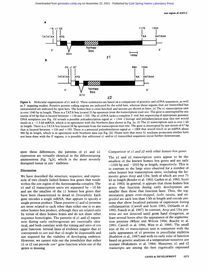
z2 Figure 4. Molecular organization of zl and z2. These summaries are based on a comparison of genomic and cDNA sequences, as well as 5' mapping studies. Putative protein coding regions are indicated by the solid bars, whereas those regions that are transcribed but untranslated are indicated by open bars. The homeo box is cross-hatched, and introns are shown as lines. (a) The zl transcription unit is over 1340 bp in length. There is a TATA box located 25 bp upstream from the transcription start site. The gene is interrupted by one intron of 64 bp that is located between + 138 and + 202. The zl cDNA lacks a complete 3' end, but sequencing of appropriate genomic DNA templates (see Fig. l b) reveals a possible polyadenylation signal at + 1344. Cleavage and polyadenylation near this site would result in a ---1.3-kb mRNA, which is in agreement with the Northern data shown in Fig. 2a. (b) The z2 transcription unit is over 1 kb in length. There is a TATA box located 28 bp upstream from the transcription start site. The gene is interrupted by one intron of 67 bp that is located between + 126 and + 193. There is a potential polyadenylation signal at + 1004 that would result in an mRNA about 900 bp in length, which is in agreement with Northern data (see Fig. 2b). Please note that since S1 nuclease protection studies have not been done with the 3' regions, it is possible that additional zl and/or z2 transcribed sequences occur further downstream.
spite these differences, the patterns of zl and z2 expression are virtually identical in the differentiating amnioserosa (Fig. 7g, h), which is the most severely disrupted tissue in z e n - embryos.
D i s c u s s i o n
We have described the structure, sequence, and expres- sion of two closely linked homeo box genes that reside within the zen region of the Antennapedia complex. The zl and z2 transcription units are separated by ---10 kb and are the smallest of the 11 homeo box genes that have been characterized in detail in Drosophila. Each gene encodes a single mRNA, that appears to specify a single protein product. These putative zl and z2 proteins are more related to each other than either one is to any other homeo box product, although they are related only by virtue of their homeo boxes and do not share other sequence homologies. The patterns of zl and z2 expres- sion during early embryogenesis are essentially iden- tical, and both correlate with the timing and sites of zen gene function. Several lines of evidence suggest that zl corresponds to zen and that z2 might be dispensable and not required for the viability of developing embryos. However, we cannot rule out the possibility that either zl or z2 can provide zen ÷ gene function when one of the genes is missing.
Comparison of z l and z2 with other homeo box genes
The zl and z2 transcription units appear to be the smallest of the known homeo box genes and are only ---1350 bp and ~-1010 bp in length, respectively. This is in contrast to the large sizes observed for a number of other homeo box transcription units, including the ho- meotic genes Antp and Ubx, both of which are over 75 kb in length (Bender et al. 1983; Garber et al. 1983; Scott et al. 1983). In general, it appears that those homeo box genes that function during early development are smaller than those that function later. Thus, the seg- mentat ion genes even-skipped, fushi tarazu, and en- grailed are each less than 5 kb in length and encode pro- teins that show localized patterns of expression during cellularization (Carroll and Scott 1985; DiNardo et al. 1985; Frasch et al. 1987). In contrast, Antp and Ubx pro- teins are not detected until germ band elongation, at least several hours after the appearance of the segmenta- tion proteins (White and Wilcox 1984; Beachy et al. 1985; Carroll et al. 1986; Wirz et al. 1986). The small size of the zl transcription unit is consistent with the early appearance of z l proteins in precellular embryos (Rushlow et al., 1987) and with its early time of function based on genetic analyses of a temperature-sensitive zen mutan t (Wakimoto et al. 1984). Moreover, zl and z2 transcripts are among the first zygotically expressed
GENES & DEVELOPMENT 1273
Cold Spring Harbor Laboratory Press on November 22, 2021 - Published by genesdev.cshlp.orgDownloaded from

Rushlow et al.
homeo box products that are detected during early devel- opment and show dorsally restricted patterns of expres- sion by cleavage stage 10-11 (Doyle et al. 1986; C. Rushlow, unpubl.).
Despite their close linkage, similar structures, and es-
sentially identical patterns of expression, the zl and z2 coding sequences are quite divergent. They do not share significant homologies outside the homeo box, although both proteins possess similar amino acid compositions. For example, both proteins possess high contents of
AAGCCTCTAGATTGCCCATTGCCAATGGCCG~GAGTCGTCGGACT~CTAGTCTCCGAGGTGCCAGTCGGGCAG O
TCCAGCGACGAAGGGATT TACGACTCC.CGC~C~T TAGGCAA~T T T TT TCC~CAACGATGCGTCGGCGGAAT TGACC T G +1
~TATGT~GTCTACCTACAAGCCCTCC~ATAT/~.,CATGATGTTTGCTTAGGACGACAGATCATTCCACTTTTGAC ~ • -
MetSerSerValMetHisTyrTyrProValHisGlnAlaLysValGly 16 AGCTCACTCGGCAGTACCAGAAAAAATATGTCATCCGTCATGCACTACTATCCAGTTCACCAGGCTAAAGTC_C~_
SerTyrSerAlaAspProSerGluValLysTyrSerAspLeuIleTyrGlyHisHisHisAspValAsnProIle 41 TCC_TAT T_CA_GCAGA_T~C T TCCGAGGT TAAATACAGTGA]T TGATCTATGGTCATCATCACGATGT TAACCCCATC
GlyLeuP roP roAsnTyrAsnGlnMetAsnSe rAsnP roThrThrLeuAsnAspHisCys SerP roGlnHisVa i 66 GGTCTGCCTCCCAACTACAACCAGATGAACTCCAACCCCACCACCCTCAACGATCACTGCTCCCCGCAGCATGTG
HisGlnGlnHisValSerSerAspGluAsnLeuProSerGlnProAsnHisAspSerGlnArgValLy~eu~.'ys 91 CATCAGCAGCATGTGTCATCGGACGAGAATCTGCCATCTCAGCCCAACCACGACTCGCAGAGGGTGAAGIGTTAAG
Arg~erArgThrA~aPheThr~er~a]~G~'nLeu~a~G~uLeuG~uAsnG~uP~heLys~erAsnMet'Tyr~.euTyr i16 CGATCACGGACTGCCTTCACCAGTG~CC~CT~TGG~CTTGAG~CG^GTTC~GA~~CATGTACTTGTAC
. . -- . . , ,
ArgThrArgArgileGluileAiaGlnArgLeuSerLeuCys-GluArgGlnValLysfleTrpPheGinAsnArg 141 AGGACGCGCAGGATTGAGATTGCTCAGCGgTTGTCqCTGTGgGAACGCCAGGTGAAGATCTGGTTCCAGAACCGA
ArgMetLysPheLysLysAspI'l~inGlyHisArgGluProLysSerAsnAlaLysLeuAlaGlnProGlnAla 166 CG,AATGA~,, GT TCAAGAA ,GGACATA~CAAGGTCACCGCGAGCCCAAGTCCAACGCCAAGT TGGCTCAACCCCAGGC T
GluGlnSerAlaHisArgGlyIleValLysArgLeuMetSerTyrSerGlnAspProArgGluGlyThrAlaAla 191 GAGCAGAGTGCCCACCGAGC4ZATAGTGAAGCGCCTCATGTCCTACTCCCAGGATCCCAGGGA~ACAGCGGCA
A l a G l u L y s A r g P r o M e t M e t A l a V a l A l a P r o V a l A s n P r o L y s P r o A s p T y r G l n A l a S e r G l n L y s M e t L y s 216 GCAGAAAAGCGTCCTATGATGGCAGT~TCCTGTGAATCCAAAGCCAGACTATCAAGCCAGCCAGAAAATGAAG
T h r G l u A l a S e r T h r A s n A s n G l y M e t C y s S e r S e r A l a A s p L e u S e r G l u I l e L e u G l u H i s L e u A l a G l n T h r 241 ACCGAAGCATCAACCAACAACCK?4:ATGTGCTCCAGCGCCGACCTCAGCGAGATCTTGGAGCATCTGGCTCAAACT
T h r A l a A l a P r o G l n V a l S e r T h r A l a T h r S e r S e r T h r G l y T h r S e r T h r A s n S e r A l a S e r S e r S e r S e r S e r 266 ACAGCTGCACCACAGGTCAGCACTGCCACATCTAGCACAGGAACATCTACCAACTCGGCTTCTAGCTCCTCTTCG
G l y H i s T y r S e r T y r A s n V a l A s p L e u V a l L e u G l n S e r I l e L y s G l n A s p L e u G l u A l a A l a A l a G l n A l a T r p 291 GGCCACTATTCCTACAACGTGGATTTGGTTCTGCAGAGCATCAAGCAGGATTTGGAGC4ZAGCGGCCCAGC4ZTTGG
S e r L y s S e r L y s S e r A l a P r o I l e L e u A l a T h r G l n S e r T r p H i s P r o S e r S e r G l n S e r G l n V a l P r o T h r S e r 316 TCCAAGTCTAAATCCGCGCCTATCCTGGCCACTCAATCCTGGCATCCCAGTTCCCAAAGTCAGGTCCCCACGAGC
V a l H i s A l a A l a P r o S e r M e t A s n L e u S e r T r p G l y G l u P r o A l a A l a L y s S e r A r g L y s L e u S e r V a l A s n H i s 341 GTGCATGCGGCTCCCTCGATGAATCTGTCGT~GAGCCTGCTGCCAAGTCGAGAAAGCTGAGCGTTAATCAC
MetAsnProCysValThrSerTyrAsnTyrProAsnSTOP ATGAACCCGTGCGTAACCTCCTACAATTATCCCAACTAATTTTGAGTTAAAAAAGTTATACTAAGTTTTACTATA
AATTTTTGTTGGAGCTAAGCAACCGTCTCTGC4ZCCTTGGACGAAAAATTCTAACCGAACTCAGTGACAAAAGCCA
TGAATATTCTTAAATTATCGTGAAGTTTAACCATTTTTTTAATAGATAGATTTTAAATAAA
Figure 5. Nucleotide sequence of zl. The complete nucleotide sequence of the zl cDNA is shown. This cDNA is not full length and lacks sequences from both termini. The nucleotide sequence of these missing regions was obtained from genomic DNAs, which are underlined in the above sequence. The nucleotide labeled + 1 corresponds to the transcription start site. There is a well-conserved TATA sequence located 25 bp upstream from the transcription start site. The first AUG is located 52 bp downstream from the start site and begins an open reading frame of 353 amino acid residues. The amino acid sequence of this reading frame is shown above the nucleotide sequence. The numbers in the right column refer to the amino acid residues. The homeo domain is boxed {amino acid residues 90-149). The vertical arrow between nucleotides 138 and 139 indicates the location of the splice junctions of a 64-bp intron. The dashed underline delineates the nucleotide sequence of the oligomer used for the primer extension analysis shown in Fig. 3b, and the circles bracket the S1 nuclease probe used for the protection experiment shown in Fig. 3a.
1274 GENES & DEVELOPMENT
Cold Spring Harbor Laboratory Press on November 22, 2021 - Published by genesdev.cshlp.orgDownloaded from

z e n region of ANT-C
serine and alanine residues and contain short, inter- rupted stretches of polyalanine and polyserine. Support for an evolutionary linkage of the two genes is provided by the observation that the z l and z2 homeo boxes are closely related and share 75%-amino-acid identity.
The zl and z2 homeo boxes are distinct from the 16 other Drosophila homeo boxes that have been reported (reviewed in Levine and Harding 1987). These different homeo boxes have been grouped into three classes: the Antennapedia class, the engrailed class, and the paired class. The zl and z2 horneo boxes are slightly more re- lated to the Antennapedia class (McGinnis et al. 1984; Scott and Weiner 1984) than to the engrailed and paired classes (Fjose et al. 1985; Poole et al. 1985; Frigerio et al.
1986). zl and z2 share -~60%-amino-acid identity with members of the Antennapedia class, ~-50% identity with the engrailed class, and only ~-40% identity with the paired class. There is a highly conserved region of the zl and z2 homeo domains that is identical to the corre- sponding portions of the homeo domains associated with the different genes of the Antennapedia class. This region consists of 14 amino acid residues (residues 131-144 of the z l protein sequence shown in Fig. 5 and residues 84 -97 of the z2 protein sequence shown in Fig. 6) and includes the putative DNA binding recognition helix (Laughon and Scott 1984; Desplan et al. 1985). Therefore, we would expect the zl and z2 proteins to show similar specificities in DNA binding; both of them
TAGA_TCCAGACGTAACATTAATATCGCCTGAATAATATTTCTCCGTGTGACGCTACTTAGAAGTACGTCSGT
T_TCTCTGAAGCCTGGAACTCACCTTGAT~CGGGGCCAGGC~CAAAAGGACGCGACTCTACCAGCCCAGACCA
~T~.T~A AT T TC~AA~T~T~CAC~C~ACA~A~G~Cg~~C~ATCCGTCACG~GGGCTT~T
+1 GGGc~ATATAAgCAAATAA~GAGAAT~TAGA~TATCTAGTTAAAAAACATCGCCCACAGCGGTCAGAAGCC
~,. _-
MetPheAlaIleGlnSerGluAsnTyrPheValAspAsnTyrSerVal 16 AACAAATCGCGCTCTTCAAGAAAAATGTTTGCCATTCAAAGCGAAAACTATTTTGTGGACAATTACTCAGTC
Se rAspLeuMetMet TyrP roCysValGluPheAsnVa 1GluAlaAlaArgThrAlaThrThrArgSe rSer 40 AGTGATTTAATGATGTATCCTTGCGTCGAGTTCAACGTAGAAGCCGCTCGCACGGCAACAACAAGGTCTTCG
Glunys~ rLysArgSe rArgTh rAl aPhe Se rSe rLeuSlnneu I leGluLeuG'luArgGluPheHis Leu 64 GAGAAA~CAAAGAGATCACGCACG~CAT ~CAG~AGTCT TCAGCTAATCGAAC TAGAAAC~GAGT TCCATCTC
As ~Lys TyrLeuAlaArgTh rArgArg'I leGluI leSe"rGlnArgLeuAiaLeuTh rGluArgGinvaliys 88 AACAAATAT T TAGCCCGAACCAGGCGAATCGAGATATCACAGCGT T T GGCAC TCACAGAGAGACAGGT TAAG
I leT rp@heGlnAsn~rgArgMetLys LeuLys Lys serTh~snArgLysGlyAla I IeGIyAI a LeuTh r 112 AT C TGGT TCCAAAATCGTCGCATGAAAC TCAAGAAG~CGACC~KATAGGAAGGGTGCCATTGGAGCGCTGACC
ThrSerIleProLeuSerSerGlnSerSerGluAspLeuGlnLysAspAspGlnIleValGluArgLeuLeu 136 ACATCTATTCCACTTTCTTCCCAGTCGAGCGAAGATCTTCAAAAGGACGACCAGATCGTAGAGCGTCTCCTT
A r g T y r A l a A s n T h r A s n V a l G l u T h r A l a P r o L e u A r g G l n V a l A s p H i s G l y V a l L e u G l u G l u G l y G l n 160 CGATATGCCAACACAAATGTGGAAACAGCTCCACTCCGGCAGGTTGATCACGGTGTCCTGGAGGAGGGTCAA
I l e T h r P r o P r o T y r G l n S e r T y r A s p T y r L e u H i s G l u P h e S e r P r o G l u P r o M e t A l a L e u P r o G l n L e u 184 ATTACACCACCCTATCAGAGCTACGATTACCTCCATGAATTTTCTCCTGAGCCCATGGCTCTTCCCCAACTG
ProPheAsnGluPheAspAlaAsnTrpAlaSerSerTrpLeuGlyLeuGluProThrIleProIleAlaGlu 208 CCCTTCAATGAATTCGATGCTAACT~GAGTTCATGC42T~CTTGAGCCAACGATTCCTATCGCAGAA
AsnValIleGluHisAsnThrGlnAspGlnProMetIleGlnAsnPheCysTrpAspSerAsnSerSerSer 232 AATGTAATTGAACACAATACACAGGATCAGCCAATGATACAGAATTTCTGCTGGGACTCGAATAGCTCTTCT
AlaSerSerSerAspIleLeuAspValAspTyrAspPheIleGlnAsnLeuLeuAsnPheSTOP GCTTCATCGTCGGATATTTTGGATGTTGACTATGACTTTATTCAAAATTTGTTGAACTTTTAAATATATCTT
TGACTATATGTTTAGGCAACTGTACATACATACATATATAATCATTGTATTATACGAGCAATATCTTTCTGT
GACTACAT TAT TAT TACTTATTTTGGATGAAATAAA
Figure 6. Nucleotide sequence of z2. The complete nucleotide sequence of the z2 cDNA is shown. This cDNA is nearly full length and lacks only 6 nucleotides from the 5' end. The nucleotide sequence of the missing region, which is underlined in the above sequence, was obtained from genomic DNAs. The nucleotide labeled + 1 corresponds to the transcription start site. There is a well-conserved TATA sequence located 28 bp upstream from the start site. The first AUG is located 67 bp downstream from the start site and begins an open reading frame of 252 amino acid residues. The amino acid sequence of this reading frame is shown above the nucleotide sequence. The numbers in the right column refer to the amino acid residues. The vertical arrow between nucleotides 126 and 127 indicates the location of the splice junctions of a 67-bp intron. The homeo domain is boxed (amino acid residues 43-102). The dashed underline delineates the nucleotide sequence of the primer used for the experiment shown in Fig. 3c.
GENES & DEVELOPMENT 1275
Cold Spring Harbor Laboratory Press on November 22, 2021 - Published by genesdev.cshlp.orgDownloaded from

Rushlow et al.
- ~ % . . . . . .
D l U I ~ .
e ~ " " , ) . ...... .- P c " ,
m h C F .
g C F - - " " "
CF
F i g u r e 7. Distribution of zl and z2 transcripts in wild-type embryos. All embryos are oriented so that anterior is to the left. Sagittal sections (a-f) are oriented so that dorsal is up. The embryos on the left (a,c,e,g)were hybridized with a zl cDNA probe, whereas sections on the right (b,d,f,h)were hybridized with a z2 eDNA probe. (a,b) Adjacent sagittal sections of a cleavage stage 14 embryo, zl and z2 transcripts are broadly distributed on the dorsal surface, as well as the anterior and posterior poles. (c,d) Adjacent sagittal sections through a gastrulating embryo, zl and z2 transcripts have been lost from the poles and are restricted to the dorsal surface. (e,f) Adjacent sections through an embryo undergoing germ band elongation, zl and z2 transcripts are localized to the amnioserosa and presumptive optic lobe. In addition, zl transcripts are detected in a subset of the pole cells within the posterior midgut invagination. (g,h) Adjacent horizontal sections through an embryo of similar age to that shown in e and f. High levels of the zl and z2 transcripts are detected in the amnioserosa. Abbreviations: (CF) Cephalic furrow; (PC)pole cells; (PMG)posterior midgut invagination.
should be more like the Antennapedia class homeo box proteins as compared with members of the other classes.
Aside from the homeo box coding region, neither zl nor z2 contains significant sequence homologies with any of the eight other homeo box genes for which com- plete sequence data are available (Laughon and Scott 1984; Poole et al. 1985; Laughon et al. 1986; Macdonald et al. 1986; Schneuwly et al. 1986; Stroeher et al. 1986; Frigerio et al. 1986; Mlodzik and Gehring 1987; Regulski et al. 1987; Coleman et al. 1987; Frasch et al. 1987). Moreover, the overall structures of the putative zl and z2 proteins are distinct from most of the other homeo box proteins. In particular, eight of the nine known homeo box proteins contain homeo domains located near the carboxyl terminus. In contrast, the zl and z2
homeo domains are located in the amino-terminal half of the respective proteins. The pair-rule gene even- s k i p p e d is the only other homeo box gene that encodes a homeo domain with a similar amino-terminal location {Macdonald et al. 1986; Frasch et al. 1987).
W h i c h gene corresponds to zen?
The patterns of zl and z2 expression correlate well with the t iming and sites of zen ÷ gene function. The pheno- critical period of zen activity was shown to occur be- tween 2 and 4 hr after fertilization (Wakimoto et al. 1984), which corresponds to the t ime when there are peak levels of zl and z2 transcripts. The complex z e n - phenotype seen in advanced-stage mutant embryos ap-
1276 GENES & DEVELOPMENT
Cold Spring Harbor Laboratory Press on November 22, 2021 - Published by genesdev.cshlp.orgDownloaded from

zen region of ANT-C
pears to result from a disruption of the dorsal ectoderm during gastrulation (C. Rushlow, unpubl.). In particular, the dorsal-most ectoderm, which normally differentiates into the amnioserosa, is transformed toward a more ven- tral cell type in z e n - . We have shown that both z l and z2 transcripts accumulate primarily in the dorsal ecto- derm of wild-type embryos and are observed in the cells that comprise the amnioserosa during germ band elonga- tion. Moreover, zl and z2 transcripts are also detected in the optic lobe primordium, which is another dorsally de- rived structure that is absent in z e n - mutants (Waki- moto et al. 1984). zl transcripts (but not z2) are also de- tected in a subset of the pole cells, which are an unex- pected site of expression since previous genetic studies do not indicate an obvious role for z e n + activity in these cells (Wakimoto et al. 1984). A more rigorous determina- tion of any role for z e n in pole cells would involve pole cell transplantation studies using z e n - embryos as donors.
It is not clear from the expression patterns whether z 1, z2, or both genes correspond to zen . However, here we have shown that the zl transcription unit, in the ab- sence of z2 sequences, can rescue z e n - embryos by P transformation (Fig. la; Table 1). The 4.5-kb genomic DNA fragment that was used for these studies includes only a limited amount of flanking sequences (1.6 kb of 5' and 1.5 kb of 3' flank), which nonetheless appears to contain the c i s - regu la tory elements required for normal function. The efficiency of rescue with P-zl transposons integrated at diverse chromosome locations suggests that flanking sequences exert little, if any, position ef- fects on the level of zl expression from the transposon. Each of five different transformed lines provided effi- cient rescue, whereby over 50% of the expected number of " z e n - " adults were observed (see legend to Table 1). These results are in contrast with rescue studies done with the homeo box gene f u s h i t a r a z u (ftz), which is also located within the ANT-C. The rescue of f t z - embryos required an extensive amount of 5'-flanking sequences (over 6 kb), and there were position effects on the ex- pression of the f t z transposon as well as a relatively poor efficiency of rescue (Hiromi et al. 1985).
The demonstrat ion that the zl gene can rescue z e n -
embryos suggests that zl is a critical component of z e n ÷
gene function. Several lines of evidence suggest that the z2 gene might be dispensable. The zenw36/Df(3R) L I N
mutants that were used for the rescue studies appear to be both z l - and z2-. z e n w36 homozygotes show a marked underexpression of both zl and z2 transcripts, and Df (3R) L I N homozygotes do not express z l and show a grossly abnormal pattern of z2 expression (C. Rushlow and M. Levine, in prep.). Thus, it would appear that z l - , z2- embryos that contain a single copy of the zl gene are fully viable. Further support for this observa- tion stems from genetic studies which indicate that z2- embryos are viable and do not show a z e n mutant pheno- type (M.A. Pultz and T.C. Kaufman, pets. comm.).
It is intriguing that z2 might be dispensable and not required for normal development. The pattern of z2 ex- pression is virtually identical to zl and corresponds to that expected for a z e n product. Moreover, z2 does not
Table 1. Rescue of zen mutan t s with a P-zl transposon
Percentage of Transposon Number of expected
Line Linkage copy number survivors survivors ~
1 II 2 7 56 2 II ND 40 2642 3 III 1 0 3 4 II, III 2 17 112 5 II ND 13 96 6 II 1 14 72
Males carrying at least one copy of the P-zl transposon were crossed to zen w36 r e/TM3 Sb females. Several of the Sb + male progeny were crossed to Df(3R) LIN e/TM3 Sb females, e Sb + progeny were scored as survivors and correspond to zenw36/
Df(3R)LIN trans-heterozygotes. 1 Of the progeny from the final cross (above), 12.5% should cor- respond to mutant trans-heterozygotes since only 50% of the Sb ÷ male progeny from the first cross contain a copy of the zl transposon. The values in this column represent the percentage of the expected number of mutant trans-heterozygotes that ac- tually survived to adulthood. 2 This value exceeds 100% since the transformed line used for rescue contains additional copies of the zl transposon on the X and/or fourth chromosome. 3 There are no survivors expected from this cross since the transformed line contains a single integration on the third chro- mosome, which does not segregate with the zen mutant alleles.
appear to be a pseudogene since the nucleotide sequence of the z2 transcription unit strongly suggests that it en- codes a protein. Thus, it appears that the z2 gene has been selected at the level of protein function, even though it might not be required for normal develop- ment.
There are at least two explanations for this discrep- ancy. First, it is possible that the z2 gene confers a selec- tive advantage to flies in nature that is not obvious when they are cultured in the laboratory. For example, perhaps the z 1 and z2 proteins function more effectively together than does the zl protein alone. Alternatively, it is pos- sible that z2 is not dispensable and that either zl alone or z2 alone can provide z e n + gene function. A critical test of this latter possibility is to determine whether embryos that are z l - and z2 + are viable. In an effort to identify such a situation, we examined the expression of zl and z2 in embryos homozygous for z e n w36, a s well as in D f ( 3 R ) L I N homozygotes. Surprisingly, as indicated above, both of these z e n mutants show altered patterns of zl and z2 expression. Additional experiments, partic- ularly involving the use of P transformation using z2 DNA sequences, will be required to resolve this issue.
There are other examples of closely linked homeo box genes that show essentially identical patterns of expres- sion, including the segmentation genes e n g r a f t e d (en)
and i n v e c t e d { i n v ) ( C o l e m a n et al. 1987) and the segment polarity gene g o o s e b e r r y (Bopp et al. 1986). The signifi- cance of such gene duplications is unclear. It is possible that duplicated, dispensable genes such as z2 represent members of the homeo box gene family that are in the process of acquiring novel functions.
GENES & DEVELOPMENT 1277
Cold Spring Harbor Laboratory Press on November 22, 2021 - Published by genesdev.cshlp.orgDownloaded from

Rushlow et al.
Materials and m e t h o d s
Fly strains
zen W36 is an EMS-induced zen "point" mutation that is cytolo- gically normal (Wakimoto et al. 1984). The deficiency Df(3R)LIN uncovers several genes within the ANT-C, in- cluding Deformed, bicoid, and zen (Frohnhofer and Ntisslein- Volhard, 1986). Df(3R)LIN fails to complement zen W36 (C. Rushlow, unpubl.).
P transformation
Germ line transformation was done essentially as described by Rubin and Spradling (1982). A helper P element called p~r25.7WC (Karess and Rubin 1984)was used. A 4.5-kb XbaI- SacI genomic DNA fragment containing the coding region as well as 1.6 kb of 5' sequence and 1.5 kb of 3' sequence was inserted into the NotI site of pDM30, which is a modification of the Carnegie 20 vector (a gift from D. Mismer). ry s°6 embryos were injected with 50-100 ~g/ml of helper plasmid and 300 ~g/ml of the zl /P transposon.
Cloned DNAs
The zl and z2 cDNAs were isolated in a screen of a 3- to 12-hr cDNA library cloned in ~, gtl0 (Poole et al. 1985).
Sequencing
DNA fragments were cloned into the bacteriophage M13 vectors (Messing 1983), m p l 8 + and m p l 9 + (New England Biolabs). Sequencing was performed by the dideoxy method (Sanger 1977)using [3sS]dATP (New England Nuclear). The single-standed zl DNA fragment that was used for the S1 nu- clease protection experiment shown in Figure 3a was se- quenced by the chemical cleavage method (Maxam and Gilbert 19771.
RNA Extraction
RNA was extracted using a hot phenol-SDS method (Scott et al. 1983) from wild-type embryos at 0-2, 2-3, 3-6, 6-12, and 12-24 hr after fertilization. Poly(A) + RNA was selected on oligo(dT)-cellulose (Sigma) columns, fractionated on an agarose-formaldehyde gel, and transferred to nitrocellulose (Maniatis et al. 1982). Approximately 0.5 ~g of poly(A) ÷ RNA was loaded per lane. [32P]UTP-labeled single-stranded RNA probes were synthesized by T7 transcription from the zl and z2 cDNAs cloned into pGEM1 (Promega). Hybridization was per- formed at 60°C for 24 hr in 50% formamide, 5 x SSC, 5 x Den° hardt's, 50 mM PO4, 0.1% SDS, 100 ~g/ml carrier salmon sperm DNA, 100 ~xg/ml carrier tRNA. The blots were washed 4 x 5 min in 2 x SSC, 0.1% SDS at room temperature and then 3 x 15 min in 0.1 x SSC, 0.1% SDS at 65°C.
Transcript mapping
Primer extension analyses were performed essentially as de- scribed by McKnight and Kingsbury (1982). The nucleotide primers shown in Figure 3 were 32P-labeled at their 5' termini with polynucleotide kinase, and each hybridized with 2 ~g of embryonic poly(A) + RNA in 0.5 M NaC1, 10 mM PIPES (pH 6.4) for 6 hr at 55°C. The pr imer-RNA hybrid templates were ex- tended with reverse transcriptase for 1 hr at 42°C in 50 mM Tris (pH 8.0), 10 mM DTT, 6 mM MgC12; 24 ~g/ml actinomycin D
0.5 mM deoxy-NTPs. The products were electrophoresed in a 6% polyacrylamide-8 M urea gel. The DNA probe used for S1 nuclease protection experiments was obtained from a 316-bp genomic DNA fragment. After 32P-labeling with polynucleotide kinase, the probe was electrophoresed in a nondenaturing gel to separate the strands. Single-stranded probe was hybridized with poly(A) + RNA for 12 hr at 37°C in 0.25 M NaC1, 30 mM NaOAc; 1 mM ZnSO4; 5% glycerol and digested with 25, 50, and 100 units of S1 nuclease. Digestions were done at 37°C for 30 rain. The resulting products were electrophoresed in a 6% polyacryl- amide-8 M urea gel.
In situ hybridization
Tissue sections were prepared as previously described (Harding et al. 1986). 3sS-Labeled antisense RNA probes were synthe- sized by T7 transcription from zl and z2 cDNAs and hybridized to tissue sections as described by Ingham et al. (19851.
A c k n o w l e d g m e n t s
We thank Thom Kaufman and Christiane Niisslein-Volhard for providing fly stocks. We also thank Allen Laughon, Mary Anne Pultz, Mark Seeger, Thorn Kaufman, and Tom Kornberg for sharing unpublished results. We are indebted to Janice Fischer for help with the transformation experiments. This work was funded by grants from the National Institutes of Health, Searle Scholars Program, and the American Cancer Society.
N o t e added in proof
These sequence data have been submitted to the EMBL/Gen Bank Data Libraries under accession number Y00213.
References
Beachy, P.A., S.L. Helfand, and D.S. Hogness. 1985. Segmental distribution of bithorax complex proteins during Drosophila development. Nature 313: 545-551.
Bender, W., P. Spierer, and D.S. Hogness. 1983. Chromosomal walking and jumping to isolate DNA from the Ace, rosy, and bithorax complex in Drosophila melanogaster. ]. Mol. Biol. 168: 17-33.
Carroll, S.B. and M.P. Scott. 1985. Localization of the fushi tarazu protein during Drosophilia embryogenesis. Cell 43: 47-57.
Carroll, S.B., R.A. Laymon, M.A. McCutcheon, P.D. Riley, and M.P. Scott. 1986. The localization and regulation of Anten- napediea protein expression in Drosophila embryos. Cell 47:113-122 .
Campos-Ortega, J. and V. Hartenstein. 1985. The embryonic de- velopment of Drosophila melanogaster, pp. 31-36. Springer-Verlag, Berlin, Heidelberg.
Coleman, K.G., S.J. Poole, M.P. Weir, W.C. Soeller, and T. Kornberg. 1987. The invected gene of Drosophila: Sequence analysis and expression studies reveal a close kinship to the engrailed gene. Genes Dev. 1: 19-28.
Desplan, C., J. Theis, and P.H. O'Farrell. 1985. The Drosophila developmental gene, engrailed, encodes a sequence-specific DNA binding activity. Nature 318: 630-635.
DiNardo, S., J.M. Kuner, J. Theis, and P.H. O'Farrell. 1985. De- velopment of embryonic pattern in D. melanogaster as re- vealed by accumulation of the nuclear engrailed protein. Cell 43: 59-69.
Doyle, H.J., R. Harding, T. Hoey, and M. Levine. 1986. Tran-
1278 GENES & DEVELOPMENT
Cold Spring Harbor Laboratory Press on November 22, 2021 - Published by genesdev.cshlp.orgDownloaded from
z e n region of ANT-C
scripts encoded by a homeo box gene are restricted to dorsal tissues of Drosophila embryos. Nature 323: 76-79.
Fjose, A., W.J. McGinnis, and W.J. Gehring. 1985. Isolation of a homeo box-containing gene from the engrailed region of Drosophila and the spatial distribution of its transcripts. Nature 313: 284-289.
Frasch, M., T. Hoey, C. Rushlow, H. Doyle, and M. Levine. 1987. Characterization and localization of the even-skipped protein of Drosophila. EMBO J. 6: 749-759.
Frigerio, G., M. Burri, D. Bopp, S. Baumgartner, and M. Noll. 1986. Structure of the segmentation gene paired and the Drosophila PRD gene set as part of a gene network. Cell 47: 735- 746.
Frohnhofer, H.G. and C. Niisslein-Volhard. 1986. Organization of anterior pattern in the Drosophila embryo by the ma- ternal gene bicoid. Nature 324: 120-125.
Garber, R.L., A. Kuroiwa, and W.J. Gehring. 1983. Genomic and cDNA clones of the homeotic gene Antennapedia in Droso- phila. EMBO J. 2: 2027-2034.
Harding, K., C. Rushlow, H.J., Doyle, T., Hoey, and M. Levine. 1986. Cross-regulatory interactions among pair-rule genes in Drosophila. Science 229: 1236-1242.
Hiromi, Y., A. Kuroiwa, and W.J. Gehring. 1985. Control ele- ments of the Drosophila segmentation gene fushi tarazu. Cell 43: 603-613.
Ingham, P.W., K.R. Howard, and D. Ish-Horowicz. 1985. Tran- scription pattern of the Drosophila segmentation gene hairy. Nature 318: 439-445.
Karess, R.E. and G. Rubin. 1984. Analysis of P transposable ele- ment functions in Drosophila. Cell 38: 135-146.
Laughon, A. and M.P. Scott. 1984. Sequence of a Drosophila segmentation gene: Protein structure homology with DNA- binding proteins. Nature 310:25-31.
Laughon, A., A.M. Boulet, J.R. Bermingham, R.A. Laymon, and M.P. Scott. 1986. Structure of transcripts from the homeotic Antennapediea gene of Drosophila melanogaster: Two pro- motors control the major protein-coding region. Mol. Cell. Biol. 6: 4676-4689.
Levine, M. and K. Harding. 1987. Spatial regulation of homeo box gene expression in Drosophila. Oxford Survey on Eu- karyotic Genes, vol. 3 Oxford University Press (in press).
Macdonald, P.M., P.N. Ingham, and G. Struhl. 1986. Isolation, structure, and expression of even-skipped: a second pair-rule gene of Drosophila containing a homeo box. Cell 47: 721- 734.
Maniatis, T., E.F. Fritsch, and J. Sambrook. 1982. Molecular cloning: A laboratory manual. Cold Spring Harbor Press, Cold Spring Harbor, New York.
Maxam, A.M. and W. Gilbert. 1977. A new method for se- quencing DNA. Proc. Natl. Acad. Sci. 74: 560-565.
McGinnis, W., M.S. Levine, E. Hafen, S. Kuroiwa, and W.J. Gehring. 1984. A conserved DNA sequence in homeotic genes of the Drosophila Antennapedia and bithorax com- plexes. Nature 308: 428-433.
McKnight, S.L, and R. Kingsbury. 1982. Transcriptional control signals of a eukaryotic protein coding gene. Science 217: 316-324.
Messing, J. 1983. A new pair of M13 vectors for selecting either DNA strand of double digest restriction fragments. Methods Enzymol. 101: 20-28.
Mlodzik, M. and W. Gehring. 1987. Expression of the caudal gene in Drosophila: Formation of an RNA and protein gra- dient during early embryogenesis. Cell 48: 465-478.
Poole, S.J., L.M. Kauvar, B. Drees, and T. Kornberg. 1985. The engrailed locus of Drosophila: Structural analysis of an em- bryonic transcript. Cell 40: 37-43.
Poulson, D.F. 1950. Histogenesis, organogenesis and differen- tiation in the embryo of Drosophila melanogaster. In Bi- ology of Drosophila (ed. M. Demerec), pp. 168-274. Wiley, New York.
Regulski, M., N. McGirmis, R. Chadwick, and W. McGinnis. 1987. Developmental and molecular analysis of Deformed: A homeotic gene controlling Drosophila head development• EMBO J. 6: 767-777.
Rubin, G.M. and A.C. Spradling. 1982. Genetic transformation of Drosophila with transposable element vectors• Science 218: 348-353.
1983. Vectors for P element-mediated gene transfer in Drosophila. Nucleic Acids Res. 11" 6341-6351.
Rushlow, C., M. Frasch, H. Doyle, and M. Levine. 1987. Mate- rial regulation of zerknf~llt, a homoeobox gene controlling differentiation of dorsal tissues in Drosophila. Nature (in press).
Sanger, F., S. Nicklen, and A.R. Coulson. 1977. DNA se- quencing with chain-terminating inhibitors. Proc. Natl. Acad. Sci. 74: 5463-5467.
Schneuwly, S., A. Kuroiwa, P. Baumgartner, and W.J. Gehring. 1986. Structural organization and sequence of the homeotic gene Antennapedia of Drosophila melanogaster. EMBO J. S: 733- 739.
Scott, M.P. and A.J. Weiner. 1984. Structural relationships among genes that control development: Sequence homology between the Antennapedia, Ultrabithorax, and fushi tarazu loci of Drosophila. Proc. Natl. Acad. Sci. 81: 4115-4119.
Scott, M.P., A.J. Weiner, B.A. Polisky, T.I. Hazelrigg, B.A. Plisky, V. Pirrotta, F. Scalenghe, and T.C. Kaufman. 1983. The molecular organization of the Antennapedia complex of Drosophila. Cell 35: 763-776.
Stroeher, V.L., E.M. Jorgensen, and R.L. Garber. 1986. Multiple transcripts from the Antennapedia gene of Drosophila. Mol. Cell. Biol. 6: 4667-4675.
Wakimoto, B.T., F.R. Turner, and T.C. Kaufman. 1984. Defects in embryogenesis in mutants associated with the Antenna- pedia gene complex of Drosophila melanogaster. Dev. Biol. 102: 147-172.
White, R.A.H. and M. Wilcox. 1985. Regulation of the distribu- tion of Ultrabithorax proteins in Drosophila. Nature 318: 563-567.
Wirz, J., L.I. Fessler, and W.J. Gehring. 1986. Localization of the Antennapedia protein in Drosophila embryos and imaginal discs. EMBO I. 5: 3327-3334.
GENES & DEVELOPMENT 1279
Cold Spring Harbor Laboratory Press on November 22, 2021 - Published by genesdev.cshlp.orgDownloaded from

10.1101/gad.1.10.1268Access the most recent version at doi: 1:1987, Genes Dev.
C Rushlow, H Doyle, T Hoey, et al. gene complex in Drosophila.Molecular characterization of the zerknüllt region of the Antennapedia
References
http://genesdev.cshlp.org/content/1/10/1268.full.html#ref-list-1
This article cites 37 articles, 7 of which can be accessed free at:
License
ServiceEmail Alerting
click here.right corner of the article or
Receive free email alerts when new articles cite this article - sign up in the box at the top
Copyright © Cold Spring Harbor Laboratory Press
Cold Spring Harbor Laboratory Press on November 22, 2021 - Published by genesdev.cshlp.orgDownloaded from