Molecular biology Protein condensation enables chromatin ...
3
2. Neuhöfer, P. et al. Nature 597, 715–719 (2021). 3. Fischer, C. G. & Wood, L. D. J. Pathol. 246, 395–404 (2018). 4. Kipling, D. & Cooke, H. J. Nature 347, 400–402 (1990). 5. van Heek, N. T. et al. Am. J. Pathol. 161, 1541–1547 (2002). A.M. declares competing interests. See go.nature. com/3hvzmkm for details. This article was published online on 15 September 2021. groups of cells, which recapitulates the pattern of sporadic KRAS mutation found in human pancreatic cells. However, a caveat to this mouse system is that the effect of telomere maintenance on cancer might differ between mice and humans, given that inbred mouse strains have considerably longer telomeres than do humans 4 . Nevertheless, the concept that Tert expression is an early event in the for- mation of pancreatic tumours is consistent with results in humans showing that telomere shortening is a nearly universal event even in the earliest human pancreatic precancers, and might be the prelude to TERT activation 5 . The identification of KRAS mutations in pop- ulations of human acinar cells by Neuhöfer et al. also provides indirect support for the hypoth- esis of an acinar cell of origin. That said, the hypothesis that TERT-expressing human aci- nar cells acquire KRAS mutations and then give rise to precancer is not testable with existing technologies, given the lack of a reliable way to visualize TERT expression in individual cells. Moreover, the role of KRAS-mutant acinar cells as a possible source of human precancers remains to be defined more clearly. Neverthe- less, these manipulations of a mouse-model system provide an important foundation on which to design the analyses of human tissue that are needed to confirm or refute the hypoth- eses put forward by Neuhöfer and colleagues. Understanding the origin of pancreatic cancer is crucial for the design of data-driven screening and cancer-prevention strate- gies. If the authors’ results are validated in human samples, further characterization of TERT-expressing acinar cells might point to new strategies to intercept early stages of pan- creatic tumour formation in high-risk individ- uals through depletion of the cells of origin. Laura D. Wood is in the Departments of Pathology and Oncology, Sol Goldman Pancreatic Cancer Research Center, Johns Hopkins University School of Medicine, Baltimore, Maryland 21231, USA. Figure 1 | Early events on a pathway towards the formation of a pancreatic tumour. Neuhöfer et al. 2 report using a mouse model to shed light on factors that might initiate the formation of cancer in the pancreas. The pancreas contains acinar and ductal cells, and the authors developed a system to study the pattern of expression of the enzyme Tert in the pancreas. Tert can enable dividing cells to avoid genomic damage, by aiding in the maintenance of telomere structures (not shown) at the ends of chromosomes. The authors report that a small subset of acinar cells express Tert. If such cells also gain a mutation in the gene Kras, their division is promoted (clonal expansion) compared with that of other acinar cells. Such a cell lineage further expands in cell number in response to inflammation. The authors suggest that the formation of this type of cellular lineage might set the stage for other molecular changes that lead to the formation of a precancer. Kras mutation Tert-expressing pancreatic acinar cell Clonal expansion of Tert-expressing cells Precancer Pancreatic acinar cell Pancreatic ductal cell Inflammation Further molecular alterations Anirban Maitra is at the Sheikh Ahmed Center for Pancreatic Cancer Research, University of Texas MD Anderson Cancer Center, Houston, Texas 77030, USA. e-mail: [email protected] 1. Grimont, A., Leach, S. D. & Chadwani, R. Cell. Mol. Gastroenterol. Hepatol. https://doi.org/10.1016/j. jcmgh.2021.07.014 (2021). In a process called liquid–liquid phase separa- tion, proteins and nucleic acids self-organize into liquid-like droplets called condensates 1 . These dynamic, membraneless compartments enable molecules to co-localize at the optimal concentrations needed for different reactions, excluding undesired molecules, and thereby controlling complex biochemical processes in space and time 2 . A growing number of proteins have been described that contain intrinsically disordered regions (IDRs) and can therefore form condensates, and an increasing number of processes have been reported to be con- trolled by liquid–liquid phase separation. On page 726, Shi et al. 3 add UTX to this list of proteins, and tumour suppression to the processes controlled by liquid–liquid phase separation. In cells, DNA is wrapped around histone proteins to form a complex called chromatin. UTX is a chromatin regulator, and controls diverse processes including development, cell differentiation and cancer suppression 4,5 . It catalyses the removal of transcriptionally repressive dimethyl and trimethyl groups from lysine amino-acid residue 27 of the H3 histone protein (an amino acid dubbed H3K27), although many of its functions seem to be independent of this demethylase activity 6 . UTX also binds to various proteins through its tetratricopeptide repeat domains, includ- ing: MLL3 and MLL4, which are the cata- lytic components of larger complexes 4 ; the lysine acetyltransferase enzymes CREBBP and p300; and several proteins that directly remodel chromatin 4–6 , such as CHD4 and SMARCA4. MLL3- and MLL4-containing complexes add monomethyl and dimethyl groups to lysine 4 of H3 histones (H3K4), Molecular biology Protein condensation enables chromatin control David Lara-Astiaso & Brian J. P. Huntly The protein UTX regulates the DNA–protein complex chromatin to suppress tumour growth. Data suggest that the ability of UTX to condense into liquid-like droplets underlies its chromatin-regulating ability. See p.726 642 | Nature | Vol 597 | 30 September 2021 News & views ©2021SpringerNatureLimited.Allrightsreserved.
Transcript of Molecular biology Protein condensation enables chromatin ...
2. Neuhöfer, P. et al. Nature 597, 715–719 (2021).3. Fischer, C. G. & Wood, L. D. J. Pathol. 246, 395–404
(2018).4. Kipling, D. & Cooke, H. J. Nature 347, 400–402 (1990).5. van Heek, N. T. et al. Am. J. Pathol. 161, 1541–1547 (2002).
A.M. declares competing interests. See go.nature.com/3hvzmkm for details.This article was published online on 15 September 2021.
groups of cells, which recapitulates the pattern of sporadic KRAS mutation found in human pancreatic cells. However, a caveat to this mouse system is that the effect of telomere maintenance on cancer might differ between mice and humans, given that inbred mouse strains have considerably longer telomeres than do humans4. Nevertheless, the concept that Tert expression is an early event in the formation of pancreatic tumours is consistent with results in humans showing that telomere shortening is a nearly universal event even in the earliest human pancreatic precancers, and might be the prelude to TERT activation5.
The identification of KRAS mutations in populations of human acinar cells by Neuhöfer et al. also provides indirect support for the hypothesis of an acinar cell of origin. That said, the hypothesis that TERTexpressing human acinar cells acquire KRAS mutations and then give rise to precancer is not testable with existing technologies, given the lack of a reliable way to visualize TERT expression in individual cells. Moreover, the role of KRAS-mutant acinar cells as a possible source of human precancers remains to be defined more clearly. Nevertheless, these manipulations of a mousemodel system provide an important foundation on which to design the analyses of human tissue that are needed to confirm or refute the hypotheses put forward by Neuhöfer and colleagues.
Understanding the origin of pancreatic cancer is crucial for the design of datadriven screening and cancerprevention strategies. If the authors’ results are validated in human samples, further characterization of TERTexpressing acinar cells might point to new strategies to intercept early stages of pancreatic tumour formation in highrisk individuals through depletion of the cells of origin.
Laura D. Wood is in the Departments of Pathology and Oncology, Sol Goldman Pancreatic Cancer Research Center, Johns Hopkins University School of Medicine, Baltimore, Maryland 21231, USA.
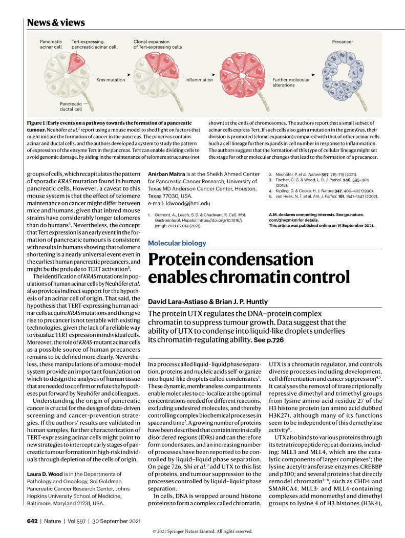
Figure 1 | Early events on a pathway towards the formation of a pancreatic tumour. Neuhöfer et al.2 report using a mouse model to shed light on factors that might initiate the formation of cancer in the pancreas. The pancreas contains acinar and ductal cells, and the authors developed a system to study the pattern of expression of the enzyme Tert in the pancreas. Tert can enable dividing cells to avoid genomic damage, by aiding in the maintenance of telomere structures (not
shown) at the ends of chromosomes. The authors report that a small subset of acinar cells express Tert. If such cells also gain a mutation in the gene Kras, their division is promoted (clonal expansion) compared with that of other acinar cells. Such a cell lineage further expands in cell number in response to inflammation. The authors suggest that the formation of this type of cellular lineage might set the stage for other molecular changes that lead to the formation of a precancer.
Kras mutation
Tert-expressingpancreatic acinar cell
Clonal expansionof Tert-expressing cells
PrecancerPancreaticacinar cell
Pancreaticductal cell
Inflammation Further molecular alterations
Anirban Maitra is at the Sheikh Ahmed Center for Pancreatic Cancer Research, University of Texas MD Anderson Cancer Center, Houston, Texas 77030, USA.e-mail: [email protected]
1. Grimont, A., Leach, S. D. & Chadwani, R. Cell. Mol. Gastroenterol. Hepatol. https://doi.org/10.1016/j.jcmgh.2021.07.014 (2021).
In a process called liquid–liquid phase separation, proteins and nucleic acids selforganize into liquidlike droplets called condensates1. These dynamic, membraneless compartments enable molecules to colocalize at the optimal concentrations needed for different reactions, excluding undesired molecules, and thereby controlling complex biochemical processes in space and time2. A growing number of proteins have been described that contain intrinsically disordered regions (IDRs) and can therefore form condensates, and an increasing number of processes have been reported to be controlled by liquid–liquid phase separation. On page 726, Shi et al.3 add UTX to this list of proteins, and tumour suppression to the processes controlled by liquid–liquid phase separation.
In cells, DNA is wrapped around histone proteins to form a complex called chromatin.
UTX is a chromatin regulator, and controls diverse processes including development, cell differentiation and cancer suppression4,5. It catalyses the removal of transcriptionally repressive dimethyl and trimethyl groups from lysine aminoacid residue 27 of the H3 histone protein (an amino acid dubbed H3K27), although many of its functions seem to be independent of this demethylase activity6.
UTX also binds to various proteins through its tetratricopeptide repeat domains, including: MLL3 and MLL4, which are the catalytic components of larger complexes4; the lysine acetyltransferase enzymes CREBBP and p300; and several proteins that directly remodel chromatin4–6, such as CHD4 and SMARCA4. MLL3 and MLL4containing complexes add monomethyl and dimethyl groups to lysine 4 of H3 histones (H3K4),
Molecular biology
Protein condensation enables chromatin controlDavid Lara-Astiaso & Brian J. P. Huntly
The protein UTX regulates the DNA–protein complex chromatin to suppress tumour growth. Data suggest that the ability of UTX to condense into liquidlike droplets underlies its chromatinregulating ability. See p.726
642 | Nature | Vol 597 | 30 September 2021
News & views
© 2021
Springer
Nature
Limited.
All
rights
reserved. ©
2021
Springer
Nature
Limited.
All
rights
reserved.
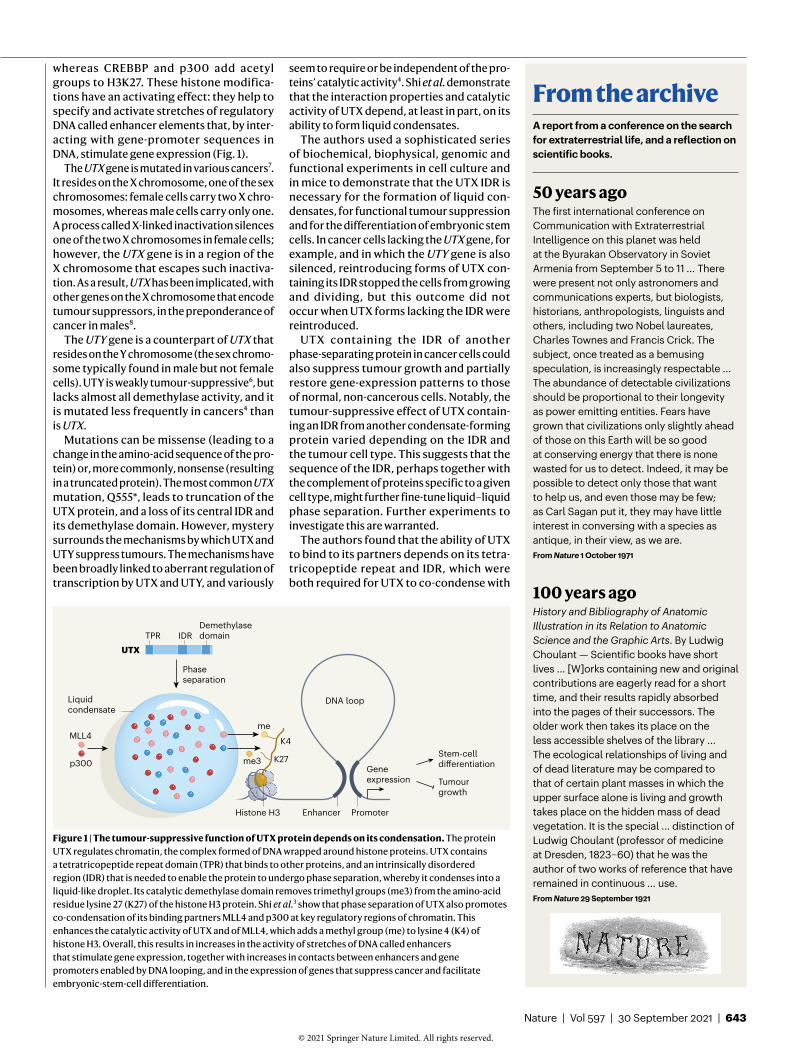
whereas CREBBP and p300 add acetyl groups to H3K27. These histone modifications have an activating effect: they help to specify and activate stretches of regulatory DNA called enhancer elements that, by interacting with genepromoter sequences in DNA, stimulate gene expression (Fig. 1).
The UTX gene is mutated in various cancers7. It resides on the X chromosome, one of the sex chromosomes: female cells carry two X chromosomes, whereas male cells carry only one. A process called Xlinked inactivation silences one of the two X chromosomes in female cells; however, the UTX gene is in a region of the X chromosome that escapes such inactivation. As a result, UTX has been implicated, with other genes on the X chromosome that encode tumour suppressors, in the preponderance of cancer in males8.
The UTY gene is a counterpart of UTX that resides on the Y chromosome (the sex chromosome typically found in male but not female cells). UTY is weakly tumoursuppressive6, but lacks almost all demethylase activity, and it is mutated less frequently in cancers4 than is UTX.
Mutations can be missense (leading to a change in the aminoacid sequence of the protein) or, more commonly, nonsense (resulting in a truncated protein). The most common UTX mutation, Q555*, leads to truncation of the UTX protein, and a loss of its central IDR and its demethylase domain. However, mystery surrounds the mechanisms by which UTX and UTY suppress tumours. The mechanisms have been broadly linked to aberrant regulation of transcription by UTX and UTY, and variously
seem to require or be independent of the proteins’ catalytic activity4. Shi et al. demonstrate that the interaction properties and catalytic activity of UTX depend, at least in part, on its ability to form liquid condensates.
The authors used a sophisticated series of biochemical, biophysical, genomic and functional experiments in cell culture and in mice to demonstrate that the UTX IDR is necessary for the formation of liquid condensates, for functional tumour suppression and for the differentiation of embryonic stem cells. In cancer cells lacking the UTX gene, for example, and in which the UTY gene is also silenced, reintroducing forms of UTX containing its IDR stopped the cells from growing and dividing, but this outcome did not occur when UTX forms lacking the IDR were reintroduced.
UTX containing the IDR of another phaseseparating protein in cancer cells could also suppress tumour growth and partially restore geneexpression patterns to those of normal, noncancerous cells. Notably, the tumoursuppressive effect of UTX containing an IDR from another condensateforming protein varied depending on the IDR and the tumour cell type. This suggests that the sequence of the IDR, perhaps together with the complement of proteins specific to a given cell type, might further finetune liquid–liquid phase separation. Further experiments to investigate this are warranted.
The authors found that the ability of UTX to bind to its partners depends on its tetratricopeptide repeat and IDR, which were both required for UTX to cocondense with
Figure 1 | The tumour-suppressive function of UTX protein depends on its condensation. The protein UTX regulates chromatin, the complex formed of DNA wrapped around histone proteins. UTX contains a tetratricopeptide repeat domain (TPR) that binds to other proteins, and an intrinsically disordered region (IDR) that is needed to enable the protein to undergo phase separation, whereby it condenses into a liquidlike droplet. Its catalytic demethylase domain removes trimethyl groups (me3) from the aminoacid residue lysine 27 (K27) of the histone H3 protein. Shi et al.3 show that phase separation of UTX also promotes cocondensation of its binding partners MLL4 and p300 at key regulatory regions of chromatin. This enhances the catalytic activity of UTX and of MLL4, which adds a methyl group (me) to lysine 4 (K4) of histone H3. Overall, this results in increases in the activity of stretches of DNA called enhancers that stimulate gene expression, together with increases in contacts between enhancers and gene promoters enabled by DNA looping, and in the expression of genes that suppress cancer and facilitate embryonicstemcell differentiation.
me3 K27
me
K4
Histone H3
UTX
Liquid condensate
MLL4
p300
Tumourgrowth
Geneexpression
Stem-celldi�erentiation
DNA loop
TPR IDRDemethylasedomain
Phaseseparation
Enhancer Promoter
A report from a conference on the search for extraterrestrial life, and a reflection on scientific books.
50 years agoThe first international conference on Communication with Extraterrestrial Intelligence on this planet was held at the Byurakan Observatory in Soviet Armenia from September 5 to 11 ... There were present not only astronomers and communications experts, but biologists, historians, anthropologists, linguists and others, including two Nobel laureates, Charles Townes and Francis Crick. The subject, once treated as a bemusing speculation, is increasingly respectable ... The abundance of detectable civilizations should be proportional to their longevity as power emitting entities. Fears have grown that civilizations only slightly ahead of those on this Earth will be so good at conserving energy that there is none wasted for us to detect. Indeed, it may be possible to detect only those that want to help us, and even those may be few; as Carl Sagan put it, they may have little interest in conversing with a species as antique, in their view, as we are.From Nature 1 October 1971
100 years agoHistory and Bibliography of Anatomic Illustration in its Relation to Anatomic Science and the Graphic Arts. By Ludwig Choulant — Scientific books have short lives ... [W]orks containing new and original contributions are eagerly read for a short time, and their results rapidly absorbed into the pages of their successors. The older work then takes its place on the less accessible shelves of the library ... The ecological relationships of living and of dead literature may be compared to that of certain plant masses in which the upper surface alone is living and growth takes place on the hidden mass of dead vegetation. It is the special ... distinction of Ludwig Choulant (professor of medicine at Dresden, 1823–60) that he was the author of two works of reference that have remained in continuous ... use. From Nature 29 September 1921
From the archive
Nature | Vol 597 | 30 September 2021 | 643
© 2021
Springer
Nature
Limited.
All
rights
reserved. ©
2021
Springer
Nature
Limited.
All
rights
reserved.
nature.com/ncomms
@NatureComms
Publish clinical research with Nature Portfolio’s flagship Open Access journal
We welcome submissions reporting influential research in all areas of medical research, from translational medicine to clinical practice, as well as studies of importance to health-related matters. Learn how our team of 100+ dedicated editors are uniquely able to support your publication journey.
A105061
MLL4 or p300 in cells. Shi et al. also show that cocondensation of UTX with MLL4 enhances the enzymatic functions of these proteins in demethylating H3K27 and methylating H3K4, respectively (Fig. 1).
Next, the authors showed that condensation affects UTX and MLL4 binding to the genome, together with the pattern of histone modifications, gene expression and the looping of DNA to allow interactions between enhancers and gene promoters. Unexpectedly, reintroducing IDRcontaining UTX in the cancer cells both positively and negatively affected the binding of UTX and MLL4 to the genome, histone modifications, DNAloop interactions and the expression of certain genes. When IDRcontaining UTX was reintroduced, many of the genes that were expressed more strongly and many of the DNAbinding events that were gained were those associated with cancer inhibition, with very few associated with cell proliferation, in keeping with the restoration of tumour suppression. Lastly, the authors found that UTY was slower to form condensates than was UTX, and they speculate that this might explain why UTY suppresses tumour growth only weakly compared with UTX.
The role of phase separation in gene transcription is well described9. However, its role in chromatin regulation is less well studied.
This and other studies (see, for example, ref. 10) suggest that liquid condensates act as a platform to coordinate the regulation of chromatin, by bringing together protein members of specific complexes at key regulatory sites. Moreover, Shi and coworkers’ study further confirms previous evidence that condensation can facilitate the catalytic activity of enzymes11. It remains to be shown whether this effect reflects the concentrations of enzymes, substrates and any cofactors in the condensates (and, if so, which ones), or other properties, such as condensateinduced changes in the conformation of enzymes.
The observation that restoring the ability of UTX to form condensates has a variable effect on chromatin and gene expression suggests that this protein has a more complex role than just activating transcription, as has been suggested previously4,6. However, a more detailed understanding is required of the direct or indirect nature of UTXmediated gene repression, its possible links to liquid–liquid phase separation and the nature of possible cocondensing proteins in this process.
Together, Shi and colleagues’ findings add tumour suppression to the list of cellular functions associated with phase separation, and suggest that condensates are a key physical platform for facilitating the regulation of
chromatin. The findings therefore reinforce the role of liquid–liquid phase separation as a dynamic means of functionally compartmentalizing the nucleus.
David Lara-Astiaso and Brian J. P. Huntly are in the Department of Haematology and the Wellcome Trust–MRC Cambridge Stem Cell Institute, University of Cambridge, Cambridge CB2 0AW, UK. B.J.P.H. is also at Cambridge University Hospitals, Cambridge, UK.e-mail: [email protected]
1. Banani, S. F., Lee, H. O., Hyman, A. A. & Rosen, M. K. Nature Rev. Mol. Cell Biol. 18, 285–298 (2017).
2. Feric, M. & Mistelli, T. Trends Cell Biol. 8, 671–685 (2021).3. Shi, B. et al. Nature 597, 726–731 (2021).4. Wang, L. & Shilatifard, A. Cancer Cell 35, 168–176 (2019).5. Tran, N., Broun, A. & Ge, K. Mol. Cell. Biol. 40, e00341-20
(2020).6. Gozdecka, M. et al. Nature Genet. 50, 883–894 (2018).7. van Haaften, G. et al. Nature Genet. 41, 521–523 (2009).8. Dunford, A. et al. Nature Genet. 49, 10–16 (2017).9. Hnisz, D., Shrinivas, K., Young, R. A., Chakraborty, A. K. &
Sharp, P. A. Cell 169, 13–23 (2017).10. Fasciani, A. et al. Nature Genet, 52, 1397–1411 (2020).11. Peeples, W. & Rosen, M. K. Nature Chem. Biol. 17, 693–702
(2021).
The authors declare no competing interests.This article was published online on 15 September 2021.
News & views
© 2021
Springer
Nature
Limited.
All
rights
reserved.


![SDG2-Mediated H3K4me3 Is Crucial for Chromatin ... · SDG2-Mediated H3K4me3 Is Crucial for Chromatin Condensation and Mitotic Division during Male Gametogenesis in Arabidopsis1[OPEN]](https://static.fdocuments.in/doc/165x107/5bc9db1c09d3f2df158b48a6/sdg2-mediated-h3k4me3-is-crucial-for-chromatin-sdg2-mediated-h3k4me3-is.jpg)