Molecular basis of telomere syndrome caused by CTC1 mutations
11
Molecular basis of telomere syndrome caused by CTC1 mutations Liuh-Yow Chen, 1 Jana Majerska ´ , and Joachim Lingner 2 Swiss Institute for Experimental Cancer Research (ISREC), School of Life Sciences, Ecole Polytechnique Fe ´de ´ rale de Lausanne (EPFL), 1015 Lausanne, Switzerland Mutations in CTC1 lead to the telomere syndromes Coats Plus and dyskeratosis congenita (DC), but the molecular mechanisms involved remain unknown. CTC1 forms with STN1 and TEN1 a trimeric complex termed CST, which binds ssDNA, promotes telomere DNA synthesis, and inhibits telomerase-mediated telomere elongation. Here we identify CTC1 disease mutations that disrupt CST complex formation, the physical interaction with DNA polymerase a-primase (pola-primase), telomeric ssDNA binding in vitro, accumulation in the nucleus, and/or telomere association in vivo. While having diverse molecular defects, CTC1 mutations commonly lead to the accumulation of internal single-stranded gaps of telomeric DNA, suggesting telomere DNA replication defects as a primary cause of the disease. Strikingly, mutations in CTC1 may also unleash telomerase repression and telomere length control. Hence, the telomere defect initiated by CTC1 mutations is distinct from the telomerase insufficiencies seen in classical forms of telomere syndromes, which cause short telomeres due to reduced maintenance of distal telomeric ends by telomerase. Our analysis provides molecular evidence that CST collaborates with DNA pola-primase to promote faithful telomere DNA replication. [Keywords: telomere syndrome; Coats Plus; dyskeratosis congenita; CST complex; DNA polymerase a-primase] Supplemental material is available for this article. Received May 24, 2013; revised version accepted September 6, 2013. Telomeres are specialized heterochromatin structures at the ends of linear chromosomes and are essential for genome stability by preventing chromosome fusion and degradation (de Lange 2009; Jain and Cooper 2010). In mammals, telomeric DNA consists of long segments of tandem (TTAGGG) repeat duplexes with a short single- stranded G-rich 39 overhang. Telomeric DNA is coated with shelterin proteins, which comprise TRF1, TRF2, RAP1, TIN2, TPP1, and POT1 (de Lange 2009). The length of telomeres determines the protective state and the cellu- lar life span. Progressive telomere shortening occurs in somatic cells due to incomplete replication of chromo- some ends by conventional DNA polymerases and nucleo- lytic processing. Telomerase, a ribonucleoprotein complex with reverse transcriptase activity, lengthens telomeres by adding telomere repeats to their 39 termini, which is essential for telomere length maintenance in germ cells and stem cells (Nandakumar and Cech 2013). The telomere G-rich sequence, with its repetitive nature, can fold into ssDNA secondary structures such as G-quadruplex struc- tures, which are believed to obstruct efficient telomere DNA replication (Bochman et al. 2012; Vannier et al. 2012). CST is a conserved trimeric complex composed of CTC1, STN1, and TEN1. Mammalian CST has been shown to associate with telomeres and is implicated in multiple steps of telomere replication (Miyake et al. 2009; Surovtseva et al. 2009; Chen et al. 2012; Gu et al. 2012; Huang et al. 2012; Stewart et al. 2012; Wu et al. 2012). Disrupting CST in human and mouse cells affects telomere duplex replication and telomere 39 overhang generation. Biochemically, CST was demonstrated to associate with DNA polymerase a-primase (pola-primase) and to be able to stimulate the enzymatic activity of DNA pola-primase (Casteel et al. 2009; Nakaoka et al. 2012). However, studies with cells depleted for CST and with mice deleted for CTC1 suggest that CST may be dispensable for general DNA replication (Gu et al. 2012; Stewart et al. 2012). Mechanistically, it remains to be elucidated how CST cooperates with DNA pola-primase for telomere replication. Apart from its functions in semiconservative DNA replication of telo- meres, CST plays a role in constraining telomerase activity for telomere length homeostasis in cancer cells (Chen et al. 2012). CST associates with and sequesters Ó 2013 Chen et al. This article is distributed exclusively by Cold Spring Harbor Laboratory Press for the first six months after the full-issue publication date (see http://genesdev.cshlp.org/site/misc/terms.xhtml). After six months, it is available under a Creative Commons License (Attribution- NonCommercial 3.0 Unported), as described at http://creativecommons.org/ licenses/by-nc/3.0/. 1 Present address: Institute of Molecular Biology, Academia Sinica, Taipei 115, Taiwan. 2 Corresponding author E-mail [email protected] Article is online at http://www.genesdev.org/cgi/doi/10.1101/gad.222893.113. GENES & DEVELOPMENT 27:2099–2108 Published by Cold Spring Harbor Laboratory Press; ISSN 0890-9369/13; www.genesdev.org 2099 Cold Spring Harbor Laboratory Press on December 5, 2021 - Published by genesdev.cshlp.org Downloaded from
Transcript of Molecular basis of telomere syndrome caused by CTC1 mutations
Molecular basis of telomere syndromecaused by CTC1 mutations
Liuh-Yow Chen,1 Jana Majerska, and Joachim Lingner2
Swiss Institute for Experimental Cancer Research (ISREC), School of Life Sciences, Ecole Polytechnique Federale de Lausanne(EPFL), 1015 Lausanne, Switzerland
Mutations in CTC1 lead to the telomere syndromes Coats Plus and dyskeratosis congenita (DC), but themolecular mechanisms involved remain unknown. CTC1 forms with STN1 and TEN1 a trimeric complex termedCST, which binds ssDNA, promotes telomere DNA synthesis, and inhibits telomerase-mediated telomereelongation. Here we identify CTC1 disease mutations that disrupt CST complex formation, the physicalinteraction with DNA polymerase a-primase (pola-primase), telomeric ssDNA binding in vitro, accumulation inthe nucleus, and/or telomere association in vivo. While having diverse molecular defects, CTC1 mutationscommonly lead to the accumulation of internal single-stranded gaps of telomeric DNA, suggesting telomere DNAreplication defects as a primary cause of the disease. Strikingly, mutations in CTC1 may also unleash telomeraserepression and telomere length control. Hence, the telomere defect initiated by CTC1 mutations is distinct fromthe telomerase insufficiencies seen in classical forms of telomere syndromes, which cause short telomeres due toreduced maintenance of distal telomeric ends by telomerase. Our analysis provides molecular evidence that CSTcollaborates with DNA pola-primase to promote faithful telomere DNA replication.
[Keywords: telomere syndrome; Coats Plus; dyskeratosis congenita; CST complex; DNA polymerase a-primase]
Supplemental material is available for this article.
Received May 24, 2013; revised version accepted September 6, 2013.
Telomeres are specialized heterochromatin structures atthe ends of linear chromosomes and are essential forgenome stability by preventing chromosome fusion anddegradation (de Lange 2009; Jain and Cooper 2010). Inmammals, telomeric DNA consists of long segments oftandem (TTAGGG) repeat duplexes with a short single-stranded G-rich 39 overhang. Telomeric DNA is coatedwith shelterin proteins, which comprise TRF1, TRF2, RAP1,TIN2, TPP1, and POT1 (de Lange 2009). The length oftelomeres determines the protective state and the cellu-lar life span. Progressive telomere shortening occurs insomatic cells due to incomplete replication of chromo-some ends by conventional DNA polymerases and nucleo-lytic processing. Telomerase, a ribonucleoprotein complexwith reverse transcriptase activity, lengthens telomeresby adding telomere repeats to their 39 termini, which isessential for telomere length maintenance in germ cells andstem cells (Nandakumar and Cech 2013). The telomereG-rich sequence, with its repetitive nature, can fold intossDNA secondary structures such as G-quadruplex struc-tures, which are believed to obstruct efficient telomereDNA replication (Bochman et al. 2012; Vannier et al. 2012).
CST is a conserved trimeric complex composed ofCTC1, STN1, and TEN1. Mammalian CST has been shownto associate with telomeres and is implicated in multiplesteps of telomere replication (Miyake et al. 2009; Surovtsevaet al. 2009; Chen et al. 2012; Gu et al. 2012; Huang et al.2012; Stewart et al. 2012; Wu et al. 2012). Disrupting CST inhuman and mouse cells affects telomere duplex replicationand telomere 39 overhang generation. Biochemically, CSTwas demonstrated to associate with DNA polymerasea-primase (pola-primase) and to be able to stimulate theenzymatic activity of DNA pola-primase (Casteel et al.2009; Nakaoka et al. 2012). However, studies with cellsdepleted for CST and with mice deleted for CTC1 suggestthat CST may be dispensable for general DNA replication(Gu et al. 2012; Stewart et al. 2012). Mechanistically, itremains to be elucidated how CST cooperates with DNApola-primase for telomere replication. Apart from itsfunctions in semiconservative DNA replication of telo-meres, CST plays a role in constraining telomeraseactivity for telomere length homeostasis in cancer cells(Chen et al. 2012). CST associates with and sequesters
� 2013 Chen et al. This article is distributed exclusively by Cold SpringHarbor Laboratory Press for the first six months after the full-issuepublication date (see http://genesdev.cshlp.org/site/misc/terms.xhtml). Aftersix months, it is available under a Creative Commons License (Attribution-NonCommercial 3.0 Unported), as described at http://creativecommons.org/licenses/by-nc/3.0/.
1Present address: Institute of Molecular Biology, Academia Sinica, Taipei115, Taiwan.2Corresponding authorE-mail [email protected] is online at http://www.genesdev.org/cgi/doi/10.1101/gad.222893.113.
GENES & DEVELOPMENT 27:2099–2108 Published by Cold Spring Harbor Laboratory Press; ISSN 0890-9369/13; www.genesdev.org 2099
Cold Spring Harbor Laboratory Press on December 5, 2021 - Published by genesdev.cshlp.orgDownloaded from
telomeric 39 overhangs after their elongation by telome-rase, thus limiting telomere extension by telomerase.
Recently, mutations in CTC1 were found to causeCoats Plus, dyskeratosis congenita (DC), and related bonemarrow failure syndromes (Anderson et al. 2012; Kelleret al. 2012; Polvi et al. 2012; Walne et al. 2013). Affectedindividuals carry biallelic CTC1 mutations, which in-clude point mutations and more severe mutations thatpresumably lead to a complete loss of function (Supple-mental Table S1). Coats Plus is a rare autosomal recessiveneurological disorder where patients develop cranial cal-cifications and cysts (Anderson et al. 2012; Polvi et al.2012). Significantly, Coats Plus patients also develop fea-tures seen with the short telomere syndrome DC, instigatedby telomerase defects, such as premature hair graying,anemia, and osteoporosis (Keller et al. 2012; Walne et al.2013). Lymphocyte telomere length in CTC1 mutant in-dividuals was reported to be short in two studies (Andersonet al. 2012; Keller et al. 2012), but this was not seen in twoother reports (Polvi et al. 2012; Walne et al. 2013). Thus, theputative telomere dysfunctions in these patients may notbe directly associated with overall telomere length lossseen in typical telomere syndromes caused by telomerasedeficiencies (Armanios and Blackburn 2012) but insteadmight relate to other structural defects of telomeres. Here,we characterize the molecular defects of disease-causingCTC1 and uncover that these mutations give rise to acommon telomeric replication defect.
Results
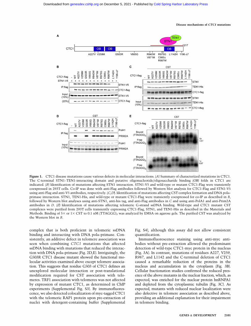
To elucidate the molecular defects caused by CTC1mutations at telomeres, we generated 11 mutations inhuman CTC1 cDNA that were reported in Coats Plus/DC patients to produce CTC1 polypeptides carrying pointmutations or small deletions (Fig. 1A; Anderson et al. 2012;Keller et al. 2012; Polvi et al. 2012; Walne et al. 2013).CTC1 forms the CST complex with STN1 and TEN1through a direct physical interaction with STN1. To assessthe interaction between CTC1 and STN1, V5-tagged STN1(STN5-V5) and wild-type or mutant Flag-tagged CTC1(CTC1-Flag) were coexpressed upon transient transfectionin HEK293T cells. Association of STN1-V5 with CTC1-Flag was detected upon immunoprecipitation with anti-Flag antibodies (Fig. 1B). The C-terminal disease mutationsCTC1-L1142H and CTC1-1196-D7 (deletion of aminoacid residues 1196–1202) disrupted the ability of CTC1to bind to STN1. This is consistent with our previousfinding that the C-terminal region of CTC1 mediatesSTN1 interaction (Chen et al. 2012; Chen and Lingner2013). Coexpression of TEN1 with CTC1-Flag and STN1partially rescued CST complex formation of CTC1-L1142H but not CTC1-1196-D7 (Fig. 1C; SupplementalFig. S1A). Therefore, CTC1 assembly into the CST com-plex involves critical residues of the CTC1 C terminusthat promote interactions with STN1. The enhancementof the CTC1–STN1 interaction by TEN1 concurred witha stabilization of the STN1 and TEN1 polypeptides uponconcomitant expression of the two factors (SupplementalFig. S1A).
CTC1 and STN1 are thought to contribute to semicon-servative DNA replication of telomeric DNA (Gu et al.2012; Huang et al. 2012; Stewart et al. 2012). They wereshown to associate with DNA pola-primase and increasethe affinity of DNA pola-primase for template DNA(Casteel et al. 2009). We therefore assessed by coimmu-noprecipitation (co-IP) the physical interaction of CTC1-Flag with the catalytic subunit PolA1 of DNA pola andthe regulatory subunit PRIM2A of DNA primase in HEK293Tcells in which STN1 and TEN1 were coexpressed (Fig. 1D).Three CTC1 point mutations (A227V, V259M, and V665G)abolished association with endogenous DNA pola-primase.A227 and V259 are part of the most N-terminal of threeputative OB (oligonucleotide/oligosaccharide-binding)folds in CTC1, whereas V665 resides in an unrecognizeddomain of CTC1. In addition, the CST complex-defectiveCTC1-1196-D7 was unable to interact with DNA pola-primase. For CTC1-L1142H, the interaction with DNApola-primase occurred only upon CST complex formation,which was dependent on ectopic coexpression of STN1and TEN1 (cf. Fig. 1D and Supplemental Fig. S1B). Together,these data suggest that the physical contacts of both CTC1and STN1/TEN1 with DNA pola-primase contribute toa stable association of CST with DNA pola-primase. CSTwas also known to physically interact with the shelterincomponents POT1–TPP1 (Wan et al. 2009; Chen et al.2012; Wu et al. 2012), but none of the CTC1 mutationstested here abolished the interaction with TPP1 or POT1as measured in co-IP experiments (Supplemental Fig. S2).
CST binds with preference to the G-rich telomericssDNA (Chen et al. 2012). Therefore, we expressed andpartially purified wild-type and mutant CST complexesupon transient transfection in HEK293T cells (Fig. 1C) forelectromobility shift assays (EMSAs) with the telomericG-strand oligonucleotide (TTAGGG)3 (Fig. 1E). EMSAwith two different concentrations of CST revealed defectsin telomeric ssDNA binding of CTC1-V665G, CTC1-R975G, CTC1-C985D, CTC1-R987W, and CTC1-1196-D7. DNA binding by CTC1-L1142H was also reduced.Therefore, in vitro binding of CSTwith telomeric G-strandssDNA relies on critical residues comprising the most C-terminal 230 amino acids of CTC1. Defects in ssDNAbinding of CTC1-L1142 and CTC1-1197-D7, which alsoshow diminished ability of STN1 interaction, are consis-tent with our previous study that CTC1 on its own doesnot bind ssDNA (Chen et al. 2012). These data supportthe notion that forming a trimeric complex is crucial forhigh-affinity binding of telomeric G-rich ssDNA by CST.
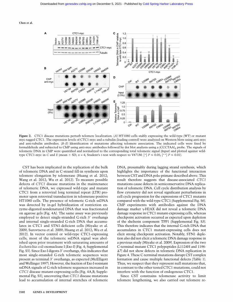
To test whether the disease mutations affect CTC1telomere association and cellular localization, we stablyexpressed myc-tagged CTC1 in HT1080 cells (Fig. 2A).Telomere association was measured by chromatin immu-noprecipitation (ChIP) using anti-myc antibodies (Fig. 2B–E). We found that the telomere association in vivo requiredcritical residues in the N-terminal and central part of theCTC1 polypeptide (A227, V259, and V665) interfacing theinteraction with DNA pola-primase and also an intact Cterminus mediating ssDNA binding and CST complexformation. This result suggests that telomere associationin vivo depends on the formation of a trimeric CST
Chen et al.
2100 GENES & DEVELOPMENT
Cold Spring Harbor Laboratory Press on December 5, 2021 - Published by genesdev.cshlp.orgDownloaded from
complex that is both proficient in telomeric ssDNAbinding and interacting with DNA pola-primase. Con-sistently, an additive defect in telomere association wasseen when combining CTC1 mutations that affectedssDNA binding with mutations that reduced the interac-tion with DNA pola-primase (Fig. 2D,E). Intriguingly, theG503R CTC1 disease mutant showed the functional mo-lecular activities examined above except telomere associa-tion. This suggests that residue G503 of CTC1 defines anunexplored molecular interaction or post-translationalmodification required for CST association with telo-meres. TRF1 association with telomeres was not affectedby expression of mutant CTC1, as determined in ChIPexperiments (Supplemental Fig. S3). By immunofluores-cence, we also detected colocalization of myc-tagged CTC1with the telomeric RAP1 protein upon pre-extraction ofnuclei with detergent-containing buffer (Supplemental
Fig. S4), although this assay did not allow consistentquantification.
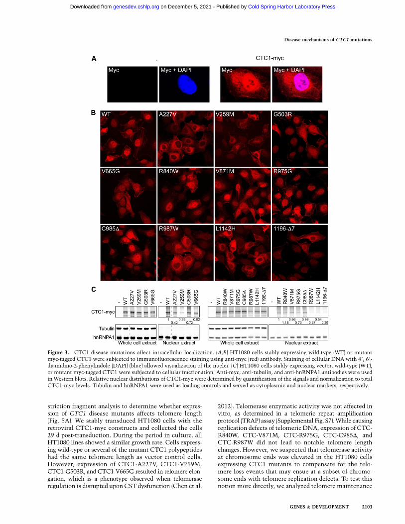
Immunofluorescence staining using anti-myc anti-bodies without pre-extraction allowed the predominantdetection of wild-type CTC1-myc protein in the nucleus(Fig. 3A). In contrast, mutations of residues A227, V259,R987, and L1142 and the C-terminal deletion of CTC1caused a remarkable reduction of the proteins in thenucleus and accumulation in the cytoplasm (Fig. 3B).Cellular fractionation studies confirmed the reduced pres-ence of the above mutants in the nuclear fraction, which, asexpected, was enriched for the nuclear protein hnRNPA1and depleted from the cytoplasmic tubulin (Fig. 3C). Asexpected, mutants with reduced nuclear localization werealso defective in telomere association as described above,providing an additional explanation for their impairmentin telomere binding.
Figure 1. CTC1 disease mutations cause various defects in molecular interactions. (A) Summary of characterized mutations in CTC1.The C-terminal STN1–TEN1-interacting domain and putative oligonucleotide/oligosaccharide binding (OB) folds in CTC1 areindicated. (B) Identification of mutations affecting STN1 interaction. STN1-V5 and wild-type or mutant CTC1-Flag were transientlycoexpressed in 293T cells. Co-IP was done with anti-Flag antibodies followed by Western blot analysis for CTC1-Flag and STN1-V5using anti-Flag and anti-V5 antibodies, respectively. (C,D). Identification of mutations affecting CST complex formation and DNA pola-primase interaction. STN1, TEN1-His, and wild-type or mutant CTC1-Flag were transiently coexpressed for co-IP as described in B,followed by Western blot analyses using anti-STN1, anti-his tag, and anti-Flag antibodies in C and using anti-PolA1 and anti-Prim2Aantibodies in D. (E) Identification of mutations affecting telomeric G-strand ssDNA binding. Wild-type and CTC1 mutant CSTcomplexes were purified from 293T cells transiently expressing CTC1-Flag, STN1, and TEN1-His as described in the Materials andMethods. Binding of 53 or 13 CST to 0.1 nM (TTAGGG)3 was analyzed by EMSA on agarose gels. The purified CST was analyzed bythe Western blot in B.
Disease mechanisms of CTC1 mutations
GENES & DEVELOPMENT 2101
Cold Spring Harbor Laboratory Press on December 5, 2021 - Published by genesdev.cshlp.orgDownloaded from
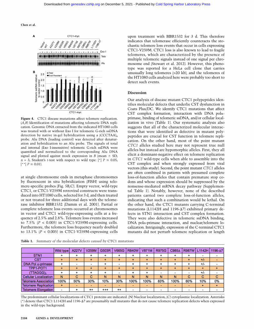
CST has been implicated in the replication of the bulkof telomeric DNA and in C-strand fill-in synthesis upontelomere elongation by telomerase (Huang et al. 2012;Wang et al. 2012; Wu et al. 2012). To measure possibledefects of CTC1 disease mutations in the maintenanceof telomeric DNA, we expressed wild-type and mutantCTC1 from a retroviral long terminal repeat (LTR) pro-moter upon retroviral transduction in telomerase-positiveHT1080 cells. The presence of telomeric G-rich ssDNAwas detected by in-gel hybridization of restriction en-zyme-digested nondenatured DNA that was fractionatedon agarose gels (Fig. 4A). The same assay was previouslyemployed to detect single-stranded G-rich 39 overhangsand internal single-stranded G-rich DNA that accumu-lates in CTC1 and STN1-deficient cells (Miyake et al.2009; Surovtseva et al. 2009; Huang et al. 2012; Wu et al.2012). In vector control or wild-type CTC1-expressingcells, most of the telomeric ssDNA signal was abol-ished upon prior treatment with saturating amounts ofEscherichia coli exonuclease I (Exo I) (Fig. 4; SupplementalFig. S5). Since Exo I digests 39 overhangs, this indicates thatmost single-stranded G-rich telomeric sequences werepresent as terminal 39 overhangs, as expected (McElligottand Wellinger 1997). However, the fraction of Exo I-resistantssDNA signals of G-rich telomere sequences increased inCTC1 disease mutant-expressing cells (Fig. 4A,B; Supple-mental Fig. S5), uncovering that CTC1 disease mutationslead to accumulation of internal stretches of telomeric
DNA, presumably during lagging strand synthesis, whichhighlights the importance of the functional interactionbetween CSTand DNA pola-primase described above. Thisresult therefore suggests that disease-associated CTC1mutations cause defects in semiconservative DNA replica-tion of telomeric DNA. Cell cycle distribution analysis byflow cytometry did not reveal significant perturbations incell cycle progression for the expressions of CTC1 mutantscompared with the wild-type CTC1 (Supplemental Fig. S6).ChIP experiments with antibodies against the DNAdamage marker g-H2AX did not reveal a telomeric DNAdamage response in CTC1 mutant-expressing cells, whereascheckpoint activation occurred as expected upon depletionof the shelterin component TPP1 (Supplemental Fig. S3).This therefore indicates that the internal G-rich DNA thataccumulates in CTC1 mutant-expressing cells does notelicit strong checkpoint activation. Notably, STN1 deple-tion also did not elicit a telomeric DNA damage response ina previous study (Miyake et al. 2009). Expression of the twoC-terminal mutant CTC1 polypeptides (L1124H and 1196-D7) did not show defects in telomeric DNA replication inFigure 4. These C-terminal mutations disrupt CSTcomplexformation and cause multiple functional defects (Table 1).Thus, we suspect that they represent null mutations that,in contrast to the other tested CTC1 mutations, could notinterfere with the function of endogenous CTC1.
Since CST constrains telomerase activity to limittelomere lengthening, we also carried out telomere re-
Figure 2. CTC1 disease mutations perturb telomere localization. (A) HT1080 cells stably expressing the wild-type (WT) or mutantmyc-tagged CTC1. The expression levels of CTC1-myc and a-tubulin (loading control) were analyzed on Western blots using anti-mycand anti-tubulin antibodies. (B–E) Identification of mutations affecting telomere association. The indicated cells were fixed byformaldehyde and subjected to ChIP using anti-myc antibodies followed by dot blot analysis using a (CCCTAA)n probe. The signals oftelomeric DNA in ChIP were quantified and normalized to the corresponding total telomeric signal (Input) and plotted against wild-type CTC1-myc in C and E (mean 6 SD; n = 4; Student’s t-test with respect to V871M: [*] P # 0.05; [**] P # 0.01).
Chen et al.
2102 GENES & DEVELOPMENT
Cold Spring Harbor Laboratory Press on December 5, 2021 - Published by genesdev.cshlp.orgDownloaded from
striction fragment analysis to determine whether expres-sion of CTC1 disease mutants affects telomere length(Fig. 5A). We stably transduced HT1080 cells with theretroviral CTC1-myc constructs and collected the cells29 d post-transduction. During the period in culture, allHT1080 lines showed a similar growth rate. Cells express-ing wild-type or several of the mutant CTC1 polypeptideshad the same telomere length as vector control cells.However, expression of CTC1-A227V, CTC1-V259M,CTC1-G503R, and CTC1-V665G resulted in telomere elon-gation, which is a phenotype observed when telomeraseregulation is disrupted upon CST dysfunction (Chen et al.
2012). Telomerase enzymatic activity was not affected invitro, as determined in a telomeric repeat amplificationprotocol (TRAP) assay (Supplemental Fig. S7). While causingreplication defects of telomeric DNA, expression of CTC-R840W, CTC-V871M, CTC-R975G, CTC-C985D, andCTC-R987W did not lead to notable telomere lengthchanges. However, we suspected that telomerase activityat chromosome ends was elevated in the HT1080 cellsexpressing CTC1 mutants to compensate for the telo-mere loss events that may ensue at a subset of chromo-some ends with telomere replication defects. To test thisnotion more directly, we analyzed telomere maintenance
Figure 3. CTC1 disease mutations affect intracellular localization. (A,B) HT1080 cells stably expressing wild-type (WT) or mutantmyc-tagged CTC1 were subjected to immunofluorescence staining using anti-myc (red) antibody. Staining of cellular DNA with 49, 69-diamidino-2-phenylindole (DAPI) (blue) allowed visualization of the nuclei. (C) HT1080 cells stably expressing vector, wild-type (WT),or mutant myc-tagged CTC1 were subjected to cellular fractionation. Anti-myc, anti-tubulin, and anti-hnRNPA1 antibodies were usedin Western blots. Relative nuclear distributions of CTC1-myc were determined by quantification of the signals and normalization to totalCTC1-myc levels. Tubulin and hnRNPA1 were used as loading controls and served as cytoplasmic and nuclear markers, respectively.
Disease mechanisms of CTC1 mutations
GENES & DEVELOPMENT 2103
Cold Spring Harbor Laboratory Press on December 5, 2021 - Published by genesdev.cshlp.orgDownloaded from
at single chromosome ends in metaphase chromosomesby fluorescent in situ hybridization (FISH) using telo-mere-specific probes (Fig. 5B,C). Empty vector, wild-typeCTC1, or CTC1-V259M retroviral constructs were trans-duced into HT1080 cells, selected for 3 d, and then treatedor not treated for three additional days with the telome-rase inhibitor BIBR1532 (Damm et al. 2001). Partial orcomplete telomere loss events occurred at chromosomesin vector and CTC1 wild-type-expressing cells at a fre-quency of 2.5% and 2.8%. Telomere loss events increasedto 7.5% (P < 0.005) in CTC1-V259M-expressing cells.Furthermore, the telomere loss frequency nearly doubledto 13.1% (P < 0.005) in CTC1-V259M-expressing cells
upon treatment with BIBR1532 for 3 d. This thereforeindicates that telomerase efficiently counteracts the sto-chastic telomere loss events that occur in cells expressingCTC1-V259M. CTC1 loss is also known to lead to fragiletelomeres, which are characterized by the presence ofmultiple telomeric signals instead of one signal per chro-mosome end (Stewart et al. 2012). However, this pheno-type was reported for a HeLa cell clone that carriesunusually long telomeres (>20 kb), and the telomeres ofthe HT1080 cells analyzed here were probably too short todetect such events.
Discussion
Our analysis of disease mutant CTC1 polypeptides iden-tifies molecular defects that underlie CST dysfunction inCoats Plus/DC. We identify CTC1 mutations that affectCST complex formation, interaction with DNA pola-primase, binding of telomeric ssDNA, and/or cellular local-ization in vivo (Table 1). Our systematic analysis alsosuggests that all of the characterized molecular interac-tions that were identified as defective in mutant poly-peptides are crucial for CST function in telomere repli-cation. On the other hand, most of the point mutantCTC1 alleles studied here may not represent true nullalleles but instead are hypomorphic alleles. First, they allelicit a dominant-negative effect on telomere replicationin CTC1 wild-type cells when able to assemble into theCST complex and when strongly expressed from viralvectors (this study). Second, the point mutant CTC1 allelesare often combined in patients with presumed completeloss-of-function alleles that contain premature stop co-dons and whose expression should be suppressed by thenonsense-mediated mRNA decay pathway (Supplemen-tal Table 1). Notably, however, none of the describedpatients carried two complete loss-of-function alleles,indicating that such a combination would be lethal. Onthe other hand, the CTC1 mutants carrying C-terminalmutations (L1142H and 1196-D7) exhibited primary de-fects in STN1 interaction and CST complex formation.They were also defective in telomeric ssDNA binding,DNA pola-primase interaction, and nuclear/telomere lo-calization. Intriguingly, expression of the C-terminal CTC1mutants did not perturb telomere replication or length
Table 1. Summary of the molecular defects caused by CTC1 mutations
The predominant cellular localizations of CTC1 proteins are indicated. (N) Nuclear localization; (C) cytoplasmic localization. Asterisks(*) denote that CTC1 L1142H and 1196-D7 are presumably null mutants that do not cause telomere replication defects when expressedin the wild-type background.
Figure 4. CTC1 disease mutations affect telomere replication.(A,B) Identification of mutations affecting telomeric DNA repli-cation. Genomic DNA extracted from the indicated HT1080 cellswas treated with or without Exo I for telomeric G-rich ssDNAdetection by native in-gel hybridization using a (CCCTAA)nprobe. Alu DNA (loading control) was detected after denatur-ation and hybridization to an Alu probe. The signals of totaland internal (Exo I-insensitive) telomeric G-rich ssDNA werequantified and normalized to the corresponding Alu DNAsignal and plotted against mock expression in B (mean 6 SD;n = 3; Student’s t-test with respect to wild type: [*] P # 0.05;[**] P # 0.01).
Chen et al.
2104 GENES & DEVELOPMENT
Cold Spring Harbor Laboratory Press on December 5, 2021 - Published by genesdev.cshlp.orgDownloaded from

maintenance, all of which suggests that they representnull mutants. Consistently, the C-terminal mutants, likeother loss-of-function alleles, are causing disease in com-bination with presumed hypomorphic alleles (Supplemen-tal Table 1).
The pleiotropy of the molecular defects of CTC1 pointmutants may be unexpected. However, with the excep-tion of the complex formation-defective CTC1 (L1142Hand 1196-D7), expression of all CTC1 mutants causedaccumulation of internal stretches of telomeric G-rich
DNA. This therefore indicates that disruption of a greatvariety of physical interactions leads to a common telo-mere defect manifested by incomplete lagging strandsynthesis during telomeric DNA replication. Our analy-sis suggests that biogenesis of functional CTC1 involves,as an early step, the assembly into a complex with STN1and TEN1. This may occur in the cytoplasm, as complex-defective CTC1 variants are cytoplasmic. Nuclear importor retention of CST appears to rely on complex formationand involve critical amino acids in the CTC1 N terminus
Figure 5. CTC1 mutations affect telomerase control and telomere maintenance. (A) Identification of mutations affecting telomere lengthhomeostasis. Indicated CTC1 proteins or mock-expressing HT1080 cells were collected 29 d after puromycin selection for telomere lengthanalysis by terminal restriction fragment assay. (B,C) Telomere FISH analysis of HT1080 cells stably expressing vector, wild-type CTC1, orCTC1-V259M in the presence or absence of BIBR1532 (15 mm, 72 h). Metaphase chromosome spreads were hybridized with a Cy3-(CCCTAA)4 PNA probe (red) and stained with DAPI (blue). Representative normal and defective mitotic telomere structures uponexpression of CTC1-V259M are shown in B. Representative metaphase chromosome spreads and quantification of chromosome aberrations(mean of the percentage of defective chromosomes per metaphase; n = 20–30) are shown in C. (White arrows) Telomere loss sites.
Disease mechanisms of CTC1 mutations
GENES & DEVELOPMENT 2105
Cold Spring Harbor Laboratory Press on December 5, 2021 - Published by genesdev.cshlp.orgDownloaded from
(A227 and V259). In the nucleus, telomere associationmay be reinforced by DNA pola-primase, as DNA pola-primase interaction mutants show reduced telomere asso-ciation of CTC1. As yeast CST is thought to recruit DNApola-primase, it will be interesting to test whether telomereassociation-defective CTC1 also impairs the recruitmentof DNA pola-primase. The ability to bind single-strandedtelomeric DNA may also enhance association with telo-meric chromatin, but this activity does not seem neces-sary (C985D) or sufficient (V259M). Finally, since none ofthe mutants showed a defect in the interaction with POT1–TPP1, it seems that POT1–TPP1 per se is not able to recruitor retain CST at telomeres.
The internal stretches of telomeric ssDNA associatedwith telomere replication defects did not elicit a detectableDNA damage response. However, incomplete laggingstrand synthesis is expected to lead to rapid telomere-shortening events in subsequent rounds of DNA repli-cation if gapped C strands serve as replication tem-plates. In addition, telomeric G-rich ssDNA can fold intoG-quadruplex structures that impede DNA replication andmay cause telomere-shortening events (Paeschke et al.2010; Vannier et al. 2012). Consistent with this notion,we observed accumulation of telomere-free ends in cellsthat expressed mutant CTC1 (V259M). We therefore pro-pose that Coats Plus/DC-causing CTC1 mutations initiatetelomere syndrome by affecting semiconservative replica-tion of telomeric DNA.
Our data also indicate that telomerase can efficientlycounteract the telomere loss events that occur uponCTC1 dysfunction. Indeed, telomerase inhibition during3 d nearly doubled the occurrence of telomere-free endsin CTC1-V259M cells. In addition, we observed that ex-pression of certain CTC1 mutations caused telomerelengthening in telomerase-positive cells, consistent withthe inhibitory role of CST in telomerase regulation (Chenet al. 2012). The mutations that caused telomere elonga-tion all showed reduced association with telomeric DNA,suggesting that a reduced presence of CST at telomeresprevented telomerase inhibition (Chen et al. 2012). A re-duced interaction of mutant CST with DNA pola-primasemay also promote telomerase, as seen in Saccharomycescerevisiae mutants in which a reduced interaction betweenCdc13 and DNA pola gives rise to longer telomeres (Qi andZakian 2000; Grossi et al. 2004). However, telomere elon-gation was only seen with a subset of CTC1 mutants withdefective telomere replication, indicating that defectivetelomere replication per se does not stimulate telomereelongation in human cells.
In the presence of telomerase, telomere replication de-fects and telomere loss events caused by CTC1 mutationsare at least partially compensated by enhanced telomeraseaction at truncated telomeres. This suggests that, in humans,CTC1 mutations may differentially affect various celltypes, depending on their telomerase status. For instance,somatic cells in adults that lack telomerase activity maybe more susceptible to CTC1 mutations than stems cellsthat express significant amounts of telomerase. This mayexplain the discrepancy of telomere-shortening pheno-types recently reported in patients with CTC1 mutations.
In summary, this study reveals that disease-causing CTC1mutations cause telomere replication defects, which cor-responds to a new type of telomere syndrome that isstrikingly different from the well-characterized ‘‘classical’’telomere syndromes, which show defects in telomerasebiogenesis or activity.
While this study was under review, a report was pub-lished on mouse CTC1 that contained the disease-associ-ated human CTC1 mutations at the corresponding posi-tions (Gu and Chang 2013). Notably, several mouse CTC1mutations did not fully recapitulate the defects observedfor mutant human CTC1. For instance, the correspondingCTC1-G503R, CTC1-V665G, and CTC1-R840W mutationsrendered the mouse CTC1 protein unstable. Therefore,an assessment of localization and molecular interactiondefects was not possible. Furthermore, we found thatresidues A227, V259, and V665 of human CTC1 are criticalfor the physical interaction with DNA pola-primase, whilethe corresponding mutations in mouse CTC1 did not affectthis interaction. In addition, Gu and Chang (2013) showedthat mutations in the central part of mouse CTC1 disruptinteraction with STN1. However, we found that the samemutations in human CTC1 preserve the abilities of CTC1to bind STN1 and form a CST complex. Instead, the diseasemutations at the C terminus of human CTC1, includingL1142H and 1196-D7, prevent its interaction with STN1.Therefore, our analysis indicates that it is crucial to analyzethe human CTC1 disease mutations with human CTC1 todecipher the defects of the disease. Our molecular andfunctional characterization of disease mutations of humanCTC1 is a first step toward understanding the diseasemechanisms, which may facilitate future developments oftherapeutic interventions.
Materials and methods
Protein expression vectors and stable cell line generation
cDNAs of CTC1, STN1, and TEN1 were subcloned into pEAK8and retroviral-based pCL vectors for mammalian expression oftagged or untagged proteins as indicated. CTC1 point mutationsand CTC1-C985D were generated by site-directed mutagenesis(Agilent, Inc.). For CTC1-1196-D7 (deletion of amino acid residues1196–1202), two overlapping cDNA fragments that skipped thedeletion region were generated by PCR and cloned into theexpression vectors by In-Fusion Cloning (Clontech). Stable celllines were generated by viral transduction and puromycin selec-tion (1 mg mL�1). All human cells were maintained at 37°C with5% CO2 in Dulbecco’s modified Eagle’s medium supplementedwith 10% FCS and penicillin/streptomycin.
Co-IP
Cells were lysed in NETN buffer (40 mM Tris-HCl at pH 8.0,100 mM NaCl, 1 mM EDTA, 0.5% NP-40) supplemented withprotease inhibitor cocktail (Sigma) for immunoprecipitationwith anti-Flag M2 affinity gel (Sigma) and subsequent Westernblot analysis as previously described (Chen et al. 2012).
Cellular fractionation
HT1080 cells were grown in 10-cm dishes and collected bytrypsinization. After washing with PBS, cells were partially
Chen et al.
2106 GENES & DEVELOPMENT
Cold Spring Harbor Laboratory Press on December 5, 2021 - Published by genesdev.cshlp.orgDownloaded from
permeabilized for 3 min on ice with 60 mL of buffer A (10 mMHEPES at pH 7.9, 10 mM KCl, 1.5 mM MgCl2, 0.34 M sucrose,10% glycerol, 1 mM DTT) supplemented with 0.1% Triton X-100.Nuclear fractions were obtained by centrifugation at 1500g for5 min. Cell fractionation was validated by Western blot analysisusing tubulin and hnRNPA1 as cytoplasmic and nuclear markers,respectively.
CST complex purification and EMSA
Protein expression and purification and EMSA were carried outas previously described (Chen et al. 2012) with minor modifica-tions. In brief, 293T cells in a 6-cm dish were transfected with2 mg of pEAK8-CTC1-Flag, 1 mg of pEAK8-STN1, and 1 mg of pEAK8-TEN1-His plasmids using Lipofectamine 2000 (Invitrogen). Threedays after transfection, cells were collected for immunoprecip-itation using anti-Flag M2 affinity gel, and the CST complex waseluted with 60 mL of NETN buffer containing 200 mg/mL Flagpeptide. For EMSA, 20-mL reactions containing 1 mL of eluateand 4 mL of eluate buffer or 5 mL of eluate, 0.1 nM [32P]-labeled(TTAGGG)3 oligonucleotide, 50 nM Tris-HCl (pH 8.0), 50 mMNaCl, 1 mM DTT, and 5% glycerol were loaded on 2.5% agarosegels for electrophoresis. Gels were dried and analyzed on a Phos-phorImager (FLA-3000, Fujifilm).
ChIP
Stable HT1080 cell lines expressing wild-type and mutant myc-tagged CTC1 protein were fixed with 1% formaldehyde, soni-cated, and subjected to analysis as previously described (Chenet al. 2012).
Immunofluorescence staining
Cells fixed with 2% paraformaldehyde were permeabilized with0.5% Triton X-100 for immunodetection of CTC1-myc andRAP1 using a-mouse anti-myc (9E10) and a-rabbit anti-RAP1primary antibodies (NB100-292, Novus), respectively. After wash-ing with phosphate-buffered saline, secondary antibodies againstmouse IgG conjugated with Alexa-Fluor 663 and rabbit IgG con-jugated with Alexa-Fluor 488 were used to recognize the primaryantibodies for visualization. Cell nuclei were visualized by DNAstaining with 49, 69-diamidino-2-phenylindole (DAPI) dye. Z-stackimages of the samples were obtained using an LSM700 confocalmicroscope (Zeiss) and processed with ImageJ software.
Terminal restriction fragment assay and telomeric G-richssDNA detection
Genomic DNA from HT1080 cells was extracted using theWizard Genomic DNA purification kit (Promega). GenomicDNA (2.5 mg) was subjected to restriction digestion with RsaIand HinfI overnight at 37°C for agarose gel electrophoresis. Gelswere dried for 1 h at 50°C, denatured with 0.8 M NaOH and 150 mMNaCl, and hybridized to a [32P]-labeled (CCCTAA)n probe fordetection of telomeric DNA. The radioisotope-labeled (CCCTAA)nprobe was generated by random prime labeling in the presence of[a32P]-dCTP using telomeric repeat sequences with a size of;600 base pairs (bp) as template. The telomeric repeat templatewas produced by self-amplification in 28 cycles of PCR using(TTAGGG)5 and (CCCTAA)5 oligonucleotides. To detect telomericG-rich ssDNA, gels were dried after electrophoresis and subjectedto hybridization with a [32P]-labeled (CCCTAA)n probe. To removetelomeric 39 overhang sequences, 1 mg of genomic DNA wastreated with 10 U of E. coli Exo I for 3 h before restriction digestionwith RsaI and HinfI. To detect Alu DNA, the gels were denatured
and hybridized with a [32P]-labeled 59-GTGATCCGCCCGCCTCGGCCTCCCAAAGTG-39 probe.
TRAP assay
Cell extracts were prepared by cells lysis in CHAPS buffer(10 mM Tris-HCl at pH 7.5, 1 mM MgCl2, 1 mM EGTA, 0.5%CHAPS, 10% glycerol, 5 mM b-mercaptoethanol, protease in-hibitors) and centrifugation at 12,000g for 20 min. Telomerasereactions were performed for 30 min at 30°C and contained in50-mL volumes of cell extract (0.1 mg or 0.3 mg), 0.1 mg of TSoligonucleotide (59-AATCCGTCGAGCAGAGTT-39), 20 mMTris-HCl (pH 8.3), 6.5 mM MgCl2, 63 mM KCl, 0.005% Tween20, 1 mM EGTA, 0.1 mg/mL BSA, 0.25-mm dNTPs, and 0.2 mL of[a-32P]dGTP (3000 Ci/mmol). Subsequently, 0.2 U of Taq poly-merase and 0.1 mg of ACX oligonucleotide (59-GCGCGGCTTACCCTTACCCTTACCTAACC-39) were added to the telome-rase reactions for 28 cycles of PCR (cycles of 30 sec at 94°C and30 sec at 60°C). TRAP reactions were separated on 12.5% poly-acrylamide gels and analyzed on a PhosphorImager (FLA-3000,Fujifilm).
Telomere FISH
HT1080 cells were treated with 0.05 mg/mL Demecolcine(Sigma) for 1 h, harvested by trypsinization, swollen in 0.056M KCl, and resuspended in fixative (methanol:acetic acid = 3:1)for metaphase spread on glass slides. Following denaturation for3 min at 80°C, hybridization was performed for 3 h at 25°C in70% formamide, 0.5% blocking reagent (Roche), 200 mM Cy3-OO-(CCCTAA)4 PNA probe (Eurogentec), and 10 mM Tris-HCl(pH 7.4). Slides were washed first with 10 mM Tris-HCl (pH 7.4)and 70% formamide and subsequently with 0.1 M Tris-HCl (pH7.4), 0.15 M NaCl, and 0.08% Tween-20. DNA was counter-stained with DAPI, and slides were mounted in ProLong Gold(Invitrogen) anti-fade reagent.
Acknowledgments
Research in the laboratory was supported by the Swiss NationalScience Foundation, a European Research Council advanced in-vestigator grant (grant agreement no. 232812), an Initial TrainingNetwork (ITN) grant (CodeAge) from the European Commission’sSeventh Framework Programme (grant agreement no. 316354),the Swiss Cancer League, and EPFL. L.-Y.C. performed most ofthe experiments with assistance from J.M., and L.-Y.C. and J.L.designed the study and wrote the paper.
References
Anderson BH, Kasher PR, Mayer J, Szynkiewicz M, JenkinsonEM, Bhaskar SS, Urquhart JE, Daly SB, Dickerson JE,O’Sullivan J, et al. 2012. Mutations in CTC1, encodingconserved telomere maintenance component 1, cause Coatsplus. Nat Genet 44: 338–342.
Armanios M, Blackburn EH. 2012. The telomere syndromes.Nat Rev Genet 13: 693–704.
Bochman ML, Paeschke K, Zakian VA. 2012. DNA secondarystructures: Stability and function of G-quadruplex struc-tures. Nat Rev Genet 13: 770–780.
Casteel DE, Zhuang S, Zeng Y, Perrino FW, Boss GR, Goulian M,Pilz RB. 2009. A DNA polymerase-a�primase cofactor withhomology to replication protein A-32 regulates DNA repli-cation in mammalian cells. J Biol Chem 284: 5807–5818.
Chen LY, Lingner J. 2013. CST for the grand finale of telomerereplication. Nucleus 4: 277–282.
Disease mechanisms of CTC1 mutations
GENES & DEVELOPMENT 2107
Cold Spring Harbor Laboratory Press on December 5, 2021 - Published by genesdev.cshlp.orgDownloaded from

Chen LY, Redon S, Lingner J. 2012. The human CST complex isa terminator of telomerase activity. Nature 488: 540–544.
Damm K, Hemmann U, Garin-Chesa P, Hauel N, Kauffmann I,Priepke H, Niestroj C, Daiber C, Enenkel B, Guilliard B, et al.2001. A highly selective telomerase inhibitor limiting hu-man cancer cell proliferation. EMBO J 20: 6958–6968.
de Lange T. 2009. How telomeres solve the end-protectionproblem. Science 326: 948–952.
Grossi S, Puglisi A, Dmitriev PV, Lopes M, Shore D. 2004. Pol12,the B subunit of DNA polymerase a, functions in bothtelomere capping and length regulation. Genes Dev 18: 992–1006.
Gu P, Chang S. 2013. Functional characterization of humanCTC1 mutations reveals novel mechanisms responsible forthe pathogenesis of the telomere disease Coats Plus. Aging
Cell doi: 10.1111/acel.12139.Gu P, Min JN, Wang Y, Huang C, Peng T, Chai W, Chang S. 2012.
CTC1 deletion results in defective telomere replication,leading to catastrophic telomere loss and stem cell exhaus-tion. EMBO J 31: 2309–2321.
Huang C, Dai X, Chai W. 2012. Human Stn1 protects telomereintegrity by promoting efficient lagging-strand synthesis attelomeres and mediating C-strand fill-in. Cell Res 22: 1681–1695.
Jain D, Cooper JP. 2010. Telomeric strategies: Means to an end.Annu Rev Genet 44: 243–269.
Keller RB, Gagne KE, Usmani GN, Asdourian GK, Williams DA,Hofmann I, Agarwal S. 2012. CTC1 mutations in a patientwith dyskeratosis congenita. Pediatr Blood Cancer 59: 311–314.
McElligott R, Wellinger RJ. 1997. The terminal DNA structureof mammalian chromosomes. EMBO J 16: 3705–3714.
Miyake Y, Nakamura M, Nabetani A, Shimamura S, Tamura M,Yonehara S, Saito M, Ishikawa F. 2009. RPA-like mammalianCtc1–Stn1–Ten1 complex binds to single-stranded DNA andprotects telomeres independently of the Pot1 pathway. Mol
Cell 36: 193–206.Nakaoka H, Nishiyama A, Saito M, Ishikawa F. 2012. Xenopus
laevis Ctc1–Stn1–Ten1 (xCST) protein complex is involvedin priming DNA synthesis on single-stranded DNA templatein Xenopus egg extract. J Biol Chem 287: 619–627.
Nandakumar J, Cech TR. 2013. Finding the end: Recruitment oftelomerase to telomeres. Nat Rev Mol Cell Biol 14: 69–82.
Paeschke K, McDonald KR, Zakian VA. 2010. Telomeres:Structures in need of unwinding. FEBS Lett 584: 3760–3772.
Polvi A, Linnankivi T, Kivela T, Herva R, Keating JP, Makitie O,Pareyson D, Vainionpaa L, Lahtinen J, Hovatta I, et al. 2012.Mutations in CTC1, encoding the CTS telomere mainte-nance complex component 1, cause cerebroretinal micro-angiopathy with calcifications and cysts. Am J Hum Genet90: 540–549.
Qi H, Zakian VA. 2000. The Saccharomyces telomere-bindingprotein Cdc13p interacts with both the catalytic subunit ofDNA polymerase a and the telomerase-associated Est1 pro-tein. Genes Dev 14: 1777–1788.
Stewart JA, Wang F, Chaiken MF, Kasbek C, Chastain PD 2nd,Wright WE, Price CM. 2012. Human CST promotes telomereduplex replication and general replication restart after forkstalling. EMBO J 31: 3537–3549.
Surovtseva YV, Churikov D, Boltz KA, Song X, Lamb JC,Warrington R, Leehy K, Heacock M, Price CM, Shippen DE.2009. Conserved telomere maintenance component 1 inter-acts with STN1 and maintains chromosome ends in highereukaryotes. Mol Cell 36: 207–218.
Vannier JB, Pavicic-Kaltenbrunner V, Petalcorin MI, Ding H,Boulton SJ. 2012. RTEL1 dismantles T loops and counteracts
telomeric G4-DNA to maintain telomere integrity. Cell 149:795–806.
Walne A, Bhagat T, Kirwan M, Gitaux C, Desguerre I, LeonardN, Nogales E, Vulliamy T, Dokal I. 2013. Mutations in thetelomere capping complex in bone marrow failure and re-lated syndromes. Haematologica 98: 334–338.
Wan M, Qin J, Songyang Z, Liu D. 2009. OB fold-containingprotein 1 (OBFC1), a human homolog of yeast Stn1, associateswith TPP1 and is implicated in telomere length regulation. JBiol Chem 284: 26725–26731.
Wang F, Stewart JA, Kasbek C, Zhao Y, Wright WE, Price CM.2012. Human CST has independent functions during telo-mere duplex replication and C-strand fill-in. Cell Rep 2:1096–1103.
Wu P, Takai H, de Lange T. 2012. Telomeric 39 overhangs derivefrom resection by Exo1 and Apollo and fill-in by POT1b-associated CST. Cell 150: 39–52.
Chen et al.
2108 GENES & DEVELOPMENT
Cold Spring Harbor Laboratory Press on December 5, 2021 - Published by genesdev.cshlp.orgDownloaded from
10.1101/gad.222893.113Access the most recent version at doi: 27:2013, Genes Dev.
Liuh-Yow Chen, Jana Majerská and Joachim Lingner
mutationsCTC1Molecular basis of telomere syndrome caused by
Material
Supplemental
http://genesdev.cshlp.org/content/suppl/2013/10/10/27.19.2099.DC1
References
http://genesdev.cshlp.org/content/27/19/2099.full.html#ref-list-1
This article cites 27 articles, 11 of which can be accessed free at:
License
Commons Creative
.http://creativecommons.org/licenses/by-nc/3.0/Creative Commons License (Attribution-NonCommercial 3.0 Unported), as described at
). After six months, it is available under ahttp://genesdev.cshlp.org/site/misc/terms.xhtmlsix months after the full-issue publication date (see This article is distributed exclusively by Cold Spring Harbor Laboratory Press for the first
ServiceEmail Alerting
click here.right corner of the article or
Receive free email alerts when new articles cite this article - sign up in the box at the top
© 2013 Chen et al.; Published by Cold Spring Harbor Laboratory Press
Cold Spring Harbor Laboratory Press on December 5, 2021 - Published by genesdev.cshlp.orgDownloaded from