Molecular Basis of Catalytic Chamber-assisted Unfolding and Cleavage of Human Insulin by
13
Molecular Basis of Catalytic Chamber-assisted Unfolding and Cleavage of Human Insulin by Human Insulin-degrading Enzyme * □ S Received for publication, January 6, 2009 Published, JBC Papers in Press, March 25, 2009, DOI 10.1074/jbc.M900068200 Marika Manolopoulou ‡1 , Qing Guo ‡1 , Enrico Malito ‡ , Alexander B. Schilling § , and Wei-Jen Tang ‡2 From the ‡ Ben-May Department for Cancer Research, University of Chicago, Chicago, Illinois 60637 and the § Proteomics and Informatics Services Facility, University of Illinois, Chicago, Illinois 60612 Insulin is a hormone vital for glucose homeostasis, and insulin- degrading enzyme (IDE) plays a key role in its clearance. IDE exhib- its a remarkable specificity to degrade insulin without breaking the disulfide bonds that hold the insulin A and B chains together. Using Fourier transform ion cyclotron resonance (FTICR) mass spec- trometry to obtain high mass accuracy, and electron capture disso- ciation (ECD) to selectively break the disulfide bonds in gas phase fragmentation, we determined the cleavage sites and composition of human insulin fragments generated by human IDE. Our time- dependent analysis of IDE-digested insulin fragments reveals that IDE is highly processive in its initial cleavage at the middle of both the insulin A and B chains. This ensures that IDE effectively splits insulin into inactive N- and C-terminal halves without breaking the disulfide bonds. To understand the molecular basis of the recogni- tion and unfolding of insulin by IDE, we determined a 2.6- A ˚ reso- lution insulin-bound IDE structure. Our structure reveals that IDE forms an enclosed catalytic chamber that completely engulfs and intimately interacts with a partially unfolded insulin molecule. This structure also highlights how the unique size, shape, charge distribution, and exosite of the IDE catalytic chamber contribute to its high affinity (100 nM) for insulin. In addition, this structure shows how IDE utilizes the interaction of its exosite with the N terminus of the insulin A chain as well as other properties of the catalytic chamber to guide the unfolding of insulin and allowing for the processive cleavages. IDE 3 is an 110-kDa zinc metalloprotease that is evolution- arily conserved from bacteria to humans (1, 2). It was first dis- covered based on its high affinity to bind insulin (100 nM) and degrade it into pieces (3, 4). Insulin is a 5.8-kDa hormone that plays a central role in glucose homeostasis and the development of diabetes in humans. Consistent with the in vitro activity of IDE for insulin degradation, loss-of-function mutations of IDE in rodents result in elevated insulin levels and glucose intoler- ance (5). In addition, a nucleotide polymorphism of the human IDE gene is linked to type 2 diabetes (6). Later studies showed that IDE can also degrade amyloid- (A), a peptide vital to the progression of Alzheimer disease (7, 8). Accumulating evidence from rodent models and human genetic analyses also indicate the physiological role of IDE in the clearance of A (5, 9 –12). Despite nearly 60 years of studies on IDE, the molecular basis by which IDE binds, unfolds, and degrades insulin has only begun to be elucidated. Different from ATP-dependent pro- teases, IDE does not require the additional energy source such as ATP to unfold, bind, and cleave its substrates (4, 13). Insulin consists of the A and B chains that are held together by two inter- and one intra-chain disulfide bonds. Remarkably, IDE does not require disulfide bond isomerase activity to unfold and cleave insulin (4). Thus, IDE needs to overcome the stability created by the disulfide bonds of insulin. Structural analysis reveals that human IDE contains a catalytic chamber formed by the internal cavity of two roughly equally sized 55-kDa N- and C-terminal halves (IDE-N and IDE-C, respectively) (2). Within this chamber, only one catalytic center exists. However, IDE cleaves insulin at multiple sites on both the insulin A and B chains to completely inactivate this hormone. It remains unclear whether the cleavages of insulin by IDE proceed in a sequential or stochastic manner. IDE represents an emerging protease family that utilizes an enclosed catalytic chamber to selectively recognize and unfold the substrates for their degradation (1). The volume of the enclosed chamber of IDE (16,000 Å 3 ) allows the preferential exclusion of peptides that are greater than 75 amino acids long. This chamber also has unique electrostatic properties; the internal cavity of IDE-N is predominantly negative, whereas that of IDE-C is positive. Inside the catalytic chamber, IDE has an exosite that is an evolutionarily conserved substrate-binding site 30 Å away from the catalytic groove. This exosite is used to anchor the N-terminal end of IDE substrates. The unique size, electrostatic potential, and exosite of Ides’ catalytic cham- ber are postulated as key factors for the selective binding and unfolding of IDE substrates (1, 2, 14). In addition, one common feature among the known IDE substrates is their higher pro- * This work was supported, in whole or in part, by National Institutes of Health Grant GM81539 (to W.-J. T.). This work was also supported by the American Health Assistance Foundation (to E. M.). The atomic coordinates and structure factors (codes 2WBY and 2WC0) have been deposited in the Protein Data Bank, Research Collaboratory for Structural Bioinformatics, Rutgers University, New Brunswick, NJ (http://www.rcsb.org/). □ S The on-line version of this article (available at http://www.jbc.org) contains supplemental Table S1 and Figs. S1–S7. 1 Both authors contributed equally to this work. 2 To whom correspondence should be addressed: Ben-May Dept. for Cancer Research, 929 East 57th St., University of Chicago, Chicago, Illinois 60637. Tel.: 773-702-4331; Fax: 773-702-4476; E-mail: [email protected]. 3 The abbreviations used are: IDE, insulin-degrading enzyme; FTICR, Fourier transform ion cyclotron resonance; ECD, electron capture dissociation; MALDI-TOF, matrix-assisted laser desorption ionization time-of-flight; MS, mass spectrometry; MS/MS, tandem mass spectrometry; HPLC, high pres- sure liquid chromatography; ITC, isothermal titration calorimetry; PDB, Protein Data Bank; A, amyloid-; ESI, electrospray ionization; IDE-CF, cys- teine-free IDE; LC, liquid chromatography. THE JOURNAL OF BIOLOGICAL CHEMISTRY VOL. 284, NO. 21, pp. 14177–14188, May 22, 2009 © 2009 by The American Society for Biochemistry and Molecular Biology, Inc. Printed in the U.S.A. MAY 22, 2009 • VOLUME 284 • NUMBER 21 JOURNAL OF BIOLOGICAL CHEMISTRY 14177 by guest on January 14, 2019 http://www.jbc.org/ Downloaded from
Transcript of Molecular Basis of Catalytic Chamber-assisted Unfolding and Cleavage of Human Insulin by

Molecular Basis of Catalytic Chamber-assisted Unfoldingand Cleavage of Human Insulin by HumanInsulin-degrading Enzyme*□S
Received for publication, January 6, 2009 Published, JBC Papers in Press, March 25, 2009, DOI 10.1074/jbc.M900068200
Marika Manolopoulou‡1, Qing Guo‡1, Enrico Malito‡, Alexander B. Schilling§, and Wei-Jen Tang‡2
From the ‡Ben-May Department for Cancer Research, University of Chicago, Chicago, Illinois 60637 and the §Proteomics andInformatics Services Facility, University of Illinois, Chicago, Illinois 60612
Insulin is a hormone vital for glucose homeostasis, and insulin-degradingenzyme(IDE)playsakeyrole in its clearance. IDEexhib-its a remarkable specificity todegrade insulinwithout breaking thedisulfidebondsthatholdthe insulinAandBchains together.UsingFourier transform ion cyclotron resonance (FTICR) mass spec-trometry to obtainhighmass accuracy, and electron capturedisso-ciation (ECD) to selectively break the disulfide bonds in gas phasefragmentation, we determined the cleavage sites and compositionof human insulin fragments generated by human IDE. Our time-dependent analysis of IDE-digested insulin fragments reveals thatIDE is highly processive in its initial cleavage at themiddle of boththe insulin A and B chains. This ensures that IDE effectively splitsinsulin into inactiveN-andC-terminalhalveswithoutbreakingthedisulfide bonds. To understand themolecular basis of the recogni-tion and unfolding of insulin by IDE, we determined a 2.6- A reso-lution insulin-bound IDE structure.Our structure reveals that IDEforms an enclosed catalytic chamber that completely engulfs andintimately interacts with a partially unfolded insulin molecule.This structure also highlights how the unique size, shape, chargedistribution, andexositeof the IDEcatalytic chambercontribute toits high affinity (�100 nM) for insulin. In addition, this structureshows how IDE utilizes the interaction of its exosite with the Nterminus of the insulin A chain as well as other properties of thecatalytic chamber toguide theunfoldingof insulinandallowing forthe processive cleavages.
IDE3 is an �110-kDa zinc metalloprotease that is evolution-arily conserved from bacteria to humans (1, 2). It was first dis-
covered based on its high affinity to bind insulin (�100 nM) anddegrade it into pieces (3, 4). Insulin is a 5.8-kDa hormone thatplays a central role in glucose homeostasis and the developmentof diabetes in humans. Consistent with the in vitro activity ofIDE for insulin degradation, loss-of-function mutations of IDEin rodents result in elevated insulin levels and glucose intoler-ance (5). In addition, a nucleotide polymorphism of the humanIDE gene is linked to type 2 diabetes (6). Later studies showedthat IDE can also degrade amyloid-� (A�), a peptide vital to theprogression of Alzheimer disease (7, 8). Accumulating evidencefrom rodent models and human genetic analyses also indicatethe physiological role of IDE in the clearance of A� (5, 9–12).Despite nearly 60 years of studies on IDE, themolecular basis
by which IDE binds, unfolds, and degrades insulin has onlybegun to be elucidated. Different from ATP-dependent pro-teases, IDE does not require the additional energy source suchas ATP to unfold, bind, and cleave its substrates (4, 13). Insulinconsists of the A and B chains that are held together by twointer- and one intra-chain disulfide bonds. Remarkably, IDEdoes not require disulfide bond isomerase activity to unfold andcleave insulin (4). Thus, IDE needs to overcome the stabilitycreated by the disulfide bonds of insulin. Structural analysisreveals that human IDE contains a catalytic chamber formed bythe internal cavity of two roughly equally sized�55-kDaN- andC-terminal halves (IDE-N and IDE-C, respectively) (2). Withinthis chamber, only one catalytic center exists. However, IDEcleaves insulin at multiple sites on both the insulin A and Bchains to completely inactivate this hormone. It remainsunclear whether the cleavages of insulin by IDE proceed in asequential or stochastic manner.IDE represents an emerging protease family that utilizes an
enclosed catalytic chamber to selectively recognize and unfoldthe substrates for their degradation (1). The volume of theenclosed chamber of IDE (�16,000 Å3) allows the preferentialexclusion of peptides that are greater than �75 amino acidslong. This chamber also has unique electrostatic properties; theinternal cavity of IDE-N is predominantly negative, whereasthat of IDE-C is positive. Inside the catalytic chamber, IDE hasan exosite that is an evolutionarily conserved substrate-bindingsite �30 Å away from the catalytic groove. This exosite is usedto anchor the N-terminal end of IDE substrates. The uniquesize, electrostatic potential, and exosite of Ides’ catalytic cham-ber are postulated as key factors for the selective binding andunfolding of IDE substrates (1, 2, 14). In addition, one commonfeature among the known IDE substrates is their higher pro-
* This work was supported, in whole or in part, by National Institutes of HealthGrant GM81539 (to W.-J. T.). This work was also supported by the AmericanHealth Assistance Foundation (to E. M.).
The atomic coordinates and structure factors (codes 2WBY and 2WC0) have beendeposited in the Protein Data Bank, Research Collaboratory for StructuralBioinformatics, Rutgers University, New Brunswick, NJ (http://www.rcsb.org/).
□S The on-line version of this article (available at http://www.jbc.org) containssupplemental Table S1 and Figs. S1–S7.
1 Both authors contributed equally to this work.2 To whom correspondence should be addressed: Ben-May Dept. for Cancer
Research, 929 East 57th St., University of Chicago, Chicago, Illinois 60637.Tel.: 773-702-4331; Fax: 773-702-4476; E-mail: [email protected].
3 The abbreviations used are: IDE, insulin-degrading enzyme; FTICR, Fouriertransform ion cyclotron resonance; ECD, electron capture dissociation;MALDI-TOF, matrix-assisted laser desorption ionization time-of-flight; MS,mass spectrometry; MS/MS, tandem mass spectrometry; HPLC, high pres-sure liquid chromatography; ITC, isothermal titration calorimetry; PDB,Protein Data Bank; A�, amyloid-�; ESI, electrospray ionization; IDE-CF, cys-teine-free IDE; LC, liquid chromatography.
THE JOURNAL OF BIOLOGICAL CHEMISTRY VOL. 284, NO. 21, pp. 14177–14188, May 22, 2009© 2009 by The American Society for Biochemistry and Molecular Biology, Inc. Printed in the U.S.A.
MAY 22, 2009 • VOLUME 284 • NUMBER 21 JOURNAL OF BIOLOGICAL CHEMISTRY 14177
by guest on January 14, 2019http://w
ww
.jbc.org/D
ownloaded from

pensity to form amyloid fibers (8). Amyloidogenic peptidestend to unfold by themselves, which could facilitate theirunfolding and subsequent cleavage by IDE. However, themolecular basis of how the catalytic chamber of IDE binds,unfolds, and cleaves insulin into pieces andhow the flexibility ofthis substrate contributes to its cleavage by IDE remain elusive.IDE is known to cut insulin atmultiple sites, and the resulting
cleavage products are quite complex (4, 15–18). Here we tookadvantage of the high mass accuracy of Fourier transform ioncyclotron resonance (FTICR)mass spectrometry and the selec-tive targeting of disulfide bonds by electron capture dissocia-tion (ECD) in our mass spectrometry (MS) analysis to unam-biguously identify IDE-degraded fragments of human insulin,as well as the time-dependent production of these fragments.We also present a 2.6- Å insulin-bound IDE structure, revealingextensive shape and charge complementarity of the partiallyunfolded insulin with the enclosed catalytic chamber and apotential path for the unfolding of insulin. Together, our dataelucidate the molecular basis by which IDE engulfs, unfolds,and effectively cleaves insulin into pieces.
EXPERIMENTAL PROCEDURES
IDE Protein Expression and Purification—The constructionof cysteine-free IDEmutant (IDE-CF) is as described (19).Wildtype human IDE and IDE mutants were expressed in Esche-richia coli Rosetta (DE3) cells (at 25 °C and 19 h, isopropyl1-thio-�-D-galactopyranoside induction) and purified by nick-el-nitrilotriacetic acid, Source-Q, and Superdex S-200 columnsas described previously (2, 14, 19).Mass Spectrometry—Enzyme reactions were carried out at
37 °C by mixing 5 �l of 1 mg/ml human insulin (RayBiotech) in20 mM HEPES buffer, pH 7.2, with 5 �l of IDE protein in anenzyme to insulin molar ratio of 1:50. Reactions were stoppedby the addition of 30 �l of stop solution (170 mM EDTA, 0.07%trifluoroacetic acid). ForMALDI/TOF analysis, 5�l of reactionsample was mixed with 5 �l of 0.1% trifluoroacetic acid andpurified using Zip-Tip C18 columns (Millipore). Sample wasthen mixed with 1 �l of matrix, �-cyano-4-hydroxycinnamicacid in 70% acetonitrile and 0.1% trifluoroacetic acid, and 0.7�lof the mixture was spotted on the metal plate. Mass spectrawere obtained either in linear or positive reflectormode using aVoyager 4700 MALDI/TOF mass spectrometer (Applied Bio-systems). The mass spectrometry data were analyzed by DataExplorer (Applied Biosystems), FindPept (ExPASy proteomicsserver), and SearchXlinks (20).For LC-FTICR-MS, the IDE-digested insulin samples (2–6
�l) were injected into a nano-HPLC system (Dionex Ultimate3000, Sunnyvale, CA) onto which was installed a Zorbax C18trapping column (300 �m inner diameter � 5 mm; AgilentTechnologies, Santa Clara, CA) running an isocratic buffer of95% 0.1% formic acid in water, 5% acetonitrile. The trappingcolumn retained the peptides injected thus allowing them to bewashed, and then the peptides were eluted onto an analyticalcolumn (75 �m inner diameter � 150 mm, 300 Å pore size;Agilent Zorbax C8) using a gradient in which a mobile phase of85% 0.1% formic acid in water, 15% acetonitrile is changed to amobile phase of 75% 0.1% formic acid in water, 25% acetonitrilein 7min, then to 45% acetonitrile in 17min, and then finally to
80% acetonitrile in 25 min. The peptides eluted from thenano column at a flow rate of 200 nl/min and were sprayedinto a Thermo LTQ-FT tandem MS instrument runningXcalibur software (version 2.2) equipped with a nanospraysource using a New Objectives (Woburn, MA) picotip nano-spray needle with an 8-�m inner diameter tip. Spectra wereacquired using positive ion nano ESI mode with the FTICRacquiring precursor spectra from 250 to 1800 m/z at a reso-lution of 50,000 at m/z 400.For ECD-MS/MS, spectrawere acquired in a data-dependent
manner using the five most intense ions with charge states of�2 or higher from each FTICRMS scan to trigger the LTQ intoperforming ECDon each of the selected precursor ions. Spectrawere recorded from the linear trap portion of the instrument.ECDwas performed within the ICR cell, allowing the system toselect precursor ions that contained a minimum of three ormore charges. The ECD parameters were 4 eV and an initialdissociation time of 100 ms, which were subject to variationbased on the actual charge state of the precursors (the higherthe charge state the shorter the dissociation time required)and two microscans per experiment at a resolution of 50,000at m/z 400.Spectra of putative disulfide-bonded peptides were con-
firmed by a subsequent run to specifically accumulate ECDMS/MS on previously detected peptides thought to contain adisulfide bond and produce dedicated tandem MS chromato-grams of only those precursor ions throughout a chromato-graphic run. A method was created using the LC conditionspreviously described to repetitively run a multiscan eventexperiment consisting of a single MS scan event using theFTICR at a resolution of 25,000 atm/z 400 and then up to threeadditional dedicated MS/MS ECD scan experiments on pre-selected peptide masses at resolution of 50,000 at m/z 400across appropriate scan ranges.Data obtained from these experiments were then extracted
and qualitatively examined to confirm the masses and chargestates of putative fragment ions arising from the direct cleavageof disulfide bonds. Accurate precursor ion masses wereextracted from theMS portion of each experiment using eitherthe program Xtract (Thermo Instruments, Waltham MA) orthe programHardklor to generate an accurate neutral mass foreach peptide. Lists of the peptide masses were submitted to theprogram SearchXLinks (20) and searched against the putativesequence of insulin, allowing for specific intramolecular cross-links between cysteines on the one or two chains of each pro-tein. The results were used to guide the analysis of possibledisulfide-bonded peptides using the ECD technique.Enzymatic Competition Assay—Enzyme activities were
assayed using a fluorogenic bradykinin-mimetic substrate ofIDE, substrate V (7-methoxycoumarin-4-yl-acetyl-RPPGF-SAFK-2,4-dinitrophenyl, R & D Systems) (2, 14, 21). Competi-tion reactions were carried out at 37 °C by mixing 90 �l of 0.5�M substrate V in 50 mM potassium phosphate buffer, pH 7.3,and 10 �l of human insulin (Sigma) at the desired concentra-tions. The reactions were initiated by the addition of 5 �l of 0.2mg/ml IDE protein. The substrate V degradation was assessedby monitoring fluorescence intensity for 10 min every 20 s on aTecan Safire2microplate reader (�ex � 327 nm, �em � 395 nm).
Chamber-assisted Degradation of Human Insulin by Human IDE
14178 JOURNAL OF BIOLOGICAL CHEMISTRY VOLUME 284 • NUMBER 21 • MAY 22, 2009
by guest on January 14, 2019http://w
ww
.jbc.org/D
ownloaded from

Isothermal Titration Calorimetry—ITC data were collectedfor the binding of insulin to IDE and IDE-CFmutant. ITC titra-tions were carried out at 25 °C using a VP-ITC instrument(MicroCal, Northampton,MA). In a typical experiment, beforethe titration, insulin and IDEwere dialyzed against 800 volumesof 20 mM Tris-HCl, pH 7.7, 50 mM NaCl at 4 °C overnight.Ligand, protein, and reference buffer solutions were degassedunder vacuum prior to experimentation. The IDE solution(1.43 ml of 6 �M in dialysis buffer) in the ITC cell was titratedwith insulin (30 �M in dialysis buffer) loaded into the ITCsyringe. The first injection (2 �l, omitted from analysis) wasfollowed by 30 injections of 10 �l with 200-s intervals betweeninjections. Experiments were carried out in triplicate.MicroCalOrigin software (version 5.0) was used to fit ITC curves using atwo-site binding model.Analytical Ultracentrifugation—Sedimentation equilibrium
experiments were carried out using an Optima XL-A analyticalultracentrifuge (Beckman Coulter) with a 60-Ti rotor at 25 °Cand 45,000 rpm, with standard Epon double-sector center-pieces equipped with quartz windows. The centerpieces werescanned from a minimum radius of 5.8 cm to a maximumradius of 7.3 cm. Sedimentation data were acquired at a wave-length of 230 nm and a radial spacing of 0.001 cm. The proteinsamples were diluted to 250–500 nM using 20 mM potassiumphosphate, pH 7.3, and insulin was added in a 1:2 molar ratiowhen desired. Data were analyzed using SEDPHAT.Insulin Iodination—Iodination of insulin was performed as
described previously in the final molar ratio of insulin to iodideof 1:5 (22, 23). The iodinated insulin was purified by SuperdexPeptide 10/300 GL column (GE Healthcare) and concentratedby ultrafiltration using Centricon� 3,000molecular weight cut-off (Millipore). The presence of iodine in insulin was verified bymass spectrometry using the Voyager 4700 MALDI/TOFmassspectrometer.Protein Crystallization—To crystallize catalytically inactive
IDE-CF-E111Q in complex with insulin or iodinated insulin,IDE-CF-E111Q was incubated with insulin in a 1:1 molar ratioat 4 °C for 1 h before application to a Superdex 200 column (GEHealthcare) previously equilibrated with a T20N50 (20mMTris-HCl, pH 8.0, 50 mM NaCl) buffer. The peak fractions of IDEwere concentrated by Centricon�. The steps of incubation andisolation of the IDE-insulin complexwere repeated five times toensure high purity and occupancy of the IDE-insulin complex.Prior to the last gel filtration step, the substrate to enzymemolar ratio was increased to 2 during the incubation step.Immediately following protein purification and concentration,1 �l of protein (16–20 mg/ml) and 1 �l of crystallization solu-tion (10–13% polyethylene glycol MME 5000, 100 mM HEPES,pH 7.0, 4–14% Tacsimate, 10% dioxane) weremixed and equil-ibrated with 500 �l of well solution at 18 °C by hanging dropvapor diffusion. Crystals appeared in 3–5 days and weresequentially equilibrated in 15 and 30% glycerol cryoprotectivesolutions containing reservoir buffer before being flash-frozenin liquid nitrogen.Data Collection and Structure Determination—Diffraction
data were collected at 100 K at the Advance Photon Source14-BM-C and 19-ID beamlines at Argonne National Labora-tory. The data set of insulin-bound IDE was collected at 13.8
keV, whereas that of iodinated insulin-bound IDEwas collectedat 8 keV for themaximal anomalous signal with the stable x-raybeam. The data sets were processed using HKL2000 (24). Thecrystals belong to space groupP65with unit cell dimensionsa�b � 263 Å and c � 90 Å and contain a dimeric complex perasymmetric unit.Molecular replacement usingA�-bound IDE-E111Q structure as a search model (PDB code 2G47) was per-formedwith the programPhaser (25). Structure refinement andrebuilding were performed using REFMAC and Coot (26, 27).The extra electron density at the catalytic chamber of IDE in theiodinated insulin-bound and insulin-bound IDEwas clearly vis-ible based on �A-weighted Fo � Fcmap calculated by CNS (28).Insulin A and B chains in the structure of iodinated insulin-bound IDE-CF-E111Q were manually built by assessing the�A-weighted 2Fo � Fc and Fo � Fc difference electron densitymaps. The anomalous difference map was used to identify andconfirm the locations of tyrosine 14 and 19 of insulin A chain.For the structure of insulin-bound IDE, the �A-weighted Fo �Fc difference map revealed that the insulin orientation in insu-lin-bound IDE was similar to that of iodinated insulin-boundIDE. Thus, an insulin molecule was placed in the catalyticchamber accordingly, and the structure of insulin-bound insu-lin was then refined. The composite omit maps were calculatedto confirm the orientation of substrates. Figureswere generatedusing PyMol (29) and CCP4MG (30). The volumes of the cata-lytic chamber of IDE and that of insulin were calculated usingVOIDOO (31), and shape complementarity was calculated asdescribed (32).
RESULTS
MALDI-TOF Analysis of the Degradation of Human Insulinby Human IDE—The determination of the precise cleavagesites and reaction products was originally derived from theHPLC profile of individually purified, reduced insulin frag-ments, and the termination of Edman degradation of porcine[125I]iodo-insulin cleaved by rat IDE (4, 15, 16). However, thesemethods suffer from low sensitivity and low throughput.Recently, the cleavage products of bovine insulin by recombi-nant rat IDE were analyzed by atmospheric pressure/matrix-assisted laser desorption ionization (MALDI)/mass spectra(MS) analysis (17, 18). However, the chosenmass spectrometrymethod has relatively low mass accuracy and resolution. Inaddition, the reaction products of cleavedA andB chains linkedby disulfide bond(s) are inferred from the mass derived fromcleavage sites of the reduced samples and not directly derivedfrom the fragmentation of precursor ions. Neither study per-formed the careful time-dependent production of IDE-de-graded fragments to determinewhether IDE cleaves insulin in asequential or stochastic manner. Moreover, the reactions wereperformed using insulin and IDE from a different species. Toour knowledge, the reaction products of human insulin cleavedby human IDE have not been reported. Thus, the precise reac-tion products of human insulin by human IDE, the time-de-pendent creation of these products, and the precise path(s) bywhich IDE unfolds insulin leading to the given cleavage prod-ucts are unclear.To study how human IDE degrades human insulin, we
expressed and purified the recombinant human IDE from
Chamber-assisted Degradation of Human Insulin by Human IDE
MAY 22, 2009 • VOLUME 284 • NUMBER 21 JOURNAL OF BIOLOGICAL CHEMISTRY 14179
by guest on January 14, 2019http://w
ww
.jbc.org/D
ownloaded from
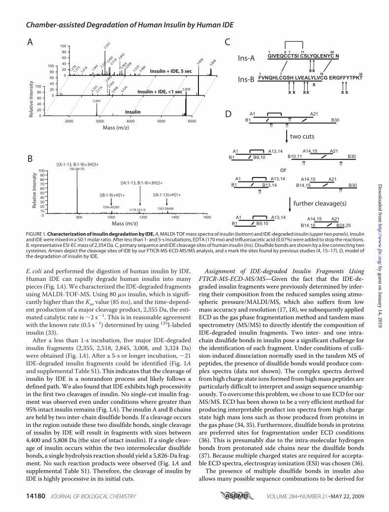
E. coli and performed the digestion of human insulin by IDE.Human IDE can rapidly degrade human insulin into manypieces (Fig. 1A).We characterized the IDE-degraded fragmentsusing MALDI-TOF-MS. Using 80 �M insulin, which is signifi-cantly higher than the Km value (85 nM), and the time-depend-ent production of a major cleavage product, 2,355 Da, the esti-mated catalytic rate is �2 s�1. This is in reasonable agreementwith the known rate (0.5 s�1) determined by using 125I-labeledinsulin (33).After a less than 1-s incubation, five major IDE-degraded
insulin fragments (2,355, 2,518, 2,845, 3,008, and 3,324 Da)were obtained (Fig. 1A). After a 5-s or longer incubation, �21IDE-degraded insulin fragments could be identified (Fig. 1Aand supplemental Table S1). This indicates that the cleavage ofinsulin by IDE is a nonrandom process and likely follows adefined path.We also found that IDE exhibits high processivityin the first two cleavages of insulin. No single-cut insulin frag-ment was observed even under conditions where greater than95% intact insulin remains (Fig. 1A). The insulin A and B chainsare held by two inter-chain disulfide bonds. If a cleavage occursin the region outside these two disulfide bonds, single cleavageof insulin by IDE will result in fragments with sizes between4,400 and 5,808 Da (the size of intact insulin). If a single cleav-age of insulin occurs within the two intermolecular disulfidebonds, a single hydrolysis reaction should yield a 5,826-Da frag-ment. No such reaction products were observed (Fig. 1A andsupplemental Table S1). Therefore, the cleavage of insulin byIDE is highly processive in its initial cuts.
Assignment of IDE-degraded Insulin Fragments UsingFTICR-MS-ECD-MS/MS—Given the fact that the IDE-de-graded insulin fragments were previously determined by infer-ring their composition from the reduced samples using atmo-spheric pressure/MALDI/MS, which also suffers from lowmass accuracy and resolution (17, 18), we subsequently appliedECD as the gas phase fragmentation method and tandemmassspectrometry (MS/MS) to directly identify the composition ofIDE-degraded insulin fragments. Two inter- and one intra-chain disulfide bonds in insulin pose a significant challenge forthe identification of each fragment. Under conditions of colli-sion-induced dissociation normally used in the tandem MS ofpeptides, the presence of disulfide bonds would produce com-plex spectra (data not shown). The complex spectra derivedfromhigh charge state ions formed fromhighmass peptides areparticularly difficult to interpret and assign sequence unambig-uously. To overcome this problem, we chose to use ECD for ourMS/MS. ECD has been shown to be a very efficient method forproducing interpretable product ion spectra from high chargestate high mass ions such as those produced from proteins inthe gas phase (34, 35). Furthermore, disulfide bonds in proteinsare preferred sites for fragmentation under ECD conditions(36). This is presumably due to the intra-molecular hydrogenbonds from protonated side chains near the disulfide bonds(37). Because multiple charged states are required for accepta-ble ECD spectra, electrospray ionization (ESI) was chosen (36).The presence of multiple disulfide bonds in insulin also
allows many possible sequence combinations to be derived for
FIGURE 1. Characterization of insulin degradation by IDE. A, MALDI-TOF mass spectra of insulin (bottom) and IDE-degraded insulin (upper two panels). Insulinand IDE were mixed in a 50:1 molar ratio. After less than 1- and 5-s incubations, EDTA (170 mM) and trifluoroacetic acid (0.07%) were added to stop the reactions.B, representative ESI-EC mass of 2,354 Da. C, primary sequence and IDE cleavage sites of human insulin (Ins). Disulfide bonds are shown by a line connecting twocysteines. Arrows depict the cleavage sites of IDE by our FTICR-MS-ECD-MS/MS analysis, and x mark the sites found by previous studies (4, 15–17). D, model ofthe degradation of insulin by IDE.
Chamber-assisted Degradation of Human Insulin by Human IDE
14180 JOURNAL OF BIOLOGICAL CHEMISTRY VOLUME 284 • NUMBER 21 • MAY 22, 2009
by guest on January 14, 2019http://w
ww
.jbc.org/D
ownloaded from

any given observed peptide mass. For example, there are 11possibilities that are within 4 ppm error tolerance for the2,355-Da species identified byMALDI-TOF, amajor product ofIDE-degraded insulin (Fig. 1A and supplemental Fig. S1). Thus,ECD-MS/MS is vital to unambiguously assign the correct A/Bchains within each insulin fragment. IDE-degraded insulin waspassed through an HPLC C18 column to allow for some sepa-ration of cleavage products and therefore ensuremaximal sam-pling of low abundance insulin fragments. FTICR mass spec-trometry was used for its compatibility with ECD and the highmass accuracy and high resolution detection of both precursorand product ion spectra (38).Using LC-ESI-FTICR-MS-ECD-MS/MS, we identified 10
IDE-degraded insulin fragments (Table 1, Fig. 1B, and supple-mental Fig. S2). This is based on the excellent match of theobserved mass from FTICR-MS of IDE-degraded insulin frag-ments with the predicted mass (�ppm �12) (Table 1). Theidentity of fragments from insulin A and B chains was revealedby ions from ECD-MS/MS (Fig. 1B, supplemental Fig. S2, andTable 1). For example, the insulin fragment withmass 2355.031Damatches well with the predictedmass of an insulin fragmentcontaining A1–A13 and B1–B9 peptides linked by one intra-and one inter-molecular disulfide bonds (�ppm � 3). The ionwith themass of 2,355Dawas analyzed simultaneously by data-dependent ECD-MS/MS inwhich selection and activationwerefavored for a precursor ion with at least three or more charges.ECD induces the breakage of the intermolecular disulfide bondto reveal ions that match well with A1–A13 and B1–B9 pep-tides (�ppm � �0.2 and 0.3, respectively). For some insulinfragments, we were only able to obtain FTICR-MS data, pre-sumably due to their low abundance and/or the dependence oflocal environment around the targeted disulfide bond. How-ever, the mass accuracy from FTICR allows the unambiguousassignment of these fragments (supplemental Table S1).Model for the Path of Human Insulin Degradation by Human
IDE—Based on our mass spectrometry study, the major cleav-age sites of human insulin by recombinant human IDE are dis-
tributed to four discrete regions, each with two cleavage sitesthat are adjacent to each other (Fig. 1C). Three of these arelocated in the middle of either the A or B chain of insulin, andthe last one is near the end of the B chain. No cleavage occursnear the N-terminal ends of either A or B chain. No clearsequence preference emerges from these sites. These sitesmatched well with those sites derived from the HPLC profileand the termination of Edman degradation of porcine[125I]iodo-insulin cleaved by rat IDE (4, 15, 16) andMS analysisof bovine insulin cleaved by recombinant rat IDE (17). Differentfrom previous findings, we did not observe the cleavagebetween B16 and B17 of human insulin by human IDE. It isworth noting that this site is the preferred binding and cleavagesite of isolated insulin B chain by IDE (2).The novel method of directly acquiring highly accurate
masses both for the nonreduced insulin A-B chain fragmentand for the individual A and B chain fragments of any given ionspecies allows the unambiguous assignment of the insulin frag-ments. Using such assignment, we then compared the cleavageproducts of insulin after a brief incubationwith IDE (�1 s) withthose from longer incubations (5 s (Fig. 1A) or longer (data notshown)). At the less than 1-s incubation, we observed fivemajorproducts, 2,355, 2,518, 2,845, 3,008, and 3,324 Da, correspond-ing to insulin fragments of A1–13/B1–9, A1–14/B1–9, A15–21/B14–30, A14–21/B14–30, and A15–21/B10–30, respec-tively (Fig. 1A). All of these fragments can be created by twocleavages of insulin, one at chain A (either A13–14 or A14–15)and the other in the middle of chain B (B9–10, B10–11, B13–14, or B14–15). Under this incubation, no insulin fragment isderived from the cleavage at the C-terminal end of chain B(B24-B25 or B25–26; Fig. 1A, Table 1, and supplemental Fig.S1). The accumulation of insulin fragments containing theC-terminal cleavage of chainB occurred only after a longer timeof incubation (5 s shown in Fig. 1A or longer (not shown)). Thisindicates that the cleavages at the C-terminal end of chain B(B24–25 and B25–26) occur later than those in the middle ofinsulin A and B chains. We also did not observe insulin frag-
TABLE 1Summary of mass spectrometry analysis of disulfide-bond linked IDE-degraded insulin fragments
Ins-A Ins-B A-B(observed)M � H
A-B(calculated)M � H Errora A (observed)
m/zA (calculated)
m/z Errora B (observed)m/z
B (calculated)m/z Errora
ppm ppm ppmN-terminal insulin fragmentsb,c1–13 1–9 2355.031 2355.024 3.0 1353.585 1353.585 �0.2 1004.463 1004.462 0.31–13 1–10 2492.095 2492.082 0.7 1353.588 1353.585 2.0 1141.495 1141.521 �22.71–14 1–9 2518.089 2518.087 0.8 1516.630 1516.648 �12.1 1004.463 1004.462 0.71–14 1–10 2655.178 2655.146 12.0 1516.640 1516.648 �5.4 1141.550 1141.521 25.1
C-terminal insulin fragmentsd14–21 14–30 3008.402 3008.396 2.0 1046.426 1046.425 0.9 982.996 983.001 �5.115–21 10–30 3323.578 3323.587 �2.4 883.389 883.362 30.3 1222.181 1222.128 43.6
C-terminal insulin peptides with an additional C-terminal cleavage at the insulin B chaine14–21 14–25 2418.092 2418.089 1.2 1046.432 1046.425 7.0 1374.684 1374.688 �3.014–21 14–24 2271.042 2271.021 9.2 1046.434 1046.425 8.6 1227.629 1227.620 7.614–21 15–25 2347.044 2347.052 �3.4 1046.417 1046.425 �8.1 1303.646 1303.651 �3.315–21 14–25 2255.019 2255.026 �3.1 883.361 883.362 �1.6 1374.684 1374.688 �3.0
a Error (in ppm) � ((M � H)obs � (M � H)calc)/(M � H)calc�106.b Assignment is shown of the most prominent insulin peaks generated after 5 min of incubation with IDE using LC-ESI-FTICR-MS-ECD-MS/MS.c Four insulin fragments resulting from the cleavages at either A13–14 or A14–15 once and at either B9–10 or B10–11 once were observed. Insulin fragment containing A1–13and B1–13 was observed, which is 1 of four possibilities from the cleavages at either B13–14 or B14–15 in conjunction with a cleavage in chain A (supplemental Table S1).
d Four insulin fragments resulting from the cleavages at either A13–14 or A14–15 once and at either B113-14 or B14–15 once were observed. Insulin fragment containingA15–21 andB10–30was also observed, which is one of four possibilities from the cleavages at either B9–10 or B10–11 in conjunctionwith a cleavage in chainA (supplementalTable S1).
e Out of eight possible insulin fragments, all were observed by either FTICR-MS or MALDI-TOF-MS (supplemental Table S1).
Chamber-assisted Degradation of Human Insulin by Human IDE
MAY 22, 2009 • VOLUME 284 • NUMBER 21 JOURNAL OF BIOLOGICAL CHEMISTRY 14181
by guest on January 14, 2019http://w
ww
.jbc.org/D
ownloaded from

ments with multiple cuts at chain B without a cut in insulin Achain (supplemental Table S1).From the time-dependent formation of insulin fragments by
IDE, we constructed a model on how IDE sequentially cleavesinsulin (Fig. 1D). IDE initiates the degradation of insulin bycleaving once at chain A and at least once in themiddle of chainB. Such cleavages result in insulin fragments that contain eitherN-terminal or C-terminal portions of insulin (Fig. 1D and sup-plemental Table S1). The resulting insulin fragments are sub-sequently cleaved by IDE at the known cleavage sites of insulinB chain. This leads to the generation of insulin fragments withgreatermass diversity as seen in the longer incubationwith IDE.Characterization of Cysteine-free IDE Mutant—The struc-
ture of insulin-bound IDE would provide insights into themolecular basis of the high affinity interaction between IDE andinsulin and on the sequential cleavages of insulin. Unfortu-nately, the previously defined crystallization condition of
human IDE requires the use of a reducing agent, tris(2-carboxy-ethyl)phosphine, presumably to protect IDE from oxidativeinactivation (2). Under this condition, we only obtained thestructure of IDE in complex with insulin B chain even thoughinsulin was added, and the minimally required amount ofreducing agent was used (2). To eliminate the need for a reduc-ing agent, we constructed a human IDEmutant, IDE-CF, that isfree of cysteine residues (supplemental Fig. S3) (19). The invitro enzymatic assay showed that IDE-CF had catalytic activitycomparable with wild type IDE in degrading insulin (Fig. 2Aand supplemental Fig. S4). In addition, the ability of insulin tocompete with the cleavage of a model substrate by IDE-CF iscomparable with that of wild type IDE (Fig. 2B).We also used ITC to evaluate the binding affinity of IDE to
insulin (Fig. 2C). To avoid the degradation of insulin by IDE, aglutamate 111 to glutamine mutation was introduced into thecatalytic site. Our ITC data fit well with a two-binding sites
FIGURE 2. Characterization of cysteine-free IDE. A, MALDI-TOF mass spectra of insulin alone (bottom), insulin after 5 s of incubation with cysteine-freeIDE (IDECF, middle) and wild type IDE (IDEwt, top). The peak with molecular mass of 2,904 Da represents the doubly charged state of undigested insulin.B, competition of substrate V degradation by IDEwt and IDE-CF by insulin. Substrate V (450 nM) was mixed with 1 �g of IDEwt or IDE-CF in the indicatedconcentrations of insulin (0 –10 �M), and fluorescence intensity was monitored for 10 min at 37 °C. Results are means � S.D. and are representative ofthree independent experiments performed in duplicate. C, isothermal calorimetry titration of insulin binding to IDEwt-E111Q and IDE-CF-E111Q withraw data (top) and the integrated heat signals (bottom). The solid line represents a calculated curve using the two-site binding model. Experiments werecarried out in triplicate at 25 °C with IDE at 6 �M in 20 mM Tris-HCl, pH 7.7, 50 mM NaCl. The single spike corresponds to the heat exchange upon theaddition of 10 �l of insulin (30 �M). Experiments were carried out in triplicate. D, sedimentation coefficient distributions for IDE-E111Q in the absenceand presence (1:2 molar ratio) of insulin.
Chamber-assisted Degradation of Human Insulin by Human IDE
14182 JOURNAL OF BIOLOGICAL CHEMISTRY VOLUME 284 • NUMBER 21 • MAY 22, 2009
by guest on January 14, 2019http://w
ww
.jbc.org/D
ownloaded from

model, and the estimated dissociation constant of the two sitesare 10 and 140 nM. Although the functional unit of IDE is amonomer, hydrodynamic analyses reveal that, dependent uponthe experimental conditions, IDE can shift to a dimer and evenhigher oligomers (21). Using analytical ultracentrifugation, wefound that insulin can shift the equilibrium of monomeric IDEto be mostly a dimer (Fig. 2D). The presence of two insulin-binding sites of IDE might reflect the dimeric state of theenzyme. Similar to wild type IDE-E111Q, our ITC data showedthat IDE-CF-E111Q also has two insulin-binding sites withcomparableKd values (20 and 280 nM). Together, our data indi-cate that the cysteinemutations do not alter the binding affinityof IDE for insulin or the ability of IDE to cleave insulin. Thus,IDE-CF represents an excellent model to investigate insulinbinding.Structure of IDE-Insulin Complex—We subsequently solved
crystal structures of the catalytically inactive IDE-CF-E111Q incomplex with human insulin and iodinated insulin at 2.6 and2.8 Å resolution, respectively (Table 2, supplemental Fig. S5,and supplemental Fig. S6). Anomalous signals from iodineatoms of the iodinated insulin were used as a guide in themodelbuilding of insulin (supplemental Fig. S5). Here we focus on theinsulin-bound IDE structure because iodinated insulin sits inthe catalytic chamber of IDE in a virtually identical fashion as
insulin. In accordance with our previously determined IDE-E111Q structures, IDE-CF is a dimer, and the four structurallyhomologous domains of each monomer form a large, enclosedcatalytic chamber with a total volume of �1.6 � 104 Å3 (Fig. 3and supplemental Fig. S7), which is slightly larger than that ofthe T-state of an insulin monomer (�1.2 � 104 Å3) (2, 39). Aninsulin molecule is completely encapsulated by each IDE mon-omer (Fig. 3A). Electron densitymaps for themajority of insulinA chain (A1–A20, 20 of 21 residues) and over three-fifths ofinsulin B chain (B1-B19, 19 of 30 residues) are clearly visibleinside the catalytic chamber of IDE, where the peptide occupies�64% of the space (Fig. 3B). The remaining portion of insulin(A21 and B20–B30) is not visible in the electron density maps,presumably due to disorder because intact, nondegraded insu-lin was found in insulin-bound IDE crystals by mass spectrom-etry (supplemental Fig. S6).Our insulin-bound IDE structure reveals that the charge
complementarity contributes significantly to the interaction ofinsulin with the catalytic chamber of IDE. IDE-N and IDE-Cserve as the functional units to form the catalytic chamber (Fig.3C) (21). The inner catalytic chamber of IDE-C is highly posi-tively charged, which complements well with the negativelycharged surface of human insulin, an acidic protein with anisoelectric point of 5.3 (Fig. 3C). The N-terminal regions ofinsulin A and B chains make intimate interactions with theinner chamber of IDE-N (Fig. 3C and supplemental Fig. S7).
FIGURE 3. Structure of insulin-bound IDE. A, global view of the structure ofinsulin-bound IDE-CF-E111Q monomer. IDE domains 1– 4 (IDE-D1 to IDE-D4)are colored green, blue, yellow, and red, respectively. Insulin A and B chains arecolored magenta and cyan, respectively. The zinc ion is colored gray. B, com-posite omit map of insulin contoured at 1.5�. C, electrostatic surface repre-sentation of insulin and IDE. The molecular surface is color-coded as calcu-lated by Adaptive Poisson-Boltzmann Solver. The molecular surface is coloredas calculated by Adaptive Poisson-Boltzmann Solver (51) (��6 kT in red, 0 kTin white, and �6 kT in blue). The interaction surface between the N- andC-terminal domains of IDE and insulin is marked by yellow dashed lines basedon the contact residues displayed using CCP4 molecular graphics (30).
TABLE 2Data collection and structure refinement statistics ofsubstrate-bound IDE
IDE-insulin IDE-iodinated insulinData collectionBeamline APS 14-BMC APS 19IDWavelength 0.90020 Å 1.54980 ÅSpace group P65 P65Cell dimensiona 262.3 Å 263.2 Åb 262.3 Å 263.2 Åc 90.6 Å 90.8 Å
Resolution 50-2.6 Å 50-2.8 ÅRsym
a (%) 12.6 (49.6)b 11.0 (46.5)I/� 21.7 (4.8)b 18.2 (3.6)Redundancyc 7.0 (6.5)b 5.4 (4.6)Completeness (%) 99.9 (100.0)b 99.6 (98.3)Figure of merit 0.871 0.873Unique reflections 109,017 98,447
RefinementRwork
d 0.164 0.170Rfree
e 0.218 0.221No. of atomsProtein 16,204 16,235Water 721 360
B-factorsIDE 27.5 36.4Substrate 49.2 52.4Water 31.7 36.4
Root mean square deviationsBond lengths 0.015 Å 0.016 ÅBond angles 1.851° 1.941°
Ramachandran plotFavorable 91.0/88.7% 89.0/88.6%Allowed 9.0/11.3% 11.0/11.4%Generously allowed 0/0% 0/0%Disallowed 0/0% 0/0%
PDB code 2WBY 2WC0a Rmerge � (I � �I�/�I�.b The outer resolution shell is shown. Values in parentheses indicate the highestresolution shell.
c Nobs/Nunique.d Rwork � hkl�Fobs� � k �Fcalc�/hkl�Fobs�.e Rfree, calculated the same as for Rwork but on the 5% data excluded from the refine-ment calculation.
Chamber-assisted Degradation of Human Insulin by Human IDE
MAY 22, 2009 • VOLUME 284 • NUMBER 21 JOURNAL OF BIOLOGICAL CHEMISTRY 14183
by guest on January 14, 2019http://w
ww
.jbc.org/D
ownloaded from

Molecular Details of the Interac-tion of Insulin with the CatalyticChamber of IDE—Insulin makesextensive contacts with the catalyticchamber of IDE (buried surface of1,758 Å2) with excellent surfacecomplementarity (surface comple-mentarity score � 0.67) (supple-mental Fig. S7) (32). The interactionof insulin with IDE-N accounts forthe predominant buried surface(1,355 Å2; surface complementarityscore � 0.71). The N-terminal endof insulin A chain forms a �-sheetwith IDE strand �12 within an evo-lutionarily conserved exosite of IDEat domain 2, which is crucial forbinding of IDE substrates (Fig. 4A)(2, 14). In addition, the N-terminalend of insulin B chain binds to thecatalytic cavity at domain 1 of IDE(Fig. 4A). There is a network ofhydrogen bonds, salt bridges, andvan derWaals interactions of insulinwith both IDE-N and IDE-C (Fig.4A) (2, 14).The N-terminal end (B1–B8) of
IDE-bound insulin B chain adoptsan extended conformation, a hall-mark of insulin T-state that is thephysiologically active form of thishormone (39, 40). The overall struc-ture of IDE-bound insulin is similarto that of the crystal structure ofT-state insulin (Fig. 4B) (39).Because of its interaction with thecatalytic chamber of IDE, themono-meric, IDE-bound insulin is partiallyunfolded judging by the loss of an�-helix, the gain of a disorderedloop, and the increased exposure ofhydrophobic residues (Fig. 4B).Insulin structures have been deter-mined both by x-ray crystallographyandNMR, and our IDE-bound insu-lin structure deviates significantlyfrom structures of wild type insulinand insulin analogs at three discreteregions (41). By binding to theexosite of IDE, the N-terminal endof insulin A chain is converted froman�-helix to a�-strand. In addition,the C-terminal segment of insulin Bchain (B20–B30) is disordered,which differs from the tight interac-tion of this segment with the central�-helix of insulin B chain in crystalstructures of T-state insulin (39).
Chamber-assisted Degradation of Human Insulin by Human IDE
14184 JOURNAL OF BIOLOGICAL CHEMISTRY VOLUME 284 • NUMBER 21 • MAY 22, 2009
by guest on January 14, 2019http://w
ww
.jbc.org/D
ownloaded from

Furthermore, the interaction of Phe1 at the N-terminal of theinsulin B chain (Phe1B) with the catalytic cavity of IDE pullsPhe1B away from its interaction with the hydrophobic patchformed by Leu17B and Val18B (Fig. 4B). The surface charge dis-tribution of the partially unfolded insulin inside the IDE cata-lytic chamber also differs significantly from that of T-state insu-lin (Fig. 4C). Most noticeably, the surface facing the catalyticchamber changes from a weakly positive to predominantlynegative.Our insulin-bound IDE structure likely represents how insu-
lin binds to the catalytic chamber of IDE prior to its completeunfolding. The N-terminal end of the insulin B chain binds tothe catalytic cavity at domain 1 of IDE with the peptide bondbetween Phe1B and Val2B near the catalytic zinc ion, resultingin an unfavorable interaction for water-mediated attack (Fig. 4,A and B). This is consistent with our observation that no cleav-age is observed at the N-terminal end of insulin chain B (Fig.1A). This also agrees with the distance between the catalytic ionand the observed cleavage sites of insulin. As described above,human IDE cleaves four discrete regions of human insulin, eachcontaining two sites. These include one in the middle of Achain, two in the middle of B chain, and the other at the end ofB chain. One cleavage in the A chain and another in the middleof B chain are required for the initial cleavages of insulin. Con-sistent with this, sites at the A chain are closest to the catalyticsite (13 and 15 Å, respectively), and those sites in the middle ofB chain are only slightly farther away (22, 20, 20, and 18 Å forB9–10, B10–11, B13–14, and B14–15, respectively). Althoughthe cleavage sites at the end of B chain (B24–25 and B25–26) inour structure are disordered, their position would be farthestfrom the catalytic site of IDE. This is consistent with our massspectrometry data suggesting that the cleavages at the end of Bchain occur only after the initial cleavages in the middle ofinsulin A and B chains.
DISCUSSION
The degradation of human insulin by human IDE results in acomplex mixture of disulfide bond-linked insulin fragments.The advance of proteomics-based mass spectrometry allowssystemic study of the time-dependent production of the mix-ture of human insulin fragments cleaved by human IDE. Inaddition to using FTICR for its highly accurate mass determi-nation of nonreduced insulin fragments, we also employedECDas the gas phase fragmentationmethod to accurately iden-tify fragments of insulin A and B chains from 10 precursor ionsof the given insulin fragments. ECD-MS/MS spectra provedvital for our study because the commonly used peptide IDmethod, collision-induced dissociation-MS/MS, createdextremely complex spectra due to the presence of disulfide
bond(s) and suffered from the ineffective production of b and yions around the disulfide bond(s) requisite for unambiguouspeptide identification. Although ECD is postulated to be aneffective method in the analysis of the labile post-translationalmodifications of proteins such as disulfide bonds (36), to ourknowledge this is the first time that ECD-MS/MS is successfullyused to decipher the composites of complexmixtures of biolog-ical samples. Further analysis of the composition of productions and themolecular basis of disulfide bond breakage of insu-lin fragments from the ECD-MS/MS spectra will undoubtedlyprovide new insights into the chemical principles and futureusage of ECD in proteomics analyses in biology and medicine.Our mass spectrometry analysis reveals that IDE cuts in the
middle of both the insulin A and B chains before cleaving theC-terminal end of the insulin B chain. Furthermore, IDE ishighly processive to digest the middle of both the insulin A andB chains to produce N- and C-terminal halves of insulin frag-ments. Insulin has both inter and intra-chain disulfide bonds,so single cleavage of insulin by IDE will still allow the A and Bchains to be held together for its binding and activation of insu-lin receptor. However, because both N- and C-terminal por-tions of the residues in the insulinA andB chains are involved inbinding to the insulin receptor, either N- or C-terminal insulinfragment would be active in receptor binding and signal trans-duction (42–46). Thus, the processive cleavages of insulin byIDE ensure the complete inactivation of insulin without theneed for subsequent binding and cleavage of the resulting insu-lin fragments (Fig. 1D).Our insulin-bound IDE structure represents the first struc-
ture of insulin in complex with its biologically relevant proteinpartners. Three major conformational switches found in theIDE-bound insulin structure suggest insulin is in an intermedi-ate unfolded state (Fig. 4,B andD). The disruption of an�-helixat the N terminus of the insulin A chain, the exposure of hydro-phobic residues near the N terminus of the insulin B chain, andthe disordered C-terminal loop of the insulin B chain are indi-cators of this partially unfolded conformation as all threechanges are hallmarks of protein unfolding. Accumulating evi-dence indicates that protein folding follows certain routes, andthe folding intermediates have substantial structural heteroge-neity (47). Substrate unfolding inside the catalytic chamber ofIDE likely follows the same principles. The lack of electron den-sity outside the active site and exosite observed for other IDE-substrate complexes is consistent with the substantial disorderor heterogeneity of IDE substrates inside the catalytic chamberof IDE (2).The binding of the N terminus of the insulin A chain to the
exosite likely plays a key role in the catalytic chamber-assisted
FIGURE 4. Characterization of the IDE-insulin interaction. A, details of the interaction between insulin and IDE. Additional hydrogen bond networks areshown in the three right panels for clarity. IDE and insulin are colored as in Fig. 3. B, comparison of insulin in its free T-state form (PDB code 1G7A) and IDE-boundform. C, comparison of surface charge distribution of the partially unfolded insulin in insulin-bound IDE with T-state insulin modeled into the catalytic chamberof IDE. Surface representation of the substrate binding chamber of IDE was generated by the software Voidoo (31). The outer surface of IDE and the substratechamber are colored pale green and dark green. The electrostatic surface representations of the IDE-bound insulin and T-state insulin models are calculated byAdaptive Poisson-Boltzmann Solver. D, comparison of IDE-bound insulin structure with the solution structures of insulin AlaA2-DKP. Insulin AlaA2-DKP has an Ileto Ala mutation in A2 residue of insulin-DKP, which is an engineered monomeric insulin. Left shows the comparison of IDE-bound insulin (colored in red andsalmon for insulin A and B chain, respectively) with an exemplary NMR structure of insulin AlaA2-DKP (PDB code 1K3M; colored in green and lime green for insulinA and B chain, respectively), and an exemplary NMR structure of insulin-DKP (PDB code 2JMN; colored in blue and light blue for insulin A and B chain,respectively). For a fair comparison, solution structures of insulin AlaA2-DKP and insulin-DKP are also shown in the middle and right, respectively.
Chamber-assisted Degradation of Human Insulin by Human IDE
MAY 22, 2009 • VOLUME 284 • NUMBER 21 JOURNAL OF BIOLOGICAL CHEMISTRY 14185
by guest on January 14, 2019http://w
ww
.jbc.org/D
ownloaded from

unfolding of insulin. When compared with 100 x-ray crystal-lographic and NMR structures of wild type insulin and insulinanalogs, IDE-bound insulin is significantly different in theN-terminal end of the A chain as well as C-terminal tail of the Bchain (Fig. 4, B and D). Interestingly, the IDE-bound insulinstructure has an interesting resemblance of theNMRstructuresof insulin that bear an isoleucine to alanine mutation at residue2 of the insulin A chain (Fig. 4D) (48). This single amino acidsubstitution is known to cause the decreased thermodynamicstability of insulin, which leads to disorder in the N-terminal Achain segment and more divergent C-terminal B chain tail.Thus, it is likely that the anchoring of theN-terminal A chain tothe exosite of IDE that significantly alters the structure of theN-terminal end of insulin A chain as well also has a similareffect in reducing the thermodynamic stability of insulin tofacilitate its unfolding.Our insulin-bound IDE structure also reveals that the shape,
size, charge distributions, conformational flexibility, andexosite binding of insulin all contribute to its tight binding(�100 nM) to IDE. During the proteolytic cycle, both IDE andits substrates undergo conformational switches (Fig. 5). IDEhasopen and closed conformational states (IDEo and IDEc), and theIDE substrates need to interact favorably with both states. TheIDEc state is stabilized by extensive contacts between IDE-Nand IDE-C (1, 2, 14), making the IDE chamber inaccessible tosubstrates. Only in the IDEo state can initial binding with thesubstrate occur. The dipolar charge distribution of insulin per-mits good charge complementarity with the catalytic chamberof IDE in the IDEo state, with either the positively or negativelycharged internal surfaces of IDE-C or IDE-N, respectively. Therepositioning of IDE-N and IDE-C by the conformationalswitch of IDEo to IDEc allows insulin to interact simultaneouslywith the IDE-N and IDE-C surfaces of the catalytic chamber.The size and shape of insulin are also compatible with the fitinto the chamber of IDE. As revealed by our insulin-bound IDEstructure, the anchoring of the N-terminal end of the insulin Achain to the exosite of IDE occurs despite the lack of interactionof the targeted cleavage sites of insulinwith the catalytic groove.This highlights the importance of the exosite interaction indetermining the binding affinity of substrates (19).In conjunction with our insulin-bound IDE structure, our
data also allow us to construct a working model of how IDEprocessively cuts insulin into pieces (Fig. 5). Using MALDI-TOF-MS/MS and LC-FTICR-MS-ECD-MS/MS, we found thatthe initial cleavages of insulin by IDE occur in at least two loca-tions, one in the middle of A chain and the other in the middleof B chain (Fig. 1D), resulting in the biologically inactive N- andC-terminal insulin fragments. Because IDE only has one cata-lytic center and the two selective cleavages are located at sepa-rate chains of insulin, IDE must have a mechanism to restrainthe exit of singly cut insulin from the catalytic chamber so thatthe second cleavage can proceed immediately.In our model, we postulate that the first cleavage of insulin
occurs at one of two sites at the insulin A chain (Fig. 1D and Fig.4B). This is based on the fact that these sites are closest to thecatalytic site among all known cleavage sites. Furthermore, theyare located in themiddle of a loop that generally has a less stablestructure than an �-helix, where the four cleavage sites of insu-
lin B chain are located. The further unfolding of insulin fromthe partially unfolded insulin intermediate shown in our insu-lin-bound IDE structure allows one of the two adjacent cleavagesites on insulin A chain to swing into the catalytic site of IDE forcleavage (Fig. 5B). The N terminus of the insulin A chain isanchored to the highly conserved exosite of IDE (1, 2, 14). Theexosite and other regions of the IDE catalytic chamber thatcontact insulin work as a reverse chaperone to guide insulinalong the defined unfolding route(s). The structural heteroge-neity present during the unfolding of insulin likely explains whyit is a probabilistic process for IDE to cut either site in theinsulin A chain. A cleavage at the insulin A chain does notseparate insulin into two pieces. IDE substrates are known tointeract with both IDE-N and IDE-C, which can serve to holdthese two domains together (2, 14). Thus, the interaction of
FIGURE 5. Model of insulin degradation by IDE. A, model for the conforma-tional switch of IDE in the catalytic cycle of insulin degradation. Insulin iscaptured by IDE, and the structure of IDEC � insulin depicts the state of thebinding. Once insulin is captured by IDEo, the conformational switch of IDEentraps insulin inside the catalytic chamber. This leads to the partiallyunfolded insulin depicted as IDEc � insulin. For insulin digestion, insulinneeds to undergo conformational changes. In this closed state, IDE likely per-forms at least two cycles of the unfolding and a single cleavage to generatethe N- and C-terminal fragments depicted in Fig. 1D before releasing thecleavage products by switching back to an open state. IDEo either goes backto the substrate-free IDEc or captures another substrate (insulin or the result-ing insulin fragments) for the next cycle. The substrate-free closed and theopen state (IDEc and IDEo, respectively) are modeled based on substrate-freeIDEC (PDB code 2JG4) and E. coli pitrilysin structure (PDB code 1Q2L). Thefree-state insulin and IDE-bound insulin are depicted as red schematics. IDE-Nand IDE-C are depicted as transparent blue and gray surfaces, respectively.The loop hinge of IDE is depicted as red ribbon. B, three possible states ofinsulin are shown, T-state (left), IDE-bound partially unfolded state (middle),and IDE-bound fully unfolded state (right). For comparison, the regions thatundergo the major conformational changes are colored in red, and those thatdo not are colored yellow.
Chamber-assisted Degradation of Human Insulin by Human IDE
14186 JOURNAL OF BIOLOGICAL CHEMISTRY VOLUME 284 • NUMBER 21 • MAY 22, 2009
by guest on January 14, 2019http://w
ww
.jbc.org/D
ownloaded from

insulin with the internal chamber still allows insulin to holdIDE-N and IDE-C together tomaintain the IDEc state. The highstability of the IDEc state also contributes to prolong theentrapment of insulin inside the catalytic chamber (14).The next cleavage occurs at one of four sites located on the
central helix of the insulin B chain (Fig. 4B). Not only are thesesites farther away from the reaction center than those on theinsulin A chain, but their access to the catalytic site is stericallyhindered by the presence of the loop containing the cleavagesites of the insulin A chain. Thus, a cleavage in the insulin Achain would allow these sites on the insulin B chain to gainaccess to the catalytic site of IDE. Such cleavage could alsotrigger a conformational switch of the central �-helix of theinsulin B chain to a loop, facilitating access to the catalyticgroove of IDE (Fig. 5B). Again, the structural heterogeneity ofthe unfolding path allows any of the four sites to interact withand be cut by the catalytic site of IDE in a stochastic manner.Such cleavage will create N- and C-terminal fragments of insu-lin that are no longer covalently joined so that they cannotinteract with IDE-N and IDE-C simultaneously and tie thesedomains together. This permits the formation of IDEo, allowingthe dissociation of insulin fragments. The released insulin frag-ments can subsequently bind IDEo and are subject to furthercleavages at other sites in the insulin B chain, particularly at itsC-terminal end. The validity of this model awaits future bio-physical and biochemical studies.In summary, our analyses describe the molecular basis by
which IDE recognizes, unfolds, and processively degrades insu-lin. They also highlight the role of the size and charge of the IDEcatalytic chamber as well as the conformational flexibility ofsubstrates as determinants of the substrate specificity of thisprotease. In addition, they reveal how the highly conservedexosite of IDE could anchor the N termini of IDE substrates,guiding the unfolding and cleavage of substrates in the catalyticchamber. Our model of how IDE processively degrades insulinalso provides the framework for designing IDE-insensitiveinsulin. Such modified insulin would have a prolonged half-lifein circulation, which could be further developed for bettermanagement of diabetes (5). Furthermore, the comparison ofthe insulin-bound IDE structure with A�-bound IDE offers aninsight into the engineering of IDE that preferentially degradesA�. Such engineered IDE might serve as a protein-based ther-apeutic for the clearance of A� in the treatment of Alzheimerdisease (49, 50).
Acknowledgments—We are grateful to the staff of Structural BiologyCenter and BioCars at Advanced Photon Source, Argonne NationalLaboratory, and Elena Solomaha at the Biophysics Facility, Univer-sity of Chicago, for help in data collection.We thank Todd Funke, YaoBian, and Ray Hulse for the helpful discussions and critical reviews.Use of the Advanced Photon Source was supported by the UnitedStates Department of Energy, Office of Basic Energy Sciences, underContract W-31-109-ENG-38. Use of proteomics service facility wassupported by the Chicago Biomedical Consortium.
REFERENCES1. Malito, E., Hulse, R. E., and Tang, W. J. (2008) Cell. Mol. Life Sci. 65,
2574–2585
2. Shen, Y., Joachimiak, A., Rosner,M. R., andTang,W. J. (2006)Nature 443,870–874
3. Mirsky, I. A., and Broh-Kahn, R. H. (1949) Arch. Biochem. 20, 1–94. Duckworth,W.C., Bennett, R. G., andHamel, F. G. (1998)Endocr. Rev. 19,
608–6245. Farris, W., Mansourian, S., Chang, Y., Lindsley, L., Eckman, E. A., Frosch,
M. P., Eckman, C. B., Tanzi, R. E., Selkoe, D., and Guenette, S. (2003) Proc.Natl. Acad. Sci. U. S. A. 100, 4162–4167
6. Sladek, R., Rocheleau, G., Rung, J., Dina, C., Shen, L., Serre, D., Boutin, P.,Vincent, D., Belisle, A., Hadjadj, S., Balkau, B., Heude, B., Charpentier, G.,Hudson, T. J., Montpetit, A., Pshezhetsky, A. V., Prentki, M., Posner, B. I.,Balding, D. J., Meyre, D., Polychronakos, C., and Froguel, P. (2007)Nature445, 881–885
7. Kurochkin, I. V., and Goto, S. (1994) FEBS Lett. 345, 33–378. Kurochkin, I. V. (2001) Trends Biochem. Sci. 26, 421–4259. Kim, M., Hersh, L. B., Leissring, M. A., Ingelsson, M., Matsui, T., Farris,
W., Lu, A., Hyman, B. T., Selkoe, D. J., Bertram, L., and Tanzi, R. E. (2007)J. Biol. Chem. 282, 7825–7832
10. Leissring, M. A., Farris, W., Chang, A. Y., Walsh, D. M., Wu, X., Sun, X.,Frosch, M. P., and Selkoe, D. J. (2003) Neuron 40, 1087–1093
11. Miller, B. C., Eckman, E. A., Sambamurti, K., Dobbs, N., Chow, K. M.,Eckman, C. B., Hersh, L. B., and Thiele, D. L. (2003) Proc. Natl. Acad. Sci.U. S. A. 100, 6221–6226
12. Tanzi, R. E., and Bertram, L. (2005) Cell 120, 545–55513. Baker, T. A., and Sauer, R. T. (2006) Trends Biochem. Sci. 31, 647–65314. Im, H., Manolopoulou, M., Malito, E., Shen, Y., Zhao, J., Neant-Fery, M.,
Sun, C. Y., Meredith, S. C., Sisodia, S. S., Leissring, M. A., and Tang, W. J.(2007) J. Biol. Chem. 282, 25453–25463
15. Davies, J. G.,Muir, A. V., Rose, K., andOfford, R. E. (1988)Biochem. J. 249,209–214
16. Duckworth, W. C., Hamel, F. G., Peavy, D. E., Liepnieks, J. J., Ryan, M. P.,Hermodson,M. A., and Frank, B. H. (1988) J. Biol. Chem. 263, 1826–1833
17. Grasso, G., Rizzarelli, E., and Spoto, G. (2007) J. Mass Spectrom. 42,1590–1598
18. Grasso, G., Rizzarelli, E., and Spoto, G. (2008) Biochim. Biophys. Acta1784, 1122–1126
19. Malito, E., Ralat, L. A., Manolopoulou, M., Tsay, J. L., Wadlington, N. L.,and Tang, W. J. (2008) Biochemistry 47, 12822–12834
20. Wefing, S., Schnaible, V., and Hoffmann, D. (2006) Anal. Chem. 78,1235–1241
21. Li, P., Kuo,W. L., Yousef,M., Rosner,M. R., andTang,W. (2006)Biochem.Biophys. Res. Commun. 343, 1032–1037
22. Frank, B. H., Beckage, M. J., and Willey, K. A. (1983) J. Chromatogr. 266,239–248
23. Hamlin, J. L., and Arquilla, E. R. (1974) J. Biol. Chem. 249, 21–3224. Otwinowski, Z., and Minor, W. (1997)Methods Enzymol. 276, 307–32625. McCoy, A. J., Grosse-Kunstleve, R.W., Storoni, L. C., andRead, R. J. (2005)
Acta Crystallogr. Sect. D Biol. Crystallogr. 61, 458–46426. Murshudov, G. N., Vagin, A. A., and Dodson, E. J. (1997)Acta Crystallogr.
Sect. D Biol. Crystallogr. 53, 240–25527. Emsley, P., and Cowtan, K. (2004) Acta Crystallogr. Sect. D Biol. Crystal-
logr. 60, 2126–213228. Brunger, A. T., Adams, P. D., Clore, G. M., DeLano, W. L., Gros, P.,
Grosse-Kunstleve, R.W., Jiang, J. S., Kuszewski, J., Nilges,M., Pannu,N. S.,Read, R. J., Rice, L. M., Simonson, T., and Warren, G. L. (1998) ActaCrystallogr. Sect. D Biol. Crystallogr. 54, 905–921
29. Brunger, A. T., Adams, P. D., Clore, G. M., DeLano, W. L., Gros, P.,Grosse-Kunstleve, R.W., Jiang, J. S., Kuszewski, J., Nilges,M., Pannu,N. S.,Read, R. J., Rice, L. M., Simonson, T., and Warren, G. L. (1998) ActaCrystallogr. D Biol. Crystallogr. 54, 905–921
30. Potterton, E., McNicholas, S., Krissinel, E., Cowtan, K., and Noble, M.(2002) Acta Crystallogr. Sect. D Biol. Crystallogr. 58, 1955–1957
31. Kleywegt, G. J., and Jones, T. A. (1994) Acta Crystallogr. Sect. D Biol.Crystallogr. 50, 178–185
32. Lawrence, M. C., and Colman, P. M. (1993) J. Mol. Biol. 234, 946–95033. Chesneau, V., and Rosner, M. (2000) Protein Expression Purif. 19, 91–9834. Zubarev, R. A. (2004) Curr. Opin. Biotechnol. 15, 12–1635. Zubarev, R. A., Kelleher, N. L., and McLafferty, F. W. (1998) J. Am Chem.
Chamber-assisted Degradation of Human Insulin by Human IDE
MAY 22, 2009 • VOLUME 284 • NUMBER 21 JOURNAL OF BIOLOGICAL CHEMISTRY 14187
by guest on January 14, 2019http://w
ww
.jbc.org/D
ownloaded from

Soc. 120, 3265–326636. Zubarev, R. A., Kruger, N. A., Fridriksson, E. K., Mark, L. A., Horn, D. M.,
Carpenter, B. K., and McLafferty, F. W. (1999) J. Am Chem. Soc. 121,2857–2862
37. Uggerud, E. (2004) Int. J. Mass Spectrom. 234, 45–5038. Page, J. S., Masselon, C. D., and Smith, R. D. (2004)Curr. Opin. Biotechnol.
15, 3–1139. Derewenda, U., Derewenda, Z., Dodson, E. J., Dodson, G. G., Reynolds,
C. D., Smith, G. D., Sparks, C., and Swenson, D. (1989) Nature 338,594–596
40. Olsen, H. B., Ludvigsen, S., and Kaarsholm, N. C. (1996) Biochemistry 35,8836–8845
41. Huang, K., Chan, S. J., Hua, Q. X., Chu, Y. C.,Wang, R. Y., Klaproth, B., Jia,W., Whittaker, J., De Meyts, P., Nakagawa, S. H., Steiner, D. F., Katsoyan-nis, P. G., and Weiss, M. A. (2007) J. Biol. Chem. 282, 35337–35349
42. McKern,N.M., Lawrence,M. C., Streltsov, V. A., Lou,M. Z., Adams, T. E.,Lovrecz, G. O., Elleman, T. C., Richards, K. M., Bentley, J. D., Pilling, P. A.,Hoyne, P. A., Cartledge, K. A., Pham, T. M., Lewis, J. L., Sankovich, S. E.,Stoichevska, V., Da Silva, E., Robinson, C. P., Frenkel, M. J., Sparrow, L. G.,
Fernley, R. T., Epa, V. C., and Ward, C. W. (2006) Nature 443, 218–22143. Ward, C.W., Lawrence,M. C., Streltsov, V. A., Adams, T. E., andMcKern,
N. M. (2007) Trends Biochem. Sci. 32, 129–13744. Glendorf, T., Sorensen, A. R., Nishimura, E., Pettersson, I., and Kjeldsen,
T. (2008) Biochemistry 47, 4743–475145. Kristensen, C., Kjeldsen, T., Wiberg, F. C., Schaffer, L., Hach, M., Have-
lund, S., Bass, J., Steiner, D. F., and Andersen, A. S. (1997) J. Biol. Chem.272, 12978–12983
46. Mayer, J. P., Zhang, F., and DiMarchi, R. D. (2007) Biopolymers 88,687–713
47. Udgaonkar, J. B. (2008) Annu. Rev. Biophys. 37, 489–51048. Xu, B., Hua, Q. X., Nakagawa, S. H., Jia, W., Chu, Y. C., Katsoyannis, P. G.,
and Weiss, M. A. (2002) Protein Sci. 11, 104–11649. DeMattos, R. B., Bales, K. R., Cummins, D. J., Paul, S. M., and Holtzman,
D. M. (2002) Science 295, 2264–226750. Nicoll, J. A., Wilkinson, D., Holmes, C., Steart, P., Markham, H., and
Weller, R. O. (2003) Nat. Med. 9, 448–45251. Baker, N. A., Sept, D., Joseph, S., Holst,M. J., andMcCammon, J. A. (2001)
Proc. Natl. Acad. Sci. U. S. A. 98, 10037–10041
Chamber-assisted Degradation of Human Insulin by Human IDE
14188 JOURNAL OF BIOLOGICAL CHEMISTRY VOLUME 284 • NUMBER 21 • MAY 22, 2009
by guest on January 14, 2019http://w
ww
.jbc.org/D
ownloaded from

TangMarika Manolopoulou, Qing Guo, Enrico Malito, Alexander B. Schilling and Wei-Jen
Insulin by Human Insulin-degrading EnzymeMolecular Basis of Catalytic Chamber-assisted Unfolding and Cleavage of Human
doi: 10.1074/jbc.M900068200 originally published online March 25, 20092009, 284:14177-14188.J. Biol. Chem.
10.1074/jbc.M900068200Access the most updated version of this article at doi:
Alerts:
When a correction for this article is posted•
When this article is cited•
to choose from all of JBC's e-mail alertsClick here
Supplemental material:
http://www.jbc.org/content/suppl/2009/03/26/M900068200.DC1
http://www.jbc.org/content/284/21/14177.full.html#ref-list-1
This article cites 51 references, 11 of which can be accessed free at
by guest on January 14, 2019http://w
ww
.jbc.org/D
ownloaded from