Molecular basis of action of a small molecule positive...
57
MOL #114082 -1- Molecular basis of action of a small molecule positive allosteric modulator (PAM)-agonist at the type 1 cholecystokinin holoreceptor Aditya J. Desai, Ingrid Mechin, Karthigeyan Nagarajan, Celine Valant, Denise Wootten, Polo C.H. Lam, Andrew Orry, Ruben Abagyan, Anil Nair, Patrick M. Sexton, Arthur Christopoulos, Laurence J. Miller Department of Molecular Pharmacology and Experimental Therapeutics, Mayo Clinic, Scottsdale, AZ 85259 (A.J.D., L.J.M.) In silico Drug Discovery Department, Icagen Inc. Tucson Innovation Center, Oro Valley, AZ 85755 (I.M, K.N., A.N.) Drug Discovery Biology Theme, Monash Institute of Pharmaceutical Sciences and Department of Pharmacology, Monash University, Parkville 3052, Australia (C.V., D.W., P.M.S., A.C., L.J.M.) Molsoft LLC, La Jolla, CA 92037 (P.C.H.L., A.O., R.A.) Skaggs School of Pharmacy and Pharmaceutical Sciences, University of California, San Diego, La Jolla, CA 92037 (R.A.) School of Pharmacy, Fudan University, Shanghai 201203, China (P.M.S.) This article has not been copyedited and formatted. The final version may differ from this version. Molecular Pharmacology Fast Forward. Published on December 27, 2018 as DOI: 10.1124/mol.118.114082 at ASPET Journals on May 22, 2020 molpharm.aspetjournals.org Downloaded from
Transcript of Molecular basis of action of a small molecule positive...

MOL #114082 -1-
Molecular basis of action of a small molecule positive allosteric modulator (PAM)-agonist
at the type 1 cholecystokinin holoreceptor
Aditya J. Desai, Ingrid Mechin, Karthigeyan Nagarajan, Celine Valant, Denise Wootten, Polo C.H.
Lam, Andrew Orry, Ruben Abagyan, Anil Nair, Patrick M. Sexton, Arthur Christopoulos, Laurence J.
Miller
Department of Molecular Pharmacology and Experimental Therapeutics, Mayo Clinic, Scottsdale, AZ 85259 (A.J.D., L.J.M.) In silico Drug Discovery Department, Icagen Inc. Tucson Innovation Center, Oro Valley, AZ 85755 (I.M, K.N., A.N.) Drug Discovery Biology Theme, Monash Institute of Pharmaceutical Sciences and Department of Pharmacology, Monash University, Parkville 3052, Australia (C.V., D.W., P.M.S., A.C., L.J.M.) Molsoft LLC, La Jolla, CA 92037 (P.C.H.L., A.O., R.A.) Skaggs School of Pharmacy and Pharmaceutical Sciences, University of California, San Diego, La Jolla, CA 92037 (R.A.) School of Pharmacy, Fudan University, Shanghai 201203, China (P.M.S.)
This article has not been copyedited and formatted. The final version may differ from this version.Molecular Pharmacology Fast Forward. Published on December 27, 2018 as DOI: 10.1124/mol.118.114082
at ASPE
T Journals on M
ay 22, 2020m
olpharm.aspetjournals.org
Dow
nloaded from

MOL #114082 -2-
Running title: CCK receptor PAM-agonist
Address for correspondence:
Laurence J. Miller, M.D. Mayo Clinic 13400 East Shea Blvd, Scottsdale, AZ 85259 Tel.: (480) 301-4217 Fax: (480) 301-8387 E-mail: [email protected]
Number of text pages: 40
Number of figures: 13
Number of references: 66
Words in Abstract: 216
Words Introduction: 706
Words Discussion: 1352
ABREVIATIONS: BDZ-1, (S)-1-(3-iodophenyl)-3-(1-methyl-2-oxo-5-phenyl-2,3-dihydro-1H-
benzo[e][1,4]diazepin-3-yl)urea); BDZ-2, (R)-1-(3-iodophenyl)-3-(1-methyl-2-oxo-5-phenyl-2,3-
dihydro-1H-benzo[e][1,4] diazepin-3-yl)urea); CCK, cholecystokinin; CCK1R or CCK2R, type 1 or
2 CCK receptor; CHO, Chinese hamster ovary; ECL, extracellular domain; ICM, internal coordinate
mechanics; KRH, Krebs-Ringer-HEPES; PAM, positive allosteric modulator; TM, transmembrane
domain.
This article has not been copyedited and formatted. The final version may differ from this version.Molecular Pharmacology Fast Forward. Published on December 27, 2018 as DOI: 10.1124/mol.118.114082
at ASPE
T Journals on M
ay 22, 2020m
olpharm.aspetjournals.org
Dow
nloaded from

MOL #114082 -3-
ABSTRACT
Allosteric modulation of receptors provides mechanistic safety while effectively achieving
biological endpoints otherwise difficult or impossible to obtain by other means. The theoretical case
has been made for the development of a positive allosteric modulator (PAM) of the type 1
cholecystokinin receptor (CCK1R) having minimal intrinsic agonist activity to enhance meal-induced
satiety for the treatment of obesity, while reducing the risk of side effects and/or toxicity.
Unfortunately, such a drug does not currently exist. In this work, we have identified a PAM-agonist of
the CCK1R, SR146131, and determined its putative binding mode and receptor activation mechanism
by combining molecular modeling, chimeric CCK1R/CCK2R constructs, and site-directed
mutagenesis. We probed the structure-activity relationship of analogs of SR146131 for impact on
agonism versus cooperativity of the analogs. This identified structural features that might be
responsible for binding affinity and potency while retaining PAM activity. SR146131 and several of
its analogs were docked into the receptor structure, which had the natural endogenous peptide agonist,
CCK, already in the bound state (by docking), providing a refined structural model of the intact
CCK1R holoreceptor. Both SR146131 and its analogs exhibited unique probe-dependent
cooperativity with orthosteric peptide agonists and were simultaneously accommodated in this model,
consistent with the derived structure-activity relationships. This provides improved understanding of
the molecular basis for CCK1R-directed drug development.
This article has not been copyedited and formatted. The final version may differ from this version.Molecular Pharmacology Fast Forward. Published on December 27, 2018 as DOI: 10.1124/mol.118.114082
at ASPE
T Journals on M
ay 22, 2020m
olpharm.aspetjournals.org
Dow
nloaded from

MOL #114082 -4-
Introduction
Cholecystokinin (CCK) has multiple important physiologic functions, including stimulating
gallbladder contraction and pancreatic exocrine secretion, regulating gastrointestinal transit, and
inducing satiety in response to ingestion of a meal. Indeed, the satiety effect of CCK, mediated by the
type 1 CCK receptor (CCK1R), has been demonstrated in both animal models (Chaudhri et al., 2008;
Gibbs et al., 1973) and humans (Kissileff et al., 1981), supporting this receptor as a possible target for
the treatment of obesity. Substantial efforts to develop small molecule agonists of the CCK1R have
been undertaken by major pharmaceutical companies (Aquino et al., 1996; Berger et al., 2008; Elliott
et al., 2010), however none of the agents have advanced beyond Phase II clinical trials. These efforts
have been hampered by on-target side effects of potent and longer duration agonists, with lower
potency agents having insufficient efficacy. A robust therapeutic window is particularly important
because of the anticipated scale and duration of use.
A possible strategy to circumvent the safety and tolerability concerns is the development of a
pure positive allosteric modulator (PAM) of the CCK1R that is devoid of intrinsic agonist activity
(Desai et al., 2015a; Desai et al., 2016a; Desai et al., 2016b; Desai et al., 2015b; Miller and Desai,
2016). Such an agent would bind to the receptor at an allosteric site that is distinct from the natural
orthosteric ligand binding site, allowing concurrent occupation and potentiation of biological
responses to CCK. This would occur only during the limited and finite period of time when nutrients
enter the duodenum to stimulate the release of endogenous hormone, resulting in amplification of the
natural effects of CCK. Such a mechanism of action would be predicted to lead to an early peak in
satiety, reducing meal size and contributing to body weight reduction, while, in the meantime,
minimizing the side effects observed with full agonists of the CCK1R and avoiding the tolerance
often experienced with such agonists.
A small molecule ligand-binding site within the CCK1R has been localized to a pocket high
within the helical transmembrane domain using receptor mutagenesis, fluorescence, and photoaffinity
labeling approaches (Cawston et al., 2012; Desai et al., 2015c; Hadac et al., 2006; Harikumar et al.,
This article has not been copyedited and formatted. The final version may differ from this version.Molecular Pharmacology Fast Forward. Published on December 27, 2018 as DOI: 10.1124/mol.118.114082
at ASPE
T Journals on M
ay 22, 2020m
olpharm.aspetjournals.org
Dow
nloaded from

MOL #114082 -5-
2013). This pocket assumes distinct conformations conferring different molecular determinants of
ligand binding in its inactive state occupied by antagonist ligands (Cawston et al., 2012), versus its
active state occupied by agonist ligands (Desai et al., 2015c; Harikumar et al., 2013).
We previously proposed an experimental strategy in which a key chemical group (isopropyl),
identified in small molecule structure-activity studies as critical for its agonist activity, would be
changed or eliminated to reduce its intrinsic agonist activity, while theoretically stabilizing a
conformation that reduces the energy barrier of activation, and possibly exhibiting PAM activity
(Desai et al., 2015b). However, the first application of this strategy to GI181771X, an allosteric
agonist lacking intrinsic PAM activity, was unsuccessful in achieving PAM activity, and instead
resulted in a negative allosteric modulator (NAM) of CCK action (Desai et al., 2015b). We postulated
that these analogs could adopt poses in the allosteric pocket that would be distinct from that of the
parent compound (Desai et al., 2015b).
In the current work, we identified the small molecule ligand, SR146131 (Bignon et al., 1999),
as a selective CCK1R PAM-agonist that could form a promising template for differentiation of
structure-activity driving intrinsic efficacy versus allosteric enhancement. SR146131 differed from an
antagonist in the same series, SR27897, possessing a cyclohexylethyl group attached to its thiazol
ring, as well as two methyl groups on the indole ring and two methoxy groups on the phenyl ring
(Gouldson et al., 2000). Moreover, SR146131 enhanced the potency of functional responses to the
orthosteric ligand, CCK. Docking of the compound into a previously developed molecular model of
CCK1R in its active state (Harikumar et al., 2013), predicted key interactions between the 5-methoxy
group on the phenyl ring of SR146131 with transmembrane segment residue Leu 7.39 of CCK1R that
we had previously linked to the agonist activity of GI181771X. Consequently, we studied a series of
structural analogs to provide insights into the molecular basis of agonism and cooperativity.
Included in this series of compounds was an analog with attenuated agonist activity, reflected
in lower binding affinity, which retained its positive cooperativity on CCK action. This ligand was
docked into a refined molecular model that accommodated simultaneous binding of natural CCK
This article has not been copyedited and formatted. The final version may differ from this version.Molecular Pharmacology Fast Forward. Published on December 27, 2018 as DOI: 10.1124/mol.118.114082
at ASPE
T Journals on M
ay 22, 2020m
olpharm.aspetjournals.org
Dow
nloaded from

MOL #114082 -6-
peptide. Within the compound series, the PAM activity was correlated with its intrinsic efficacy, thus
identifying structural features for binding affinity independent of efficacy and allosteric modulation.
While future work will focus on the potential to separate intrinsic efficacy and positive modulation,
the current study advances our understanding of the molecular determinants for modulator binding
that may be useful in the design and development of ligands with tailored molecular properties.
This article has not been copyedited and formatted. The final version may differ from this version.Molecular Pharmacology Fast Forward. Published on December 27, 2018 as DOI: 10.1124/mol.118.114082
at ASPE
T Journals on M
ay 22, 2020m
olpharm.aspetjournals.org
Dow
nloaded from
MOL #114082 -7-
Materials and Methods
Materials. Ham’s F-12 medium, OptiMEM medium, L-glutamine, Lipofectamine LTX and
PLUS reagents were from Invitrogen (Carlsbad, CA). Quest Fluo-8-AM was from AAT Bioquest Inc.
(Sunnyvale, CA). Fetal Clone II tissue culture medium supplement was from Hyclone Laboratories
(Logan, UT). Microscint, Unifilter 96-well microplates with bonded GF/B filters were from
PerkinElmer Life and Analytical Sciences (Shallon, CT). Costar 96-well V bottom assay plates and
the black assay plates with clear bottoms were from Corning (Corning, NY). All other reagents were
analytical grade.
Ligands. SR146131, 2-[4-(4-chloro-2,5-dimethoxyphenyl)-5-(2-cyclohexyl-ethyl)-thiazol-2-
ylcarbam-oyl]-5,7-dimethyl-indol-1-yl-1-acetic acid, was provided by Sanofi (structure shown in Fig.
1A). This ligand is a potent full agonist at the CCK1R (Bignon et al., 1999; Gouldson et al., 2000).
Cholecystokinin octapeptide (CCK-26-33, based on the numbering of CCK-33; also known as CCK-8
and commonly identified as CCK) was purchased from Peninsula Laboratories (Belmont, CA). The
O-phenylethyl ester analog of CCK previously demonstrated to be a partial agonist at the CCK1R
(CCK-OPE) (Gaisano et al., 1989) was synthesized in our laboratory. The CCK1R-selective 1,4-
benzodiazepine radioligand (125I-BDZ-1, (S)-1-(3-iodophenyl)-3-(1-methyl-2-oxo-5-phenyl-2,3-
dihydro-1H-benzo[e][1,4]diazepin-3-yl)urea) and the CCK2R-selective radioligand (125I-BDZ-2, (R)-
1-(3-iodophenyl)-3-(1-methyl-2-oxo-5-phenyl-2,3-dihydro-1H-benzo[e][1,4] diazepin-3-yl)urea) were
prepared as we described previously (Akgun et al., 2009). These were radioiodinated using oxidative
techniques with iodobeads (Pearce, Rockford, IL), and the products were purified to homogeneity on
reversed-phase HPLC (Powers et al., 1988). Both radioligands had specific radioactivities of
approximately 2000 Ci/mmol.
CCK Receptor-Bearing Cell Lines and Membrane Preparation. Chinese Hamster Ovary
(CHO) cell lines stably expressing a series of CCK receptor constructs that have been previously
characterized (Cawston et al., 2012; Desai et al., 2015b; Harikumar et al., 2013) were utilized in the
current studies. The cell lines were passaged approximately two times per week, and were maintained
This article has not been copyedited and formatted. The final version may differ from this version.Molecular Pharmacology Fast Forward. Published on December 27, 2018 as DOI: 10.1124/mol.118.114082
at ASPE
T Journals on M
ay 22, 2020m
olpharm.aspetjournals.org
Dow
nloaded from

MOL #114082 -8-
at 37°C in Ham’s F-12 medium supplemented with 5% Fetal Clone II in a humidified environment
containing 5% carbon dioxide. As described previously (Hadac et al., 1996), receptor-bearing
membrane fractions were prepared using homogenization and sucrose-density gradient centrifugation,
with the resulting particulate fraction suspended in Krebs-Ringer-HEPES (KRH) medium (in mM, 25
HEPES, pH 7.4, 104 NaCl, 5 KCl, 1.5 CaCl2, 1.0 KH2PO4, 1.2 MgSO4, 1.2 MgCl2) containing 0.01%
soybean trypsin inhibitor and 1 mM phenylmethylsulfonyl fluoride and stored at -80°C until use.
Receptor Binding Assays. Individual radioligand competition-binding assays were
performed in duplicate using benzodiazepine radioligands (Cawston et al., 2012; Desai et al., 2015b;
Harikumar et al., 2013). Briefly, ~5-7 μg membranes were added to a 100 μl assay volume in a clear
96-well plate containing increasing concentrations of SR146131 and ~20-25 pM radioligand, in KRH
medium and incubated for 60 min at room temperature. The reaction was then terminated by vacuum
filtration using Unifilter-96 well microplates with bonded GF/B filters in a Filtermate Harvester
(PerkinElmer, Waltham, MA). Non-saturable binding was determined using the corresponding
unlabeled ligand, 1 μM BDZ-1 or BDZ-2. The plates were washed six times using 0.9% NaCl and
0.2% bovine serum albumin and then dried, with radioactivity quantified using a Top CountNXTTM
instrument (Packard, Meriden, CT) after addition of 30 μl Microscint.
Intracellular Calcium Assays. CCK receptor-mediated biological responses were measured
by quantifying intracellular calcium increases in response to SR146131 (Desai et al., 2014; Desai et
al., 2015b). Briefly, cells were seeded in sterile clear-bottom black 96-well tissue culture plates 24 h
before the assay to achieve 75-80 percent confluence at the time of the assays. Cells were loaded with
0.75 μM Fluo 8 AM dye in KRH medium containing 2.5 mM probenecid for 45 min at 37°C in the
dark. They were washed once and the assay was performed in a Flexstation 3.0 plate reader
(Molecular Devices, Sunnyvale, CA) using robotic addition of the appropriate agonist ligand.
Intracellular calcium responses were quantified by measuring fluorescence emission intensity at 525
nm after excitation at 485 nm, with data collection every 4 sec over a 120 sec period, while
maintaining a constant temperature of 37°C. Concentration-response curves were constructed by
plotting the peak responses. To test their positive allosteric effect, fixed concentrations of compounds
This article has not been copyedited and formatted. The final version may differ from this version.Molecular Pharmacology Fast Forward. Published on December 27, 2018 as DOI: 10.1124/mol.118.114082
at ASPE
T Journals on M
ay 22, 2020m
olpharm.aspetjournals.org
Dow
nloaded from

MOL #114082 -9-
were added simultaneously with increasing concentrations of CCK, or the partial agonist, CCK-OPE
that is a phenethyl ester analog of CCK (Gaisano et al., 1989), and intracellular calcium
measurements were taken as mentioned above.
Molecular Modeling. SR146131, together with 159 known CCK1R agonists and 1739 other
CCK1R ligands (not identified as agonists) collected from ChEMBL_15, were docked to the
previously-generated CCK1R agonist receptor models (Desai et al., 2015c; Harikumar et al., 2013)
using MolSoft ICM 3.8 (Totrov and Abagyan, 1997). The best agonist receptor model achieved an
area under ROC curve (AUC) value of 87%, effectively differentiating the agonists from other
ligands. We clustered the docking poses of all agonists using the ICM atomic property field (APF)
method (Totrov, 2008) with a distance cutoff of 0.4. The majority of the CCK1R agonists docked into
two distinct docking pose families, consistent with their chemotype (benzodiazepine core: 67
members; piperazine core: 64 members), indicating the general applicability and consistency of the
receptor model for docking. SR146131 achieved an ICM docking score of -39, more favorable than
the ICM docking score of -32 for a typical binder.
We also developed a refined CCK1R homology model using the recently solved structure of
the orexin-1 receptor in complex with the selective antagonist SB-674042 (PDB code: 4ZJ8) (Yin et
al., 2016) as a template, since this receptor is closer in sequence to CCK1R than the A2a adenosine
receptor used in our previous CCK1R model. The orexin-1 receptor has 33% sequence identity/53%
homology with CCK1R, versus 26% sequence identity/42% homology for the A2a adenosine
receptor. Although 4ZJ8 is in an antagonist-bound conformation, it also offers the advantage of
presenting a pre-formed ligand binding site. The starting homology model was generated using Prime
(Jacobson et al., 2004) after multiple sequence alignment of several human and mouse class A GPCR
sequences. The NMR structure of the amino terminus of CCK1R (PDB code: 1D6G (Pellegrini and
Mierke, 1999)) was then grafted to the core helical bundle domain. Extracellular loops for the new
model were sampled using Loop Modeler from MOE (Labute, 2010) and the N-amino terminus was
sampled by Macromodel (Watts et al., 2014). From the numerous N-amino termini and loop
combinations obtained, we retained and minimized those in agreement with previously reported
This article has not been copyedited and formatted. The final version may differ from this version.Molecular Pharmacology Fast Forward. Published on December 27, 2018 as DOI: 10.1124/mol.118.114082
at ASPE
T Journals on M
ay 22, 2020m
olpharm.aspetjournals.org
Dow
nloaded from

MOL #114082 -10-
experimental data (Ding et al., 2001; Ding et al., 2002; Dong et al., 2009; Gigoux et al., 1998; Gigoux
et al., 1999; Giragossian and Mierke, 2001; Hadac et al., 1998; Ji et al., 1997; Kennedy et al., 1997)
and without substantial clashes.
We performed induced-fit docking of the CCK peptide in the best new model (out of several
prioritized models), taking into account the mutational data and the reported potency/efficacy of
peptide on the CCK1R mutants (Ding et al., 2002; Gigoux et al., 1998; Gigoux et al., 1999),
intermolecular Nuclear Overhauser Effects (NOEs) observed between CCK and the receptor
(Giragossian and Mierke, 2001; Kennedy et al., 1997), as well as photoaffinity labeling data (Ding et
al., 2001; Dong et al., 2009; Hadac et al., 1998; Ji et al., 1997). These considerations are reviewed in
Supplemental Table 1. The CCK1R-CCK peptide complex structure was subsequently used to dock
SR146131.
The three-molecule complex (CCK1R + CCK peptide + SR146131) was then subjected to 1
μs molecular dynamics simulation using Desmond (Bowers et al., 2006) after placing a POPC
membrane system and addition of explicit solvent, TIP3P water system together with Cl- ions to
neutralize the system. In addition, 0.15M NaCl salt was also added to the system. The transmembrane
segments annotated in the Uniprot entry for CCK1R were used for the placement of the POPC bilayer.
Twenty-six frames from the simulation were selected at even intervals of 40 ns, starting at the initial
frame, for docking of compound 12 using Glide (Schrödinger LLC., USA) (Friesner et al., 2004)
without any constraints. A maximum of six poses was retained for each docking, and only those poses
with a docking score better than -9.0 were subjected to binding mode analysis. For these poses, we
observed a binding mode where the acid group of compound 12 interacts with Arg 6.58 about 42% of
the time and the same group interacts with Asn 6.55 for about 24% of the time. We also found that
Tyr 4.63 interacts with the same acid group in a substantial number (33%) of binding poses, either as
sole partner or in addition to Asn 6.55. We also observed in a small number (6%) of cases some
interactions with Tyr 4.60.
This article has not been copyedited and formatted. The final version may differ from this version.Molecular Pharmacology Fast Forward. Published on December 27, 2018 as DOI: 10.1124/mol.118.114082
at ASPE
T Journals on M
ay 22, 2020m
olpharm.aspetjournals.org
Dow
nloaded from

MOL #114082 -11-
There is strong mutational data supporting the critical roles of Arg 6.58 and Asn 6.55 in
SR146131 binding (Gouldson et al., 2000 and Escrieut et al., 2002), so we decided to focus on these
residues for further analysis. Supplemental Table 2 summarizes the data describing the functional
impact of CCK1R mutations on SR146131 action, and the ligand-receptor residue distances in our
models. Two representative frames, each leading consistently to one of these two binding modes,
were selected and used as starting points for docking of SR146131 and its analogs using Glide. Here
also, no constraints were employed for docking of these compounds to the CCK peptide-bound
receptor structure. The top poses for docked SR146131 and its analogs were then submitted to Prime-
mmGBSA calculations (Sherman et al., 2006). This was used to refine ranking based on relative
binding affinities (ΔG) and to evaluate structural changes on the receptor occurring after binding of
this series of ligands, both in the presence and absence of the CCK peptide. Our models differ from all
previously-described molecular models of this receptor since they simultaneously accommodate
binding of both the CCK peptide and a small molecule ligand, here SR146131 and its analogs. This
also may be helpful in understanding subtle differences in the functional behavior of different ligands.
Data Analysis and Statistics. All data were analyzed using Prism 6 or 7 (GraphPad
Software, Inc., San Diego, CA). In all analyses, the data were unweighted, with the mean of replicates
in each experiment considered as an individual point. Throughout the manuscript, data are presented
as the mean of these values ± S.E.M. with the number of replicates of independent experiments noted.
Concentration-response data were analyzed using the three-parameter logistic equation (May et al.,
2007), E = Bottom + (Top-Bottom)[A]/[A]+[EC50], where Bottom represents the E value in the
absence of ligand, Top represents the maximal stimulation in the presence of ligand, [A] is the molar
concentration of ligand, and EC50 represents the molar concentration of ligand required to generate a
response halfway between Top and Bottom. The competitive model was utilized for the analysis of
the binding data, with data corrected for radioligand occupancy using the Cheng-Prusoff equation
(Cheng and Prusoff, 1973). Differences in receptor binding and signalling parameters between various
constructs were statistically evaluated using one way ANOVA with Dunnett’s multiple comparison
post-test, with p<0.05 considered to be statistically significant.
This article has not been copyedited and formatted. The final version may differ from this version.Molecular Pharmacology Fast Forward. Published on December 27, 2018 as DOI: 10.1124/mol.118.114082
at ASPE
T Journals on M
ay 22, 2020m
olpharm.aspetjournals.org
Dow
nloaded from

MOL #114082 -12-
Radioligand equilibrium binding curves with allosteric ligands were fitted to either a one-site
inhibition mass action curve to determine inhibitor potency (IC50) estimates, which were then
converted to Ki values as appropriate, or an allosteric ternary complex model (Eq. 1) to derive
estimates of allosteric modulator affinity (KB) and cooperativity between the compound and
radioligand (α); where α>1 denotes positive cooperativity, 0<α<1 denotes negative cooperativity and
α=1 denotes neutral cooperativity (Christopoulos and Kenakin, 2002; Ehlert, 1988).
Eq. 1
𝐸𝐸 = 𝐵𝐵𝑚𝑚𝑚𝑚𝑚𝑚 [𝐴𝐴]
[𝐴𝐴] + (𝐾𝐾𝐴𝐴) � 𝐾𝐾𝐵𝐵 + [𝐵𝐵]𝐾𝐾𝐵𝐵 + 𝛼𝛼[𝐵𝐵]�
where KA and KB represent the equilibrium dissociation constant of the radioligand and interacting
ligand, respectively, and [A] and [B] denote their concentrations. Bmax is the maximum number of
binding sites on the cell. Dissociation kinetic data were fit to a one-phase exponential decay function
to derive the apparent rate constant of dissociation (Koff) in the absence or presence of each
compound.
Functional studies for the interaction of orthosteric agonist and allosteric modulator was
either fitted to a three parameter logistic equation or an operational model of allosterism and agonism
(Leach et al., 2007) (Eq. 2) to derive estimates of affinity and combined binding/efficacy
cooperativity:
Eq. 2
𝐸𝐸 =𝐸𝐸𝑚𝑚(𝜏𝜏𝐴𝐴[𝐴𝐴](𝐾𝐾𝐵𝐵 + 𝛼𝛼𝛼𝛼[𝐵𝐵]) + 𝜏𝜏𝐵𝐵[𝐵𝐵]𝐾𝐾𝐴𝐴)𝑛𝑛
([𝐴𝐴]𝐾𝐾𝐵𝐵 + 𝐾𝐾𝐴𝐴𝐾𝐾𝐵𝐵 + 𝐾𝐾𝐴𝐴[𝐵𝐵] + 𝛼𝛼[𝐴𝐴][𝐵𝐵])𝑛𝑛 + (𝜏𝜏𝐴𝐴[𝐴𝐴](𝐾𝐾𝐵𝐵 + 𝛼𝛼𝛼𝛼[𝐵𝐵] + 𝜏𝜏𝐵𝐵[𝐵𝐵]𝐾𝐾𝐴𝐴)𝑛𝑛
where Em is the maximum possible response for the system, [A] and [B] are the concentration of the
orthosteric agonist and allosteric modulator, respectively, τA and τB are the signaling efficacy of the
respective ligands, KA and KB is the equilibrium dissociation constant for the respective ligands, n is a
transducer slope factor linking occupancy to response, α is the cooperativity factor (as described
This article has not been copyedited and formatted. The final version may differ from this version.Molecular Pharmacology Fast Forward. Published on December 27, 2018 as DOI: 10.1124/mol.118.114082
at ASPE
T Journals on M
ay 22, 2020m
olpharm.aspetjournals.org
Dow
nloaded from

MOL #114082 -13-
above) and β is an empirical scaling factor describing the allosteric effect of the modulator on the
orthosteric agonist signaling efficacy.
This article has not been copyedited and formatted. The final version may differ from this version.Molecular Pharmacology Fast Forward. Published on December 27, 2018 as DOI: 10.1124/mol.118.114082
at ASPE
T Journals on M
ay 22, 2020m
olpharm.aspetjournals.org
Dow
nloaded from

MOL #114082 -14-
Results
Subtype Selectivity of SR14131 at Wild Type CCK Receptors. Consistent with previous
reports (Bignon et al., 1999; Gouldson et al., 2000), SR146131 displayed concentration-dependent
agonism, stimulating maximal intracellular calcium responses in the CCK1R-bearing CHO-CCK1R
cells similar to that stimulated by natural CCK, while lacking agonism in the CCK2R-bearing CHO-
CCK2R cell line over equivalent concentrations (Fig. 1B). This compound displayed high potency on
CCK1R, with an EC50 of 43 ± 2 pM (mean ± S.E.M., n=6), and substantial functional subtype
selectivity over CCK2R (Fig. 1B).
Competition-binding studies using radiolabeled non-peptidyl antagonist benzodiazepines
(radioiodinated BDZ-1 and BDZ-2 for CCK1R and CCK2R, respectively), which bind to an
intrahelical allosteric site (Cawston et al., 2012), were performed to assess the binding properties of
SR146131 at both CCKR subtypes. SR146131 fully competed for binding of the 125I-BDZ-1 at
CCK1R, yielding a pKi of 8.30 ± 0.09 (n=6) using a competitive binding model (Fig. 1C), whereas it
displayed incomplete inhibition (~50%) of the saturable binding of 125I-BDZ-2 at CCK2R (n=4) (Fig.
1C), indicative of an allosteric mode of interaction between the two ligands. We recently
demonstrated that similar behavior of another CCK receptor ligand was due to interactions across a
receptor homodimer, and this is likely also true for the SR146131 compound (Desai et al., 2015c). To
account for the allosteric nature of the binding, these data were subsequently analyzed using an
allosteric ternary complex model (Christopoulos and Kenakin, 2002). The derived constant for
SR146131 binding to the free CCK1R (pKB) was 8.24 ± 0.08 (n=5), and this was different from that at
CCK2R (7.20 ± 0.17, n=4, p < 0.05, unpaired t-test). The cooperativity constants with the iodinated
radioligand, Log αBDZ-1 or Log αBDZ-2, were also lower at CCK1R than at the CCK2R, respectively (-
1.22 ± 0.10 vs. -0.36 ± 0.07, p < 0.05, unpaired t-test) indicating stronger negative cooperativity at the
CCK1R compared to CCK2R.
Allosteric Action of SR146131 at CHO-CCK1R cells. We examined possible PAM activity
of SR146131 by measuring dose-response curves of both CCK and a phenethyl ester analog of CCK
This article has not been copyedited and formatted. The final version may differ from this version.Molecular Pharmacology Fast Forward. Published on December 27, 2018 as DOI: 10.1124/mol.118.114082
at ASPE
T Journals on M
ay 22, 2020m
olpharm.aspetjournals.org
Dow
nloaded from

MOL #114082 -15-
(CCK-OPE), which is a partial agonist at the CCK1R, in CHO-CCK1R cells in the absence or
presence of increasing concentrations of SR146131. These analyses confirmed that SR146131 is a
PAM-agonist since it potentiated both CCK- and CCK-OPE-dependent intracellular calcium
mobilization (Fig. 2A-B). The strong intrinsic agonism of SR146131 makes it difficult to apply
operational models to quantify the degree of allosteric modulation, since a maximal system response
is reached at concentrations much lower than the predicted pKB of SR146131. In these interaction
studies, low concentrations of SR146131 were used, and these data were analyzed with the
operational model of allosterism by fixing the pKB to the affinity of SR146131 defined in radioligand
binding to yield composite (affinity and efficacy) cooperativity estimates. One caveat with fixing the
pKB in this analysis is that the assay conditions for radioligand binding (equilibrium conditions and
performed in membranes) and the functional assay (non-equilibrium and performed in whole cells)
are not equivalent. Since the estimated pKB can differ under different experimental conditions, and
pKB and alpha are linked in the operational analysis, under/over estimation of this value can influence
the derived cooperativity estimates. Nonetheless, this analysis confirmed the high intrinsic agonist
efficacy of SR146131 with consistent efficacies predicted from both data sets with operational Log τB
values of 2.41 ± 0.06 (τB = 263) derived from the CCK interaction curve and 2.32 ± 0.11 (τB = 209)
from the CCK-OPE analysis. Consistent with visual observation of the data, cooperativity estimates
from this analysis predict that SR146131 has greater cooperativity with CCK-OPE (Log αβCCK-OPE =
3.72 ± 0.08 (αβCCK-OPE = 5248)) than with CCK (Log αβCCK = 2.75 ± 0.25 (αβCCK = 562)).
SR146131 Function at CCK1R/CCK2R Chimeric Constructs. An experimental structure-
activity relationship approach for the small molecule ligand-binding site was used to interrogate the
molecular basis of ligand-receptor and/or peptide interaction of SR146131. We have previously
utilized this approach for molecular understanding of the action of other CCKR small molecule
antagonists (Cawston et al., 2012), and agonists (Desai et al., 2015c; Harikumar et al., 2013), using
CCK1R/CCK2R constructs with exchanged intrahelical allosteric pocket residues between these
receptors. Receptor density of stably expressed constructs and effect of mutation on BDZ and CCK
ligand affinities have been previously reported (Cawston et al., 2012; Desai et al., 2015c; Harikumar
This article has not been copyedited and formatted. The final version may differ from this version.Molecular Pharmacology Fast Forward. Published on December 27, 2018 as DOI: 10.1124/mol.118.114082
at ASPE
T Journals on M
ay 22, 2020m
olpharm.aspetjournals.org
Dow
nloaded from

MOL #114082 -16-
et al., 2013). In the first series of studies, we used receptor constructs exchanging individual TM2,
TM3, TM6 and TM7 segments of CCK1R with the corresponding residues of CCK2R, as well as the
converse constructs (Cawston et al., 2012; Desai et al., 2015b; Harikumar et al., 2013) (Fig. 3A, Table
1). In the case of the CCK1R-based chimeras, the TM7 construct (CCK1R L7.39H; all residue
numbers are labelled according to the Ballesteros and Weinstein class A numbering scheme
(Ballesteros and Weinstein, 1992)) showed greatest loss in ligand-induced intracellular calcium
mobilization, followed by the TM2 construct (CCK1R N2.61T). Of note, at the TM3- and TM6-based
chimeras (CCK1R T3.28V, T3.29S, and I6.51V, F6.52Y) there were small but statistically significant
right-shifts in EC50 values. Interestingly, of the converse CCK2R-based constructs with exchanged
residues of CCK1R, only the TM7 and TM6 constructs exhibited a partial gain-of-function in
response to SR146131 (Fig. 3B, Table 1).
Competition-binding experiments utilizing 125I-BDZ-1 or 125I-BDZ-2 radioligands on the
CCK receptor chimeras (Fig. 3C, Table 1) demonstrated that, in the CCK1R-based chimeras, the TM7
construct had greatest impact on binding, followed by the TM2 and TM6 constructs, whereas the TM3
construct lead to an increase in SR146131 pIC50 values. These data were analyzed with the
operational allosteric model to yield values of pKB and Log α for the respective radioligands, 125I-
BDZ-1 or 125I-BDZ-2, illustrating that the chimeras impacted both affinity and binding cooperativity
(Table 1). The CCK2R-based constructs exhibited the converse increase or decrease in binding
inhibition, although this was only statistically significant for the TM6 and TM7 chimeras (Fig. 3D,
Table 1). For the TM6 chimera the effect was primarily driven by increased negative cooperativity,
while the TM7 chimera altered both affinity and cooperativity (Table 1). Of note, as reported in our
previous studies (Cawston et al., 2012; Harikumar et al., 2013), the CCK2R TM2 chimera showed a
complete loss of binding of 125I-BDZ-2 and hence could not be assessed. Overall, these data suggested
a greater importance of residues constituting the CCK1R TM7 (Leu 7.39) and TM2 (Asn 2.61)
constructs in docking of SR146131.
Modeling and Docking of SR146131 in the CCK1R Helical Bundle. SR146131 was
initially docked into our previously reported model of the active conformation of the helical bundle of
This article has not been copyedited and formatted. The final version may differ from this version.Molecular Pharmacology Fast Forward. Published on December 27, 2018 as DOI: 10.1124/mol.118.114082
at ASPE
T Journals on M
ay 22, 2020m
olpharm.aspetjournals.org
Dow
nloaded from

MOL #114082 -17-
CCK1R (Harikumar et al., 2013) (Fig. 4). The best-fitting, most energetically favorable model
predicted that the compound docked within the same intramembranous intrahelical pocket of the
CCK1R previously predicted for GI181771X (Harikumar et al., 2013), with the thiazole ring of
SR146131 and the benzodiazepine ring of GI181771X occupying the same space (Fig. 4, 5). The
main difference in these models is that GI181771X makes closer interactions with TM6, including
two hydrogen bonds with Asn 6.55 and Arg 6.58 (Fig. 5B). The pocket occupied by SR146131 was
formed by TM2, TM3, TM6 and TM7, with the indole ring oriented toward the extracellular region
and the carboxylate group facing TM6 and making interactions with Asn 6.55. This model predicted
that the residues Met 3.32, Val 3.36 and Trp 6.48 formed the steric bulk lining the bottom of the
pocket. Residues Asn 2.61 and Asn 2.65 and Tyr 7.43 formed a hydrogen-bonding network
connecting TM2 and TM7 (Fig. 4, 5). Furthermore, Ser 7.42 formed two intrahelical hydrogen bonds
with the backbone of TM7 (Fig. 4, 5A).
As noted earlier, the agonist SR146131 and the closely-related antagonist SR27897 differ by
the presence of two methyl groups on the indole ring, an additional cyclohexylethyl group attached to
the thiazol ring, and two methoxy groups on the phenyl ring of SR146131 (Gouldson et al., 2000).
Since the two methyl groups on the indole ring are predicted to face the extracellular surface, they are
unlikely to contribute any molecular interaction important for agonist activity. In contrast, the
cyclohexylethyl group and the phenyl ring are predicted to bind deep inside the pocket (Fig. 4, 5).
According to this model, the cyclohexylethyl group is predicted to point toward the pocket formed by
TM3, TM5, and TM6 to make key interactions with Val 3.36 (contact area 21 Å2), Trp 6.48 (17 Å2),
and Phe 6.52, and the phenyl ring is accommodated in the hydrophobic pocket formed by TM2, TM3,
and TM7. This model predicts important interactions of the methoxy groups on the phenyl ring with
residues Thr 3.29 and Leu 7.39 that were important for SR146131 function as observed in the
CCK1R/CCK2R chimeric receptor data. The model also predicts that the 2-methoxy group interacts
with Thr 3.29, Tyr 3.30, and Met 3.32 (40 Å2), and the 5-methoxy group can interact with Leu 7.39,
Ser 7.42, Asn 2.61, and Met 3.32 (Fig. 4, 5A).
This article has not been copyedited and formatted. The final version may differ from this version.Molecular Pharmacology Fast Forward. Published on December 27, 2018 as DOI: 10.1124/mol.118.114082
at ASPE
T Journals on M
ay 22, 2020m
olpharm.aspetjournals.org
Dow
nloaded from

MOL #114082 -18-
Characterization of Receptor Mutants Predicted to Be Important for SR146131
Activity. Based on the proposed binding hypothesis by docking of SR146131, we identified
additional receptor residues that were predicted to be in spatial proximity to the ligand, and thus could
have functional importance. Of these, we used alanine-replacement mutants for Met 3.32, Val 3.36,
and Trp 6.48 that were used in an earlier study (Bmax, pmol/mg: CCK1R WT, 5.0 ± 1.0; M3.32A,
151 ± 63; V3.36A, 0.9 ± 0.4; W6.48A, 0.8 ± 0.2) (Desai et al., 2015c), and also generated an alanine-
replacement mutant for Ser 7.42 (Bmax, pmol/mg: 82.6 ± 12.0). Biological activity and binding
parameters for SR146131 at these CCK1R constructs are reflected in Fig. 6 and Table 2. Because of
the differences in levels of receptor construct expression, we include Log τB values normalized to
levels of cell surface binding, Bmax, in Table 2 to reflect intrinsic efficacy of SR146131 at each of the
receptor constructs relative to that at wild type CCK1R. This difference was statistically significant
for three of the constructs, with efficacy decreased for M3.32A and increased for V3.36A, and
S6.48A.
Functional Effects of Modification of SR146131. In an attempt to reduce the agonist
activity of SR146131 while still maintaining its PAM activity, we utilized structurally-related
compounds that were available from Sanofi with modifications on the phenyl ring that was predicted
to be located at an equivalent position to the isopropyl group of GI181771X (Table 3). Of these,
compounds with appreciable structural differences in this part of the molecule that retained the PAM
activity included compound 2 (methyl group), compound 4 (hydrogen), and compound 12 (ethyl
group) (Fig. 7). These compounds showed 1.3-, 1.3-, and 1.2-fold lower potencies than SR146131,
respectively, at the CCK1R WT (Table 3). Using 125I-BDZ-1 as radiolabel, IC50 values of compounds
2, 4, and 12 were determined to be 3.65-, 5.0-, and 32.0-times lower than SR146131, respectively (pKi
values: 7.65 ± 0.03, compound 2; 7.52 ± 0.04, compound 4; 6.70 ± 0.02, compound 12, n=3). Of note,
the pIC50 of compound 2 was different from compound 12 (p<0.05; One way ANOVA, Dunnett’s
post-test). Application of the operational model for allosterism revealed that compound 2 and
compound 4 had markedly diminished efficacy and cooperativity (compound 4 exhibiting no PAM
activity), with greatest impact seen with compound 4 (compound 2, Log τB = 0.42, Log αβ = 0.76;
This article has not been copyedited and formatted. The final version may differ from this version.Molecular Pharmacology Fast Forward. Published on December 27, 2018 as DOI: 10.1124/mol.118.114082
at ASPE
T Journals on M
ay 22, 2020m
olpharm.aspetjournals.org
Dow
nloaded from

MOL #114082 -19-
compound 4, Log τB = 0.02, Log αβ = 0.06) (Fig. 8A, B). Compound 12 was unique among these
SR146131 analogs in that it retained efficacy and positive allosteric modulatory activity on CCK
action that was similar to that of the parental compound (Fig. 9A), with a log τB value of 2.27 ± 0.04
and Log αβ of 2.41 ± 0.19 (αβ = 257). Of note, this effect was also seen on cells expressing 11-fold
lower CCK1R WT on their surface, Log τB = 1.17 and Log αβ = 1.41). Interestingly, like SR146131,
compound 12 exhibited stronger PAM effect when using the partial agonist, CCK-OPE, as the
orthosteric ligand (Fig 9B). The quantitative parameters for this effect were Log αβ = 4.18 ± 0.08,
and Log τB = 2.28 ± 0.36.
Enhanced Molecular Model for Docking of SR146131 to the CCK1R Holoreceptor.
Using the available structure-function data and the new model derived from the ligand-bound orexin-1
receptor as template, we have generated several refined models of CCK1R by loop refinement,
molecular dynamic simulations and ensemble dockings. Based on this, we were able to develop a
model that allowed the simultaneous occupation of both CCK peptide and SR146131 or its analogs.
We believe that this is the first reported CCK1R model with natural peptide ligand and a small
molecule modulator ligand bound simultaneously. In these models, the two ligands reside adjacent to
each other, with predicted ligand-ligand interactions that likely contribute to some of the biological
behavior of the tested small molecules (Figs. 10 and 11). The synergistic effect of the peptide and the
small molecule ligand binding could be deciphered by sampling of the extracellular domain and the
loops, molecular dynamics simulations and mmGBSA calculations, which had not been previously
exploited for this receptor. We have utilized these new models to guide interpretation of the
cooperativity of both CCK peptide and the small molecule ligands. Two of these models,
corresponding to docking poses of SR146131 and analogues into two representative frames from the
MD simulations mentioned in the Methods section, are discussed below.
Molecular Modeling and Binding Hypothesis for SR146131 and its Analogs. Initial
models of the ternary complex between CCK1R, CCK peptide and SR146131 were further subjected
to molecular dynamics simulations to generate refined binding site and CCK peptide conformations
for the 13 structurally-related analogs of SR146131 discussed above. Two putative binding modes for
This article has not been copyedited and formatted. The final version may differ from this version.Molecular Pharmacology Fast Forward. Published on December 27, 2018 as DOI: 10.1124/mol.118.114082
at ASPE
T Journals on M
ay 22, 2020m
olpharm.aspetjournals.org
Dow
nloaded from

MOL #114082 -20-
SR146131 were observed from the molecular dynamics simulations with about the same frequency.
Changes in binding mode seem driven by slight structural variations in the receptor and CCK peptide
over the course of the MD simulations. We decided to investigate both binding modes in more detail
and performed docking studies for all 14 compounds in both pocket conformations using Glide
(Schrödinger LLC., USA) (Friesner et al., 2004). Top poses for these active compounds were
consistent with the original SR146131 putative binding mode in both cases with highly negative
docking scores, reinforcing our confidence in the new model. For all 14 ligands studied and in both
binding modes, the thiazole ring of the small molecule ligand exhibits pi stacking with the tryptophan
(Trp 30) of the CCK peptide, indicating potential direct interaction between the endogenous agonist
and the small molecule ligands as a component of their cooperativity. Figures 10 and 11 exemplify the
two putative binding modes for docked compound 12 by itself and in comparison to docked
SR146131 and are discussed in detail below. General observations are also given for other
representative analogs studied in this paper.
Binding mode 1 (Fig. 10A), where the acid group of the compounds is involved in a salt
bridge with Arg 6.58, gave the best docking scores and most consistent poses for all 14 compounds.
This binding mode is in reasonable agreement with most of the mutational data reported to date that
has impact on the binding of SR146131 to CCK1R. This involves loss of activity of SR146131 at
receptor constructs with mutations such as, C94L (Cys 2.57 to Leu), F97A (Phe 2.60 to Ala), G122L
(Gly 3.33 to Leu), V125A (Val 3.36 to Ala), F198A (part of ECL2), I329A(F) (Ile 6.51 to Ala or
Phe), R336A(D)(M) (Arg 6.58 to Ala or Asp or Met), and I352A (Ile 7.35 to Ala) and L356A (Leu
7.39 to Ala). All of these residues are in the close vicinity of this ligand’s proposed binding site (see
Fig. 10 for orientations of these residues, except Gly 3.33).
However, the effect of the N333A (Asn 6.55 to Ala) mutation to the loss of binding affinity
of SR146131 to CCK1R could not be easily rationalized by this binding hypothesis, although the
docked compounds are within 6.4Å of Asn 6.55. Additional induced effects and thus stronger
interactions of Asn 6.55 with the ligand may be possible since this residue is within TM6, known to
This article has not been copyedited and formatted. The final version may differ from this version.Molecular Pharmacology Fast Forward. Published on December 27, 2018 as DOI: 10.1124/mol.118.114082
at ASPE
T Journals on M
ay 22, 2020m
olpharm.aspetjournals.org
Dow
nloaded from

MOL #114082 -21-
exhibit considerable movement and to play important functional roles in many GPCRs (Latorraca et
al., 2017; Rasmussen et al., 2011).
Binding mode 2 (Fig. 10B), when compared to binding mode 1, shows a flipped orientation of
the indole moiety. In this case, the acid group of the ligand is involved in a hydrogen bond with the
side chain of Asn 6.55, while the indole scaffold has a stacking interaction with the side chain of Arg
6.58. In this binding mode (Fig. 11B), we observe an additional difference between docked SR146131
and compound 12 of approximately 1 Å shift of the indole scaffold due to the additional methyl group
at position 7 and the proximity of the side chain of Asn 6.55. This binding mode also corresponds to
good docking scores, although less negative, and increased variability in docking poses. Due to these
reasons, we prioritized binding mode 1 and the Prime-mmGBSA calculations discussed in the next
section focus on binding mode 1.
The lower half of the compounds, below the amide linker, exhibits similar orientations and
interactions in both binding modes, independent of the orientation of the indole moiety: Cys 2.57, Val
3.36, Trp 6.48 and Ile 6.51 are involved in hydrophobic interactions with the cyclohexyl moiety, in
agreement with both new mutational data reported in this paper and historical experimental data
(Gouldson et al., 2000). Detailed analysis of compound 12 binding modes 1 and 2 predict that Asn
2.61, Met 3.32 and Ser 7.42 form interactions with both the cyclohexyl and the substituted benzyl
group. Phe 2.60, Asn 2.65, Phe 198, Leu 7.39 and Tyr 7.43 delimit the substituted benzene ring
binding region. In addition, this new model sheds light on one potentially interesting aspect of PAM
regulation: in both binding modes 1 and 2, the substituted benzene of compound 12 is near CCK
peptide residue Trp 30, where they show stacking interactions (in addition to the stacking of the
thiazole ring to Trp 30 within the CCK peptide observed for all representatives) (Fig. 10). Differences
were observed for the interactions of benzene ring substituents to the receptor in the two models.
Particularly to compound 12 vs SR146131, the substitution in position 5, (ethyl vs methoxy groups)
led to differences in interaction energies with Leu 7.39 and Ser 7.42. Similar changes were seen in
the interaction with Asn 2.65 with substitutions at position 4 (Cl vs methoxy).
This article has not been copyedited and formatted. The final version may differ from this version.Molecular Pharmacology Fast Forward. Published on December 27, 2018 as DOI: 10.1124/mol.118.114082
at ASPE
T Journals on M
ay 22, 2020m
olpharm.aspetjournals.org
Dow
nloaded from

MOL #114082 -22-
Prime-mmGBSA Calculations for SR146131 and its Analogs in Binding Mode 1. In an
attempt to better understand the effect of substitutions around SR146131 we ran Prime-mmGBSA
induced-fit calculations for all compounds docked in CCK1R with previously described binding mode
1, both in the presence and absence of CCK peptide. Figure 12 highlights the differences in CCK1R
conformations between bound compound 10 (pure allosteric agonist) and compound 12 and
SR146131 (PAM-agonists), as a result of Prime-mmGBSA calculations in the presence of CCK
peptide. Within TM7, changes in Tyr 7.43 seem to have a downstream impact on Thr 7.44 and Ser
7.45 (not shown for clarity), suggesting a potential signal transduction mechanism through TM7.
Further changes down TM7 were not observed, probably due to the 10 Å radius limit selected for the
Prime-mmGBSA induced-fit calculation. The most striking differences were observed for TM2,
probably driven by the combination of changes relative to Asn 2.61 and Asn 2.65, and to a lesser
extent Pro 2.64. The top of TM2 was pushed the most in the complex with compound 10 and the least
in the complex with SR146131. This shift in TM2 had impact on ECL1 position. In addition, Phe 198,
which is present in ECL2, was also slightly shifted as well as the loop overall. ECL1 and ECL2
participate in CCK peptide binding and the predicted differences in ligand conformations would
provide a different environment for the peptide, leading to potential changes in affinity, residence
time and consequent functional effects.
Figure 13 summarizes the calculated relative binding affinities for each ligand with or without
CCK peptide present in the binding site. In general, we see a decrease in relative binding affinities
(∆G) with or without CCK peptide, which is in correlation with decrease in potency as determined
experimentally (see Table 3). We observe a more favorable (more negative) ∆G in the presence of
CCK peptide for approximately half of the compounds, including SR146131 and compound 12, both
with PAM-agonist properties. Compound 10 and compound 4, both experimentally shown to lack
PAM activity, were the only two compounds with a more favorable ∆G in absence of CCK peptide.
The other compounds did not display any large change in ∆G (Fig. 13). This provides qualitative
insights into the complexes with various compounds, however, a more quantitative assessment of
This article has not been copyedited and formatted. The final version may differ from this version.Molecular Pharmacology Fast Forward. Published on December 27, 2018 as DOI: 10.1124/mol.118.114082
at ASPE
T Journals on M
ay 22, 2020m
olpharm.aspetjournals.org
Dow
nloaded from
MOL #114082 -23-
binding affinities and functional effects would require more detailed calculations with large scale
conformational sampling using molecular dynamics simulations and replica exchange methods.
This article has not been copyedited and formatted. The final version may differ from this version.Molecular Pharmacology Fast Forward. Published on December 27, 2018 as DOI: 10.1124/mol.118.114082
at ASPE
T Journals on M
ay 22, 2020m
olpharm.aspetjournals.org
Dow
nloaded from

MOL #114082 -24-
Discussion
CCK induces satiety by acting on the CCK1R present on axonal projections of vagal afferent
neurons present in the gut (Li and Owyang, 1994; Smith et al., 1981; Smith et al., 1985). This has
provided a rationale for attempts to develop agonists of this receptor as non-caloric satiety agents,
potentially useful for the management of obesity (Aquino et al., 1996; Berger et al., 2008; Elliott et
al., 2010). Several synthetic agonists that have been developed for this purpose, however, none has
gained regulatory approval for this application. This is mainly due to failure to reach the desired
efficacy in humans (Jordan et al., 2008), with more potent analogs often possessing side effects, and
longer duration agonists avoided due to concerns about potential trophic effects (Hoshi and Logsdon,
1993; Smith and Solomon, 2014).
We have proposed a possible alternative strategy to circumvent the concerns about side
effects and possible toxicity of a highly potent full agonist, by working to develop CCK1R ligands
with pure positive allosteric modulator activity for CCK, without possessing any intrinsic agonist
activity (Desai et al., 2015a; Desai et al., 2016a; Desai et al., 2017; Desai et al., 2016b; Desai et al.,
2015b; Miller and Desai, 2016). In this approach, the modulator would be expected to remain ‘silent’
in the absence of CCK, but then to act as a PAM to enhance the activity of CCK at the CCK1R only
during the brief interval after meals when the hormone is released, thereby minimizing on-target side
effects and possible toxicity. It also reduces the likelihood of desensitization. Indeed, this strategy has
been successfully utilized at other receptors, including the G protein-coupled calcium-sensing
receptor and the ionotropic GABA-A receptor (Barker et al., 1986; Nemeth et al., 2004). In addition
to the usefulness of high throughput screening for such candidates, we have also postulated an
alternate strategy where the determinants of agonist activity of an existing synthetic agonist can be
removed, while retaining the components for causing a conformational change of the receptor to
stabilize G protein association by reducing the energy barrier to achieve the active state (Desai et al.,
2015b).
This article has not been copyedited and formatted. The final version may differ from this version.Molecular Pharmacology Fast Forward. Published on December 27, 2018 as DOI: 10.1124/mol.118.114082
at ASPE
T Journals on M
ay 22, 2020m
olpharm.aspetjournals.org
Dow
nloaded from

MOL #114082 -25-
In the current study we demonstrate that SR146131, which is a CCK1R-specific, highly
potent agonist (Bignon et al., 1999), also possesses PAM activity, hence categorizing it as a PAM-
agonist. Thus, this compound is distinct from the previously characterized 1,5-benzodiazepine
agonist, GI181771X (Desai et al., 2015b). Subsequent use of CCK1R-CCK2R TM chimeric
constructs enabled development of a molecular model for compound binding to the CCK1R where
SR146131 was accommodated in the same agonist pocket as that predicted for GI181771X. Of note,
this modeling predicted different docking poses of SR146131 compared to previous models that were
developed prior to our current understanding of class A GPCR crystal structures (Gouldson et al.,
2000).
Since the receptor conformation elucidated in initial docking of SR146131 was not
substantially different from that achieved using GI181771X, we hypothesized that the region critical
for agonist activity could also be in analogous parts of both ligands. In the case of GI181771X, the
isopropyl group interacts with Leu 7.39, which is a major determinant of the agonist activity
(Harikumar et al., 2013). The portion of SR146131 that was predicted to be adjacent to this part of the
receptor is the phenyl ring. Indeed, while that ring is present in the structurally-related antagonist,
SR27897, it does not include the two methoxy groups present on the agonist. For this reason, these
were a focus of interest in attempts to reduce agonist activity of the parent PAM-agonist.
We examined thirteen compounds that were structurally similar or related to SR146131 with
substitutions on the phenyl ring and observed that the intrinsic agonist activity was reduced by
varying degrees. We selected compounds 2, 4, and 12 from the series based on single point PAM
assays testing the effect of different concentrations of compounds on the EC50 value of CCK. Further
experiments with these compounds revealed that compound 12 exhibited the most notable positive
cooperativity with CCK. This cooperativity was maintained in cells with both high and low levels of
receptor expression, with the amount of intrinsic agonist activity reduced in the setting of low receptor
expression, consistent with Monod-Wyman-Changeux mechanism model, as has been described for
M1 muscarinic acetylcholine receptor PAMs (Canals et al., 2012).
This article has not been copyedited and formatted. The final version may differ from this version.Molecular Pharmacology Fast Forward. Published on December 27, 2018 as DOI: 10.1124/mol.118.114082
at ASPE
T Journals on M
ay 22, 2020m
olpharm.aspetjournals.org
Dow
nloaded from

MOL #114082 -26-
Similar to SR146131, the degree of PAM activity of compound 12 was predicted to be greater
when enhancing the action of the partial agonist, CCK-OPE. These data are illustrative of probe-
dependency in allosterism that follows the Monod-Wyman-Changeux model, and is consistent with
some M1 muscarinic acetylcholine receptor PAMs (Canals et al., 2012). This enhancement of a weak
response of a partial agonist has also been recorded for other receptors such as the GABAB receptor
where the PAM exhibited an increase in Emax along with increase in potency of a partial agonist, as
opposed to only a left-shift in EC50 for a full agonist (Maksay et al., 2000). Greater enhancement,
relative to the GLP-1, of oxyntomodulin, or the GLP-1 metabolite GLP-1(9-36)NH2 that are weak or
partial agonists, has also been observed with small molecule PAMs at the GLP1-R (Koole et al.;
Wootten et al., 2012). It is important to note that CCK-OPE is not a natural metabolite of CCK, and
therefore, these data do not hold physiologic importance, however it is an important tool to understand
the compound pharmacology. Based on these observations, it might be interesting to explore whether
a combination therapy with a partial agonist and this type of PAM would have therapeutic advantages
over use of only the PAM itself.
It is important to note that in the case of SR146131 and the structurally-related compounds
tested in this study, the observed potency tracked with the affinity of the compounds resulting in
PAM-agonists with varying degrees of activity. For these compounds, the PAM activity is dependent
on their ability to stabilize an “active” conformation of the receptor that provides a lower energy
barrier threshold for activation by the co-bound orthosteric peptide; this is linked to their intrinsic
agonist activity. Prime-mmGBSA calculations on the complexes of small molecules bound to the
receptor-peptide system show differences in ∆G for these analogs, which is in qualitative agreement
with their observed agonist or PAM-agonist functional effects. However, as this series of compounds
appear to follow the Monod-Wyman-Changeaux model of allosterism (Canals et al., 2012), it may be
difficult to dissect the agonist activity from the PAM activity to achieve a pure PAM without intrinsic
agonist activity. This phenomenon is also an issue for certain scaffolds of M1 muscarinic
acetylcholine receptor PAM-agonists (Miao et al., 2016), but can be overcome with alternative
scaffolds (Khajehali et al., 2018).
This article has not been copyedited and formatted. The final version may differ from this version.Molecular Pharmacology Fast Forward. Published on December 27, 2018 as DOI: 10.1124/mol.118.114082
at ASPE
T Journals on M
ay 22, 2020m
olpharm.aspetjournals.org
Dow
nloaded from
MOL #114082 -27-
Cinacalcet, a PAM of the calcium-sensing receptor, has rather small PAM activity for calcium
at this receptor and had nonetheless been approved by the FDA in 2004. However, in this particular
case, where the serum concentrations of the endogenous ligand are tightly regulated, only a small
change in the activity of the agonist is necessary, and even in some cases better than a large effect that
could stimulate side-effects. Of note, the small shift in potency by Cinacalcet is complemented by a
higher cooperativity of the compound (Davey et al., 2012). However, such level of detail on fine-
tuning the CCK1R function physiologically does not yet exist. Therefore, further experimentation is
required to understand the desired magnitude of PAM activity at the CCK1R and will likely require
identification of a novel scaffold with no intrinsic efficacy.
In conclusion, we report that SR146131 is a PAM-agonist that binds to an allosteric pocket of
the CCK1R (Harikumar et al., 2013). Based on the proposed binding pose for SR146131 within this
pocket, we postulate that the methoxy groups are important for the agonist activity. By studying
analogs of SR146131 with different phenyl substitutions both in vitro and in silico, we gained further
insights into the mode of small molecule ligand binding and receptor activation, and the interactions
between CCK1R, PAMs and CCK that drive agonism and cooperative behavior. These insights are
important for future drug design towards a PAM without intrinsic agonist activity that might be useful
for the therapy of obesity.
This article has not been copyedited and formatted. The final version may differ from this version.Molecular Pharmacology Fast Forward. Published on December 27, 2018 as DOI: 10.1124/mol.118.114082
at ASPE
T Journals on M
ay 22, 2020m
olpharm.aspetjournals.org
Dow
nloaded from

MOL #114082 -28-
Acknowledgements
The authors would like to thank Sanofi for providing analogs of SR146131, Dr. David Thorpe
for initiating this collaboration, and Dr. Maoqing Dong for help in manuscript preparation.
Authorship Contributions
Participated in research design: Desai, Mechin, Sexton, Christopoulos, Miller
Conducted experiments: Desai, Mechin, Nagarajan, Lam, Nair
Performed data analysis: Desai, Mechin, Nagarajan, Valant, Wootten, Nair, Sexton, Christopoulos,
Miller
Wrote or contributed to the writing of the manuscript: Desai, Mechin, Nagarajan, Valant, Wootten,
Lam, Orry, Abagyan, Nair, Sexton, Christopoulos, Miller
This article has not been copyedited and formatted. The final version may differ from this version.Molecular Pharmacology Fast Forward. Published on December 27, 2018 as DOI: 10.1124/mol.118.114082
at ASPE
T Journals on M
ay 22, 2020m
olpharm.aspetjournals.org
Dow
nloaded from

MOL #114082 -29-
References
Akgun E, Korner M, Gao F, Harikumar KG, Waser B, Reubi JC, Portoghese PS and Miller LJ (2009)
Synthesis and in vitro characterization of radioiodinatable benzodiazepines selective for type
1 and type 2 cholecystokinin receptors. J Med Chem 52:2138-2147.
Aquino CJ, Armour DR, Berman JM, Birkemo LS, Carr RA, Croom DK, Dezube M, Dougherty RW,
Jr., Ervin GN, Grizzle MK, Head JE, Hirst GC, James MK, Johnson MF, Miller LJ, Queen
KL, Rimele TJ, Smith DN and Sugg EE (1996) Discovery of 1,5-benzodiazepines with
peripheral cholecystokinin (CCK-A) receptor agonist activity. 1. Optimization of the agonist
"trigger". J Med Chem 39:562-569.
Ballesteros JA and Weinstein H (1992) Analysis and refinement of criteria for predicting the structure
and relative orientations of transmembranal helical domains. Biophys J 62:107-109.
Barker JL, Harrison NL and Mariani AP (1986) Benzodiazepine pharmacology of cultured
mammalian CNS neurons. Life Sci 39:1959-1968.
Berger R, Zhu C, Hansen AR, Harper B, Chen Z, Holt TG, Hubert J, Lee SJ, Pan J, Qian S, Reitman
ML, Strack AM, Weingarth DT, Wolff M, Macneil DJ, Weber AE and Edmondson SD
(2008) 2-Substituted piperazine-derived imidazole carboxamides as potent and selective
CCK1R agonists for the treatment of obesity. Bioorg Med Chem Lett 18:4833-4837.
Bignon E, Alonso R, Arnone M, Boigegrain R, Brodin R, Gueudet C, Heaulme M, Keane P, Landi M,
Molimard JC, Olliero D, Poncelet M, Seban E, Simiand J, Soubrie P, Pascal M, Maffrand JP
and Le Fur G (1999) SR146131: a new potent, orally active, and selective nonpeptide
cholecystokinin subtype 1 receptor agonist. II. In vivo pharmacological characterization. J
Pharmacol Exp Ther 289:752-761.
Bowers KJ, Chow E, Xu H, Dror RO, Eastwood MP, Gregersen BA, Klepeis JL, Kolossváry I,
Moraes MA, Sacerdoti FD, Salmon JK, Shan Y and Shaw DE (2006) Scalable algorithms for
molecular dynamics simulations on commodity clusters. Proc ACM/IEEE Conf
Supercomputing:Tampa, Florida, Nov 11–17, 2006.
This article has not been copyedited and formatted. The final version may differ from this version.Molecular Pharmacology Fast Forward. Published on December 27, 2018 as DOI: 10.1124/mol.118.114082
at ASPE
T Journals on M
ay 22, 2020m
olpharm.aspetjournals.org
Dow
nloaded from

MOL #114082 -30-
Canals M, Lane JR, Wen A, Scammells PJ, Sexton PM and Christopoulos A (2012) A Monod-
Wyman-Changeux mechanism can explain G protein-coupled receptor (GPCR) allosteric
modulation. J Biol Chem 287:650-659.
Cawston EE, Lam PC, Harikumar KG, Dong M, Ball AM, Augustine ML, Akgun E, Portoghese PS,
Orry A, Abagyan R, Sexton PM and Miller LJ (2012) Molecular basis for binding and
subtype selectivity of 1,4-benzodiazepine antagonist ligands of the cholecystokinin receptor. J
Biol Chem 287:18618-18635.
Chaudhri OB, Salem V, Murphy KG and Bloom SR (2008) Gastrointestinal satiety signals. Annu Rev
Physiol 70:239-255.
Cheng Y and Prusoff WH (1973) Relationship between the inhibition constant (K1) and the
concentration of inhibitor which causes 50 per cent inhibition (I50) of an enzymatic reaction.
Biochem Pharmacol 22:3099-3108.
Christopoulos A and Kenakin T (2002) G protein-coupled receptor allosterism and complexing.
Pharmacol Rev 54:323-374.
Davey AE, Leach K, Valant C, Conigrave AD, Sexton PM and Christopoulos A (2012) Positive and
negative allosteric modulators promote biased signaling at the calcium-sensing receptor.
Endocrinology 153:1232-1241.
Desai AJ, Dong M, Harikumar KG and Miller LJ (2015a) Impact of ursodeoxycholic acid on a
CCK1R cholesterol-binding site may contribute to its positive effects in digestive function.
Am J Physiol 309:G377-386.
Desai AJ, Dong M, Harikumar KG and Miller LJ (2016a) Cholecystokinin-induced satiety, a key gut
servomechanism that is affected by the membrane microenvironment of this receptor. Int J
Obes Supp 6:S22-S27.
Desai AJ, Dong M, Langlais BT, Dueck AC and Miller LJ (2017) Cholecystokinin responsiveness
varies across the population dependent on metabolic phenotype. Am J Clin Nutr 106:447-456.
Desai AJ, Dong M and Miller LJ (2016b) Beneficial effects of beta-sitosterol on type 1
cholecystokinin receptor dysfunction induced by elevated membrane cholesterol. Clin Nutr
35:1374-1379.
This article has not been copyedited and formatted. The final version may differ from this version.Molecular Pharmacology Fast Forward. Published on December 27, 2018 as DOI: 10.1124/mol.118.114082
at ASPE
T Journals on M
ay 22, 2020m
olpharm.aspetjournals.org
Dow
nloaded from

MOL #114082 -31-
Desai AJ, Harikumar KG and Miller LJ (2014) A type 1 cholecystokinin receptor mutant that mimics
the dysfunction observed for wild type receptor in a high cholesterol environment. J Biol
Chem 289:18314-18326.
Desai AJ, Henke BR and Miller LJ (2015b) Elimination of a cholecystokinin receptor agonist 'trigger'
in an effort to develop positive allosteric modulators without intrinsic agonist activity. Bioorg
Med Chem Lett 25:1849-1855.
Desai AJ, Lam PC, Orry A, Abagyan R, Christopoulos A, Sexton PM and Miller LJ (2015c)
Molecular Mechanism of Action of Triazolobenzodiazepinone Agonists of the Type 1
Cholecystokinin Receptor. Possible Cooperativity across the Receptor Homodimeric
Complex. J Med Chem 58:9562-9577.
Ding XQ, Dolu V, Hadac EM, Holicky EL, Pinon DI, Lybrand TP and Miller LJ (2001) Refinement
of the structure of the ligand-occupied cholecystokinin receptor using a photolabile amino-
terminal probe. J Biol Chem 276:4236-4244.
Ding XQ, Pinon DI, Furse KE, Lybrand TP and Miller LJ (2002) Refinement of the conformation of a
critical region of charge-charge interaction between cholecystokinin and its receptor. Mol
Pharmacol 61:1041-1052.
Dong M, Lam PC, Pinon DI, Abagyan R and Miller LJ (2009) Elucidation of the molecular basis of
cholecystokinin Peptide docking to its receptor using site-specific intrinsic photoaffinity
labeling and molecular modeling. Biochemistry 48:5303-5312.
Ehlert FJ (1988) Estimation of the affinities of allosteric ligands using radioligand binding and
pharmacological null methods. Mol Pharmacol 33:187-194.
Elliott RL, Cameron KO, Chin JE, Bartlett JA, Beretta EE, Chen Y, Jardine Pda S, Dubins JS,
Gillaspy ML, Hargrove DM, Kalgutkar AS, LaFlamme JA, Lame ME, Martin KA, Maurer
TS, Nardone NA, Oliver RM, Scott DO, Sun D, Swick AG, Trebino CE and Zhang Y (2010)
Discovery of N-benzyl-2-[(4S)-4-(1H-indol-3-ylmethyl)-5-oxo-1-phenyl-4,5-dihydro-6H-
[1,2,4]tri azolo[4,3-a][1,5]benzodiazepin-6-yl]-N-isopropylacetamide, an orally active, gut-
selective CCK1 receptor agonist for the potential treatment of obesity. Bioorg Med Chem Lett
20:6797-6801.
This article has not been copyedited and formatted. The final version may differ from this version.Molecular Pharmacology Fast Forward. Published on December 27, 2018 as DOI: 10.1124/mol.118.114082
at ASPE
T Journals on M
ay 22, 2020m
olpharm.aspetjournals.org
Dow
nloaded from

MOL #114082 -32-
Escrieut C, Gigoux V, Archer E, Verrier S, Maigret B, Behrendt R, Moroder L, Bignon E, Silvente-
Poirot S, Pradayrol L, Fourmy D. (2002) The biologically crucial C terminus of
cholecystokinin and the non-peptide agonist SR-146,131 share a common binding site in the
human CCK1 receptor. Evidence for a crucial role of Met-121 in the activation process. J Biol
Chem 277:7546-7555.
Friesner RA, Banks JL, Murphy RB, Halgren TA, Klicic JJ, Mainz DT, Repasky MP, Knoll EH,
Shelley M, Perry JK, Shaw DE, Francis P and Shenkin PS (2004) Glide: a new approach for
rapid, accurate docking and scoring. 1. Method and assessment of docking accuracy. J Med
Chem 47:1739-1749.
Gaisano HY, Klueppelberg UG, Pinon DI, Pfenning MA, Powers SP and Miller LJ (1989) Novel tool
for the study of cholecystokinin-stimulated pancreatic enzyme secretion. J Clin Invest 83:321-
325.
Gibbs J, Young RC and Smith GP (1973) Cholecystokinin decreases food intake in rats. J Comp
Physiol Psychol 84:488-495.
Gigoux V, Escrieut C, Silvente-Poirot S, Maigret B, Gouilleux L, Fehrentz JA, Gully D, Moroder L,
Vaysse N and Fourmy D (1998) Met-195 of the cholecystokinin-A receptor interacts with the
sulfated tyrosine of cholecystokinin and is crucial for receptor transition to high affinity state.
J Biol Chem 273:14380-14386.
Gigoux V, Maigret B, Escrieut C, Silvente-Poirot S, Bouisson M, Fehrentz JA, Moroder L, Gully D,
Martinez J, Vaysse N and Fourmy AD (1999) Arginine 197 of the cholecystokinin-A receptor
binding site interacts with the sulfate of the peptide agonist cholecystokinin. Protein Sci
8:2347-2354.
Giragossian C and Mierke DF (2001) Intermolecular interactions between cholecystokinin-8 and the
third extracellular loop of the cholecystokinin A receptor. Biochemistry 40:3804-3809.
Gouldson P, Legoux P, Carillon C, Delpech B, Le Fur G, Ferrara P and Shire D (2000) The agonist
SR 146131 and the antagonist SR 27897 occupy different sites on the human CCK(1)
receptor. Eur J Pharmacol 400:185-194.
This article has not been copyedited and formatted. The final version may differ from this version.Molecular Pharmacology Fast Forward. Published on December 27, 2018 as DOI: 10.1124/mol.118.114082
at ASPE
T Journals on M
ay 22, 2020m
olpharm.aspetjournals.org
Dow
nloaded from

MOL #114082 -33-
Hadac EM, Dawson ES, Darrow JW, Sugg EE, Lybrand TP and Miller LJ (2006) Novel
benzodiazepine photoaffinity probe stereoselectively labels a site deep within the membrane-
spanning domain of the cholecystokinin receptor. J Med Chem 49:850-863.
Hadac EM, Ghanekar DV, Holicky EL, Pinon DI, Dougherty RW and Miller LJ (1996) Relationship
between native and recombinant cholecystokinin receptors: role of differential glycosylation.
Pancreas 13:130-139.
Hadac EM, Pinon DI, Ji Z, Holicky EL, Henne RM, Lybrand TP and Miller LJ (1998) Direct
identification of a second distinct site of contact between cholecystokinin and its receptor. J
Biol Chem 273:12988-12993.
Harikumar KG, Cawston EE, Lam PC, Patil A, Orry A, Henke BR, Abagyan R, Christopoulos A,
Sexton PM and Miller LJ (2013) Molecular basis for benzodiazepine agonist action at the
type 1 cholecystokinin receptor. J Biol Chem 288:21082-21095.
Hoshi H and Logsdon CD (1993) Both low- and high-affinity CCK receptor states mediate trophic
effects on rat pancreatic acinar cells. Am J Physiol 265:G1177-1181.
Jacobson MP, Pincus DL, Rapp CS, Day TJ, Honig B, Shaw DE and Friesner RA (2004) A
hierarchical approach to all-atom protein loop prediction. Proteins 55:351-367.
Ji Z, Hadac EM, Henne RM, Patel SA, Lybrand TP and Miller LJ (1997) Direct identification of a
distinct site of interaction between the carboxyl-terminal residue of cholecystokinin and the
type A cholecystokinin receptor using photoaffinity labeling. J Biol Chem 272:24393-24401.
Jordan J, Greenway FL, Leiter LA, Li Z, Jacobson P, Murphy K, Hill J, Kler L and Aftring RP (2008)
Stimulation of cholecystokinin-A receptors with GI181771X does not cause weight loss in
overweight or obese patients. Clin Pharmacol Ther 83:281-287.
Kennedy K, Gigoux V, Escrieut C, Maigret B, Martinez J, Moroder L, Frehel D, Gully D, Vaysse N
and Fourmy D (1997) Identification of two amino acids of the human cholecystokinin-A
receptor that interact with the N-terminal moiety of cholecystokinin. J Biol Chem 272:2920-
2926.
This article has not been copyedited and formatted. The final version may differ from this version.Molecular Pharmacology Fast Forward. Published on December 27, 2018 as DOI: 10.1124/mol.118.114082
at ASPE
T Journals on M
ay 22, 2020m
olpharm.aspetjournals.org
Dow
nloaded from

MOL #114082 -34-
Khajehali E, Valant C, Jorg M, Tobin AB, Conn PJ, Lindsley CW, Sexton PM, Scammells PJ and
Christopoulos A (2018) Probing the binding site of novel selective positive allosteric
modulators at the M1 muscarinic acetylcholine receptor. Biochem Pharmacol 154:243-254.
Kissileff HR, Pi-Sunyer FX, Thornton J and Smith GP (1981) C-terminal octapeptide of
cholecystokinin decreases food intake in man. Am J Clin Nutr 34:154-160.
Koole C, Wootten D, Simms J, Valant C, Sridhar R, Woodman OL, Miller LJ, Summers RJ,
Christopoulos A and Sexton PM Allosteric ligands of the glucagon-like peptide 1 receptor
(GLP-1R) differentially modulate endogenous and exogenous peptide responses in a
pathway-selective manner: implications for drug screening. Mol Pharmacol 78:456-465.
Labute P (2010) LowModeMD--implicit low-mode velocity filtering applied to conformational search
of macrocycles and protein loops. J Chem Inf Model 50:792-800.
Latorraca NR, Venkatakrishnan AJ and Dror RO (2017) GPCR Dynamics: Structures in Motion.
Chem Rev 117:139-155.
Leach K, Sexton PM and Christopoulos A (2007) Allosteric GPCR modulators: taking advantage of
permissive receptor pharmacology. Trends Pharmacol Sci 28:382-389.
Li Y and Owyang C (1994) Endogenous cholecystokinin stimulates pancreatic enzyme secretion via
vagal afferent pathway in rats. Gastroenterology 107:525-531.
Maksay G, Thompson SA and Wafford KA (2000) Allosteric modulators affect the efficacy of partial
agonists for recombinant GABA(A) receptors. Br J Pharmacol 129:1794-1800.
May LT, Avlani VA, Langmead CJ, Herdon HJ, Wood MD, Sexton PM and Christopoulos A (2007)
Structure-function studies of allosteric agonism at M2 muscarinic acetylcholine receptors.
Mol Pharmacol 72:463-476.
Miao Y, Goldfeld DA, Moo EV, Sexton PM, Christopoulos A, McCammon JA and Valant C (2016)
Accelerated structure-based design of chemically diverse allosteric modulators of a
muscarinic G protein-coupled receptor. Proc Natl Acad Sci USA 113:E5675-5684.
Miller LJ and Desai AJ (2016) Metabolic Actions of the Type 1 Cholecystokinin Receptor: Its
Potential as a Therapeutic Target. Trends Endocrinol Metab 27:609-619.
This article has not been copyedited and formatted. The final version may differ from this version.Molecular Pharmacology Fast Forward. Published on December 27, 2018 as DOI: 10.1124/mol.118.114082
at ASPE
T Journals on M
ay 22, 2020m
olpharm.aspetjournals.org
Dow
nloaded from

MOL #114082 -35-
Nemeth EF, Heaton WH, Miller M, Fox J, Balandrin MF, Van Wagenen BC, Colloton M, Karbon W,
Scherrer J, Shatzen E, Rishton G, Scully S, Qi M, Harris R, Lacey D and Martin D (2004)
Pharmacodynamics of the type II calcimimetic compound cinacalcet HCl. J Pharmacol Exp
Ther 308:627-635.
Pellegrini M and Mierke DF (1999) Molecular complex of cholecystokinin-8 and N-terminus of the
cholecystokinin A receptor by NMR spectroscopy. Biochemistry 38:14775-14783.
Powers SP, Pinon DI and Miller LJ (1988) Use of N,O-bis-Fmoc-D-Tyr-ONSu for introduction of an
oxidative iodination site into cholecystokinin family peptides. Int J Pept Protein Res 31:429-
434.
Rasmussen SG, DeVree BT, Zou Y, Kruse AC, Chung KY, Kobilka TS, Thian FS, Chae PS, Pardon
E, Calinski D, Mathiesen JM, Shah ST, Lyons JA, Caffrey M, Gellman SH, Steyaert J,
Skiniotis G, Weis WI, Sunahara RK and Kobilka BK (2011) Crystal structure of the beta2
adrenergic receptor-Gs protein complex. Nature 477:549-555.
Sherman W, Day T, Jacobson MP, Friesner RA and Farid R (2006) Novel procedure for modeling
ligand/receptor induced fit effects. J Med Chem 49:534-553.
Smith GP, Jerome C, Cushin BJ, Eterno R and Simansky KJ (1981) Abdominal vagotomy blocks the
satiety effect of cholecystokinin in the rat. Science 213:1036-1037.
Smith GP, Jerome C and Norgren R (1985) Afferent axons in abdominal vagus mediate satiety effect
of cholecystokinin in rats. Am J Physiol 249:R638-641.
Smith JP and Solomon TE (2014) Cholecystokinin and pancreatic cancer: the chicken or the egg? Am
J Physiol 306:G91-G101.
Totrov M (2008) Atomic property fields: generalized 3D pharmacophoric potential for automated
ligand superposition, pharmacophore elucidation and 3D QSAR. Chem Biol Drug Des 71:15-
27.
Totrov M and Abagyan R (1997) Flexible protein-ligand docking by global energy optimization in
internal coordinates. Proteins Suppl 1:215-220.
Watts KS, Dalal P, Tebben AJ, Cheney DL and Shelley JC (2014) Macrocycle conformational
sampling with MacroModel. J Chem Inf Model 54:2680-2696.
This article has not been copyedited and formatted. The final version may differ from this version.Molecular Pharmacology Fast Forward. Published on December 27, 2018 as DOI: 10.1124/mol.118.114082
at ASPE
T Journals on M
ay 22, 2020m
olpharm.aspetjournals.org
Dow
nloaded from

MOL #114082 -36-
Wootten D, Savage EE, Valant C, May LT, Sloop KW, Ficorilli J, Showalter AD, Willard FS,
Christopoulos A and Sexton PM (2012) Allosteric modulation of endogenous metabolites as
an avenue for drug discovery. Mol Pharmacol 82:281-290.
Yin J, Babaoglu K, Brautigam CA, Clark L, Shao Z, Scheuermann TH, Harrell CM, Gotter AL,
Roecker AJ, Winrow CJ, Renger JJ, Coleman PJ and Rosenbaum DM (2016) Structure and
ligand-binding mechanism of the human OX1 and OX2 orexin receptors. Nat Struct Mol Biol
23:293-299.
This article has not been copyedited and formatted. The final version may differ from this version.Molecular Pharmacology Fast Forward. Published on December 27, 2018 as DOI: 10.1124/mol.118.114082
at ASPE
T Journals on M
ay 22, 2020m
olpharm.aspetjournals.org
Dow
nloaded from
MOL #114082 -37-
Footnotes
This work was partially supported by the Mayo Clinic and by a grant from the Flinn
Foundation. D.W., P.M.S. and A.C. are Career Development, Principal, and Senior Principal
Research Fellows, respectively, of the National Health and Medical Research Council of Australia.
Address correspondence to: Laurence J. Miller, M.D., Mayo Clinic, 13400 East Shea Blvd,
Scottsdale, AZ 85259. E-mail: [email protected]
This article has not been copyedited and formatted. The final version may differ from this version.Molecular Pharmacology Fast Forward. Published on December 27, 2018 as DOI: 10.1124/mol.118.114082
at ASPE
T Journals on M
ay 22, 2020m
olpharm.aspetjournals.org
Dow
nloaded from

MOL #114082 -38-
Figure Legends
Fig. 1. Properties of SR146131. Shown is the structure of SR146131 (A), along with intracellular
calcium responses generated at both subtypes of CCK receptors stably expressed on CHO cells (B).
Data are represented as means ± S.E.M. of eight experiments. Shown also are the data from
competition-binding studies with the allosteric antagonist radioligands, 125I-BDZ-1 at the CCK1R or
125I-BDZ-2 at the CCK2R stably expressed on CHO cell membranes (C). Non-saturable binding was
determined by using 1 μM unlabeled BDZ. Data are represented as means ± S.E.M. of data from six
experiments for CCK1R and five experiments for CCK2R.
Fig. 2. Effect of increasing concentrations of SR146131 on CCK- and CCK-OPE-stimulated
intracellular calcium responses. Shown are CCK-stimulated intracellular calcium dose-response
curves on CHO-CCK1R cells in the absence or presence of increasing concentrations of SR146131.
Data represent means ± S.E.M. of data from six (0), four (1e-12), seven (1e-11), and nine (3.16e-11)
experiments (A). Shown also are the data generated from similar experiments performed using the
partial agonist, CCK-OPE, on the same cells (B). Values are expressed as percentages of the
intracellular calcium responses to maximum stimulation achieved by 0.1 mM ATP. Data represent
means ± S.E.M. of data from seven (0), eight (1e-13), and four (3.16e-13), six (1e-12), and seven (3.16e-
12) experiments.
Fig. 3. Biological activity and competition-binding studies of SR146131 at CCK1R- and CCK2R-
based chimeric receptor constructs. The dose-response curves shown here represent the ability of
SR146131 to stimulate intracellular calcium responses in CHO cells bearing CCK1R (A) or CCK2R
(B) wild type and chimeric constructs. Data are represented in percentage responses relative to the
maximum stimulation achieved by 0.1 mM ATP. Shown also are the competition-binding curves for
SR146131 at CCK1R-based (C) or CCK2R-based (D) TM chimeric constructs using the allosteric
antagonist radioligands, 125I-BDZ-1 (for CCK1R) and 125I-BDZ-2 (for CCK2R). Values represent
percentages of maximal saturable binding that were observed in the absence of competitor. Non-
This article has not been copyedited and formatted. The final version may differ from this version.Molecular Pharmacology Fast Forward. Published on December 27, 2018 as DOI: 10.1124/mol.118.114082
at ASPE
T Journals on M
ay 22, 2020m
olpharm.aspetjournals.org
Dow
nloaded from

MOL #114082 -39-
saturable binding was determined by using 1 μM unlabeled BDZ-1 or BDZ-2, as appropriate for the
radioligand. Data are expressed as means ± S.E.M. of data from six experiments.
Fig. 4. Docking of SR146131 at the CCK1R. Shown is the best performing model for docked binding
poses of SR146131 (green stick) to CCK1R (grey ribbon and stick) (A, B). The viewpoint in (A) is
from the side within the membrane with TM1 cut away, and that in (B) is from above the membrane.
Part of TM7 has been cut away for optimal viewing of the interactions. This model in PDB format is
provided in Supplementary Data file, S1.
Fig. 5. Comparison between docking of SR146131 and GI181771X at the CCK1R. The key predicted
interactions between SR146131 (green stick) and CCK1R that were studied by mutation are shown in
light grey stick and ribbon in (A). Residues Trp 6.48 (W326A), Met 3.32 (M121A), and Val 3.36
(V125A) make contact area interaction of 17 Å2, 40 Å2, and 21 Å2, with SR146131. Ser 7.42
(S359A) and Asn 2.65 (N102A) do not make contacts with SR146131, but make hydrogen bond
interactions with other side-chains (purple spheres) which are predicted to stabilize the conformation
of the pocket. Ser 7.42 makes two hydrogen bonds with the backbone atoms of TM7 and Asn 2.65 is
one part of a chain of hydrogen bonds with Asn 2.61 and Tyr 7.43. Panel (B) shows a comparison of
the docked binding pose of SR146131 (green stick) and GI181771X (purple stick): part of TM7 has
been cut away for optimal viewing of the interactions. The pose of GI181771X is consistent with that
reported in Harikumar et al., 2013 and docks in a similar region of the pocket as SR146131, with the
thiazole ring of SR146131 and the benzodiazepine ring of GI181771X occupying the same space. The
main differences are that GI181771X makes closer interactions with TM6 including two hydrogen
bonds (small colored spheres) with Asn 6.55 and Arg 6.58. Inset: a close-up of the isopropyl group
important for agonist activity of GI181771X. In SR146131, the carbon atom of the 5-methoxy group
overlays this region of GI181771X. This model in PDB format is provided in Supplementary Data
files, S1 (SR146131) and S2 (GI181771X).
Fig. 6. Impact of selected receptor mutations on SR146131 biological activity and binding. Shown
are the effects of alanine replacements of the residues predicted by the molecular model as potentially
This article has not been copyedited and formatted. The final version may differ from this version.Molecular Pharmacology Fast Forward. Published on December 27, 2018 as DOI: 10.1124/mol.118.114082
at ASPE
T Journals on M
ay 22, 2020m
olpharm.aspetjournals.org
Dow
nloaded from

MOL #114082 -40-
interacting with SR146131, on the biological activity (A) and binding (B) of the compound. Data are
expressed as means ± S.E.M. of data from eleven (WT), seven (M121A), six (V125A), seven
(W326A), and seven (S359A) experiments (A), and ten (WT), six (M121A, V125A, W326A), and
eight (S359A) experiments (B).
Fig. 7. Structures of compounds 2, 4, and 12. Shown is the region of SR146141 that is analogous to
the isopropyl group of GI181771X, which has been shown to be critical for its agonist activity, shaded
in gray (A), along with structures of compound 2 (B), 4 (C), and 12 (D).
Fig. 8. Effect of increasing concentrations of compounds 2 and 4 on CCK-stimulated intracellular
calcium responses. Shown are CCK-stimulated intracellular calcium dose-response curves on CHO-
CCK1R cells in the absence or presence of increasing concentrations of compound 2 (A), and
compound 4 (B), with values expressed as percentages of the response to maximum stimulation
achieved by 0.1 mM ATP. Data represent means ± S.E.M. of data from five (10e-9, 5e-9, 2.5e-9, 1e-9),
three (0.1e-9), and six (0) experiments (A), and nine experiments for all doses (B).
Fig. 9. Effect of increasing concentrations of compound 12 on CCK- and CCK-OPE-stimulated
calcium responses. Shown are effects of increasing concentrations of compound 12 on CCK-
stimulated intracellular calcium dose-response curves in CHO cells stably expressing CCK1R at 19.2
± 2.5 pmol/mg (A). Shown also are the data generated from analogous experiments performed using
the partial agonist, CCK-OPE (B). Values are expressed as percentages of the response to maximum
stimulation achieved by 0.1 mM ATP. Data represent means ± S.E.M. of data from eleven (0, 0.5e-9,
1e-9), nine (0.25e-9), and twelve (0.05e-9, 0.1e-9) experiments (A), and six (3.16e-11), nine (1e-12, 3.16e-
12, 1e-11, 1e-10), seven (1e-13, 3.16e-13) and fourteen (0) experiments (B).
Fig. 10. Model of CCK and compound 12 bound to the CCK1R. Shown is a representation of the
ternary structures of the CCK1R (in light grey) docked with the CCK peptide (in green) and the
positive allosteric modulator compound 12 (in cyan) for binding mode 1 (A), where the acid group is
involved in a salt bridge with Arg 6.58, and for binding mode 2 (B), where the acid group is involved
in a H-bond with Asn 6.55 side chain. In both cases, potential H-bonds are represented with light blue
This article has not been copyedited and formatted. The final version may differ from this version.Molecular Pharmacology Fast Forward. Published on December 27, 2018 as DOI: 10.1124/mol.118.114082
at ASPE
T Journals on M
ay 22, 2020m
olpharm.aspetjournals.org
Dow
nloaded from

MOL #114082 -41-
dashed lines. Some helices and residues were clipped for clarity. These models in PDB format are
provided in Supplementary Data files, S3 (mode 1) and S4 (mode 2).
Fig. 11. Comparison between the docking of compound 12 and SR146131 at CCK1R. A comparison
of receptor interacting residues in the docked binding poses of SR146131 (purple stick) and
compound 12 (cyan stick) is shown for binding mode 1 (A), and 2 (B). CCK is shown in light green.
The differences between both compounds in substitutions on the benzene ring postulated to be
important for its agonist activity, led to differences in interaction with Leu 7.39 and Ser 7.42, as well
as Asn 2.65. Some helices and residues were clipped for clarity. These models in PDB format are
provided in Supplementary Data files S3 (mode 1, compound 12), S4 (mode 2, compound 12), S5
(mode 1, SR146131) and S6 (mode 2, SR146131).
Fig. 12. Comparison between the Prime-mmGBSA calculations for SR146131 (ago-PAM), compound
10 (allosteric agonist) and compound 12 (PAM). CCK1R-SR146131 complex is shown in blue,
CCK1R-compound 10 complex shown in green and CCK1R-compound 12 complex shown in
magenta. Differences in side chain orientations in the vicinity of the substituted benzene ring are
shown, while all other CCK1R residues are hidden for clarity. The CCK peptide is represented by an
orange ribbon in all three cases.
Fig. 13. Prime-mmGBSA values for all compounds in the presence or absence of CCK. Compounds
in this graph are illustrated in the order of decreasing potency of agonist activity. Blue bars
correspond to Prime-mmGBSA calculations in the presence of CCK peptide, while red bars
correspond to the same calculations after removal of the CCK peptide. Prime-mmGBSA relative
binding energies (in Kcal/mol) are noted below each bar.
This article has not been copyedited and formatted. The final version may differ from this version.Molecular Pharmacology Fast Forward. Published on December 27, 2018 as DOI: 10.1124/mol.118.114082
at ASPE
T Journals on M
ay 22, 2020m
olpharm.aspetjournals.org
Dow
nloaded from

MOL #114082 -42-
TABLE 1
Pharmacology of SR146131 at WT and chimeric CCK1R and CCK2R
1Equilibrium binding was established using the CCK1R-selective small molecule radioligand, 125I-BDZ-1.
2Equilibrium binding was established using the CCK2R-selective small molecule radioligand, 125I-BDZ-2.
Data are mean ± S.E.M. from a minimum of 4 independent experiments conducted in duplicate. pKb and Log α were derived from fitting to an allosteric model of binding (Eq. 1). Differences in receptor binding and signalling parameters between constructs were evaluated using one way ANOVA with Dunnett’s post-test. * p<0.05, **p<0.01, relative values compared with wild type receptor. NDB: No detectable binding, NR: No response.
CCK1R-based chimeric receptors1
Receptor abbreviations pIC50 pEC50 pKb Log α
CCK1R WT CCK1R 8.10 ± 0.09 10.59 ± 0.11 8.20 ± 0.08 -1.20 ± 0.10
N2.61T CCK1R TM2 6.85 ± 0.04** 9.70 ± 0.11** 7.30 ± 0.11** -0.50 ± 0.04**
T3.28V, T3.29S CCK1R TM3 9.13 ± 0.07** 10.13 ± 0.09* 9.14 ± 0.08** (α is close to 0)**
I6.51V, F6.52Y CCK1R TM6 7.26 ± 0.04** 10.12 ± 0.06* 7.60 ± 01** -0.64 ± 0.08**
L7.39H CCK1R TM7 6.33 ± 0.20** 8.00 ± 0.10** 7.24 ± 0.11** -0.40 ± 0.05**
CCK2R-based chimeric receptors2
CCK2R WT CCK2R 6.23 ± 0.13 NR 7.20 ± 0.17 -0.36 ± 0.07
T2.61N CCK2R TM2 NDB NR NDB NDB
V3.28T, S3.29T CCK2R TM3 5.94 ± 0.09 NR 7.06 ± 0.28 -0.24 ± 0.08
V6.51I, Y6.52F CCK2R TM6 7.03 ± 0.13** < 6.0 7.40 ± 0.12 -0.61 ± 0.05*
H7.39L CCK2R TM7 8.03 ± 0.09** < 6.0 8.11 ± 0.08** -1.11 ± 0.06**
This article has not been copyedited and formatted. The final version may differ from this version.Molecular Pharmacology Fast Forward. Published on December 27, 2018 as DOI: 10.1124/mol.118.114082
at ASPE
T Journals on M
ay 22, 2020m
olpharm.aspetjournals.org
Dow
nloaded from

MOL #114082 -43-
TABLE 2
SR146131 binding parameters and potency for CCK1R-based single mutant constructs
1intrinsic efficacy of SR146131 in each of the cell lines, compared with that in WT CCK1R. *p<0.05,** p<0.01, **** p<0.0001 relative values compared with wild type CCK1 receptor using one-way ANOVA, Dunnett’s post-test.
CCK1R-based
chimeric receptors
Receptor abbreviations
pIC50 pEC50 Log τB corrected
for Bmax1
CCK1R WT CCK1R 8.08 ± 0.10 (n=10) 10.34 ± 0.11
(n=8) 0.76 ± 0.09
M3.32A CCK1R M121A 9.61 ± 0.15** (n=5) 9.90 ± 0.16*
(n=4) -1.62 ± 0.06****
V3.36A CCK1R V125A 7.97 ± 0.13 (n=6) 9.60 ± 0.05**
(n=5) 1.83 ± 0.11****
W6.48A CCK1R W326A
8.14 ± 0.08 (n=6) 9.30 ± 0.06**
(n=4) 1.44 ± 0.04****
S7.42A CCK1R S359A 8.43 ± 0.07 (n=8) 10.92 ± 0.10**
(n=5) 0.52 ± 0.09
This article has not been copyedited and formatted. The final version may differ from this version.Molecular Pharmacology Fast Forward. Published on December 27, 2018 as DOI: 10.1124/mol.118.114082
at ASPE
T Journals on M
ay 22, 2020m
olpharm.aspetjournals.org
Dow
nloaded from

MOL #114082 -44-
TABLE 3
Substitutions at ‘R’ in the compounds structurally-related to SR146131. The series is arranged in descending order of potency
pEC50 values represent means ± SEM, * p<0.05, **p<0.01, ***p<0.001 calculated using one-way ANOVA, Dunnett’s post-test.
Positions 2 3 4 5 6 Indole Substitutions pEC50 Emax
SR 146131 Methoxy
Cl Methoxy
5,7-dimethyl 10.50±0.20
10 Methoxy
Me
Methoxy
10.17±0.17 (n=5) 124.4±7.7
12 Methoxy
Methoxy Ethyl
5-methyl 8.81±0.12* (n=5) 103.7±4.6
9 Methoxy
Me MethoxyMe
8.77±0.16* (n=5) 104.5±5.7
7 Me
Me Methoxy
8.60±0.13* (n=5) 97.9±1.1
3 Methoxy
Cl Cl
8.30±0.10* (n=5) 113.2±6.0
11 Methoxy
Methoxy Me
5,7-dimethyl 8.30±0.10* (n=5) 102.9±4.4
4 Methoxy
Methoxy
8.22±0.14* (n=5) 92.5±5.4
2 Methoxy
Me Me
8.17±0.14* (n=4) 94.7±6.4
8 Methoxy dioxalane 7.72±0.18* (n=5) 73.7±9.4
1 Methoxy
Me Cl
7.58±0.13* (n=4) 92.0±10.0
13 Cl
7.28±0.16* (n=5) 61.6±8.5**
5 Methoxy Methoxy Me
7.02±0.23* (n=3) 25.6±4.5***
6 Me Me Methoxy
6.85±0.18* (n=3) 19.0±8.5***
This article has not been copyedited and formatted. The final version may differ from this version.Molecular Pharmacology Fast Forward. Published on December 27, 2018 as DOI: 10.1124/mol.118.114082
at ASPE
T Journals on M
ay 22, 2020m
olpharm.aspetjournals.org
Dow
nloaded from

MOL #114082 -45-
Figure 1
This article has not been copyedited and formatted. The final version may differ from this version.Molecular Pharmacology Fast Forward. Published on December 27, 2018 as DOI: 10.1124/mol.118.114082
at ASPE
T Journals on M
ay 22, 2020m
olpharm.aspetjournals.org
Dow
nloaded from

MOL #114082 -46-
Figure 2
This article has not been copyedited and formatted. The final version may differ from this version.Molecular Pharmacology Fast Forward. Published on December 27, 2018 as DOI: 10.1124/mol.118.114082
at ASPE
T Journals on M
ay 22, 2020m
olpharm.aspetjournals.org
Dow
nloaded from

MOL #114082 -47-
Figure 3
This article has not been copyedited and formatted. The final version may differ from this version.Molecular Pharmacology Fast Forward. Published on December 27, 2018 as DOI: 10.1124/mol.118.114082
at ASPE
T Journals on M
ay 22, 2020m
olpharm.aspetjournals.org
Dow
nloaded from

MOL #114082 -48-
Figure 4
This article has not been copyedited and formatted. The final version may differ from this version.Molecular Pharmacology Fast Forward. Published on December 27, 2018 as DOI: 10.1124/mol.118.114082
at ASPE
T Journals on M
ay 22, 2020m
olpharm.aspetjournals.org
Dow
nloaded from

MOL #114082 -49-
Figure 5
This article has not been copyedited and formatted. The final version may differ from this version.Molecular Pharmacology Fast Forward. Published on December 27, 2018 as DOI: 10.1124/mol.118.114082
at ASPE
T Journals on M
ay 22, 2020m
olpharm.aspetjournals.org
Dow
nloaded from

MOL #114082 -50-
Figure 6
This article has not been copyedited and formatted. The final version may differ from this version.Molecular Pharmacology Fast Forward. Published on December 27, 2018 as DOI: 10.1124/mol.118.114082
at ASPE
T Journals on M
ay 22, 2020m
olpharm.aspetjournals.org
Dow
nloaded from

MOL #114082 -51-
Figure 7
This article has not been copyedited and formatted. The final version may differ from this version.Molecular Pharmacology Fast Forward. Published on December 27, 2018 as DOI: 10.1124/mol.118.114082
at ASPE
T Journals on M
ay 22, 2020m
olpharm.aspetjournals.org
Dow
nloaded from

MOL #114082 -52-
Figure 8
This article has not been copyedited and formatted. The final version may differ from this version.Molecular Pharmacology Fast Forward. Published on December 27, 2018 as DOI: 10.1124/mol.118.114082
at ASPE
T Journals on M
ay 22, 2020m
olpharm.aspetjournals.org
Dow
nloaded from

MOL #114082 -53-
Figure 9
This article has not been copyedited and formatted. The final version may differ from this version.Molecular Pharmacology Fast Forward. Published on December 27, 2018 as DOI: 10.1124/mol.118.114082
at ASPE
T Journals on M
ay 22, 2020m
olpharm.aspetjournals.org
Dow
nloaded from

MOL #114082 -54-
Figure 10
This article has not been copyedited and formatted. The final version may differ from this version.Molecular Pharmacology Fast Forward. Published on December 27, 2018 as DOI: 10.1124/mol.118.114082
at ASPE
T Journals on M
ay 22, 2020m
olpharm.aspetjournals.org
Dow
nloaded from

MOL #114082 -55-
Figure 11
This article has not been copyedited and formatted. The final version may differ from this version.Molecular Pharmacology Fast Forward. Published on December 27, 2018 as DOI: 10.1124/mol.118.114082
at ASPE
T Journals on M
ay 22, 2020m
olpharm.aspetjournals.org
Dow
nloaded from
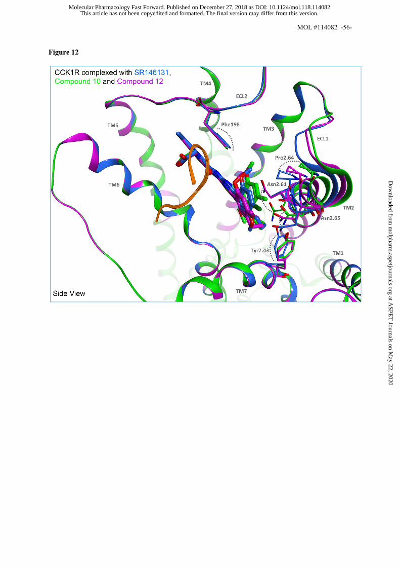
MOL #114082 -56-
Figure 12
This article has not been copyedited and formatted. The final version may differ from this version.Molecular Pharmacology Fast Forward. Published on December 27, 2018 as DOI: 10.1124/mol.118.114082
at ASPE
T Journals on M
ay 22, 2020m
olpharm.aspetjournals.org
Dow
nloaded from

MOL #114082 -57-
Figure 13
This article has not been copyedited and formatted. The final version may differ from this version.Molecular Pharmacology Fast Forward. Published on December 27, 2018 as DOI: 10.1124/mol.118.114082
at ASPE
T Journals on M
ay 22, 2020m
olpharm.aspetjournals.org
Dow
nloaded from