Molecular Basis Altered Red Blood Cell Membrane Properties in
10
Molecular Basis of Altered Red Blood Cell Membrane Properties in Southeast Asian Ovalocytosis: Role of the Mutant Band 3 Protein in Band 3 Oligomerization and Retention by the Membrane Skeleton By Shih-Chun Liu, Jiri Palek, Scott J. Yi, Pilarin E. Nichols, Laura H. Derick, Shyh-Shin Chiou, Dominick Amato, James D. Corbett, Michael R. Cho, and David E. Golan Southeast Asian ovalocytosis (SA01 is anasymptomatic trait characterized by rigid, poorly deformable red cells that resist invasion by several strains of malaria parasites. The underlying molecular genetic defect involves simple hetero- zygous state for a mutant band3 protein, which contains a deletion of amino acids 400 through 408, linked with a Lys 56-to-Glu substitution (band 3-Memphis polymorphism). To elucidate the contribution of the mutant SA0 band 3 protein to increased SA0 red blood cell (RBC) rigidity, we examined the participation of the mutant SA0 band 3 protein in in- creased band 3 attachment t o the skeleton and band 3 oligo- merization. We found first that SA0 RBC skeletons retained more band 3 than normal cells and that this increased reten- tion preferentially involved the mutant SA0 band 3 protein. Second, SA0 RBCs contained a higher percentage of band 3 oligomer-ankyrin complexes than normal cells, and these oligomers were preferentially enriched by the mutant SA0 protein. At theultrastructural level, the increased oligomer OUTHEAST ASIAN ovalocytosis (SAO) is a domi- nantly inherited trait characterized by the presence of oval-shaped red blood cells (RBCs) that are rigid and resis- tant to invasion by several strains of malaria parasites."' The underlying molecular defect involves a deletion of nine codons (400 through 408) in the erythroid band 3 protein gene; this defect is tightly linked to the Lys 56-to-Glu substi- tution (band 3-Memphis p~lymorphism).~"~ It is of consider- able interest that the primary molecular defect in S A 0 can be assigned to band 3 protein,I3 the major RBC integral membrane protein, because membrane deformability has been believed to be regulated principally by the properties of the membrane skeleton.'4"6 Band 3 is the most abundant protein of the RBC mem- brane, consisting of two domains with distinct structure and f~nction.".'~ The N-terminal 41-kD cytoplasmic domain of band 3 (cdb3) anchors the membrane skeleton to the mem- brane via interactions with ankyrin, and it binds hemoglobin and several glycolytic enzymes. The C-terminal 56-kD mem- brane domain consists of 14 membrane-spanning a helices connected by endoplasmic and ectoplasmic loops. The main function of the membrane domain is to mediate anion ex- change across the membrane. Several mutations of the hu- man band 3 have recently been described, including Glu 40- to-Lys and Pro 327-to-Arg substitutions, respectively, leading to spherocytic hemolytic anemia with protein 4.2 defi~iency,".~' Pro 868-to-Leu substitution reported in asso- ciation with acanthocytosis and increased anion transport," and a diverse group of band 3 mutations thatleadto the phenotype of hereditary spherocytosis and band 3 defi- cienc~.~'.~~ Several biochemical, biophysical, and ultrastructural ab- normalities ofband 3 in S A 0 RBCs have been reported, including increased band 3 association with membrane skele- ton,'3 increased propensity to form oligomers and to aggre- gate in the membrane,25.26 decreased anion transport,27and S Blood, Vol 86, No 1 (July I), 1995: pp 349-358 formation of SA0 RBCs was reflected by stacking of band 3-containing intramembrane particles (IMP) into longitudinal strands. The IMP stacking was not reversed by treating SA0 RBCs in alkaline pH (pH 11). which is known to weaken an- kyrin-band 3 interactions, or by removing the cytoplasmic domain of band 3 from SA0 membranes with trypsin. Fi- nally, we found that band 3 protein in intact SA0 RBCs ex- hibited a markedly decreased rotational mobility, presum- ably reflecting the increased oligomerization and the membrane skeletal association of the SA0 band 3 protein. We propose that the mutant SA0band 3 has an increased propensity to form oligomers, which appear as longitudinal strands of IMP and exhibit increased association with mem- brane skeleton. This band 3 oligomerization underlies the increase in membrane rigidity by precluding membrane skeletal extension, which is necessary for membrane deformation. 0 1995 by The American Society of Hematology. reduced lateral and rotational mobilities."~'3~26~28~z9 However, it has not been established whether or not these abnormalities directly reflect altered properties of the mutant S A 0 protein, or conversely, whether they represent nonspecific conse- quences of other alterations of S A 0 RBCs, such as increased adenosine triphosphate (ATP) consumption resulting in a premature ATP depleti~n.~' In normal RBCs, ATP depletion leads to a formation of high-molecular-weight aggregates of membrane proteins3' and increased retention of membrane skeletal proteins by the RBC membrane.32 The role of ATP depletion is particularly important in light of the data that the normalization of ATP stores of SA0 RBCs leads to a decrease in the resistance of S A 0 RBCs to malaria parasite invasion.3' From the Department of Biomedical Research, St Elizabeth's Medical Center of Boston, Tufts University Medical School, Boston, MA; Mt Sinai Hospital, University of Toronto, Toronto, Canada; the Departments of Medicine and of Biological Chemistry and Mo- lecular Pharmacology, Harvard Medical School, Boston, MA; and the Hematology-Oncology Division, Brigham and Women's Hospi- tal, Boston, MA. Submitted October 24, 1994; accepted February 16, 1995. Supported by National Institutes of Health (NIH)Research Grants No. HL 37462 and HL 27215, the UNDP/World BanWWHO Special Program for Research and Training in Tropical Diseases (J.P. and S.-C.L.), NIH Research Grants No. HL-32854 and HL-15157 (D.E.G.) and Training Grant No. HL-07623 (J.D.C.), and a Bristol- Meyers Squibb Fellowship in Clinical Pharmacology (M.R.C.). Address reprint requests to Shih-Chun Liu, PhD, Department of Biomedical Research, St Elizabeth's Medical Center of Boston, 736 Cambridge St, Boston, MA 02135. The publication costs of this article were defrayed in part by page charge payment. This article must therefore be hereby marked "advertisement" in accordance with 18 U.S.C. section 1734 solely to indicate this fact. 0 1995 by The American Society of Hematology. ooo6-4971/95/8601-0001$3.00/0 349
Transcript of Molecular Basis Altered Red Blood Cell Membrane Properties in
Molecular Basis of Altered Red Blood Cell Membrane Properties in Southeast Asian Ovalocytosis: Role of the Mutant Band 3 Protein in Band 3 Oligomerization and Retention by the Membrane Skeleton
By Shih-Chun Liu, Jiri Palek, Scott J. Yi, Pilarin E. Nichols, Laura H. Derick, Shyh-Shin Chiou, Dominick Amato, James D. Corbett, Michael R. Cho, and David E. Golan
Southeast Asian ovalocytosis (SA01 is an asymptomatic trait characterized by rigid, poorly deformable red cells that resist invasion by several strains of malaria parasites. The underlying molecular genetic defect involves simple hetero- zygous state for a mutant band 3 protein, which contains a deletion of amino acids 400 through 408, linked with a Lys 56-to-Glu substitution (band 3-Memphis polymorphism). To elucidate the contribution of the mutant SA0 band 3 protein to increased SA0 red blood cell (RBC) rigidity, we examined the participation of the mutant SA0 band 3 protein in in- creased band 3 attachment to the skeleton and band 3 oligo- merization. We found first that SA0 RBC skeletons retained more band 3 than normal cells and that this increased reten- tion preferentially involved the mutant SA0 band 3 protein. Second, SA0 RBCs contained a higher percentage of band 3 oligomer-ankyrin complexes than normal cells, and these oligomers were preferentially enriched by the mutant SA0 protein. At the ultrastructural level, the increased oligomer
OUTHEAST ASIAN ovalocytosis (SAO) is a domi- nantly inherited trait characterized by the presence of
oval-shaped red blood cells (RBCs) that are rigid and resis- tant to invasion by several strains of malaria parasites."' The underlying molecular defect involves a deletion of nine codons (400 through 408) in the erythroid band 3 protein gene; this defect is tightly linked to the Lys 56-to-Glu substi- tution (band 3-Memphis p~lymorphism).~"~ It is of consider- able interest that the primary molecular defect in SA0 can be assigned to band 3 protein,I3 the major RBC integral membrane protein, because membrane deformability has been believed to be regulated principally by the properties of the membrane skeleton.'4"6
Band 3 is the most abundant protein of the RBC mem- brane, consisting of two domains with distinct structure and f~nction.".'~ The N-terminal 41-kD cytoplasmic domain of band 3 (cdb3) anchors the membrane skeleton to the mem- brane via interactions with ankyrin, and it binds hemoglobin and several glycolytic enzymes. The C-terminal 56-kD mem- brane domain consists of 14 membrane-spanning a helices connected by endoplasmic and ectoplasmic loops. The main function of the membrane domain is to mediate anion ex- change across the membrane. Several mutations of the hu- man band 3 have recently been described, including Glu 40- to-Lys and Pro 327-to-Arg substitutions, respectively, leading to spherocytic hemolytic anemia with protein 4.2 defi~iency,".~' Pro 868-to-Leu substitution reported in asso- ciation with acanthocytosis and increased anion transport," and a diverse group of band 3 mutations that lead to the phenotype of hereditary spherocytosis and band 3 defi- c i e n c ~ . ~ ' . ~ ~
Several biochemical, biophysical, and ultrastructural ab- normalities of band 3 in SA0 RBCs have been reported, including increased band 3 association with membrane skele- ton,'3 increased propensity to form oligomers and to aggre- gate in the membrane,25.26 decreased anion transport,27 and
S
Blood, Vol 86, No 1 (July I), 1995: pp 349-358
formation of SA0 RBCs was reflected by stacking of band 3-containing intramembrane particles (IMP) into longitudinal strands. The IMP stacking was not reversed by treating SA0 RBCs in alkaline pH (pH 11). which is known to weaken an- kyrin-band 3 interactions, or by removing the cytoplasmic domain of band 3 from SA0 membranes with trypsin. Fi- nally, we found that band 3 protein in intact SA0 RBCs ex- hibited a markedly decreased rotational mobility, presum- ably reflecting the increased oligomerization and the membrane skeletal association of the SA0 band 3 protein. We propose that the mutant SA0 band 3 has an increased propensity to form oligomers, which appear as longitudinal strands of IMP and exhibit increased association with mem- brane skeleton. This band 3 oligomerization underlies the increase in membrane rigidity by precluding membrane skeletal extension, which is necessary for membrane deformation. 0 1995 by The American Society of Hematology.
reduced lateral and rotational mobilities."~'3~26~28~z9 However, it has not been established whether or not these abnormalities directly reflect altered properties of the mutant SA0 protein, or conversely, whether they represent nonspecific conse- quences of other alterations of SA0 RBCs, such as increased adenosine triphosphate (ATP) consumption resulting in a premature ATP depleti~n.~' In normal RBCs, ATP depletion leads to a formation of high-molecular-weight aggregates of membrane proteins3' and increased retention of membrane skeletal proteins by the RBC membrane.32 The role of ATP depletion is particularly important in light of the data that the normalization of ATP stores of SA0 RBCs leads to a decrease in the resistance of SA0 RBCs to malaria parasite invasion.3'
From the Department of Biomedical Research, St Elizabeth's Medical Center of Boston, Tufts University Medical School, Boston, MA; Mt Sinai Hospital, University of Toronto, Toronto, Canada; the Departments of Medicine and of Biological Chemistry and Mo- lecular Pharmacology, Harvard Medical School, Boston, MA; and the Hematology-Oncology Division, Brigham and Women's Hospi- tal, Boston, MA.
Submitted October 24, 1994; accepted February 16, 1995. Supported by National Institutes of Health (NIH) Research Grants
No. HL 37462 and HL 27215, the UNDP/World BanWWHO Special Program for Research and Training in Tropical Diseases (J.P. and S.-C.L.), NIH Research Grants No. HL-32854 and HL-15157 (D.E.G.) and Training Grant No. HL-07623 (J.D.C.), and a Bristol- Meyers Squibb Fellowship in Clinical Pharmacology (M.R.C.).
Address reprint requests to Shih-Chun Liu, PhD, Department of Biomedical Research, St Elizabeth's Medical Center of Boston, 736 Cambridge St, Boston, MA 02135.
The publication costs of this article were defrayed in part by page charge payment. This article must therefore be hereby marked "advertisement" in accordance with 18 U.S.C. section 1734 solely to indicate this fact. 0 1995 by The American Society of Hematology. ooo6-4971/95/8601-0001$3.00/0
349

350 LIU ET AL
To elucidate the contribution of the mutant S A 0 band 3 protein to the increase in membrane rigidity, we have com- bined three independent approaches. First, using size exclu- sion high-performance liquid chromatography (HPLC), we have analyzed the oligomeric forms of band 3 and the enrich- ment of the skeleton-associated band 3 oligomers by the SA0 mutant protein. Second, we have used polarized fluo- rescence depletion to measure the rotational mobility of band 3 in the membranes of intact RBCs. Third, using freeze- fracture electron microscopy, we have visualized the distri- bution of intramembrane particles (IMP), which are princi- pally composed of band 3 protein. We find first that the band 3 protein in S A 0 RBCs, but not in normal -CS, forms high-molecular-weight oligomers with high band 3-to-an- kyrin stoichiometry and that these oligomers are selectively enriched in the mutant SA0 band 3 protein. Second, we report that the mutant SA0 protein is preferentially retained by the skeleton. Third, we find that the rotational mobility of the band 3 protein in intact SA0 red cells is markedly reduced compared with band 3 in normal RBCs and that this reduction is manifested by a decrease in the rapidly rotating fraction of band 3, as well as a marked increase in the rota- tionally immobile fraction. Finally, on ultrastructural exami- nation, we find that the band 3-containing IMP stack into longitudinal strands and that this stacking is unaffected by membrane expansion by detergents, proteolytic removal of cdb3 from the membrane, or chymotryptic dissection of the third external loop of the transmembrane domain. We con- clude that the mutant S A 0 band 3 protein undergoes linear stacking in the intact membrane, thus restricting lateral and rotational mobilities of band 3 molecules. Because mem- brane deformation requires extension of the skeletal network as well as lateral movement of band 3 molecules in the plane of the membrane, we propose that the increased oligomeriza- tion and membrane skeletal association of the S A 0 band 3 protein rigidifies the S A 0 RBC membrane by precluding the skeletal reorganization necessary for RBC deformation.
MATERIALS AND METHODS
Extractability of band 3 from RBC membranes by the nonionic detergent octaethylene glycol n-dodecyl monoether (C12E8). Blood from individuals with heterozygous SA0 was collected in citrate- phosphate-dextrose anticoagulant bags or tubes and shipped on ice overnight to Boston, MA, for evaluation. RBCs were sedimented at 2,000 rpm for I O minutes and washed with phosphate-buffered saline (PBS).
RBC ghosts (3 mg protein per milliliter), prepared by hypotonic ly~is ,3~ were mixed with equal volumes of 0.5% CI2E8 in isotonic buffer (5 mmol/L NaPi, pH 7.4; 150 mmol/L NaCI) or hypotonic buffer (5 mmoVL NaPi, pH 7.4) at 4°C for 10 minutes. After centrifu- gation at 150,000g for 30 minutes, the supernatants and pellets were dissolved in sodium dodecyl sulfate (SDS) and analyzed by SDS- polyacrylamide gel electrophoresis (PAGE) according to Agre et al.34 Band 3-to-spectrin ratios in isotonic and hypotonic skeletal shells were analyzed by densitometric gel scans. The values were obtained from four experiments.
Retention of the normal and mutant band 3 in SA0 membrane skeletons. To assess whether or not band 3 retained by the SA0 skeleton is selectively enriched in mutant band 3 protein, we pre- treated intact S A 0 and band 3-Memphis RBCs with chymotrypsin (0.5 mg/mL) in PBS buffer at 37°C for 90 minutes.35 Membrane
prepared from these cells was extracted with C,*E8 (0.5%) under isotonic conditions. The extract supernatants and insoluble skeletons were prepared as described above, dissolved in SDS, and analyzed using 7.5% to 15% Laemmli gels.36 In normal cells, chymotryptic digestion produced an N-terminal60-kD segment of band 3 resulting from cleavage at Tyr 553." In SA0 RBCs containing the linked band 3-Memphis polymorphism, chymotryptic digestion produced both the 60-kD band (the normal allele) and an abnormal 63-kD band.3R For comparison, samples from a normal individual with mor- phologically normal RBCs and the band 3-Memphis polymorphism were analyzed in parallel. The relative abundance of mutant to nor- mal band 3 in supernatant and skeleton fractions was assessed by measuring the ratio of 63-kD to total (60-kD + 63-kD) fragments by densitometric gel scans.
Size exclusion HPLC of band 3 extracted from spectrin-depleted membranes by C12Es RBC ghosts, prepared by hypotonic lysis, were incubated in 0.1 mmoVL NaPi, pH 8.0, buffer at 37°C to remove ~pectrin.'~ The spectrin-depleted vesicles (2.0 to 2.5 mgl mL protein, final concentration) were dissolved in 0.5% ClzEn in hypotonic buffer ( 5 mmoVL NaPi, pH 7.4) at 4°C. After centrifuga- tion at 150,OOOg for 30 minutes, the supernatants (45 to 225 pg protein in 50 to 250 pL buffer) were analyzed by size-exclusion HPLC using a TSK-4000 SW,, column (7.8 X 300 mm; Tosohaas, Tokyo, Japan), as described previo~sly.~" The standard elution buffer contained 0.01% CI2E8, 100 mmom NaCI, and 5 mmoVL NaPi, pH 7.0. Protein in HPLC fractions was concentrated by centrifugation of 0.5- to 2.0-mL aliquots in a 10,000-molecular weight (mol wt) cutoff ultrafiltration unit (Millipore, Lexington, MA) at 5,000 rpm for 15 to 20 minutes. The concentrated supernatant was removed, dissolved in SDS, and analyzed by SDS-PAGE according to Laem- ~nl i .~ '
Enrichment of bund 3 oligomers by the SA0 bund 3 protein. To assess whether or not SA0 band 3 oligomers are selectively enriched in mutant band 3 protein, we pretreated intact SA0 and normal RBCs with chymotrypsin as described above. Spectrin-depleted membrane vesicles were derived from these cells and extracted with CI2Ex, and the solubilized supernatants were analyzed by size-exclusion HPLC, as described above. The relative abundance of mutant to normal band 3 in different band 3-containing HPLC peaks was assessed by measuring the ratio of 63-kD to total (60-kD + 63-kD) fragments after SDS-PAGE, as described above.
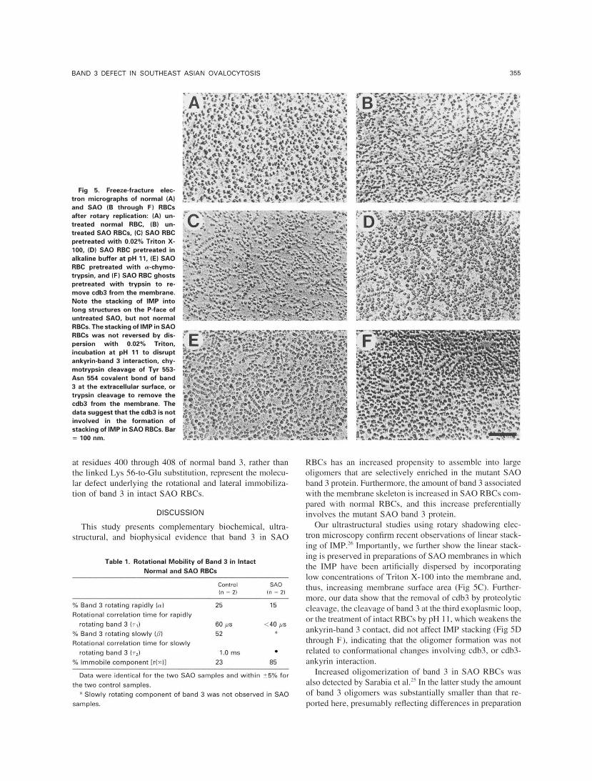
Freeze-fructure electron microscopy. Washed RBCs from blood collected within 24 hours were fixed in 1.75% glutaraldehyde in 0.1 m o m sodium cacodylate buffer, pH 7.4, for 1.5 hours at 0°C. The fixed cells were washed five times in 0.05 m o m sodium cacodylate, pH 7.4, glycerinated (final glycerol concentration, 33%), frozen in freon, and transferred to liquid nitrogen. The samples were fractured in a freeze-etch unit (model BAF 400D; Balzers, Hudson, NH) at - I 10°C. Samples were rotary-shadowed at a 25" angle with plati- num-carbon and at a 90" angle with arbo on.^' Replicas were viewed in a transmission electron microscope (model JEM 100s; JEOL USA, Peabody, MA) at an accelerating voltage of 80 kV.
To test if the IMP stacking persisted at low IMP density, we treated SA0 RBCs with prehemolytic concentration of Triton X- 100 (0.02%) at 0°C for IO minutes before these cells were fixed and examined by freeze-fracture electron microscopy. Further, we examined if the IMP stacking in SA0 could be reversed by (1) incubation of S A 0 RBCs in PBS at pH 11 .O @"c, 30 minutes), which was believed to weaken the ankyrin-band 3 interaction>2 (2) chymotryptic digestion (0.5 mg/mL, 37°C 60 minutes) of intact SA0 RBCs to cleave band 3 extracellularly at Tyr 553," or (3) trypsin digestion (5 pg/mL, O T , 30 minutes) of isolated SA0 ghosts (3 mg protein per milliliter) to remove cdb3 from the membrane." Specific band 3 cleavages by the proteolytic enzymes in the above

BAND 3 DEFECT IN SOUTHEAST ASIAN OVALOCYTOSIS 351
experiments were verified by SDS-PAGE of the resultant membrane specimen.
Band 3 rotational mobility in intact RBCs. RBC band 3 was fluorescently labeled, as previously described!’ Briefly, fresh RBCs from blood collected within 24 hours were washed in high potassium PBS (KF’BS; 140 mmoVL KCI, 15 mmoVL NaPi, 10 mmoVL glu- cose, pH 7.4) and mixed with 0.25 mg/mL (final concentration) eosin-5-maleimide for 12 minutes at room temperature. Cells were then washed three times in KPBS with 1% bovine serum albumin (BSA). Under these conditions, greater than 80% of the membrane- associated fluorescence was covalently bound to band 3 in normal and SA0 RBCs. The stoichiometry of labeling was approximately 1 eosin molecule per band 3 monomer in both normal and S A 0 cells, in agreement with the results of Tilley et alzs and Che et
The technique of polarized fluorescence depletion was used to measure the rotational mobility of eosin-labeled band 3 in mem- branes of intact RBCs, as previously de~cribed.4~ Briefly, recovery of fluorescence after a ground state depletion laser pulse depended both on the triplet lifetime(s) of the fluorophore and on the rotational relaxation time(s) of band 3. In addition, the fraction of band 3 molecules that was rotationally immobile on the time scale of the experiment was obtained from the residual anisotropy of the fluores- cence intensities excited by parallel and perpendicular probe beams. Using our laser microscopy photometer, two exponential compo- nents of anisotropy decay and a residual anisotropy could be re- solved, so data were fitted by nonlinear least-squares analysis to the equation r(t) = r(m) + a * e (-t/Tl) + p * e (-thZ), where r(t) is the anisotropy at time t, r(m) is the residual anisotropy, and a and p are the fractions of molecules with rotational correlation times T, and T ~ , respectively. For anisotropy decay curves in which it was apparent that T, < 40 ,us, r (0) was set equal to the anisotropy value determined using eosin-labeled RBCs fixed with 1% glutaraldehyde. Band 3 in the membranes of fixed cells exhibited no rotational mobility, and the anisotropy, typically 0.27 to 0.33, was time-invari- ant. This procedure allowed the reliable determination of a but not T~ in such cases.
The preparation of intact RBC samples for polarized fluorescence depletion experiments and the time-resolved photon-counting laser microscope used in such experiments have been described in detail!’
RESULTS
Preferential association of mutant band 3 protein with S A 0 membrane skeleton. Previous studies showed con- flicting data on the retention of band 3 in membrane skele- tons. In one study using extraction with Triton X-100, no differences were observed in band 3 retention between SA0 RBCs and normal RBCs.” In another report, increased reten- tion of band 3 by the skeleton was observed and it was attributed to a nonspecific trapping of band 3 by the SA0 skeleton.I2 Therefore, we first examined differences between normal RBCs and SA0 cells in the retention of band 3 by the skeleton, using a mild nonionic detergent, C12Es.25240
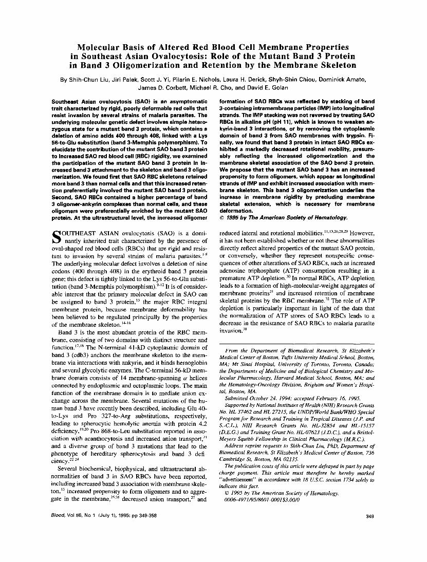
SA0 membranes and normal RBC membranes (3 mg pro- tein per milliliter) were extracted with equal volumes of 0.5% CI2E8 to determine the proportion of band 3 that was retained by the detergent-insoluble membrane skeleton. Band 3-to-spectrin ratios of the resulting skeletal shells were analyzed by SDS-PAGE followed by dye quantitation after elution from the gels. Increased retention of band 3 in SA0 membrane skeletons was detected under both isotonic and hypotonic extraction conditions (Fig 1). After isotonic C12ES extraction, 59% of band 3 was retained in SA0 skeletons,
compared with 38% in normal skeletons. No ankyrin were released from the skeleton under these conditions. After ex- traction under hypotonic conditions, 35% of band 3 was retained in SA0 skeletons, compared with 21% in normal skeletons. The band 3 retention in ClzE8 shells was not sig- nificantly affected by the use of higher extraction volumes (five instead of one) of 0.5% CI2E8, or the concentration of CIPE8 (0.1% to l%), or additional washes with 0.5% C12Es (data not shown).
To explore the possible role of the mutant SA0 band 3 in the increased retention of band 3 in SA0 membrane skele- tons, we investigated whether or not the S A 0 membrane skeletons were selectively enriched in the mutant SA0 band 3 protein. We took advantage of the band 3-Memphis poly- m o r p h i ~ m ~ ~ . ~ ~ that is tightly linked with the SA0 muta- tion’.” and the availability of subjects carrying the band 3- Memphis polymorphism without other associated band 3 abnormalities. After chymotryptic cleavage of band 3 protein at Tyr 553 in intact cells, the band 3-Memphis polymorphism produces a 63-kD proteolytic fragment, rather than the nor- mal 60-kD fragment. The decrease in electrophoretic mobil- ity of the proteolytic fragment of band 3-Memphis is caused by a change in a single amino acid, Lys 56 to Glu 56.45.46 Figure 2 depicts the analysis of mutant band 3 in SA0 mem- brane skeletons. The percentage of 63-kD fragment in the original SA0 ghosts was 40%. In contrast, the amount of the 63-kD fragment was increased in the C12E8-insoluble pellet (52%), whereas it was diminished in the C12E8-soluble supernatant (32%). Thus, the fraction of band 3 associated with the skeleton of SA0 RBCs was enriched in mutant band 3 protein. For comparison, we examined RBCs of an asymptomatic carrier of band 3-Memphis and found no en- richment of the skeletal fraction by the 63-kD band 3 frag- ment. The percentage of 63-kD fragment was 50% in band 3-Memphis ghosts, the C12E8-soluble supernatant, and the insoluble pellet (Fig 2).
Enrichment of band 3 oligomers by the mutant S A 0 pro- tein. To elucidate the molecular basis of increased reten- tion of the mutant protein by the membrane skeleton, we analyzed the dimeric and oligomeric states of band 3 protein in SA0 RBCs and normal RBCs using size-exclusion HPLC. Band 3 oligomeric states were analyzed in the native mem- brane by extracting spectrin-depleted inside-out vesicles with the nonionic detergent CIZE8 and centrifuging at 150,000g for 30 minutes. In control cells, the detergent extracted ap- proximately 73% of band 3 from the membrane, leaving the remainder of band 3 protein in the pellet fraction. The amount of ankyrin and protein 4.2 extracted from control cells under such conditions was 83% and 72%, respectively. The amounts of band 3 protein (69%), ankyrin (72%), and protein 4.2 (63%) solubilized from SA0 RBCs were all slightly lower than the corresponding amounts extracted from control cells (data not shown).
In both normal and SA0 RBCs, HPLC separation resolved the ClzEs extract into two major protein peaks and one deter- gent peak (Fig 3). The slower, more abundant peak was identical in mobility to that of band 3 dimers, whereas the faster peak represented complexes containing ankyrin and band 3 oligomers. Often, a small but variable amount of
352 LIU ET AL
5 - 6 -
7 -
I
W G S P W G S P
- . ".
WG S P W G S P
void volume peak (Vo) also appeared in HPLC profiles. The identification of the band 3 dimer peak on size-exclusion HPLC agreed with that determined previously by using sedi- mentation velocity analysis and partial specific volume deter- mination?" Importantly, differences between normal RBCs and SA0 RBCs were found in the relative distribution of the band 3 dimer and ankyrinhand 3 oligomer peaks. The amount of the ankyrinhand 3 oligomer peak (including the overlapping Vo peak) was considerably greater in SA0 RBCs (62%) than in normal cells (42%). There was a corre- sponding decrease in the dimer fraction of band 3 protein in S A 0 cells (38%) compared with normal cells (58%).
In both normal and S A 0 RBCs, the band 3 dimer fraction contained not only band 3 but also protein 4.2 and a small amount of protein 4. I , as shown in SDS-polyacrylamide gels (Fig 3). Further, glycophorin A was detected in a fraction that eluted slightly slower than the band 3 dimer fraction (data not shown). Notably, no ankyrin was detected in the band 3 dimer fraction in either normal or S A 0 RBCs. In both S A 0 and normal RBCs, the band 3 oligomer fraction contained not only band 3 but also protein 4.2 and ankyrin. However, in contrast to the band 3 dimer fraction, the band 3 oligomer fraction was devoid of glycophorin and protein 4.1. Importantly, the band 3-to-ankyrin stoichiometry in the oligomer fraction of S A 0 RBCs differed from normal cells. Densitometric scans of gels corresponding to the oligomer fraction showed that SA0 RBCs contained 6.0 copies of band 3 for each copy of ankyrin, whereas normal cells con- tained about 4.4 copies of band 3 for each copy of ankyrin. The molar ratio of protein 4.2 to band 3 was 1:6 in both dimer and oligomer fractions from both S A 0 and normal RBCs.
Fig 1. Extractability of band 3 from SA0 mem- branes by C12Ea. (A) CIZEa extraction of normal and SA0 ghosts was performed under isotonic condi- tions. (B) C12Es extraction of ghosts was performed under hypotonic conditions. (C) Band 3:spectrin ratio from C1,Ea-insoluble normal and S A 0 shells. Note that the band 3:spectrin ratio was markedly in- creased in S A 0 skeletons compared with normal skeletons, indicating substantial band 3 retention.
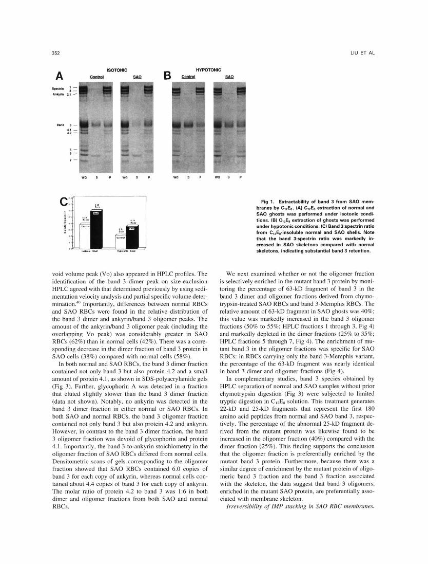
We next examined whether or not the oligomer fraction is selectively enriched in the mutant band 3 protein by moni- toring the percentage of 63-kD fragment of band 3 in the band 3 dimer and oligomer fractions derived from chymo- trypsin-treated S A 0 RBCs and band 3-Memphis RBCs. The relative amount of 63-kD fragment in S A 0 ghosts was 40%; this value was markedly increased in the band 3 oligomer fractions (50% to 55%; HPLC fractions l through 3, Fig 4) and markedly depleted in the dimer fractions (25% to 35%; HPLC fractions 5 through 7, Fig 4). The enrichment of mu- tant band 3 in the oligomer fractions was specific for S A 0 RBCs: in RBCs carrying only the band 3-Memphis variant, the percentage of the 63-kD fragment was nearly identical in band 3 dimer and oligomer fractions (Fig 4).
In complementary studies, band 3 species obtained by HPLC separation of normal and S A 0 samples without prior chymotrypsin digestion (Fig 3) were subjected to limited tryptic digestion in CI2ES solution. This treatment generates 22-kD and 25-kD fragments that represent the first 180 amino acid peptides from normal and S A 0 band 3, respec- tively. The percentage of the abnormal 25-kD fragment de- rived from the mutant protein was likewise found to be increased in the oligomer fraction (40%) compared with the dimer fraction (25%). This finding supports the conclusion that the oligomer fraction is preferentially enriched by the mutant band 3 protein. Furthermore, because there was a similar degree of enrichment by the mutant protein of oligo- meric band 3 fraction and the band 3 fraction associated with the skeleton, the data suggest that band 3 oligomers, enriched in the mutant S A 0 protein, are preferentially asso- ciated with membrane skeleton.
Irreversibility of IMP stacking in S A 0 RBC membranes.

BAND 3 DEFECT IN SOUTHEAST ASIAN OVALOCYTOSIS 353
A SAO
5-
6-
7-
WG S P
tion. Therefore, we examined the distribution of IMP using - Band 3 Memphis rotary shadowing, which provides a better visualization of
particles in a close proximity to one another.
m di&& IMP in P- and E-fracture faces of S A 0 red cells were 3,640 Using rotary shadowing, we found that the densities of
5 120/p.m' and 430 2 20/pm', respectively, which were similar to IPM densities in normal cells of 3,680 2 100/pm' and 470 2 30/p.mZ, respectively. The density was of the same magnitude as the number of band 3 dimers in the
"-
normal RBC membrane, namely 600,000 band 3 dimers per cell:' However, the distribution of the IMP in S A 0 red cells was not uniform, with many particles stacked together to form linear strands that resembled strings of beads (Fig 5B). Analysis of IMP in 12 0.1-pm' fields of fractured P face showed that 17% to 19% of band 3 dimers were stacked into such structures. The structures were 50 to 250 nm in
"- length (average, 94 nm) and contained 4 to 21 IMP each (average, eight IMP). IMP organized into stacked structures 150 to 200 nm in length were unique to S A 0 RBCs: we did not find any such structures in spherocytic RBCs from pa- tients with hereditary spherocytosis (HS), including HS asso-
WG S P
Band 3 Memphls
0 0 5 0
x
$ 4 0
i a 3 0
2 o WO S P WC S P
Fig 2. Analysis of mutant band 3 in SA0 membrane skeletons. Intact SA0 and band 3-Memphis RBCs were treated with chymotryp- sin t o cleave the band 3 quantitatively into the 35-kD [amino acids (AA) 554 to 9111 fragment and the complementary 63-kD or 60-kD fragments (AA 1 to 5531. The 63-kD and 60-kD fragments were derived from the mutant and normal alleles of band 3, respectively. White ghosts (WG) prepared from these cells were extracted with C12E8 (0.5%) under isotonic conditions. (A) Extract supernatants (S) and insoluble skeletons (P) were dissolved in SDS and analyzed using Laemmli gels. (B) The ratio of 63-kD to total 160-kD + 63-kD) frag- ments in the supernatant and skeleton fractions was analyzed by densitometric gel scans. Note that the percentage of 63-kD fragment was 50% in band 3-Memphis ghosts, the ClZE8-soluble supernatant, and the insoluble pellet. In contrast, the percentage of 63-kD frag- ment was 40% in SA0 ghosts, markedly increased in the ClZE8-insolu- ble pellet (52%). and depleted in the C,,E8-soluble supernatant (32%).
Band 3 protein is the principal constituent of IMP. Recently, a linear stacking of IMP has been demonstrated in S A 0 RBC membrane'6 using a unidirection shadowing technique. However. because of the very high density of IMP in the membrane and the inherent limitations of unidirectional shadowing, it is difficult to exclude artifactual IMP aggrega-
Control E3
E3 DIMER EWANK. DIMER .
ii 2 3.0 7
N g 2.0
5 1.0
m n
D a 0.0
0.0 2.5 5.0 7.5 10.0 12.5 15.0 Elution Volume (ml)
ANK.-
83-
4.2-
t = r
BWANK. DIMER E3
-q 7
ANK."' ~
- 4.1
4.0 B3 DIMER 4.0
3.0 1 ,
B3 - P; - 4.1 2.0 4.2-
l .o 1
. -
0.0 0.0 2.5 5.0 7 5 10.0 12.5 15.0 I 1 0.0 2.5 5.0 7 5 10.0 12.5 15.0
Elutlon Volume (ml) .-. -
Fig 3. Size-exclusion HPLC of C& extracts from spectrin-de- pleted membrane vesicles. Normal and SA0 RBC ghosts were incu- bated in low salt buffer t o remove spectrin. The spectrin-depleted vesicles were dissolved in 0.5% C,*Es in hypotonic buffer at 4°C. Sam- ples were centrifuged at 150,000gfor 30 minutes. Supernatants were collected and analyzed by size-exclusion HPLC using a TSK-4000 SW column. The elution buffer contained 0.01% ClZE8, 100 mmol/L NaCI, and 5 mmol/L NaPi, pH 7.0. Note the marked increase in the amount of band 3 oligomer peak (B3/ANK) eluting at 6.0 t o 7.9 mL in the ClZEs extract from SA0 vesicles as compared with the amount eluting from normal vesicles. Note also the marked decrease in the band 3 dimer peak (B3 DIMER) eluting at 9.0 mL from SA0 as compared with normal vesicles. The protein composition of each peak was analyzed using a 10% Laemmli gel. V., void volume peak; G, glyco- phorin A; R, residual globin.
354
B 6 0
0 5 0 0 4 4 0
3 0
2 0 I
, . . . . . . Ghost F r l Fr2 F r 3 F r 4 F r 5 F r 6 Fr7
~ O l l p o m s r Fr.cllonr.* U-Dlmer Fraction*+
Fig 4. Distribution of mutant and normal band 3 in dimer and oligomer fractions of band 3 from S A 0 and band 3-Memphis RBCs. Intact SA0 and band 3-Memphis RBCs were treated with chymotryp- sin to cleave band 3 extracellularly. Spectrin-depleted membrane ves- icles derived from these chymotrypsin-pretreated cells were ex- tracted with CI2E8 and analyzed by HPLC, as described in the legend to Fig 3. (A) SDS-polyacrylamide gels of HPLC fractions correspond- ing to oligomers and dimers of band 3. (B) The ratio of 63-kD to total (60-kD + 63-kD) fragments in S A 0 and band 3-Memphis ghosts and in the respective HPLC fractions was analyzed by densitometric gel scans. Note that the relative amount of 63-kD fragment was 50% in both band 3-Memphis ghosts and in oligomer and dimer species of band 3 fractionated by HPLC. In contrast, the relative amount of 63- kD fragment was 40% in SA0 ghosts, but was markedly enriched in the band 3 oligomer fractions [fractions (Fr) l to 31 and markedly depleted in the dimer fractions (Fr 5 to 7).
ciated with band 3 deficiency. in elliptocytic RBCs con- taining mutant spectrins, or in normal cells (data not shown). The abnormal stacking of IMP in S A 0 membranes became even more evident in cells pretreated with low concentrations (0.02%) of Triton X-l00 before freeze-fracture (Fig SC). At such low concentrations, Triton X-l00 reduced somewhat the IMP density in P-fracture face from 3.640 f 120/pm2 to 3,340 5 145 pm' without a detectable change of the size of individual IMP. It is likely that Triton, at prehemolytic concentrations, expands the membrane without disturbing the linear stacking of IMP present in the S A 0 membrane. However, this modest expansion of RBC surface area did not produce an obvious increase in cell dimensions or cell shape (data not shown).
We next attempted to disaggregate the linear strands of IMP by subjecting S A 0 RBCs to treatment that disrupts band 3-ankyrin interaction or produces proteolytic cleavage of band 3 leading to a release of cdb3 from the plasma membrane (Fig SD through F). These conditions included ( l ) treatment of S A 0 RBCs at elevated pH (eg, pH I l ) ,
LIU ET AL
which weakens the ankyrin-to-band 3 interaction" (Fig SD), (2) pretreatment of S A 0 RBCs with a-chymotrypsin, which cleaves band 3 at Tyr SS3 in the third exoplasmic (Fig SE), and (3) treatment of S A 0 RBC ghosts with trypsin to remove cytoplasmic domain of band 3 from the membrane by cleavage at residue Lys 36017 (Fig SF). None of these conditions leads to a disassembly of the stacked IMP, im- plying that the stacking of IMP in S A 0 membranes does not require the covalent bond between Tyr S53 and Asn 554. the tight interaction between ankyrin and band 3, or the presence of cytoplasmic domain of band 3. The definitive site(s) involved in the stacking of S A 0 band 3 remains to be established.
Rotcrtioncd itnmobili;crtior~ of hclnrl 3 in intact S A 0 RBC menrhrcrne. Measurements of membrane protein rotational mobility provide an independent assessment of the state of protein self-association and degree of protein association with the membrane skeleton. We used the polarized fluores- cence depletion technique to measure the rotational mobility of eosin maleimide-labeled band 3 in membranes of intact normal and S A 0 RBCs. Unlike other systems that require a relatively large phosphorescence signal from many optically transparent cells to measure rotational mobility (in the case of RBCs, this requirement necessitates the use of white ghosts rather than intact cells), the technique used here took advantage of the sensitivity of fluorescence detection to per- form measurements on several hundred intact RBCs in one to three layers on a microscope slide. The instrument design4? avoided the problem of inner filter effects, as excitation and emission light paths were not filtered by the hemoglobin in intact cells. In addition, the system used here allowed performance of band 3 lateral and rotational mobility mea- surements on matched samples of intact normal and S A 0 RBCs. Lateral mobility was measured using the fluorescence photobleaching recovery technique, which used the same optical and electronic apparatus as that used for rotational mobility measurements.
In agreement with previous rapidly rotating (25%), slowly rotating (S2%), and rotationally immobile (23%) forms of band 3 were observed in the membranes of intact normal RBCs. In contrast, intact S A 0 cells manifested a very large ( S % ) rotationally immobile band 3 fraction and a small (15%) rapidly rotating band 3 fraction. The slowly rotating band 3 fraction was absent in S A 0 cells (Table 1 ). These data are similar to reports of band 3 rota- tional mobility in ghosts prepared from S A 0 RBCs,".'" sug- gesting that the physical state of band 3 molecules is not markedly perturbed by the preparation of ghosts from intact S A 0 cells. As previously reported,'? the lateral mobility of band 3 in S A 0 RBCs was also markedly reduced as com- pared with that in normal cells. The profound rotational and lateral immobilization of band 3 in S A 0 cells is consistent with the increased band 3 oligomerization and increased band 3 binding to the membrane skeleton that were demon- strated using ultrastructural and biochemical techniques. In intact RBCs carrying only the band 3-Memphis variant, the rotational and lateral mobilities of band 3 were not signifi- cantly different from those in control RBCs (data not shown). These data further suggest that the nine-amino acid deletion
BAND 3 DEFECT IN SOUTHEAST ASIAN OVALOCYTOSIS 355
Fig 5. Freeze-fracture elec- tron micrographs of normal (AI and SA0 (B through F) RBCs after rotary replication: (A) un- treated normal RBC, (B) un- treated SA0 RBCs, (Cl SA0 RBC pretreated with 0.02% Triton X- 100, (D) SA0 RBC pretreated in alkaline buffer at pH 11, (E) SA0 RBC pretreated with a-chymo- trypsin, and (F1 SA0 RBC ghosts pretreated with trypsin to re- move cdb3 from the membrane. Note the stacking of IMP into long structures on the P-face of untreated SAO, but not normal RBCs. The stacking of IMP in SA0 RBCs was not reversed by dis- persion with 0.02% Triton, incubation at pH 11 t o disrupt ankyrin-band 3 interaction, chy- motrypsin cleavage of Tyr 553- Asn 554 covalent bond of band 3 at the extracellular surface, or trypsin cleavage to remove the cdb3 from the membrane. The data suggest that the cdb3 is not involved in the formation of stacking of IMP in SA0 RBCs. Bar = 100 nm.
at residues 400 through 408 of normal band 3, rather than the linked Lys 56-to-Glu substitution, represent the molecu- lar defect underlying the rotational and lateral immobiliza- tion of band 3 in intact S A 0 RBCs.
DISCUSSION
This study presents complementary biochemical, ultra- structural, and biophysical evidence that band 3 in S A 0
Table l . Rotational Mobility of Band 3 in Intact Normal and SA0 RBCs
Control SA0 In = 21 In = 2 )
% Band 3 rotating rapidly (m) 25 15 Rotational correlation time for rapidly
% Band 3 rotating slowly (0) 52 * Rotational correlation time for slowly
rotating band 3 (TJ 1 .O ms % Immobile component [r(x)l 23 85
rotating band 3 (T,) 60 v s <40 v s
Data were identical for the two SA0 samples and within 55% for
* Slowly rotating component of band 3 was not observed in SA0 the two control samples.
samples.
RBCs has an increased propensity to assemble into large oligomers that are selectively enriched in the mutant S A 0 band 3 protein. Furthermore, the amount of band 3 associated with the membrane skeleton is increased in S A 0 RBCs com- pared with normal RBCs, and this increase preferentially involves the mutant S A 0 band 3 protein.
Our ultrastructural studies using rotary shadowing elec- tron microscopy confirm recent observations of linear stack- ing of IMP." Importantly, we further show the linear stack- ing is preserved in preparations of S A 0 membranes in which the IMP have been artificially dispersed by incorporating low concentrations of Triton X- IO0 into the membrane and, thus, increasing membrane surface area (Fig 5C). Further- more, our data show that the removal of cdb3 by proteolytic cleavage, the cleavage of band 3 at the third exoplasmic loop, or the treatment of intact RBCs by pH 1 I , which weakens the ankyrin-band 3 contact, did not affect IMP stacking (Fig 5D through F), indicating that the oligomer formation was not related to conformational changes involving cdb3, or cdb3- ankyrin interaction.
Increased oligomerization of band 3 in S A 0 RBCs was also detected by Sarabia et al.'? In the latter study the amount of band 3 oligomers was substantially smaller than that re- ported here, presumably reflecting differences in preparation
356
Normal
LIU ET AL
results of Moriyama et al.” These investigators did find an increased band 3 retention by the skeleton, but they did not detect a preferential retention of the mutant band 3 in S A 0 spectrin shells. The reason for this discrepancy is not clear. It is possible that in the study by Moriyama et all2 the amount of band 3 retained in Triton shells was relatively low, thereby diminishing the accuracy of the subsequent quantitative mea- surement of mutant S A 0 band 3 in these shells. The validity of our data demonstrating the enrichment of mutant band 3 in spectrin shells from S A 0 membranes is further strength- ened by our finding of the complementary reduction of mu- tant band 3 in the C12E,-soluble supernatant (Fig 2) .
Finally, our study is consistent with previous reports of markedly decreased band 3 rotational mobility in ghosts pre- pared from S A 0 Rotational immobilization could be caused by increased band 3 self-association (aggre- gation) in the plane of the membrane or by increased direct binding interactions between the cytoplasmic domain of band 3 and membrane skeletal proteins. Several lines of evidence point to the relative importance of the former over the latter mechanism in regulating band 3 rotation in S A 0 RBCs. First, there is conclusive ultrastructural and biochemi- cal evidence for increased band 3 oligomerization in S A 0 RBCs. Second, theoretical studies predict that the rotational mobility of particles in membranes is especially sensitive to particle size.5’ Linear oligomers, eight IMP in length (on average), are expected to have rotational diffusion coeffi- cients approximately 100-fold less than those of an individ- ual IMP, and thus to appear rotationally immobile on the time scale of the experiments reported here. Third, Che et alzh found that removal of spectrin and actin from S A 0 ghost membranes has a minimal effect on band 3 rotational mobility and that further removal of ankyrin and protein 4.1 does not increase band 3 rotational mobility to the level manifested by band 3 in similarly treated membranes from normal cells. These data suggest that the rotational and lat- eral immobilization of band 3 in S A 0 RBCs is caused by the mutant S A 0 band 3 oligomerization, which is mediated by conformational changes in the transmembrane domain resulting from the deletions of amino acid residues 400 through 408. On the other hand, it is unlikely that increased binding interactions between the cytoplasmic domain of band 3 and membrane skeletal proteins, especially ankyrin, contribute to band 3 immobilization, as evidenced by our data that IMP stacking was unaffected by removal of cdb3 by proteolytic cleavage or by weakening of the ankyrin-band 3 contact (Fig 5D and F).
It is not known whether the linear band 3 aggregates in S A 0 membranes are composed exclusively of mutant band 3 or whether they contain both normal and mutant band 3. About 25% of the mutant band 3 was detected in the dimer fraction (Fig 4), which presumably represents band 3 mole- cules that are not bound to the membrane skeleton. In con- trast, both the skeleton-associated and the oligomeric band 3 fractions were enriched in the mutant protein. This finding implies that S A 0 band 3 is more prone than normal band 3 to form oligomers. Further, as the presence of 40% mutant band 3 molecules in S A 0 membranes leads to an increase in the rotationally immobile component of band 3 from 23%
SA0
Q Band 3 oligomen 0 Bmd 3 d i m n 0 Junctional complexes of actin
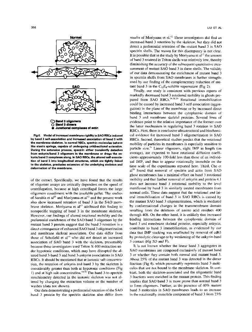
Fig 6. Model of increased membrane rigidity in SA0 RBCs induced by band 3 self-association and increased association of band 3 with the membrane skeleton. In normal RBCs, spectrin molecules behave like elastic springs, capable of undergoing unidirectional extension. During the extension process, spectrin either transiently detaches from ankyrinlband 3 oligomers in the membrane or drags the an- kyrin/band 3 complexes along. In SA0 RBCs, the altered self-asrocia- tion of band 3 into longitudinal structures, which are tightly linked to the skeleton, precludes extension of the underlying skeleton and deformation of the membrane.
of the extract. Specifically, we have found that the results of oligomer assays are critically dependent on the speed of centrifugation, because at high centrifugal forces the large oligomers cosediment with the insoluble pellet. The studies of Sarabia et alZ5 and Moriyama et all2 and the present work also show increased retention of band 3 in the S A 0 mem- brane skeleton. Moriyama et all2 attributed this finding to nonspecific trapping of band 3 in the membrane skeleton. However, our findings of altered rotational mobility and the preferential enrichment of the S A 0 band 3 oligomers by the mutant band 3 protein suggest that the band 3 retention is a direct consequence of enhanced S A 0 band 3 oligomerization and membrane skeletal association. Our data differ from those of Schofield et all0 who did not detect an increased association of S A 0 band 3 with the skeleton, presumably because these investigators used Triton X- 100 extraction un- der hypotonic conditions, which may have disrupted the al- tered band 3-band 3 and band 3-ankyrin associations in S A 0 RBCs. It should be mentioned that at isotonic salt concentra- tion, the retention of ankyrin and band 3 by the skeleton is considerably greater than both at hypotonic conditions (Fig 1) and at high salt on cent ration.^^.^^ The band 3-to-spectrin stoichiometry detected in the isotonic skeleton was not al- tered by changing the extraction volume or the number of washes (data not shown).
Our data demonstrating a preferential retention of the S A 0 band 3 protein by the spectrin skeleton also differ from
BAND 3 DEFECT IN SOUTHEAST ASIAN OVALOCYTOSIS 357
to 85% (Table l), it is likely that mutant band 3 is capable of forming at least some hetero-oligomers. However, we cannot exclude the possibility that a small amount of mutant band 3 does form homodimers or homo-oligomers in SAO.
On the basis of these studies, we propose the following mechanism by which the S A 0 band 3 protein mutation leads to a marked increase in membrane rigidity (Fig 6). The pre- viously held hypothesis suggested that membrane deform- ability is principally regulated by the skeleton and, in particu- lar, by the propensity of spectrin to undergo unidirectional extension without rupture. In this model, spectrin molecules act as elastic ~prings. '~ , '~ .~* While acknowledging this im- portant property of spectrin, we propose that membrane de- formability also requires that band 3 oligomers, which repre- sent the principal attachment points of the skeleton to the membrane, are laterally and rotationally mobile. Aggrega- tion and stacking of such connecting points into longitudinal strands, such as occurs in the case of the SA0 band 3 muta- tion, precludes both lateral mobility of the band 3 protein and, consequently, the lateral extension of the underlying skeleton. Membrane skeletal immobilization, in turn, pro- duces a marked decrease in membrane deformability. This model can explain most, if not all, abnormalities found in SA0 RBCs, including the high-molecular-weight protein ag- gregation, the lateral and rotational immobilization of band 3, and the increased membrane rigidity.
ACKNOWLEDGMENT
We thank Diana Fraser for manuscript typing and Donna Marie Mironchuk for artwork.
REFERENCES 1. Lie-Injo LE: Hereditary ovalocytosis and hemoglobin E-ovalo-
cytosis in Malayan aborigines. Nature 208:1329, 1965 2. Amato D, Booth PB: Hereditary ovalocytosis in Melanesians.
Papua New Guinea Med J 20:26, 1977 3. Cattani JA, Gibson FD, Alpers MP, Crane GG: Hereditary
ovalocytosis and reduced susceptibility to malaria in Papua New Guinea. Trans R SOC Trop Med Hyg 81:705, 1987
4. Mohandas N, Lie-Injo LE, Friedman M, Mak JW: Rigid mem- branes of Malayan ovalocytes: A likely genetic barrier against ma- laria. Blood 63: 1385, 1984
5. Saul A, Lamont G, Sawyer WH, Kidson C: Decreased mem- brane deformability in Melanesian ovalocytes from Papua New Guinea. J Cell Biol 98:1348, 1984
6. Kidson C, Lamont G, Saul A, Nurse G T Ovalocytic erythro- cytes from Melanesians are resistant to invasion by malaria parasites in culture. Proc Natl Acad Sci U S A 78:5829, 1981
7. Hadley T, Saul A, Lamont G, Hudson DE, Miller LH, Kidson C: Resistance of Melanesian elliptocytes (ovalocytes) to invasion by Plasmodium knowlesi and Plasmodium falciparum malaria parasites in vitro. J Clin Invest 71:780, 1983
8. Serjeantson S, Bryson K, Amato D, Babona D: Malaria and hereditary ovalocytosis. Hum Genet 37:161, 1977
9. Jarolim P, Palek J, Amato D, Hassan K, Sapak P, Nurse GT, Rubin HL, Zhai S, Sahr K, Liu SC: Deletion in erythrocyte band 3 gene in malaria-resistant Southeast Asian ovalocytosis. Proc Natl Acad Sci U S A 88:11022, 1991
10. Schofield A E , Tanner MJA, Pinder JC, Clough B, Bayley PM, Nash GB, Dluzewski AR, Reardon DM, Cox T M , Wilson RJM, Gratzer WB: Basis of unique red cell membrane properties in heredi- tary ovalocytosis. J Mol Biol 223:949, 1992
11. Mohandas N, Winardi R, Knowles D, Leung A, Parra M, George E, Conboy J, Chasis J: Molecular basis for membrane rigidity of hereditary ovalocytosis: A novel mechanism involving the cyto- plasmic domain of band 3. J Clin Invest 89:686, 1992
12. Moriyama R, Ideguchi H, Lombard0 CR, Van Dort HM, Low PS: Structural and functional characterization of band 3 from South- east Asian ovalocytes. J Biol Chem 267:25792, 1992
13. Liu SC, Zhai S, Palek J, Golan DE, Amato D, Hassan K, Nurse GT, Babone D, Coetzer T, Jarolim P, Zaik M, Borwein S: Molecular defect of the band 3 protein in Southeast Asian ovalo- cytosis. N Engl J Med 323:1530, 1990
14. Lux SE: Spectrin-actin membrane skeleton of normal and abnormal red blood cells. Semin Hematol 16:21, 1979
15. Mohandas N, Chasis JA, Shohet SB: The influence of mem- brane skeleton on red cell deformability, membrane material proper- ties, and shape. Semin Hematol 20:225, 1983
16. Berk DA, Hochmuth RM, Waugh RE: Viscoelastic properties and rheology, in Agre P, Parker JC (eds): Red Blood Cell Mem- branes: Structure, Function, and Clinical Implications. New York, NY, Marcel Dekker, 1989, p 423
17. Low PS: Structure and function of the cytoplasmic domain of band 3: Center of erythrocyte membrane-peripheral protein inter- actions. Biochim Biophys Acta 864:145, 1986
18. Salhany JM: Erythrocyte Band 3 Protein. Boca Raton, FL, CRC, 1990
19. Jarolim P, Palek J, Rubin HL, Prchal S T , Korsgren C, Cohen CM: Band 3 Tuscalossa: Pro'" + Arg'" substitution in the cyto- plasmic domain of erythrocyte band 3 protein associated with spherocytic hemolytic anemia and partial deficiency of protein 4.2. Blood 80523, 1992
20. Rybicki AC, Qiu JJH, Musto S, Rosen NL, Nagel RL, Schwartz RS: Human erythrocyte protein 4.2 deficiency associated with hemolytic anemia and a homozygous @glutamic acid + lysine substitution in the cytoplasmic domain of band 3 (band 3Mon'eR"rr). Blood 81:2155, 1993
21. Bruce LJ, Kay MMB, Lawrence C, Tanner MJA: Band 3 HT, a human red-cell variant associated with acanthocytosis and increased anion transport, carries the mutation Pro-868 + Leu in the membrane domain of band 3. Biochem J 293:317, 1993
22. Jarolim P, Rubin HL, Liu SC, Cho MR, Brabec V, Derick LH, Yi SJ, Saad STO, Alper S, Brugnara C, Golan DE, Palek J: Duplication of 10 nucleotides in the erythroid band 3 (AE1) gene in a kindred with hereditary spherocytosis and band 3 protein deficiency (band 3PRAGUE). J Clin Invest 93:121, 1994
23. Jarolim P, Rubin HL, Brabec V, Chrobak L, Zolotarev AS, Alper SL, Brugnara C, Wichterle H, Palek J: Mutations of conserved arginines in the membrane domain of erythroid B3 (AEI) lead to a decrease in membrane-associated B3 and to the phenotype of heredi- tary spherocytosis. Blood 85:634, 1994
24. Jarolim P, Murray J, Rubin HL, Palek J, and the Hemolytic Anemia Study Group: Molecular characterization of hereditary sphe- rocytosis with band 3 deficiency. Blood 84:362a, 1994
25. Sarabia VE, Casey JR, Reithmeier RAF: Molecular character- ization of the band 3 protein in Southeast Asian ovalocyte. J Biol Chem 268:10676, 1993
26. Che A, Cherry RJ, Bannister LH, Dluzewski AR: Aggregation of band 3 in hereditary ovalocytic red blood cell membranes. Elec- tron microscopy and protein rotational diffusion studies. J Cell Sci 105:655, 1993
27. Schofield AE, Rearden DM, Tanner MJA: Defective anion transport activity of the abnormal band 3 in hereditary ovalocytic red cells. Nature 335:836, 1992
28. Tilley L, Nash GB, Jones GL, Sawyer WH: Decreased rota- tional diffusion of band 3 in Melanesian ovalocytes from Papua, New Guinea. J Membrane Biol 121:59, 1991
358 LIU ET AL
29. Tilley L, McPherson RA, Jones GL, Sawyer WH: Structural organization of band 3 in Melanesian ovalocytosis. Biochim Biophys Acta 1181:83, 1993
30. Dluzewski AR, Nash GB, Wilson MM, Reardon DM, Gratzer WB: Invasion of hereditary ovalocytes by Plasmodium falciparum in vitro and its relation to intracellular ATP concentration. Mol Biochem Parasitol 55:1, 1992
31. Liu SC, Palek J: Metabolic dependence of protein arrange- ment in human erythrocyte membranes. 11. Crosslinking of major proteins in ghosts from fresh and ATP-depleted red cells. Blood 54:1117, 1979
32. Lux SE, John KM, Ukena TE: Diminished spectrin extraction from ATP-depleted human erythrocytes. Evidence relating spectrin to changes in erythrocyte shape and deformability. J Clin Invest 61:815, 1978
33. Dodge JT, Mitchell C, Hanahan DJ: The preparation and chemical characteristics of hemoglobin free ghosts of human eryth- rocytes. Arch Biochem Biophys 100:119, 1963
34. Agre P, Casella JF, Zinkham WH, McMillan C, Bennett V: Partial deficiency of erythrocyte spectrin in hereditary spherocytosis. Nature 314:380, 1985
35. Steck TL, Ramos B, Strapazon E: Proteolytic digestion of band 3, the predominant transmembrane polypeptide of the human erythrocyte membrane. Biochemistry 15:1154, 1976
36. Laemmli UK: Cleavage of structural proteins during the as- sembly of the head of bacteriophage T4. Nature 227:680, 1970
37. Lux SE, John K M , Kopito RR, Lodish HF: Cloning and char- acterization of band 3, the human erythrocyte anion-exchange protein (AE1). Proc Natl Acad Sci U S A 86:9089, 1989
38. Mueller TJ, Morrison M: Detection of a variant of protein 3, the major transmembrane protein of the human erythrocyte. J Biol Chem 252:6573, 1977
39. Liu SC, Palek J, Prchal J, Castleberry RP: Altered spectrin dimer-dimer association and instability of erythrocyte membrane skeletons in hereditary pyropoikilocytosis. J Clin Invest 68:597,198 1
40. Casey JR, Reithmeier RAF: Analysis of the oligomeric state of band 3, the anion transport protein of the human erythrocyte membrane, by size exclusion high performance liquid chromatogra-
phy. Oligomeric stability and origin of heterogeneity. J Biol Chem 266:15726, 1991
41. Margaritis LH, Elgsaeter A, Branton D: Rotary replication for freeze-etching. J Cell Biol 72:47, 1977
42. Thevenin BJM, Low PS: Kinetics and regulation of the an- kyrin-band 3 interaction of the human red cell membrane. J Biol Chem 265:16166, 1990
43. Corbett JD, Golan DE: Band 3 and glycophorin are progres- sively aggregated in density fractionated sickle and normal red blood cells: Evidence from rotational and lateral mobility studies. J Clin Invest 91:208, 1993
44. Ranney HM, Rosenbery GH, Momson M: Frequencies of band 3 variants of human red cell membranes in some different populations. Br J Haematol 76:262, 1990
45. Jarolim P, Rubin H L , Zhai J, Sahr KE, Liu SC, Mueller TJ, Palek J: Band 3-Memphis: A widespread polymorphism with abnormal electrophoretic mobility of erythrocyte band 3 protein caused by substitution AAG + GAG (Lys + Glu) in codon 56. Blood 80:1592, 1992
46. Yannoukakos D, Vasseur C, Driancourt C, Blouquit Y, Delau- nay J, Wajeman H, Bursaux E: Human erythrocyte band 3 polymor- phism (band 3-Memphis). Characterization of the structural modifi- cation (lys 56 + glu) by protein chemistry methods. Blood 78:1117, 1991
47. Steck TL: The organization of proteins in the human red blood cell membrane. J Cell Biol 62:l-19, 1974
48. Matayoshi ED, Jovin TM: Rotational diffusion of band 3 in erythrocyte membranes. 1. Comparison of ghosts and intact cells. Biochemistry 30:3525, 1991
49. Kunimoto M, Shibata K, Miura T: Comparison of the cy- toskeleton fractions of rat red blood cells prepared with non-ionic detergents. J Biochem 105:190, 1989
50. Sheetz MP: Integral membrane protein interaction with Triton cytoskeletons of erythrocytes. Biochim Biophys Acta 557: 122, 1979 SI. Saffman PG, Delbruck M: Brownian motion in biological
membranes. Proc Natl Acad Sci USA 72:3111, 1975 52. Waugh RE: Temperature dependence of the yield shear resul-
tant and the plastic viscosity coefficient of erythrocyte membrane. Biophys J 39:273, 1982