
MOLECULAR ASPECTS OF CELLULAR REGULATION
26
MOLECULAR ASPECTS OF CELLULAR REGULATION VOLUME 6 General Editor PHILIP COHEN Dundee ELSEVIER AMSTERDAM - NEW YORK - OXFORD
Transcript of MOLECULAR ASPECTS OF CELLULAR REGULATION

M O L E C U L A R ASPECTS OF C E L L U L A R R E G U L A T I O N
V O L U M E 6
G e n e r a l E d i t o r
PHILIP COHEN
D u n d e e
ELSEVIER A M S T E R D A M - NEW YORK - OXFORD

THE H O R M O N A L CONTROL OF G E N E TRANSCRIPTION
E d i t e d by
PHILIP COHEN
a n d
J. GORDON FOULKES
1991 ELSEVIER
A M S T E R D A M - NEW YORK - OXFORD

xiii
Contents
E d i t o r 's f o r e w o r d v
L i s t of c o n t r i b u t o r s vii
C o n t e n t s xiii
Section I . I n t r o d u c t i o n 1
C h a p t e r 1 . A n o v e r v i e w of t r a n s c r i p t i o n , by N . H e i n t z a n d C. D e s p l a n 3
1. Introduction 3 2. Anatomy of the transcription unit 3
2.1. The eukaryotic promoter 4 2.2. The enhancer 7
3. The structure and function of R N A Polymerase II 8 4. RNA Polymerase II transcription initiation 11
4.1. In vitro transcription 12 4.2. TFIID 12 4.3. TFIIA 14 4.4. TFIIB 14 4.5. TFIIE 14 4.6. TFIIF-RAP30/74 14 4.7. Sil 15 4.8. Formation of the R N A Polymerase II initiation complex 15 4.9. Template committed complex 16 4.10. Pre-initiation complex 16 4.11. Activated initiation complex 17 4.12. Elongation complex 17 4.13. Regulation of the formation of the initiation complex 17 4.14. Action of general transcription factors on TFIID 18 4.15. Inhibition of transcription by specific factors 19 4.16. Release of elongation pauses 19
5. Transcription termination by R N A Polymerase II 19 6. Concluding remarks 21
xiv
References 21
C h a p t e r 2. R e g u l a t i o n of e u k a r y o t i c R N A P o l y m e r a s e I I t r a n s c r i p t i o n by sequence-specific D N A - b i n d i n g p r o t e i n s , by W. H e r r 25
1. Introduction 25 2. Bipartite and universal activation 26 3. Activators as developmental regulators 27 4. The different modules for D N A binding and activation 28
4.1. DNA-binding domains 28 4.2. TheHTHmotif 30 4.3. Zincfingers 31 4.4. Leucine zipper and H L H motifs 34 4.5. Independent DNA-binding domains within a single protein 36 4.6. Flexible sequence recognition by eukaryotic DNA-binding proteins 37 4.7. Activation domains 37 4.8. DNA-binding domains as activating regions 39
5. Enhancer Organization: the SV40 enhancer 41 6. Levels of synergism between enhancer-bound transcription factors 45 7. Mechanism of enhancer function 46 8. Differential transcriptional activation by Oct-1 and Oct-2: two proteins that bind to the same
D N A sequence 46 8.1. Differential positive and negative regulation of transcription by the Oct factors 48 8.2. The C-termini of Oct-1 and Oct-2 specify different transcriptional activation potentials 49 8.3. Oct-2 phosphorylation correlates with transcriptional activation potential 50
9. Conclusions and perspectives 51 Acknowledgements 52 References 52
C h a p t e r 3. H o r m o n a l effects o n gene s u p p r e s s i o n m e d i a t e d t h r o u g h t r a n s a c t i n g f a c t o r s a n d c h r o m a t i n s t r u c t u r e , by L H . W i l l i a m s a n d J . D . H a l e y 5 7
1. Introduction 57 2. Chromatin structure and suppression of transcription 58
2.1. Phased nucleosomes 58 2.2. DNase I-sensitive sites 59 2.3. Nucleoskeleton 60 2.4. D N A methylation 61 2.5. Transcriptional interference 61
3. Gene suppression mediated by trans-acting factors 62 3.1. Gene extinction 62 3.2. Suppression of three different classes of genes 64 3.3. Role of suppression in class 1 gene transcription 65

XV
3.4. Suppressors modulate transcription ofclass 2 genes 65 3.5. The role of suppression in class 3 gene transcription 66
4. Suppression of gene transcription by Steroid hormone receptors 68 5. Viral factors may mimic host cell suppressors 69 6. Gene suppression by transcriptional termination and blocked R N A elongation 70 7. Conclusions 71 References 72
C h a p t e r 4. A n o v e r v i e w of S i g n a l t r a n s d u c t i o n , by P . J . P a r k e r 77
1. Introduction 77 2. Cell surface receptors 79
2.1. G protein-linked receptors 79 2.2. Receptors with catalytic functions 80
2.2.1. Protein-tyrosine kinase receptors 81 2.2.2. CD45 and L A R 83 2.2.3. Guanylate cyclase receptors 84
2.3. Ion Channels 84 2.4. Receptor-protein interactions 85
3. Second messenger generation 85 3.1. Gs-adenylate cyclase 86 3.2. Transducin-cGMP Phosphodiesterase 87 3.3. Inositol-specific phospholipase C 87 3.4. Phospholipase A 2 89 3.5. Other G protein-linked signalling Systems 89
4. Second messenger receptors 90 4.1. cAMP-dependent protein kinase 91 4.2. cGMP receptors 91 4.3. Protein kinase C 92 4.4. Ca 2 +calmodulin 93
5. Non-linear signal transduction 94 References 95
Section I I . R e g u l a t i o n by h o r m o n e r e c e p t o r - D N A - b i n d i n g p r o t e i n s : t h e S t e r o i d s u p e r f a m i l y 9 9
C h a p t e r 5. M e c h a n i s m s of r e g u l a t i o n of gene t r a n s c r i p t i o n by S t e r o i d r e c e p t o r s , by M . - J . T s a i a n d B . W. O ' M a l l e y 1 0 1
1. Introduction 101 2. Structure of the Steroid receptor 101 3. A model for Steroid hormone regulation of target genes 104 4. Conversion of Steroid receptors from the 8-1 OS complex to the 4-5S complex 105 5. Receptor-DNA interactions 106

xvi
6. Role of Steroid hormone receptor in the induction of target gene transcription 108 7. The role of hormone in receptor function 110 8. Synergistic interaction of Steroid response elements 111 9. Conclusions and perspective 113 References 113
C h a p t e r 6. R e g u l a t i o n of t r a n s c r i p t i o n by g l u c o c o r t i c o i d s , by M . B e a t o , U. Brüggemeier, G. C h a l e p a k i s , R . J . G . H a c h e , M . Kalff, B . P i n a , E . S l a t e r a n d M . Truss 1 1 7
1. Introduction 117 2. Positive regulation of transcription: the mouse mammary tumor virus (MMTV) promoter 118
2.1. Binding of the glucocorticoid receptor to the M M T V promoter 119 2.2. Role of D N A topology on hormone inducibility and receptor binding to D N A 120 2.3. Role of nuclear factor I in transcriptional activation 121 2.4. Role of chromatin structure in M M T V transcription 122
3. Negative regulation of transcription by glucocorticoid hormones 124 4. Conclusions 125 Acknowledgements 126 References 126
C h a p t e r 7. R e g u l a t i o n of gene t r a n s c r i p t i o n by t h y r o i d h o r m o n e s a n d r e t i n o i c a c i d , by C.K. G l a s s a n d M . G . R o s e n f e l d 1 2 9
1. Introduction 129 2. Regulation of gene transcription by thyroid hormones 131
2.1. Biochemical properties of thyroid hormone receptors 131 2.2. Specific interactions of thyroid hormone receptor with target genes 133 2.3. Thyroid hormone receptor genes 138
2.3.1. The c-erb A alpha gene 138 2.3.2. The beta T 3 receptor gene 140
2.4. Functional analysis of erb A gene products 142 2.4.1. DNA-binding properties of erb A gene products 142 2.4.2. Properties of the hormone-binding domain of c-erb A gene products 142 2.4.3. Transcriptional properties of erb A gene products 143 2.4.4. Post-transcriptional modifications o fT 3 receptors 145
3. Regulation of gene transcription by the retinoic acid receptor 145 3.1. Biological actions of retinoids 145 3.2. Cellular proteins that bind retinoids 146
3.2.1. Identification of cDNAs encoding retinoic acid receptors 147 3.3. Regulation of gene expression by retinoic acid 149 3.4. Retinoic acid receptor-DNA interactions 150 3.5. Biochemical studies of the retinoic receptor 151
4. Summary and future directions 152

xvii
References 153
Section I I I . R e g u l a t i o n of t r a n s c r i p t i o n by second messenger Systems 1 5 9
C h a p t e r 8. C h a r a c t e r i s t i c s of t h e c A M P response u n i t , by M . R . M o n t m i n y , G . A . Gonzales a n d K . K . Yamamoto 1 6 1
Ack no wledgements 171 References 171
C h a p t e r 9. P r o b i n g c A M P - r e g u l a t e d gene e x p r e s s i o n w i t h a r e c o m b i n a n t p r o t e i n k i n a s e I n h i b i t o r , by J . R . G r o v e a n d J . A v r u c h 173
1. Introduction 173 2. Protein kinase inhibitor 174
2.1. Protein kinase inhibitor: structure-function relationships 175 2.2. Construction and expression of genes encoding PKI peptides 176
3. PKI(l-31) inhibits cAMP-stimulated gene expression specifically 180 4. PKI( 1 -31) does not inhibit protein kinase C activity in the intact cell 184 5. PKI( 1-31) identifies the recruitment of PK-A regulated gene expression by other second
messenger pathways 187 6. PK-A participates in the regulation of gene expression at basal levels of cAMP 189 7. Potential mechanisms involved in the tuning of cAMP-regulated gene expression 192 8. Conclusions 194 Ackno wledgements 195 References 195
C h a p t e r 1 0 . U r o k i n a s e - t y p e P l a s m i n o g e n a c t i v a t o r gene r e g u l a t i o n as a m o d e l System f o r s t u d y i n g t r a n s c r i p t i o n a l a c t i v a t i o n by t h e c A M P - d e p e n -dent p r o t e i n k i n a s e p a t h w a y , by Y. N a g a m i n e , M . S . A l t u s , J . - i . N a k a g a w a , D . P e a r s o n a n d D . v o n der Ahe 1 9 7
1. Introduction 197 2. uPA expression in L L C - P K t cells 198
2.1. uPA activity induced by cAMP in L L C - P K t cells 198 2.1.1. Increase in uPA activity due to an increase in uPA mRNA concentration 200 2.1.2. The uPA gene as the primary target of calcitonin action 201
2.2. Analysis of the uPA gene promoter 201 2.2.1. Homologous cell-free transcription 202 2.2.2. Gene expression from stably integrated templates 206

xviii
2.2.3. Protein-protein interaction in the cAMP-responsive region 209 2.2.4. A role of protein phosphorylation on DNA-protein interactions 210
2.3. Negative regulation of uPA gene expression 212 2.3.1. Negative regulation at transcriptional and post-transcriptional levels 212
2.4. Genetic approach 215 3. Perspective 217 Acknowledgements 218 References 219
C h a p t e r I L H o r m o n a l a n d l i v e r - s p e c i f i c c o n t r o l of e x p r e s s i o n of t h e t y r o s i n e a m i n o t r a n s f e r a s e gene, by D . N i t s c h , S. R u p p e r t , G. Kelsey, A . Schedl, F . W e i n , A . F . S t e w a r t , U. Strähle, W. Schmid, C. de Vack, A . Reik, M . B o s h a r t a n d G. Schütz 223
1. The tyrosine aminotransferase gene as a model System to study the interplay of Controlling genes and hormone-signalling pathways 223
2. Two distinct trans-acting loci control overlapping sets of liver-specific genes 225 3. Alterations in chromatin structure indicate cis-acting regulatory sequences of the T A T gene 227 4. The glucocorticoid enhancer of the T A T gene 228 5. Cell-specific expression of the T A T gene 230
5.1. The — 3.6 enhancer is composed of a cAMP-responsive sequence and a liver cell-specific dement 230
5.2. Reversal of extinction by cAMP 232 6. Summary 232 References 234
C h a p t e r 1 2 . The A P - 1 c o m p l e x a n d i t s r o l e i n t r a n s c r i p t i o n a l c o n t r o l by p r o t e i n k i n a s e C, by M . K a r i n 235
1. Introduction 235 2. Jun-fos interactions 236 3. Transcriptional control ofc-jun expression 241 4. Post-translational control of AP-1 activity 244 5. Negative regulation of c-jun expression 247 6. The role of AP-1 in cellular proliferation and differentiation 249 Acknowledgements 250 References 251

xix
Section I V . R e g u l a t i o n of t r a n s c r i p t i o n by P o l y p e p t i d e h o r m o n e s 255
C h a p t e r 1 3 . Genes i n d u c e d by serum g r o w t h f a c t o r s , by L . F . L a u a n d D . N a t h a n s 2 5 7
1. Introduction 257 2. Detection of genes and proteins induced by serum growth factors 258
2.1. Detection of inducible proteins 259 2.2. Detection of induced mRN As 259
3. Activation and expression of immediate early genes 260 3.1. Kinetics and patterns of mRNA changes 260 3.2. Transcriptional activation 263 3.3. Transcriptional repression 264 3.4. mRNA stability 264 3.5. Mechanisms of gene activation 266
4. Proteins encoded by immediate early genes 267 4.1. Known or probable transcription factors 268
4.1.1. Jun family members 268 4.1.2. Fos family members 269 4.1.3. Nuclear proteins containing zinc fingers 269
4.1.3.1. Zif268 (NGF-IA,erg-l,krox24) 270 4.1.3.2. Krox20 270 4.1.3.3. Nur77(NGF-IB,N10) 270
4.1.4. Other nuclear proteins 271 4.1.4.1. Myc 271 4.1.4.2. Serum-response factor (SRF) 272
4.2. Potential cytokines 272 4.3. Integral membrane proteins 273
4.3.1. Mouse tissue factor (mTF) 273 4.3.2. Glucose transporter 273
4.4. Cytoskeletal and extracellular matrix proteins 274 4.5. Other immediate early proteins 274
5. Delayed early genes and proteins 274 5.1. Biosynthetic enzymes 275
5.1.1. Ornithine decarboxylase 275 5.1.2. Asparagine synthetase 275
5.2. Secreted proteins 276 5.2.1. Proliferin (MRP) 276 5.2.2. Cathepsin L (MEP) 276 5.2.3. TIMP(16C8;phorbin) 276 5.2.4. Osteopontin 277 5.2.5. Stromelysin (transin) 277
5.3. Proteins with oncogenic potential 278 5.3.1. p53 278 5.3.2. Ras 278 5.3.3. Myb 279
5.4. Other proteins 279 6. Relationship of immediate early genes identified in fibroblasts to genes induced by other
extracellular signalling agents 279
XX
7. Prospects 281 Acknowledgements 283 References 284
C h a p t e r 1 4 . F o s a n d J u n : i n t e r m e d i a r y t r a n s c r i p t i o n f a c t o r s , by T. C u r r a n 295
1. Introduction 295 2. Origins 296
2.1. fos 296 2.2. jun 297
3. Cellular immediate-early genes 298 3.1. fos and j u n gene families 298 3.2. Dimerization and D N A binding 299
4. 5'-control regions of c-fos 301 4.1. Serum response dement (SRE) 301 4.2. SIFRE 303 4.3. CRE/CaRE 303 4.4. Repression of c-fos transcription 304
Acknowledgements 305 References 305
C h a p t e r 1 5 . R e g u l a t i o n of t r a n s c r i p t i o n by i n s u l i n , by D . K . G r a n n e r a n d R . M . O ' B r i e n 3 0 9
1. Introduction 309 2. Insulin can regulate gene expression at several Steps 311 3. Positive effects of insulin on gene transcription 312
3.1. Glyceraldehyde-3-phosphate dehydrogenase (GAPDH) 312 3.2. c-fos 313 3.3. Glucokinase 314 3.4. Gene 33 314 3.5. Prolactin 315 3.6. Fatty acid synthase 316 3.7. Lipoprotein lipase 316 3.8. Glucose transporter 316 3.9. a-Amylase 317
4. Negative effects of insulin on gene transcription 318 4.1. Phosphoenolpyruvate carboxykinase 318 4.2. Growth hormone 320 4.3. Insulin receptor 321
5. Other genes 322 5.1. Albumin 322 5.2. Glutamine synthetase 322 5.3. Adipsin and glycerol-3-phosphate dehydrogenase 322

xxi
5.4. Tyrosine aminotransferase 323 5.5. Miscellaneous effects 323
6. Permissive effects of insulin on gene transcription 324 6.1. Pyruvate kinase 324 6.2. Casein 324 6.3. Ovalbumin 325
7. Criteria used to establish that insulin per se affects gene transcription 325 8. Conclusion 327 Acknowledgements 328 References 328
C h a p t e r 1 6 . R e g u l a t i o n of t r a n s c r i p t i o n by t r a n s f o r m i n g g r o w t h factor-ß, by D . R . E d w a r d s a n d J . K . H e a t h 333
1. Introduction 333 2. Transduction o f T G F ^ signals 334 3. An overview of the effects of TGFß on gene expression 335
3.1. Connective tissue formation and wound-healing responses 335 3.2. Mechanistic considerations 339
4. Mechanisms of transcriptional effects 340 4.1. Role of nuclear factor I 340 4.2. Regulation through fos-binding sequences 342
5. Conclusions and future directions 343 Acknowledgements 344 References 344
C h a p t e r 17. T r a n s c r i p t i o n a l r e g u l a t i o n of i n t e r f e r o n - i n d u c i b l e genes, by G.C. Sen 3 4 9
1. The interferon system 349 2. Interferons and their genes 350 3. Induction of IFN synthesis 350 4. Interferon actions 352 5. IFN-inducible proteins and genes 353 6. IFN-receptors 354 7. Signal transduction 357 8. Regulation of IFN-inducible gene expression 358 9. Cis-acting sequences 360 10. Trans-acting factors 361 11. Analysis of IFN action in variant cell lines 364 12. Mechanism of induction of the IFN genes 365 13. Synergism among inducers 367 14. Double-stranded R N A and the interferon system 368 15. Future perspectives 370

XXII
Acknowledgements References
Section V. R e g u l a t i o n of t r a n s c r i p t i o n by e n v i r o n m e n t a l stress
C h a p t e r 1 8 . T r a n s c r i p t i o n a l r e g u l a t i o n of heat shock genes, by R.E. K i n g s t o n
1. Introduction 2. Mutational analysis of heat shock Promoters
2.1. The heat shock dement 2.2. Complexity and location of the HSE 2.3. The T A T A dement 2.4. Elements involved in basal regulation of HSP Promoters
3. Detection and properties of heat shock factor 3.1. Inducible or constitutive binding by HSF 3.2. Activation of HSF 3.3. Other modifications of HSF following heat shock 3.4. Binding of HSF to D N A
4. Mechanism of transcriptional activation by HSF 4.1. Does HSF function alone?
5. CloningofHSF 6. Establishment of heat shock genes in chromatin 7. Perspectives Acknowledgements References
C h a p t e r 1 9 . T r a n s c r i p t i o n a l r e g u l a t i o n of i m m u n o g l o b u l i n gene e x p r e s s i o n , by T . A . L i b e r m a n n a n d D . B a l t i m o r e
1. Introduction 2. Regulatory elements of immunoglobulin genes
2.1. The promoter 2.2. The enhancer
2.2.1. Immunoglobulin heavy chain enhancer 2.2.1.1. The octamer dement 2.2.1.2. The /*B dement 2.2.1.3. The TT dement 2.2.1.4. The E box elements 2.2.1.5. Other enhancer elements
2.2.2. Immunoglobulin light chain enhancers 2.2.2.1. The intronic enhancer 2.2.2.2. The 3' enhancer

xxiii
3. DNA-binding proteins 413 3.1. Octamer-binding factors 413 3.2. E box-binding proteins 415
References 418
C h a p t e r 2 0 . The p h y s i o l o g y of t h e N F - k B t r a n s c r i p t i o n f a c t o r , by P . A . B a e u e r l e a n d D . B a l t i m o r e 423
1. Introduction 423 2. KB sequence motifs as inducible transcriptional enhancers 423 3. Characteristics of the N F - K B transcription factor 425
3.1. Post-translational activation ofNF-/cB 425 3.2. In vitro activation ofNF-/cB 426 3.3. The role ofl/cB 426 3.4. The purified NF-/cB protein 427
4. Factors related to N F - K B 428 4.1. H2TF1/KBF1 428 4.2. HIVEN86 429 4.3. EBP-1 430 4.4. An LPS-inducible protein 430 4.5. A putative cytokine factor 430 4.6. Other k/cB-like factors' 431
5. ActivatorsofNF-KB 431 5.1. Activators of protein kinases 431 5.2. Inhibitors of protein synthesis 432 5.3. Double-stranded RNA 432 5.4. Lectins 433 5.5. DNAdamage 433 5.6. Cytokines 433 5.7. Viral transactivators 434 5.8. The parasite Theileria parva 435
6. Genes activated by the N F - K B transcription factor 436 6.1. The K immunoglobulin light chain 436 6.2. Cytokines 436 6.3. Cell surface receptors 438 6.4. Major histocompatibility antigens and associated proteins 439 6.5. Acute phase proteins 440 6.6. Viruses 440
7. Concluding remarks 441 7.1. How can a ubiquitous transcription factor activate genes in a tissue-specific fashion? 441 7.2. NF-/cB as a mediator of immune response, inflammatory and distress Signals 442
References 444

xxiv
Section VI. T r a n s c r i p t i o n a l r e g u l a t i o n a n d development 4 4 7
C h a p t e r 2 1 . T r a n s c r i p t i o n a l c o n t r o l of Drosophila embryogenesis, by M . L e v i n e a n d J . L . M a n l e y 4 4 9
1. Introduction 449 2. The initiation of gene expression along the dorsal-ventral axis 450 3. Localized expression along the anterior-posterior axis 454 4. Regulation of gene activity in cultured cells 461 5. Transcriptional activities ofhomeobox proteins 462 References 467
C h a p t e r 2 2 . The r e g u l a t i o n of t r a n s c r i p t i o n d u r i n g mouse embryogenesis, by P . W . J . R i g b y 4 7 1
1. Introduction 471 2. Mouse development 472 3. The analysis of transcription during pre-implantation development 474 4. The exploitation of viruses for the study of transcriptional regulation in embryonal Carcino
ma cells 476 4.1. Papovaviruses 476 4.2. C-type retroviruses 477 4.3. Adenoviruses 479
5. Homeobox proteins 480 6. Regulation of known transcription factors during development 481
6.1. Octamer-binding proteins 481 6.2. Other POU homeobox proteins 483
7. Regulation of R N A Polymerase III transcription during development 484 8. Future perspectives 485 Acknowledgements 486 References 486
Subject i n d e x 4 8 9

c 1 9 9 1 Elsevier Science Publishers B . V. ( B i o m e d i c a l D i v i s i o n ) T h e h o r m o n a l control r e g u l a t i o n of gene transcription P . Cohen <& J . G . F o u l k e s , etlitors 223
CHAPTER11
Hormonal and liver-specific control of expression of the tyrosine aminotransferase
gene
DORIS NITSCH, SIEGFRIED RUPPERT, GAVIN KELSEY, ANDREAS SCHEDL, F A L K WEIH,
A . FRANCIS STEWART, U W E STRÄHLE, WOLFGANG SCHMID, CAROL DE V A C K ,
ANDREAS REIK, MICHAEL BOSHART AND GÜNTHER SCHÜTZ
1. The tyrosine aminotransferase gene as a model system to study the interplay of Controlling genes and hormone-signalling pathways
From a Single cell a variety of cell types and tissues with differential structures and functions develop. To understand the underlying processes of differentiation it is nec-essary to elucidate the mechanisms of selective activation of those genes that give cells their characteristic structures and functions. Of particular importance for differentiation processes is the establishment of patterns of gene activity by regulatory genes. As a model system we have chosen the tyrosine aminotransferase (TAT) gene [1] and analysed the regulatory processes which determine its cell- and developmental-specif-ic expression. We are characterizing the control sequences that determine cell-specific expression and the proteins that interact with these regulatory elements in Order to understand their mode of action. T A T gene expression is an attractive system with which to analyse the molecular mechanism of a developmental process for the rea-sons summarized and shown in Fig. 1:
(1) Expression of the T A T gene is cell type-specific. As far as is known, only the parenchymal cells of the liver are able to synthesize the enzyme [1].
(2) Expression of the gene is regulated developmentally: the enzyme is not expressed before birth. Enzyme synthesis Starts around birth and attains its maximal level within a few hours [2]. This developmental activation is likely to be dependent on glucocorticoids and glucagon, the effect of which is mediated by c A M P [3].
(3) A particularly interesting aspect of this system is the fact that the activity of the gene is influenced by two genetically defined trans-acting loci as deduced from analysis of a set of mouse mutations and by analysis of expression in somatic cell hybrids.

224
E X P R E S S I O N IN S P E C I F I C C E L L S
H E P A T O C Y T E S P E C I F I C E X P R E S S I O N
C O N T R O L O F D E V E L O P M E N T A L A C T I V A T I O N
P E R I N A T A L ACT IVAT ION
T I S S U E - S P E C I F I C E X T I N G U I S H E R
( F@<5-tf )
C O N T R O L IN R E S P O N S E T O M E T A B O L I C R E Q U I R E M E N T S
INDUCTION B Y G L U C O C O R T I C O I D S A N D c A M P
c - L O C U S L I N K E D T Ä Ä N S - A C T I V A T O R (« I I )
Fig. 1. Control of expression of the tyrosine aminotransferase gene.
(a) The albino lethal mutations of mice are characterized by a set of overlapping deletions within the region of the albino locus (c-locus) on chromosome 7 [4]. It has been shown that several liver-specific enzymes are expressed at a strongly reduced level in homozygous mutant animals [4]. One of the genes affected by the mutation is the T A T gene. The transcription rate of the T A T gene is severely reduced [5], sug-gesting that the factor indicated by the mutation is involved in cell-specific expression of the gene [6]. Since the structural gene for T A T is not deleted and has been mapped to chromosome 8 [7], it has been concluded that the albino lethal deletions remove a regulatory locus [6], which we denote as alf (factor indicated by the albino lethal mutations).
(b) The second trans-acting factor involved in T A T gene regulation is indicated by the phenomenon of extinction [8, 9]. In intertypic hybrids of TAT-expressing he-patoma cells and -non-expressing fibroblasts, T A T gene expression is shut off - a phenomenon termed 'extinction'. The work of Keith Fournier and his colleagues [9, 10] has shown that distinct loci are responsible for the extinction of specific gene expression. For extinction of T A T gene expression a locus named 'Tse-V (tissue-specif-ic extinguisher 1) which maps on mouse chromosome 11 is operative as a dominant negative regulator [9]. Since the T A T gene is encoded on chromosome 8 [7], this clear-ly indicates that the extinguishing factor also acts in trans.
In order to understand these complex regulatory processes involved in T A T gene regulation and the interplay between Controlling genes and signalling molecules, we are characterizing in detail those elements that are important for cell-specific expression as well as for responsiveness towards glucocorticoids and c A M P [11-13]. We are particularly interested in determining the targets for the factors indicated by the

225
albino lethal mutations and the tissue-specific extinguisher Tse-1 and the mode of action of these two trans-acting factors. By gene transfer into hepatoma cells, by characterization of proteins interacting with regulatory sequences of the gene using in vivo and in vitro footprinting methods and finally by isolation and characterization of these proteins, we are dissecting the mechanisms underlying the cell- and devel-opmental-specific expression of the T A T gene.
2. Two distinct trans-acting loci control overlapping sets of liver-specific genes
Mice homozygous for deletions around the albino locus on chromosome 8 fail to ac-tivate expression of a set of neonatal liver functions and die a few hours after birth. The perinatal lethal phenotype is associated with the reduction in activity of a number of enzymes expressed in liver (glucose-6-phosphatase, tyrosine aminotransferase, serine dehydratase and phosphoenolpyruvate carboxykinase), as well as with ultrastructural abnormalities of hepatocytes and cells of the proximal convoluted tu-bules of the kidney [4, 14]. However, a large number of other enzymes remain unin-fluenced by the mutation, attesting to its specific nature [4]. In the case of phosphoenolpyruvate carboxykinase and T A T , it has been established that the decreased enzyme activities result from lower levels of steady-state m R N A [7, 15], which are due to a decreased transcriptional rate as deduced from nuclear run-on assays [5].
In order to understand the perinatal lethal phenotype more fully and to elucidate the nature and function of the product of the perinatal survival locus, we have at-tempted to identify additional genes influenced by the albino lethal mutations. Using differential c D N A Screening we have collected and characterized genes whose cell type-specific transcription is affected in the mutants [5]. With this approach nine distinct genes could be identified, designated 4 X1 to X 9 \ none of which maps to the locus indicated by the albino lethal deletions [5]. This permits the conclusion that none encodes the transactivating protein indicated by the mutant phenotype. Some of the gene products encoded by these m R N A s have been identified by sequence analysis (see legend to Fig. 2). 15 genes, so far, have been identified whose expression is affected by alf (Fig. 2). Most of the genes are expressed in liver only. Some (X5, ASS and A S L ) , however, are expressed in all tissues analysed (liver, kidney, brain, heart, lung and skin), but the effect of the deletion is apparent only in liver. Interest-ingly, the m R N A for C / E B P , a transactivator with restricted tissue distribution [16], is also influenced by alf and is reduced in liver, but not in other tissues.
A l l the genes found to be affected by the albino lethal mutation are induced by c A M P and glucocorticoids, C / E B P being the only exception [5]. However, expression of the genes encoding proteins involved in mediation of hormonal signalling (the glucocorticoid receptor, the c A M P response element-binding protein (CREB) or protein kinase A) is not affected by the mutation [5].

226
Tse-1 alf
Fig. 2. Co-ordinate regulation of overlapping sets of genes by two distinct transacting loci. All the genes listed are regulated by the factor indicated by the albino lethal mutation (denoted alf). A subset of these genes is also regulated by the tissue-specific extinguisher 1 ( T s e - 1 ) . Since the genes coding for X7 and X9 are not expressed in the hepatoma cell lines analysed, evidence for co-regulation by Tse-1 cannot be estab-lished. X1-X9 designate the mouse cDNA clones isolated by subtractive cDNA hybridization. The follow-ing abbreviations are used: T A T (tyrosine aminotransferase), CPSI (carbamoylphosphate synthetasc I), ASS (argininosuccinate-synthetase), SDH (serine dehydratase), PEPCK (phosphoenolpyruvate carboxykinase), aFIB (a-chain of fibrinogen), SPI (serine protease inhibitor), A L D B (aldolase B), ASL (argininosuc-cinate-lyase), C/EBP (CCAAT box/enhancer-binding protein).
Interestingly, a subset of the genes affected by the mutation is negatively controlled by the tissue-specific extinguisher locus Tse-1 [5, 10]. As discussed in more detail below, 7te-/-dependent extinction can be relieved by c A M P and a Tlse-V-responsive dement co-resides with a c A M P response element (CRE) [12, 17]. This raises the in-teresting possibility that a functional antagonism between negative control mediated by Tse-1 and hormonal induction might play a role in the hormone-dependent perinatal activation of this set of liver genes [2].
In summary, these results show that two trans-acting factors regulate expression of overlapping sets of liver-specific genes. These sets of genes are also characterized by the observations that most of them are activated shortly after birth and that their transcription rates are influenced by both glucocorticoids and c A M P . Since the ulti-mate demonstration of the function of the gene indicated by the albino lethal mutation depends upon its isolation, we are now attempting to clone it from the knowl-edge of its chromosomal location [18].

227
3. Alterations in chromatin structure indicate cis-acting regulatory sequences of the T A T gene
To understand the various levels of control of the T A T gene it is imperative to iden-tify the regulatory regions of the gene. We have used DNasel hypersensitive site analysis as a means to identify ds-regulatory elements in the T A T upstream region. By comparing the DNasel-hypersensitive regions found in expressing and non-express-ing cells with the enhancing activity of the sequences comprising the hypersensitive regions in gene transfer assays into these cells, we could establish that specific chromatin changes relate to the function of these sequences [11, 19, 20]. This correla-tive analysis of hypersensitive sites (HS) and transcriptional enhancers of the T A T gene was performed in hepatoma cells expressing the T A T gene (Tse-1'"), in hepato-ma cells carrying a short segment of a human fibroblast chromosome with an active Tse-1 locus which have a strongly reduced T A T m R N A level [10] and in fibroblasts in which the gene is inactive (Fig. 3).
DNasel-hypersensitive sites 3.6 and 11 kb upstream of the initiation site indicate liver-specific enhancers, they are absent in fibroblasts [19]. Both sites are fully devel-oped in Tse-1 ~ and Tse-1+ hepatoma cells. However, the enhancer at —3.6 kb is inactive in cells expressing Tse-1 [12, 19]. As will be discussed below, the target for Tse-1 action is the c A M P response element, which is crucial to enhancer function but which by itself does not constitute enhancing activity. Obliteration of its func-tional activity by Tse-1 thus does not lead to disappearance of the HS, in agreement with the complex structure of this enhancer with binding sites for several proteins [12]. The activity of the enhancer at —11 kb is not affected by Tse-1 [19]. This site is hypersensitive in foetal livers several days before the T A T gene is transcriptionally activated (F. Stewart and G . Yeoh, unpubl. data). This element might function during development to establish an active T A T locus, similar to the role of the dominant control region of the /?-globin gene Cluster [21].
- 1 1 - 3 . 6 - 2 . 5 P basal
inducible TAT mRNA
cell line activity HS activity HS activity HS HS level
HEPATOMA (Tse-1 ~) present present + present present high
HEPATOMA (Tse-1 V present _ present + present present, very low but modified
FIBROBLAST - absent - absent + absent absent u n -detectable
Fig. 3. Correlation of DNasel-hypersensitive sites (HS) and enhancer function of the tyrosine aminotransferase gene. The enhancer activities were analysed by transient transfection assays in different cell lines as indicated on the left. Sequences at —11 and —3.6 kb act as liver cell-specific enhancers, the element at — 2.5 kb as a glucocorticoid-inducible enhancer. P denotes the promoter.

228
The glucocorticoid-inducible enhancer [11] is characterized by a hormone-depend-ent HS at —2.5 kb. This site is rapidly induced following glucorticoid administration [11, 20] and maintained as long as the inducer is present. Removal of glucocorticoids leads to rapid disappearance of the site. The HS is not inducible in fibroblasts, but the enhancer is active in a transfected reporter plasmid [11], indicating additional controls in the endogenous gene.
The HS at —3.6 and —2.5 kb have been analysed in detail. As discussed below, this analysis reveals the complex nature of far upstream enhancers and gives an in-sight into the molecular interactions occurring between constitutive and inducible factors.
4. The glucocorticoid enhancer of the T A T gene
Our understanding of how Steroid hormones affect gene expression has advanced considerably in recent years [22-24]. Our interest focused on the identification and characterization of elements required for steroid-dependent transcription of the T A T
B
I N D U C I B I L 1 T Y R E C E P T O R B I N D I N G
| G R E | - *
— | G R E | 1 G R E | + +
- < C A C C C > - | G R E | + +• +
-<^CAaT>--| G R E | + + n. d.
Fig. 4. The glucocorticoid-dependent enhancer of the tyrosine aminotransferase gene has a modular structure. The glucocorticoid-dependent enhancer of the T A T gene (schematically outlined in A) is located 2500 bp upstream of the Start site of transcription and is composed of two GREs and sites for transcription factors recognizing the C C A A T and C A C C C sequence. (B) A Single G R E is not sufficient for induction from a distance. Multiple GREs or a combination of a G R E with binding sites for factors recognizing the C C A A T and C A C C C elements are required to constitute a hormone-inducible enhancer. The receptor binds in a co-operative fashion to a G R E dimer when compared to a monomer (indicated by + + + + +). Binding affinity to a G R E is not influenced by the presence of a C A C C C element.

229
gene [l 1]. By gene transfer experiments, genomic footprinting analyses and in vitro binding studies with the purified glucocorticoid receptor we characterized glucocorti-coid-responsive elements of the T A T gene and defined the role of the hormone in the interaction of the receptor with its recognition sequence.
The element of the T A T gene required for glucocorticoid induction is located 2.5 kb upstream of the Start site of transcription (Figs. 3, 4). These sequences contain two glucocorticoid receptor-binding sites which affect expression of the gene in a syn-ergistic fashion [11] and C C A A T and C A C C C box-related sequences (Fig. 4) [25]. Although the proximal receptor-binding site has no inherent capacity to stimulate transcription, when present in conjunction with the distal glucocorticoid response element (GRE) , this element enhances glucocorticoid induction of gene expression synergistically [11]. Using the genomic footprinting technique we were able to show that the receptor binds to its target sequence only after induction with hormone. Concomitant with alterations in DNA-pro te in interaction in vivo at the G R E changes are seen at a C A C C C box-related sequence in the vicinity of the G R E [26].
The Observation that the G R E s are found in close vicinity to binding sites for other transcription factors prompted a more detailed analysis of the dependence of receptor function on other transcription factors [25, 27]. It became clear that a Single G R E is not capable of mediating hormone inducibility when positioned far upstream of the Start site of transcription. Inducibility can, however, be attained by multimerizing the steroid-responsive sequences (Fig. 4), as found in the glucocorticoid-dependent enhancer of the T A T gene [25, 27]. Alternatively, a single steroid-responsive element can function in combination with a binding site for other transcription factors. We have demonstrated that the transcription factors interacting with the C C A A T and C A C C C boxes which are found in the vicinity of the T A T G R E s , as well as N F 1 -and Spl-binding sites, can fulfil this synergistic action [25, 27]. Interestingly, these combinations of a G R E with different transcription factor-binding sites show strong cell type-dependent induction of expression. For example, the combination of a G R E with a C A C C C box is most active in hepatoma cells. This correlates well with the Observation that concomitant with receptor binding at the G R E in vivo, alterations in pro te in-DNA interaction occur at the C A C C C box [26].
What is the molecular basis for the functional synergy of glucocorticoid-responsive sequences in vivo? By detailed analysis in vitro of receptor binding to one or two receptor-binding sites using various techniques, we demonstrated that the functional synergy of G R E s is based on co-operative binding of the receptor to these elements (Fig. 4). Measurement of the relative affinity showed a > 10-fold higher affinity of the glucocorticoid receptor to a duplicated G R E when compared to a single element. Thus, co-operative binding of the receptor to two adjacent G R E s is a basis for the synergism observed at the level of transcription [28]. However, no evidence for co-operative binding of the receptor and NF1 or the C A C C C box-recognizing protein has been found in similar analyses (C. de Vack, unpubl. data).
The D N A elements of the T A T gene which mediate induction by glucocorticoids

230
are also capable of mediating response to progesterone and testosterone when trans-fected in appropriate reporter constructs into cells that contain the corresponding hormone receptors [27, 29]. This finding is surprising since these hormones control vastly different processes in different target cells. In view of the fact that the receptors mediating the effects of glucocorticoids, progesterone, testosterone and aldosterone interact with the same sequence it was of interest to determine whether the presence or absence of a Steroid receptor is sufficient to control the transcriptional activity of genes that harbour a Steroid response element. To address this question we in-troduced an expression vector for the progesterone receptor into hepatoma cells in which this receptor is not synthesized. By stably expressing the progesterone receptor in hepatoma cells we could demonstrate that the T A T gene becomes responsive to progesterone [30]. Thus, we conclude that steroid-specific effects, at least in part, are determined by differential expression of the respective receptors in target tissues. This is at least one mechanism by which steroid-specific gene activation is achieved.
Mutational analysis of response elements has also shown that the sequences required for estradiol- and ecdysone-dependent expression are closely related to those required for glucocorticoid/progesterone induction (Fig. 5) [27, 29, 31]. The similari-ty of the Steroid response elements has its counterpart in the similarity of amino acid residues in the DNA-binding region of the corresponding receptor molecules [22-24]. In line with this close relationship of response elements is the recent demonstration that three amino acid substitutions in the estradiol receptor are sufficient to alter its target specificity to that of the glucocorticoid receptor [32].
5. Cell-specific expression of the T A T gene
5 . 1 . The — 3 . 6 enhancer i s composed of a c A M P - r e s p o n s i v e sequence a n d a l i v e r c e l l -specific element
As summarized above we have identified two elements which are important for liver cell-specific expression of the T A T gene located at —3.6 and — 11 kb. This conclu-sion is based on gene transfer experiments into hepatoma and non-hepatoma cell lines [19]. To characterize the enhancer at —3.6 kb in more detail a series of 5' and 3' internal deletion mutants was analysed which defined a sequence of 80 bp as abso-lutely essential for enhancer function of the element located 3.6 kb upstream [12]. To define the regulatory sequences a series of clustered point mutations was estab-lished. Analysis of these mutants showed that mutations in two regions destroyed enhancer function entirely [12]. Each of the two sequences was inactive by itself in front of a heterologous promoter. However, if multimerized, each element could function as a strong transcriptional activator (Fig. 6). One of these elements has a sequence similar to the c A M P response element (CRE) found in cAMP-inducible genes [33, 34]. This element confers strong c A M P induction and interacts in vivo
231
T - G - T - A - C - A - N N N - T - G - T - T - C - T G l u c o c o r t i c o i d , P r o g e s t e r o n e T e s t o s t e r o n e , A l d o s t e r o n e
A - G - G - T - C - A - N N N - T - G - A - C - C - T
A - G - G - G - T - T - N N N - T - G - C - A - C - T E c d y s o n e
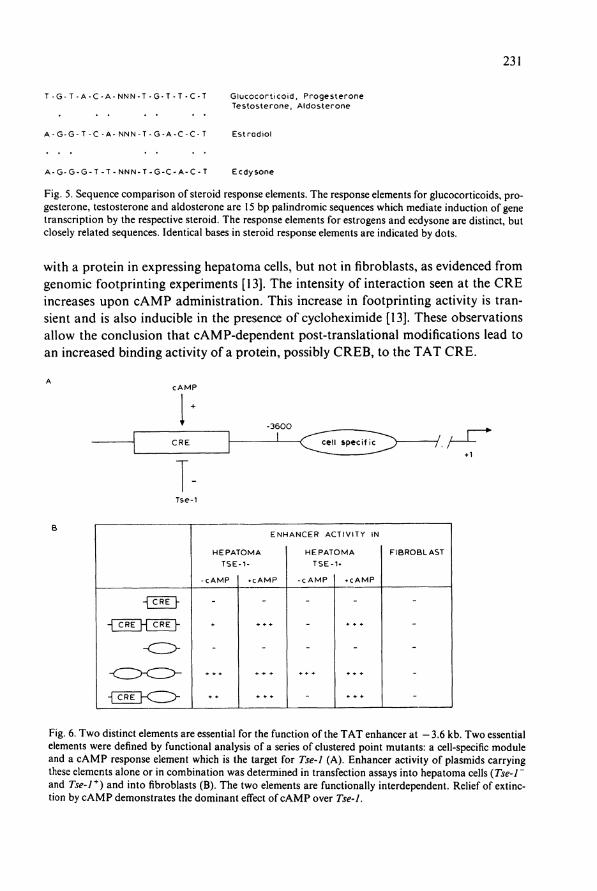
Fig. 5. Sequence comparison of Steroid response elements. The response elements for glucocorticoids, progesterone, testosterone and aldosterone are 15 bp palindromic sequences which mediate induction of gene transcription by the respective Steroid. The response elements for estrogens and ecdysone are distinct, but closely related sequences. Identical bases in Steroid response elements are indicated by dots.
with a protein in expressing hepatoma cells, but not in fibroblasts, as evidenced from genomic footprinting experiments [13]. The intensity of interaction seen at the C R E increases upon c A M P administration. This increase in footprinting activity is tran-sient and is also inducible in the presence of cycloheximide [13]. These observations allow the conclusion that cAMP-dependent post-translational modifications lead to an increased binding activity of a protein, possibly C R E B , to the T A T C R E .
- 3 6 0 0
C R E 1 , C R E < ^ c e i l speci fTT^) /, / L-• — ' +1
E N H A N C E R ACTIVITY IN
H E P A T O M A H E P A T O M A
T S E - 1 - T S E - 1 »
C A M P * c A M P C A M P » c A M P
F I B R O B L A S T
ÜRF}<z>
Fig. 6. Two distinct elements are essential for the function of the T A T enhancer at - 3.6 kb. Two essential elements were defined by functional analysis of a series of clustered point mutants: a cell-specific module and a cAMP response element which is the target for Tse-1 (A). Enhancer activity of plasmids carrying these elements alone or in combination was determined in transfection assays into hepatoma cells (Tse-1 ~ and Tse-1+ ) and into fibroblasts (B). The two elements are functionally interdependent. Relief of extinction by c A M P demonstrates the dominant effect of cAMP over Tse-1.

232
The multimer of the second essential element behaves as a cell type-specific activa-tor of transcription [12]. This element is active in liver cells, but inactive in any other cell type tested so far. In conjunction with the element carrying the C R E sequence, an enhancing element with all the regulatory properties of the T A T gene enhancer can be regenerated (Fig. 6) [12].
5.2. R e v e r s a l of e x t i n c t i o n by c A M P
The c A M P response element of the —3.6 kb enhancer is also a target for negative regulation by Tse-1 [12] (Fig. 6). In a hepatoma microcell hybrid line which contains only a small segment of the human fibroblast chromosome carrying Tse-1 [10] admin-istration of c A M P is able to overcome extinction of expression of the transfected re-porter gene, thus revealing a functional antagonism between Tse-1 and the c A M P signal transduction pathway. In vivo footprinting revealed characteristic changes in D M S reactivity at the C R E . In the presence of a functional Tse-1 locus, protein-D N A interaction at the C R E sequence is abolished. This binding can be recovered by addition of c A M P [12]. Thus, the extinguished State of the T A T gene is character-ized by the absence of binding at the C R E . As might be expected from the relief of extinction by c A M P , this footprint reappears after administration of the inducer.
As hormones acting via the c A M P pathway are thought to be critically involved in turning on T A T gene expression around birth, this suggests that the functional antagonism between Tse-1 and the c A M P pathway might form the basis of a molecu-lar switch governing the onset of T A T gene expression at birth. The strong increase of gluconeogenic hormones around birth may trigger the expression of the T A T gene [1, 2]. This is in line with previous experience that T A T enzyme activity can be prema-turely induced before birth by administration of glucagon in utero [2].
In order to understand the mechanism involved in the cell-specific expression of the T A T gene, and in particular to understand the mode of action of the product of the Tse-1 locus, we are presently isolating and characterizing the proteins involved in recognition of the C R E sequence and the liver cell-specific element. Analysis of possible modifications of the CRE-binding protein in response to c A M P and the tissue-specific extinguisher might give an understanding of the biochemical basis of activation of these Controlling proteins.
6, Summary
Two different signal transduction pathways control the activity of the T A T gene via two inducible enhancers (Fig. 7). These enhancers are composed of constitutive and inducible elements which appear to be crucial for cell-specific and hormone-depend-ent transcription. The effect of glucagon via the c A M P transduction pathway most likely involves specific phosphorylation events [35]. As shown from genomic foot-

233
Glucoco r t i co id - i nduc ib l e enhance r
-2.5
-3.6
~ i T T T n 1 1 1 1 1 F ~ n 1 r kb -12 -11 -10 -9 -8 -7 -6 -5 -4 -3 -2 -1
C e l l - s p e c i f i c enhancer C e l l - s p e c i f i c enhancer
- T s e -1 r e s p o n s i v e - c A M P r e s p o n s i v e
11 lr t ^ 4̂ DNase I HSs
P r o m o t e r
Fig. 7. Several enhancers determine the cell-specific and hormone-dependent activation of the T A T gene. The 5'-flanking region of the T A T gene is outlined with DNasel-hypersensitive regions and enhancers indicated. Shaded and filled arrows point to DNasel-hypersensitive sites observed in T A T gene-expressing hepatoma lines. The filled arrows indicate functionally identified elements; P indicates the promoter. The dependence of the DNasel HS at the G R E on glucocorticoids is indicated by the double arrow.
printing experiments this phosphorylation may be the basis of the increased affinity of a DNA-binding protein recognizing the C R E . This C R E is essential for function of the liver-specific enhancer at —3.6 kb in hepatoma cells and it has to co-operate with a protein recognizing a liver cell-specific module. Since the c A M P response element is a target for the tissue-specific extinguisher it may contribute, in conjunction with a cell-specific enhancer, to the cell-specific and developmentally timed expression of the gene. The enhancer at —3.6 kb is characterized by a constitutive chroma-tin-hypersensitive site, possibly due to the fact that not only the CRE-binding protein determines the active State of this enhancer. The enhancer at — 11 kb is not influenced in its activity by c A M P and glucocorticoids but is liver cell-specific. A D N a sel-hypersensitive region is present in fetal livers, possibly indicating that this distal element may function early in development to establish an activated locus.
The glucocorticoid-dependent enhancer is characterized by a hormone-dependent DNasel-hypersensitive site which is induced by interaction of the glucocorticoid receptor with its binding site (Fig. 7). The glucocorticoid-dependent enhancer is characterized by the presence of two receptor-binding sites plus binding sites for addition-al transcription factors recognizing the C C A A T and the C A C C C motif. This combination of glucocorticoid receptor recognition sequences with transcription factor binding sites might be involved in the cell-specific modulation of activity of the T A T gene. It is most remarkable that the sequences involved in hormonal control

234
function in conjunction with elements which display preferential activity in liver cells. This unique combination may be crucial for the precisely timed onset of expression of the T A T gene at birth in the parenchymal cells of the liver.
References
1. Granner, D.K. and Beale, E .G. (1985) In: Biochemical Actions of Hormones, Vol. XII, (Litwack, G . , ed.), pp. 89-138, Academic Press, New York.
2. Greengard, O. (1970) In: Mechanisms of Hormone Action, Vol. I (Litwack, G. , ed.) pp. 53-85, Academic Press, New York.
3. Ruiz-Bravo, N. and Ernest, M.J. (1982) Proc. Natl. Acad. Sei. USA 79, 365-368. 4. Gluecksohn-Waelsch, S. (1979) Cell 16, 225-237. 5. Ruppert, S., Boshart, M . , Bosch, F.X., Schmid, W., Fournier, R.E.K, and Schütz, G . (1990), Cell
61,895-904. 6. Gluecksohn-Waelsch, S. (1987) Trends Genet. 3, 123-127. 7. Schmid, W., Müller, G. , Schütz, G . and Gluecksohn-Waelsch, S. (1985) Proc. Natl. Acad. Sei. USA
82, 2866-2869. 8. Schneider, J.A. and Weiss, M.C. (1971) Proc. Natl. Acad. Sei. USA 68, 127-131. 9. Killary, A . M . and Fournier, R.E.K. (1984) Cell 38, 523-534.
10. Lern, J., Chin, A .C. , Thayer, M.J. , Leach, R.J. and Fournier, R.E.K. (1988) Proc. Natl. Acad. Sei. USA 85, 7302-7306.
11. Jantzen, H .M. , Strähle, U. , Gloss, B., Stewart, F., Schmid, W., Boshart, M . , Miksicek, R. and Schütz, G.(I987) Cell 49, 29-38.
12. Boshart, M . , Weih, F., Schmidt, A. and Schütz, G . (1990), Cell 61, 905-916. 13. Weih, F., Stewart, F., Boshart, M . , Nitsch, D. and Schütz, G. (1990), Genes Devel. 4, 1437-1449. 14. Trigg, M.J. and Gluecksohn-Waelsch, S. (1973) J. Cell Biol. 58, 549-563. 15. Loose, D.S., Shaw, P.A., Krauter, K.S., Robinson, C , Englard, S., Hanson, R.W. and Gluecksohn-
Waelsch, S. (1986) Proc. Natl. Acad. Sei. USA, 83, 5184-5188. 16. Friedman, A.D. , Landschulz, H.W. and McKnight, S.L. (1989) Genes Dev. 3, 1314^1322. 17. Thayer, M.J. and Fournier, R.E.K. (1989) Mol. Cell. Biol. 9, 2837-2846. 18. Kelsey, G.D., Ruppert, S., Boshart, M . , Schedl, A., Schmid, W. and Schütz, G . (1989) In: Vectors
as Tools for the Study of Normal and Abnormal Growth and Differentiation, N A T O ASI Ser., Vol. H34, pp. 47-62, Springer-Verlag.
19. Nitsch, D., Stewart, A .F . , Boshart, M . , Mestril, R., Weih, F. and Schütz, G. (1990), Mol. Cell Biol. 10, 3334-3342.
20. Becker, P.B., Renkawitz, R. and Schütz, G . (1984) EMBO J. 3, 2015-2020. 21. Grosveld, F., van Assendelft, G.B., Greaves, D.R. and Kollias, G. (1987) Cell 51, 975-985. 22. Evans, R. (1988) Science 240, 889-895. 23. Green, S. and Chambon, P. (1988) Trends Genet. 4, 309-314. 24. Beato,M. (1989) Cell 56, 332-344. 25. Strähle, U. , Schmid, W. and Schütz, G . (1988) EMBO J. 7, 3389-3395. 26. Becker, P.B., Gloss, B., Schmid, W., Strähle, U . and Schütz, G. (1986) Nature 324, 686-688. 27. Strähle, U. , Münsterberg, A., Mestril, R., Klock, G. , Ankenbauer, W., Schmid, W. and Schütz, G .
(1988) Cold Spring Harbor Symp. Quant. Biol. 53, 835-841. 28. Schmid, W., Strähle, U. , Schütz, G. , Schmitt, J. and Stunnenberg, H. (1989) E M B O J. 8, 2257-2263. 29. Strähle, U . , Klock, G . and Schütz, G . (1987) Proc. Natl. Acad. Sei. USA 84, 7871-7875. 30. Strähle, U . , Boshart, M . , Klock, G. , Stewart, F. and Schütz, G . (1989) Nature 339, 629-632. 31. Klock, G. , Strähle, U . and Schütz, G . (1987) Nature 329, 734^736. 32. Mader, S., Kumar, U. , de Verneuil, H. and Chambon, P. (1989) Nature 338, 271-274. 33. Karin, M . (1989) TIG 5, 65-67. 34. Roesler, W.J., Vandenbark, G.R. and Hanson, R.W. (1988) J Biol. Chem. 263, 9063-9066. 35. Gonzalez, G.A. and Montminy, M.R. (1989) Cell 59, 675-680.


