Molecular and Phenotypic Evaluation of Lichtheimia corymbifera
9
JOURNAL OF CLINICAL MICROBIOLOGY, Dec. 2009, p. 3862–3870 Vol. 47, No. 12 0095-1137/09/$12.00 doi:10.1128/JCM.02094-08 Copyright © 2009, American Society for Microbiology. All Rights Reserved. Molecular and Phenotypic Evaluation of Lichtheimia corymbifera (Formerly Absidia corymbifera) Complex Isolates Associated with Human Mucormycosis: Rehabilitation of L. ramosa Dea Garcia-Hermoso, 1 Damien Hoinard, 1 Jean-Charles Gantier, 1 Fre ´de ´ric Grenouillet, 2 Franc ¸oise Dromer, 1 and Eric Dannaoui 1,3 * Institut Pasteur, Unite ´ de Mycologie Mole ´culaire, Centre National de Re ´fe ´rence Mycologie et Antifongiques, and CNRS URA3012, Paris 75724 Cedex 15, France 1 ; De ´partement de Mycologie-Parasitologie, CHU Jean Minjoz, Besanc ¸on 25030, France 2 ; and Universite ´ Paris Descartes, Faculte ´ de Me ´decine, AP-HP, Ho ˆpital Europe ´en Georges Pompidou, Unite ´ de Parasitologie-Mycologie, Paris 75015, France 3 Received 31 October 2008/Returned for modification 15 January 2009/Accepted 8 September 2009 Thirty-eight isolates (including 28 isolates from patients) morphologically identified as Lichtheimia corym- bifera (formerly Absidia corymbifera) were studied by sequence analysis (analysis of the internal transcribed spacer [ITS] region of the ribosomal DNA, the D1-D2 region of 28S, and a portion of the elongation factor 1 [EF-1] gene). Phenotypic characteristics, including morphology, antifungal susceptibility, and carbohydrate assimilation, were also determined. Analysis of the three loci uncovered two well-delimited clades. The maximum sequence similarity values between isolates from both clades were 66, 95, and 93% for the ITS, 28S, and EF-1 loci, respectively, with differences in the lengths of the ITS sequences being detected (763 to 770 bp for isolates of clade 1 versus 841 to 865 bp for isolates of clade 2). Morphologically, the shapes and the sizes of the sporangiospores were significantly different among the isolates from both clades. On the basis of the molecular and morphological data, we considered isolates of clade 2 to belong to a different species named Lichtheimia ramosa because reference strains CBS 269.65 and CBS 270.65 (which initially belonged to Absidia ramosa) clustered within this clade. As neotype A. corymbifera strain CBS 429.75 belongs to clade 1, the name L. corymbifera was conserved for clade 1 isolates. Of note, the amphotericin B MICs were significantly lower for L. ramosa than for L. corymbifera (P < 0.005) but were always <0.5 g/ml for both species. Among the isolates tested, the assimilation of melezitose was positive for 67% of the L. ramosa isolates and negative for all L. corymbifera isolates. In conclusion, this study reveals that two Lichtheimia species are commonly associated with mucormycosis in humans. Mucormycosis is a life-threatening infection that occurs in immunocompromised patients, diabetic patients with keto- acidosis, and immunocompetent patients after trauma expo- sure to contaminated soil (7, 18). The filamentous fungi responsible for these infections belong to the Mucorales order. About 20 different species have been shown to be pathogenic for humans (4). According to a recent review (19), the species that were the most frequent encountered were Rhizopus spp., Mucor spp., and Cunninghamella spp., while Apophysomyces elegans and Absidia spp. accounted for 6% and 5% of the cases, respectively. The true frequency is, however, difficult to assess because surveys are rare and determination of the species of the Zygomycetes class by standard mycological methods remains difficult. Indeed, all the genera and species within the family Mucoraceae (the Absidia, Rhizopus, Mucor, Rhizomucor, and Apophysomyces genera) shared similar morphological characteristics (6). The precise identification to the species level often requires the specific expertise usually available only at reference laboratories. The availability of molecular tools for taxo- nomic and identification purposes has changed the picture. Sequencing of various DNA targets has facilitated the rec- ognition of phylogenetic species within the Zygomycetes (27, 28) and provided tools for DNA bar coding of these fungi (22). A revision of the genus Absidia was recently performed on the basis of phylogenetic, physiological, and morpholog- ical characteristics (10). A new family (Mycocladiaceae) and the genus Mycocladus were proposed to accommodate the three species Mycocladus corymbifer (formerly Absidia corymbifera), M. blakesleeana, and M. hyalospora. More re- cently, it was suggested that additional nomenclatural changes were necessary, and the names Lichtheimiaceae and Lichtheimia were proposed for the family and the genus, respectively (11). The intraspecific variability of Lichtheimia corymbifera (formerly A. corymbifera) has been poorly evaluated so far. After the analysis of a small number of clinical isolates, we recently reported that some of the isolates morphologically identified as L. corymbifera had divergent internal transcribe spacer (ITS) sequences (21). Subsequently, the use of mo- lecular identification on a routine basis for all isolates of the Zygomycetes collected at the French National Reference Center for Mycoses and Antifungals allowed us to uncover intraspecific sequence variability among isolates morpholog- ically identified to be L. corymbifera. To further characterize the atypical isolates, we used three different DNA targets, * Corresponding author. Mailing address: Institut Pasteur, Unite ´ de Mycologie Mole ´culaire, Centre National de Re ´fe ´rence Mycologie et Antifongiques, and CNRS URA3012, 25, rue du Roux, Paris 75724 Cedex 15, France. Phone: 33 1 40 61 32 50. Fax: 33 1 45 68 84 20. E-mail: [email protected]. Published ahead of print on 16 September 2009. 3862 on April 12, 2019 by guest http://jcm.asm.org/ Downloaded from
Transcript of Molecular and Phenotypic Evaluation of Lichtheimia corymbifera

JOURNAL OF CLINICAL MICROBIOLOGY, Dec. 2009, p. 3862–3870 Vol. 47, No. 120095-1137/09/$12.00 doi:10.1128/JCM.02094-08Copyright © 2009, American Society for Microbiology. All Rights Reserved.
Molecular and Phenotypic Evaluation of Lichtheimia corymbifera(Formerly Absidia corymbifera) Complex Isolates Associated
with Human Mucormycosis: Rehabilitation of L. ramosa�
Dea Garcia-Hermoso,1 Damien Hoinard,1 Jean-Charles Gantier,1 Frederic Grenouillet,2Francoise Dromer,1 and Eric Dannaoui1,3*
Institut Pasteur, Unite de Mycologie Moleculaire, Centre National de Reference Mycologie et Antifongiques, and CNRS URA3012,Paris 75724 Cedex 15, France1; Departement de Mycologie-Parasitologie, CHU Jean Minjoz, Besancon 25030, France2; and
Universite Paris Descartes, Faculte de Medecine, AP-HP, Hopital Europeen Georges Pompidou, Unite deParasitologie-Mycologie, Paris 75015, France3
Received 31 October 2008/Returned for modification 15 January 2009/Accepted 8 September 2009
Thirty-eight isolates (including 28 isolates from patients) morphologically identified as Lichtheimia corym-bifera (formerly Absidia corymbifera) were studied by sequence analysis (analysis of the internal transcribedspacer [ITS] region of the ribosomal DNA, the D1-D2 region of 28S, and a portion of the elongation factor 1�[EF-1�] gene). Phenotypic characteristics, including morphology, antifungal susceptibility, and carbohydrateassimilation, were also determined. Analysis of the three loci uncovered two well-delimited clades. Themaximum sequence similarity values between isolates from both clades were 66, 95, and 93% for the ITS, 28S,and EF-1� loci, respectively, with differences in the lengths of the ITS sequences being detected (763 to 770 bpfor isolates of clade 1 versus 841 to 865 bp for isolates of clade 2). Morphologically, the shapes and the sizesof the sporangiospores were significantly different among the isolates from both clades. On the basis of themolecular and morphological data, we considered isolates of clade 2 to belong to a different species namedLichtheimia ramosa because reference strains CBS 269.65 and CBS 270.65 (which initially belonged to Absidiaramosa) clustered within this clade. As neotype A. corymbifera strain CBS 429.75 belongs to clade 1, the nameL. corymbifera was conserved for clade 1 isolates. Of note, the amphotericin B MICs were significantly lowerfor L. ramosa than for L. corymbifera (P < 0.005) but were always <0.5 �g/ml for both species. Among theisolates tested, the assimilation of melezitose was positive for 67% of the L. ramosa isolates and negative for allL. corymbifera isolates. In conclusion, this study reveals that two Lichtheimia species are commonly associatedwith mucormycosis in humans.
Mucormycosis is a life-threatening infection that occurs inimmunocompromised patients, diabetic patients with keto-acidosis, and immunocompetent patients after trauma expo-sure to contaminated soil (7, 18). The filamentous fungiresponsible for these infections belong to the Mucoralesorder. About 20 different species have been shown to bepathogenic for humans (4). According to a recent review(19), the species that were the most frequent encounteredwere Rhizopus spp., Mucor spp., and Cunninghamella spp.,while Apophysomyces elegans and Absidia spp. accounted for6% and 5% of the cases, respectively. The true frequency is,however, difficult to assess because surveys are rare anddetermination of the species of the Zygomycetes class bystandard mycological methods remains difficult. Indeed, allthe genera and species within the family Mucoraceae (theAbsidia, Rhizopus, Mucor, Rhizomucor, and Apophysomycesgenera) shared similar morphological characteristics (6).The precise identification to the species level often requiresthe specific expertise usually available only at referencelaboratories. The availability of molecular tools for taxo-
nomic and identification purposes has changed the picture.Sequencing of various DNA targets has facilitated the rec-ognition of phylogenetic species within the Zygomycetes (27,28) and provided tools for DNA bar coding of these fungi(22). A revision of the genus Absidia was recently performedon the basis of phylogenetic, physiological, and morpholog-ical characteristics (10). A new family (Mycocladiaceae) andthe genus Mycocladus were proposed to accommodate thethree species Mycocladus corymbifer (formerly Absidiacorymbifera), M. blakesleeana, and M. hyalospora. More re-cently, it was suggested that additional nomenclaturalchanges were necessary, and the names Lichtheimiaceae andLichtheimia were proposed for the family and the genus,respectively (11).
The intraspecific variability of Lichtheimia corymbifera(formerly A. corymbifera) has been poorly evaluated so far.After the analysis of a small number of clinical isolates, werecently reported that some of the isolates morphologicallyidentified as L. corymbifera had divergent internal transcribespacer (ITS) sequences (21). Subsequently, the use of mo-lecular identification on a routine basis for all isolates of theZygomycetes collected at the French National ReferenceCenter for Mycoses and Antifungals allowed us to uncoverintraspecific sequence variability among isolates morpholog-ically identified to be L. corymbifera. To further characterizethe atypical isolates, we used three different DNA targets,
* Corresponding author. Mailing address: Institut Pasteur, Unite deMycologie Moleculaire, Centre National de Reference Mycologie etAntifongiques, and CNRS URA3012, 25, rue du Roux, Paris 75724Cedex 15, France. Phone: 33 1 40 61 32 50. Fax: 33 1 45 68 84 20.E-mail: [email protected].
� Published ahead of print on 16 September 2009.
3862
on April 12, 2019 by guest
http://jcm.asm
.org/D
ownloaded from
which allowed us to confirm that L. corymbifera is a speciescomplex.
MATERIALS AND METHODS
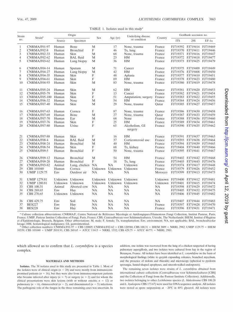
Isolates. The 38 isolates used in this study are presented in Table 1. Most ofthe isolates were of clinical origin (n � 28) and were mostly from immunocom-promised patients (n � 16), but they were also from immunocompetent patientswho became infected after injury (n � 7) or surgery (n � 2) and for whom theclinical presentations were skin lesions (with or without osteitis; n � 12) orpulmonary (n � 6), rhinocerebral (n � 2), and disseminated (n � 5) infections.The pathogenic role of the fungus in the three remaining cases was uncertain. In
addition, one isolate was recovered from the lung of a chicken suspected of havingpulmonary aspergillosis, and two isolates were cultured from hay in the region ofBesancon, France. All isolates have been identified as L. corymbifera on the basis ofmorphological findings (white to greyish expanding colonies, branched mycelium,and the presence of stolons and rhizoids) and microscopy (spherical to pyriformsporangia, funnel-shaped apophyses, and smooth-walled endospores).
The remaining seven isolates were strains of L. corymbifera obtained frominternational culture collections (Centraalbureau voor Schimmelcultures [CBS]and the Collection of Fungi from the Pasteur Institute Collection). Additionally,two isolates belonging to other Lichtheimia species (L. blakesleeana CBS 100.28and L. hyalospora CBS 173.67) were used for DNA sequence analysis. All isolateswere stored as spore suspensions at �20°C in 40% glycerol. All isolates were
TABLE 1. Isolates used in this studya
Strainno. Strainb
OriginSex Age (yr) Underlying disease
or condition CountryGenBank accession no.
Source Specimen ITS 28S EF-1�
1 CNRMA/F01-97 Human Bone M 17 None, trauma France FJ719392 FJ719414 FJ7194692 CNRMA/F02-8 Human Bronchial F 46 Tx, lung France FJ719370 FJ719411 FJ7194463 CNRMA/F02-33 Human Skin M 68 None, trauma France FJ719371 FJ719416 FJ7194474 CNRMA/F02-62 Human BAL fluid M 23 HM France FJ719372 FJ719418 FJ7194775 CNRMA/F03-62 Human Lung biopsy M 36 HM France FJ719373 FJ719425 FJ719479
6 CNRMA/F04-14 Human Sputum M 71 Cancer France FJ719375 FJ719409 FJ7194497 CNRMA/F04-27 Human Lung biopsy M 42 HM France FJ719376 FJ719408 FJ7194508 CNRMA/F04-35 Human Skin F 48 Aplasia France FJ719377 FJ719410 FJ7194519 CNRMA/F04-61 Human Skin F 69 HM France FJ719378 FJ719423 FJ71948010 CNRMA/F04-93 Human Skin M 83 None, trauma France FJ719380 FJ719419 FJ719478
11 CNRMA/F05-24 Human Skin M 62 HM France FJ719381 FJ719420 FJ71945312 CNRMA/F05-79 Human Skin F 13 Cancer France FJ719382 FJ719421 FJ71945413 CNRMA/F05-100 Human Skin F 51 Amputation, surgery France FJ719383 FJ719422 FJ71945514 CNRMA/F06-32 Human Nose M 54 HM France FJ719384 FJ719424 FJ71945615 CNRMA/F07-40 Human Skin M 29 None, trauma Qatar FJ719385 FJ719427 FJ719457
16 CNRMA/F07-63 Human Cornea F 87 None, trauma France FJ719386 FJ719430 FJ71945817 CNRMA/F07-69 Human Bone M 27 None, trauma Qatar FJ719387 FJ719433 FJ71945918 CNRMA/F07-70 Human Ear M 68 None France FJ719388 FJ719436 FJ71946019 CNRMA/F07-76 Human Skin F 25 HM France FJ719389 FJ719434 FJ71946120 CNRMA/F07-80 Human Urine F 65 Alcoholism, GI
surgeryFrance FJ719390 FJ719435 FJ719462
21 CNRMA/F07-88 Human Skin F 16 HM France FJ719391 FJ719437 FJ71946322 CNRMA/F08-4 Human BAL fluid M 37 Corticosteroid use France FJ719393 FJ719438 FJ71946423 CNRMA/F08-24 Human Bronchial M 40 HM France FJ719394 FJ719439 FJ71946524 CNRMA/F08-54 Human Skin F 68 Tx, kidney France FJ719404 FJ719440 FJ71946625 CNRMA/F09-5 Human Bronchial F 50 None, trauma France FJ719395 FJ719441 FJ719467
26 CNRMA/F09-12 Human Bronchial M 54 HM France FJ719402 FJ719442 FJ71946827 CNRMA/F09-20 Human Bronchial F 18 Tx, lung France FJ719403 FJ719443 FJ71947628 CNRMA/F03-82 Animal Lung, chicken NA NA Unknown France FJ719374 FJ719417 FJ71944829 CBS 101040 Human Cornea Unknown Unknown Unknown France FJ719379 FJ719426 FJ71945230 UMIP 1129.75 Env Outdoor air NA NA NA Morocco FJ719399 FJ719413 FJ719473
31 UMIP 1279.81 Unknown Unknown Unknown Unknown Unknown Unknown FJ719400 FJ719412 FJ71948132 UMIP 1280.81 Unknown Unknown Unknown Unknown Unknown Unknown FJ719401 FJ719415 FJ71947433 CBS 100.31 Animal Aborted cow NA NA NA NA FJ719398 FJ719429 FJ71947234 CBS 269.65 Env Hay NA NA NA NA FJ719405 FJ719432 FJ71947535 CBS 270.65 Unknown Unknown NA NA NA NA FJ719406 FJ719445 FJ719482
36 CBS 429.75 Env Soil NA NA NA NA FJ719407 FJ719444 FJ71948337 BES227 Env Hay NA NA NA France FJ719397 FJ719428 FJ71947038 BES228 Env Hay NA NA NA France FJ719396 FJ719431 FJ719471
a Culture collection abbreviations: CNRMA/F, Centre National de Référence Mycologie et Antifongiques-Filamentous Fungi Collection, Institut Pasteur, Paris,France; UMIP, Pasteur Institut Collection of Fungi, Paris, France; CBS, Centraalbureau voor Schimmelcultures, Utrecht, The Netherlands; IHEM, Institut d’Hygieneet d’Epidémiologie, Brussels, Belgium. Other abbreviations: M, male; F, female; Tx, transplantation; Env, environment; NA, not applicable; BAL, bronchoalveolarlavage; HM, hematological malignancy; GI, gastrointestinal.
b Other collection numbers: CNRMA/F01-97 � CBS 120805; CNRMA/F02-62 � CBS 120580; CBS 100.31 � IHEM 3809 � NRRL 2982; UMIP 1129.75 � IHEM10339; CBS 101040 � UMIP 2018.91; CBS 269.65 � ATCC 11613 � NRRL 1332; CBS 429.75 � ATCC 46771 � NRRL 2981.
VOL. 47, 2009 LICHTHEIMIA CORYMBIFERA COMPLEX 3863
on April 12, 2019 by guest
http://jcm.asm
.org/D
ownloaded from

subcultured for 3 to 6 days on 2% malt extract agar (MEA) at 30°C for macro-scopic and microscopic examination.
Molecular study. (i) Extraction, amplification, and sequencing. Mycelium wasgrown in 20 ml of RPMI 1640 medium with L-glutamine but without sodiumbicarbonate (Sigma-Aldrich, Saint Quentin Fallavier, France) buffered to pH 7.0with 0.165 M morpholinepropanesulfonic acid (Sigma-Aldrich). After 48 h ofcontinuous agitation (100 rpm) at 30°C, the mycelium was recovered, washedtwice with a 0.9% NaCl solution, and stored at �20°C until extraction.
Genomic DNA extraction was performed as described previously (22) withapproximately 200 mg of mycelium, and the DNA was stored at �20°C. Thecomplete ITS1-5.8S-ITS2 region of the ribosomal DNA (rDNA) was amplifiedwith primer pair V9D (5�-TTAAGTCCCTGCCCTTTGTA-3�) and LS266 (5�-GCATTCCCAAACAACTCGACTC-3�) (9). The D1-D2 region of the large-subunit rDNA was amplified with primer pair NL-1 (5�-GCATATCAATAAGCGGAGGAAAAG-3�) and NL-4 (5�-GGTCCGTGTTTCAAGACGG-3�) (14).A small region of the 5� elongation factor 1� (EF-1�) nuclear gene was amplifiedwith primers MEF-11 (5�-AAGAAGATTGGTTTCAACCC-3�) and MEF-41(5�-GCACCGATTTGACCAGGRTGG-3�) (17).
The PCR amplification of ITS and 28S was done as described previously (22)in an iCycler thermocycler (Bio-Rad, Hercules, CA). For the amplification withprimers MEF-11 and MEF-41, the PCR mixture (50 �l) contained 3 �l of theextracted genomic DNA, 1� PCR buffer (Roche Diagnostics GmbH, Mann-heim, Germany), 3 mM MgCl2, 0.25 �M of each primer, 0.25 mM of eachdeoxynucleoside triphosphate (Roche), and 1.25 U of AmpliTaq DNA polymer-ase (Roche). The PCR conditions were predenaturation at 94°C for 5 min; 40cycles at 95°C for 30 s, 52°C for 30 s, and 72°C for 1 min; and a final incubationat 72°C for 7 min. The PCR products were then sequenced at the Institut Pasteursequencing facility by using a BigDye Terminator (version 1.1) kit (AppliedBiosystems, Foster City, CA) and each of the primer pairs used for amplificationon an ABI Prism 3730 XL DNA analyzer (Applied Biosystems).
(ii) Sequence analysis. A consensus sequence was computed from the forwardand reverse sequences by using the ChromasPro program (version 1.33;Technelysium, Helensvale, Queensland, Australia), and multiple-sequence align-ments were performed with the Clustal W program (26). The analysis treatedgaps (indels) as a fifth state character. To determine the percentage of identicalresidues between each pair of sequences, identity matrices for each set of datawere generated with BioEdit software (Isis Therapeutics, Carlsbad, CA). Thepercent similarity represents the number of identical sites divided by the lengthof the longest sequence (sites at which a gap was present in both sequences wereremoved). Single-locus cladograms were constructed by the neighbor-joiningmethod with the pairwise-deletion option (20) in the MEGA (version 3.1) com-puter program (13). A combined three-locus analysis was also performed. Rhi-zomucor pusillus (CNRMA/F09-7) was chosen as the outgroup, and the robust-ness of the branches was assessed by bootstrap analysis with 1,000 replicates.
Carbon source assimilation profiles. Carbon source assimilation profiles weredetermined with a commercial kit (ID32C system; bioMerieux, Marcy, l’Etoile,France), as described previously (23). Briefly, isolates were cultured for 7 days onSabouraud agar slants at 30°C to obtain sufficient sporulation. The spores weretransferred to API C medium (bioMerieux) to achieve a final concentration of5 � 105 spores/ml, and 135 �l was distributed into each well. The results wereread visually after 72 h of incubation at 30°C. Weak growth was consideredpositive. A functional analysis by use of an agglomerative clustering method (byuse of the unweighted-pair group method with arithmetic mean algorithm) wasperformed with BioloMICS (Biological Manager for Identification, Classificationand Statistics) software (version 7.2.5; BioAware, Hannut, Belgium) to group theisolates and the carbon assimilation results at the same time.
In vitro susceptibility testing. All isolates were subcultured on Sabourauddextrose agar (supplemented with 0.02% chloramphenicol) prior to testing toensure purity and viability. Pure powders of known potency of amphotericin B(Sigma-Aldrich), voriconazole (Pfizer Central Research, Sandwich, United King-dom), itraconazole (Janssen-Cilag, Issy-les-Moulineaux, France), posaconazole(Schering-Plough Research Institute, Kenilworth, NJ), flucytosine (Sigma-Al-drich), terbinafine (Novartis Pharma AG, Basel, Switzerland), caspofungin(Merck & Co., Inc., Rahway, NJ), and micafungin (Astellas Pharma, Osaka,Japan) were used. In vitro susceptibility was determined by a broth microdilutiontechnique, according to the guidelines of the Antifungal Susceptibility TestingSubcommittee of the European Committee on Antibiotic Susceptibility Testingfor the testing of conidium-forming molds (24), but with some modifications.Briefly, microplates containing the eight antifungal drugs were prepared inbatches and stored frozen at �20°C. The final concentrations were 0.125 to 64mg/liter for flucytosine and 0.015 to 8 mg/liter for all other drugs. Testing wasperformed in RPMI 1640 medium supplemented with 2% glucose for all drugsexcept amphotericin B, which was tested in AM3 medium, with a final inoculum
size of 105 CFU/ml. MIC endpoints were determined on an automated micro-plate reader spectrophotometer (Multiscan RC-351; Labsystems Oy, Helsinki,Finland) after 24 h or 48 h of incubation (an optical density of �0.15 wasrequired for the drug-free control wells) at 35°C. The MIC endpoint was definedas a reduction in growth of 80% or more compared to the amount of growth inthe drug-free well for all drugs except amphotericin B, for which an endpoint ofa 90% reduction was used. Two reference strains, Candida krusei ATCC 6258and C. parapsilosis ATCC 22019, were included in each set of determinations toensure quality control.
Morphological study. A detailed morphological study was performed with 11isolates (5 isolates randomly chosen from each clade [see below] plus isolateCNRMA/F05-100). Isolates were cultured on MEA at 30°C, and the macroscopicmorphology was described after 3 to 4 days of incubation. Microscopic exami-nation was done with cultures grown for 5 to 9 days after they were mounted inwater with 1% gelatin. The different structures (sporangia, columellae, andsporangiospores) were examined. The sporangiospores were measured with aDM LB2 optical microscope (Leica Microsystemes SAS, Rueil-Malmaison,France) with interferential contrast and a Leica D5000 microscope coupled withLeica Application Suite software, which comprises the Multifocus and the In-teractive Measurement modules (precision, 0.01 �m). For each isolate, approx-imately 100 sporangiospores were measured, and the ratio between the lengthand the width was calculated.
Statistical analysis. The distributions of the MICs were compared by a non-parametric test (Mann-Whitney). The mean spore length, width, and length/width ratio were calculated for the L. corymbifera isolates (n � 613) and the L.ramosa isolates (n � 601) and were compared by an unpaired t test. Analyseswere performed with Prism (version 3.00) software for Windows (GraphPadSoftware, San Diego, CA). Statistical significance was defined as a P value of�0.05.
RESULTS
Molecular data. The sequences of the whole ITS1-5.8S-ITS2region, the D1-D2 domain of 28S, and a partial region of theEF-1� gene were determined for the 38 isolates (total length,approximately 68,000 bp). For the ITS locus, the sequences(starting at the ITS1 primer position and ending at the ITS4primer position) ranged from 741 to 865 nucleotides in length:613 to 617 nucleotides for the 28S D1-D2 domain and 439nucleotides for the EF-1� locus. Two well-delimited cladeswere obtained with all of the single-locus distance trees gen-erated (Fig. 1 to 3) and with the tree obtained when the threeloci were combined (Fig. 4). As all individual isolates weregrouped together in one clade for each locus, it was possible toconsider the two clades two different species, in accordancewith the principles of the genealogical concordance of phylo-genetic species recognition (25).
Analysis of the ITS data matrix revealed a high degree ofnucleotide sequence similarity (more than 98%) within clade 1,with the exception of that for isolate CNRMA/F05-100, whichshowed nucleotide sequence differences of more than 20%with the sequences of the other clade 1 isolates. Within clade2, the maximum difference was observed between the subgroupconsisting of isolates CNRMA/F02-8 and CNRMA/F04-35 andthe rest of the clade 2 isolates (91.5 to 93.5% similarity). Thesizes of the ITS sequences differed between the two clades (763to 770 bp and 841 to 865 bp for clade 1 and clade 2, respec-tively). Greater than 99% similarity within the clade 1 se-quences and only small variations (�2%) within the clade 2sequences were observed when the 28S domain sequenceswere analyzed, and differences of less than 2% were observedwithin each clade when the EF-1� locus was analyzed (Table2). The highest degree of sequence variability between clade 1and clade 2 was observed for the ITS locus (34%, 5%, and 7%
3864 GARCIA-HERMOSO ET AL. J. CLIN. MICROBIOL.
on April 12, 2019 by guest
http://jcm.asm
.org/D
ownloaded from

variability for the ITS, 28S, and EF-1� gene regions, respec-tively).
Clade 1 corresponded to Lichtheimia corymbifera because itincluded neotype strain Absidia corymbifera CBS 429.75. Clade2 isolates were designated L. ramosa because reference isolatesCBS 269.65 and CBS 270.65 (which initially belonged to thespecies Absidia ramosa) (8) clustered within this clade.
Comparative morphology of L. corymbifera and L. ramosaisolates. After 3 to 4 days of incubation on MEA, colonies ofall isolates were expanding but differences in the growth pat-terns were observed. The L. corymbifera isolates exhibitedcompact growth, while the L. ramosa isolates had a more effusemycelium. No significant differences in the morphologies of the
sporangia and columellae or the branching patterns of thesporangiophores were observed between the two species (Fig.5). The sporangiospores of the L. corymbifera isolates weresmooth and hyaline, whereas those of the L. ramosa isolateswere smooth but slightly colored. More importantly, the spo-rangiospores of the L. corymbifera isolates were ellipsoid (2.73by 2.24 �m), while those of the L. ramosa isolates were longellipsoid (3.06 by 2.18 �m) (Fig. 5), with significant (P 0.0001) differences in terms of length, width, and the length/width ratio (1.41 versus 1.22, respectively) being observed.
Comparison of other phenotypic characteristics between thetwo species. Melezitose and palatinose were assimilated by67% and 33% of the L. ramosa isolates, respectively, while
FIG. 1. Neighbor-joining analysis based on the complete sequences of ITS1-5.8S-ITS2. Bootstrap values from 1,000 replicates are indicated atthe nodes separating the two clades. Rhizomucor pusillus was used as the outgroup. The bar indicates the number of substitutions per site. *,neotype strain of Absidia corymbifera; **, reference strains of Absidia ramosa.
VOL. 47, 2009 LICHTHEIMIA CORYMBIFERA COMPLEX 3865
on April 12, 2019 by guest
http://jcm.asm
.org/D
ownloaded from

none of the L. corymbifera isolates tested assimilated those twocarbon sources (Table 3). There were no additional differencesin carbon source assimilation profiles that could discriminatebetween the two species.
The susceptibilities of the clinical isolates to eight antifungaldrugs were determined. All isolates exhibited high flucytosineMICs (�64 �g/ml), caspofungin MICs (�8 �g/ml), and mica-fungin MICs (�8 �g/ml); and all but one isolate had a highvoriconazole MIC (�8 �g/ml). Differences in the itraconazoleMICs (range, 0.25 to 16 �g/ml), posaconazole MICs (range,0.125 to 4 �g/ml), and terbinafine MICs (range, 0.125 to 2�g/ml) were observed among the isolates; but there were nosignificant differences by species. A significant difference in theamphotericin B MIC distribution was observed between thetwo species (0.125 to 0.5 �g/ml for the L. corymbifera isolatesversus 0.03 to 0.25 �g/ml for the L. ramosa isolates; P 0.005).
It should be noted, however, that the MIC50 differed by only 2log2 dilutions.
Finally, there was no difference between the two species interms of the underlying diseases of the patients from whomthey were recovered (hematological malignancies, solid cancer,organ transplantation, or a lack of immunosuppression) or theclinical presentations that they caused (cutaneous, pulmonary,and disseminated infections).
The main molecular and phenotypic characteristics that dif-ferentiated the L. ramosa isolates from the L. corymbiferaisolates are presented in Table 3.
DISCUSSION
Recently, a revision of the genus Absidia on the basis of thephylogenetic, physiological, and morphological characteristics
FIG. 2. Neighbor-joining analysis based on partial 28S sequences. Bootstrap values from 1,000 replicates are indicated at the nodes separatingthe two clades. Rhizomucor pusillus was used as the outgroup. The bar indicates the number of substitutions per site. *, neotype strain of Absidiacorymbifera; ** reference strains of Absidia ramosa.
3866 GARCIA-HERMOSO ET AL. J. CLIN. MICROBIOL.
on April 12, 2019 by guest
http://jcm.asm
.org/D
ownloaded from

of 16 species was conducted (10), and nomenclatural changeswere proposed (11). The three thermotolerant Absidia species(A. corymbifera, A. blakesleeana, and A. hyalospora) are nowclassified in the genus Lichtheimia. L. corymbifera was the onlyspecies pathogenic for humans. Although L. corymbifera isreported to be responsible for only 5% of the human cases ofzygomycosis (19), this figure should be considered with cautionbecause of a lack of surveys and because identifications aremostly based on morphology (12). The use of molecular iden-tification (2) will be important for an accurate assessment ofthe epidemiology.
Phylogenetic species recognition in the Mucorales order isperformed by sequencing rDNA genes (18S, 28S, and ITS), aswell as the actin and EF-1� genes (10, 17, 27, 28). For identi-fication (DNA bar coding) of this group of fungi, ITS is a good
molecular target (22). The recent guidelines published by theClinical and Laboratory Standards Institute (3) recommendthe use of ITS sequencing as a first-line method for the iden-tification of species within the Mucorales, an approach that wasfurther approved by another international consortium of ex-perts (1). Our routine use of ITS sequencing for the molecularidentification of filamentous fungi allowed us to notice thatsome isolates morphologically identified as L. corymbifera haddivergent ITS sequences (21). To further characterize theseisolates, two other loci (28S and EF-1�) were sequenced for allthe isolates initially identified as L. corymbifera. On the basis ofthose data, the morphospecies L. corymbifera appeared to be aspecies complex that included at least two clades. Due to thelow level of sequence similarity (maximums, 66, 95, and 93%for ITS, 28S, and EF-1�, respectively) between the two clades
FIG. 3. Neighbor-joining analysis based on partial EF-1� sequences. Bootstrap values from 1,000 replicates are indicated at the nodesseparating the two clades. Rhizomucor pusillus was used as the outgroup. The bar indicates the number of substitutions per site. *, neotype strainof Absidia corymbifera; **, reference strains of Absidia ramosa.
VOL. 47, 2009 LICHTHEIMIA CORYMBIFERA COMPLEX 3867
on April 12, 2019 by guest
http://jcm.asm
.org/D
ownloaded from
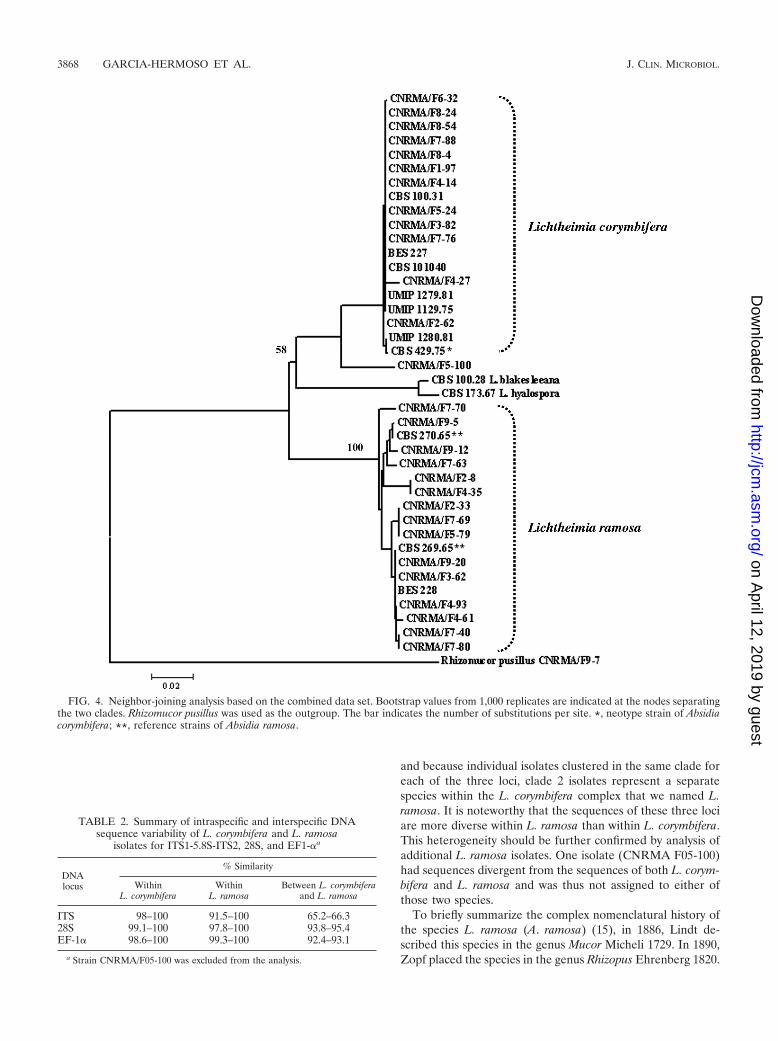
and because individual isolates clustered in the same clade foreach of the three loci, clade 2 isolates represent a separatespecies within the L. corymbifera complex that we named L.ramosa. It is noteworthy that the sequences of these three lociare more diverse within L. ramosa than within L. corymbifera.This heterogeneity should be further confirmed by analysis ofadditional L. ramosa isolates. One isolate (CNRMA F05-100)had sequences divergent from the sequences of both L. corym-bifera and L. ramosa and was thus not assigned to either ofthose two species.
To briefly summarize the complex nomenclatural history ofthe species L. ramosa (A. ramosa) (15), in 1886, Lindt de-scribed this species in the genus Mucor Micheli 1729. In 1890,Zopf placed the species in the genus Rhizopus Ehrenberg 1820.
FIG. 4. Neighbor-joining analysis based on the combined data set. Bootstrap values from 1,000 replicates are indicated at the nodes separatingthe two clades. Rhizomucor pusillus was used as the outgroup. The bar indicates the number of substitutions per site. *, neotype strain of Absidiacorymbifera; **, reference strains of Absidia ramosa.
TABLE 2. Summary of intraspecific and interspecific DNAsequence variability of L. corymbifera and L. ramosa
isolates for ITS1-5.8S-ITS2, 28S, and EF1-�a
DNAlocus
% Similarity
WithinL. corymbifera
WithinL. ramosa
Between L. corymbiferaand L. ramosa
ITS 98–100 91.5–100 65.2–66.328S 99.1–100 97.8–100 93.8–95.4EF-1� 98.6–100 99.3–100 92.4–93.1
a Strain CNRMA/F05-100 was excluded from the analysis.
3868 GARCIA-HERMOSO ET AL. J. CLIN. MICROBIOL.
on April 12, 2019 by guest
http://jcm.asm
.org/D
ownloaded from

In 1903, Vuillemin described the genus Lichtheimia, whichcomprised the type species L. corymbifera (29) and L. ramosa.In 1908, however, Lendner placed both species in the genusAbsidia Van Thieghem 1876. Despite the morphological dif-ferences underlined by Ellis and Hesseltine (8), subsequentstudies proved this distinction difficult and A. ramosa was re-duced to being synonymous with A. corymbifera (16).
Both species infect humans and cannot be differentiated interms of the hosts that they infect or the types of disease thatthey cause. L. corymbifera and L. ramosa are very similar bothmacroscopically and microscopically, but some differences thatdelineate the two species were uncovered. First, by culturingisolates on MEA plates at 30°C for 3 to 4 days, compact growth
characterizes L. corymbifera, while more effuse growth is sug-gestive of L. ramosa. The sporangiospores of L. corymbifera aresmooth, hyaline, and ellipsoidal when they are mature, whilethose of L. ramosa are smooth, lightly colored, and more el-lipsoidal, a finding consistent with the earlier description byEllis and Hesseltine (8). Carbohydrate assimilation can beused for Zygomycetes identification (23) but was not useful forthe differentiation of L. corymbifera from L. ramosa. Indeed,only if palatinose or melezitose assimilation were positivecould we suspect the species to be L. ramosa. Likewise, theantifungal susceptibility profiles were undistinguishable be-tween the two Lichtheimia species, whereas they can be used todistinguish some species within other genera (5).
The results of the present study clearly show that molecular,biological, and morphological characteristics support the sep-aration of the two species, even if their detection by classicalmethods remains difficult. In conclusion, L. ramosa representsa species distinct from L. corymbifera and is thus another Li-chtheimia species responsible for mucormycosis in humans.
ACKNOWLEDGMENTS
We are grateful to Monique Coutanson and Bernard Papierok fromthe Pasteur Institute Collection of Fungi for providing some of thereference strains. Other clinical isolates were studied as part as anationwide survey of invasive mycosis in France. Members of theFrench Mycoses Study Group who sent isolates used in this study wereas follows (in alphabetical order by city in France): C. Duhamel(Caen), D. Pons (Clermont-Ferrand), E. Forget (Clichy), F. Dalle(Dijon), B. Sendid (Lille), F. de Monbrison (Lyon), F. Gay-Andrieu(Nantes), M. Gari-Toussaint (Nice), C. Lacroix (Paris), C. Kauffmann-Lacroix (Poitiers), D. Toubas (Reims), P. Cahen (Suresnes), and F.
FIG. 5. Morphological characteristics of L. corymbifera isolates (CNRMA/F08-54) (A and B) and L. ramosa isolates (CNRMA/F05-79) (C andD). (A and C) Sporangia. Magnifications, �400. (B and D) Sporangiospores. Magnifications, �1,000.
TABLE 3. Main molecular, morphological, and physiologicalcharacteristics of L. corymbifera and L. ramosa isolatesa
Characteristic L. corymbifera L. ramosa
ITS sequence length (bp)b 763–770 841–865Growth Compact EffuseSporangiospores Ellipsoid Long ellipsoidAssimilation of melezitose (% of
isolates)0 67
% of isolates for which amphotericinB MIC is �0.125 �g/ml
20 87
a The numbers of isolates used for determination of these characteristics were10 for growth and sporangiospore size, 37 for ITS sequence length and assimi-lation profile, and 26 for antifungal susceptibility testing. Strain CNRMA/F05-100 was excluded from the analysis.
b The number of base pairs of the region located between primers ITS1 andITS4.
VOL. 47, 2009 LICHTHEIMIA CORYMBIFERA COMPLEX 3869
on April 12, 2019 by guest
http://jcm.asm
.org/D
ownloaded from
Benaoudia (Troyes). We are also grateful to Saad J. Taj-Aldeen(Doha, Qatar) for sharing some clinical isolates and to Gabriel Rebouxfor providing two environmental isolates. We thank Laure Diancourtand Coralie Tran from the Institut Pasteur sequencing program fortechnical help.
We thank the Institut Pasteur sequencing program for financialsupport (Genopole PF-8).
REFERENCES
1. Balajee, S. A., A. M. Borman, M. E. Brandt, J. Cano, M. Cuenca-Estrella, E.Dannaoui, J. Guarro, G. Haase, C. C. Kibbler, W. Meyer, K. O’Donnell,C. A. Petti, J. L. Rodriguez-Tudela, D. Sutton, A. Velegraki, and B. L.Wickes. 2009. Sequence-based identification of Aspergillus, Fusarium, andMucorales in the clinical mycology laboratory: where are we and whereshould we go from here? J. Clin. Microbiol. 47:877–884.
2. Balajee, S. A., L. Sigler, and M. E. Brandt. 2007. DNA and the classical way:identification of medically important molds in the 21st century. Med. Mycol.45:475–490.
3. Clinical and Laboratory Standards Institute. 2008. Interpretive criteria formicroorganism identification of bacteria and fungi by DNA target sequenc-ing; approved guideline. Document MM18-A. Clinical and Laboratory Stan-dards Institute, Wayne, PA.
4. Dannaoui, E., and D. Garcia-Hermoso. 2007. The Zygomycetes, p. 159–183.In K. Kavanagh (ed.), New insights in fungal pathogenicity. Springer Science,Dordrecht, The Netherlands.
5. Dannaoui, E., J. Meletiadis, J. W. Mouton, J. F. Meis, and P. E. Verweij.2003. In vitro susceptibilities of Zygomycetes to conventional and new anti-fungals. J. Antimicrob. Chemother. 51:45–52.
6. de Hoog, G. S., and G. Guarro (ed.). 1995. Atlas of clinical fungi. Centraal-bureau voor Schimmelcultures, Baarn, The Netherlands.
7. Dromer, F., and M. R. McGinnis. 2002. Zygomycosis, p. 297–308. In E.Anaissie, M. R. McGinnis, and M. A. Pfaller (ed.), Clinical mycology.Churchill Livingstone, New York, NY.
8. Ellis, J. J., and C. W. Hesseltine. 1966. Species of Absidia with ovoid spo-rangiospores. II. Sabouraudia 5:59–77.
9. Gerrits van den Ende, A. H. G., and G. S. de Hoog. 1999. Variability andmolecular diagnostics of the neurotropic species Cladophialophora bantiana.Stud. Mycol. 43:151–162.
10. Hoffmann, K., S. Discher, and K. Voigt. 2007. Revision of the genus Absidia(Mucorales, Zygomycetes) based on physiological, phylogenetic, and mor-phological characters; thermotolerant Absidia spp. form a coherent group,Mycocladiaceae fam. nov. Mycol. Res. 111:1169–1183.
11. Hoffmann, K., G. Walther, and K. Voigt. 2009. Mycocladus vs. Lichtheimia:a correction (Lichtheimiaceae fam. nov., Mucorales, Mucoromycotina). My-col. Res. 113:277–278.
12. Kontoyiannis, D. P., M. S. Lionakis, R. E. Lewis, G. Chamilos, M. Healy, C.Perego, A. Safdar, H. Kantarjian, R. Champlin, T. J. Walsh, and I. I. Raad.2005. Zygomycosis in a tertiary-care cancer center in the era of Aspergillus-active antifungal therapy: a case-control observational study of 27 recentcases. J. Infect. Dis. 191:1350–1360.
13. Kumar, S., K. Tamura, and M. Nei. 2004. MEGA3: integrated software formolecular evolutionary genetics analysis and sequence alignment. Brief.Bioinform. 5:150–163.
14. Kurtzman, C. P., and C. J. Robnett. 1997. Identification of clinically impor-tant ascomycetous yeasts based on nucleotide divergence in the 5� end of thelarge-subunit (26S) ribosomal DNA gene. J. Clin. Microbiol. 35:1216–1223.
15. Naumov, N. A. 1939. Cles des Mucorinees (Mucorales), Encyclopedie my-cologigue, vol. IX. (P. Lechevalier [ed.]) Paris, France.
16. Nottebrock, H., H. J. Scholer, and M. Wall. 1974. Taxonomy and identifi-cation of mucormycosis-causing fungi. I. Synonymity of Absidia ramosa withA. corymbifera. Sabouraudia 12:64–74.
17. O’Donnell, K., F. Lutzoni, T. J. Ward, and G. L. Benny. 2001. Evolutionaryrelationships among mucoralean fungi (Zygomycota): evidence for familypolyphyly on a large scale. Mycologia 93:286–296.
18. Ribes, J. A., C. L. Vanover-Sams, and D. J. Baker. 2000. Zygomycetes inhuman disease. Clin. Microbiol. Rev. 13:236–301.
19. Roden, M. M., T. E. Zaoutis, W. L. Buchanan, T. A. Knudsen, T. A.Sarkisova, R. L. Schaufele, M. Sein, T. Sein, C. C. Chiou, J. H. Chu, D. P.Kontoyiannis, and T. J. Walsh. 2005. Epidemiology and outcome of zygo-mycosis: a review of 929 reported cases. Clin. Infect. Dis. 41:634–653.
20. Saitou, N., and M. Nei. 1987. The neighbor-joining method: a new methodfor reconstructing phylogenetic trees. Mol. Biol. Evol. 4:406–425.
21. Schwarz, P., S. Bretagne, A. S. Delannoy, F. Dromer, O. Lortholary, and E.Dannaoui. 2005. Sequence-based identification of zygomycetes species ofmedical interest. Clin. Microbiol. Infect. 11(Suppl. 2):478.
22. Schwarz, P., S. Bretagne, J. C. Gantier, D. Garcia-Hermoso, O. Lortholary,F. Dromer, and E. Dannaoui. 2006. Molecular identification of zygomycetesfrom culture and experimentally infected tissues. J. Clin. Microbiol. 44:340–349.
23. Schwarz, P., O. Lortholary, F. Dromer, and E. Dannaoui. 2007. Carbonassimilation profiles as a tool for identification of zygomycetes. J. Clin.Microbiol. 45:1433–1439.
24. Subcommittee on Antifungal Susceptibility Testing (AFST) of the ESCMIDEuropean Committee for Antimicrobial Susceptibility Testing (EUCAST),J. L. Rodriguez-Tudela, M. C. Arendrup, S. Arikan, F. Barchiesi, J. Bille, E.Chryssanthou, M. Cuenca-Estrella, E. Dannaoui, D. W. Denning, J. P.Donnelly, W. Fegeler, C. Lass-Florl, C. Moore, M. Richardson, P. Gaustad,A. Schmalreck, A. Velegraki, and P. Verweij. 2008. EUCAST technical noteon the method for the determination of broth dilution minimum inhibitoryconcentrations of antifungal agents for conidia-forming moulds. Clin. Mi-crobiol. Infect. 14:982–984.
25. Taylor, J. W., D. J. Jacobson, S. Kroken, T. Kasuga, D. M. Geiser, D. S.Hibbett, and M. C. Fisher. 2000. Phylogenetic species recognition and spe-cies concepts in fungi. Fungal Genet. Biol. 31:21–32.
26. Thompson, J. D., D. G. Higgins, and T. J. Gibson. 1994. CLUSTAL W:improving the sensitivity of progressive multiple sequence alignment throughsequence weighting, position-specific gap penalties and weight matrix choice.Nucleic Acids Res. 22:4673–4680.
27. Voigt, K., E. Cigelnik, and K. O’Donnell. 1999. Phylogeny and PCR identi-fication of clinically important Zygomycetes based on nuclear ribosomal-DNA sequence data. J. Clin. Microbiol. 37:3957–3964.
28. Voigt, K., and J. Wostemeyer. 2001. Phylogeny and origin of 82 zygomycetesfrom all 54 genera of the Mucorales and Mortierellales based on combinedanalysis of actin and translation elongation factor EF-1alpha genes. Gene270:113–120.
29. Vuillemin, P. 1903. Le genre Tieghemella et la serie de Absidiees. Bull. Soc.Mycol. Fr. 19:119–127.
3870 GARCIA-HERMOSO ET AL. J. CLIN. MICROBIOL.
on April 12, 2019 by guest
http://jcm.asm
.org/D
ownloaded from