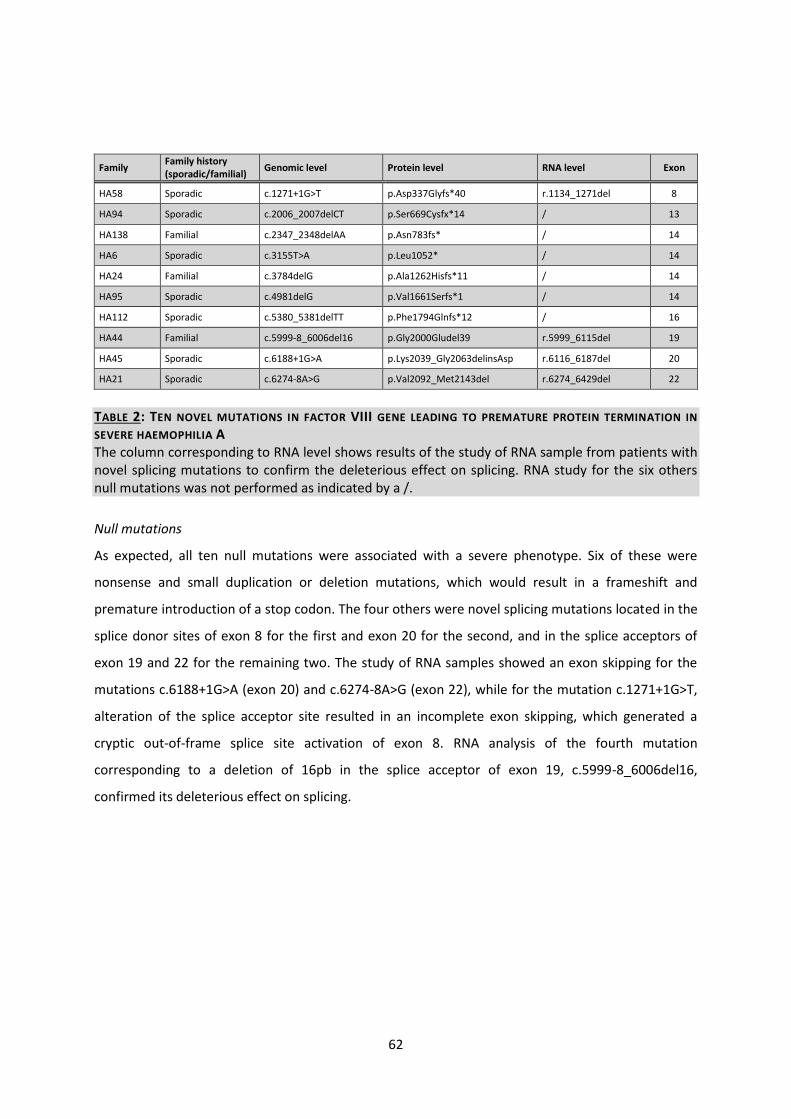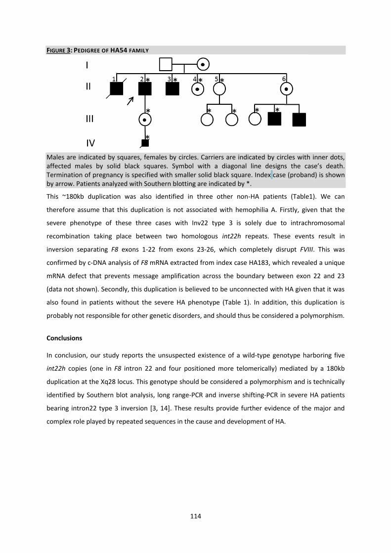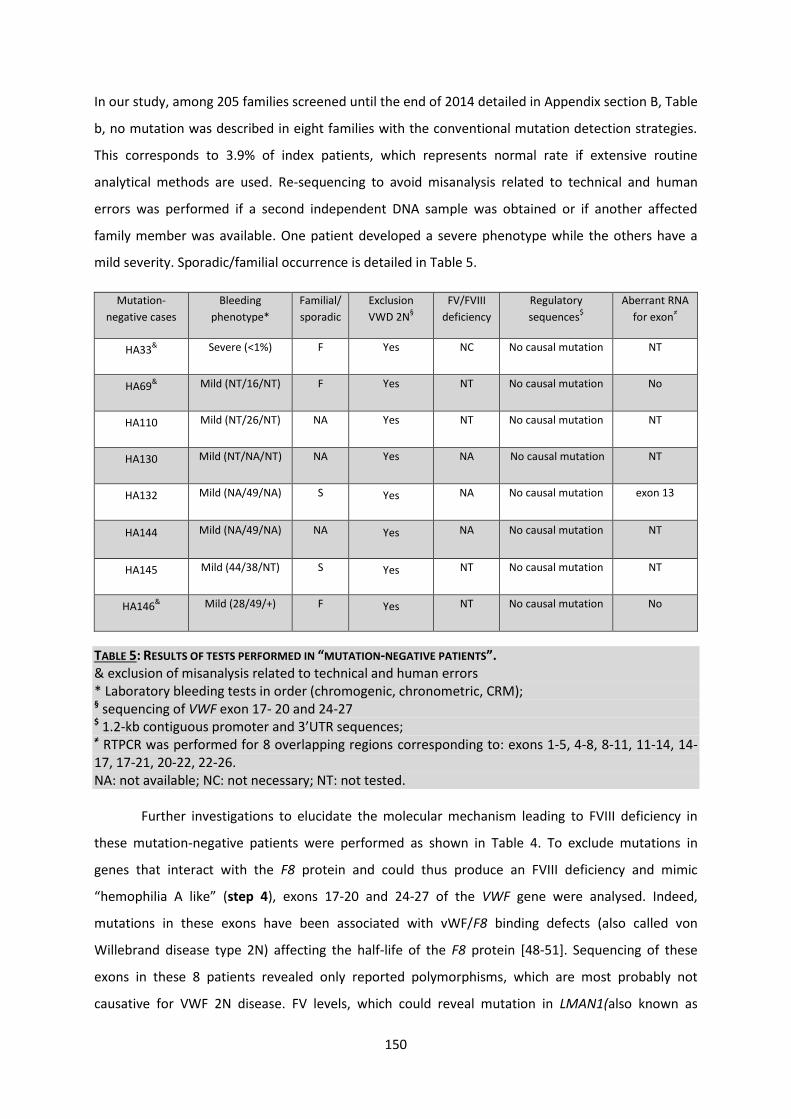
Molecular analysis of simple and complex genetic variants ...
219
UNIVERSITE CATHOLIQUE DE LOUVAIN (UCL) INSTITUTE OF EXPERIMENTAL AND CLINICAL RESEARCH (IREC) SERVICE D’HÉMATOLOGIE, CLINIQUES UNIVERSITAIRE SAINT-LUC CENTRE DE GÉNÉTIQUE HUMAINE, CLINIQUES UNIVERSITAIRE SAINT-LUC Molecular analysis of simple and complex genetic variants in a cohort of patients with hemophilia A: Mechanisms and diagnostic implications Nathalie Lannoy December 2015 Thesis submitted in fulfilment of the requirements for the degree of «Docteur en sciences biomédicales et pharmaceutiques» Promoter : Professor Cédric Hermans Co-promoter : Professor Miikka Vikkula
Transcript of Molecular analysis of simple and complex genetic variants ...

UNIVERSITE CATHOLIQUE DE LOUVAIN (UCL)
INSTITUTE OF EXPERIMENTAL AND CLINICAL RESEARCH (IREC)
SERVICE DrsquoHEacuteMATOLOGIE CLINIQUES UNIVERSITAIRE SAINT-LUC
CENTRE DE GEacuteNEacuteTIQUE HUMAINE CLINIQUES UNIVERSITAIRE SAINT-LUC
Molecular analysis of simple and complex genetic variants in a cohort of patients with hemophilia A
Mechanisms and diagnostic implications
Nathalie Lannoy
December 2015
Thesis submitted in fulfilment of the requirements for the degree of laquoDocteur en sciences biomeacutedicales et pharmaceutiquesraquo
Promoter Professor Ceacutedric Hermans Co-promoter Professor Miikka Vikkula
Jury members
Chair
Prof Luc Bertrand Universiteacute catholique de Louvain (UCLouvain) Institute of Experimental and Clinical Research (IREC) Pole of cardiovascular research
Promoter
Prof Ceacutedric Hermans Universiteacute catholique de Louvain (UCLouvain) Cliniques universitaires Saint-Luc Uniteacute de Thrombose et Heacutemostase Service drsquoheacutematologie Bruxelles Belgique
Co-Promoter
Prof Miikka Vikkula Universiteacute catholique de Louvain de Duve Institute Laboratory of Human Molecular Genetics Bruxelles Belgique
Members of the examination committee
Prof Jean-Noeumll Octave Universiteacute catholique de Louvain (UCLouvain) Institute Of NeuroScience (IoNS) Pocircle Cellulaire et moleacuteculaire
Prof Christiane Vermylen Universiteacute catholique de Louvain Cliniques universitaires Saint-Luc Service dheacutematologie et oncologie peacutediatrique Bruxelles Belgique
Prof Laurent Knoops Universiteacute catholique de Louvain Cliniques universitaires Saint-Luc Service dheacutematologie adulte Bruxelles Belgique
Prof Christine Vinciguerra Universiteacute Claude Bernard Groupement hospitalier Edouard Herriot Service drsquoheacutematologie biologique Lyon France
Summary
Haemophilia A (HA) is caused by widespread mutations in the factor VIII gene Although genetic
alterations responsible for HA can now be identified in a vast majority of patients molecular
diagnostic remains challenging in some patients This thesis addresses important issues in the
diagnostic and molecular mechanisms of some frequent or rare genetic alterations associated with
HA It demonstrates the value of computational and molecular approaches for predicting the
causality of unreported missense mutations It also shows that the overrepresentation of specific
missense mutations in mild HA can be explained by a founder effect rather than by an independent
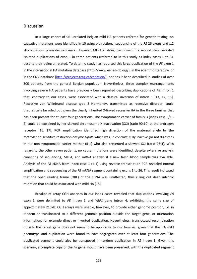
occurrence Also it highlights how complex genetic rearrangements such as tandem inversion
duplication within intron 1 and exons 1-22 duplications involving intron 22 homologous repeats
(int22h) can be detected and their occurrence best explained Finally it demonstrates for the first
time the existence of wild-type human genotype harbouring five int22h copies identified in the rare
intron22 inversion type 3 of the factor VIII gene
Keywords Hemophilia A F8 mutation analysis duplication breakpoints intron 22 inversion Copy Number Variation genotyping CGH array MLPA int22h homologous repeats
Reacutesumeacute
Lheacutemophilie A (HA) est due agrave des mutations du gegravene F8 codant pour le facteur VIII de la coagulation
Bien que les mutations geacuteneacutetiques responsables de lHA puissent deacutesormais ecirctre identifieacutees pour la
majoriteacute des patients le diagnostic moleacuteculaire reste probleacutematique chez certains sujets atteints
Cette thegravese aborde les techniques drsquoidentification et drsquointerpreacutetation de plusieurs deacutefauts geacuteneacutetiques
concernant quelques nucleacuteotides ou incluant de larges remaniements Elle propose des strateacutegies
bio-informatiques et moleacuteculaires pour eacutevaluer la causaliteacute de mutations ponctuelles nouvellement
deacutecrites Elle montre eacutegalement que la surrepreacutesentation des quelques mutations faux-sens peut
ecirctre expliqueacutee par un effet fondateur plutocirct que par un eacuteveacutenement de novo reacutecurrent Ensuite elle
suggegravere des hypothegraveses pour expliquer les meacutecanismes moleacuteculaires de certains reacutearrangements
geacuteneacutetiques complexes tels qursquoune duplication en miroir pour la duplication de lrsquoexon 1 et une
participation de deux seacutequences homologues int22h pour la duplication des exons 1 agrave 22
Finalement elle montre pour la premiegravere fois qursquoil existe des geacutenotypes humains normaux contenant
cinq copies de seacutequences homologues int22h qui sont techniquement visibles dans un type rare de
lrsquoinversion de lrsquointron 22
Mots-cleacutes Heacutemophilie A analyse moleacuteculaire gegravene F8 inversion de lrsquointron 22 copies homologues int22h duplication puce dhybridation geacutenomique comparative geacutenotypage variabiliteacute du nombre de copies MLPA
Remerciements
Une thegravese est loin drsquoecirctre un long fleuve tranquille Pourtant agrave lrsquoeacutecriture de ces quelques lignes je
nrsquoai aucun regret de cette aventure magnifique passionnante et particuliegraverement enrichissante tant
sur le plan humain que celui de la connaissance
Je suis aussi convaincue que cette thegravese est loin decirctre un travail solitaire Jamais je naurais pu
reacutealiser ce travail doctoral sans le soutien dun grand nombre de personnes dont la geacuteneacuterositeacute la
bonne humeur et linteacuterecirct manifesteacutes agrave leacutegard de mes recherches mont permis de progresser dans
cette phase deacutelicate du laquochercheurraquo En espeacuterant nrsquooublier personne je tiens agrave remercier
Mon promoteur le Professeur Ceacutedric Hermans Service de Thrombose et Heacutemostase des cliniques
universitaires Saint-Luc
Pas facile pour un heacutematologue de comprendre le langage et les meacuteandres de la geacuteneacutetique Pourtant
tu nrsquoas pas heacutesiteacute agrave mrsquoaccorder ta confiance Mille mercis drsquoavoir accepteacute ce challenge et cette
aventure Mille mercis aussi pour ton enseignement ta rigueur scientifique ta disponibiliteacute ta
patience ton enthousiasme et surtout pour tes qualiteacutes humaines drsquoeacutecoute et de compreacutehension
tout au long de ce travail doctoral De cette expeacuterience jrsquoai appris eacutenormeacutement en espeacuterant qursquoelle
sera un tremplin pour un nouveau futur plus serain et plus respectueux
Mon co-promoteur le Professeur Miikka Vikkula laboratoire de geacuteneacutetique moleacuteculaire humaine
de lrsquoInstitut de Duve
Vous avez eacuteteacute agrave la fois mon promoteur et mon chef de service pendant deux ans A mes yeux et agrave
ceux de beaucoup drsquoautres vous ecirctes lrsquoexemple de la reacuteussite dans ce domaine exigeant qursquoest le
monde acadeacutemique et la ʺRechercheʺ Merci de mrsquoavoir fait partager votre expertise dans des
entrevues privileacutegieacutees deacutenicheacutees dans votre agenda plus que chargeacute
Les Membres du Jury
Jrsquoexprime ma gratitude au Professeur Luc Bertrand drsquoavoir preacutesideacute le jury de mon comiteacute
drsquoencadrement Vous avez toujours eacuteteacute prompt et disponible pour reacutepondre agrave mes questions et mes
doutes Il en va de mecircme pour le Professeur Christiane Vermylen le Professeur Jean-Noeumll Octave
le Professeur Laurent Knoops et le Professeur Alexandre Persu drsquoavoir accepteacute de faire partie de ce
comiteacute et drsquoavoir suggeacutereacute maints conseils lors de nos reacuteunions En effet vos critiques et vos
reacuteflexions eacuteclaireacutees ont largement contribueacute agrave ameacuteliorer ma thegravese en vue de sa publication Je
remercie aussi chaleureusement le Professeur Christine Vinciguerra drsquoavoir fait le voyage jusque
dans notre Belgique et drsquoavoir accepteacute de relire cette thegravese Jrsquoai appreacutecieacute vos remarques et
commentaires Jrsquoespegravere que lrsquoavenir nous permettra de nous revoir et de participer agrave des projets
communs
Un encadrement scientifique est indispensable pour lrsquoaboutissement drsquoune thegravese de doctorat
Pourtant une structure technique est tout aussi primordiale pour son bon deacuteroulement et sa
reacutealisation Le preacutesent travail est le fruit drsquoune collaboration eacutetroite avec de nombreux chercheurs et
techniciens de diffeacuterents laboratoires qursquoil me tient agrave cœur de remercier
Ainsi mes premiers remerciements vont naturellement et prioritairement agrave Isabelle Abinet Pendant
plus de 28 ans nous avons travailleacute ensemble Ce qui fait un paquet drsquoanalyses effectueacutees de mises
au point drsquoinnovations techniques de tregraves bons moments et drsquoautres plus sombres Jrsquoai appreacutecieacute
tout au long de ces anneacutees ta rigueur de travail ta conscience pofessionelle ton esprit critique et
ton expertise
Je remercie ensuite Arnaud Bosmans Meacutelanie Vast Jean-Luc Guerin et Vincent Kayndaszyk pour
leur aide technique Marie Ravoet et Claude Bandelier pour leur soutien dans les phases deacutelicates de
la soumission drsquoarticles Je ne voudrais certainement pas oublier Bernard Grisart du centre de
geacuteneacutetique de lrsquoIPG Ton enthousiasme et tes connaissances autour des laquo duplications raquo ont permis la
publication de plusieurs articles Merci agrave Vinciane Dideberg Ceacutecile Libioulle et au docteur Steacutephanie
Gaillez du centre de geacuteneacutetique de Liegravege au docteur Catheline Vilain du centre de geacuteneacutetique de lrsquoULB
aux docteurs Elisabeth Ronge-Collard Jean-Marc Minon Marie-Franccediloise Dresse et Michel
Reginster de mrsquoavoir fait confiance en mrsquoenvoyant des preacutelegravevements de patients agrave analyser
Je remercie eacutegalement chaleureusement toutes les personnes du service drsquoHeacutematologie des cliniques
universitaires Saint-Luc les docteurs Catherine Lambert et Sophie Dupont Eva Devricis Dominique
Pothen et Dominique Verkissen de mrsquoavoir soutenue tout au long de ces anneacutees de thegravese sans
oublier aussi les patients et leurs familles Sans eux cette thegravese nrsquoaurait pu aboutir
Je nrsquooublie pas tous les theacutesards mes camarades de galegravere inscrits eux aussi en thegravese Laurent
Pitance Seacutebastien Lobet Damien Lederer Seacuteverine Henrard et Xavier Pepermans Je nrsquoai qursquoune
phrase agrave leur dire ʺIl faut lrsquoavoir veacutecu pour savoir ce que ccedila repreacutesenteʺ
Ce travail naurait pu ecirctre meneacute agrave bien sans laide de diffeacuterents financeurs qui au travers de leur
soutien mateacuteriel ont reconnu mon travail et mrsquoont fait confiance Je remercie ainsi les socieacuteteacutes
Pfizer Bayer Baxalta et la Fondation Saint-Luc
Mes remerciements vont enfin agrave ma famille et mes amis qui avec cette question reacutecurrente laquoquand
est-ce que tu la soutiens cette thegraveseraquo mrsquoont permis de ne jamais deacutevier de mon objectif final Merci
agrave mes parents leur preacutesence et leurs encouragements sont pour moi les piliers fondateurs de ce que
je suis et de ce que je fais Merci agrave ma sœur Pascale et son mari Thierry mon fregravere Pierre-Paul et sa
femme Anne-Catherine mes nombreux oncles tantes neveux et niegraveces cousins et cousines Merci
aussi agrave Pierre-Jean Doye et Marc Deckers drsquoavoir eacuteteacute les confidents et teacutemoins de freacutequentes
peacuteriodes de doute
Pour terminer je remercie mes trois filles Florence Caroline Virginie et mon cher mari Patrick Je
ne pourrais jamais assez les remercier eux qui mrsquoont soutenue remotiveacutee durant ces anneacutees et qui
ont accepteacute les sacrifices neacutecessaires agrave la reacutealisation de ce travail doctoral
Mille mercis agrave vous tous
ʺNotre richesse collective est faite de notre diversiteacute Lrsquoʺautreʺ individu ou socieacuteteacute nous est preacutecieux dans la mesure ougrave il nous est indispensableʺ
Albert Jacquard ELOGE DE LA DIFFERENCE La geacuteneacutetique et les hommes
ʺ Faites que le recircve deacutevore votre vie afin que la vie ne deacutevore pas votre recircve rdquo
Antoine de Saint-Exupeacutery
Table of contents
Introduction 1
Part one General introduction 4
Part two Mechanisms and consequences of mutations Principles of genetic variations and molecular diseases Application to haemophilia A 21
Aim of the study and experimental strategy 41
Results 45
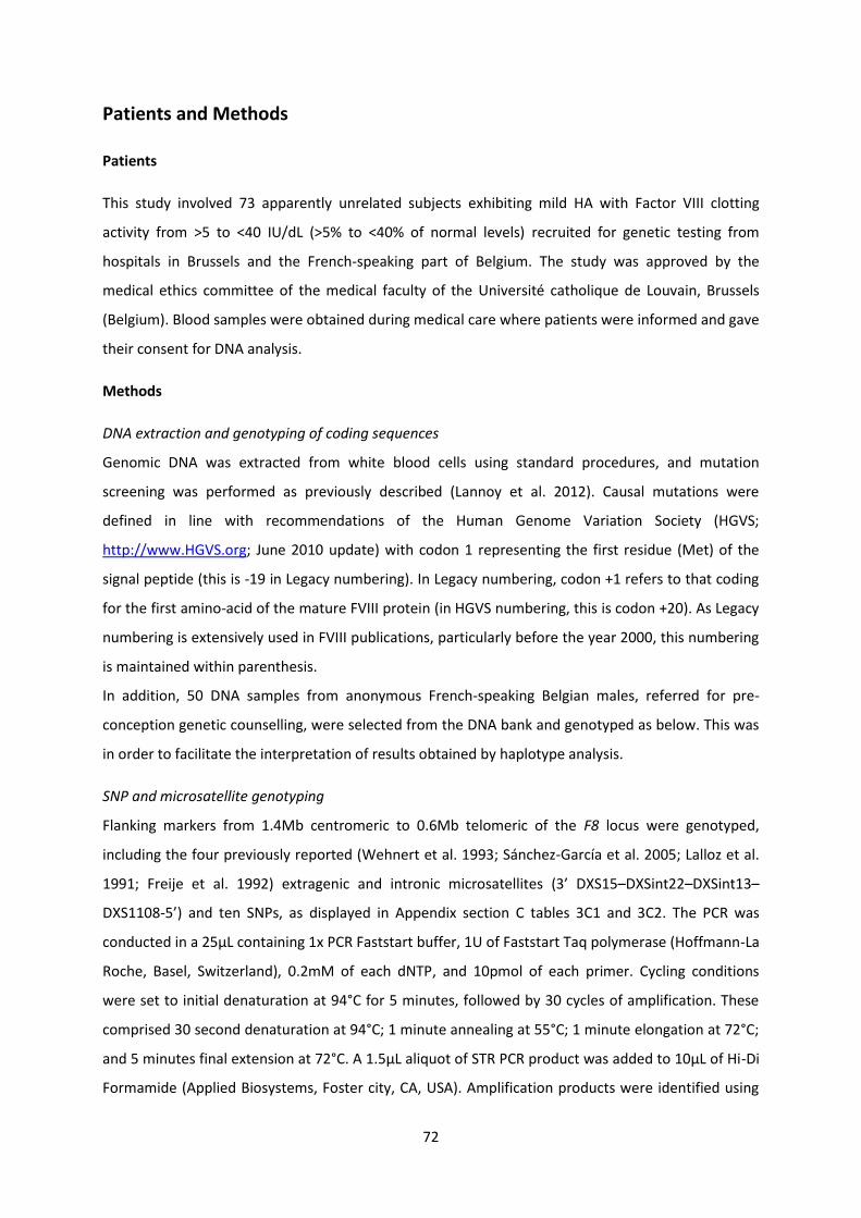
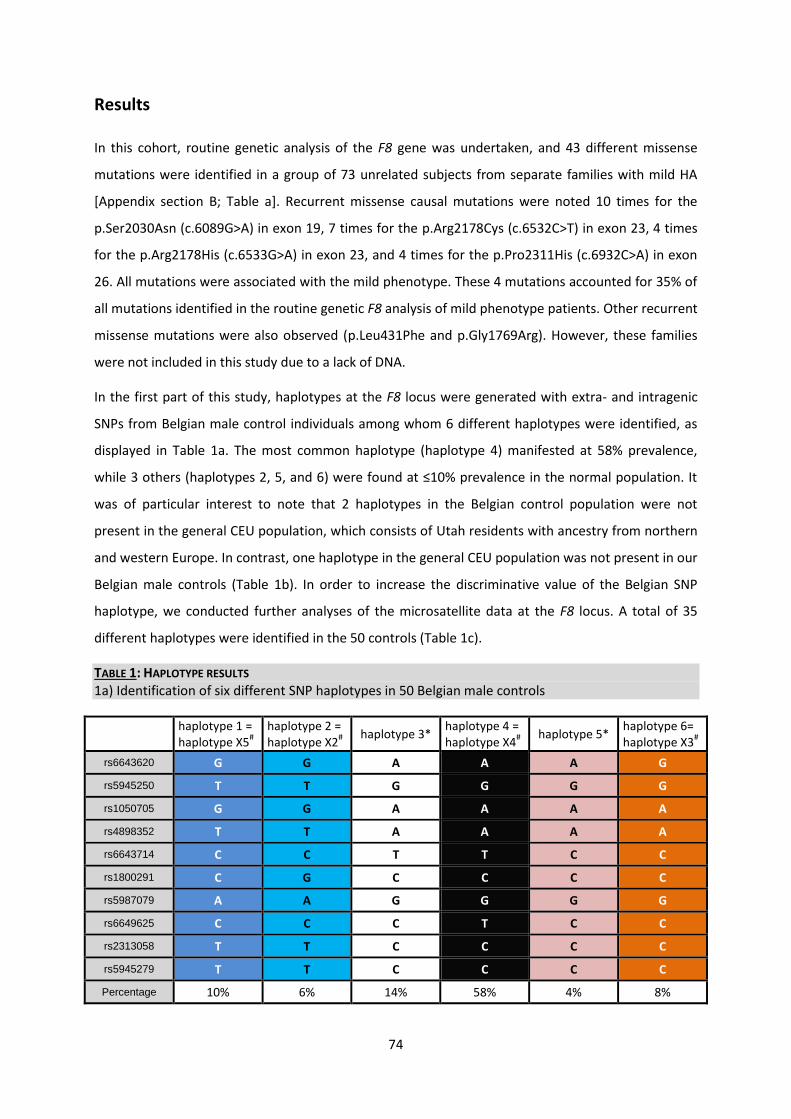
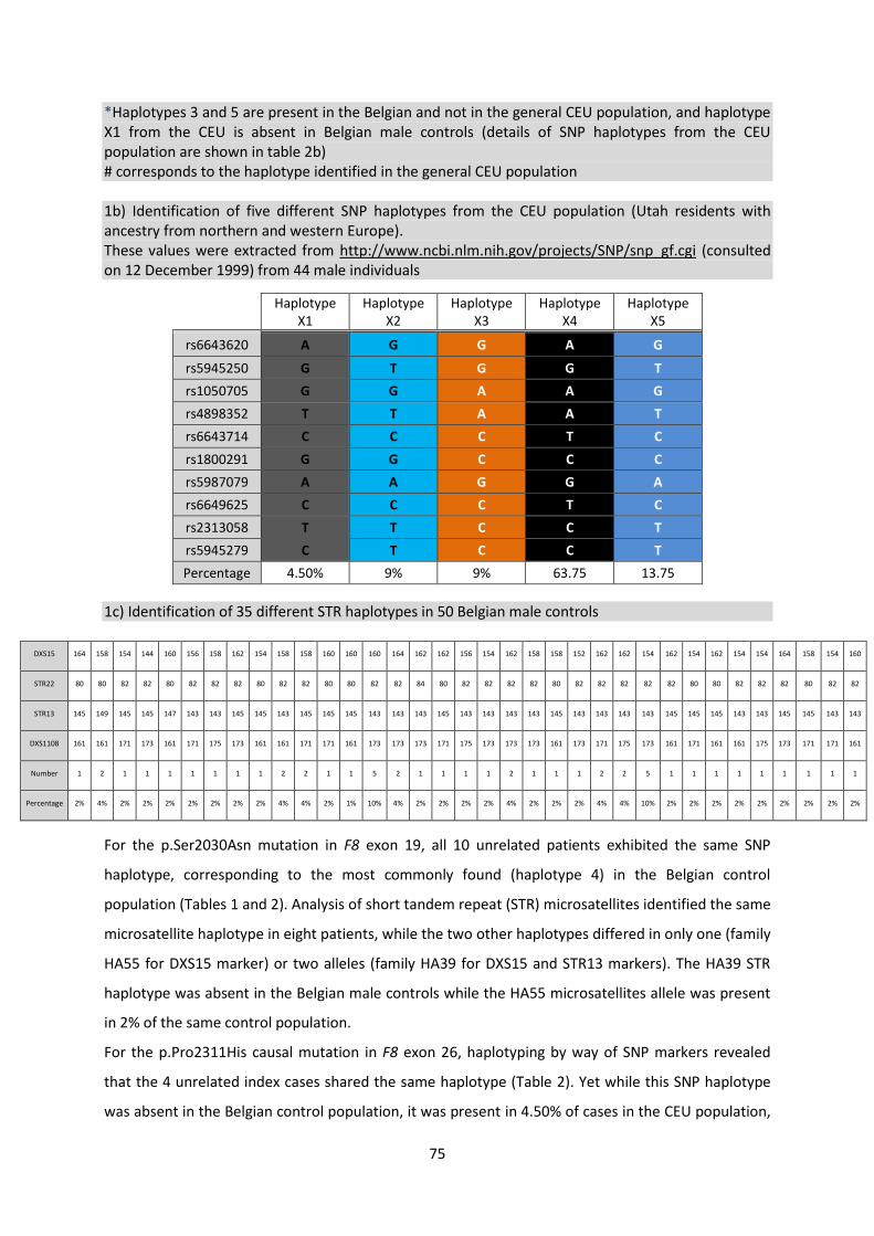
Section one F8 alterations in a cohort of patients with hemophilia A
Chapter 1 Computational and molecular approaches for predicting unreported causal missense
mutations in Belgian patients with Haemophilia A [Lannoy et al Haemophilia 2012
18e331-339] 49
Chapter 2 Overrepresentation of missense mutations in mild hemophilia A patients from Belgium
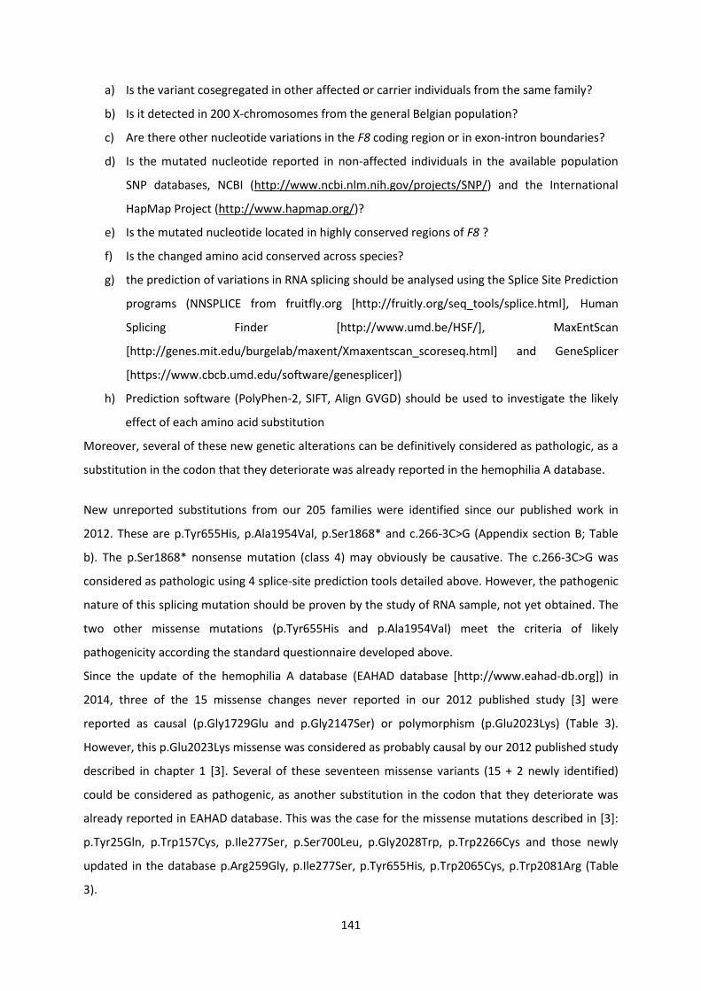
mechanisms and diagnostic implications [Lannoy et al Thromb Res 2015 1351057-1063] 69
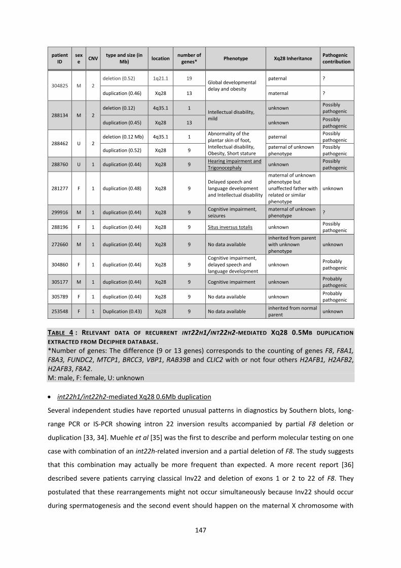
Section two Chromosomal alterations at the F8 locus producing larges duplication mechanisms and implications in the diagnosis of hemophilia A
Chapter 3 Intron 22 homologous regions are implicated in exons 1-22 duplications of the factor 8 gene [Lannoy et al Eur J Human Genet 2013 21970-976] 89
Chapter 4 Five int22h homologous copies at the Xq28 locus identified in intron22 inversion type 3 of the Factor VIII gene [Lannoy et al Thromb Res In press] 109
Chapter 5 Tandem inversion duplication within F8 intron 1 associated with mild Hemophilia A [Lannoy et al Haemophilia 2015 21516-522] 119
Discussion and perspectives 137
Appendixes
A) Details of methods 159
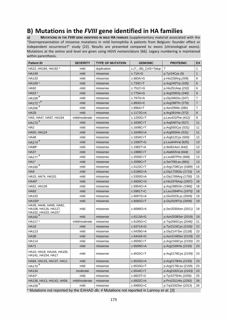
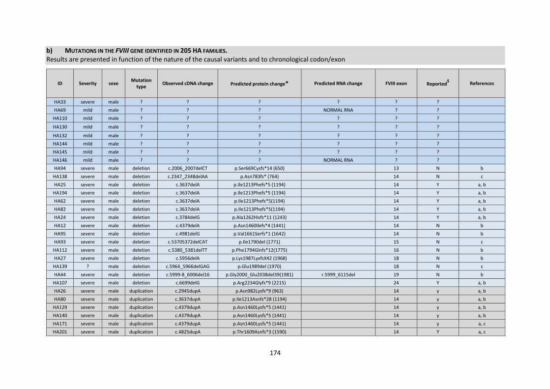
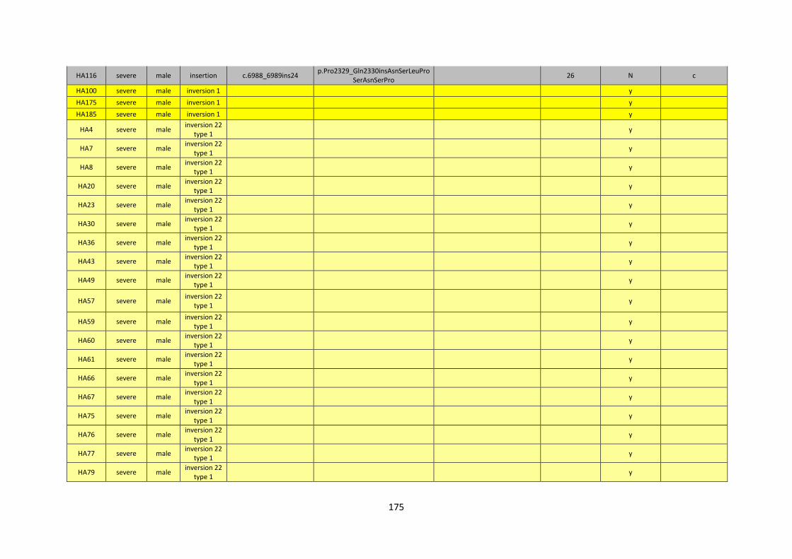
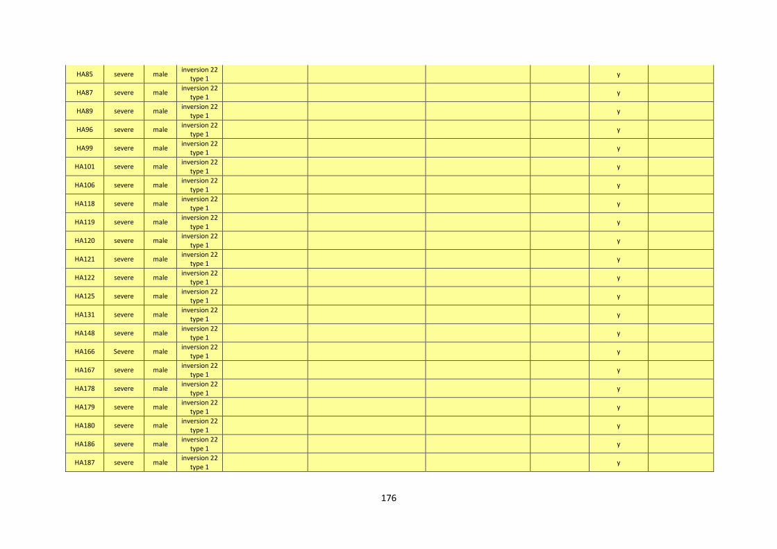
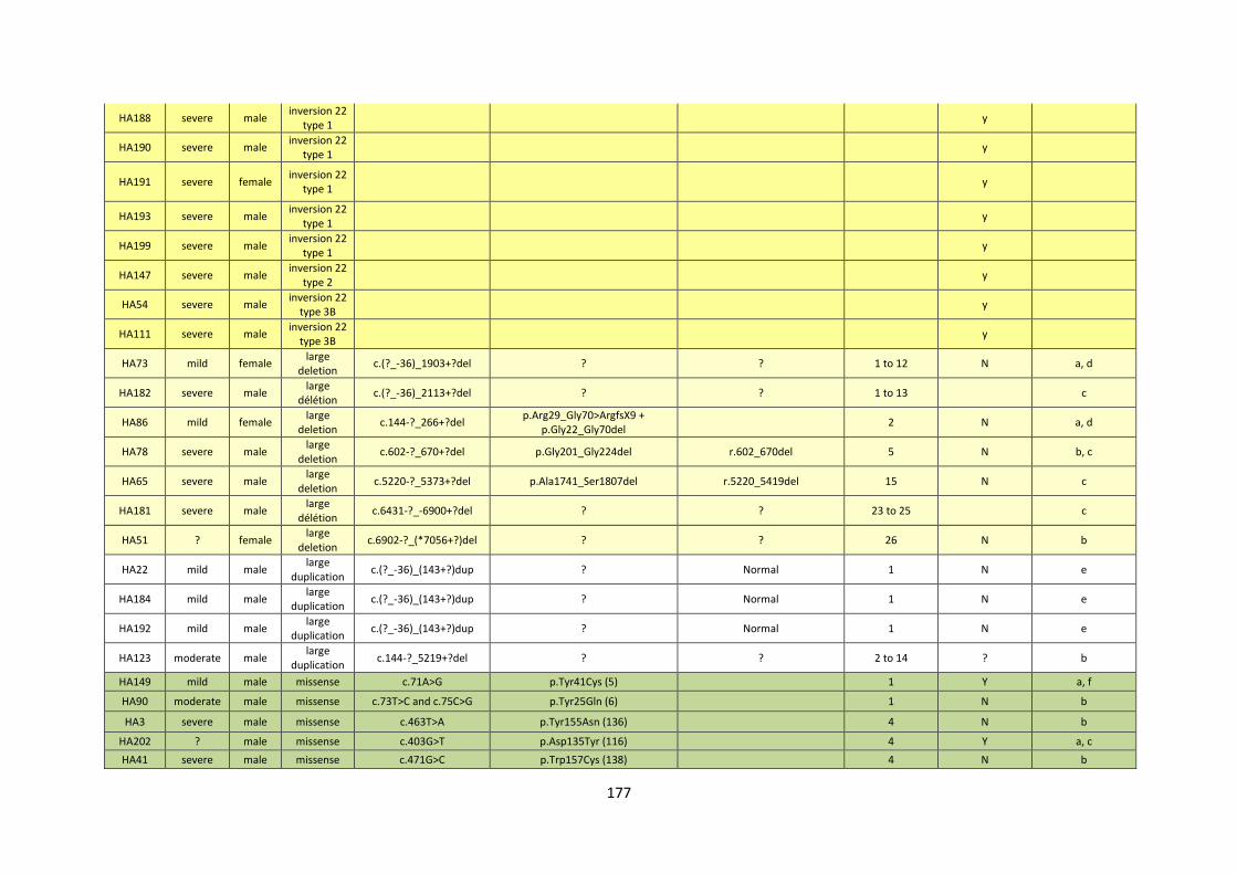
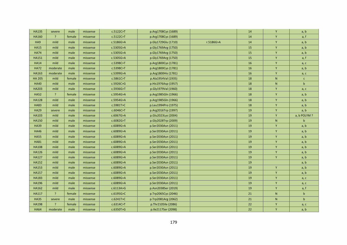
B) Mutations in the FVIII gene identified in HA families 173
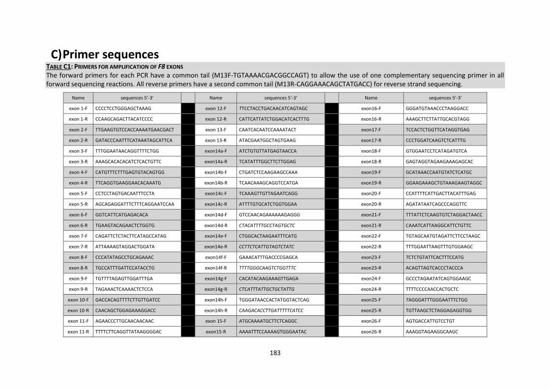
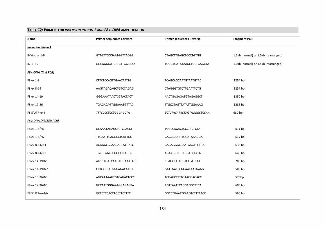
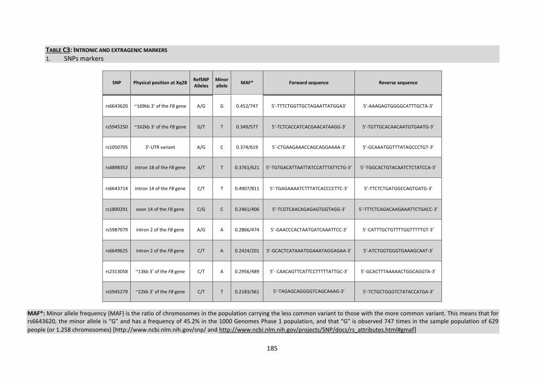
C) Primer sequences 183
References of general discussionperspectives and appendixes 189
List of abbreviations percentage
degC degree Celsius
5rsquoUTR 5rsquo-untranslated region
3rsquoUTR 3rsquo-untranslated region
A1 A2 A3 B C1 and C2 domains of FVIII
ABI Applied Biosystems
APTT Activated Partial Thromboplastin Time
BER base excision repair
bp basepair
BU Bethesda Unit
Bcl I Bacillus caldolyticus
cDNA complementary DNA
CE capillary electrophoresis
chrX chromosome X
CGH comparative genomic hybridization
CLIC2 gene encoding chloride intracellular channel 2
CNVs copy number variations
CRM cross-reacting material
CSGE conformational sensitive gel electrophoresis
CpG cytosine-phosphate-guanine
DB data base
DDAVP 1-deamino-8-D-arginine vasopressin (Desmopressin)
deaza-dGTP 7acute-deaza-deoxyguanosine triphosphate
dH2O distilled water
DGGE denaturing gradient gel electrophoresis
DHPLC denaturing high performance liquid chromatography
DMSO dimethyl sulfoxide
DNA deoxyribose nucleic acid
dNTP deoxynucleotide triphosphate
DSBs double-strand breaks
DTT Dithiothreithol
EDTA Ethylenediaminetetraacetic acid
FISH fluorescence in situ hybridization
FoSTeS fork stalling and template switching
F8 gene encoding FVIII
F9 gene encoding FIX
FV clotting factor V
FVIII clotting factor VIII
HA hemophilia A
HB hemophilia B
Kb kilo bases
Hg19 Human Genome version 19
HR Homologous Recombination
HRM high resolution melting
Hrs hours
ID intellectual disability
IS-PCR inverse shifting-PCR
LCRs low-copy repeats
LINES long interspersed elements
LMAN1 lectin mannose-binding 1
LR-PCR long-range polymerase chain reaction
MAQ Multiplex amplicon quantification
Mb Mega base
MECP2 gene encoding methyl CpG binding protein 2
microg microgram
microl microlitre
Mg milligram
MgCl2 Magnesium chloride
Min minutes
MLPA multiplex ligation-dependent probe amplification
MMBIR microhomology-mediated break-induced replication
MMEJ mediated end joining
MMR mismatch repair
mRNA messenger RNA
nm nanometer
NHEJ non-homologous DNA end joining
NGS next generation sequencing
OMIM Online Mendelian Inheritance in Man
ON over night
ORF Open reading Frame
PCR polymerase chain reaction
pH potential hydrogen
qPCR quantitative PCR
qRT-PCR quantitative real-time PCR
rcf relative centrifugal force
RT room temperature
RT-PCR reverse transcriptase polymerase chain reaction
SD Segmental duplication
SB Southern blot
SINES short interspersed elements
SDS Sodium dodecylsulfate
SNPs Single nucleotide polymorphisms
SNVs single nucleotide variants
STRs short tandem repeats
TAE Tris-acetate-EDTA
Taq Thermus aquaticus
Tel Telomere
TMLHE gene encoding trimethyllysine dioxygenase
UCSC University of California Santa Cruz
USA United States of America
UTR untranslated region
VNTR variable number tandem repeat
VWD von Willebrand disease
VWF von Willebrand factor
WT wild type
1
INTRODUCTION
2
3
Introduction
Blood is essential for life This precious body liquid is composed of cells and plasmatic
components which allows for numerous functions so essential to life ranging from transport of
oxygen carbon dioxide nutrients vitamins metabolites waste products hormones and components
of the cellular as well as humoral immune system to homeostatic functions Moreover one of its
elementary tasks is to prevent its own loss namely by the mechanism of hemostasis ndash the cessation
of blood leakage from damaged vessels The hemostatic challenge requires a delicate balance
between hemostasis and thrombosis A ldquoperfect clotrdquo not only seals the injured vessel at the site of
damage to stop bleeding but also must ensure that it is sufficiently labile to be degraded at the end
of this process The deficit of one element of this delicate balance leads to the pathologic process of
abnormal bleeding or on the contrary to the formation of persistent clot
The main actor of this thesis is one of the coagulation factors the FVIII involved in clot production in
the process of hemostasis In case of missing inconsistent or reduced FVIII protein caused by
mutations in the F8 clinical symptoms of hemophilia A (HA) appear The presented work includes
molecular analysis in HA patients beyond routine diagnosis to identify mechanisms behind simple
variants or gross genomic rearrangements Therefore the first section of the introduction entitled
ldquogeneral introductionrdquo will discuss the history of hemophilia the basics of blood coagulation the
main bleeding disorders and provide overview of the clinical diagnosis and management of
hemophilia In the second section general mechanisms and consequences of genetic variations are
developed and applied to hemophilia A
4
Part one General introduction
1 History of hemophilia
Incidences of excessive or abnormal bleeding were first recorded hundreds of years ago The Talmud
a collection of Jewish rabbinical writings on laws and traditions from the 2nd century AD stated that
baby boys did not have to be circumcised if two of their brothers had previously died from the
procedure [1] The Arab physician considered as the greatest medieval surgeon Abu al Qasim (936ndash
1013) who lived in Al-Andalus described families whose male relatives died from uncontrolled
bleeding after only minor traumas [2] In 1803 Dr John Conrad Otto (1774-1844) an American
physician was the first to publish an article recognizing that a hemorrhagic bleeding disorder
primarily affected men passed down by healthy females and ran in certain families In 1813 John
Hay published an article in The New England Journal of Medicine where he discussed how the
affected male could pass the disorder to the unaffected daughters [3] The term hemophilia is
derived from the term hemorrhaphilia postulated in 1828 by Friedrich Hopff a student at the
University of Zurich and his professor Dr Schonlein [4] In 1937 Patek and Taylor two doctors from
Harvard discovered anti-hemophilic globulin [5] In 1947 Pavlosky from Buenos Aires found
hemophilia A and hemophilia B to be separate diseases by doing a lab test This test was done by
transferring the blood of one hemophiliac to another hemophiliac The fact that this corrected the
clotting problem showed that there was more than one form of hemophilia
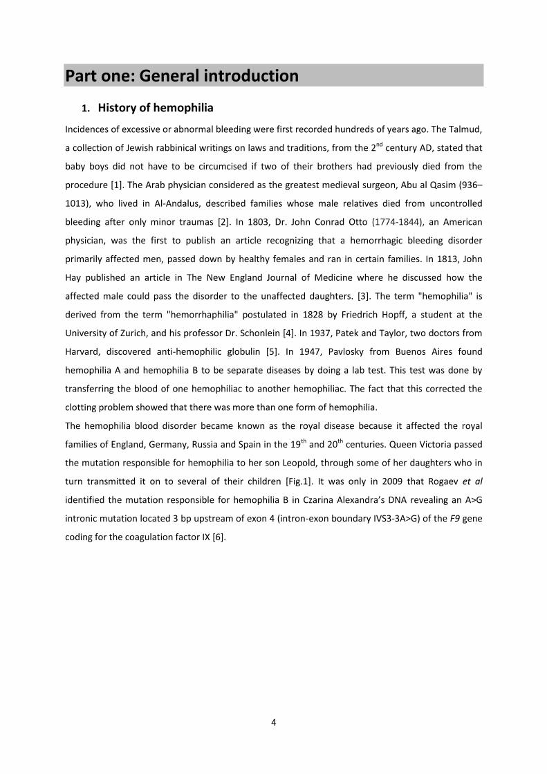
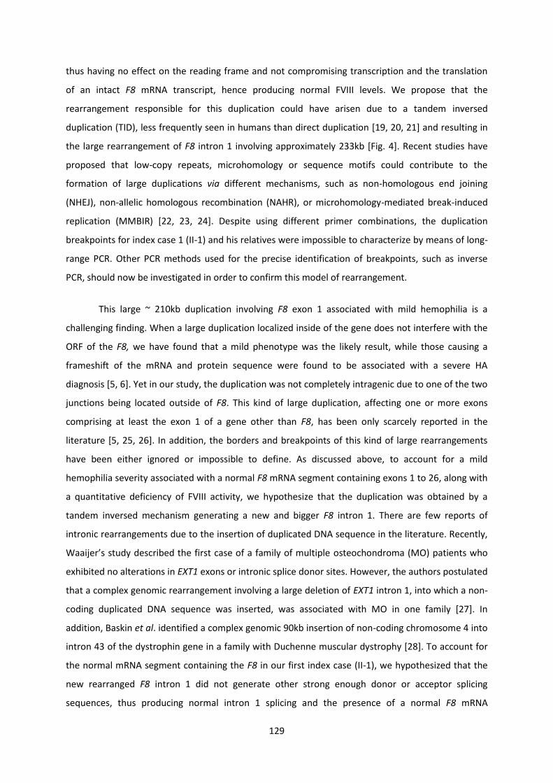
The hemophilia blood disorder became known as the royal disease because it affected the royal
families of England Germany Russia and Spain in the 19th and 20th centuries Queen Victoria passed
the mutation responsible for hemophilia to her son Leopold through some of her daughters who in
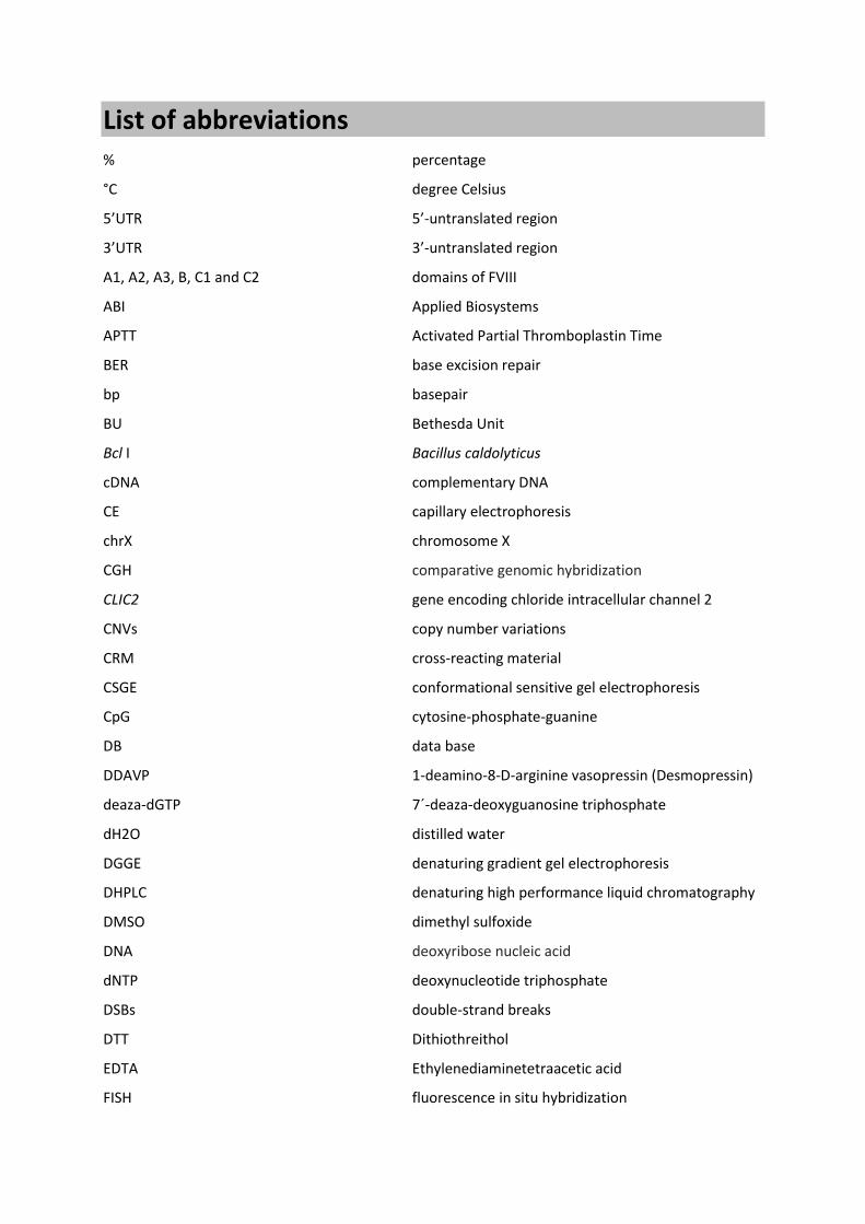
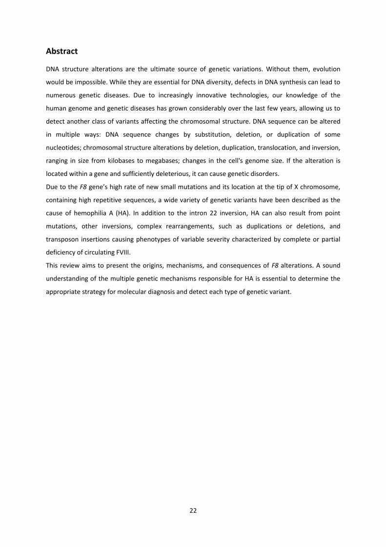
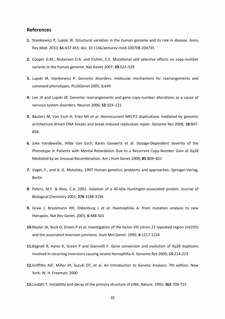
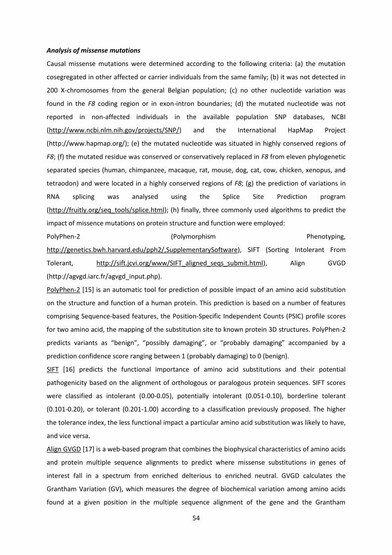
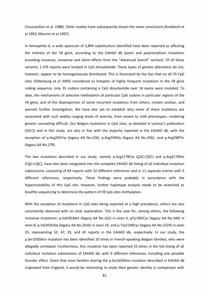
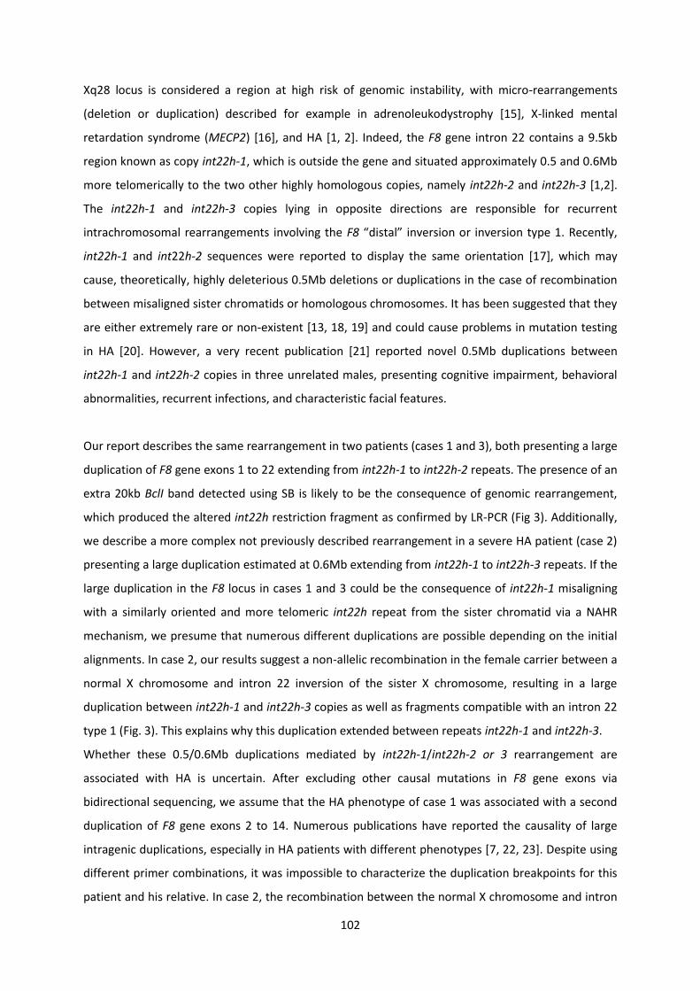
turn transmitted it on to several of their children [Fig1] It was only in 2009 that Rogaev et al
identified the mutation responsible for hemophilia B in Czarina Alexandrarsquos DNA revealing an AgtG
intronic mutation located 3 bp upstream of exon 4 (intron-exon boundary IVS3-3AgtG) of the F9 gene
coding for the coagulation factor IX [6]
5
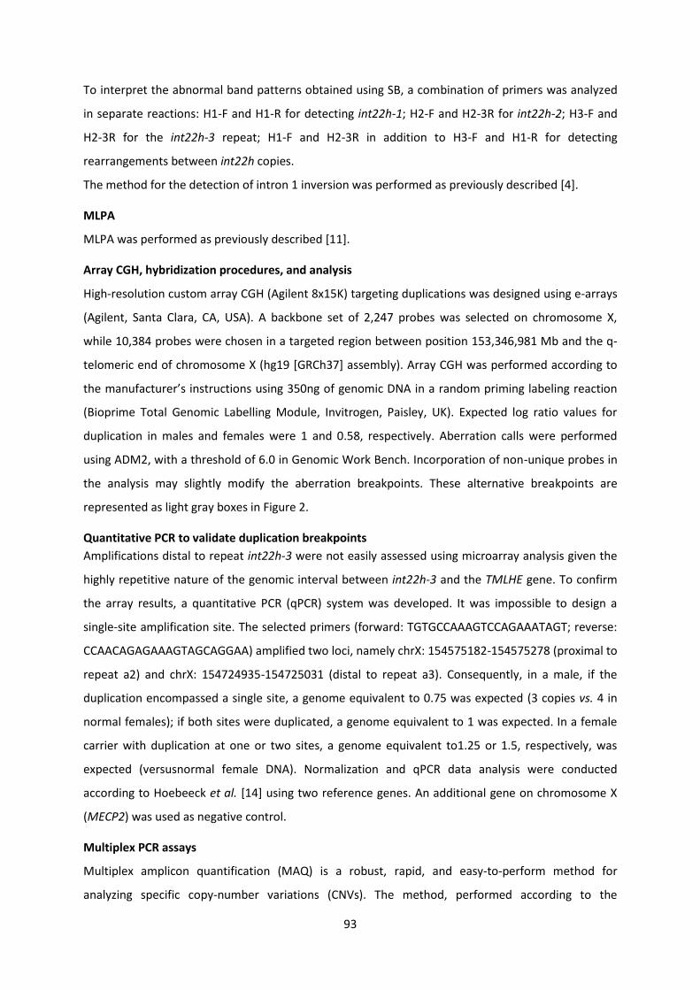
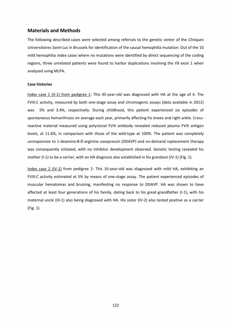
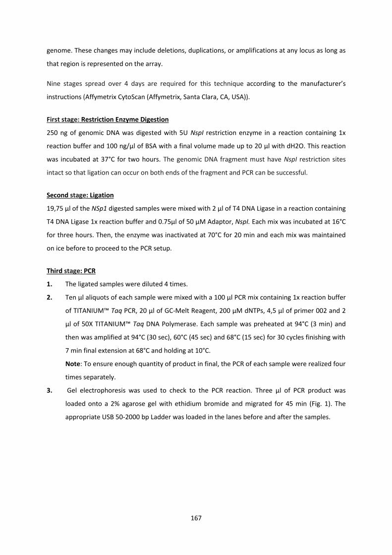
FIGURE 1 QUEEN VICTORIA GRANDMOTHER OF EUROPE Queen Victoria of England (1837-1901) was a hemophilia carrier Her eighth child Leopold three of her grandchildren and seven of her great-grandchildren were affected by the disease Due to princely alliances the disease spread through most European royal families notably in Germany Spain and Russia
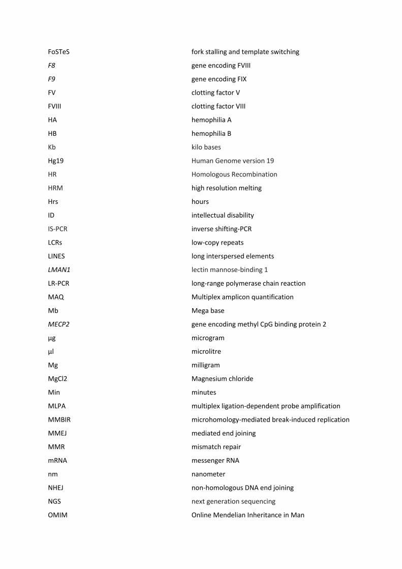
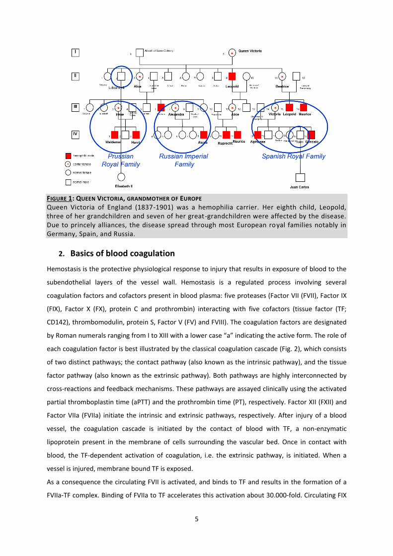
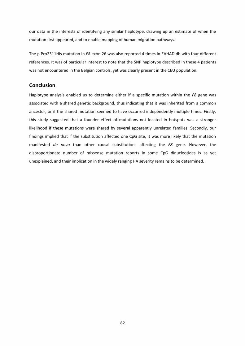
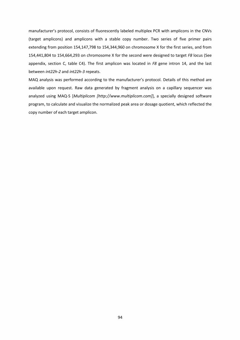
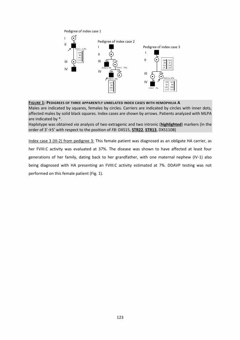
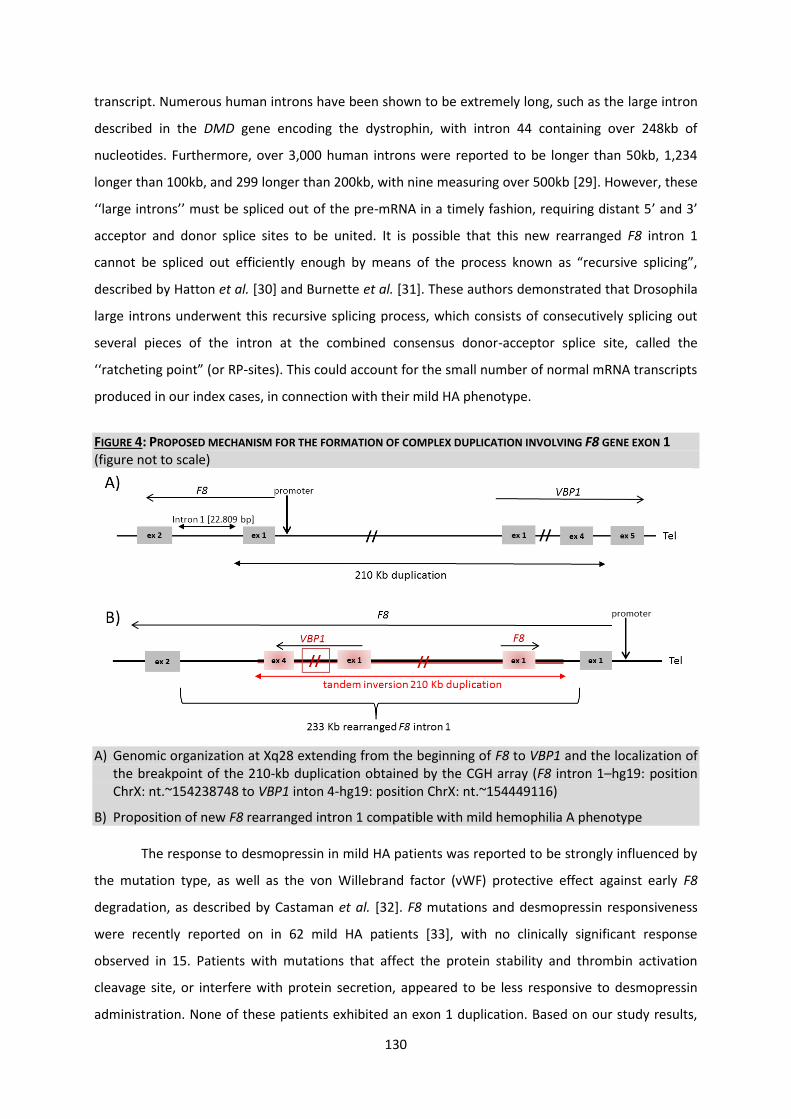
2 Basics of blood coagulation
Hemostasis is the protective physiological response to injury that results in exposure of blood to the
subendothelial layers of the vessel wall Hemostasis is a regulated process involving several
coagulation factors and cofactors present in blood plasma five proteases (Factor VII (FVII) Factor IX
(FIX) Factor X (FX) protein C and prothrombin) interacting with five cofactors (tissue factor (TF
CD142) thrombomodulin protein S Factor V (FV) and FVIII) The coagulation factors are designated
by Roman numerals ranging from I to XIII with a lower case ldquoardquo indicating the active form The role of
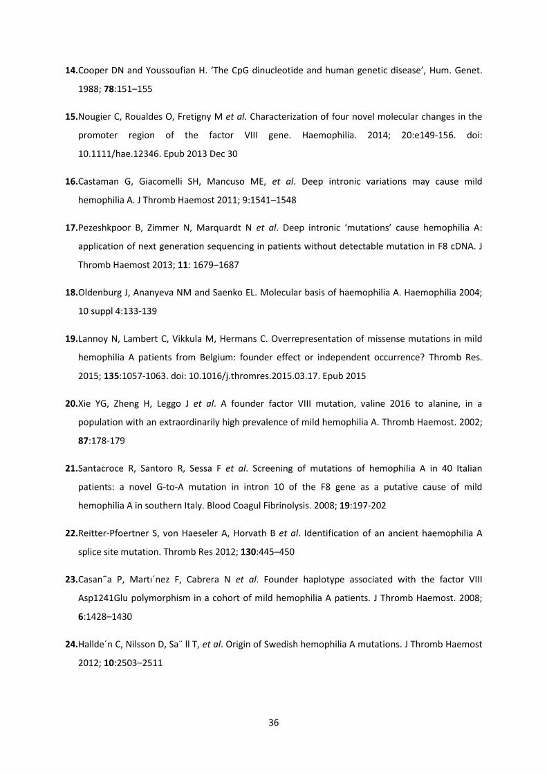
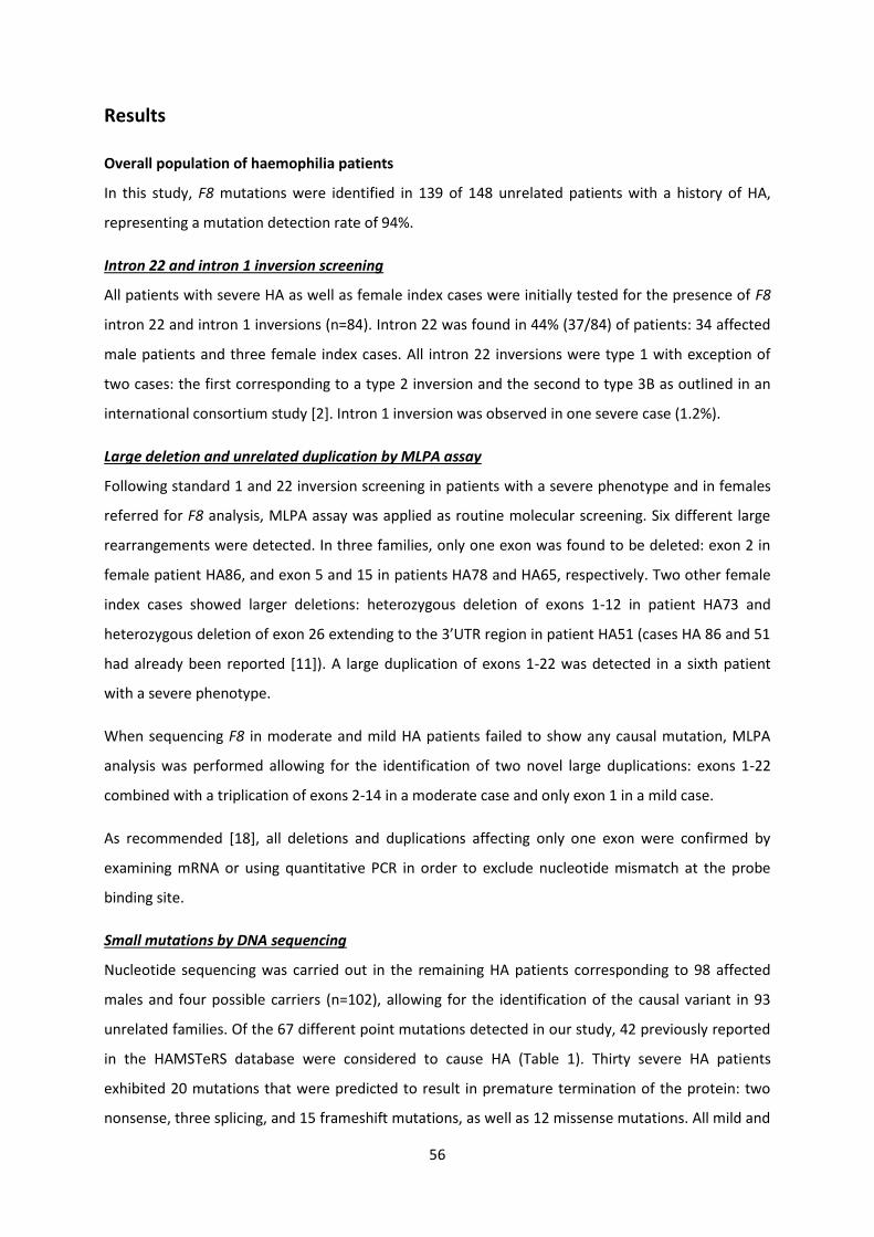
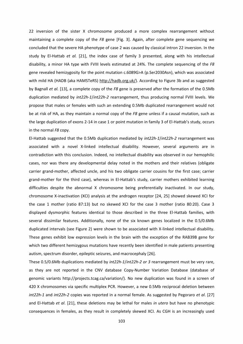
each coagulation factor is best illustrated by the classical coagulation cascade (Fig 2) which consists
of two distinct pathways the contact pathway (also known as the intrinsic pathway) and the tissue
factor pathway (also known as the extrinsic pathway) Both pathways are highly interconnected by
cross-reactions and feedback mechanisms These pathways are assayed clinically using the activated
partial thromboplastin time (aPTT) and the prothrombin time (PT) respectively Factor XII (FXII) and
Factor VIIa (FVIIa) initiate the intrinsic and extrinsic pathways respectively After injury of a blood
vessel the coagulation cascade is initiated by the contact of blood with TF a non-enzymatic
lipoprotein present in the membrane of cells surrounding the vascular bed Once in contact with
blood the TF-dependent activation of coagulation ie the extrinsic pathway is initiated When a
vessel is injured membrane bound TF is exposed
As a consequence the circulating FVII is activated and binds to TF and results in the formation of a
FVIIa-TF complex Binding of FVIIa to TF accelerates this activation about 30000-fold Circulating FIX
6
and FX are converted by the FVIIa-TF complex to active serine proteases FIXa and FXa which in turn
activate TF-bound FVII via a feedback mechanism (Fig 2) FIXa and FXa then assemble with their
nonenzymatic protein cofactors FVIIIa and FVa The FVIIIa-FIXa complex termed tenase or intrinsic
Xase generates even more FXa whereas the FVa-FXa complex (prothrombinase) converts
prothrombin (FII) to active thrombin (FIIa) which works in a positive feedback loop itself Thrombin
can activate FV and FVIII which activates FXI FXI itself activates in turn FIX and allows activation of
more thrombin This will lead to releases of FVIII from being bound to von Willebrand Factor (vWF)
Subsequently after sufficient amount of thrombin is present in the circulation fibrinogen is activated
and becomes fibrin In the presence of activated factor 13 (FXIIIa) fibrin is polymerized and a fibrin
clot is formed at the site of injury
The functional activity of FVIII is measured in vitro by determination of the clotting time of the blood
plasma sample with depleted endogenous FVIII and added FVIII in the solution under study
FIGURE 2 Simplified scheme of the procoagulation ldquocascaderdquo of proteolytic reactions initiated by damage of blood vessel walls and culminating in the creation of a fibrin mesh Black arrows represent the conversion or activation of coagulation factors dotted thin lines show additional enzymatic functions Coagulation factors are abbreviated by ldquoFrdquo and their Roman number FVIIIa works as a cofactor for FIX in the active tenase complex to activate FX Ca2+ - calcium ions HMWK ndash high molecular weight kininogen PL ndash phospholipid membrane VWF ndash von Willebrand factor See text for details on factors and mechanisms (figure published by Muumlhle C [7)
7
3 Principal bleeding disorders
Bleeding disorders are a group of disease that results in the inability of the blood to form a clot
properly They are characterized by extended bleeding after injury surgery trauma or menstruation
Improper clotting can be caused by defects in blood components such as platelets andor clotting
factors If any of them are defective or deficient blood clotting is affected and a mild moderate or
severe bleeding disorder can result Some bleeding disorders can be inherited or acquired Others
can occur from such conditions as cirrhosis of the liver HIV leukemia and vitamin K deficiency They
also can result from certain medications that thin the blood including aspirin heparin and warfarin
The most common hereditary coagulation abnormality described in humans with an incidence of
roughly 1 is von Willebrand disease (VWD) arising from a qualitative or quantitative deficiency of
von Willebrand factor (VWF) which is required for platelet adhesion as well as stabilization of FVIII It
is thus a defect of primary hemostasis associated with a secondary defect in coagulation factor VIII
and therefore is sometimes classified under hemophilia [8] Hemophilia is the most common form
of congenital deficiencies in a blood coagulation factor It is subdivided into hemophilia A
(OMIM306700) caused by the lack or defective function of FVIII and the less frequent hemophilia B
(OMIM306900) due to a deficiency in FIX The incidence of hemophilia A and B is approximately 1
in 5000 and 1 in 30000 male live births respectively and no ethnic or geographic predisposition has
been defined The F8 and F9 genes are located on the X chromosome giving them the status of X-
linked disease which stands in contrast to the other coagulation factor genes that are predominantly
inherited as autosomal recessive traits Hemophilia passed on through females who carry the
defective genes Half of the sons of a carrier are affected with HA and half of the daughters are
carriers again In contrast all daughters of a hemophilic man are carriers whereas all sons are
healthy with respect to hemophilia The female carriers usually do not experience problems or only
with very mild symptoms of the disease However symptomatic females have been documented
and the proposed mechanisms include unbalanced X chromosome inactivation or the presence of a
true homozygous offspring due to consanguinity or to compound heterozygosity from an affected
father and a carrier mother [9-11] Female individuals diagnosed with Turner syndrome may suffer
from HA similarly to hemizygous males [12] Approximately 60 of hemophilia A cases have a family
history the remainder is sporadic due to de novo mutations In 90 of these families the mutation
has arisen in the parents or grandparents [13] It has been reported that mutations causing
hemophilia A originate 36 times more frequently in male germ cells than female germ cells
However this sex ratio of mutation frequencies differs for each type of mutation The mutation rate
for point mutations is 5-10-fold higher and inversions more than 10-fold higher in male germ cells
while deletions are more than 5-fold higher in female germ cells [14] Therefore a mother of a
8
sporadic case of hemophilia has a high risk of being a carrier of the disorder Although the prevalence
does not vary much between populations it could be underestimated in developing countries due to
poor diagnosis [15]
Low levels of factor XI (also known as Rosenthal syndrome) caused by mutations in the gene for FXI
and parahemophilia or Owren hemophilia due to a deficiency of FV are rare
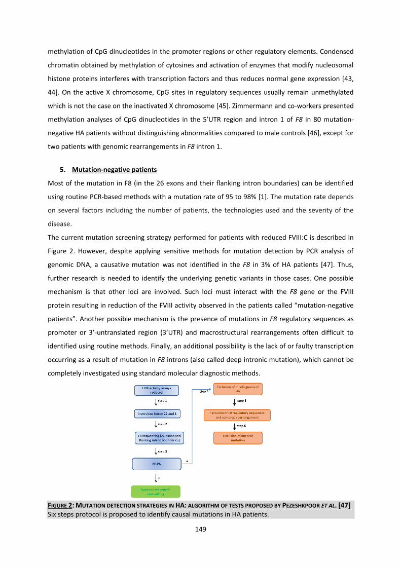
4 Clinical features diagnosis and management of hemophilia
Clinical Features
The most common bleeding sites are joints (80 of bleeding) muscles and the gastrointestinal
mucosa Ankles are the most commonly affected joints in children whereas knees and elbows are
more often involved in adults Quadriceps and iliopsoas bleeding are the most common sites of
muscle hematomas Abdominal wall bleeding and gastrointestinal mucosal bleeding can occur
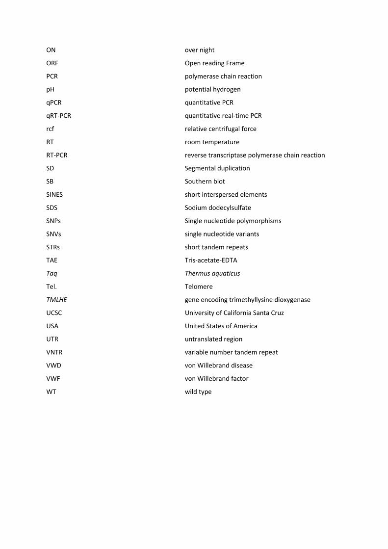
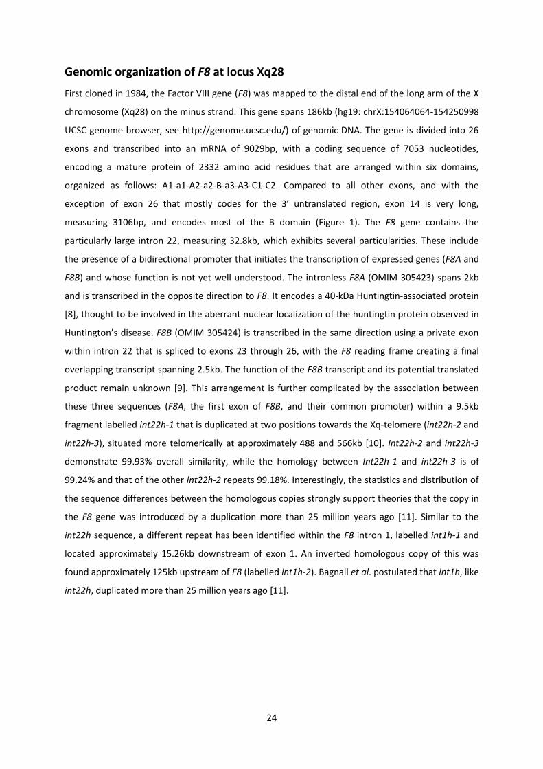
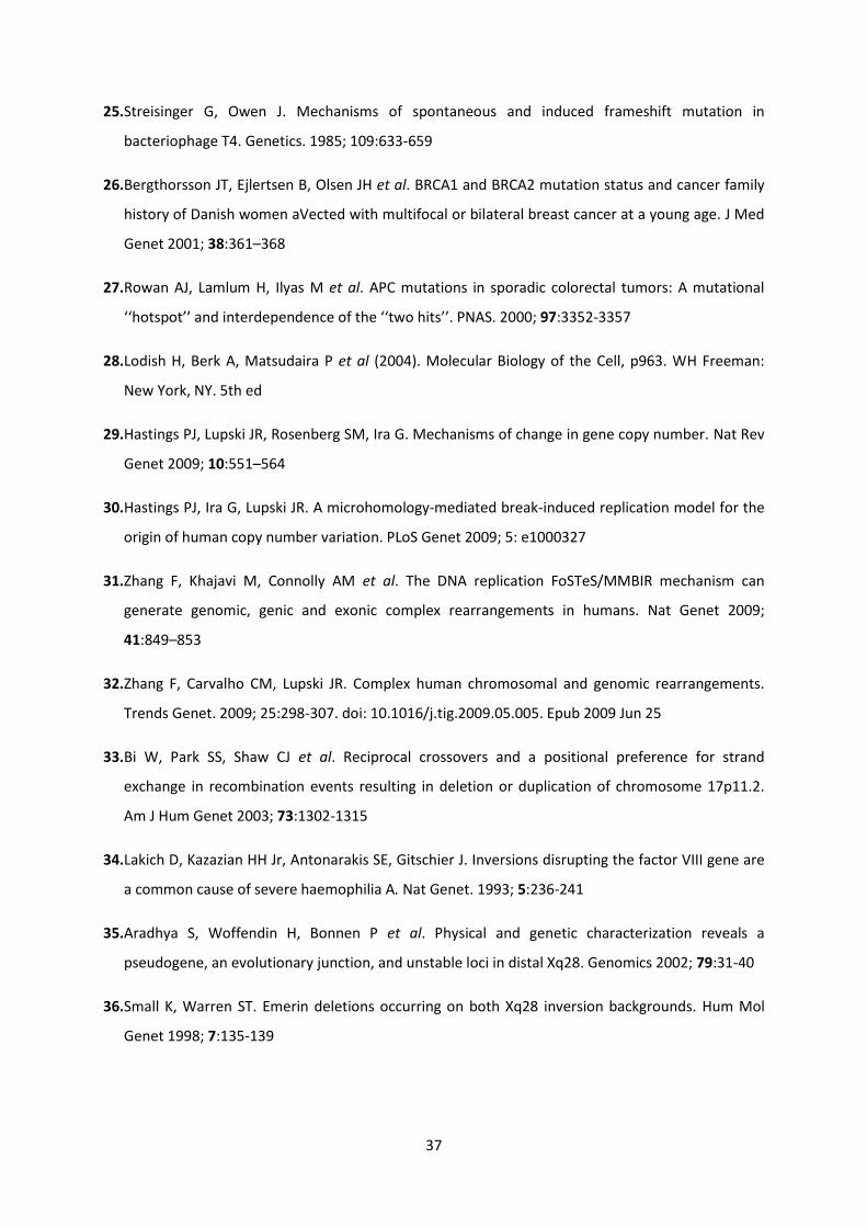
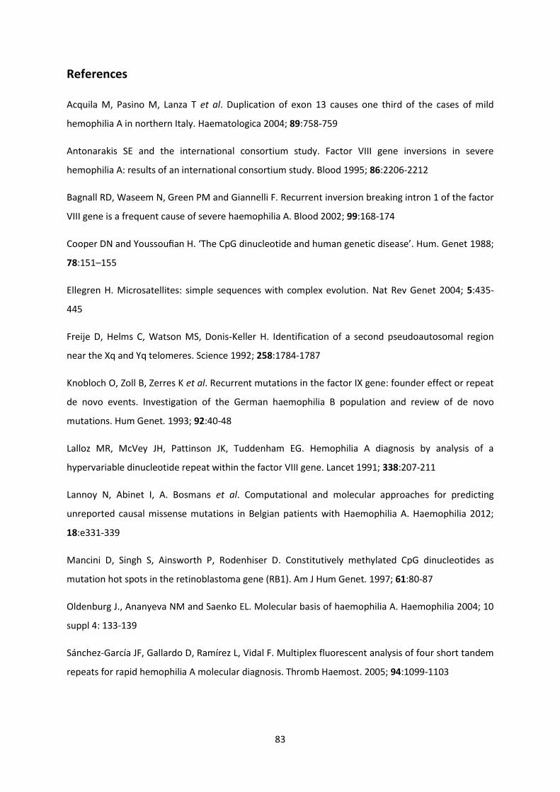
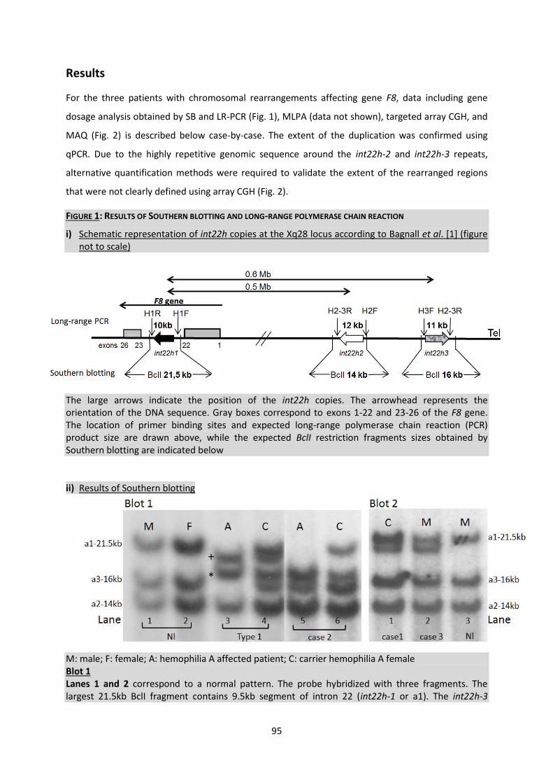
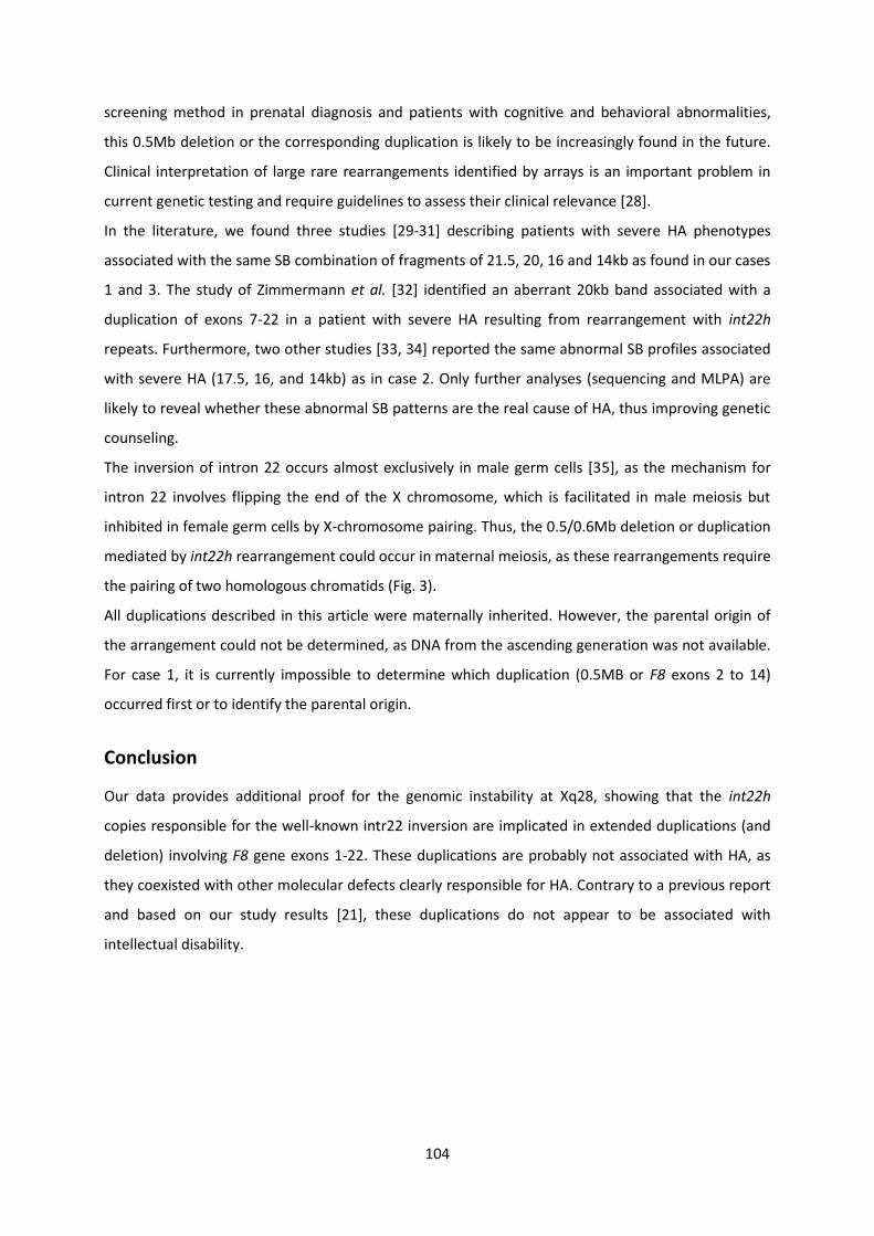
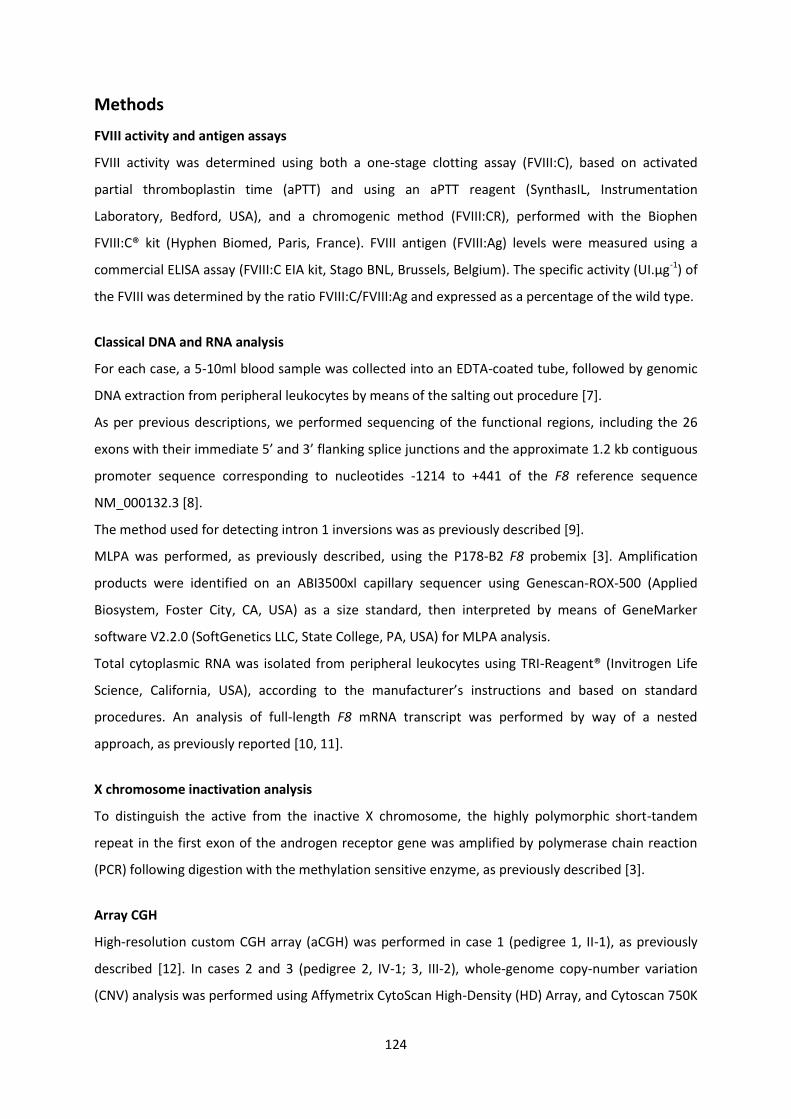
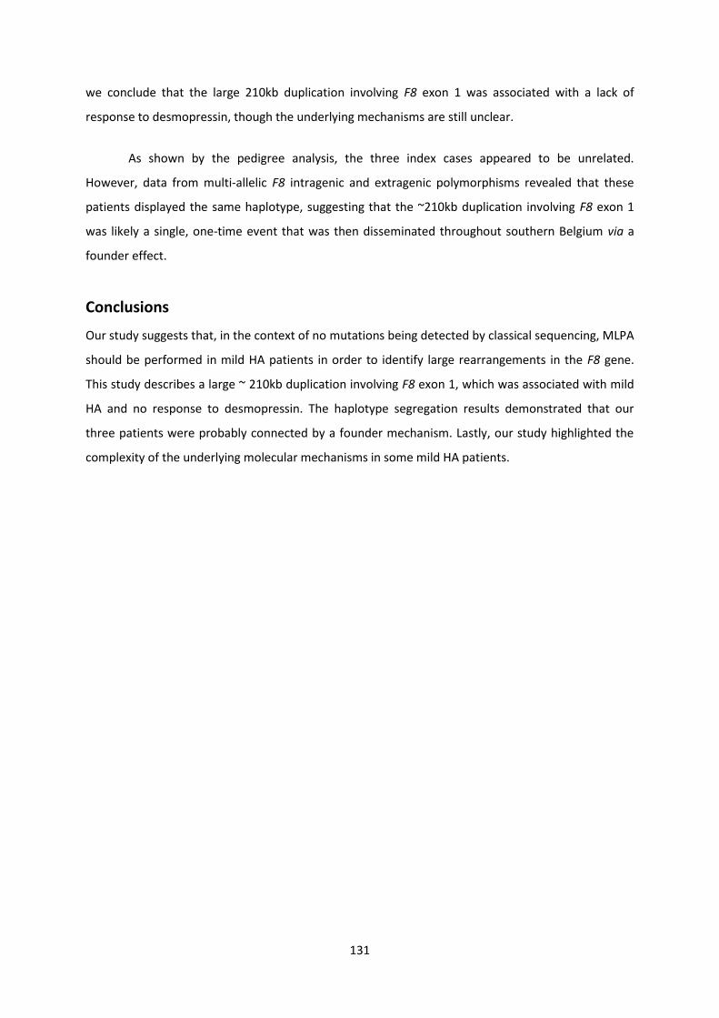
Depending on the amount of clotting factor in the blood three classes of HA severities have been
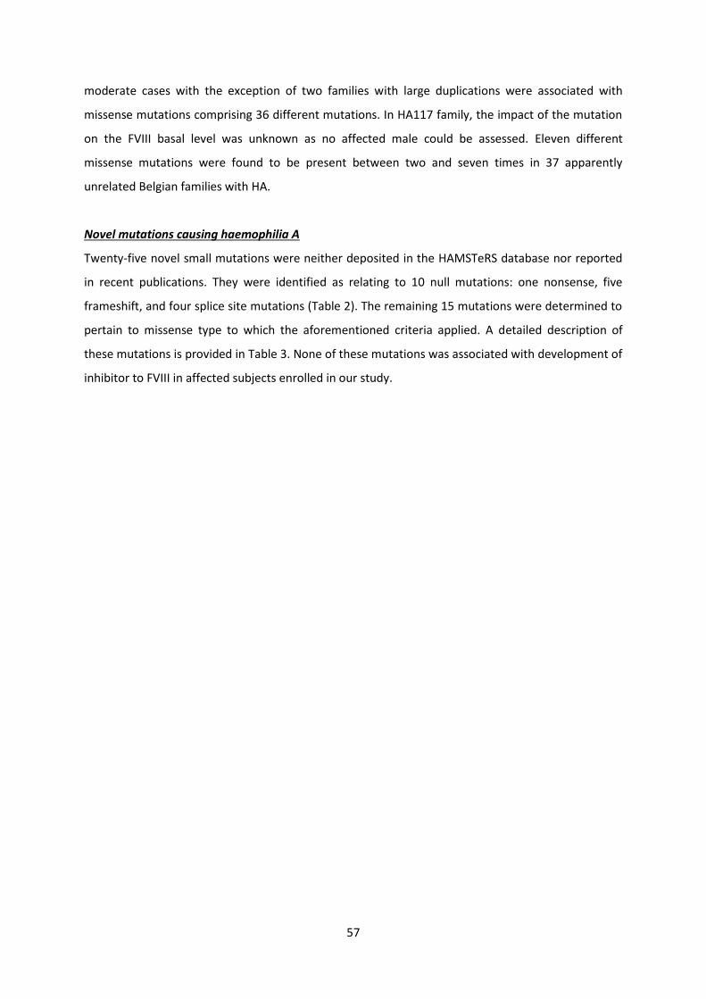
defined compared to the normal range of 50-150 [Fig 3] severely affected individuals have lt1
IUdL (less than 1 of normal) moderate gt1 - lt5 IUdL (1-5 of normal) and mild gt5 - lt40 IUdL
(gt5 - lt40 of normal) (16)] These classes represent 40 10 and 50 of patients with HA People
with severe hemophilia usually bleed frequently into their muscles or joints particularly the knee
ankle hip and elbow associated with pain They may bleed one to two times per week Bleeding is
often spontaneous which means it happens for no obvious reason People with moderate
hemophilia bleed less frequently about once a month They are often recognized by prolonged
bleeding after surgery bad injury or dental extraction A person with moderate hemophilia will
rarely experience spontaneous bleeding People with mild hemophilia usually bleed only as a result
of surgery or major injury They do not bleed often and in fact may never have a bleeding problem
Female carriers with heterozygous mutation may exhibit various FVIII plasma levels and bleeding
symptoms [17] that have been partly attributed to different X chromosome inactivation levels
described by Lyon in 1962 [9]
9
FIGURE 3 CLASSIFICATION OF HEMOPHILIA BASED ON THE ACTIVITY LEVELS OF CLOTTING FACTOR VIII Three classes of HA severities have been defined compared to the normal range of 50-150
Diagnosis
Screening Tests
Activated Partial Thromboplastin Time (APTT) test or ldquoone-stagerdquo assay
This test measures how long it takes for blood to clot It measures the clotting ability of factors VIII
(F8) IX (F9) XI (F11) and XII (F12) If any of these clotting factors are too low it takes longer than
normal for the blood to clot In cases of a long APTT an equal mixture of normal and test plasma
should be tested (ie a mixture of 1 part test and 1 part normal plasma called a 5050 mix) If the
APTT corrects by more than 50 of the difference between the clotting times of the normal and test
plasma a factor deficiency is indicated Poor correction suggests an inhibitor possibly to one of the
clotting factors in the system or of the non-specific type such as lupus anticoagulant
The one-stage factor FVIII assay is relatively simple to perform and accurate It does have limitations
including susceptibility to interference from preactivation of factor VIII or anti-phospholipid
antibodies as well as misleading results when assaying recombinant factor VIII In addition some F8
mutations can lead to discrepant 1-stage2-stage [chromogenic] FVIII assay results [18]
Two-Stage amp Chromogenic Factor Assays
Two main alternatives to the one stage assay the 2-stage APTT based assay and the chromogenic
assay exist Both are based around an initial step to produce factor Xa in a quantity proportional to
the amount of factor VIII present and a second step to assay the amount of factor Xa and so deduce
the amount of factor VIII present The 2-stage assay is rarely performed today but remains an
important test Results by the chromogenic and 2-stage factor VIII assays are usually equivalent For
most patients the FVIII calculated by either the chromogenic 2-stage or 1 stage APTT based assay will
10
be also comparable However some genetic mutations of the F8 gene produce discrepant results
between 1-stage and 2-stage chromogenic assays particularly those which impede interaction of
the A1 and A2 domains [18]
The chromogenic assay is now the reference method for potency determination of FVIII concentrates
as recommended by European Pharmacopoeia and the ISTH Subcommittee for FVIII and FIX [19]
FVIII cross-reacting material
Factor VIII protein in plasma can be measured by polyclonal antigen ELISA assays Depending on the
mutation that influences FVIII level and activity hemophiliacs can be classified into three broad
categories with major influence on their risk of developing FVIII antibodies (see section 6) [20-21]
(1) Cross-reacting material (CRM)-negative patients lacking immunologically detectable FVIII antigen
levels (in about 50 )
(2) CRM-positive patients with considerable but presumably dysfunctional FVIII protein levels
(about 5 ) caused by missense or other small mutations
(3) An intermediate group of CRM-reduced patients (about 45 ) possibly as a consequence of
inefficient secretion or faster clearance of FVIII
The majority of mutations are CRM-negative and probable affect the folding and stability of the
protein for example in case of mRNA degradation due to nonsense-mediated decay [22] Others
mechanisms associated with CRM-reduced are described the pPhe672del mutation prevents the
formation of the tightly packed structure of FVIII resulting in aberrant folding and a block to
transport through the secretory pathway [23] and the pArg2326Gln mutant FVIII protein showed
reduced secretion with increased intracellular degradation [24]
Mutations associated with CRM-positive such as pIle585Thr and pSer577Phe have been described
inducing defective interaction with factor IXa [23]
Management
Replacement coagulation
Choices for replacing factor include recombinant versus plasma-derived factor VIII Plasma-derived
concentrates vary in purity and must undergo viral inactivation procedures First-generation
recombinant products have demonstrated efficacy in clinical trials however they continue to carry a
theoretic risk of viral transmission because of the added human albumin necessary for factor
stabilization Second-generation recombinant products do not require albumin stabilization Factor IX
replacement has traditionally been with prothrombin complex concentrates (PCCs) that contain
factors II VII and X as well as IX and were associated with thrombotic risk Newer plasma-derived
factor IX concentrates are effective and they undergo viral inactivation Recombinant factor IX
11
concentrates such as Benefix are also effective and have no added albumin hence eliminating a
theoretic risk of viral infection
Bleeding episodes in mild HA patients may be successfully treated or prevented either by
desmopressin (1-deamino-8-D-arginine vasopressin DDAVP) to temporarily boost plasma levels of
VWF and FVIII in good responder patients with mild severity or with FVIII concentrates However the
response to desmopressin in mild HA patients was reported to be strongly influenced by the
mutation type that affect the protein stability and thrombin activation cleavage site or interfere with
protein secretion and the von Willebrand factorrsquos (vWF) protective effect against early F8
degradation [25 26]
5 FVIII gene expression features
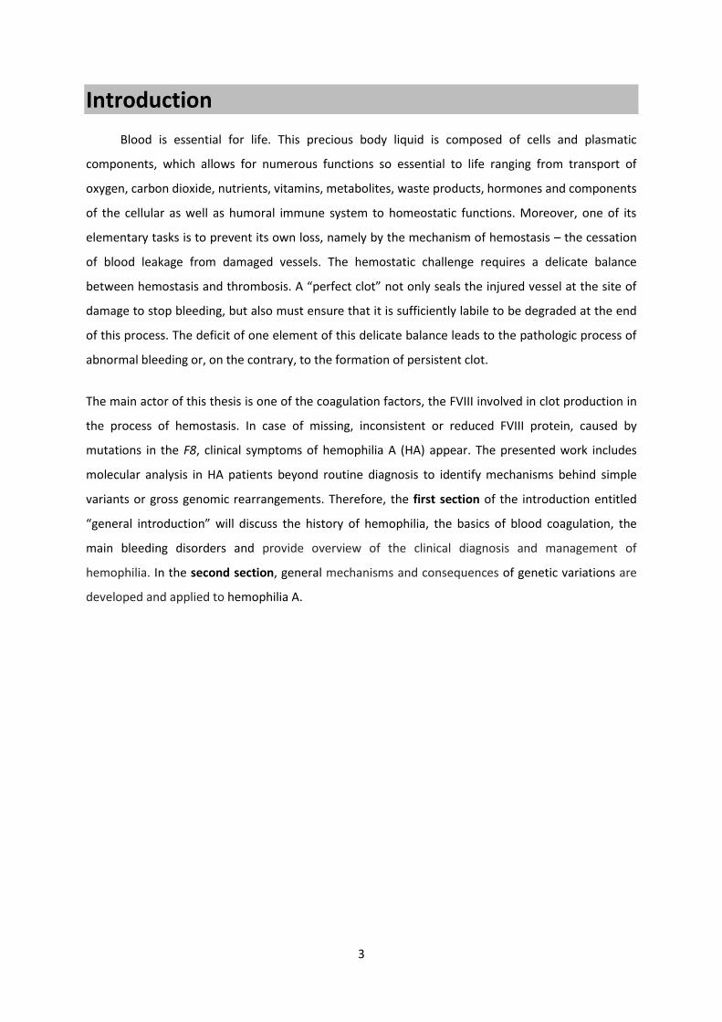
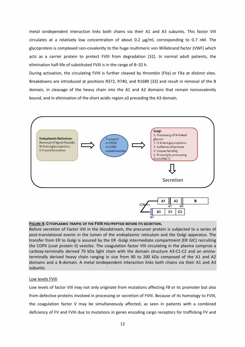
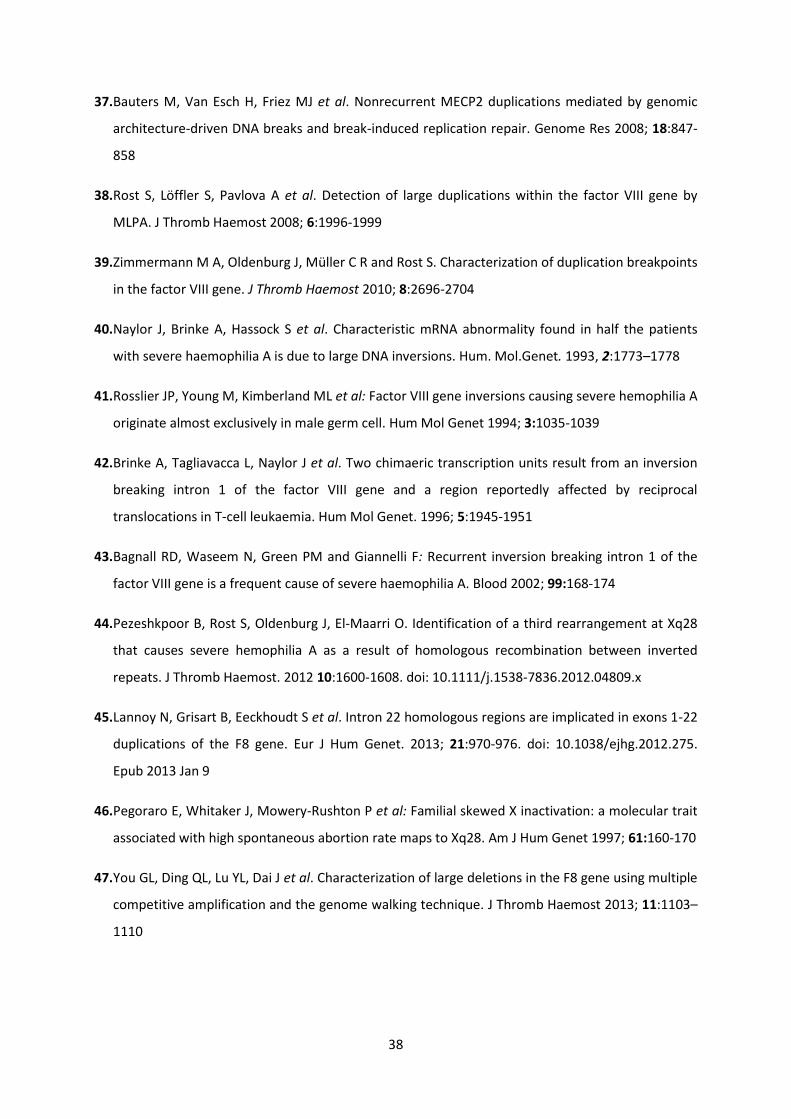
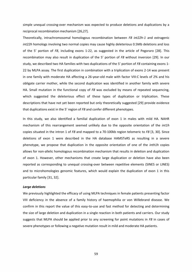
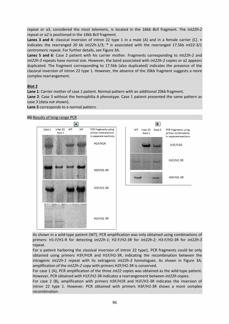
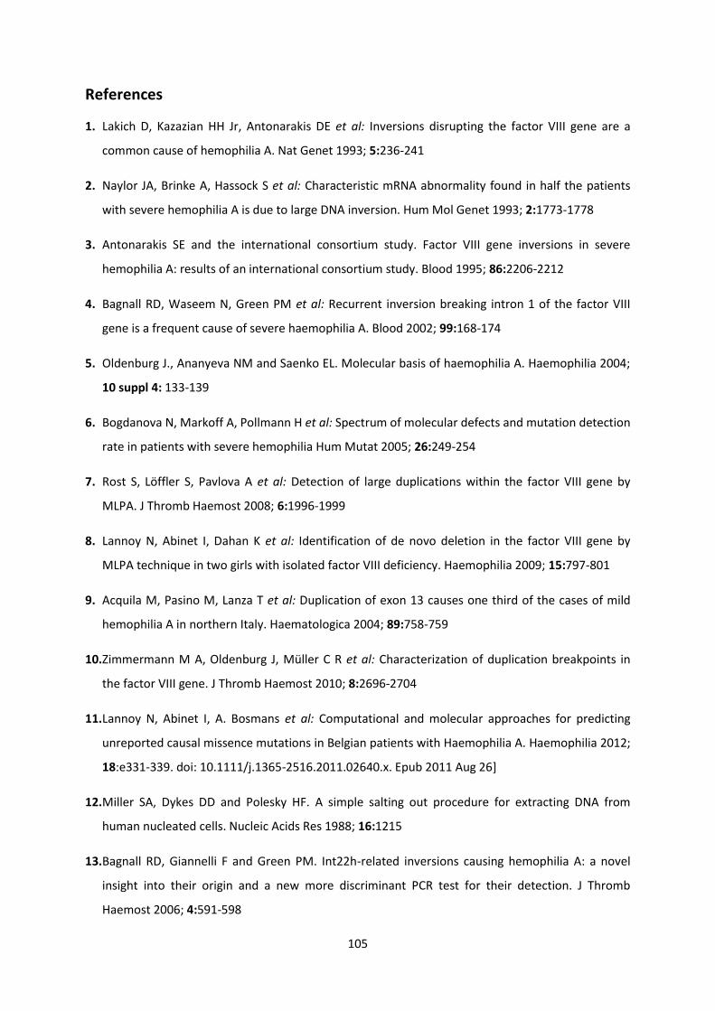
Post-translational modifications of the FVIII precursor protein in cytoplasm
Expression of the FVIII gene is tissue-specific and is mostly observed in liver cells The highest level of
the mRNA and FVIII protein have been detected in liver sinusoidal cells significant amounts of FVIII
are also present in hepatocytes and in Kupffer cells [27 28] The FVIII precursor protein is subject to a
series of post-translational events [Fig 4] In the lumen of the endoplasmic reticulum (ER) where the
FVIII polypeptide can remain for 15 min to several days processing of the 19-amino-acid-long signal
peptide N-glycosylation and disulfide bond formation occur first Then the FVIII polypeptide is
transferred from ER to the Golgi apparatus via the ER-Golgi intermediate compartment (ERGIC) FVIII
and FV are recruited to this compartment by binding to the transmembrane protein (cargo receptor)
ER GIC-53 also known as LMAN1 (lectin mannose-binding 1) and ensures mannose-selective
calcium-dependent binding and transport of glycoproteins from the ER to the Golgi apparatus [29]
The mutations resulting in the loss of LMAN1 function or disturbing the interaction between LMAN1
and the component of the transport complex MCFD2 (multiple coagulation factor deficiency protein
2) cause inherited coagulopathy a combined deficiency of factor V and factor VIII The FVIII level in
the plasma of patients with mutant LMAN1 decreases to 5ndash30 of its normal level [30] In the Golgi
apparatus high-mannose N-glycans of the FVIII molecule are modified O-glycosylation and sulfation
of tyrosine residues occur in the trans-Golgi The final stage of FVIII processing in the trans-Golgi
prior to secretion in the plasma involves proteolysis of the single-chain precursor at residues R1313
and R1648 giving rise to a light and a heavy chain [31]
Factor VIII in the bloodstream
The coagulation factor VIII secreted in the plasma comprises a carboxy-terminally derived 79 kDa
light chain with the domain structure A3-C1-C2 and an amino-terminally derived heavy chain ranging
in size from 90 to 200 kDa composed of the A1 and A2 domains and a B-domain of variable length A
12
metal iondependent interaction links both chains via their A1 and A3 subunits This factor VIII
circulates at a relatively low concentration of about 02 μgml corresponding to 07 nM The
glycoprotein is complexed non-covalently to the huge multimeric von Willebrand factor (VWF) which
acts as a carrier protein to protect FVIII from degradation [32] In normal adult patients the
elimination half-life of substituted FVIII is in the range of 8ndash32 h
During activation the circulating FVIII is further cleaved by thrombin (FIIa) or FXa at distinct sites
Breakdowns are introduced at positions R372 R740 and R1689 [33] and result in removal of the B
domain in cleavage of the heavy chain into the A1 and A2 domains that remain noncovalently
bound and in elimination of the short acidic region a3 preceding the A3 domain
FIGURE 4 CYTOPLASMIC TRAFFIC OF THE FVIII POLYPEPTIDE BEFORE ITS SECRETION Before secretion of Factor VIII in the bloodstream the precursor protein is subjected to a series of post-translational events in the lumen of the endoplasmic reticulum and the Golgi apparatus The transfer from ER to Golgi is assured by the ER -Golgi intermediate compartment (ER GIC) recruiting the COPII (coat protein II) vesicles The coagulation factor VIII circulating in the plasma comprise a carboxy-terminally derived 79 kDa light chain with the domain structure A3-C1-C2 and an amino-terminally derived heavy chain ranging in size from 90 to 200 kDa composed of the A1 and A2 domains and a B-domain A metal iondependent interaction links both chains via their A1 and A3 subunits Low levels FVIII
Low levels of factor VIII may not only originate from mutations affecting F8 or its promoter but also
from defective proteins involved in processing or secretion of FVIII Because of its homology to FVIII
the coagulation factor V may be simultaneously affected as seen in patients with a combined
deficiency of FV and FVIII due to mutations in genes encoding cargo receptors for trafficking FV and
13
FVIII from the endoplasmic reticulum to the Golgi apparatus for subsequent secretion (MCFD2 or
LMAN1) [34-36] Another hemophilia phenotype has been identified in a patient with a mutation in
VWF affecting its binding to FVIII (also called von Willebrand disease type 2N) and thereby reducing
the half-life of FVIII from the normal 12 hours to one hour [37]
Low FVIII expression is principally caused by inefficient expression of the mRNA [38] a significant
proportion of protein misfolding with subsequent intracellular degradation and inefficient transport
of the primary translation product from the endoplasmic reticulum (ER) to the Golgi [24 39]
6 Complication of hemophilia
Development of an immune response to supplemented FVIII remains the most serious and
challenging complication in the treatment of patients with severe HA (approximately 30 of the
patients) FVIII inhibitors are immunoglobulin IgG (IgG1 and IgG4) antibodies that neutralize FVIII
procoagulant activity in plasma Inhibitors can be quantified in the Bethesda assay by their ability to
neutralize FVIII activity of normal plasma [40] and are usually classified according to their levels in
plasma as a ldquohigh-titerrdquo inhibitors those with the highest activity gt5 Bethesda Units (BU)ml or a low-
titer inhibitor type In hemophilia A approximately 60-70 of inhibitors are high titer inhibitors and
the remainders are low titer Some patients develop transient inhibitors (usually low titer inhibitors
that never exceed a titer of 5 BUml and disappear spontaneously with time) [16]
Various predisposing factors may increase the risk to develop FVIII inhibitory antibodies that usually
appear within 50 exposure days [41] These depend mainly on the type of mutation [42] Indeed
splice site and missense mutations are associated with a relatively low risk whereas around 21 of
patients with the recurrent intron 22 inversion develop FVIII-inhibitory antibodies Inhibitor
prevalence is highest in HA patients with large deletions until 88 in patients with deletions
encoding multiple domains Other genetics factors include severity of the FVIII deficiency family
history of inhibitors patient ethnicity as well as mutations and polymorphisms in immune response
genes [43-47] In mildmoderate hemophilia A a few missense mutations potentially contributing to
a higher risk of inhibitor development have also been described [48]
7 Modifier genes in hemophilia A
The phenotype of hereditary disorder depends on a number of variables
Gene sequence factors type of mutation and associated of polymorphisms
Epigenetic factors that can influence gene regulation and expression by the way of
enhancers silencers alternative promoters imprinting and methylation
RNA factors such as alternative splicing or antisense regulation
14
Protein metabolism factors involved in the balance between the formation and the
degradation of the protein Interindividual differences in genes involved in protein stability
(proteolytic enzymes or chaperones) could account for phenotypic variation of patients with
the same mutation For example the missense pArg2169His has been reported 103 in the
HAMSTeRS database [httpwwweahad-dborgversion 13] with FVIIIC activity varying
from lt1 to 40 of normal plasma values This large variability in these multiple reports
suggests that additional factors besides the defined mutation in the F8 gene influence
circulating FVIII protein levels as discussed in the previously section [34-36]
Environmental factors that influence cell context
It is therefore apparent that the gene mutation alone does not always determine the final outcome
In HA severely affected patients usually show a significant number of spontaneous or traumatic
bleeding episodes and require on average more than 60 000ndash80 000 units of replacement factor per
year However there are patients with similar levels of factor activity whose bleeding episodes are
very sporadic and factor utilization is much lower Several studies suggested that the coinheritance of
the FV Leiden mutation [49 50] the variant G20210A (PT20210A) in the prothrombin gene [5152]
could influence the phenotype of severe HA patients sharing an identical FVIIIC mutation Given that
the mechanisms of coagulation and fibrinolysis are very complex and involve a large number of
genes multiple genetic variants influence the final phenotype of an individual
15
References
1 Rosner F Hemophilia in the Talmud and rabbinic writings Ann Intern Med 1969 70 833-837
2 Case of the Week 175 University of Utah Medical Library Archived from the original on 19 May
2011
3 Nilsson IM Haemophilia--then and now Sydsvenska medicinhistoriska sallskapets arsskrift
1994 31 33ndash52
4 Hay J Account of a remarkable haeligmorrhagic disposition existing in many individuals of the
same family N Engl J Med Surg 1813 2 221ndash225
5 Chapter 38 Coagulation Factors V and VIII by GC White and GE Gilbert in Blood principles and
practice of hematology 2nd edition 2003 Eds Robert I Handin Samuel E Lux Thomas P Stosse
6 Rogaev EI Grigorenko AP Faskhutdinova G et al Genotype analysis identifies the cause of the
royal disease Science 2009 326 817 [Epub 2009 Oct 8]
7 Muumlhle C Molecular basis and characteristics of the polyclonal antibody response to exogenous
coagulation factor VIII in patients with hemophilia A Doctoral thesis
8 Sadler JE von Willebrand factor two sides of a coin J Thromb Haemost 2005 31702-1709
9 Lyon MF Sex chromatin and gene action in the mammalian X-chromosome Am J Hum Genet
1962 14135-148
10 Preethi S Nair S Shetty and Kanjaksha Ghosh A homozygous female hemophilia A Indian J Hum
Genet 2012 18 134ndash136
11 Seeler RA Vnencak-Jones CL Basset LM et al Severehaemophilia A in a female a compound
heterozygote with nonrandom X-inactivation Haemophilia 1999 5445-449
12 Panarello C Acquila M Caprino D et al Concomitant Turner syndrome and hemophilia A in a
female with an idic(X)(p11) heterozygous at locus DXS52 Cytogenet Cell Genet 1992 59241-242
13 Becker J Schwaab R Moumlller-Taube A et al Characterization of the factor VIII defect in 147
patients with sporadic hemophilia A family studies indicate a mutation type-dependent sex ratio
of mutation frequencies Am J Hum Genet 1996 58657-670
14 Oldenburg J Ananyeva N M and Saenko E L Molecular basis of haemophilia A Haemophilia
2004 10 suppl 4 133-139
16
15 Graw J Brackmann HH Oldenburg J et al Haemophilia A from mutation analysis to new
therapies Nat Rev Genet 2005 6488-501
16 White GC 2nd et al Definitions in hemophilia Recommendation of the scientific subcommittee on
factorVIII and factor IX of the scientific and standardization committee of the International Society
on Thrombosis and Haemostasis Thromb Haemost 2001 85 560
17 Mauser Bunschoten EP van Houwelingen JC Sjamsoedin Visser EJ et al Bleeding symptoms in
carriers of hemophilia A and B Thromb Haemost 1988 59349-352
18 Oldenburg J Pavlova A Discrepancy between one-stage and chromogenic factor VIII activity assay
results can lead to misdiag-nosis of haemophilia A phenotype Hamostaseologie 2010 30207ndash
211
19 Raut S Sands D Heath AB Barrowcliffe TW Variability in factor VIII concentrate measurement
results from SSC field collaborative studies J Thromb Haemost 2003 1 1927ndash1934
20 McGinniss MJ Kazazian HH Jr Hoyer LW et al Spectrum of mutations in CRM-positive and CRM-
reduced hemophilia A Genomics 1993 15392-398
21 Lazarchick J and Hoyer LW Immunoradiometric measurement of the factor VIII procoagulant
antigen J Clin Invest 1978 62 1048-1052
22 David D Santos IM Johnson K et al Analysis of the consequences of premature termination
codons within factor VIII coding sequences J Thromb Haemost 2003 1139-146
23 Amano K Sarkar R Pemberton S et al The molecular basis for cross-reacting material-positive
hemophilia A due to missense mutations within the A2-domain of factor VIII Blood 1998 91538-
548
24 Pipe SW Kaufman RJ Factor VIII C2 domain missense mutations exhibit defective trafficking of
biologically functional proteins J Biol Chem 1996 27125671
25 Castaman G Mancuso ME Giacomelli SH et al Molecular and phenotypic determinants of the
response to desmopressin in adult patients with mild hemophilia A J Thromb Haemost 2009
71824-1831 Epub 2009 Aug 28
26 Nance D Fletcher SN Bolgiano DC et al Factor VIII mutation and desmopressin-responsiveness in
62 patients with mild haemophilia A Haemophilia 2013 19720-726 doi 101111hae12173
Epub 2013 May 28
17
27 Do H Healey JF Waller EK Lollar P Expression of factor VIII by murine liver sinusoidal endothelial
cells J Biol Chem 1999 27419587-19592
28 Hollestelle MJ Thinnes T Crain K et al Tissue distribution of factor VIII gene expression in vivo--a
closer look Thromb Haemost 2001 86855-861
29 Moussalli M Pipe SW Hauri HP et al Mannose-dependent endoplasmic reticulum (ER)-Golgi
intermediate compartment-53-mediated ER to Golgi trafficking of coagulation factors V and VIII J
Biol Chem 1999 27432539-32542
30 Zhang B Cunningham MA Nichols WC et al Bleeding due to disruption of a cargo-specific ER-to-
Golgi transport complex Nat Genet 2003 34220-225
31 Lenting PJ van Mourik JA Mertens K The life cycle of coagulation factor VIII in view of its
structure and function Blood 1998 923983-3996
32 Butenas S and Mann KG Blood coagulation Biochemistry (Mosc) 2002 673-12
33 Eaton D Rodriguez H Vehar GA Proteolytic processing of human factor VIII Correlation of
specific cleavages by thrombin factor Xa and activated protein C with activation and inactivation
of factor VIII coagulant activity Biochemistry 1986 25505-512
34 Cunningham MA Pipe SW Zhang B et al LMAN1 is a molecular chaperone for the secretion of
coagulation factor VIII J Thromb Haemost 2003 12360-2367
35 Zhang B McGee B Yamaoka JS et al Combined deficiency of factor V and factor VIII is due to
mutations in either LMAN1 or MCFD2 Blood 2006 1071903-1907 Epub 2005 Nov 22
36 Nichols WC Terry VH Wheatley MA et al ERGIC-53 gene structure and mutation analysis in 19
combined factors V and VIII deficiency families Blood 1999 932261-2266
37 Mazurier C Dieval J Jorieux S et al A new von Willebrand factor (vWF) defect in a patient with
factor VIII (FVIII) deficiency but with normal levels and multimeric patternsof both plasma and
platelet vWF Characterization of abnormal vWFFVIII interaction Blood1990 75 20-26
38 Kaufman RJ Wasley LC Davies MV et al Effect of von Willebrand factor coexpression on the
synthesis and secretion of factor VIII in Chinese hamster ovary cells Mol Cell Biol 1989 9 1233-
1242
18
39 Dorner AJ Bole DG Kaufman RJ The relationship of N-linked glycosylation and heavy
chainbinding protein association with the secretion of glycoproteins J Cell Biol 1987 1052665-
2674
40 Verbruggen B Novakova I Wessels H et al The Nijmegen modification of the Bethesda assay for
factor VIIIC inhibitors improved specificity and reliability Thromb Haemost 1995 73247-251
41 Kreuz W Ettingshausen CE Auerswald G et al Epidemiology of inhibitors and current treatment
strategies Haematologica 2003 88EREP04
42 Oldenburg J El-Maarri O and Schwaab R Inhibitor development in correlation to factor VIII
genotypes Haemophilia 2002 8 Suppl 2 23-29
43 Goodeve AC Peake IR The molecular basis of hemophilia A genotype-phenotype relationships
and inhibitor development Semin Thromb Hemost 2003 2923-30
44 Oldenburg J Schroder J Brackmann HH et al Environmental and genetic factors influencing
inhibitor development Semin Hematol 2004 41(Suppl 1)82-88
45 Viel KR Ameri A Abshire TC et al Inhibitors of factor VIII in black patients with hemophilia N Engl
J Med 2009 3601618-1627
46 Astermark J Wang X Oldenburg J et al Polymorphisms in the CTLA-4 gene and inhibitor
development in patients with severe hemophilia A J Thromb Haemost 2007 5263-265
47 Astermark J Oldenburg J Carlson J et al Polymorphisms in the TNFA gene and the risk of inhibitor
development in patients with hemophilia A Blood 2006 1083739-3745
48 DrsquoOiron R Pipe SW Jacquemin M Mildmoderate haemophilia A new isights into molecular
mechanisms and inhibitor development Haemophilia 2008 14(Suppl 3) 138-146
49 Nichols WC Amano K Cacheris PM et al Moderation of hemophilia A phenotype by the factor V
R506Q mutation Blood 1996 88 1183ndash1187
50 Lee DH Walker IR Teitel J et al Effect of the factor V Leiden mutation on the clinical expression of
severe hemophilia A Thromb Haemost 2000 83 387ndash391
51 Tizzano EF Soria JM Coll I et al The Prothrombin 20210A allele influences clinical manifestations
of haemophilia A patients with Intron 22 inversion and without inhibitors Haematologica 2002
87 279ndash285
19
52 Escuriola Ettingshausen C Halimeh S Kurnik K et al Symptomatic onset of severe hemophilia A in
childhood is dependent on the presence of prothrombotic risk factors Thromb Haemost 2001 85
218ndash220
20
21
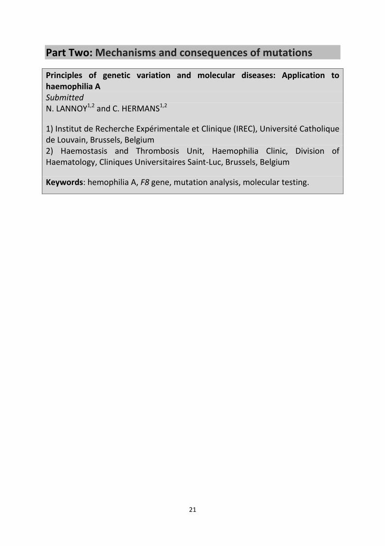
Part Two Mechanisms and consequences of mutations
Principles of genetic variation and molecular diseases Application to haemophilia A Submitted N LANNOY12 and C HERMANS12
1) Institut de Recherche Expeacuterimentale et Clinique (IREC) Universiteacute Catholique de Louvain Brussels Belgium 2) Haemostasis and Thrombosis Unit Haemophilia Clinic Division of Haematology Cliniques Universitaires Saint-Luc Brussels Belgium
Keywords hemophilia A F8 gene mutation analysis molecular testing
22
Abstract
DNA structure alterations are the ultimate source of genetic variations Without them evolution
would be impossible While they are essential for DNA diversity defects in DNA synthesis can lead to
numerous genetic diseases Due to increasingly innovative technologies our knowledge of the
human genome and genetic diseases has grown considerably over the last few years allowing us to
detect another class of variants affecting the chromosomal structure DNA sequence can be altered
in multiple ways DNA sequence changes by substitution deletion or duplication of some
nucleotides chromosomal structure alterations by deletion duplication translocation and inversion
ranging in size from kilobases to megabases changes in the cells genome size If the alteration is
located within a gene and sufficiently deleterious it can cause genetic disorders
Due to the F8 genersquos high rate of new small mutations and its location at the tip of X chromosome
containing high repetitive sequences a wide variety of genetic variants have been described as the
cause of hemophilia A (HA) In addition to the intron 22 inversion HA can also result from point
mutations other inversions complex rearrangements such as duplications or deletions and
transposon insertions causing phenotypes of variable severity characterized by complete or partial
deficiency of circulating FVIII
This review aims to present the origins mechanisms and consequences of F8 alterations A sound
understanding of the multiple genetic mechanisms responsible for HA is essential to determine the
appropriate strategy for molecular diagnosis and detect each type of genetic variant
23
Introduction
In the last 25 years of the twentieth century our knowledge about human genetic variations was
primarily limited to restriction identifying single nucleotide and microsatelliteminisatellite variants
by means of traditional polymer chain reaction- (PCR) based DNA sequencing In recent years the
rapid development and expanded use of microarray technologies such as array comparative
genomic hybridization (aCGH) and next-generation sequencing (NGS) have led to the discovery of
submicroscopic structural variations that are not identified by classical sequencing and not visible
using traditional light microscopes The size of these rearrangements termed copy-number
variations (CNVs) is estimated to range from kilobases (kb) to megabases (mb) These
rearrangements can involve deletions duplications or insertions of DNA sections and account for a
significant amount of the individual variability within species It is estimated that over 13 of the
human genome is affected by numerous CNVs These appear to be the main source of genetic
diversity competing with the single nucleotide variants (SNVs) [1] On account of this it is clear that
the sequence of a gene can be altered in a number of ways small-scale mutations such as those
affecting a gene in one or a small number of nucleotides including point mutations (missense
nonsense splicing and small deletionsduplications) large-scale mutations that alter the
chromosomal structure causing large duplicationsdeletions translocations inversions and
insertions Thus if the variant is sufficiently deleterious to affect the gene structure and the protein
synthesis associated with it this causes genetic disease [2-4]
This study sought to review the different variants reported in the F8 gene which encodes the
coagulation Factor VIII and their underlying mechanisms Hemophilia A (HA) is an X-linked congenital
bleeding disorder caused by a lack or dysfunction of coagulation Factor VIII and is classified as
severe (lt1) moderate (1ndash5) or mild (5ndash40) according to the FVIII plasma activity The genetic
alterations responsible for hemophilia can be classified into three categories The first consists of
alterations that change the sequence of the genersquos components or gene variants including the
promoter exons and introns The large size of the F8 gene results in a high rate of new small
mutations (25x10-5 to 42x10-5 versus the median mutation rate estimated at approximately 1x10-
6genecell division) The F8 location at the tip of X chromosome (Xq28) which contains high
repetitive sequences in close proximity to each other renders this gene region prone to
rearrangement of the second category classified as chromosome variants thus accounting for the
wide variety of large genetic alterations observed in HA patients [5-7] The third source of DNA
damage corresponds to the insertion of mobile elements termed ldquotransposonsrdquo which has also
been described among HA patients
24
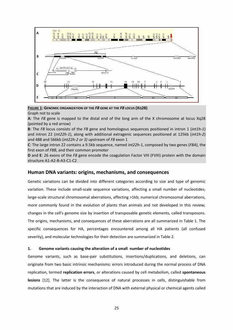
Genomic organization of F8 at locus Xq28
First cloned in 1984 the Factor VIII gene (F8) was mapped to the distal end of the long arm of the X
chromosome (Xq28) on the minus strand This gene spans 186kb (hg19 chrX154064064-154250998
UCSC genome browser see httpgenomeucscedu) of genomic DNA The gene is divided into 26
exons and transcribed into an mRNA of 9029bp with a coding sequence of 7053 nucleotides
encoding a mature protein of 2332 amino acid residues that are arranged within six domains
organized as follows A1-a1-A2-a2-B-a3-A3-C1-C2 Compared to all other exons and with the
exception of exon 26 that mostly codes for the 3rsquo untranslated region exon 14 is very long
measuring 3106bp and encodes most of the B domain (Figure 1) The F8 gene contains the
particularly large intron 22 measuring 328kb which exhibits several particularities These include
the presence of a bidirectional promoter that initiates the transcription of expressed genes (F8A and
F8B) and whose function is not yet well understood The intronless F8A (OMIM 305423) spans 2kb
and is transcribed in the opposite direction to F8 It encodes a 40-kDa Huntingtin-associated protein
[8] thought to be involved in the aberrant nuclear localization of the huntingtin protein observed in
Huntingtonrsquos disease F8B (OMIM 305424) is transcribed in the same direction using a private exon
within intron 22 that is spliced to exons 23 through 26 with the F8 reading frame creating a final
overlapping transcript spanning 25kb The function of the F8B transcript and its potential translated
product remain unknown [9] This arrangement is further complicated by the association between
these three sequences (F8A the first exon of F8B and their common promoter) within a 95kb
fragment labelled int22h-1 that is duplicated at two positions towards the Xq-telomere (int22h-2 and
int22h-3) situated more telomerically at approximately 488 and 566kb [10] Int22h-2 and int22h-3
demonstrate 9993 overall similarity while the homology between Int22h-1 and int22h-3 is of
9924 and that of the other int22h-2 repeats 9918 Interestingly the statistics and distribution of
the sequence differences between the homologous copies strongly support theories that the copy in
the F8 gene was introduced by a duplication more than 25 million years ago [11] Similar to the
int22h sequence a different repeat has been identified within the F8 intron 1 labelled int1h-1 and
located approximately 1526kb downstream of exon 1 An inverted homologous copy of this was
found approximately 125kb upstream of F8 (labelled int1h-2) Bagnall et al postulated that int1h like
int22h duplicated more than 25 million years ago [11]
25
FIGURE 1 GENOMIC ORGANIZATION OF THE F8 GENE AT THE F8 LOCUS (XQ28) Graph not to scale A The F8 gene is mapped to the distal end of the long arm of the X chromosome at locus Xq28 (pointed by a red arrow) B The F8 locus consists of the F8 gene and homologous sequences positioned in intron 1 (int1h-1) and intron 22 (int22h-1) along with additional extragenic sequences positioned at 125kb (int1h-2) and 488 and 566kb (int22h-2 or 3) upstream of F8 exon 1 C The large intron 22 contains a 95kb sequence named int22h-1 composed by two genes (F8A) the first exon of F8B and their common promoter D and E 26 exons of the F8 gene encode the coagulation Factor VIII (FVIII) protein with the domain structure A1-A2-B-A3-C1-C2
Human DNA variants origins mechanisms and consequences
Genetic variations can be divided into different categories according to size and type of genomic
variation These include small-scale sequence variations affecting a small number of nucleotides
large-scale structural chromosomal aberrations affecting gt1kb numerical chromosomal aberrations
more commonly found in the evolution of plants than animals and not developed in this review
changes in the cells genome size by insertion of transposable genetic elements called transposons
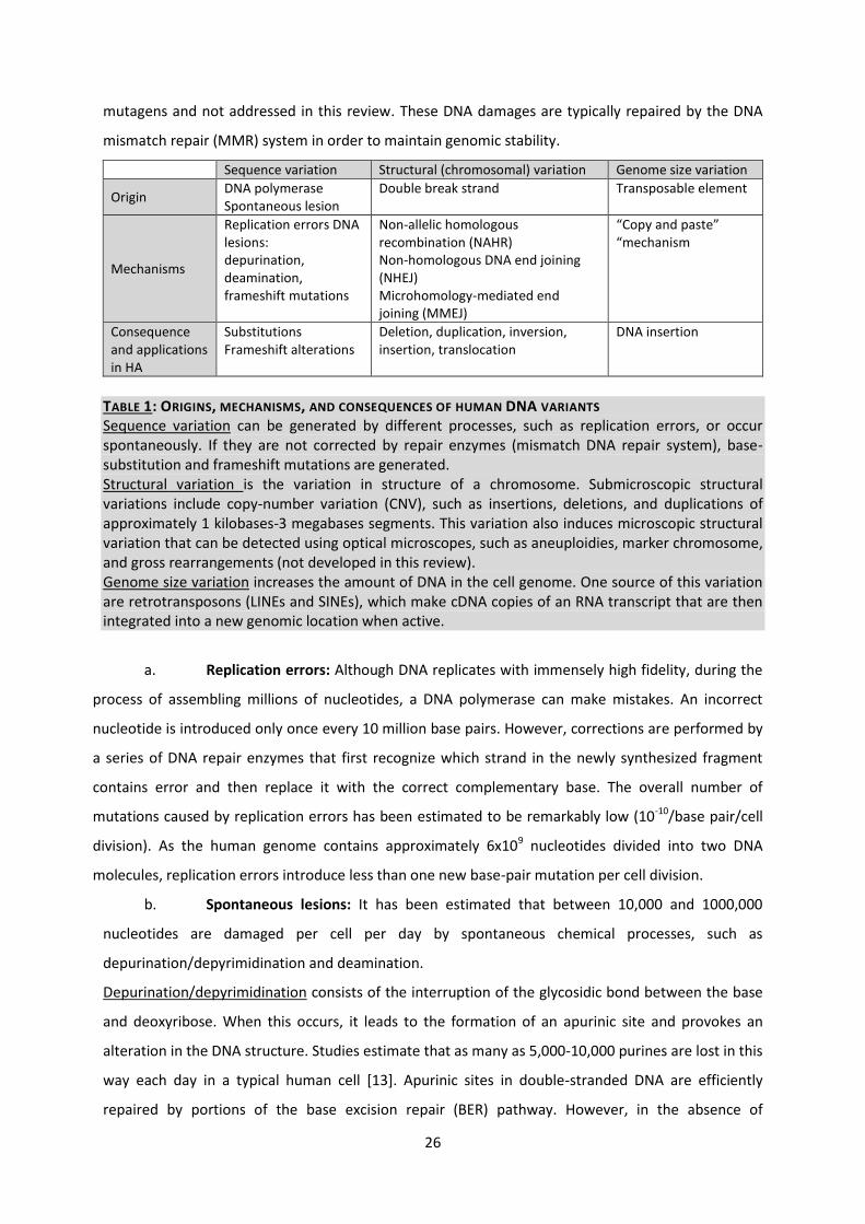
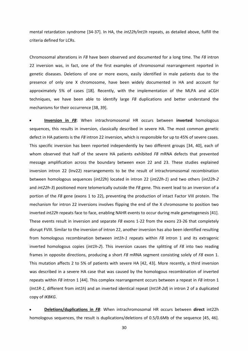
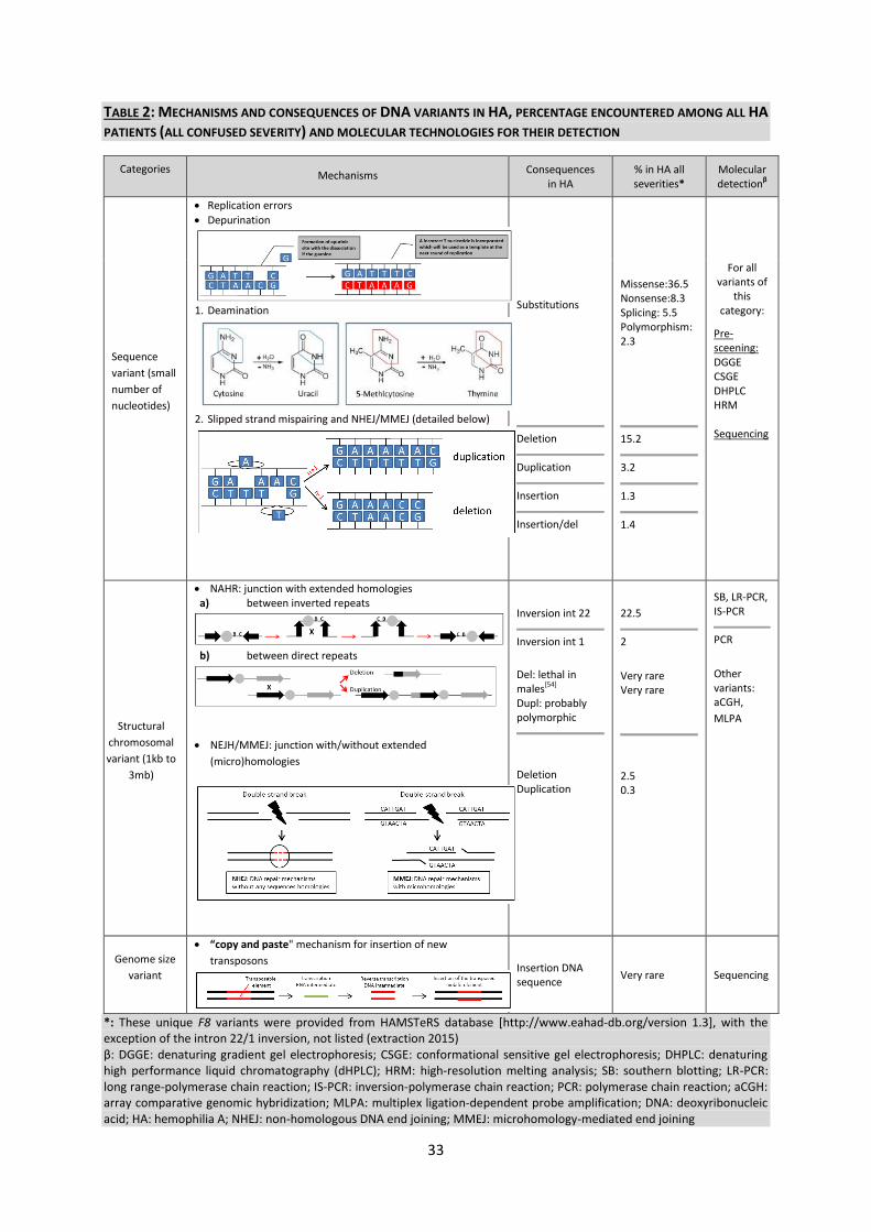
The origins mechanisms and consequences of these aberrations are all summarized in Table 1 The
specific consequences for HA percentages encountered among all HA patients (all confused
severity) and molecular technologies for their detection are summarized in Table 2
1 Genome variants causing the alteration of a small number of nucleotides
Genome variants such as base-pair substitutions insertionsduplications and deletions can
originate from two basic intrinsic mechanisms errors introduced during the normal process of DNA
replication termed replication errors or alterations caused by cell metabolism called spontaneous
lesions [12] The latter is the consequence of natural processes in cells distinguishable from
mutations that are induced by the interaction of DNA with external physical or chemical agents called
26
mutagens and not addressed in this review These DNA damages are typically repaired by the DNA
mismatch repair (MMR) system in order to maintain genomic stability
Sequence variation Structural (chromosomal) variation Genome size variation
Origin DNA polymerase Spontaneous lesion
Double break strand Transposable element
Mechanisms
Replication errors DNA lesions depurination deamination frameshift mutations
Non-allelic homologous recombination (NAHR) Non-homologous DNA end joining (NHEJ) Microhomology-mediated end joining (MMEJ)
ldquoCopy and pasterdquo ldquomechanism
Consequence and applications in HA
Substitutions Frameshift alterations
Deletion duplication inversion insertion translocation
DNA insertion
TABLE 1 ORIGINS MECHANISMS AND CONSEQUENCES OF HUMAN DNA VARIANTS Sequence variation can be generated by different processes such as replication errors or occur spontaneously If they are not corrected by repair enzymes (mismatch DNA repair system) base-substitution and frameshift mutations are generated Structural variation is the variation in structure of a chromosome Submicroscopic structural variations include copy-number variation (CNV) such as insertions deletions and duplications of approximately 1 kilobases-3 megabases segments This variation also induces microscopic structural variation that can be detected using optical microscopes such as aneuploidies marker chromosome and gross rearrangements (not developed in this review) Genome size variation increases the amount of DNA in the cell genome One source of this variation are retrotransposons (LINEs and SINEs) which make cDNA copies of an RNA transcript that are then integrated into a new genomic location when active
a Replication errors Although DNA replicates with immensely high fidelity during the
process of assembling millions of nucleotides a DNA polymerase can make mistakes An incorrect
nucleotide is introduced only once every 10 million base pairs However corrections are performed by
a series of DNA repair enzymes that first recognize which strand in the newly synthesized fragment
contains error and then replace it with the correct complementary base The overall number of
mutations caused by replication errors has been estimated to be remarkably low (10-10base paircell
division) As the human genome contains approximately 6x10⁹ nucleotides divided into two DNA
molecules replication errors introduce less than one new base-pair mutation per cell division
b Spontaneous lesions It has been estimated that between 10000 and 1000000
nucleotides are damaged per cell per day by spontaneous chemical processes such as
depurinationdepyrimidination and deamination
Depurinationdepyrimidination consists of the interruption of the glycosidic bond between the base
and deoxyribose When this occurs it leads to the formation of an apurinic site and provokes an
alteration in the DNA structure Studies estimate that as many as 5000-10000 purines are lost in this
way each day in a typical human cell [13] Apurinic sites in double-stranded DNA are efficiently
repaired by portions of the base excision repair (BER) pathway However in the absence of
27
information from the complementary strand BER can add an incorrect base at the apurinic site
resulting in a substitution mutation
The deamination of the cytosine consists of the removal of the amine group by hydrolysis reaction
This is a common process and results in the replacement of cytosine by uracil Deamination can also
occur with guanine and adenine leading to the production of xanthine and hypoxanthine
respectively The deamination of the cytosine can be easily repaired by the uracil-DNA glycosylase
which recognizes the uracil residues in the DNA as well as by the BER mechanism
However the deamination of 5-methylcytosine which generates thymine and ammonia poses a
particular problem since it is not recognized by the uracil-DNA glycosylase enzyme as thymine is a
normal base of DNA and thus is not repaired The major form of DNA modification in the human
genome involves the methylation of cytosine residues especially when 5-methylcytosine
immediately precedes a guanine such as in the dinucleotide 5rsquo-CG-3rsquo The spontaneous deamination
of 5-methylcytosine in the CG doublet gives rise to CgtT or GgtA substitutions depending on the
strand of DNA in which the 5-methylcytosine is located This type of mutation thus represents over
30 of all single nucleotide substitutions in the human genome and they occur at a rate that is 25
times greater than that of any other single nucleotide mutation [14] This is why the CG dinucleotide
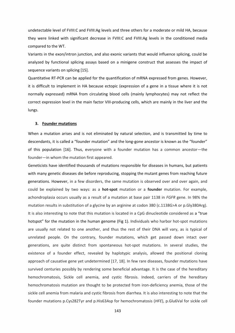
represents a ldquotrue hotspotrdquo for mutation in the human genome
Classical HA is caused by widespread deleterious mutations in the F8 gene The mutations that create
significant disruption in F8 lead to severe disease whereas those that alter apparently ldquominorrdquo
regions of the Factor VIII protein result in mild to moderate disease HA also presents with genetic
heterogeneity with over 2015 variants described in the HAMSTeRS database [httpwwweahad-
dborgversion 13] These 2015 variants correspond to 5472 individual case reports and include
substitutions as well as small and large duplicationsdeletions The vast majority of these variations
are substitutions producing missense nonsense in the coding sequence and splice defects of the
mRNA when located in the exonintron junction Recently substitutions in the promoter and deep
intron have also been reported as associated with mild HA [15-17]
The F8 gene is composed of 70 cytosine-phosphate-guanine (CpG) sites that are not mutated in the
same proportion [18 19] However nearly 35 of all F8 substitution mutation reports (point and
polymorphism mutations providing missense nonsense and silent effects) occur at one of these CG
dinucleotides The Lannoy et al study suggested that if the same recurrent substitution shared by
unrelated HA patients is located somewhere in the F8 gene the founder effect is the more probable
mechanism than for those positioned precisely in the 5rsquo-CG-3rsquo dinucleotide [19] A number of reports
have in fact described F8 mutations that are present in high frequencies in specific populations
28
c6104TgtC (pVal2035Alanine) c1538-18GgtA c788-14TgtG and c3780CgtG (pAsp1260Glu) [20-23]
These substitutions were not located in the 5rsquo-CG-3rsquo dinucleotide and have in all cases been
reported in subjects exhibiting the mild HA phenotype and who share a common ancestor Through
haplotyping patients sharing the same mutation in a group of HA-positive Swedish families 22
were revealed to have a common ancestor [24] This study also revealed that some mutations could
date back to the Middle Ages When investigating the location of these Swedish mutations in
patients who apparently share a common ancestor they were all found to be located outside of a 5rsquo-
CG-3rsquo dinucleotide site (c67 c121 c1195 c1244 c1595 c2211 c3146 c5093 c6371 c6658
c6680 and c6932) except for those located at c1834 and c5806
Spontaneous frameshift mutations Streisinger et al observed that frameshift mutations in
bacteriophages tended to occur in areas with runs of repeats of one nucleotide He proposed that
these frameshifts were the result of slipped mispairing or strand slippage between the template
DNA strand and the newly synthesized strand during DNA replication [25] Subsequent studies on
genes taken from other organisms including humans have since demonstrated that runs of repeated
nucleotides are indeed hotspots for frameshift mutations [26 27]
Another mutation hotspot has thus been identified in the F8 gene associated with frameshift
deletion or duplication It has in fact been shown that 25 of all F8 small deletionsduplications
affect one of the two adenine stretches composed of eight or nine adenine nucleotides within exon
14 (at codons 1210-1213 and 1458-1460) [18] The remaining 75 of small
deletionsduplicationsinsertions are the result of poor DNA breaks repairs by the non-homologous
DNA end joining (NHEJ) and microhomology-mediated end joining (MMEJ) pathways described
below
2 Chromosomal variant providing inversions and CNVs
In human cells both normal metabolic activities and environmental factors such as UV light and
radiation can cause DNA damage like double-strand breaks (DSBs) resulting in as many as a million
individual molecular lesions per cell per day [28] There are three principal mechanisms that enable
DSB repair thereby ensuring genome stability is maintained homologous recombination (HR)
between homologous sequences the non-homologous DNA end joining (NHEJ) and microhomology-
mediated end joining (MMEJ) pathways
The homologous recombination mechanism HR is critical both for repairing DNA lesions in
mitosis and for chromosomal pairing and exchange during meiosis It is also the most accurate
mechanism as it requires extensive DNA sequence identity and uses an intact copy of the DNA from
the sister chromatid or homologue chromosome to repair the break
29
The non-homologous or microhomology recombination mechanisms In addition to the HR
pathways there are other mechanisms that serve to repair DSBs which use little or no homology
sequences and are termed non-homologous recombination mechanisms NHEJ is referred to as non-
homologous due to the break ends being directly ligated without the need for a homologous
template This contrasts with HR pathways which require a homologous sequence to guide the
repair The Microhomology-mediated end joining (MMEJ) mechanism should be distinguished from
NHEJ by its use of 5ndash25 base-pair microhomologous sequences in order to align the broken strands
prior to joining them Other DNA replication-based mechanisms such as fork stalling and template
switching (FoSTeS) or microhomology-mediated break-induced replication (MMBIR) have been
proposed to repair DSBs [29-32]
When homology is not used to ensure that the DBSs are rejoined in the correct positions the process
can cause damage in the chromosome and induce CNVs The detection of chromosomal changes
requires specialized technologies such as aCGH multiplex ligation dependent probe amplification
(MLPA) and NGS Taking advantage of these new bio-molecular technologies recent studies have
demonstrated that the human genome is also composed of high-order sequences that predispose it
to genome instability and CNV formation [1] These genomic architectural features are primarily
highly homologous sequences located throughout the human genome named low-copy repeats
(LCRs) or segmental duplications (SDs) Typically they measure 10-300kb in length and exhibit a
gt97 sequence identity yet are not alleles Though rare in most mammals LCRs comprise a large
portion of the human genome estimated at up to 5 due to a significant expansion that occurred
during primate evolution over the past 25-40 million years
Due to their high degree of sequence identity these non-allelic copies (LCRs) provide a substrate for
genomic rearrangement via non-allelic homologous recombination (NAHR) when misalignment
occurs during meiosis or mitosis In this way if a crossover forms when high homologous sequences
are positioned in non-allelic positions and in direct orientation the result is duplication and deletion
of sequences between the repeats These can subsequently segregate from each other at the next
cell division thus changing the copy number in both daughter cells Crossing over during
intrachromosomal recombination between inverted repeats leads to the inversion of the sequence
located between them The NAHR mechanism occurring between repeats on different
chromosomes can lead to chromosomal translocation In summary this NAHR mechanism is one of
the principal processes for recurrent rearrangements [33] The Xq28 locus is considered a
rearrangement hotspot for NAHR and is associated with several disease phenotypes such as
hemophilia A incontinentia pigmenti Emery-Dreifuss muscular dystrophy and the MECP2 X-linked
30
mental retardation syndrome [34-37] In HA the int22hint1h repeats as detailed above fulfill the
criteria defined for LCRs
Chromosomal alterations in F8 have been observed and documented for a long time The F8 intron
22 inversion was in fact one of the first examples of chromosomal rearrangement reported in
genetic diseases Deletions of one or more exons easily identified in male patients due to the
presence of only one X chromosome have been widely documented in HA and account for
approximately 5 of cases [18] Recently with the implementation of the MLPA and aCGH
techniques we have been able to identify large F8 duplications and better understand the
mechanisms for their occurrence [38 39]
Inversion in F8 When intrachromosomal HR occurs between inverted homologous
sequences this results in inversion classically described in severe HA The most common genetic
defect in HA patients is the F8 intron 22 inversion which is responsible for up to 45 of severe cases
This specific inversion has been reported independently by two different groups [34 40] each of
whom observed that half of the severe HA patients exhibited F8 mRNA defects that prevented
message amplification across the boundary between exon 22 and 23 These studies explained
inversion intron 22 (Inv22) rearrangements to be the result of intrachromosomal recombination
between homologous sequences (int22h) located in intron 22 (int22h-1) and two others (int22h-2
and int22h-3) positioned more telomerically outside the F8 gene This event lead to an inversion of a
portion of the F8 gene (exons 1 to 22) preventing the production of intact Factor VIII protein The
mechanism for intron 22 inversions involves flipping the end of the X chromosome to position two
inverted int22h repeats face to face enabling NAHR events to occur during male gametogenesis [41]
These events result in inversion and separate F8 exons 1-22 from the exons 23-26 that completely
disrupt FVIII Similar to the inversion of intron 22 another inversion has also been identified resulting
from homologous recombination between int1h-1 repeats within F8 intron 1 and its extragenic
inverted homologous copies (int1h-2) This inversion causes the splitting of F8 into two reading
frames in opposite directions producing a short F8 mRNA segment consisting solely of F8 exon 1
This mutation affects 2 to 5 of patients with severe HA [42 43] More recently a third inversion
was described in a severe HA case that was caused by the homologous recombination of inverted
repeats within F8 intron 1 [44] This complex rearrangement occurs between a repeat in F8 intron 1
(Int1R-1 different from int1h) and an inverted identical repeat (Int1R-2d) in intron 2 of a duplicated
copy of IKBKG
Deletionsduplications in F8 When intrachromosomal HR occurs between direct int22h
homologous sequences the result is duplicationsdeletions of 0506Mb of the sequence [45 46]
31
Other alterations of the F8 structure such as large deletions or duplications are relatively
uncommon in F8 compared to the small genic variants and account for approximately 5 and 007
respectively [18 39] Exon deletion in F8 in male hemophiliacs can be detected through repeated
failures to amplify the gene using PCR a process that is not possible in females due to their second
normal X chromosome Over 206 large deletions have been reported in the Hemophilia A Mutation
database These deletions range from just a few hundreds of bases up to sequences of over 210kb
thus constituting deletion of the entire gene The molecular characterization of these deletion
breakpoints in the F8 gene indicated that NHEJ and microhomology-mediated replication-dependent
recombinations (MMEJ FoSTeS or MMBIR) represent the probable causative mechanisms in these
deletions [47] For large F8 duplications the breakpoints of 10 large F8 duplications were
characterized in the study of Zimmermann et al [39] In nine duplications at least one of the two
breakpoints was mapped within LINEs (long interspersed elements) or SINEs (short interspersed
elements) Molecular mechanisms such as NHEJ MMEJ MMBIR or FoSTeS may mediate the
formation of these duplications through sequence motifs microhomologies and larger stretches of
homology such as Alu repeat sequences
3 Genome size variation
Transposon-induced mutations are the last category of mutations Transposable elements (TEs)
also known as ldquojumping genesrdquo or ldquotransposonsrdquo were first discovered by Barbara McClintock a
USA geneticist and Nobel laureate in 1950 These represent DNA sequences that can move or jump
position within the genome As a result of this movement these insertions provide a source of
genetic variation and in rare cases cause mutations that lead to disease TEs are categorized into
two major classes based on their distinct mechanisms of transposition Only the retrotransposons
remain actively mobile in the human genome These are represented by the LINEs which represent
the most abundant autonomous retrotransposons in humans accounting for approximately over one
million copies and representing 19 of human DNA as well as the SINEs or Alu sequences These
retrotransposons have the ability to duplicate and insert themselves into a new location in the
genome by means of a copy and paste mechanism These LINEs and SINEs still have a significant
impact on the human genome and their jumping can lead to sporadic cases of disease such as
hemophilia B muscular dystrophy and neurofibromatosis Type 1 if they insert themselves into the
functional gene [48-50]
In hemophilia A rearrangements like the insertion of LINE sequences of several kb (38 and 23kb
respectively) into F8 exon 14 have been described in a few unrelated sporadic cases of HA [51]
32
Polymorphism versus causal mutation
One of the major genetic challenges is to distinguish causal change from polymorphism Due to the
high rate of new small mutations that occur in the F8 gene unreported substitutions are frequently
identified during molecular analysis screening Special care should thus be taken when novel
substitutions are identified before concluding that they are responsible for HA disease Guidelines
have been published to help predict what consequences these new F8 variants will have on protein
function [52]
Numerous identified human CNVs represent benign polymorphic variants Interpreting the clinical
relevance of copy number variation is thus a complex exercise and requires both cytogenetic and
molecular genetic expertise as well as the involvement of an experienced geneticist [53] Due to its
location at the tip of the X chromosome which contains high repetitive sequences the F8 locus has
been widely reported to harbor CNVs with the vast majority being deleterious However the
duplication of 0506kb sections involving F8 gene exons 1-22 is probably not associated with HA or
intellectual disability [45]
33
TABLE 2 MECHANISMS AND CONSEQUENCES OF DNA VARIANTS IN HA PERCENTAGE ENCOUNTERED AMONG ALL HA
PATIENTS (ALL CONFUSED SEVERITY) AND MOLECULAR TECHNOLOGIES FOR THEIR DETECTION
Categories Mechanisms
Consequences in HA
in HA all severities
Molecular detectionβ
Sequence
variant (small
number of
nucleotides)
Replication errors
Depurination
1 Deamination
2 Slipped strand mispairing and NHEJMMEJ (detailed below)
Substitutions
Deletion
Duplication
Insertion
Insertiondel
Missense365 Nonsense83 Splicing 55 Polymorphism23
152
32
13
14
For all variants of
this category
Pre-sceening DGGE CSGE DHPLC HRM Sequencing
Structural
chromosomal
variant (1kb to
3mb)
NAHR junction with extended homologies a) between inverted repeats
b) between direct repeats
NEJHMMEJ junction withwithout extended
(micro)homologies
Inversion int 22
Inversion int 1
Del lethal in males[54] Dupl probably polymorphic
Deletion Duplication
225
2
Very rare Very rare
25 03
SB LR-PCR IS-PCR
PCR
Other variants aCGH
MLPA
Genome size
variant
ldquocopy and paste mechanism for insertion of new
transposons
Insertion DNA sequence
Very rare Sequencing
These unique F8 variants were provided from HAMSTeRS database [httpwwweahad-dborgversion 13] with the exception of the intron 221 inversion not listed (extraction 2015) β DGGE denaturing gradient gel electrophoresis CSGE conformational sensitive gel electrophoresis DHPLC denaturing high performance liquid chromatography (dHPLC) HRM high-resolution melting analysis SB southern blotting LR-PCR long range-polymerase chain reaction IS-PCR inversion-polymerase chain reaction PCR polymerase chain reaction aCGH array comparative genomic hybridization MLPA multiplex ligation-dependent probe amplification DNA deoxyribonucleic acid HA hemophilia A NHEJ non-homologous DNA end joining MMEJ microhomology-mediated end joining
34
Conclusion
Our knowledge of the human genome has grown considerably over the last 10 years advanced by
the identification of CNVs It has now become clear that the human genome mutates recombines
deletes and duplicates to ensure its survival and evolution adapting to on-going environmental
changes However if a DNA variant affects the structure of a gene protein synthesis can be changed
to such a degree that it no longer functions properly and leads to disease
Due to the high rate of new small mutations in the F8 gene as well as its location at the tip of the X
chromosome alterations in F8 are frequent and are caused by the three molecular mechanisms
associated with genetic diversity Our understanding of these causal mutations and their mechanisms
is essential to determine the optimal strategy of molecular diagnostics for HA cases given that each
type of genetic change requires a different technique for detection
35
References
1 Stankiewicz P Lupski JR Structural variation in the human genome and its role in disease Annu
Rev Med 2010 61437-455 doi 101146annurev-med-100708-204735
2 Cooper GM Nickerson DA and Eichler EE Mutational and selective effects on copy-number
variants in the human genome Nat Genet 2007 39S22ndashS29
3 Lupski JR Stankiewicz P Genomic disorders molecular mechanisms for rearrangements and
conveyed phenotypes PLoSGenet 2005 1e49
4 Lee JA and Lupski JR Genomic rearrangements and gene copy-number alterations as a cause of
nervous system disorders Neuron 2006 52103ndash121
5 Bauters M Van Esch H Friez MJ et al Nonrecurrent MECP2 duplications mediated by genomic
architecture-driven DNA breaks and break-induced replication repair Genome Res 2008 18847-
858
6 Joke Vandewalle Hilde Van Esch Karen Govaerts et al Dosage-Dependent Severity of the
Phenotype in Patients with Mental Retardation Due to a Recurrent Copy-Number Gain at Xq28
Mediated by an Unusual Recombination Am J Hum Genet 2009 85809ndash822
7 Vogel F and A G Motulsky 1997 Human genetics problems and approaches Springer-Verlag
Berlin
8 Peters MF amp Ross CA 2001 Isolation of a 40-kDa Huntingtin-associated protein Journal of
Biological Chemistry 2001 2763188-3194
9 Graw J Brackmann HH Oldenburg J et al Haemophilia A from mutation analysis to new
therapies Nat Rev Genet 2005 6488-501
10 Naylor JA Buck D Green P et al Investigation of the factor VIII intron 22 repeated region (int22h)
and the associated inversion junctions Hum Mol Genet 1995 41217-1224
11 Bagnall R Ayres K Green P and Giannelli F Gene conversion and evolution of Xq28 duplicons
involved in recurring inversions causing severe hemophilia A Genome Res 2005 15214-223
12 Griffiths AJF Miller JH Suzuki DT et al An Introduction to Genetic Analysis 7th edition New
York W H Freeman 2000
13 Lindahl T Instability and decay of the primary structure of DNA Nature 1993 362709-715
36
14 Cooper DN and Youssoufian H lsquoThe CpG dinucleotide and human genetic diseasersquo Hum Genet
1988 78151ndash155
15 Nougier C Roualdes O Fretigny M et al Characterization of four novel molecular changes in the
promoter region of the factor VIII gene Haemophilia 2014 20e149-156 doi
101111hae12346 Epub 2013 Dec 30
16 Castaman G Giacomelli SH Mancuso ME et al Deep intronic variations may cause mild
hemophilia A J Thromb Haemost 2011 91541ndash1548
17 Pezeshkpoor B Zimmer N Marquardt N et al Deep intronic lsquomutationsrsquo cause hemophilia A
application of next generation sequencing in patients without detectable mutation in F8 cDNA J
Thromb Haemost 2013 11 1679ndash1687
18 Oldenburg J Ananyeva NM and Saenko EL Molecular basis of haemophilia A Haemophilia 2004
10 suppl 4133-139
19 Lannoy N Lambert C Vikkula M Hermans C Overrepresentation of missense mutations in mild
hemophilia A patients from Belgium founder effect or independent occurrence Thromb Res
2015 1351057-1063 doi 101016jthromres20150317 Epub 2015
20 Xie YG Zheng H Leggo J et al A founder factor VIII mutation valine 2016 to alanine in a
population with an extraordinarily high prevalence of mild hemophilia A Thromb Haemost 2002
87178-179
21 Santacroce R Santoro R Sessa F et al Screening of mutations of hemophilia A in 40 Italian
patients a novel G-to-A mutation in intron 10 of the F8 gene as a putative cause of mild
hemophilia A in southern Italy Blood Coagul Fibrinolysis 2008 19197-202
22 Reitter-Pfoertner S von Haeseler A Horvath B et al Identification of an ancient haemophilia A
splice site mutation Thromb Res 2012 130445ndash450
23 Casan˜a P Martıacutenez F Cabrera N et al Founder haplotype associated with the factor VIII
Asp1241Glu polymorphism in a cohort of mild hemophilia A patients J Thromb Haemost 2008
61428ndash1430
24 Halldeacuten C Nilsson D Sauml ll T et al Origin of Swedish hemophilia A mutations J Thromb Haemost
2012 102503ndash2511
37
25 Streisinger G Owen J Mechanisms of spontaneous and induced frameshift mutation in
bacteriophage T4 Genetics 1985 109633-659
26 Bergthorsson JT Ejlertsen B Olsen JH et al BRCA1 and BRCA2 mutation status and cancer family
history of Danish women aVected with multifocal or bilateral breast cancer at a young age J Med
Genet 2001 38361ndash368
27 Rowan AJ Lamlum H Ilyas M et al APC mutations in sporadic colorectal tumors A mutational
lsquolsquohotspotrsquorsquo and interdependence of the lsquolsquotwo hitsrsquorsquo PNAS 2000 973352-3357
28 Lodish H Berk A Matsudaira P et al (2004) Molecular Biology of the Cell p963 WH Freeman
New York NY 5th ed
29 Hastings PJ Lupski JR Rosenberg SM Ira G Mechanisms of change in gene copy number Nat Rev
Genet 2009 10551ndash564
30 Hastings PJ Ira G Lupski JR A microhomology-mediated break-induced replication model for the
origin of human copy number variation PLoS Genet 2009 5 e1000327
31 Zhang F Khajavi M Connolly AM et al The DNA replication FoSTeSMMBIR mechanism can
generate genomic genic and exonic complex rearrangements in humans Nat Genet 2009
41849ndash853
32 Zhang F Carvalho CM Lupski JR Complex human chromosomal and genomic rearrangements
Trends Genet 2009 25298-307 doi 101016jtig200905005 Epub 2009 Jun 25
33 Bi W Park SS Shaw CJ et al Reciprocal crossovers and a positional preference for strand
exchange in recombination events resulting in deletion or duplication of chromosome 17p112
Am J Hum Genet 2003 731302-1315
34 Lakich D Kazazian HH Jr Antonarakis SE Gitschier J Inversions disrupting the factor VIII gene are
a common cause of severe haemophilia A Nat Genet 1993 5236-241
35 Aradhya S Woffendin H Bonnen P et al Physical and genetic characterization reveals a
pseudogene an evolutionary junction and unstable loci in distal Xq28 Genomics 2002 7931-40
36 Small K Warren ST Emerin deletions occurring on both Xq28 inversion backgrounds Hum Mol
Genet 1998 7135-139
38
37 Bauters M Van Esch H Friez MJ et al Nonrecurrent MECP2 duplications mediated by genomic
architecture-driven DNA breaks and break-induced replication repair Genome Res 2008 18847-
858
38 Rost S Loumlffler S Pavlova A et al Detection of large duplications within the factor VIII gene by
MLPA J Thromb Haemost 2008 61996-1999
39 Zimmermann M A Oldenburg J Muumlller C R and Rost S Characterization of duplication breakpoints
in the factor VIII gene J Thromb Haemost 2010 82696-2704
40 Naylor J Brinke A Hassock S et al Characteristic mRNA abnormality found in half the patients
with severe haemophilia A is due to large DNA inversions Hum MolGenet 1993 21773ndash1778
41 Rosslier JP Young M Kimberland ML et al Factor VIII gene inversions causing severe hemophilia A
originate almost exclusively in male germ cell Hum Mol Genet 1994 31035-1039
42 Brinke A Tagliavacca L Naylor J et al Two chimaeric transcription units result from an inversion
breaking intron 1 of the factor VIII gene and a region reportedly affected by reciprocal
translocations in T-cell leukaemia Hum Mol Genet 1996 51945-1951
43 Bagnall RD Waseem N Green PM and Giannelli F Recurrent inversion breaking intron 1 of the
factor VIII gene is a frequent cause of severe haemophilia A Blood 2002 99168-174
44 Pezeshkpoor B Rost S Oldenburg J El-Maarri O Identification of a third rearrangement at Xq28
that causes severe hemophilia A as a result of homologous recombination between inverted
repeats J Thromb Haemost 2012 101600-1608 doi 101111j1538-7836201204809x
45 Lannoy N Grisart B Eeckhoudt S et al Intron 22 homologous regions are implicated in exons 1-22
duplications of the F8 gene Eur J Hum Genet 2013 21970-976 doi 101038ejhg2012275
Epub 2013 Jan 9
46 Pegoraro E Whitaker J Mowery-Rushton P et al Familial skewed X inactivation a molecular trait
associated with high spontaneous abortion rate maps to Xq28 Am J Hum Genet 1997 61160-170
47 You GL Ding QL Lu YL Dai J et al Characterization of large deletions in the F8 gene using multiple
competitive amplification and the genome walking technique J Thromb Haemost 2013 111103ndash
1110
39
48 Mukherjee S Mukhopadhyay A Banerjee D et al Molecular pathology of haemophilia B
identification of five novel mutations including a LINE 1 insertion in Indian patients Haemophilia
2004 10259-263
49 Yoshida K Nakamura A Yazaki M et al Insertional mutation by transposable element L1 in the
DMD gene results in X-linked dilated cardiomyopathy Hum Molec Genetics 1998 71129-1132
50 Wallace MR Andersen LB Saulino AM et al A de novo Alu insertion results in neurofibromatosis
type 1 Nature 1991 353864-866
51 Kazazian HH Jr Wong C Youssoufian H et al Haemophilia A resulting from de novo insertion of L1
sequences represents a novel mechanism for mutation in man Nature 1988 332164-166
52 Practice Guidelines for the Molecular Diagnosis of Haemophilia A Guidelines prepared by Steve
Keeney Mike Mitchell and Anne Goodeve on behalf of the UK Haemophilia Centre Doctorsrsquo
Organisation (UKHCDO) the Haemophilia Genetics Laboratory Network and the Clinical Molecular
Genetics Society All network members contributed to amending and approving the text
UPDATED July 2010
httpcmgswebsharedhostingzencoukBPGsBest_Practice_Guidelineshtm
53 Kearney HM Thorland EC Brown KK et al A Working Group of the American College of Medical
Genetics (ACMG) Laboratory Quality Assurance Committee American College of Medical Genetics
standards and guidelines for interpretation and reporting of postnatal constitutional copy number
variants Genetics in Medicine 2011 13680ndash685 doi101097GIM0b013e3182217a3a
54 Pegoraro E Whitaker J Mowery-Rushton P et al Familial skewed X inactivation a molecular trait
associated with high spontaneous abortion rate maps to Xq28 Am J Hum Genet 1997 61160-170
40
41
Aim of the study and experimental strategy
Results developed throughout this work were presented in two parts
The first part mainly studies the F8 gene by establishing a registry of variations from 148 families
with molecular biology technologies (Southern blot sequencing MLPA RT-PCR genotyping) At the
end of the thesis a total of more than 200 patients apparently unrelated have been analyzed As
expected the nature and the percentage of the identified mutations correspond to those published
in the literature However several specific variations that have never been reported previously
require a delicate interpretation to conclude on their deleterious effect or not For this other
approaches reported in the first chapter were undertaken as the study of cDNA bioinformatics or
family cosegregation Another finding of this study is the recurrence (up to more than 10 times) of
four missense mutations in families with mild hemophilia A With allelic segregation analyzes we
have suggested in chapter 2 that an identical mutation shared in several unrelated families is
probably associated with a founder mutation if this mutation does not affect a CpG sequence
The second part of this thesis will focus on the study of three different chromosomal duplications
located in the F8 gene and at its Xq28 locus The first identified in two patients with HA and a third
patient presenting intellectual disability and dysmorphic features involves duplication of 05 or 06
Megabases (Mb) including F8 exons 1 to 22 Molecular analysis of this rearrangement with Southern
blot long-range PCR and aCGH detailed in chapter 3 have been shown to involve repetitive highly
homologous sequences called int22h positioned in direct position in the formation of this type of
duplication by a NAHR mechanism These sequences are present in three copies in the general
population one being located in F8 intron 22 the two extragenic others positioned side by side more
telomerically The genotype-phenotype correlation of these three patients lead to the conclusion
that these specific rearrangements are probably not responsible for HA and should be carefully
considered in genetic counseling while continuing to investigate the causal mutation of hemophilia
However controversy about the causality of the int22h1int22h2-mediated Xq28 duplication in
intellectual disability persists in the scientific literature In chapter 4 we are interested in the third
type of inversion of intron 22 Rarely encountered this inversion is a variation of types 1 and 2 easily
recognized by abnormal profile when it is revealed by Southern blot The three types of inversion
cause a severe phenotype of HA since the 3rsquo extremity of the F8 gene (exons 23-26) is separated
from the rest of the gene (exons 1-22) during a intrachromosomal illegitimate recombination
between two int22h repeats With aCGH technology an approximate 180kb duplication delimited
on either side of the two extragenic copies int22h was found in DNA of three unrelated HA patients
(one with type 3A the two others with type 3B) This duplication in size and location was also
42
reported in the DECIPHER database that classifies this rearrangement as likely benign This
allowed us to hypothesize that there are probably in the general population polymorphic genotypes
with five int22h copies generated by 180kb duplication This rearrangement is not responsible of the
intron 22 inversion but it is technically visible by the method of Southern blot in HA patients with the
third type of inversion Finally the third rearrangement of 210kb explained in the fifth chapter
involves duplication of F8 exon 1 This duplication never reported was found in mild HA patients of
three apparently unrelated families After showing normal sequence of the mRNA and quantitative
deficiency of FVIII activity we postulated that the rearrangement responsible for this duplication
could have arisen due to a tandem inversed duplication producing novel 233kb F8 intron 1 instead of
normally 22kb present in the gene This new intron could provide small amounts of intact F8 mRNA
transcripts which explain the mild severity of patients of these families The family segregation
analyses also suggested that the ~210kb duplication involving F8 exon 1 was likely a single one-time
event that was then disseminated via a founder effect
43
44
45
RESULTS
46
47
SECTION 1
F8 alterations in a cohort of patients with
hemophilia A
48
49
Chapter 1
Computational and molecular approaches for predicting unreported causal missense mutations in Belgian patients with Haemophilia A Haemophilia 2012 18e331-339 Nathalie LANNOY1 Isabelle ABINET1 Arnaud BOSMANS1 Catherine LAMBERT2 Christiane VERMYLEN3 and Cedric HERMANS2
1Center of Human Genetics Cliniques Universitaires Saint-Luc Brussels Belgium 2Haemostasis and Thrombosis Unit Haemophilia Clinic Division of Haematology Cliniques Universitaires Saint-Luc Brussels Belgium 3Department of Pediatrics Cliniques Universitaires Saint-Luc Brussels Belgium
Keywords Haemophilia A gene F8 causal missense mutation bioinformatics approaches genetic counselling HAMSTeRS database
50
Summary
Introduction
Haemophilia A (HA) is caused by widespread mutations in the factor VIII gene The high spontaneous
mutation rate of this gene means that roughly 40 of HA mutations are private This study aimed to
describe the approaches used to confirm private disease-causing mutations in a cohort of Belgian HA
patients
Methods
We studied 148 unrelated HA families for the presence of intron 22 and intron 1 inversion by
Southern blotting and polymerase chain reaction (PCR) Multiplex ligation-dependent probe
amplification (MLPA) assay was used to detect large genomic rearrangements Detection of point
mutations was performed by DNA sequencing Predicting the causal impact of new non-synonymous
changes was studied by two general strategies 1) molecular approaches such as family
cosegregation evaluation of the implicated codon based on phylogenic separated species and
absence of the mutation in the general Belgian population 2) bioinformatics approaches to analyse
the potential functional consequences of missense mutations
Results
Among the 148 HA patients in addition to common intron 22 and intron 1 inversions as well as large
deletions or duplications 67 different point mutations were identified of which 42 had been
reported in the HAMSTeRS database and 25 were novel including 10 null variants for which RNA
analyses confirmed the causal effect of 4 mutations located in a splice site consensus and 15
missense mutations whose causality was demonstrated by molecular approaches and bio-
informatics
Conclusions
This article reports several strategies to evaluate the deleterious consequences of unreported F8
substitutions in a large cohort of HA patients
51
Introduction
Haemophilia A (HA) (OMIM 306700) an X-linked hereditary disease is one of the most common
coagulation disorders with a worldwide incidence of approximately one in 5000 male newborns
Factor VIII gene (F8) maps to the distal end of the long arm of the X-chromosome (Xq28) and spans
186kb of genomic DNA that is divided into 26 exons (approximately 9kb cDNA) containing 2332
amino acids residues that are arranged within six domains organized as A1-a1-A2-a2-B-a3-A3-C1-C2
More than 1000 mutations within the FVIII coding region and splice site junctions including more
than 580 missense mutations have been identified and reported in the major HA mutation database
[1] The type of mutation in F8 predicts HA severity Mutations that produce a significant disruption
in factor VIII lead to severe disease whereas mutations that alter apparently ldquominorrdquo regions of the
factor VIII protein result in mild to moderate disease Typically the most common genetic defect in
HA patients is the F8 intron 22 inversion which is responsible for up to 45 of severe cases [2] An
additional inversion of intron 1 of F8 affecting 2 to 5 of patients with severe HA has been described
[3] The large size of F8 predisposes to deletions and duplications which respectively account for
approximately 5 and 1 [4 5] of characterised mutations in patients with severe HA Single base
pair changes spread all over exons of F8 In severe types they account for 15 of missense
mutations and approximately 25 of other variation types that result in non-sense splice junction
mutations and small deletions or duplications while they comprise almost 100 of moderate and
minor forms
In spite of multiple techniques no mutation is detected in 1 to 2 of HA patients suggesting that
other still-unknown regulatory regions are responsible for the disease [6 7]
The main objective of this study was to describe the current approaches used to confirm disease-
causing mutations in 25 small private mutations that produce mild to severe phenotype changes in
Belgian HA patients To date these mutations have not been reported in HA databases
52
Patients and methods
Patients
Diagnosis of HA was carried out on the basis of the following laboratory analysis FVIII activity using
the one-stage coagulative method (FVIIIC) and chromogenic substrate assays
Haemophilia severity was defined by FVIIIC levels in plasma according to which individuals with
severe HA had lt001 IUl (lt1 of normal) moderate HA 001-005 IUl (1 to 5 of normal) and mild
HA 005-040 IUl (5 to 40 of normal) [8]
In our cohort of 148 Belgian HA subjects mainly followed within the Haemostasis and Thrombosis
Unit of the Cliniques Universitaires Saint Luc Brussels Belgium 74 (50) had severe nine (6)
moderate and 55 (37) mild phenotype Ten (7) of the patients were females presenting with mild
disease or normal factor VIII level who were referred for analysis due to their fathers haemophilia
history (n=6) or their presentation of de novo FVIII (or first case in the family) deficiency and bleeding
symptoms (n=4) As they were the first family cases they acted as case indices
DNA collection
A blood sample (5-10ml) was collected in EDTA from 148 unrelated Belgian patients with HA
followed by a genomic DNA purification from peripheral leucocytes using the salting-out procedure
Detection of intron 1 and intron 22 inversions
Intron 22 inversion was detected by southern blotting In short 7microg of genomic DNA from patients
was digested with BclI and thereafter DNA fragments were electrophoresed in a 08 agarose gel for
20 hours at 35V and then transferred to nylon membranes The membranes were probed with the
09kb EcoRISstI fragment from plasmid p4826 (American Type Culture Collection [ATCC] catalogue
ndeg57203)
The PCR amplification of the F8 intron 1 region and the intron 1 h repeats were performed on 100ng
of genomic DNA in a 25microl reaction volume containing 1x PCR Faststart buffer 1U of Faststart Taq
polymerase (Hoffmann-La Roche Basel Switzerland) 02mM of each dNTP and 10pmol of 9F 9CR
and int1h-2F primers as previously described by Bagnall [3] After the initial denaturing step at 95degC
for 5 min 30 cycles of PCR were carried out at 94degC for 30 sec 65degC for 30 sec and 72degC for 2 min
Finally 5microl of PCR product were electrophoresed for 1 hour in an 8 polyacrylamide gel
MLPA
In order to detect large genomic rearrangements in F8 a specifically designed SALSA MLPA P178 FVIII
kit (ltMRC-Holland Amsterdam The Netherlands) was used for screening [9] The P178 probe mix
contains probes for each of the 26 exons of F8 as well as 11 control probes for sequences located in
other genes More than one probe was present for exons 1 7 12 14 and 26 This probe mix did not
53
contain a probe for exon 22B which was only present in an alternative transcript Procedures had
been described previously [10] and applied according to the manufacturerrsquos protocol Amplification
products were identified on an ABI 3130xl capillary sequencer (Applied Biosystem Foster City CA
USA) using Genescan-ROX-500 as a size standard and were interpreted using GeneMarker software
v15 (SoftGenetics LLC State College PA USA) that normalises data in relation to control peaks from
other genes Peak ratios of lt070 were considered to be deletions and ratios gt140 to be duplications
DNA samples from healthy individuals were used as negative controls
Detection of point mutations by DNA sequencing
All known functional regions including the 26 exons with their immediate 5rsquo and 3rsquo flanking splice
junctions the approximately 12 kb of contiguous promoter sequence corresponding to nucleotide -
1214 to +441 of the F8 reference sequence NM_000132 httpwwwncbinlmnihgov) and the
309bp of flanking 3rsquo-genomic DNA of F8 gene were amplified by the polymerase chain reaction (PCR)
and sequenced as previously described [11 12] Primers for screening exons 17-20 and 24-27 of von
Willebrand factor (vWF) gene were created using Primer 3 software v030 (SourceForge Inc
Massachusets USA) Forward and reverse sequences and amplification conditions were available on
request All primers were synthesized using Sigma Proligo or Eurogentec Amplified DNA fragments
were purified and subjected to direct cycle sequence analysis using the Taq dye-deoxy terminator
method and an ABI PRISM 3130xl Genetic Analyzer sequencer (Applied Biosystem Foster City CA
USA) according to the manufacturerrsquos instructions All the nucleotide changes identified were
confirmed by repeating the PCR and sequencing reactions
Sequence variation nomenclature used to generate the factor VIII genotype (the nucleotide number
was assigned according to the F8 cDNA sequence from A of the initiator ATG site as +1 (NM_000132))
and protein changes are reported according to international recommendations for the description of
sequence variants on the Human Genome Variation Society (HGVS httpwwwHGVSorg update
June 2010) Variants are also reported in italics and in parentheses according to the numbering of
HAMSTeRS (httpeuropiumcscmrcacuk) since this numbering is used in most haemophilia A
reports All identified genomic variations were screened against HAMSTeRS and Alamutreg-Mutation
Interpretation Software (httpwwwinteractive-biosoftwarecom) which displays information from
the Human Gene Mutation Database available to HGMDreg Professional subscribers
RNA Analysis
Total cytoplasmic RNA was isolated from peripheral leucocytes using Tri reagent (Invitrogen Life
Science California USA) according to the manufacturerrsquos protocol based on standard procedures
Primers and conditions had been previously reported [13 14]
54
Analysis of missense mutations
Causal missense mutations were determined according to the following criteria (a) the mutation
cosegregated in other affected or carrier individuals from the same family (b) it was not detected in
200 X-chromosomes from the general Belgian population (c) no other nucleotide variation was
found in the F8 coding region or in exon-intron boundaries (d) the mutated nucleotide was not
reported in non-affected individuals in the available population SNP databases NCBI
(httpwwwncbinlmnihgovprojectsSNP) and the International HapMap Project
(httpwwwhapmaporg) (e) the mutated nucleotide was situated in highly conserved regions of
F8 (f) the mutated residue was conserved or conservatively replaced in F8 from eleven phylogenetic
separated species (human chimpanzee macaque rat mouse dog cat cow chicken xenopus and
tetraodon) and were located in a highly conserved regions of F8 (g) the prediction of variations in
RNA splicing was analysed using the Splice Site Prediction program
(httpfruitlyorgseq_toolssplicehtml) (h) finally three commonly used algorithms to predict the
impact of missence mutations on protein structure and function were employed
PolyPhen-2 (Polymorphism Phenotyping
httpgeneticsbwhharvardedupph2SupplementarySoftware) SIFT (Sorting Intolerant From
Tolerant httpsiftjcviorgwwwSIFT_aligned_seqs_submithtml) Align GVGD
(httpagvgdiarcfragvgd_inputphp)
PolyPhen-2 [15] is an automatic tool for prediction of possible impact of an amino acid substitution
on the structure and function of a human protein This prediction is based on a number of features
comprising Sequence-based features the Position-Specific Independent Counts (PSIC) profile scores
for two amino acid the mapping of the substitution site to known protein 3D structures PolyPhen-2
predicts variants as ldquobenignrdquo ldquopossibly damagingrdquo or ldquoprobably damagingrdquo accompanied by a
prediction confidence score ranging between 1 (probably damaging) to 0 (benign)
SIFT [16] predicts the functional importance of amino acid substitutions and their potential
pathogenicity based on the alignment of orthologous or paralogous protein sequences SIFT scores
were classified as intolerant (000-005) potentially intolerant (0051-010) borderline tolerant
(0101-020) or tolerant (0201-100) according to a classification previously proposed The higher
the tolerance index the less functional impact a particular amino acid substitution was likely to have
and vice versa
Align GVGD [17] is a web-based program that combines the biophysical characteristics of amino acids
and protein multiple sequence alignments to predict where missense substitutions in genes of
interest fall in a spectrum from enriched delterious to enriched neutral GVGD calculates the
Grantham Variation (GV) which measures the degree of biochemical variation among amino acids
found at a given position in the multiple sequence alignment of the gene and the Grantham
55
Deviation (GD) which reflects the lsquobiochemical distancersquo of the mutant amino acid from the
observed amino acid at a particular position (given by GV)If (GV = 0) and (GD gt 0) the position of
interest is invariant (100 conservation) so any mutation at the position is predicted as deleterious
The classifier provides ordered grades ranging from the most likely pathogenic to least likely
pathogenic
56
Results
Overall population of haemophilia patients
In this study F8 mutations were identified in 139 of 148 unrelated patients with a history of HA
representing a mutation detection rate of 94
Intron 22 and intron 1 inversion screening
All patients with severe HA as well as female index cases were initially tested for the presence of F8
intron 22 and intron 1 inversions (n=84) Intron 22 was found in 44 (3784) of patients 34 affected
male patients and three female index cases All intron 22 inversions were type 1 with exception of
two cases the first corresponding to a type 2 inversion and the second to type 3B as outlined in an
international consortium study [2] Intron 1 inversion was observed in one severe case (12)
Large deletion and unrelated duplication by MLPA assay
Following standard 1 and 22 inversion screening in patients with a severe phenotype and in females
referred for F8 analysis MLPA assay was applied as routine molecular screening Six different large
rearrangements were detected In three families only one exon was found to be deleted exon 2 in
female patient HA86 and exon 5 and 15 in patients HA78 and HA65 respectively Two other female
index cases showed larger deletions heterozygous deletion of exons 1-12 in patient HA73 and
heterozygous deletion of exon 26 extending to the 3rsquoUTR region in patient HA51 (cases HA 86 and 51
had already been reported [11]) A large duplication of exons 1-22 was detected in a sixth patient
with a severe phenotype
When sequencing F8 in moderate and mild HA patients failed to show any causal mutation MLPA
analysis was performed allowing for the identification of two novel large duplications exons 1-22
combined with a triplication of exons 2-14 in a moderate case and only exon 1 in a mild case
As recommended [18] all deletions and duplications affecting only one exon were confirmed by
examining mRNA or using quantitative PCR in order to exclude nucleotide mismatch at the probe
binding site
Small mutations by DNA sequencing
Nucleotide sequencing was carried out in the remaining HA patients corresponding to 98 affected
males and four possible carriers (n=102) allowing for the identification of the causal variant in 93
unrelated families Of the 67 different point mutations detected in our study 42 previously reported
in the HAMSTeRS database were considered to cause HA (Table 1) Thirty severe HA patients
exhibited 20 mutations that were predicted to result in premature termination of the protein two
nonsense three splicing and 15 frameshift mutations as well as 12 missense mutations All mild and
57
moderate cases with the exception of two families with large duplications were associated with
missense mutations comprising 36 different mutations In HA117 family the impact of the mutation
on the FVIII basal level was unknown as no affected male could be assessed Eleven different
missense mutations were found to be present between two and seven times in 37 apparently
unrelated Belgian families with HA
Novel mutations causing haemophilia A
Twenty-five novel small mutations were neither deposited in the HAMSTeRS database nor reported
in recent publications They were identified as relating to 10 null mutations one nonsense five
frameshift and four splice site mutations (Table 2) The remaining 15 mutations were determined to
pertain to missense type to which the aforementioned criteria applied A detailed description of
these mutations is provided in Table 3 None of these mutations was associated with development of
inhibitor to FVIII in affected subjects enrolled in our study
58
Discussion
Our study investigated the first series of HA patients analysed for F8 genomic mutations in Belgium
The causative mutation within F8 was identified in 94 of families Despite repeated sequencing of
all coding exons and their splice site junctions the approximately 12kb promoter and 03kb 3rsquoUTR
region no mutation was detected in two severe (one sporadic one with family history) and seven
mild HA patients with a FVIII level ranging between 15 and 38 (5 sporadic 2 with family history)
The von Willebrand disease type 2N mutation was excluded in all these patients by analysing exons
17-20 and 24-27 of the vWF gene where mutations have been associated with vWFF8 binding
defects [19-22] As expected the analysis of RNA when available (one severe and two mild HA
patients) did not show rearrangements that excluded mutations located deep in the introns and
either affected normal splicing or led to mechanisms causing some unknown rearrangements of F8 as
the cause of HA
Inversions
As expected 44 of severe HA was associated with intron 22 inversion Recent data revealed that
int22h-2 was oriented in the same direction as int22h-1 while int22h-3 was in the opposite direction
to both [23] which allowed for intrachromosomal homologous recombination between int22h-1 and
int22h-3 (F8 ldquodistalrdquo inversion or type 1 inversion) In addition data showed [24 25] that int22h-2
and int22h-3 formed an imperfect palindrome separated by a 65kb loop containing the BclI sites that
characterise int22h-2 and int22h-3 Homologous recombination between int22h-1 and int22h-2 (F8
ldquoproximalrdquo inversion or type 2 inversion) required prior recombination between int22h-2 and int22h-
3 by means of a polymorphic inversion that created alleles where the int22h-2 repeat was the most
telomeric and was in inverse orientation to int22h-1 The frequency of this polymorphic allele may
explain the rarity of the int22h-2 inversion which is five times rarer than that of type 1 [2] In our
study all severe HA patients documented as having inversions exhibited a type 1 inversion except for
one case with a type 2 inversion In one family with severe HA a Southern blot pattern of the intron
22 inversion differing from type 1 and 2 inversions was associated with form 3B as previously
suggested [2] This kind of inversion was explained by the presence of at least two copies of the
tandem palindromic int22h-2 and int22h-3 where the copy int22h-2 was the most telomeric and had
the opposite orientation to int22h-1
Large duplications
Large duplications have been reported in many diseases resulting from Non-Allelic Homologous
Recombination (NAHR) between low-copy repeats oriented in the same position By consequence a
59
simple unequal crossing-over mechanism was expected to produce deletions and duplications by a
reciprocal recombination mechanism [2627]
Theoretically intrachromosomal homologous recombination between F8 int22h-1 and extragenic
int22h homologs involving two normal copies may cause highly deleterious 05Mb deletions and loss
of the 5rsquo portion of F8 including exons 1-22 as suggested in the article of Pegoraro [28] This
recombination may also result in duplication of the 5rsquo portion of F8 without inversion [29] In our
study we described two HA families with two duplications of the 5rsquo portion of F8 containing exons 1-
22 by MLPA assay The first duplication in combination with a triplication of exons 2-14 was present
in one family with moderate HA affecting a 26-year-old male with factor VIIIC levels of 2 and his
obligate carrier mother while the second duplication was identified in another family with severe
HA Small mutation in the functional copy of F8 was excluded by means of repeated sequencing
which suggested the deleterious effect of these types of duplication or triplication These
descriptions that have not yet been reported but only theoretically suggested [29] provide evidence
that duplications exist in the 5rsquo region of F8 and confer different phenotypes
In this study we also identified a familial duplication of exon 1 in males with mild HA NAHR
mechanism of this rearrangement seemed unlikely due to the opposite orientation of the int1h
copies situated in the intron 1 of F8 and mapped to a 70-100kb region telomeric to F8 [3 30] Since
deletions of exon 1 were described in the HA database HAMSTeRS as resulting in a severe
phenotype we propose that duplication in the opposite orientation of one of the inth1h copies
allows for non-allelic homologous recombination mechanism that results in deletion and duplication
of exon 1 However other mechanisms that create large duplication or deletion have also been
reported as corresponding to unequal crossing-over between repetitive elements (SINES or LINES)
and to microhomologies genomic features which would explain the duplication of exon 1 in this
particular family [31 32]
Large deletions
We previously highlighted the efficacy of using MLPA techniques in female patients presenting factor
VIII deficiency in the absence of a family history of haemophilia or von Willebrand disease We
confirm in this report the value of this easy-to-use and fast method for detecting and determining
the size of large deletion and duplication in a single reaction in both patients and carriers Our study
suggests that MLPA should be applied prior to any screening for point mutations in F8 in cases of
severe phenotypes or following a negative mutation result in mild and moderate HA patients
60
Missense mutations already reported
Missense mutations are the most prevalent mutation type responsible for all severities of HA
whereby the severity depends on the location of the mutation Our study confirms the well-known
correlation between genotype and phenotype As expected most patients with a severe phenotype
carry a molecular defect predicting a null allele while missense mutations were found in nearly all
patients with a non-severe phenotype
Missense mutations were located throughout F8 with the exception of exons 2 3 5 10 and 17 In
agreement with the HAMSTeRS mutation database our Belgian series detected very few missense
mutations in exon 14 that represents about half of the coding region and of which codons 741 to
1648 encodes for the F8 B domain a region lacking pro-coagulant activity that is spliced out from the
mature protein These missense mutations pVal727Phe and pArg1708Cys identified in the F8 exon
14 are located in A2 and a3 domains respectively
Unpublished small mutations
Based on the patient data investigated in this paper a total of 25 novel genetic alterations in F8 ie
10 null and 15 missense mutations were not included in the HAMSTeRS database nor identified in
recent reports [33-36]
SEVERITY TYPE OF MUTATION
GEMONIC PROTEINIC EXON
HA117 (female) Unknownsect missense c6195GgtC pTrp2065Cys (pTrp2046Cys) 21
HA149 mild missense c71AgtG pTyr24Cys (pTyr5Cys) 1
HA133 mild missense c683AgtG pHis228Arg (pHis209Arg) 6
HA109plusmn mild missense c733CgtT pArg245Trp (pArg226Trp) 6
HA92 mild missense c751CgtG pHis251Asp (pHis232Asp) 6
HA53 mild missense c775AgtG pArg259Gly (pArg240Gly) 6
HA28 mild missense c1172GgtA pArg391His (pArg372His) 8
HA5 HA47 HA97 mild moderate missense c1293GgtT pLeu431Phe (pLeu412Phe) 9
HA2plusmn mild missense c1648CgtT pArg550Cys (pArg531Cys) 11
HA50 HA124 (female) mild missense c1649GgtA pArg550His (pArg531His) 11
HA48plusmn mild missense c1834CgtT pArg612Cys (pArg593Cys) 12
HA88 mild missense c1982TgtA pIle661Asn (pIle642Asn) 13
HA37 mild missense c1988CgtT pAla663Val (pAla644Val) 13
HA1plusmn mild missense c2099CgtT pSer700Leu (pSer681Leu) 13
HA9 mild missense c5186GgtA pGly1729Glu (pGly1710Glu) 14
HA15 HA74 mild missense c5305GgtA pGly1769Arg (pGly1750Arg) 15
HA40 mild missense c5926CgtG pHis1976Asp (pHis1957Asp) 18
HA52 (female) HA128 mild missense c5954GgtA pArg1985Gln (pArg1966Gln) 18
HA83 mild missense c5981TgtA pLeu1994Pro (pLeu1975Pro) 18
HA103 mild missense c6067GgtA pGlu2023Lys (pGlu2004Lys) 19
HA150 mild missense c6082GgtT pGly2028Trp (pGly2009Trp) 19
HA39HA46 HA81 HA108 HA126 HA127 HA55
mild missense c6089GgtA pSer2030Asn (pSer2011Asn) 19
HA19 mild missense c6371AgtG pTyr2124Cys (pTyr2105Cys) 22
HA113 mild missense c6439GgtA pGly2147Ser (pGly2128Ser) 23
61
HA38 mild missense c6443AgtG pAsn2148Ser (pAsn2129Ser) 23
HA114plusmn mild missense c6505CgtT pArg2169Cys (pArg2150Cys) 23
HA71 mild missense c6506GgtA pArg2169His (pArg2150His) 23
HA10 HA18 HA104 HA105 HA141 HA150 HA17 (female) plusmn
mild missense c6532CgtT pArg2178Cys (pArg2159Cys) 23
HA84 HA115 HA137 HA11
mild missense c6533CgtA pArg2178His (pArg2159His) 23
HA134plusmn mild missense c6544CgtT pArg2182Cys (pArg2163Cys) 23
HA42 mild missense c6823TgtC pTyr2275His (pTyr2256His) 25
HA136 HA13 HA142 HA56
mild moderate missense c6932CgtA pPro2311His (pPro2292His) 26
HA90 moderate missense c73TgtC et c75CgtG pTyr25Gln (pTyr6Gln) 1
HA91 moderate missense c992TgtG pIle331Ser (pIle312Ser) 7
HA68 moderate missense c2059CgtT pLeu687Phe (pLeu668Phe) 13
HA14 HA72plusmn moderate missense c5398CgtT pArg1800Cys (pArg1781Cys) 16
HA64 moderate missense c6350TgtG pIle2117Ser (pIle2098Ser) 22
HA3 severe missense c463TgtA pTyr155Asn (pTyr136Asn) 4
HA41 severe missense c471GgtC pTrp157Cys (pTrp138Cys) 4
HA98 severe missense c757GgtT pVal253Phe (pVal234Phe) 6
HA102 severe missense c830TgtG pIle277Ser (pIle258Ser) 7
HA58 severe splicing c1271+1GgtT pAsp337GlyfsX40 (pAsp318GlyfsX40) 8
HA143plusmn severe missense c1171CgtT pArg391Cys (pArg372Cys) 8
HA16 severe missense c1814AgtG pTyr605Cys (pTyr586Cys) 12
HA94 severe deletion c2006_2007delCT pSer669CysfsX14 (pSer650CysfsX14) 13
HA138 severe deletion c2347_2348delAA pAsn783fsX (pAsn764fsX) 14
HA32 severe missense c2179GgtT pVal727Phe (pVal708Phe) 14
HA26 severe duplication c2945dupA pAsn982Lysfs9 (pAsn963Lysfs9) 14
HA6 severe nonsense c3155TgtA pLeu1052 (pLeu1033) 14
HA80 severe duplication c3637dupA pIle1213Asnfs28(pIle1194Asnfs28) 14
HA25 HA62 HA82 severe deletion c3637delA pIle1213Phefs5 (pIle1194PhefsX5) 14
HA24 severe deletion c3784delG pAla1262Hisfs11 (pAla1243Hisfs11) 14
HA12 severe deletion c4379delA pAsn1460Ilefs4 (pAsn1441Ilefs4) 14
HA129 HA140 severe duplication c4379_4380dupA pAsn1460Lysfs5 (pAsn1441Lysfs5) 14
HA95 severe deletion c4981delG pVal1661Serfs1 (pVal1642Serfs1) 14
HA135plusmn severe missense c5122CgtT pArg1708Cys (pArg1689Cys) 14
HA112 severe deletion c5380_5381delTT pPhe1794Glufs12 (pPhe1775Glufs12) 16
HA27 severe deletion c5956delA pLys1987Lysfs42 (pLys1968LysfsX42) 18
HA44 severe deletion c5999-8_6006del16 pGly2000Gludel39 (pGly1981Gludel39) 19
HA29plusmn severe missense c6046CgtT pArg2016Trp (pArg1997Trp) 19
HA45 severe splicing c6188+1GgtA pLys2039_Gly2063delinsAsp (pLys2020_Gly2044delinsAsp)
20
HA35 severe missense c6241TgtC pTrp2081Arg (pTrp2062Arg) 21
HA21 severe splicing c6274-8AgtG pVal2092_Met2143del (pVal2073_Met2124del)
22
HA63plusmn severe nonsense c6496CgtT pArg2166Ter (pArg2147Ter) 23
HA31 severe missense c6683GgtA pArg2228Gln (pArg2209Gln) 24
HA107 severe deletion c6699delG pArg2234Glyfs9(pArg2215Glyfs9) 24
HA70 severe missense c6797GgtT pGly2266Val (pGly2247Val) 25
TABLE 1 SIXTY-SEVEN DIFFERENT POINT MUTATIONS DETECTED BY DNA SEQUENCING Mutations are reported in two different nomenclature forms as suggested by HGVS and as reported in HAMSTeRS in italic and in parentheses Mutations not reported by the HAMSTeRS or by the Alamutreg-Mutation Interpretation Software plusmn Mutations localised in a CpG dinucleotide sect Only female carriers tested
62
Family Family history (sporadicfamilial)
Genomic level Protein level RNA level Exon
HA58 Sporadic c1271+1GgtT pAsp337Glyfs40 r1134_1271del 8
HA94 Sporadic c2006_2007delCT pSer669Cysfx14 13
HA138 Familial c2347_2348delAA pAsn783fs 14
HA6 Sporadic c3155TgtA pLeu1052 14
HA24 Familial c3784delG pAla1262Hisfs11 14
HA95 Sporadic c4981delG pVal1661Serfs1 14
HA112 Sporadic c5380_5381delTT pPhe1794Glnfs12 16
HA44 Familial c5999-8_6006del16 pGly2000Gludel39 r5999_6115del 19
HA45 Sporadic c6188+1GgtA pLys2039_Gly2063delinsAsp r6116_6187del 20
HA21 Sporadic c6274-8AgtG pVal2092_Met2143del r6274_6429del 22
TABLE 2 TEN NOVEL MUTATIONS IN FACTOR VIII GENE LEADING TO PREMATURE PROTEIN TERMINATION IN
SEVERE HAEMOPHILIA A The column corresponding to RNA level shows results of the study of RNA sample from patients with novel splicing mutations to confirm the deleterious effect on splicing RNA study for the six others null mutations was not performed as indicated by a
Null mutations
As expected all ten null mutations were associated with a severe phenotype Six of these were
nonsense and small duplication or deletion mutations which would result in a frameshift and
premature introduction of a stop codon The four others were novel splicing mutations located in the
splice donor sites of exon 8 for the first and exon 20 for the second and in the splice acceptors of
exon 19 and 22 for the remaining two The study of RNA samples showed an exon skipping for the
mutations c6188+1GgtA (exon 20) and c6274-8AgtG (exon 22) while for the mutation c1271+1GgtT
alteration of the splice acceptor site resulted in an incomplete exon skipping which generated a
cryptic out-of-frame splice site activation of exon 8 RNA analysis of the fourth mutation
corresponding to a deletion of 16pb in the splice acceptor of exon 19 c5999-8_6006del16
confirmed its deleterious effect on splicing
63
TABLE 3 DESCRIPTION AND EVALUATION OF CAUSALITY OF FIFTEEN NOVEL MISSENSE MUTATIONS IN FACTOR VIII GENE IN SEVERE AND MODERATEMILD HAEMOPHILIA A a) Family history of Haemophilia FVIII activity levels description of mutations and clinical severity
Family history FVIIIC Mutation
Clinical severity Nucleotide Protein Exon Domain
HA90 Sporadic 3 c73TgtC and c75CgtG pTyr25Gln 1 A1 Moderate
HA41 Familial lt 1 c471GgtC pTrp157Cys 4 A1 Severe
HA109 Familial 46 c733CgtT pArg245Trp 6 A1 Mild
HA53 Sporadic 40 c775AgtG pArg259Gly 6 A1 Mild
HA102 Familial lt 1 c830TgtG pIle277Ser 7 A1 Severe
HA88 Familial 28 c1982TgtA pIle661Asn 13 A2 Mild
HA1 Sporadic 35 c2099CgtT pSer700Leu 13 A2 Mild
HA9 Familial 20 c5186GgtA pGly1729Glu 14 B Mild
HA103 Familial 41 c6067GgtA pGlu2023Lys 19 C1 Mild
HA150 Familial 17 c6082GgtT pGly2028Trp 19 C1 Mild
HA117 Familial Unknown c6195GgtC pTrp2065Cys 21 C1 Unknown
HA35 Sporadic lt 1 c6241TgtC pTrp2081Arg 21 C1 Severe
HA113 Familial 18 c6439GgtA pGly2147Ser 23 C1 Mild
HA70 Familial lt 1 c6797GgtT pGly2266Val 25 C2 Severe
HA42 Familial 22 c6823TgtC pTyr2275His 25 C2 Mild
Only female carriers tested b) Predicted deleterious effect based on multiple alignment and algorithms studies
Family history
Mutation at protein level
Ex Clinical severity
Multiple Alignment PolyPhen prediction score SIFT prediction Align-GVGD
HA90 Familial pTyr25Gln 1 Moderate YYYYYYYYYYY identical probably damaging (1) Affect function most likely
HA41 Familial pTrp157Cys 4 Severe WWWWWWWWWWW
identical probably damaging (1) Affect function most likely
HA109 Familial pArg245Trp 6 Mild RRRPREQDHKD
dissimilar benign (0012) Affect function most likely
HA53 Sporadic pArg259Gly 6 Mild RRRSRRRRSSA
dissimilar probably damaging (1) Affect function most likely
HA102 Familial pIle277Ser 7 Severe IIIMIIIIIII identical possibly damaging (093) Affect function most likely
HA88 Familial pIle661Asn 13 Mild IIIVVVVVVVA dissimilar possibly damaging (082) Affect function most likely
HA1 Sporadic pSer700Leu 13 Mild SSSSSSSSSKE similar probably damaging (099) Affect function most likely
HA9 Familial pGly1729Glu 14 Mild GGGGGGGGGDG
identical probably damaging (099) Affect function most likely
HA103 Familial pGlu2023Lys 19 Mild EEEEEEEEEEE identical possibly damaging (086) Affect function most likely
HA150 Familial pGly2028Trp 19 Mild GGGGGGGGGEG
identical probably damaging (1) Affect function most likely
HA117 Familial pTrp2065Cys 21 Unknown WWWWWWWWWCW
identical probably damaging (099) Affect function most likely
HA35 Sporadic pTrp2081Arg 21 Severe WWWWWWWWWFW
identical probably damaging (1) Affect function most likely
HA113 Familial pGly2147Ser 23 Mild GGGGGGGGAGG
identical probably damaging (1) Affect function most likely
HA70 Familial pGly2266Val 25 Severe GGGGGGGGGGG
identical probably damaging (1) Affect function most likely
HA42 Familial pTyr2275His 25 Mild YYYFFYYYYFFM
dissimilar probably damaging (099) Affect function most likely
Multiple alignments Each novel missense mutation was compared with sequences from 11 species described above in order to examine the conservation of amino acids All substitutions in severe moderatemild and indefinite clinical severity were classified as ldquoidenticalrdquo if the amino acid was
64
conserved in all 11 phylogenic separated species Human Homo sapiens Primates 1) chimpanzee Pan troglodytes 2) macaque Macaca Rodent 1) rat Rattus norvegicus 2) mouse Mus musculus Laurasiatheria 1) dog Canis faliliaris 2) cat Felis catus 3) cow Bos taurus Bird chicken Gallus gallus domesticus Amphibians xenopus Xenopus laevis and Fish tetraodon tetraodon nigroviridis ldquosimilarrdquo if conserved in nine of the species or dissimilar if it conserved in eight or less species
Missense mutations
Two-thirds of the 15 novel missense mutations affect residues within domains A1 or C1 with five
changes occurring in each domain The pathological consequences of these mutations need to be
examined Although the structure-function relationships of some missense mutations are known or
can be deduced eg alteration of vWF binding site thrombin cleavage or interaction with co-factor
FIXa and FX the structural consequences of most mutations are still undefined Therefore
characterising the F8 causal mutations is crucial not only to better understand the structure-function
relationship of the FVIII molecule but also to give reliable genetic counselling to patients and their
families
One of the most difficult tasks in genetics consists of distinguishing a deleterious mutation from
polymorphism that has no consequence on the protein Thus a number of methods based on genetic
analyses are employed 1) genotype-phenotype correlation 2) segregation of the mutation in
affected patients from a same family 3) localisation of mutations in highly conserved regions of the
gene and in several animal species with a divergent evolution 4) absence of mutations description in
non-affected individuals and in the available population SNP database in order to exclude known
polymorphisms 5) finally change in the polarity or load of amino-acid substitutions that makes it
possible to deduce the causal character of such substitutions The potential functional consequences
of missense mutations must also be examined by bioinformatics approaches as multiple
computational methods have been developed to estimate the deleterious effects of amino acid
substitutions (Fig 1) However it is clear that the pathogenic nature of each novel missense
mutation should be proven by means of the recombinant expression of the mutant F8 although this
analysis is difficult due to the large number of novel mutations in F8 and thus its time-consuming
nature
None of the 15 missense mutations described in this study were found in the 200 X-chromosomes
from the general Belgian population or in the SNP databases All were situated in the highly
conserved regions of F8 The majority of these mutations are transmitted over several generations
and cosegregate in other affected patients or carriers from the same family Moreover several of
these new genetic alterations can be definitively considered as pathologic as a substitution in the
codon that they deteriorate was already reported in HAMSTeRS database This is the case for the
65
missense mutations described in this study pTyr25Gln pTrp157Cys pSer700Leu pGly2028Trp
and pTrp2266Cys
Relying on in silico structural analysis all of the new missense substitutions appeared to have a
significant impact on the protein structure Indeed none of these mutations were found to induce
theoretically alternative splicing and all were predicted to affect protein function with the algorithm
SIFT Align GVGD In addition there was a good correlation between amino-acid conservation
through the phylogenic separated species predictions using the algorithm PolyPhen-2 and the
severe and moderate aspects of HA This correlation also applied to the minor phenotype of the
disease with the exception of pArg245Trp mutation for which the algorithm PolyPhen-2 predicted a
benign effect on the factor VIII function
Since amino-acid polymorphism of FVIII is rare our 15 new missense mutations in HA patients may
be considered as causal in general practice especially when no other variant is detected using
complete F8 sequencing and bioinformatics approaches support the deleterious effects of such
mutation
The strategies proposed in this study to evaluate the deleterious impact of genetic variations of the
FVIII gene have some limitations which should be emphasized One could indeed assume that any
approach based on amino-acid species homologies or predicted change algorithms or even molecular
modelling would miss effects of point mutations on mRNA stability or of missence variants leading to
decreased protein secretion both shown to account for low circulating factor levels associated with
severe hemophilic mutations Indeed few studies have been conducted to understand the reason
why missence mutations are associated with severe phenotype in some patients Different strategies
could be proposed to understand the underlying mechanisms expression by qPCR to explore the F8
mRNA stability sequencing of the mRNA to exclude a splicing defect and CRM (cross-reacting
material) quantification to estimate the level of protein secretion
Conclusion
This study showed the genotype-phenotype correlation in 139 out of 148 unrelated patients among
who 25 showed new deleterious variants corresponding to 10 null and 15 missense mutations that
had never been reported Among the 10 null mutations RNA analyses confirmed the causal effect of
mutations located in a splice site consensus In addition we described two general strategies
currently available to estimate the deleterious effects of amino acid substitutions and assess the
potential causality of 15 new missense mutations identified in F8
66
References
1 HAMSTeRS Database [accessed August 06 2007] httphadborguk
2 Antonarakis SE and the international consortium study Factor VIII gene inversions in severe
Haemophilia A results of an international consortium study Blood 1995 862206-2212
3 Bagnall RD Waseem N Green PM Giannelli F Recurrent inversion breaking intron 1 of the factor
VIII gene is a frequent cause of severe Haemophilia A Blood 2002 99168-174
4 Oldenburg J Ananyeva NM Saenko EL Molecular basis of haemophilia A Haemophilia 2004 10
(suppl 4)133-139
5 Rost S Loumlffler S Pavlova A et al Detection of large duplications within the factor VIII gene by
MLPA J Thromb Haemost 2008 61996-1999
6 Oldenburg J El-Maarri O New insight into the molecular basis of Haemophilia A Int J Hematol
2006 8396-102
7 El-Maarri O Singer H Klein C Watzka M Herbiniaux U Brackmann HH et al Lack of F8 mRNA a
novel mechanism leading to Haemophilia A Blood 2006 1072759-2765
8 White GC 2nd Rosendaal F Aledort LM et al Factor VIII and Factor IX Subcommittee Definitions
in Haemophilia Recommendation of the scientific subcommittee on factor VIII and factor IX of the
scientific and standardization committee of the International Society on Thrombosis and
Haemostasis Thromb Haemost 2001 85560
9 Miller SA Dykes DD Polesky HF A simple salting out procedure for extracting DNA from human
nucleated cells Nucleic Acids Res 1988 161215
10 Schouten JP McElgunn CJ Waaijer R et al Relative quantification of 40 nucleic acid sequences by
multiplex ligation-dependent probe amplification Nucleic Acids Res 2002 30 e57
11 Lannoy N Abinet I Dahan K Hermans C Identification of de novo deletion in the factor VIII gene
by MLPA technique in two girls with isolated factor VIII deficiency Haemophilia 2009 15797-801
12 Oldenburg J Ivaskevicius V Rost S et al Evaluation of DHPLC in the analysis of Haemophilia A J
Biochem Biophys Methods 2001 4739-51
13 Viel K Ameri A Abshire T et al A sequence variation scan of the coagulation factor VIII (FVIII)
structural gene and associations with plasma FVIII activity levels Blood 2007 1093713-24
67
14 El-Maarri O Herbiniaux U Graw J et al Analysis of mRNA in Haemophilia A patients with
undetectable mutations reveals normal splicing in the factor VIII gene J Thromb Haemost 2005
3332-339
15 Adzhubei IA Schmidt S Peshkin L et al A method and server for predicting damaging missence
mutations Nat Methods 2010 7248-249
16 Kumar P Henikoff S and Ng PC Predicting the effects of coding non-synonymous variants on
protein function using the SIFT algorithm Nature Protocols 2009 41073-1081
17 Tavtigian SV Deffenbaugh AM Yin L et al Comprehensive statistical study of 452 BRCA1 missense
substitutions with classification of eight recurrent substitutions as neutral J Med Genet 2006
43295-305
18 Sellner LN Taylor GR MLPA and MAPH New techniques for detection of gene deletions Human
Mutat 2004 23 13-419
19 Casonato A Sartorello F Cattini M G et al An Arg760Cys mutation in the consensus sequence of
the von Willebrand factor propeptide cleavage site is responsible for a new von Willebrand
disease variant Blood 2003 101151-156
20 Hilbert L Jorieux S Proulle V et al Two novel mutations Q1053H and C1060R located in the D3
domain of von Willebrand factor are responsible for decreased FVIII-binding capacity Br J
Haematol 2003 120627-632
21 Hilbert L DOiron R Fressinaud E et al First identification and expression of a type 2N von
Willebrand disease mutation (E1078K) located in exon 25 of von Willebrand factor gene J Thromb
Haemost 2004 22271-2273
22 Hilbert L Nurden P Caron C et al Type 2N von Willebrand disease due to compound
heterozygosity for R854Q and a novel R763G mutation at the cleavage site of von Willebrand
factor propeptide Thromb Haemost 2006 96290-294
23 Ross MT Grafham DV Coffey AJ et al The DNA sequence of the human X chromosome Nature
2005 5236-241
24 Bagnall R Giannelli F and Green P Polymorphism and hemophilia A causing inversions in distal
Xq28 a complex picture J Thromb Haemost 2005 3 2598-2599
25 Bagnall R Giannelli F and Green P Int22h-related inversions causing hemophilia A a novel insight
into their origin and a new more discriminant PCR test for their detection J Thromb Haemost
2006 4591-598
68
26 Chance P Abbas N Lensch M et al Two autosomal dominant neuropathies result from reciprocal
DNA duplicationdeletion of a region on chromosome 17 Hum Mol Genet 1994 3 223-228
27 Shaw C and Lupski J Implications of human genome architecture for rearrangement-base
disorders the genomic basis of disease Hum Mol Genet 2004 13 Spec No 1R57-64
28 Pegoraro E Whitaker J Mowery-Rushton P et al Familial skewed X inactivation a molecular trait
associated with high spontaneous abortion rate maps to Xq28 Am J Hum Genet 1997 61160-
170
29 Green P Hill M Bowen D and the UK Haemophilia Centre Doctorsrsquo Organisation Haemophilia
Genetics Network Duplications involving int22h-1 of the factor VIII gene a cause for concern in
genetic testing for hemophilia A J Thromb Haemost 2007 52155-2156
30 Bagnall R Ayres K Green P and Giannelli F Gene conversion and evolution of Xq28 duplicons
involved in recurring inversions causing severe hemophilia A Genome Res 2005 15214-223
31 Hu X Ray P and Worton R Mechanisms of tandem duplication in the Duchenne muscular
dystrophy gene include both homologous and nonhomologous intrachromosomal recombination
EMBO J 1991 102471-2477
32 Zimmermann M A Oldenburg J Muumlller C R and Rost S Characterization of duplication breakpoints
in the factor VIII gene J Thromb Haemost 2010 82696-2704
33 David D Ventura C Moreira I et al The spectrum of mutations and molecular pathogenesis of
hemophilia A in 181 Portuguese patients Haematologica 2006 91840-843
34 Lin SY Su YN Hung CC et al Mutation spectrum of 122 hemophilia A families from Taiwanese
population by LD-PCR DHPLC multiplex PCR and evaluating the clinical application of HRM BMC
Med Genet 2008 953
35 Riccardi F Tagliaferri A Martorana D et al Spectrum of F8 gene mutations in haemophilia A
patients from a region of Italy identification of 23 new mutations Haemophilia 2010 16791-800
36 Reitter S Sturn R Horvath B et al Spectrum of causative mutations in patients with haemophilia
A in Austria Thromb Haemost 2010 10478-85
69
Chapter 2
Overrepresentation of missense mutations in mild hemophilia A patients from Belgium founder effect or independent occurrence Thromb Res 2015 1351057-1063 doi 101016jthromres20150317 Epub 2015 Mar 17 N LANNOY12 C LAMBERT3 M VIKKULA14 and C HERMANS23
1) Center of Human Genetics Universiteacute Catholique de Louvain Cliniques Universitaires Saint-Luc Brussels Belgium 2) Institut de Recherche Expeacuterimentale et Clinique (IREC) Universiteacute catholique de Louvain Brussels Belgium 3) Hemostasis and Thrombosis Unit Hemophilia Clinic Division of Hematology Cliniques Universitaires Saint-Luc Brussels Belgium 4) Laboratory of Human Molecular Genetics of the Duve Institute Universiteacute catholique de Louvain Brussels Belgium
Keywords hemophilia A Factor VIII Genetics of Thrombosis and Haemostasis mutation analysis microsatellites
70
Summary
Roughly 40 of observed mutations responsible for hemophilia A (HA) are novel and present in
either a single family or a limited number of unrelated families During routine diagnostic analysis of
73 unrelated Belgian patients with mild HA 4 out of 43 different mutations (pSer2030Asn
pArg2178Cys pArg2178His and pPro2311His) were detected in more than one family
representing 35 of total identified mutations
To discriminate between an independent recurrence or a founder effect an analysis of intra- and -
extragenic single nucleotide polymorphisms (SNPs) and short tandem repeats (STRs) flanking the F8
gene was conducted
SNP haplotype and microsatellite analysis revealed strong evidence that pSer2030Asn and
pPro2311His mutations were probably associated with a founder effect The two other mutations
localized in an F8 cytosine-phosphate-guanine (CpG) site likely resulted from recurrent de novo
events
This study suggests that missense mutations producing C-to-T or G-to-A substitutions in CpG
dinucleotide can occur de novo with more repetition than other causal substitutions that do not
affect the CpG site Analysis of F8 database implied that CpG sites throughout the F8 gene are not all
mutated with the same frequency Causes are still unknown and remain to be identified
71
Introduction
Hemophilia A (HA) (OMIM 306700) an X-linked hereditary disease is one of the most common
coagulation disorders caused by a spectrum of mutations in the F8 gene With the exception of
intron 1 and 22 inversions present in approximately 45 of severe hemophilia A cases (Antonarakis
et al1995 Bagnall et al 2002) more than 2015 unique variants spread throughout the F8 genersquos 26
exons have been identified and recorded in the international hemophilia A mutation database
[httpwwweahad-dborgFebruary 2014 update]
Due to the high rate of spontaneous F8 gene mutations approximately 40 of families with
phenotypes of variable severity have affected individuals being either sporadic or confined to one
family branch (Oldenburg et al 2004) However several publications have described F8 mutations
that are highly prevalent in specific populations sharing the same haplotype indicating the existence
of a single common ancestor Examples of this can be seen with the c6104TgtC or pVal2035Alanine
(legacy AA No2016) mutation identified in an isolated population in rural Newfoundland (Xie et al
2002) the exon 13 duplication located in northern Italy (Acquila et al 2004) and the novel G-to-A
mutation (c1538-18GgtA) in intron 10 of the F8 gene which was identified as a putative cause of mild
hemophilia A in southern Italy (Santacroce et al 2008) All these mutations were associated with the
mild HA phenotype
Within the setting of a study comprising 73 apparently unrelated French-speaking Belgian HA families
with mild severity screened for F8 gene mutations 4 different missense mutations were detected as
occurring between 4 and 10 times By way of haplotype analysis constructed with single nucleotide
polymorphisms (SNPs) and microsatellites repeats this study sought to assess the origins of these
mutations We investigated whether they represent a recurrent molecular defect occurring
repeatedly de novo or whether they may have manifested as a single molecular event that was then
disseminated throughout French-speaking Belgium via a founder effect This study and analysis of the
EAHAD database (EAHAD db) will attempt to demonstrate that certain F8 CpG sites (CpG standing
for mdashCmdashphosphatemdashGmdash) are more susceptible to mutations than others and that mutations
frequently reported outside of a CpG site are possibly the result of a founder effect
72
Patients and Methods
Patients
This study involved 73 apparently unrelated subjects exhibiting mild HA with Factor VIII clotting
activity from gt5 to lt40 IUdL (gt5 to lt40 of normal levels) recruited for genetic testing from
hospitals in Brussels and the French-speaking part of Belgium The study was approved by the
medical ethics committee of the medical faculty of the Universiteacute catholique de Louvain Brussels
(Belgium) Blood samples were obtained during medical care where patients were informed and gave
their consent for DNA analysis
Methods
DNA extraction and genotyping of coding sequences
Genomic DNA was extracted from white blood cells using standard procedures and mutation
screening was performed as previously described (Lannoy et al 2012) Causal mutations were
defined in line with recommendations of the Human Genome Variation Society (HGVS
httpwwwHGVSorg June 2010 update) with codon 1 representing the first residue (Met) of the
signal peptide (this is -19 in Legacy numbering) In Legacy numbering codon +1 refers to that coding
for the first amino-acid of the mature FVIII protein (in HGVS numbering this is codon +20) As Legacy
numbering is extensively used in FVIII publications particularly before the year 2000 this numbering
is maintained within parenthesis
In addition 50 DNA samples from anonymous French-speaking Belgian males referred for pre-
conception genetic counselling were selected from the DNA bank and genotyped as below This was
in order to facilitate the interpretation of results obtained by haplotype analysis
SNP and microsatellite genotyping
Flanking markers from 14Mb centromeric to 06Mb telomeric of the F8 locus were genotyped
including the four previously reported (Wehnert et al 1993 Saacutenchez-Garciacutea et al 2005 Lalloz et al
1991 Freije et al 1992) extragenic and intronic microsatellites (3rsquo DXS15ndashDXSint22ndashDXSint13ndash
DXS1108-5rsquo) and ten SNPs as displayed in Appendix section C tables 3C1 and 3C2 The PCR was
conducted in a 25microL containing 1x PCR Faststart buffer 1U of Faststart Taq polymerase (Hoffmann-La
Roche Basel Switzerland) 02mM of each dNTP and 10pmol of each primer Cycling conditions
were set to initial denaturation at 94degC for 5 minutes followed by 30 cycles of amplification These
comprised 30 second denaturation at 94degC 1 minute annealing at 55degC 1 minute elongation at 72degC
and 5 minutes final extension at 72degC A 15microL aliquot of STR PCR product was added to 10microL of Hi-Di
Formamide (Applied Biosystems Foster city CA USA) Amplification products were identified using
73
an ABI3130xl capillary sequencer by employing the Genescan-ROX-500 (Applied Biosystem Foster
City CA USA) size standard and were interpreted with GeneMarker software V15 (SoftGenetics LLC
State College PA USA) Amplified SNP DNA fragments were purified and subjected to direct cycle
sequence analysis using the Taq di-deoxy terminator method and an ABI PRISM 3130xl Genetic
Analyzer sequencer in accordance with the manufacturerrsquos instructions
74
Results
In this cohort routine genetic analysis of the F8 gene was undertaken and 43 different missense
mutations were identified in a group of 73 unrelated subjects from separate families with mild HA
[Appendix section B Table a] Recurrent missense causal mutations were noted 10 times for the
pSer2030Asn (c6089GgtA) in exon 19 7 times for the pArg2178Cys (c6532CgtT) in exon 23 4 times
for the pArg2178His (c6533GgtA) in exon 23 and 4 times for the pPro2311His (c6932CgtA) in exon
26 All mutations were associated with the mild phenotype These 4 mutations accounted for 35 of
all mutations identified in the routine genetic F8 analysis of mild phenotype patients Other recurrent
missense mutations were also observed (pLeu431Phe and pGly1769Arg) However these families
were not included in this study due to a lack of DNA
In the first part of this study haplotypes at the F8 locus were generated with extra- and intragenic
SNPs from Belgian male control individuals among whom 6 different haplotypes were identified as
displayed in Table 1a The most common haplotype (haplotype 4) manifested at 58 prevalence
while 3 others (haplotypes 2 5 and 6) were found at le10 prevalence in the normal population It
was of particular interest to note that 2 haplotypes in the Belgian control population were not
present in the general CEU population which consists of Utah residents with ancestry from northern
and western Europe In contrast one haplotype in the general CEU population was not present in our
Belgian male controls (Table 1b) In order to increase the discriminative value of the Belgian SNP
haplotype we conducted further analyses of the microsatellite data at the F8 locus A total of 35
different haplotypes were identified in the 50 controls (Table 1c)
TABLE 1 HAPLOTYPE RESULTS 1a) Identification of six different SNP haplotypes in 50 Belgian male controls
haplotype 1 = haplotype X5
haplotype 2 = haplotype X2
haplotype 3 haplotype 4 = haplotype X4
haplotype 5 haplotype 6= haplotype X3
rs6643620 G G A A A G
rs5945250 T T G G G G
rs1050705 G G A A A A
rs4898352 T T A A A A
rs6643714 C C T T C C
rs1800291 C G C C C C
rs5987079 A A G G G G
rs6649625 C C C T C C
rs2313058 T T C C C C
rs5945279 T T C C C C
Percentage 10 6 14 58 4 8
75
Haplotypes 3 and 5 are present in the Belgian and not in the general CEU population and haplotype X1 from the CEU is absent in Belgian male controls (details of SNP haplotypes from the CEU population are shown in table 2b) corresponds to the haplotype identified in the general CEU population 1b) Identification of five different SNP haplotypes from the CEU population (Utah residents with ancestry from northern and western Europe) These values were extracted from httpwwwncbinlmnihgovprojectsSNPsnp_gfcgi (consulted on 12 December 1999) from 44 male individuals
Haplotype X1
Haplotype X2
Haplotype X3
Haplotype X4
Haplotype X5
rs6643620 A G G A G
rs5945250 G T G G T
rs1050705 G G A A G
rs4898352 T T A A T
rs6643714 C C C T C
rs1800291 G G C C C
rs5987079 A A G G A
rs6649625 C C C T C
rs2313058 T T C C T
rs5945279 C T C C T
Percentage 450 9 9 6375 1375
1c) Identification of 35 different STR haplotypes in 50 Belgian male controls
DXS15 164 158 154 144 160 156 158 162 154 158 158 160 160 160 164 162 162 156 154 162 158 158 152 162 162 154 162 154 162 154 154 164 158 154 160
STR22 80 80 82 82 80 82 82 82 80 82 82 80 80 82 82 84 80 82 82 82 82 80 82 82 82 82 82 80 80 82 82 82 80 82 82
STR13 145 149 145 145 147 143 143 145 145 143 145 145 145 143 143 143 145 143 143 143 143 145 143 143 143 143 145 145 145 143 143 145 145 143 143
DXS1108 161 161 171 173 161 171 175 173 161 161 171 171 161 173 173 173 171 175 173 173 173 161 173 171 175 173 161 171 161 161 175 173 171 171 161
Number 1 2 1 1 1 1 1 1 1 2 2 1 1 5 2 1 1 1 1 2 1 1 1 2 2 5 1 1 1 1 1 1 1 1 1
Percentage 2 4 2 2 2 2 2 2 2 4 4 2 1 10 4 2 2 2 2 4 2 2 2 4 4 10 2 2 2 2 2 2 2 2 2
For the pSer2030Asn mutation in F8 exon 19 all 10 unrelated patients exhibited the same SNP
haplotype corresponding to the most commonly found (haplotype 4) in the Belgian control
population (Tables 1 and 2) Analysis of short tandem repeat (STR) microsatellites identified the same
microsatellite haplotype in eight patients while the two other haplotypes differed in only one (family
HA55 for DXS15 marker) or two alleles (family HA39 for DXS15 and STR13 markers) The HA39 STR
haplotype was absent in the Belgian male controls while the HA55 microsatellites allele was present
in 2 of the same control population
For the pPro2311His causal mutation in F8 exon 26 haplotyping by way of SNP markers revealed
that the 4 unrelated index cases shared the same haplotype (Table 2) Yet while this SNP haplotype
was absent in the Belgian control population it was present in 450 of cases in the CEU population
76
as reported in the international HapMap project (Haplotype Map of the human genome
httpwwwncbinlmnihgovprojectsSNPsnp_gfcgi consulted on 12 December 2009) These
results were confirmed by STR haplotype except for the 139bpSTR allele in family HA56
TABLE 2 SNP AND STR HAPLOTYPE RESULTS FOR FAMILIES SHARING IDENTICAL MUTATIONS The color of SNP Haplotypes is identical as described in the controls Due to the impossibility of associating microsatellite alleles with an SNP haplotype they will be colorless Mutation pSer2030Asn in F8 exon 19 identified in ten families
Markers Position allele in pb for microsatelite and SNP allele
DXS15 extragenic 158 152 158 152 158 158 158 158 158 158
rs6643620 extragenic A A A A A A A A A A
rs5945250 extragenic G G G G G G G G G G
rs1050705 3rsquo-UTR A A A A A A A A A A
STR22 intron 22 82 82 82 82 82 82 82 82 82 82
rs4898352 intron 18 A A A A A A A A A A
rs6643714 intron 14 T T T T T T T T T T
rs1800291 exon 14 C C C C C C C C C C
STR13 intron 13 143 141 143 143 143 143 143 143 143 143
rs5987079 intron 2 G G G G G G G G G G
rs6649625 intron 2 T T T T T T T T T T
rs2313058 extragenic C C C C C C C C C C
rs5945279 extragenic C C C C C C C C C C
DXS1108 extragenic 175 175 175 175 175 175 175 175 175 175
Family ndeg HA126 HA39 HA46 HA55 HA127 HA81 HA108 HA152 HA153 HA157
pPro2311His mutation in F8 exon 26 identified in four families The SNP haplotype of these families is not present in the Belgian control population
Markers Position allele in pb for microsatelite and SNP allele
DXS15 extragenic 162 162 162 162
rs6643620 extragenic A A A A
rs5945250 extragenic G G G G
rs1050705 3rsquo-UTR G G G G
STR22 intron 22 78 78 78 78
rs4898352 intron 18 T T T T
rs6643714 intron 14 C C C C
STR13 intron 13 141 141 139 141
rs1800291 exon 14 G G G G
rs5987079 intron 2 A A A A
rs6649625 intron 2 C C C C
rs2313058 extragenic T T T T
rs5945279 extragenic C C C C
DXS1108 extragenic 173 173 173 173
Family ndeg HA13 HA136 HA56 HA142
77
For the two other exon 23 mutations at the same codon namely pArg2178Cys and pArg2178His
genotyping of unrelated patients by means of SNP and STR markers clearly revealed 3 different
haplotypes in each mutation type However several families exhibited an identical genotype with
the sole exception of the 143bpSTR13 allele in family HA10 the 160bpDXS15 allele in family HA17
and the 164bpDXS15 allele in family HA137 (Table 2)
pArg2178Cys mutation in F8 exon 23 identified in seven families
Markers Position allele in pb for microsatellite and SNP allele
DXS15 extragenic 160 160 160 158 162 162 160
rs6643620 extragenic A A A A G G G
rs5945250 extragenic G G G G G G G
rs1050705 3rsquo-UTR A A A A A A A
DXSint22 intron 22 82 82 82 82 80 80 80
rs4898352 intron 18 A A A A A A A
rs6643714 intron 14 T T T T C C C
rs1800291 exon 14 C C C C C C C
DXSint13 intron 13 143 141 141 143 145 145 145
rs5987079 intron 2 G G G G G G G
rs6649625 intron 2 C C C T C C C
rs2313058 extragenic C C C C C C C
rs5945279 extragenic C C C C C C C
DXS1108 extragenic 175 175 175 173 171 171 171
Family ndeg HA10 HA104 HA105 HA18 HA141 HA154 HA17
p Arg2178His mutation in F8 exon 23 identified in four families
Markers Position allele in pb for microsatelite and SNP allele
DXS15 extragenic 164 162 162 158
rs6643620 extragenic A A G A
rs5945250 extragenic G G G G
rs1050705 3rsquo-UTR A A A A
DXSint22 intron 22 82 82 80 82
rs4898352 intron 18 A A A A
rs6643714 intron 14 T T C T
rs1800291 exon 14 C C C C
DXSint13 intron 13 143 143 145 145
rs5987079 intron 2 G G G G
rs6649625 intron 2 T T C C
rs2313058 extragenic C C C C
rs5945279 extragenic C C C C
DXS1108 extragenic 173 173 171 173
Family ndeg HA137 HA84 HA115 HA11
78
To integrate these findings in a larger study we exploited substitution mutation reports (point and
polymorphism mutations providing missense nonsense and silent effects) available in the EAHAD db
(ldquoAdvanced Searchrdquo sectionFebruary 2014 release) A wide spectrum of 3894 mutations which
were identified in the coding sequence was reported to affect the entirety of the F8 gene This
number represents the total of separate reports in unrelated individuals unless otherwise stated
Among these missense mutations 35 codons containing a CpG dinucleotide in a total of 16 exons
were mutated This represented only 15 of the F8 protein sequence (2322 aa) When we calculated
the total point mutations (n=1376 Table 3) which affected these CpG dinucleotides an
overrepresentation was observed equal to more than 35 of all 3894 mutations reported in the F8
gene Nevertheless the number of mutations reported in these 35 codons varied greatly with a
range of one (codons 362 437 446 1500 1671 2016 or 2109) to 137 descriptions (codon 612)
79
TABLE 3 NUMBER OF 1375 SUBSTITUTIONS MUTATIONS IN 33 CpG SITES OF THE F8 CODING SEQUENCE WERE
REPORTED AND EXTRACTED FROM ldquoADVANCED SEARCHrdquo IN THE EAHAD DB
Exon number Position in codon Aminoacid Missence mutation Number of reports Severity
1 15 (-5) arginine CGAgtTGA 14 severe
7 301 (282) arginine
CGCgtTGC 14 severemoderatemild
CGCgtCAC 28 severemoderatemild
308 (289) serine TCGgtTTG 28 mildsevere
8
355 (336) arginine CGAgtTGA 27 severe
362 (343) alanine GCGgtGCA 1 polymorphism
391 (372) β
arginine CGCgtTGC 15 severemoderatemild
CGCgtCAC 25 severemoderatemild
9
437 (418) arginine CGGgtTGG 1 mild
446 (427) arginine CGAgtTGA 22 severemoderate
CGAgtCAA 1 severe
458 (439) arginine CGTgtTGG 2 mild
10 503 (484) arginine CGTltCAT 2 polymorphism
11
546 (527) β
arginine CGGgtTGG 45 mildmoderate
550 (531)β arginine
CGCgtTGC 58 mildmoderate
CGCgtCAC 44 mild
12 602 (583) arginine CGAgtTGA 20 severemoderate
612 (593) β
arginine CGCgtTGC 137 severemoderatemild
14
717 (698) arginine CGCgtCAG 5 mild
CGCgtTGC 43 mildmoderate
814 (795) arginine CGAgtTGA 32 severe
1500 (1481) arginine CCGgtCCA 1 polymorphism
1671 (1652) arginine CGTgtCAT 1 severe
1708 (1689) β
arginine CGCgtTGC 31 severemoderatemild
CGCgtCAG 19 mild
1715 (1696) arginine CGAgtCAA 3 severemild
CGAgtTGA 13 severe
15 1768 (1749) arginine CGTgtTGT 2 mild
CGTgtCAT 6 mild
16 1800 (1781) β
arginine CGTgtTGT 13 severemoderatemild
CGTgtCAT 32 severemoderatemild
18
1960 (1941) arginine CGAgtCAA 23 severemoderatemild
CGAgtTGA 20 severe
1985 (1966) β
arginine CGAgtCAA 69 severemild
CGAgtTGA 31 severe
19 2016 (1997) β
arginine CGGgtTGG 61 severemoderatemild
CGGgtCAG 1 mild
22 2109 (2090) arginine CGTgtTGT 1 mild
2135 (2116) arginine CGAgtTGA 27 severe
23
2166 (2147) arginine CGAgtTGA 34 severe
2169 (2150) β
arginine CGTgtTGT 5 mild
CGTgtCAT 103 severemoderatemild
2178 (2159) arginine CGCgtTGC 69 severemoderatemild
CGCgtCAC 11 mild
2182 (2163) β
arginine CGCgtTGC 16 severemoderate
CGCgtCAC 36 severemoderatemild
24 2228 (2209) β
arginine CGAgtCAA 37 severemoderatemild
CGAgtTGA 41 severe
26
2319 (2300) proline CCGgtCTG 30 severemild
2323 (2304) arginine CGCgtTGC 20 severemoderate
CGCgtCAC 3 mild
2326 (2307) arginine CGAgtCAA 30 severemoderatemild
CGAgtTGA 23 severemoderate
16 exons 35 different codons 1376 reports
Variants reported as HGVS nomenclature Legacy numbering is noted within parenthesis β reported in Lannoy et al publication (2012)
80
Discussion
In order to determine whether the 35 recurrent four mutations found in our cohort of unrelated
mild HA patients represented a hotspot or founder mutation we analyzed the mutation positions in
the F8 gene and constructed haplotypes with intragenic and extragenic SNP and STR markers in
unrelated mild HA subjects sharing the same causal mutation The pSer2030Asn (AGCgtAAC) and
pPro2311His (CCTgtCAT) in F8 exons 19 and 26 respectively that were not located in a CpG
dinucleotide were identified 10 and 4 times in patients with FVIIIC levels ranging between 20 and
30 One patient of family HA13 developed an inhibitor following surgery Microsatellites and SNP
data indicated that the common haplotype identified in the patients was probably a founder rather
than common recurrent mutation The presence of other STR haplotypes in the HA55 and HA39
(exon 19) and HA56 (exon 26) was not significant Hereditary transmission of STR markers is unstable
due to the distance between the marker DXS15 and the F8 gene (high probability of crossing-over)
and the STR13 microsatellite mutation rate in germ line (Ellegren 2004)
The two other mutations namely pArg2178Cys (CGCgtTGC) and pArg2178His (CGCgtCAC) involved a
CpG dinucleotide These mutations were associated with the mild HA phenotype with FVIIIC levels
ranging from 9 to 43 and no inhibitor development Haplotyping revealed 3 different haplotypes
for each mutation indicating probably three de novo events and a potential shared ancestor among
those with the same mutation and genotyping
The methylation process of mammalian DNA occurs within the cytosine residues inside CpG
dinucleotide (CpG standing for mdashCmdashphosphatemdashGmdash) attaining percentages between 70 and
80 CpGs are unevenly distributed in the genome and are implicated in the process of mutagenesis
(Cooper et al 1988) Indeed the hypermutability of the CpG dinucleotide has been explained by the
spontaneously deamination of cytosine to uracil by a process known as hydraulic deamination When
this occurs guanine which was originally linked to the molecule of cytosine is found opposite the
uracil (uracil is normally complementary to adenine) Thereafter when the cell begins a new round of
DNA replication the position on uracil becomes occupied by an adenine rather than guanine would
have been there in normal times so modifying the code gene expressed by this DNA sequence The
deamination of cytosine results transitional mutations (ie CgtT) at CpGs Given the symmetry of
these CpG motifs the methylcytosine on the opposite strand may also be affected leading to GgtA
changes The theory that cytosinendashguanine (CpG) dinucleotide potentially constitutes a hotspot for
pathological mutations in the human genome was first advanced nearly 25 years ago based on the
finding that two different CGA gtTGA (Arg to Term) nonsense mutations had recurred quite
independently at different locations in the factor VIII (F8) gene thus causing hemophilia A
81
(Youssoufian et al 1988) Other studies have subsequently drawn the same conclusions (Knobloch et
al 1993 Mancini et al 1997)
In hemophilia A a wide spectrum of 3894 substitutions identified have been reported as affecting
the entirety of the F8 gene according to the EAHAD db (point and polymorphism mutations
providing missense nonsense and silent effects from the ldquoAdvanced Searchrdquo section) Of all these
variants 1376 reports were located in CpG dinucleotide These types of genetic alterations do not
however appear to be homogeneously distributed This is illustrated by the fact that on all 70 CpG
sites (Oldenburg et al 2004) considered as hotspots of highly frequent mutations in the F8 gene
coding sequence only 35 codons containing a CpG dinucleotide over 16 exons were mutated To
date the mechanisms of selective methylation of particular CpG codons in particular regions of the
F8 gene and of the disproportion of some recurrent mutations from others remain unclear and
warrant further investigation We have also yet to establish why some of these mutations are
associated with such widely ranging levels of severity from severe to mild phenotypes rendering
genetic counseling difficult Our Belgian mutations in CpG sites as detailed in Lannoyrsquos publication
(2012) and in this study are also in line with the majority reported in the EAHAD db with the
exception of pArg245Trp (legacy AA No226) pArg259Gly (legacy AA No240) and pArg298Thr
(legacy AA No279)
The two mutations described in our study namely pArg2178Cys (CGCgtTGC) and pArg2178His
(CGCgtCAC) have also been integrated into the complete EAHAD db listing of all individual mutation
submissions consisting of 69 reports with 32 different references and in 11 separate entries with 9
different references respectively These findings were probably in accordance with the
hypermutability of this CpG site However further haplotype analysis needs to be examined as
bisulfite sequencing to determine the pattern of F8 CpG sites methylation
With the exception of mutations in CpG sites being reported at a high prevalence others are also
consistently observed with no clear explanation This is the case for among others the following
missense mutations pVal181Met (legacy AA No162) in exon 4 pTyr365Cys (legacy AA No346) in
exon 8 pVal2035Ala (legacy AA No2016) in exon 19 and pTrp2248Cys (legacy AA No2229) in exon
25 representing 52 47 35 and 42 reports in the EAHAD db respectively In our study the
pSer2030Asn mutation has been identified 10 times in French-speaking Belgian families who were
allegedly unrelated Furthermore this mutation has been reported 33 times in the full listing of all
individual mutation submissions of EAHAD db with 9 different references including one possible
founder effect Given that most families sharing the pSer2030Asn mutation described in EAHAD db
originated from England it would be interesting to study their genetic identity in comparison with
82
our data in the interests of identifying any similar haplotype drawing up an estimate of when the
mutation first appeared and to enable mapping of human migration pathways
The pPro2311His mutation in F8 exon 26 was also reported 4 times in EAHAD db with four different
references It was of particular interest to note that the SNP haplotype described in these 4 patients
was not encountered in the Belgian controls yet was clearly present in the CEU population
Conclusion
Haplotype analysis enabled us to determine either if a specific mutation within the F8 gene was
associated with a shared genetic background thus indicating that it was inherited from a common
ancestor or if the shared mutation seemed to have occurred independently multiple times Firstly
this study suggested that a founder effect of mutations not located in hotspots was a stronger
likelihood if these mutations were shared by several apparently unrelated families Secondly our
findings implied that if the substitution affected one CpG site it was more likely that the mutation
manifested de novo than other causal substitutions affecting the F8 gene However the
disproportionate number of missense mutation reports in some CpG dinucleotides is as yet
unexplained and their implication in the widely ranging HA severity remains to be determined
83
References
Acquila M Pasino M Lanza T et al Duplication of exon 13 causes one third of the cases of mild
hemophilia A in northern Italy Haematologica 2004 89758-759
Antonarakis SE and the international consortium study Factor VIII gene inversions in severe
hemophilia A results of an international consortium study Blood 1995 862206-2212
Bagnall RD Waseem N Green PM and Giannelli F Recurrent inversion breaking intron 1 of the factor
VIII gene is a frequent cause of severe haemophilia A Blood 2002 99168-174
Cooper DN and Youssoufian H lsquoThe CpG dinucleotide and human genetic diseasersquo Hum Genet 1988
78151ndash155
Ellegren H Microsatellites simple sequences with complex evolution Nat Rev Genet 2004 5435-
445
Freije D Helms C Watson MS Donis-Keller H Identification of a second pseudoautosomal region
near the Xq and Yq telomeres Science 1992 2581784-1787
Knobloch O Zoll B Zerres K et al Recurrent mutations in the factor IX gene founder effect or repeat
de novo events Investigation of the German haemophilia B population and review of de novo
mutations Hum Genet 1993 9240-48
Lalloz MR McVey JH Pattinson JK Tuddenham EG Hemophilia A diagnosis by analysis of a
hypervariable dinucleotide repeat within the factor VIII gene Lancet 1991 338207-211
Lannoy N Abinet I A Bosmans et al Computational and molecular approaches for predicting
unreported causal missense mutations in Belgian patients with Haemophilia A Haemophilia 2012
18e331-339
Mancini D Singh S Ainsworth P Rodenhiser D Constitutively methylated CpG dinucleotides as
mutation hot spots in the retinoblastoma gene (RB1) Am J Hum Genet 1997 6180-87
Oldenburg J Ananyeva NM and Saenko EL Molecular basis of haemophilia A Haemophilia 2004 10
suppl 4 133-139
Saacutenchez-Garciacutea JF Gallardo D Ramiacuterez L Vidal F Multiplex fluorescent analysis of four short tandem
repeats for rapid hemophilia A molecular diagnosis Thromb Haemost 2005 941099-1103
84
Santacroce R Santoro R Sessa F et al Screening of mutations of hemophilia A in 40 Italian patients a
novel G-to-A mutation in intron 10 of the F8 gene as a putative cause of mild hemophilia A in
southern Italy Blood Coagul Fibrinolysis 2008 19197-202
Xie YG Zheng H Leggo J et al A founder factor VIII mutation valine 2016 to alanine in a population
with an extraordinarily high prevalence of mild hemophilia A Thromb Haemost 2002 87178-179
Youssoufian H Antonarakis SE Bell W et al Nonsense and missense mutations in hemophilia A
estimate of the relative mutation rate at CG dinucleotides Am J Hum Genet 1988 42718-25
Wehnert M Reiner O Caskey CT Four STR polymorphisms map to a 500 kb region between DXS15
and DXS134 Hum Mol Genet 1993 21503
85
86
87
SECTION 2
Chromosomal alterations at the F8 locus producing larges duplication
mechanisms and implications in the diagnosis of hemophilia A
88
89
Chapter 3
Intron 22 homologous regions are implicated in exons 1-22 duplications of the factor 8 gene Eur J Hum Genet 2013 21970-976 doi 101038ejhg2012275 Epub 2013 Jan 9 N LANNOY12 B GRISART3 S EECKHOUDT4 Ch VERELLEN-DUMOULIN3 C LAMBERT5 M VIKKULA16 and C HERMANS25
1) Center of Human Genetics UCLouvain Cliniques Universitaires Saint-Luc Bruxelles Belgium 2) Institut de Recherche Expeacuterimentale et Clinique (IREC) Universiteacute Catholique de Louvain Bruxelles Belgium 3) Center of Human Genetics Institut de Pathologie et de Geacuteneacutetique (IPG) Charleroi (Gosselies) Belgium 4) Haemostasis Laboratory Department of Biological Chemistry Cliniques Universitaires Saint-Luc Bruxelles Belgium 5) Haemostasis and Thrombosis Unit Haemophilia Clinic Division of Haematology Cliniques Universitaires Saint-Luc Bruxelles Belgium 6) Laboratory of Human Molecular Genetics de Duve Institute Universiteacute Catholique de Louvain Bruxelles Belgium
Keywords hemophilia F8 gene genomic duplication CGH array int22h repeats
90
Abstract
The intron 22 inversion found in up to 50 of severe hemophilia A patients results from a
recombination between three intron 22 homologous copies (int22h) This study evaluated the
implication of these copies in the formation of extended duplications comprising exons 1 to 22 of the
factor 8 (F8) gene and their association with hemophilia and mental retardation Two hemophilic
patients with moderate and severe phenotypes and a third non-hemophilic patient with
developmental delay were studied All exhibited a duplication of F8 gene exons 1-22 identified by
MLPA along with abnormal patterns on Southern blotting and unexpected long-range PCR
amplification Breakpoint analysis using array-comparative genomic hybridization was performed to
delimit the extent of these rearrangements These duplications were bounded on one side by the F8
intragenic int22h-1 repeat and on the other side by extragenic int22h-2 or int22h-3 copies However
the simultaneous identification of a second duplication containing F8 gene exons 2-14 for the
moderate patient and the classical intron 22 inversion for the severe patient are considered in this
study as the genetic causal defects of hemophilia This study shows that the well-known int22h
copies are involved in extended duplications comprising F8 gene exons 1-22 These specific
duplications are probably not responsible for hemophilia and intellectual disability but should be
carefully considered in genetic counseling while continuing to investigate the causal mutation of
hemophilia
91
Introduction
The X-linked coagulation disorder hemophilia A (HA) is caused by mutations in the factor 8 (F8) gene
which encodes coagulation factor VIII (FVIII) and is located at Xq28 the telomeric end of the long arm
of the X chromosome The most recurrent mutations associated with severe HA are intron 1 and 22
inversions in the F8 gene During male gametogenesis both inversions are mediated by
intrachromosomal homologous recombination between the highly homologous copies located in
intron 1 or 22 and other extragenic copies positioned more telomerically outside the gene [1-4]
Point mutations deletions and insertions were detected in all 26 exons of the F8 gene in HA
patients causing phenotypes of variable severity characterized by complete or partial deficiency of
circulating FVIII Large deletions in the F8 gene involving one or more exons account for about 5 of
all severe HA cases [5-6 (HADB(aka HAMSTeRS) the Hemophilia A Database httphadborguk)]
The identification of a large duplication comprising one or more F8 gene exons was recently made
possible following the introduction of multiplex ligation-dependent probe amplification (MLPA)
technology with an estimated occurrence of 1 [7-8] Large duplications are associated with
different severity of hemophilia A depending on the localization length of exons involved and on
whether the duplications generate an in-frame or out-frame protein [9-10]
In a large HA cohort from Belgium undergoing diagnostic analysis [11] two male patients with
moderate and severe phenotypes were found with an abnormal band pattern of intron 22 inversion
on Southern blotting (SB) and large duplications involving F8 gene exons 1 to 22 using MLPA These
cases were further analyzed using high-resolution custom array-comparative genomic hybridization
As supported by a third non-hemophilic patient with intellectual disability but the same duplication
of F8 gene exons 1 to 22 by MLPA a comprehensive mechanistic model was proposed to explain
these complex rearrangements
92
Materails and Methods
Case histories
Case 1 This 25-year-old male was diagnosed with HA at four months of age FVIII concentrations
measured by one-stage and chromogenic assays were between 2 and 4 and thus considered as
moderate HA The patient had a history of recurrent knee elbow and ankle hemarthroses since
infancy without inhibitor development during replacement therapy HA was observed in at least
three generations DNA samples from his obligate carrier mother and cousins of his maternal obligate
carrier were tested
Case 2 This 18-year-old patient was the only son of non-consanguineous parents without a family
history of coagulation disorders Severe HA was diagnosed at the age of 1 year on account of large
hematomas The patient had a history of recurrent knee elbow and ankle hemarthroses since
infancy No inhibitor development occurred during replacement therapy His mother was diagnosed
as a carrier female
Case 3 This 212-year-old patient was prematurely born at 3427 weeks of gestation At birth Pierre
Robin sequence with micrognathia cleft palate glossoptosis and upper airway obstruction was
diagnosed The patient was developmentally delayed at 11 months he sat upright and at 212 years
started walking saying just a few words Facial features comprised a high forehead long face upper
eyelid fullness and convergent strabismus The nasal bridge was broad and anteversion of the
nostrils was observed together with a long philtrum open mouth thin upper lip and
microrethrognatia Large ears fetal fingertip pads rocker bottom feet and pes valgus were noted
Array CGH revealed a large duplication at Xq28 located in the intron 22 homologous region (int22h)
between int22h-1 and int22h-2 repeats The patient had no coagulation disorder with normal FVIII
activity
FVIII activity and FVIII inhibitor screening
FVIII activity levels (FVIIIC) were determined by standard one-stage coagulation and chromogenic
assays FVIII inhibitor screening and titration were performed according to BethesdaNijmegen
modification assays
DNA collection
A blood sample (5-10ml) was collected in an EDTA tube and genomic DNA purification from
peripheral leucocytes was performed using salting-out procedures [12]
Detection of intron 1 and 22 inversions
The intron 22 inversion was detected using both SB [11] and a modified long-range polymerase chain
reaction (LR-PCR) adapted from Bagnall et al [13] Details of this method are available upon request
93
To interpret the abnormal band patterns obtained using SB a combination of primers was analyzed
in separate reactions H1-F and H1-R for detecting int22h-1 H2-F and H2-3R for int22h-2 H3-F and
H2-3R for the int22h-3 repeat H1-F and H2-3R in addition to H3-F and H1-R for detecting
rearrangements between int22h copies
The method for the detection of intron 1 inversion was performed as previously described [4]
MLPA
MLPA was performed as previously described [11]
Array CGH hybridization procedures and analysis
High-resolution custom array CGH (Agilent 8x15K) targeting duplications was designed using e-arrays
(Agilent Santa Clara CA USA) A backbone set of 2247 probes was selected on chromosome X
while 10384 probes were chosen in a targeted region between position 153346981 Mb and the q-
telomeric end of chromosome X (hg19 [GRCh37] assembly) Array CGH was performed according to
the manufacturerrsquos instructions using 350ng of genomic DNA in a random priming labeling reaction
(Bioprime Total Genomic Labelling Module Invitrogen Paisley UK) Expected log ratio values for
duplication in males and females were 1 and 058 respectively Aberration calls were performed
using ADM2 with a threshold of 60 in Genomic Work Bench Incorporation of non-unique probes in
the analysis may slightly modify the aberration breakpoints These alternative breakpoints are
represented as light gray boxes in Figure 2
Quantitative PCR to validate duplication breakpoints
Amplifications distal to repeat int22h-3 were not easily assessed using microarray analysis given the
highly repetitive nature of the genomic interval between int22h-3 and the TMLHE gene To confirm
the array results a quantitative PCR (qPCR) system was developed It was impossible to design a
single-site amplification site The selected primers (forward TGTGCCAAAGTCCAGAAATAGT reverse
CCAACAGAGAAAGTAGCAGGAA) amplified two loci namely chrX 154575182-154575278 (proximal to
repeat a2) and chrX 154724935-154725031 (distal to repeat a3) Consequently in a male if the
duplication encompassed a single site a genome equivalent to 075 was expected (3 copies vs 4 in
normal females) if both sites were duplicated a genome equivalent to 1 was expected In a female
carrier with duplication at one or two sites a genome equivalent to125 or 15 respectively was
expected (versusnormal female DNA) Normalization and qPCR data analysis were conducted
according to Hoebeeck et al [14] using two reference genes An additional gene on chromosome X
(MECP2) was used as negative control
Multiplex PCR assays
Multiplex amplicon quantification (MAQ) is a robust rapid and easy-to-perform method for
analyzing specific copy-number variations (CNVs) The method performed according to the
94
manufacturerrsquos protocol consists of fluorescently labeled multiplex PCR with amplicons in the CNVs
(target amplicons) and amplicons with a stable copy number Two series of five primer pairs
extending from position 154147798 to 154344960 on chromosome X for the first series and from
154441804 to 154664293 on chromosome X for the second were designed to target F8 locus (See
appendix section C table C4) The first amplicon was located in F8 gene intron 14 and the last
between int22h-2 and int22h-3 repeats
MAQ analysis was performed according to the manufacturerrsquos protocol Details of this method are
available upon request Raw data generated by fragment analysis on a capillary sequencer was
analyzed using MAQ-S [Multiplicom [httpwwwmultiplicomcom]] a specially designed software
program to calculate and visualize the normalized peak area or dosage quotient which reflected the
copy number of each target amplicon
95
Results
For the three patients with chromosomal rearrangements affecting gene F8 data including gene
dosage analysis obtained by SB and LR-PCR (Fig 1) MLPA (data not shown) targeted array CGH and
MAQ (Fig 2) is described below case-by-case The extent of the duplication was confirmed using
qPCR Due to the highly repetitive genomic sequence around the int22h-2 and int22h-3 repeats
alternative quantification methods were required to validate the extent of the rearranged regions
that were not clearly defined using array CGH (Fig 2)
FIGURE 1 RESULTS OF SOUTHERN BLOTTING AND LONG-RANGE POLYMERASE CHAIN REACTION
i) Schematic representation of int22h copies at the Xq28 locus according to Bagnall et al [1] (figure not to scale)
The large arrows indicate the position of the int22h copies The arrowhead represents the orientation of the DNA sequence Gray boxes correspond to exons 1-22 and 23-26 of the F8 gene The location of primer binding sites and expected long-range polymerase chain reaction (PCR) product size are drawn above while the expected BclI restriction fragments sizes obtained by Southern blotting are indicated below
ii) Results of Southern blotting
M male F female A hemophilia A affected patient C carrier hemophilia A female Blot 1 Lanes 1 and 2 correspond to a normal pattern The probe hybridized with three fragments The largest 215kb BclI fragment contains 95kb segment of intron 22 (int22h-1 or a1) The int22h-3
96
repeat or a3 considered the most telomeric is located in the 16kb BclI fragment The int22h-2 repeat or a2 is positioned in the 14kb BclI fragment Lanes 3 and 4 classical inversion of intron 22 type 1 in a male (A) and in a female carrier (C) + indicates the rearranged 20 kb int22h-13 is associated with the rearranged 175kb int22-31 centromeric repeat For further details see Figure 3A Lanes 5 and 6 Case 2 patient with his carrier mother Fragments corresponding to int22h-2 and int22h-3 repeats have normal size However the band associated with int22h-2 copies or a2 appears duplicated The fragment corresponding to 175kb (also duplicated) indicates the presence of the classical inversion of intron 22 type 1 However the absence of the 20kb fragment suggests a more complex rearrangement Blot 2 Lane 1 Carrier mother of case 1 patient Normal pattern with an additional 20kb fragment Lane 2 Case 3 without the hemophilia A phenotype Case 1 patient presented the same pattern as case 3 (data not shown) Lane 3 corresponds to a normal pattern iii) Results of long-range PCR
As shown in a wild-type patient (WT) PCR amplification was only obtained using combinations of primers H1-FH1-R for detecting int22h-1 H2-FH2-3R for int22h-2 H3-FH2-3R for int22h-3 repeat For a patient harboring the classical inversion of intron 22 type1 PCR fragments could be only obtained using primers H3FH1R and H1FH2-3R indicating the recombination between the intragenic int22h-1 repeat with its extragenic int22h-3 homologues As shown in Figure 3A amplification of the int22h-2 copy with primers H2FH2-3R is conserved For case 1 (A) PCR amplification of the three int22 copies was obtained as the wild-type patient However PCR obtained with H1FH2-3R indicates a rearrangement between int22h copies For case 2 (B) amplification with primers H3FH1R and H1FH2-3R indicates the inversion of intron 22 type 1 However PCR obtained with primers H3FH2-3R shows a more complex recombination
97
Case 1
A normal profile with an additional 20kb fragment was observed using SB The normal amplification
of int22h copies was obtained in LR-PCR using repeat specific primers pairs However amplification
was also obtained with the combination of H1-F and H2-3R primers A large duplication of F8 gene
exons 1 to 22 was identified using MLPA assay along with a second duplication of exons 2 to 14 Due
to insufficient patient DNA DNA samples from his obligate carrier mother presenting the same SB
and MLPA patterns were used for array CGH analysis A 05Mb duplication (log ratio=0520)
extending from intron 22 of factor F8 (position 154117967bp on genome build hg19) to repeat
sequences int22h-2 (position 154609974bp) was revealed in the vicinity of the H2AFB2 and F8A2
genes which was confirmed by MAQ assay Higher log ratios between positions 154146529-
154229174bp (0870) suggested that this region within the duplication was present in three copies
(ie ldquosecond duplicationrdquo) The breakpoints of this second 82kb duplication fell within intron 14 and
intron 1 of F8 gene respectively
Case 2
An abnormal pattern was observed using SB with normal fragments containing int22h-2 and int22h-
3 repeats as well as a 175kb band (probably duplicated) which was associated with one of the two
rearranged fragments in an intron 22 type 1 inversion This classic inversion was confirmed by the
int22h-1 rearranged 115kb fragment via LR-PCR A large duplication of F8 gene exons 1 to 22 was
identified using MLPA Array CGH analysis revealed a large duplication of 06MB extending from the
int22h-1 to int22h-3 repeats or even further distally and so nearer the TMLHE gene The region
between the int22h-2 and int22h-3 repeats was clearly duplicated but the highly repetitive nature of
the region distal to h3 did not clarify whether this duplication extended more distally given the lack
of a unique probe in this interval Thus the duplication ranged from 154118907-154722370 or
154731487bp on chromosome X Quantitative PCR using this male patientrsquos DNA with primers
hybridizing to both sides of int22h-2 and int22h-3 repeats provided a genome equivalent of 083
versus normal female DNA This suggested that the region close to the int22h-2 repeat was
duplicated while the duplication did probably not extend beyond the int22h-3 repeat in the TMLHE
gene region (2 copies close to int22h-2 + 1 copy close to int22h-3 vs 4 copies in the reference
female)
The patientrsquos mother had the same MLPA and SB profiles
Case 3
The patientrsquos SB results were the same as case 1 A large duplication of F8 gene exons 1 to 22 was
identified using MLPA His mother presented the same SB and MLPA patterns Array CGH for both
98
mother and son showed a large duplication between positions 154116088 and 154606719 which
was located between int22h-1 and int22h-2 repeats (confirmed by MAQ analysis) whereas the
interval between int22h-2 and int22h-3 repeats was not duplicated Quantitative PCR performed
using patient DNA provided a genome equivalent of 079 in line with a duplication in the proximal
region of int22h-2 repeat but not distal to int22h-3
FIGURE 2 FINE-MAPPING AND BREAKPOINT ANALYSES USING ARRAY COMPARATIVE GENOMIC HYBRIDIZATION OR
MULTIPLEX POLYMERASE CHAIN REACTION ASSAYS
Illustration of the rearrangements of the Xq28 region using custom tracks in the UCSC browser The scale is provided above The section ldquoprobesrdquo shows the distribution of probes on the targeted custom array in the selected region (6526 probes in the selected window) Each vertical line corresponds to a probe Sections ldquoqPCRrdquo and ldquoMAQ systems 1 and 2rdquo illustrate the localization of the qPCR and PCR systems used to confirm copy number changes (see article and Table 1 for details) Thereafter follow the rearrangement intervals as determined from the array CGH results for patients 1 2 and 3 and from the 2011 publication by El Hattab et al (patients with duplication or deletion in fetus and carrier females) Clearly duplicated (or deleted) intervals are depicted in dark gray while regions not clearly rearranged are shown in light gray The region with three copies (ldquotriplicationrdquo) in female patient 1 is shown in black The deletion identified by Pegoraro et al (1997) is not provided as no fine mapping information is available The section ldquorepeatsrdquo show the int22h-1 int22h-2 and int22h-3 repeats in dark gray as well as intron 22 of F8 (gray rectangle) Genes and segmental duplications (gt99 similarity) are depicted using information provided by the University of California Santa Crus (University of California Santa Crus genome browser httpgenomeucscedu)
MAQ analysis on normal DNA controls
Multiplex PCR assays were developed to detect the same 05 or 06Mb duplication extending from
int22h-1 to int22h-2 or 3 repeats on 420 X chromosomes (84 females and 252 males) No duplication
between int22h-1 and extragenic int22h sequences was found However one 05Mb heterozygous
99
deletion between int22h-1 and int22h-2 repeats was identified in a normal female This deletion
including F8 gene exon 1 to 22 was confirmed via MLPA
100
Discussion
In a large cohort of 148 Belgian HA families analyzed for F8 gene mutations we identified a
duplication of F8 gene exons 1 to 22 in two male patients with moderate and severe HA [11] A third
duplication of F8 gene exons 1 to 22 in a non-hemophilic patient suffering from intellectual disability
and Pierre Robin syndrome (case 3) was added to this study Case 1 patient with moderate HA
presented a second duplication of F8 gene exons 2-14 while case 2 with severe HA harbored the
classic inversion of intron 22 type 1 Breakpoint array CGH analyses revealed that rearrangements
were delimited by the intragenic int22h-1 repeat on one side and on the other side by int22h-2 for
cases 1 and 3 and int22h-3 copies for case 2 These results demonstrate that partial F8 gene
segments may be duplicated by mechanisms involving the same DNA repeats as those involved in the
classical intron 22 inversion Based on these results we propose a comprehensive modeling of the
mechanisms involved
FIGURE 3 NON-ALLELIC HOMOLOGOUS RECOMBINATION EVENTS BETWEEN INT22H COPIES (figures not to scale)
i) Schematic mechanism of the recurrent int22h inversion type 1 in severe hemophilia A This mode of rearrangement is facilitated in male meiosis as this mechanism involves flipping the end of the X chromosome which is inhibited in female germ cells by X chromosome pairing
101
ii) Proposed mechanisms for the formation of complex duplicationdeletion in cases 1 and 3 (a) and in case 2 (b) a) Misalignment of two normal maternal X chromatids during meiosis which may result in a non-
allelic homologous recombination mechanism in an unequal crossing-over that provides copies with the duplication of 05 Mb between the int22h-1 and int22h-2 copies or the deletion of 05 Mb The duplication maintains a normal and functional copy of the F8 gene Normal long-range polymerase chain reaction products and BclI restriction fragment sizes using Southern blotting are obtained for the three int22h copies However the duplication generates an additional fragment of 20kb observed on Southern blotting as well as amplification with the combination of primers H1F and H2-3R
b) Misalignment between one normal chromatid and another chromatid with an inversion of intron 22 in a carrier female during meiosis Unequal crossing-over by a non-allelic homologous recombination NAHR mechanism produces duplication between int22h-1 and int22h-3 copies instead of duplication between int22h-1 and int22h-2 in the mechanism proposed in part iia of this Figure This rearrangement involves the presence of two incomplete sequences of exons 1 to 22 of the F8 gene two copies of int22h-2 one copy of the int22h-3 repeats one copy associated with the rearranged fragment of 175kb in the int22-31 repeat and one copy associated with another 175kb rearranged fragment corresponding to the newly produced int22-13 repeat
102
Xq28 locus is considered a region at high risk of genomic instability with micro-rearrangements
(deletion or duplication) described for example in adrenoleukodystrophy [15] X-linked mental
retardation syndrome (MECP2) [16] and HA [1 2] Indeed the F8 gene intron 22 contains a 95kb
region known as copy int22h-1 which is outside the gene and situated approximately 05 and 06Mb
more telomerically to the two other highly homologous copies namely int22h-2 and int22h-3 [12]
The int22h-1 and int22h-3 copies lying in opposite directions are responsible for recurrent
intrachromosomal rearrangements involving the F8 ldquodistalrdquo inversion or inversion type 1 Recently
int22h-1 and int22h-2 sequences were reported to display the same orientation [17] which may
cause theoretically highly deleterious 05Mb deletions or duplications in the case of recombination
between misaligned sister chromatids or homologous chromosomes It has been suggested that they
are either extremely rare or non-existent [13 18 19] and could cause problems in mutation testing
in HA [20] However a very recent publication [21] reported novel 05Mb duplications between
int22h-1 and int22h-2 copies in three unrelated males presenting cognitive impairment behavioral
abnormalities recurrent infections and characteristic facial features
Our report describes the same rearrangement in two patients (cases 1 and 3) both presenting a large
duplication of F8 gene exons 1 to 22 extending from int22h-1 to int22h-2 repeats The presence of an
extra 20kb BclI band detected using SB is likely to be the consequence of genomic rearrangement
which produced the altered int22h restriction fragment as confirmed by LR-PCR (Fig 3) Additionally
we describe a more complex not previously described rearrangement in a severe HA patient (case 2)
presenting a large duplication estimated at 06Mb extending from int22h-1 to int22h-3 repeats If the
large duplication in the F8 locus in cases 1 and 3 could be the consequence of int22h-1 misaligning
with a similarly oriented and more telomeric int22h repeat from the sister chromatid via a NAHR
mechanism we presume that numerous different duplications are possible depending on the initial
alignments In case 2 our results suggest a non-allelic recombination in the female carrier between a
normal X chromosome and intron 22 inversion of the sister X chromosome resulting in a large
duplication between int22h-1 and int22h-3 copies as well as fragments compatible with an intron 22
type 1 (Fig 3) This explains why this duplication extended between repeats int22h-1 and int22h-3
Whether these 0506Mb duplications mediated by int22h-1int22h-2 or 3 rearrangement are
associated with HA is uncertain After excluding other causal mutations in F8 gene exons via
bidirectional sequencing we assume that the HA phenotype of case 1 was associated with a second
duplication of F8 gene exons 2 to 14 Numerous publications have reported the causality of large
intragenic duplications especially in HA patients with different phenotypes [7 22 23] Despite using
different primer combinations it was impossible to characterize the duplication breakpoints for this
patient and his relative In case 2 the recombination between the normal X chromosome and intron
103
22 inversion of the sister X chromosome produced a more complex rearrangement without
maintaining a complete copy of the F8 gene [Fig 3] Again after complete gene sequencing we
concluded that the severe HA phenotype of case 2 was caused by classical intron 22 inversion In the
study by El-Hattab et al [21] the index case of family 3 presented along with his intellectual
disability a minor HA type with FVIII levels estimated at 24 The complete sequencing of the F8
gene revealed hemizygosity for the point mutation c6089GgtA (pSer2030Asn) which was associated
with mild HA (HADB (aka HAMSTeRS) httphadborguk) According to Figure 3b and as suggested
by Bagnall et al [13] a complete copy of the F8 gene is preserved after the formation of the 05Mb
duplication mediated by int22h-1int22h-2 rearrangement thus producing normal FVIII levels We
propose that males or females with such an extending 05Mb duplicated rearrangement would not
be at risk of HA as they maintain a normal copy of the F8 gene unless if a causal mutation such as
the large duplication of exons 2-14 in case 1 or point mutation in family 3 of El-Hattabrsquos study occurs
in the normal F8 copy
El-Hattab suggested that the 05Mb duplication mediated by int22h-1int22h-2 rearrangement was
associated with a novel X-linked intellectual disability However several arguments are in
contradiction with this conclusion Indeed no intellectual disability was observed in our hemophilic
cases nor was there any developmental delay noted in the mothers and their relatives (obligate
carrier grand-mother affected uncle and his two obligate carrier cousins for the first case carrier
grand-mother for the third case) whereas in El-Hattabrsquos study carrier mothers exhibited learning
difficulties despite the abnormal X chromosome being preferentially inactivated In our study
chromosome X-inactivation (XCI) analysis at the androgen receptor [24 25] showed skewed XCI for
the case 1 mother (ratio 8713) but no skewed XCI for the case 3 mother (ratio 8020) Case 3
displayed dysmorphic features identical to those described in the three El-Hattab families with
several dissimilar features Additionally none of the six known genes localized in the 0506Mb
duplicated intervals (see Figure 2) were shown to be associated with X-linked intellectual disability
These genes exhibit low expression levels in the brain with the exception of the RAB39B gene for
which two different hemizygous mutations have recently been identified in male patients presenting
autism spectrum disorder epileptic seizures and macrocephaly [26]
These 0506Mb duplications mediated by int22h-1int22h-2 or 3 rearrangement must be very rare
as they are not reported in the CNV database Copy-Number Variation Database (database of
genomic variants httpprojectstcagcavariation) No new duplication was found in a screen of
420 X chromosomes via specific multiplex PCR However a new 05Mb reciprocal deletion between
int22h-1 and int22h-2 copies was reported in a normal female As suggested by Pegoraro et al [27]
and El-Hattab et al [21] these deletions may be lethal for males in utero but have no phenotypic
consequences in females as they result in completely skewed XCI As CGH is an increasingly used
104
screening method in prenatal diagnosis and patients with cognitive and behavioral abnormalities
this 05Mb deletion or the corresponding duplication is likely to be increasingly found in the future
Clinical interpretation of large rare rearrangements identified by arrays is an important problem in
current genetic testing and require guidelines to assess their clinical relevance [28]
In the literature we found three studies [29-31] describing patients with severe HA phenotypes
associated with the same SB combination of fragments of 215 20 16 and 14kb as found in our cases
1 and 3 The study of Zimmermann et al [32] identified an aberrant 20kb band associated with a
duplication of exons 7-22 in a patient with severe HA resulting from rearrangement with int22h
repeats Furthermore two other studies [33 34] reported the same abnormal SB profiles associated
with severe HA (175 16 and 14kb) as in case 2 Only further analyses (sequencing and MLPA) are
likely to reveal whether these abnormal SB patterns are the real cause of HA thus improving genetic
counseling
The inversion of intron 22 occurs almost exclusively in male germ cells [35] as the mechanism for
intron 22 involves flipping the end of the X chromosome which is facilitated in male meiosis but
inhibited in female germ cells by X-chromosome pairing Thus the 0506Mb deletion or duplication
mediated by int22h rearrangement could occur in maternal meiosis as these rearrangements require
the pairing of two homologous chromatids (Fig 3)
All duplications described in this article were maternally inherited However the parental origin of
the arrangement could not be determined as DNA from the ascending generation was not available
For case 1 it is currently impossible to determine which duplication (05MB or F8 exons 2 to 14)
occurred first or to identify the parental origin
Conclusion
Our data provides additional proof for the genomic instability at Xq28 showing that the int22h
copies responsible for the well-known intr22 inversion are implicated in extended duplications (and
deletion) involving F8 gene exons 1-22 These duplications are probably not associated with HA as
they coexisted with other molecular defects clearly responsible for HA Contrary to a previous report
and based on our study results [21] these duplications do not appear to be associated with
intellectual disability
105
References
1 Lakich D Kazazian HH Jr Antonarakis DE et al Inversions disrupting the factor VIII gene are a
common cause of hemophilia A Nat Genet 1993 5236-241
2 Naylor JA Brinke A Hassock S et al Characteristic mRNA abnormality found in half the patients
with severe hemophilia A is due to large DNA inversion Hum Mol Genet 1993 21773-1778
3 Antonarakis SE and the international consortium study Factor VIII gene inversions in severe
hemophilia A results of an international consortium study Blood 1995 862206-2212
4 Bagnall RD Waseem N Green PM et al Recurrent inversion breaking intron 1 of the factor VIII
gene is a frequent cause of severe haemophilia A Blood 2002 99168-174
5 Oldenburg J Ananyeva NM and Saenko EL Molecular basis of haemophilia A Haemophilia 2004
10 suppl 4 133-139
6 Bogdanova N Markoff A Pollmann H et al Spectrum of molecular defects and mutation detection
rate in patients with severe hemophilia Hum Mutat 2005 26249-254
7 Rost S Loumlffler S Pavlova A et al Detection of large duplications within the factor VIII gene by
MLPA J Thromb Haemost 2008 61996-1999
8 Lannoy N Abinet I Dahan K et al Identification of de novo deletion in the factor VIII gene by
MLPA technique in two girls with isolated factor VIII deficiency Haemophilia 2009 15797-801
9 Acquila M Pasino M Lanza T et al Duplication of exon 13 causes one third of the cases of mild
hemophilia A in northern Italy Haematologica 2004 89758-759
10 Zimmermann M A Oldenburg J Muumlller C R et al Characterization of duplication breakpoints in
the factor VIII gene J Thromb Haemost 2010 82696-2704
11 Lannoy N Abinet I A Bosmans et al Computational and molecular approaches for predicting
unreported causal missence mutations in Belgian patients with Haemophilia A Haemophilia 2012
18e331-339 doi 101111j1365-2516201102640x Epub 2011 Aug 26]
12 Miller SA Dykes DD and Polesky HF A simple salting out procedure for extracting DNA from
human nucleated cells Nucleic Acids Res 1988 161215
13 Bagnall RD Giannelli F and Green PM Int22h-related inversions causing hemophilia A a novel
insight into their origin and a new more discriminant PCR test for their detection J Thromb
Haemost 2006 4591-598
106
14 Hoebeeck J Van der Luijt R Poppe B De Smet E et al Rapid detection of VHL exon deletions using
real-time quantitative PCR J Lab Invest 2005 8524-33
15 Kutsche K Ressler B Katzera HG et al Characterization of breakpoint sequences of five
rearrangements in L1CAM and ABCD1 (ALD) genes Hum Mutat 2002 19526-535
16 Bauters M Van Esch H Friez MJ et al Nonrecurrent MECP2 duplications mediated by genomic
architecture-driven DNA breaks and break-induced replication repair Genome Res 2008 18847-
858
17 Ross MT Grafham DV Coffey AJ et al The DNA sequence of the human X chromosome Nature
2005 5236-241
18 De Brasi CD Bowen DJ Molecular characteristics of the intron 22 homologs of the coagulation
factor VIII gene an update J Thromb Haemost 2008 61822-1824
19 Bagnall RD Giannelli F Green PM Polymorphism and hemophilia A causing inversions in distal
Xq28 a complex picture J Thromb Haemost 2005 32598-2599
20 Green P Hill M Bowen DJ et al Duplications involving int22h-1 of the factor VIII gene a cause for
concern in genetic testing for hemophilia A Thromb Haemost 2007 52155-6
21 El-Hattab AW Fang P Jin W et al Int22h-1int22h-2-mediated Xq28 rearrangements intellectual
disability associated with duplications and in utero male lethality with deletions J Med Genet
2011 48840-850 Epub 2011 Oct 8
22 Hu XY Burghes AH Ray PN et al Partial gene duplication in Duchenne and Becker muscular
dystrophies J Med Genet 1988 25369ndash376
23 Murru S Casula L Pecorara M et al Illegitimate recombination produced a duplication within the
FVIII gene in a patient with mild hemophilia A Genomics 1990 7115-118
24 Allen RC Zoghbi HY Moseley AB et al Methylation of HpaII and HhaI sites near the polymorphic
CAG repeat in the human Androgen-receptor gene correlates with X chromosome inactivation
Am J Hum Genet 1999 511229-1239
25 Sharp A Robinson D Jacobs P Age-and tissue-specific variation of X chromosome inactivation
ratios in normal women Hum Genet 2000 107343-349
107
26 Giannandrea M Bianchi V Mignogna ML et al Mutations in the small GTPase gene RAB39B are
responsible for X-linked mental retardation associated with autism epilepsy and macrocephaly
Am J Hum Genet 2010 12185-195
27 Pegoraro E Whitaker J Mowery-Rushton P et al Familial skewed X inactivation a molecular trait
associated with high spontaneous abortion rate maps to Xq28 Am J Hum Genet 1997 61160-170
28 Koolen DA Pfundt R de Leeuw N et al Genomic microarrays in mental retardation a practical
workflow for diagnostic applications Hum Mutat 2009 30283-292
29 Andrikovics H Klein I Bors A et al Analysis of large structural changes of the factor VIII gene
involving intron 1 and 22 in severe hemophilia A Haematologica 2003 88778-784
30 Arruda VR Pieneman WC Reitsma PH et al Eleven novel mutations in the factor VIII gene from
Brazilian hemophilia A patients Blood 1995 863015-3020
31 Windsor S Taylor SA Lillicrap D Direct detection of a common inversion mutation in the genetic
diagnosis of severe hemophilia A Blood 1994 842202-2205
32 Zimmermann MA Oldenburg J Muumlller CR et al Unusual genomic rearrangements in introns 1 and
22 of the F8 gene Hamostaseologie 2011 31 Suppl 1S69-73
33 Yamazaki E Mohri H Inaba H et al Variant of intron 22 inversions in the factor VIII gene in severe
hemophilia A Blood Coagul Fibrinolysis 1997 8445-449
34 Enayat MS Theophilus BD Williams MD et al Another Unique Variant Pattern of Intron 22
Rearrangement in Factor Vlll Gene Seen in a Hemophilia A Family Blood 1995 852639
35 Rosslier JP Young M Kimberland ML et al Factor VIII gene inversions causing severe hemophilia A
originate almost exclusively in male germ cell Hum Mol Genet 1994 31035-1039
108
109
Chapter 4
Five int22h homologous copies at the Xq28 locus identified in intron22 inversion type 3 of the Factor VIII gene Thrombosis Research In press N Lannoy M Ravoet B Grisartsect M FretignyƗ M Vikkulayen and C Hermansamp
) Institut de Recherche Expeacuterimentale et Clinique (IREC) Universiteacute Catholique de Louvain Brussels Belgium ) Center of Human Genetics UCL Cliniques Universitaires Saint-Luc Brussels Belgium sect) Center of Human Genetics IPG Gosselies Belgium Ɨ) Service drsquoHeacutematologie Biologique Hocircpital Edouard Herriot Hospices Civils de Lyon France yen) Laboratory of Human Molecular Genetics de Duve Institute Universiteacute Catholique de Louvain Brussels Belgium amp) Haemostasis and Thrombosis Unit Haemophilia Clinic Division of Haematology UCL Cliniques Universitaires Saint-Luc Brussels Belgium
Keywords Hemophilia genomic duplication molecular genetics CGH array int22h repeats F8 gene
110
Since the Factor VIII gene (F8) was first cloned in 1984 our ability to analyze it has vastly progressed
facilitated by recent innovative technologies The heterogeneity of its genomic structure and the
various and complex molecular mechanisms affecting it were largely documented The extensive
length of the F8 gene (186kb containing 26 exons) the presence of internal repeat sequences
(localized in F8 introns 1 and 22) and its location at the extremity of chromosome X (the Xq28 locus)
all account for the wide variety of genetic abnormalities that may occur causing hemophilia A (HA)
In spite of this heterogeneity the intron 22 inversion (Inv22) is known to be the most prevalent
genetic abnormality among patients with severe HA [1 2] From what we understand of its
mechanism F8 Inv22 results from a recombination between three highly homologous copies
(int22h) one located in intron 22 of the F8 gene (int22h-1) and two extragenic copies (int22h-2 and
int22h-3) occupying more telomeric positions outside of the gene Intrachromosomal
rearrangements between int22h-1 and int22h-3 are responsible for the so-called F8 inv22 type 1
whereas F8 Inv22 type 2 is caused by the int22-h2 repeat sequence [3] The existence of an
infrequent and more complex third type of Inv22 referred to as 3A or 3B has also been described
[4] Some authors hypothesize that 3A and 3B implicate duplicated copies of int22h-3 or int22h-2
respectively although their underlying molecular mechanisms have yet to be fully elucidated [5 6]
Antonarakis et al estimated the frequency of Inv22 3A or 3B to be 005 based on the pooled data
of nearly 3000 severe hemophiliac patients [5] The three inversion types cause a severe HA
phenotype due to the 3rsquo extremity of the F8 gene (exons 23-26) being separated from the remaining
gene (exons 1-22) during an intrachromosomal illegitimate recombination between two int22h
repeats
FIGURE 1 DUPLICATION AT THE XQ28 LOCUS DETECTED BY AFFYMETRIX CYTOSCAN HD ASSAY The figure corresponds to results obtained from HA54 proband (II2)
A large duplication at the Xq28 locus was detected in all patients with Inv22 type 3 delimited to intron 1 of the CLIC2 gene (hg19 position 154542523) and repeat int22h-3 (hg19 position
111
154681275) comprising part of the coding gene CLIC2 (ONIM 300138) and genes H2AFB2 (ONIM 300445) F8A2 (ONIM 305423) Symbol A corresponds to a graphical representation of the gain identified in the Xq28 region B shows the log2 ratio intensity for each probe C is the copy number for each probe D identifies genes in this region and E shows cytogenetic bands and base pair positions onof chromosome X Genomic DNA purification from peripheral leucocytes was performed using salting-out procedures Whole-genome copy number variation (CNV) analysis was conducted using the Affymetrix CytoScan High-Density array and the Chromosomal Analysis Suite V122 (ChAS) software (Affymetrix Santa Clara CA USA) according to the manufacturerrsquos instructions
In our study we employed comparative genomic hybridization (CGH) to study the genotype of three
severe HA patients harboring F8 Inv22 type 3 (one with type 3A two with type 3B) This enabled us
to identify an approximately 180kb-long duplication delimited on either side of the two extragenic
int22h copies We also reviewed individuals who had previously presented with similar
rearrangement and discussed its probably polymorphic nature
Of 99 unrelated severe HA DNA patients investigated for the presence of an F8 gene Inv22 by
Southern blot analysis 49 unrelated severe HA patients (49) exhibited evidence of the F8 Inv22
mutation The classical Inv22 type 1 was detected in 92 (n=45) while the second and third types
were found in 2 (n=1) and 6 respectively (n=1 for Inv22 type 3A and n=2 for the 3B) (Fig S1) In
order to delimitate the quantitative genomic changes at the Xq28 locus we performed a whole-
genome copy number variation (CNV) analysis using the Affymetrix CytoScan High-Density array
(Affymetrix Santa Clara CA USA) Breakpoint analyses in all patients with Inv22 type 3 revealed the
same duplication of approximately 180kb between intron 1 of the CLIC2 gene (hg19 position
154542523-154556313) and int22h-3 repeat (at hg19 position 154680732-154739500) (Fig1) A
maximum of six genes were included within this range part of CLIC2 H2AFB2 F8A2 H2AFB3 and
F8A3 and part of TMLHE (Table1) The HA54 proband (II2 at Fig S2) was also analyzed for copy
number alteration by an X chromosome CNV-specific custom array probes (Agilent Santa Clara CA
USA) as previously described [7] The results produced using this array confirmed the presence of a
duplication between intron 1 of the CLIC2 gene (at position hg19 154543828 bp) and int22h-3
repeat (at position hg19 154724508 bp) or slightly more distal toward the TMLHE gene (position
154733993 bp) Intron 1 of CLIC2 was aligned with the whole human genome to determine if there
are sequence homologies between intron 1 of CLIC2 and the distal breakpoint High homologies
between intron 1 of CLIC2 and intron 3 of TMLHE with sequences in direct orientation were
observed These homologies do not exactly fit with the position of the breakpoint as determined by
our targeted custom array However the resolution in determining the breakpoint is not precise given
the highly repetitive nature of these regions and the relative probe paucity Furthermore quantitative
112
PCR performed using case 3 DNA shows a genome equivalent of 080 suggesting that the proximal
region of TMLHE is not actually duplicated
In addition to the three aforementioned HA cases a similar ~180kb duplication was also identified in
patients reported in the ʺDatabasE of genomiC varIation and Phenotype in Humans using Ensembl
Resourcesʺ (DECIPHER) [httpsdeciphersangeracuk] and the ʺDatabase of Genomic Variantsʺ
[httpprojectstcagcavariation] Clinical characteristics of each patient have been presented in
Table1
FIGURE 2 SOUTHERN BLOT OF HA54 FAMILY Southern blots of BclI-digested genomic DNA were carried out via a standard procedure as previously described using the 09kb EcoRISacI fragment from plasmid p4826 (ATCC catalog ndeg 57203) for hybridization process [8]
Lane 1 corresponds to a normal pattern The probe hybridized with three fragments The largest 215kb BclI fragment contains 95kb segment of intron 22 (int22h-1) The int22h-3 repeat considered the most telomeric is located in the 16kb BclI fragment The Int22h-2 repeat is positioned in the 14kb BclI fragment Lanes 2 and 3 classical inversion of intron 22 type 1 in a male (M) and in a female carrier (C) The white arrow indicates the rearranged 20 kb int22h-13 or int22h-12 The gray arrow is associated with the rearranged 175kb int22-31 centromeric repeat Lanes 4 and 5 classical inversion of intron 22 type 2 in a male (M) and in a female carrier (C) The black arrow is associated with the rearranged 155kb int22-21 centromeric repeat Lane 6 Southern blot is compatible with a rare inversion of intron 22 type 3 (B) presented in HA54 patient by the presence of rearranged fragments compatible with the type 2 inversion and by the presence of an additional normal fragment corresponding with migration of int22h-2 repeat
As expected 49 of the severe HA cases exhibited intron 22 inversion To account for the
observed 5- to 6-fold excess of type I inversions in comparison with type II Bagnall et al [3 9] have
advanced a promising hypothesis Int22h-2 and int22h-3 repeats are located in complex sequences
composed by two imperfect 95kb palindromes separated by a central 65kb segment and extended
113
duplicated sequence of 40kb These large palindrome arms can recombine frequently with each
other so as to generate an inversion of the central 65kb segment and create a polymorphism
genotype where the int22h-2 repeat is in the most telomeric position In the human population
these two structural inversion polymorphisms are present at up to 80 (int22h-123) and 20
(int22h-132) incidence respectively
In this study we have identified an approximately 180kb duplication at the Xq28 locus between
intron 1 of the CLIC2 gene and int22h-3 repeat in all patients presenting with the third type of F8
Inv22 An analysis of the ldquoDatabase of Genomic Variantsrdquo and ldquoDECIPHERrdquo which catalog structural
variations identified in healthy individuals and genomic variation data from patient analyses
respectively reported three other duplications of the same size and located at the same locus The
imprecise breakpoint distal to the int22h-2int22h-3 repeats is probably due to the highly repetitive
nature and relative probe paucity of this genomic interval rendering it difficult to access by
microarray and other types of technology employed
The human genome is not a stable entity especially when one considers the presence of the
duplicated region containing extragenic int22h repeats as described above The Xq28 locus is
considered a region at high risk of genomic instability with a multitude of reported micro-
rearrangements (deletion or duplication) [10-13] However several variants are novel or extremely
rare rendering the clinical interpretation problematic and genotype-phenotype correlations
uncertain A large number of recombination events could give rise to this duplication depending on
the initial alignments its position and the mechanisms involved It could even result from a
transposition of the duplicated part to a genomic position outside of the affected locus This latter
hypothesis did not appear to fit for one family with Inv22 type 3B (HA54) as the familyrsquos HA history
and that of the duplication were found to segregate over at least four generations (Fig S2) As
discussed above the int22h-2 and int22h-3 repeats of this complex sequence could have recombined
with each other generating two palindromic genotypes in the normal human population
Consequently it would be logical to assume that this complex sequence could also be subject to a
duplication mechanism creating a genotype of five copies in the F8 locus one in the F8 intron 22
and four positioned more telomerically Further investigation should now be undertaken in order to
identify the precise duplication breakpoints
114
FIGURE 3 PEDIGREE OF HA54 FAMILY
Males are indicated by squares females by circles Carriers are indicated by circles with inner dots affected males by solid black squares Symbol with a diagonal line designs the casersquos death Termination of pregnancy is specified with smaller solid black square Index case (proband) is shown by arrow Patients analyzed with Southern blotting are indicated by
This ~180kb duplication was also identified in three other non-HA patients (Table1) We can
therefore assume that this duplication is not associated with hemophilia A Firstly given that the
severe phenotype of these three cases with Inv22 type 3 is solely due to intrachromosomal
recombination taking place between two homologous int22h repeats These events result in
inversion separating F8 exons 1-22 from exons 23-26 which completely disrupt FVIII This was
confirmed by c-DNA analysis of F8 mRNA extracted from index case HA183 which revealed a unique
mRNA defect that prevents message amplification across the boundary between exon 22 and 23
(data not shown) Secondly this duplication is believed to be unconnected with HA given that it was
also found in patients without the severe HA phenotype (Table 1) In addition this duplication is
probably not responsible for other genetic disorders and should thus be considered a polymorphism
Conclusions
In conclusion our study reports the unsuspected existence of a wild-type genotype harboring five
int22h copies (one in F8 intron 22 and four positioned more telomerically) mediated by a 180kb
duplication at the Xq28 locus This genotype should be considered a polymorphism and is technically
identified by Southern blot analysis long range-PCR and inverse shifting-PCR in severe HA patients
bearing intron22 type 3 inversion [3 14] These results provide further evidence of the major and
complex role played by repeated sequences in the cause and development of HA
115
TABLE 1 RELEVANT DATA OF APPROXIMATIVELY 180KB DUPLICATION DELIMITED BETWEEN INTRON 1 OF THE CLIC2 GENE AND REPEAT INT22H-3
ID Sexe Phenotype Length (Mb) and position hg19 Structural variant detection
technology Genes Inheritance
Pathogenicity contribution
Remarks References
HEMOA
male Severe HA 018 (154556313 - 154739500) Affymetrix Cytoscan - HD Array 6 (part of CLIC2 H2AFB2 F8A2H2AFB3 F8A3 and part of TMLHE)
unknown Likely benign
HA phenotype is caused by intrachromosomal rearrangement between int22h-1 and int22-h3 copies
this study
HA54 male Severe HA 014 (154542523 - 154681275) 018 (154543828 - 154724508 )
Affymetrix Cytoscan - HD Array Agilent 8x15K array
3 (CLIC2 H2AFB2 F8A2) 5 (part of CLIC2 H2AFB2 F8A2 H2AFB3 F8A3 )
Familial inheritance Mother is carrier
Likely benign
HA phenotype is caused by intrachromosomal rearrangement between int22h-1 and int22-h2 copies
this study and Lannoy et al [7]
HA183 male Severe HA 012 (154556313 - 154680732) Affymetrix Cytoscan - HD Array 3 (part of CLIC2 H2AFB2 F8A2) unknown Likely benign
HA phenotype is caused by intrachromosomal rearrangement between int22h-1 and int22-h2 copies
this study
278479 male unknown 014 unknown 4 (CLIC2 H2AFB2 F8A2H2AFB3) inherited from parent with unknown phenotype
unknown NR Decipher
288623 male
Diffuse demyelination of the cerebral white matter Global developmental delay seizures
013 unknown 5 (CLIC2 H2AFB2 F8A2H2AFB3 F8A3) unknown Likely benign Presence of two others CNV This localized in 3q29 is possibly pathogenic
Decipher
nsv508817 or GM18994 (Coriell Institute)
male
Japanese from the Tokyo metropolitan area in Japan
007 (154127686 - 154197147) Optical Mapping
NM_001289 (CLIC2) NM_001017990 (H2AFB1) NM_080720 (H2AFB3) NM_001017991 (H2AFB2) NM_012151 (F8A1) NM_001007523 (F8A2) NM_001007524 (F8A3)
unknown unknown Individual from normal human population
Database of Genomic Variants and Teague et al [15]
H2AFB1 and F8A1 genes (positioned in F8 intron 22) is mentioned due to their highly homologies with the H2AFB23 and F8A23 genes NR non remarks
116
References
1 Lakich D Kazazian HH Jr Antonarakis SE Gitschier J Inversions disrupting the factor VIII gene are
a common cause of severe haemophilia A Nat Genet 1993 5236-241
2 Naylor J Brinke A Hassock S et al Characteristic mRNA abnormality found in half the patients
with severe haemophilia A is due to large DNA inversions Hum Mol Genet 1993 21773-1778
3 Bagnall RD Giannelli F Green PM Int22h-related inversions causing hemophilia A novel insight
into their origin and a new more discriminant PCR test for their detection J Thromb Haemost
2006 4591ndash598
4 Rossiter JP Young M Kimberland ML et al Factor VIII gene inversions causing severe hemophilia
A originate almost exclusively in male germ cells Hum Mol Genet 1994 31035-1039
5 Antonarakis SE and the international consortium study Factor VIII gene inversions in severe
hemophilia A results of an international consortium study Blood 1995 862206-2212
6 Schroumlder W Wehnert M Herrmann FH Intron 22 of factor VIII gene--a hot spot for structural
aberrations causing severe hemophilia A Blood 1996 873067-3068
7 Lannoy N Grissart B Eeckhoudt S et al Intron 22 homologous regions are implicated in exons 1-
22 duplications of the F8 gene Eur J Human Genet 2013 21970-976 doi 101038ejhg2012275
Epub 2013 Jan 9
8 Lannoy N Abinet I A Bosmans et al Computational and molecular approaches for predicting
unreported causal missence mutations in Belgian patients with Haemophilia A Haemophilia 2012
18e331-339
9 Bagnall RD Giannelli F Green PM Polymorphism and hemophilia A causing inversions in distal
Xq28 a complex picture J Thromb Haemost 2005 32598ndash2599
10 Kutsche K Ressler B Katzera HG et al Characterization of breakpoint sequences of five
rearrangements in L1CAM and ABCD1 (ALD) genes Hum Mutat 2002 19526-535
11 Bauters M Van Esch H Friez MJ et al Nonrecurrent MECP2 duplications mediated by genomic
architecture-driven DNA breaks and break-induced replication repair Genome Res 2008 8847ndash
858 doi 101101gr075903107
117
12 Pezeshkpoor B Oldenburg J F8 gene embedded in a region of genomic instability representing a
hotspot of complex rearrangements Haemophilia 2015 21513-515 doi 101111hae12687
Epub 2015 May 4
13 You GL Ding QL Lu YL et al Characterization of large deletions in the F8 gene using multiple
competitive amplification and the genome walking technique J Thromb Haemost 2013 111103-
1110 doi 101111jth12205
14 Rossetti LC Radic CP Larripa IB De Brasi CD Developing a new generation of tests for
genotyping hemophilia-causative rearrangements involving int22h and int1h hotspots in the
factor VIII gene J Thromb Haemost 2008 6 830-836 doi 101111j1538-7836200802926x
Epub 2008 Feb 12
15 Teague B Waterman MS Goldstein S et al High-resolution human genome structure by single-
molecule analysis Proc Natl Acad Sci U S A 20101510848-10853 doi 101073pnas0914638107
Epub 2010 Jun 1
118
119
Chapter 5
Tandem Inversion Duplication within F8 Intron 1 associated with mild hemophilia A Haemophilia 2015 21516-522 doi 101111hae12675 Epub 2015 May 11 N LANNOY12 C BANDELIER1 B GRISART3 M REGINSTER4 E RONGE-COLLARD5 M VIKKULA6 and C HERMANS27
1) Center of Human Genetics UCLouvain Cliniques Universitaires Saint-Luc Bruxelles Belgium 2) Institut de Recherche Expeacuterimentale et Clinique (IREC) Universiteacute catholique de Louvain Bruxelles Belgium 3) Center of Human Genetics Institut de Pathologie et de Geacuteneacutetique (IPG) Charleroi (Gosselies) Belgium 4) Department of Hemato-oncology Centre Hospitalier Regional de Huy Huy Belgium 5) Hemostasis Laboratory Department of Biological Chemistry Centre Hospitalier Regional de Liegravege Belgium 6) Laboratory of Human Molecular Genetics de Duve Institute Universiteacute Catholique de Louvain Bruxelles Belgium 7) Haemostasis and Thrombosis Unit Haemophilia Clinic Division of Haematology Cliniques Universitaires Saint-Luc Bruxelles Belgium
Keywords hemophilia A F8 gene duplication CGH array MLPA
120
Abstract
In approximately 90 of mild hemophilia A (HA) patients a missense mutation can be identified
using complete gene sequencing In this study multiplex ligation-dependent probe amplification
(MLPA) analysis was performed as a second step in 10 French-speaking Belgian with mild HA
presenting no detectable causal mutation by complete sequencing of the factor VIII (F8) genersquos 26
exons and its 12kb of contiguous promoter sequence This gene dosage technique enabled the
detection of exon 1 duplications of F8 in three apparently unrelated subjects Using array-
comparative genomic hybridization breakpoint analysis delimited the duplication extent to 210kb in
the F8 intron 1 and VBP1 gene intragenic position We postulated that the rearrangement
responsible for this duplication never before reported could be attributed to a symmetrical tandem
inversion duplication resulting in a large 233kb rearrangement of F8 intron 1 This rearranged intron
should lead to the production of a small number of normal mRNA transcripts in relation to the mild
HA phenotype Our analysis of the entire F8 mRNA from index case 1 particularly the segment
containing exons 1 to 9 revealed normal amplification and sequencing Reduced plasma FVIII antigen
levels caused by cross-reacting material (CRM) is associated with a quantitative deficiency of plasma
factor VIII Male patients were unresponsive to desmopressin (1-deamino-8-D-arginine vasopressin
[DDAVP]) All patients displayed identical F8 haplotypes despite not being related which suggests a
possible founder effect caused by a 210kb duplication involving F8 exon 1
121
Introduction
Hemophilia A (HA) an X-linked coagulation disorder is caused by mutations in the gene that encodes
coagulation factor VIII (F8) With the exception of intron 22 and 1 inversions point mutations
deletions and insertions are distributed heterogeneously throughout the F8 26 exons in HA patients
These are responsible for the variable severity of the different phenotypes which depends on the
residual FVIII activity ranging from complete to partial [httpwwweahad-dborg]
Other more recently reported genetic defects in HA patients have included deletions and
duplications Large deletions comprising over 50 base-pairs in F8 implicating one or more exons
account for approximately 5 of all severe HA cases [1-3] With the aid of multiplex ligation-
dependent probe amplification (MLPA) technology large duplications comprising one or more F8
exons were identified at an estimated occurrence rate of 1 [4-5] These duplications were linked to
different hemophilia severities depending on localization length of exons involved and whether the
resulting proteins were in-frame or out-of-frame An internal in-frame exon 13 duplication within the
F8 gene was described in association with the mild HA phenotype [6] while duplications responsible
for frameshift in the F8 mRNA and protein sequences have been identified in the severe HA form [4]
In this study comprising 96 apparently unrelated French-speaking Belgian mild HA families screened
for F8 mutations duplications involving only exon 1 of the gene were identified by means of MLPA in
three unrelated patients all with a mild HA phenotype DNA sequencing did not identify any
mutation These three cases were further analyzed by high-resolution custom array-comparative
genomic hybridization (array-CGH) in order to identify the borders of these rearrangements allowing
for a comprehensive mechanistic model to be created in order to account for correlating genotypes
and phenotypes
122
Materials and Methods
The following described cases were selected among referrals to the genetic center of the Cliniques
Universitaires Saint-Luc in Brussels for identification of the causal hemophilia mutation Out of the 10
mild hemophilia index cases where no mutations were identified by direct sequencing of the coding
regions three unrelated patients were found to harbor duplications involving the F8 exon 1 when
analyzed using MLPA
Case histories
Index case 1 (II-1) from pedigree 1 This 45-year-old was diagnosed with HA at the age of 4 The
FVIIIC activity measured by both one-stage assay and chromogenic assays (data available in 2012)
was 5 and 34 respectively During childhood this patient experienced six episodes of
spontaneous hemarthrosis on average each year primarily affecting his knees and right ankle Cross-
reactive material measured using polyclonal FVIII antibody revealed reduced plasma FVIII antigen
levels at 116 in comparison with those of the wild-type at 100 The patient was completely
unresponsive to 1-deamino-8-D-arginine vasopressin (DDAVP) and on-demand replacement therapy
was consequently initiated with no inhibitor development observed Genetic testing revealed his
mother (I-1) to be a carrier with an HA diagnosis also established in his grandson (IV-1) (Fig 1)
Index case 2 (IV-1) from pedigree 2 This 16-year-old was diagnosed with mild HA exhibiting an
FVIIIC activity estimated at 5 by means of one-stage assay The patient experienced episodes of
muscular hematomas and bruising manifesting no response to DDAVP HA was shown to have
affected at least four generations of his family dating back to his great-grandfather (I-1) with his
maternal uncle (III-1) also being diagnosed with HA His sister (IV-2) also tested positive as a carrier
(Fig 1)
123
FIGURE 1 PEDIGREES OF THREE APPARENTLY UNRELATED INDEX CASES WITH HEMOPHILIA A Males are indicated by squares females by circles Carriers are indicated by circles with inner dots affected males by solid black squares Index cases are shown by arrows Patients analyzed with MLPA are indicated by Haplotype was obtained via analysis of two extragenic and two intronic (highlighted) markers (in the order of 3rsquorarr5rsquo with respect to the position of F8 DXS15 STR22 STR13 DXS1108)
Index case 3 (III-2) from pedigree 3 This female patient was diagnosed as an obligate HA carrier as
her FVIIIC activity was evaluated at 37 The disease was shown to have affected at least four
generations of her family dating back to her grandfather with one maternal nephew (IV-1) also
being diagnosed with HA presenting an FVIIIC activity estimated at 7 DDAVP testing was not
performed on this female patient (Fig 1)
124
Methods
FVIII activity and antigen assays
FVIII activity was determined using both a one-stage clotting assay (FVIIIC) based on activated
partial thromboplastin time (aPTT) and using an aPTT reagent (SynthasIL Instrumentation
Laboratory Bedford USA) and a chromogenic method (FVIIICR) performed with the Biophen
FVIIICreg kit (Hyphen Biomed Paris France) FVIII antigen (FVIIIAg) levels were measured using a
commercial ELISA assay (FVIIIC EIA kit Stago BNL Brussels Belgium) The specific activity (UImicrog-1) of
the FVIII was determined by the ratio FVIIICFVIIIAg and expressed as a percentage of the wild type
Classical DNA and RNA analysis
For each case a 5-10ml blood sample was collected into an EDTA-coated tube followed by genomic
DNA extraction from peripheral leukocytes by means of the salting out procedure [7]
As per previous descriptions we performed sequencing of the functional regions including the 26
exons with their immediate 5rsquo and 3rsquo flanking splice junctions and the approximate 12 kb contiguous
promoter sequence corresponding to nucleotides -1214 to +441 of the F8 reference sequence
NM_0001323 [8]
The method used for detecting intron 1 inversions was as previously described [9]
MLPA was performed as previously described using the P178-B2 F8 probemix [3] Amplification
products were identified on an ABI3500xl capillary sequencer using Genescan-ROX-500 (Applied
Biosystem Foster City CA USA) as a size standard then interpreted by means of GeneMarker
software V220 (SoftGenetics LLC State College PA USA) for MLPA analysis
Total cytoplasmic RNA was isolated from peripheral leukocytes using TRI-Reagentreg (Invitrogen Life
Science California USA) according to the manufacturerrsquos instructions and based on standard
procedures An analysis of full-length F8 mRNA transcript was performed by way of a nested
approach as previously reported [10 11]
X chromosome inactivation analysis
To distinguish the active from the inactive X chromosome the highly polymorphic short-tandem
repeat in the first exon of the androgen receptor gene was amplified by polymerase chain reaction
(PCR) following digestion with the methylation sensitive enzyme as previously described [3]
Array CGH
High-resolution custom CGH array (aCGH) was performed in case 1 (pedigree 1 II-1) as previously
described [12] In cases 2 and 3 (pedigree 2 IV-1 3 III-2) whole-genome copy-number variation
(CNV) analysis was performed using Affymetrix CytoScan High-Density (HD) Array and Cytoscan 750K
125
Array (Affymetrix Santa Clara CA USA) respectively in accordance with the manufacturersrsquo
instructions The CNVs were analyzed using Chromosomal Analysis Suite V122 (ChAS) software
(Affymetrix) Various arrays were used during the study according to the technical evolution in time
adopted by the laboratory
Haplotype analysis
The patients were tested for the following extragenic and intronic (highlighted) markers in the order
of 3rsquorarr5rsquo with respect to the position of F8 as previously published DXS15 STR22 STR13 DXS1108
(Appendix section C Table C32) The PCR was performed in a 25microl solution containing 1 PCR FastStart
buffer 1 U of FastStart Taq polymerase (Hoffmann-La Roche Basel Switzerland) 02mM of each
dNTP and 10pmol of each primer The cycling conditions were as follows initial denaturation
temperature at 94degC for 5 minutes (min) 30 cycles of amplification and 30 seconds (sec) of
denaturation at 94degC 1 min annealing at 55degC 1 min elongation at 72degC and finally 5 min extension
at 72degC A 15microl aliquot of the PCR product was added to 10microl of Hi-Di formamide (Applied
Biosystems Foster city CA USA) Amplification products were identified on an ABI3130xl capillary
sequencer using Genescan-ROX-500 (Applied Biosystems Foster City CA USA) as a size standard
then interpreted by means of GeneMarker software V15 (SoftGenetics LLC State College PA USA)
126
Results
Molecular analysis
Of the 10 mild HA patients of our cohort (n=96) presenting negative results from coding
region sequencing and the 12kb contiguous promoter sequence three unrelated index cases were
found to have large duplications ge03kb involving F8 exon 1 by using MLPA (Fig 2) This technique
also identified the same exon 1 duplication in the three mothers (I-1 III-2 II-1 respectively) of our
subjects (II-1 IV-1 III-2) as well as in other affected relatives (Fig 1) Normal int1h-1 and inth-2
amplification bands were detected by PCR thus ruling out rearrangement involving the int1h
repeats
FIGURE 2 MLPA ANALYSIS OF SYMPTOMATIC CARRIER INDEX CASE OF FAMILY 3 (III-2)
The patient shows heterozygous duplication of exon 1 as detailed in the report obtained by the GeneMarker software The report includes A) electropherogram B) pick ratio C) report table
Analysis of the entire F8 mRNA extracted from index case 1 (II-1) particularly the segment
containing exons 1 to 9 revealed normal amplification and sequencing
In order to delimitate the breakpoints of this duplication involving F8 exon 1 CGH arrays
were conducted for all index cases The results revealed large duplications of approximately 210kb
between F8 intron 1 (position 154246965 bp 154244746 bp and 154238748 bp on genome hg19
127
build for index cases 1 [II-1] 2 [IV-1] and 3 [III-2] respectively) and VBP1 gene intragenic positions
(at hg19 position 154459572 bp 154459273 bp and 154449116 bp respectively for each case)
(Fig 3) The differences in start and end base positions among the index cases were due to the
different coverage of the arrays used
FIGURE 3 DUPLICATION AT THE XQ28 LOCUS DETECTED BY AFFYMETRIX CYTOSCAN HD ASSAY
Affymetrix CytoScan HD Array analysis of chromosome X showing the location and extent of the gain on Xq28 region (hg19 position ChrX nt154238748 nt154449116) The copy number and log2 ratio were obtained using Chromosomal Analysis Suite V122 (ChAS) software Large duplications of approximately 210 kb at Xq28 were detected in all cases The above figure corresponds to index case 3 (III-2) Symbol A corresponds to a graphical representation of the gain identified on Xq28 region B shows the log2 ratio intensity for each probe C is the copy number for each probe D indicates the genes in this region E shows the cytogenetic bands (hg19) and base-pair positions on or of the X chromosome FVIII antigen assays
In order to determine if the mild HA phenotype was caused by a quantitative or qualitative FVIII
deficiency cross-reactive material was measured using polyclonal antibody in index case 1 (II-1 from
pedigree 1) The results demonstrated reduced plasma FVIIIAg levels (116) in this case compared
to the wild-type (100)
Haplotype analysis
Analysis of extragenic and intronic markers indicated that the three index cases (II-1 IV-1 and III-2)
had identical F8 haplotypes 164bp ndash 80bp ndash 143bp ndash 171bp (Fig 1) in all likelihood due to a common
founder effect
128
Discussion
In a large cohort of 96 unrelated Belgian mild HA patients referred for genetic testing no
causative mutations were identified in 10 using bidirectional sequencing of the F8 26 exons and 12
kb contiguous promoter sequence However MLPA analysis performed in a second step revealed
isolated duplications of exon 1 in three patients (referred to in this study as index cases 1 to 3)
despite their being unrelated To date no study has reported this large duplication of the F8 exon 1
in the international HA mutation database [httpwwweahad-dborg] in the scientific literature or
in the CNV database [httpprojectstcagcavariation] nor has it been described in studies of over
300 patients from the general Belgian population Nevertheless three complex rearrangements
involving severe HA patients have previously been reported describing duplications of F8 intron 1
that contrary to our cases were associated with a classical inversion of intron 1 [13 14 15]
Recessive von Willebrand disease type 2 Normandy transmitted as recessive disorder could
theoretically be ruled out given the clearly inherited X-linked recessive HA in the three families that
has been present for at least four generations The symptomatic carrier of family 3 (index case 3III-
2) could be explained by her skewed chromosome X-inactivation (XCI) (ratio 9010) at the androgen
receptor [16 17] PCR amplification identified high digestion of the maternal allele by the
methylation-sensitive restriction enzyme HpaII which was in contrast fully inactive (or not digested)
in her non-symptomatic carrier mother (II-1) who also presented a skewed XCI (ratio 964) With
regard to the other seven patients no causal mutations were identified despite extensive analysis
consisting of sequencing MLPA and mRNA analysis if a new fresh blood sample was available
Analysis of the F8 cDNA from index case 1 (II-1) using reverse transcription PCR revealed normal
amplification and sequencing of the F8 mRNA segment containing exons 1 to 26 This result indicated
that the open reading frame (ORF) of the cDNA was unaffected thus ruling out deep intronic
mutation that could be associated with mild HA [18]
Breakpoint array CGH analyses in our index cases revealed that duplications involving F8
exon 1 were delimited to F8 intron 1 and VBP1 gene intron 4 exhibiting the same size of
approximately 210kb CGH arrays were unable however to provide either genome position ie in
tandem or translocated to a different genomic position outside the target gene or orientation
information for example direct or inverted duplication Nevertheless translocated recombination
outside the target gene does not seem to be applicable to our families given that the HA mild
phenotype and duplication were found to have segregated over at least four generations The
duplicated segment could also be transposed in tandem duplication in F8 intron 1 Given this
scenario a complete copy of the F8 gene should have been preserved with the duplicated segment
129
thus having no effect on the reading frame and not compromising transcription and the translation
of an intact F8 mRNA transcript hence producing normal FVIII levels We propose that the
rearrangement responsible for this duplication could have arisen due to a tandem inversed
duplication (TID) less frequently seen in humans than direct duplication [19 20 21] and resulting in
the large rearrangement of F8 intron 1 involving approximately 233kb [Fig 4] Recent studies have
proposed that low-copy repeats microhomology or sequence motifs could contribute to the
formation of large duplications via different mechanisms such as non-homologous end joining
(NHEJ) non-allelic homologous recombination (NAHR) or microhomology-mediated break-induced
replication (MMBIR) [22 23 24] Despite using different primer combinations the duplication
breakpoints for index case 1 (II-1) and his relatives were impossible to characterize by means of long-
range PCR Other PCR methods used for the precise identification of breakpoints such as inverse
PCR should now be investigated in order to confirm this model of rearrangement
This large ~ 210kb duplication involving F8 exon 1 associated with mild hemophilia is a
challenging finding When a large duplication localized inside of the gene does not interfere with the
ORF of the F8 we have found that a mild phenotype was the likely result while those causing a
frameshift of the mRNA and protein sequence were found to be associated with a severe HA
diagnosis [5 6] Yet in our study the duplication was not completely intragenic due to one of the two
junctions being located outside of F8 This kind of large duplication affecting one or more exons
comprising at least the exon 1 of a gene other than F8 has been only scarcely reported in the
literature [5 25 26] In addition the borders and breakpoints of this kind of large rearrangements
have been either ignored or impossible to define As discussed above to account for a mild
hemophilia severity associated with a normal F8 mRNA segment containing exons 1 to 26 along with
a quantitative deficiency of FVIII activity we hypothesize that the duplication was obtained by a
tandem inversed mechanism generating a new and bigger F8 intron 1 There are few reports of
intronic rearrangements due to the insertion of duplicated DNA sequence in the literature Recently
Waaijerrsquos study described the first case of a family of multiple osteochondroma (MO) patients who
exhibited no alterations in EXT1 exons or intronic splice donor sites However the authors postulated
that a complex genomic rearrangement involving a large deletion of EXT1 intron 1 into which a non-
coding duplicated DNA sequence was inserted was associated with MO in one family [27] In
addition Baskin et al identified a complex genomic 90kb insertion of non-coding chromosome 4 into
intron 43 of the dystrophin gene in a family with Duchenne muscular dystrophy [28] To account for
the normal mRNA segment containing the F8 in our first index case (II-1) we hypothesized that the
new rearranged F8 intron 1 did not generate other strong enough donor or acceptor splicing
sequences thus producing normal intron 1 splicing and the presence of a normal F8 mRNA
130
transcript Numerous human introns have been shown to be extremely long such as the large intron
described in the DMD gene encoding the dystrophin with intron 44 containing over 248kb of
nucleotides Furthermore over 3000 human introns were reported to be longer than 50kb 1234
longer than 100kb and 299 longer than 200kb with nine measuring over 500kb [29] However these
lsquolsquolarge intronsrsquorsquo must be spliced out of the pre-mRNA in a timely fashion requiring distant 5rsquo and 3rsquo
acceptor and donor splice sites to be united It is possible that this new rearranged F8 intron 1
cannot be spliced out efficiently enough by means of the process known as ldquorecursive splicingrdquo
described by Hatton et al [30] and Burnette et al [31] These authors demonstrated that Drosophila
large introns underwent this recursive splicing process which consists of consecutively splicing out
several pieces of the intron at the combined consensus donor-acceptor splice site called the
lsquolsquoratcheting pointrdquo (or RP-sites) This could account for the small number of normal mRNA transcripts
produced in our index cases in connection with their mild HA phenotype
FIGURE 4 PROPOSED MECHANISM FOR THE FORMATION OF COMPLEX DUPLICATION INVOLVING F8 GENE EXON 1 (figure not to scale)
A) Genomic organization at Xq28 extending from the beginning of F8 to VBP1 and the localization of the breakpoint of the 210-kb duplication obtained by the CGH array (F8 intron 1ndashhg19 position ChrX nt~154238748 to VBP1 inton 4-hg19 position ChrX nt~154449116)
B) Proposition of new F8 rearranged intron 1 compatible with mild hemophilia A phenotype
The response to desmopressin in mild HA patients was reported to be strongly influenced by
the mutation type as well as the von Willebrand factor (vWF) protective effect against early F8
degradation as described by Castaman et al [32] F8 mutations and desmopressin responsiveness
were recently reported on in 62 mild HA patients [33] with no clinically significant response
observed in 15 Patients with mutations that affect the protein stability and thrombin activation
cleavage site or interfere with protein secretion appeared to be less responsive to desmopressin
administration None of these patients exhibited an exon 1 duplication Based on our study results
131
we conclude that the large 210kb duplication involving F8 exon 1 was associated with a lack of
response to desmopressin though the underlying mechanisms are still unclear
As shown by the pedigree analysis the three index cases appeared to be unrelated
However data from multi-allelic F8 intragenic and extragenic polymorphisms revealed that these
patients displayed the same haplotype suggesting that the ~210kb duplication involving F8 exon 1
was likely a single one-time event that was then disseminated throughout southern Belgium via a
founder effect
Conclusions
Our study suggests that in the context of no mutations being detected by classical sequencing MLPA
should be performed in mild HA patients in order to identify large rearrangements in the F8 gene
This study describes a large ~ 210kb duplication involving F8 exon 1 which was associated with mild
HA and no response to desmopressin The haplotype segregation results demonstrated that our
three patients were probably connected by a founder mechanism Lastly our study highlighted the
complexity of the underlying molecular mechanisms in some mild HA patients
132
References
1 Oldenburg J Ananyeva NM and Saenko EL Molecular basis of haemophilia A Haemophilia 2004
10 suppl 4 133-139
2 Bogdanova N Markoff A Pollmann H et al Spectrum of molecular defects and mutation
detection rate in patients with severe hemophilia Hum Mutat 2005 26249-254
3 Lannoy N Abinet I Dahan K Hermans C Identification of de novo deletion in the factor VIII gene
by MLPA technique in two girls with isolated factor VIII deficiency Haemophilia 2009 15797-801
4 Rost S Loumlffler S Pavlova A et al Detection of large duplications within the factor VIII gene by
MLPA J Thromb Haemost 2008 61996-1999
5 Zimmermann MA Oldenburg J Muumlller CR and Rost S Characterization of duplication breakpoints
in the factor VIII gene J Thromb Haemost 2010 82696-2704
6 Acquila M Pasino M Lanza T et al Duplication of exon 13 causes one third of the cases of mild
hemophilia A in northern Italy Haematologica 2004 89758-759
7 Miller SA Dykes DD and Polesky HF A simple salting out procedure for extracting DNA from
human nucleated cells Nucleic Acids Res 1988 161215
8 Lannoy N Abinet I A Bosmans et al Computational and molecular approaches for predicting
unreported causal missence mutations in Belgian patients with Haemophilia A Haemophilia 2012
18e331-339
9 Bagnall RD Waseem N Green PM Giannelli F Recurrent inversion breaking intron 1 of the factor
VIII gene is a frequent cause of severe Haemophilia A Blood 2002 99168-74
10 Viel K Ameri A Abshire T et al A sequence variation scan of the coagulation factor VIII (FVIII)
structural gene and associations with plasma FVIII activity levels Blood 2007 1093713-3724
11 El-Maarri O Herbiniaux U Graw J et al Analysis of mRNA in Haemophilia A patients with
undetectable mutations reveals normal splicing in the factor VIII gene J Thromb Haemost 2005
3332-339
12 Lannoy N Grissart B Eeckhoudt S et al Intron 22 homologous regions are implicated in exons 1-
22 duplications of the F8 gene Eur J Human Genet 2013 21970-976 doi
101038ejhg2012275 Epub 2013 Jan 9
133
13 Zimmermann MA Oldenburg J Muumlller CR Rost S Unusual genomic rearrangements in introns 1
and 22 of the F8 gene Hamostaseologie 2011 31 Suppl 1S69-73
14 Sanna V Ceglia C Tarsitano M et al Aberrant F8 gene intron 1 inversion with concomitant
duplication and deletion in a severe hemophilia A patient from Southern Italy J Thromb Haemost
2013 11195-197 doi 101111jth12061
15 You G Chi K Lu Y et al Identification and characterisation of a novel aberrant pattern of intron 1
inversion with concomitant large insertion and deletion within the F8 gene Thromb Haemost
2014 112264-270 doi 101160TH13-10-0892 Epub 2014 Apr 3
16 Allen RC Zoghbi HY Moseley AB et al Methylation of HpaII and HhaI sites near the polymorphic
CAG repeat in the human Androgen-receptor gene correlates with X chromosome inactivation
Am J Hum Genet 1999 511229-1239
17 Sharp A Robinson D Jacobs P Age-and tissue-specific variation of X chromosome inactivation
ratios in normal women Hum Genet 2000 107343-349
18 Castaman G Giacomelli SH Mancuso ME et al Deep intronic variations may cause mild
hemophilia A J Thromb Haemost 2011 91541-1548 doi 101111j1538-7836201104408x
19 Van Dyke DL Miller MJ Weiss L The origin of inverted tandem duplications and phenotypic
effects of tandem duplication of the X chromosome long arm Am J Med Genet 1983 15441-450
20 Schmidt T Bartels I Liehr T et al A Family with an Inverted Tandem Duplication 5q221q232
Cytogenet Genome Res 2013 13965-70
21 Reams AB Kofoid E Kugelberg E and Roth JR Multiple pathways of duplication formation with
and without recombination (RecA) in Salmonella enterica Genetics 2012 192397-415 doi
101534genetics112142570 Epub 2012 Aug 3
22 Bauters M Van Esch H Friez MJ et al Nonrecurrent MECP2 duplications mediated by genomic
architecture-driven DNA breaks and break-induced replication repair Genome Res 2008 18847-
858 doi 101101gr075903107 Epub 2008 Apr 2
23 Gu W Zhang F Lupski JR Mechanisms for human genomic rearrangements Pathogenetics 2008
14 doi 1011861755-8417-1-4
24 Hastings PJ Lupski JR Rosenberg SM Ira G Mechanisms of change in gene copy number Nat Rev
Genet 2009 10551-564
134
25 Zhu M Li J Zhang X et al Large genomic aberrations in MSH2 and MLH1 genes are frequent in
Chinese colorectal cancer Cancer Genet Cytogenet 2005 16061-67
26 Baert-Desurmont S Buisine MP Bessenay E et al Partial duplications of the MSH2 and MLH1
genes in hereditary nonpolyposis colorectal cancer Eur J Hum Genet 2007 15383-386 Epub
2007 Jan 17
27 Waaijer CJ Winter MG Reijnders CM et al Intronic deletion and duplication proximal of the EXT1
gene a novel causative mechanism for multiple osteochondromas Genes Chromosomes Cancer
2013 52431-436 doi 101002gcc22041 Epub 2013 Jan 23
28 Baskin B Gibson WT and Ray PN Duchenne muscular dystrophy caused by a complex
rearrangement between intron 43 of the DMD gene and chromosome 4 Neuromuscul Disord
2011 21178ndash182
29 Fedorova L Fedorov A Puzzles of the human genome why do we need our introns Current
Genomics 2005 6589ndash595
30 Hatton AR Subramaniam V Lopez AJ Generation of alternative Ultrabithorax isoforms and
stepwise removal of a large intron by resplicing at exon-exon junctions Mol Cell 1998 2787ndash796
31 Burnette JM Miyamoto-Sato E Schaub MA et al Subdivision of large introns in Drosophila by
recursive splicing at nonexonic elements Genetics 2005 170661ndash674
32 Castaman G Mancuso ME Giacomelli SH et al Molecular and phenotypic determinants of the
response to desmopressin in adult patients with mild hemophilia A J Thromb Haemost 2009
71824-1831 Epub 2009 Aug 28
33 Nance D Fletcher SN Bolgiano DC et al Factor VIII mutation and desmopressin-responsiveness in
62 patients with mild haemophilia A Haemophilia 2013 19720-726
135
136
137
DISCUSSION AND PERSPECTIVES
138
139
Analysis of F8 mutations in patients with hemophilia A provides a mutual benefit for basic science
and clinical application It not only serves as a diagnostic confirmation of the disease but also aids in
the prognosis of the severity and clinical course in the prediction of the risk for development of FVIII-
inactivating antibodies [1] and in the choice of optimal treatment Moreover knowledge of the
mutation is a prerequisite for prenatal diagnosis and for carrier testing in female relatives
As we have discussed throughout this thesis new small or large variants were continually identified
during F8 molecular screening reflecting the high mutation rate of this gene We propose here to
review and discuss genomic alterations described in this work without forgetting to examine the
status of mutation-negative patients
1 Database statistics
A total of 205 apparently unrelated HA families have been analyzed in this study In this cohort 104
(51) had severe 10 (5) moderate and 75 (37) mild phenotype Two males (02) were referred
without factor FVIII value Fourteen (68) of the patients were female presenting with mild disease
or normal factor VIII level who were referred for analysis due to their fathers haemophilia history or
their presentation of de novo FVIII (first case) deficiency and bleeding symptoms (Appendix section
B Table b) The transmission of disease was clearly familial in 93 patients In 3 affected HA patients
the mutation occurred de novo according to the carrier status analysis of the mother Germinal
mosaiumlcism mutation was identified in one family with moderate HA
As expected our study confirms the predominance (n=49) of severe HA associated with intron 22
inversion (47 of the severe class) Inversion of intron 1 has been found in three unrelated severe HA
patients (3 of the severe class) However substitutions mutations (missense nonsense splicing)
are the most prevalent mutation type responsible for all severities of HA (Table 1)
TABLE 1 SPECIFIC UNIQUE VARIANTS TABLE EXTRACTED FROM
A) EAHAD database (2015 unique F8 variants)
missense (n=977)
nonsense (n=216)
splicing (n=149)
deletion (n=468)
duplication (n=97)
insertion (n=34)
indel (n=28)
140
These unique F8 variants were extracted from EAHAD database [http wwweahad-dborgversion 132015] comprising 485 (missense) 11 (nonsense) 74 (splicing) 223 (deletion) 45 (duplication) 17 (insertion) 14 (indel) and 23 (polymorphism)
These unique F8 variants were extracted from our study comprising 65 (missense) 5 (nonsense) 5 (splicing) 185 (deletion) 56 (duplication) 09 (insertion)
2 Novel variants never reported Polymorphism versus causal
Genetic testing may detect variant that are clearly pathogenic clearly neutral or variants of unclear
clinical significance Such variants present a considerable challenge to the diagnostic laboratory in
terms of interpretation and clear presentation of the implications of the result to the patient The
potential for confusion among clinicians and patients is considerable and misinterpretation may lead
to inappropriate clinical consequences Thus guidance for the interpretation of sequence variants
was elaborated by the establishment of a classification of five classes of variants based on the degree
of likelihood of pathogenicity (Table 2) Each class is associated with specific recommendations for
clinical management of at-risk relatives that will depend on the syndrome [2]
Class Description Probability of being Pathogenic
5 Definitely Pathogenic gt099 4
4 Likely Pathogenic 095ndash099
3 Uncertain 005ndash0949
2 Likely Not Pathogenic or of Little Clinical Significance 0001ndash0049
1 Not Pathogenic or of No Clinical Significance lt0001
TABLE 2 CLASSIFICATION SYSTEM FOR SEQUENCE VARIANTS IDENTIFIED BY GENETIC TESTING Five classes of variants were established based on the degree of likehood of pathogenicity
A standard questionnaire is used to systematically evaluate and record the likely pathogenicity of the
F8 unreported variant from the third class
B) Our study (103 unique F8 variants)
missense (n=67)
nonsense (n=5)
splicing (n=5)
deletion (n=19)
duplication (n=6)
insertion (n=1)
141
a) Is the variant cosegregated in other affected or carrier individuals from the same family
b) Is it detected in 200 X-chromosomes from the general Belgian population
c) Are there other nucleotide variations in the F8 coding region or in exon-intron boundaries
d) Is the mutated nucleotide reported in non-affected individuals in the available population
SNP databases NCBI (httpwwwncbinlmnihgovprojectsSNP) and the International
HapMap Project (httpwwwhapmaporg)
e) Is the mutated nucleotide located in highly conserved regions of F8
f) Is the changed amino acid conserved across species
g) the prediction of variations in RNA splicing should be analysed using the Splice Site Prediction
programs (NNSPLICE from fruitflyorg [httpfruitlyorgseq_toolssplicehtml] Human
Splicing Finder [httpwwwumdbeHSF] MaxEntScan
[httpgenesmiteduburgelabmaxentXmaxentscan_scoreseqhtml] and GeneSplicer
[httpswwwcbcbumdedusoftwaregenesplicer])
h) Prediction software (PolyPhen-2 SIFT Align GVGD) should be used to investigate the likely
effect of each amino acid substitution
Moreover several of these new genetic alterations can be definitively considered as pathologic as a
substitution in the codon that they deteriorate was already reported in the hemophilia A database
New unreported substitutions from our 205 families were identified since our published work in
2012 These are pTyr655His pAla1954Val pSer1868 and c266-3CgtG (Appendix section B Table
b) The pSer1868 nonsense mutation (class 4) may obviously be causative The c266-3CgtG was
considered as pathologic using 4 splice-site prediction tools detailed above However the pathogenic
nature of this splicing mutation should be proven by the study of RNA sample not yet obtained The
two other missense mutations (pTyr655His and pAla1954Val) meet the criteria of likely
pathogenicity according the standard questionnaire developed above
Since the update of the hemophilia A database (EAHAD database [httpwwweahad-dborg]) in
2014 three of the 15 missense changes never reported in our 2012 published study [3] were
reported as causal (pGly1729Glu and pGly2147Ser) or polymorphism (pGlu2023Lys) (Table 3)
However this pGlu2023Lys missense was considered as probably causal by our 2012 published study
described in chapter 1 [3] Several of these seventeen missense variants (15 + 2 newly identified)
could be considered as pathogenic as another substitution in the codon that they deteriorate was
already reported in EAHAD database This was the case for the missense mutations described in [3]
pTyr25Gln pTrp157Cys pIle277Ser pSer700Leu pGly2028Trp pTrp2266Cys and those newly
updated in the database pArg259Gly pIle277Ser pTyr655His pTrp2065Cys pTrp2081Arg (Table
3)
142
ID Family history Clinical severity Mutation Substitution in the same
codon reported as deleterious
ref Nucleotide Protein Exon
HA90 Sporadic Moderate c73TgtC and c75CgtG pTyr25Gln 1 c74AgtGpTyr25Cys 3
HA41 Familial Severe c471GgtC pTrp157Cys 4 c471GgtApTrp157 3
HA109 Familial Mild c733CgtT pArg245Trp 6
HA53 Sporadic Mild c775AgtG pArg259Gly 6 c775AgtTpArg259Trp 4
HA102 Familial Severe c830TgtG pIle277Ser 7 c829AgtGpIle277Val 5
HA88 Familial Mild c1982TgtA pIle661Asn 13
HA204 Familial Mild c1963TgtC pTyr655His 13 c1964AgtGpTyr655Cys 6
HA1 Sporadic Mild c2099CgtT pSer700Leu 13 c2099CgtApSer700 3
HA9 Familial Mild c5186GgtA pGly1729Glu 14 published as causal 7
HA205 Familial Unknown c5861CgtT pAla1954Val 18
HA103 Familial Mild c6067GgtA pGlu2023Lys 19 published as polymorphism 8
HA150 Familial Mild c6082GgtT pGly2028Trp 19 c6082GltApGly2028Arg 3
HA117 Familial Unknown c6195GgtC pTrp2065Cys 21 c6193TgtCpTrp2065Arg 9
HA35 Sporadic Severe c6241TgtC pTrp2081Arg 21 c6242GgtCpTrp2081Ser 10
HA113 Familial Mild c6439GgtA pGly2147Ser 23 published as causal 11
HA70 Familial Severe c6797GgtT pGly2266Val 25 c6797GgtApGly2266Glu 3
HA42 Familial Mild c6823TgtC pTyr2275His 25
TABLE 3 UPDATE OF DESCRIPTION AND EVALUATION OF CAUSALITY OF 17 NOVEL MISSENSE MUTATIONS IN FACTOR
VIII GENE IN SEVERE AND MODERATEMILD HAEMOPHILIA A Only female carriers tested
To decide whether a sequence abnormality identified in a patient is causal rather than a simple
polymorphism pathogenic mechanisms could be studied by in vitro or in vivo expression and
functional analysis
In hemophilia A expression studies have been mainly carried out to evaluate the pathogenicity and
the severity of mutations identified in DNA of potentially carrier women (with low FVIII activity
(FVIIIC) level without family history of HA or with normal FVIIIC but without index case available)
and of missense mutations within the F8 B-domain (from codons 741 to 1648) dispensable for
secretion and function of FVIII Site directed mutagenesis were performed within mammalian
expression vectors harboring either the full length human or B-domain deleted F8 cDNA After
transfection of mutant and control plasmids into COS-1 monkey kidney or Chinese hamster ovary
cells FVIII activity and FVIII antigen levels of wild type (WT) and mutant FVIII proteins were measured
in the medium or in the cell lysate [12-14]
Results with this methodology confirmed the non-pathogenic nature of the pPro2311Ser and F8 B-
domain missense variants [12-13]
In the study by Roualdes et al defect in FVIII synthesis function or combined defect in synthesis and
secretion were suspected after FVIII activity and antigen assay measurements of five new F8 variants
in HA carrier females with a low FVIII clotting assay level [14] In this work two variants were
considered to be potentially responsible for a severe HA because they were associated with an
143
undetectable level of FVIIIC and FVIIIAg levels and three others for a moderate or mild HA because
they were linked with significant decrease in FVIIIC and FVIIIAg levels in the conditioned media
compared to the WT
Variants in the exonintron junction and also exonic variants that would influence splicing could be
analyzed by functional splicing assays based on a minigene construct that assesses the impact of
sequence variants on splicing [15]
Quantitative RT-PCR can be applied for the quantification of mRNA expressed from genes However
it is difficult to implement in HA because ectopic (expression of a gene in a tissue where it is not
normally expressed) mRNA from circulating blood cells (mainly lymphocytes) may not reflect the
correct expression level in the main factor VIII-producing cells which are mainly in the liver and the
lungs
3 Founder mutations
When a mutation arises and is not eliminated by natural selection and is transmitted by time to
descendants it is called a ldquofounder mutationrdquo and the long-gone ancestor is known as the ldquofounderrdquo
of this population [16] Thus everyone with a founder mutation has a common ancestormdashthe
foundermdashin whom the mutation first appeared
Geneticists have identified thousands of mutations responsible for diseases in humans but patients
with many genetic diseases die before reproducing stopping the mutant genes from reaching future
generations However in a few disorders the same mutation is observed over and over again and
could be explained by two ways as a hot-spot mutation or a founder mutation For example
achondroplasia occurs usually as a result of a mutation at base pair 1138 in FGFR gene In 98 the
mutation results in substitution of a glycine by an arginine at codon 380 (c1138GgtA or pGly380Arg)
It is also interesting to note that this mutation is located in a CpG dinucleotide considered as a ldquotrue
hotspotrdquo for the mutation in the human genome (Fig 1) Individuals who harbor hot-spot mutations
are usually not related to one another and thus the rest of their DNA will vary as is typical of
unrelated people On the contrary founder mutations which get passed down intact over
generations are quite distinct from spontaneous hot-spot mutations In several studies the
existence of a founder effect revealed by haplotypic analysis allowed the positional cloning
approach of causative gene yet undetermined [17 18] In few rare diseases founder mutations have
survived centuries possibly by rendering some beneficial advantage It is the case of the hereditary
hemochromatosis Sickle cell anemia and cystic fibrosis Indeed carriers of the hereditary
hemochromatosis mutation are thought to be protected from iron-deficiency anemia those of the
sickle cell anemia from malaria and cystic fibrosis from diarrhea It is also interesting to note that the
founder mutations pCys282Tyr and pHis63Asp for hemochromatosis (HFE) pGlu6Val for sickle cell
144
anemia (HBB) and the ΔF508 for the cystic fibrosis (CFTR) are not positioned in a CpG sequence (Fig
1)
FIGURE 1 RECURRENT SUBSTITUTIONS IN FGFR3 HFE AND HBB GENES Mutations are described in relation to the coding DNA reference sequence
Numbers of reports have described F8 mutations that are present in specific populations at high
frequencies c6104TgtC (pVal2035Ala) c1538-18GgtA and c788-14TgtG [1921] These substitutions
are not located in a 5rsquo-CG-3rsquo dinucleotide and have in all cases been reported in subjects having a
mild HA phenotype and a common ancestor In chapter 2 we suggested that if a recurrent
substitution shared by unrelated HA patients is located in F8 founder effect is more probable if this
mutation does not affect a CpG sequence [22] The age of a founder mutation can be estimated by
determining the length of the haplotype common to all descendants Indeed a young founder
mutation (few generations) should be found in the midst of a haplotype few altered by crossing-over
in people who have it today In contrary an ancient founder mutation perhaps tens of thousands of
years old rests in a high altered haplotype in current carriers due to the possibility of genomic
rearrangement during meiosis
Given that most families sharing the pSer2030Asn mutation described in EAHAD db originated from
England it would be interesting to study their genetic identity in comparison with our data in the
interests of identifying any similar haplotype drawing up an estimate of when the mutation first
appeared and to enable mapping of human migration pathways As reported in ldquoOrigin of Swedish
hemophilia A mutationsraquo article the ESTIAGE program [23] could be used to estimate the time from
the most recent common ancestor based on the breakdown of haplotypes and assuming a common
origin of identical mutations
145
Further haplotype analysis needs to be examined to determine the pattern of F8 CpG site
methylation Indeed of the 70 CpG sites distributed throughout the F8 gene coding sequence only
35 codons over 16 exons were mutated in proportions ranging from one (codons 362 437 446
1500 1671 2016 or 2109) to 137 for the codon 612 (EAHAD database) Sodium bisulfite conversion
method to detect cytosine methylation in representative exons of F8 from different tissues could be
used to evaluate the DNA methylation status Data from different studies have suggested that
methylation is an important contributor to but not the only factor responsible for substitution
mutations in hereditary diseases [24-26]
4 Copy number variations
int22h1int22h2-mediated Xq28 05Mb duplication
Because of the presence of directly oriented 95 kb long int22h LCRs flanking the 05Mb duplication
(proximally int22h-1 and distally int22h-2) the recurrent copy number gain is likely due to a
recombination of NAHR type This mechanism of rearrangement was also proposed by El-Hattab and
Vanmarsenille et al [27 28] but no direct molecular proof was provided Despite the use of
alternative primers conditions and combinations it was not possible to characterize the duplication
breakpoints in our patients and relatives
We have postulated that these specific duplications are probably not responsible for hemophilia and
intellectual disability (ID) but should be carefully considered in genetic counseling while continuing
to investigate the causal mutation of hemophilia However discrepancy about the causality in ID of
the int22h-1int22h-2-mediated Xq28 duplication persists in the scientific literature The recent
article published by El-Hattab et al [29] suggests that the int22h-1int22h-2-mediated Xq28
duplication is associated with mental retardation syndrome confirming results obtained in the first
study [27] In their second paper the authors described 11 additional individuals (males and females
belonging to five families) and other cases reported in the scientific literature with similar or
overlapping rearrangements One of these reported cases was described by us (chapter 3) [30]
However the second El-Hattab study never mentioned our two other families presenting hemophilia
A without intellectual disability No developmental delay or behavioral problem was noted in our
two hemophiliac patients mothers and relatives (obligate carrier grand-mother affected uncle and
his two obligate carrier cousins for the first case) which are not the case of El-Hattab studies A
Vanmarsenillersquos study reported the same recurrent 05 Mb duplication at Xq28 (ChrX154109090-
154615708 (Hg19)) in four additional male patients with mild nonsyndromic intellectual disability
and behavioral problems [28] The authors demonstrated that overexpression of the Rab39b gene in
mouse primary hippocampal neurons resulted in a significant decrease in neuronal branching as well
as a decrease in the number of synapses compared to controls These findings suggested that
146
increased dosage of the Rab39b gene may cause disturbed neuronal development leading to
cognitive impairment in humans
Levylsquos study revealed one approximately same duplicated variant from one individual manrsquos DNA
under the reference variation CNVR_77 [31] The DNA donor named J Craig Venter biochemist
geneticist and entrepreneur known for being one of the first to sequence the human genome gave
full consent to provide his DNA for study via sequencing methods Several cases of this family were
documented with chronic disease including hypertension ovarian and skin cancer The CNVR_77
duplication (ChrX154009347-154929412 (Hg19)) comprises the same protein coding genes F8
FUNDC2 MTCP1 BRCC3 VBP1 RAB39B and CLIC2 and the genes H2AFB1 H2AFB2 H2AFB3 F8A1
F8A2 F8A3 as those described in the four studies [27-30] Analysis of Decipher v89 (DatabasE of
genomiC variation and Phenotype in Humans using Ensembl Resources) database
[httpsdeciphersangeracuk] which reports structural CNVs identified in patients associated with
phenotype interpretation of plausibly pathogenic and inheritance reports various cases with
duplication at Xq28 locus comprising same genes as described above (Table 4) Phenotype sexe and
inheritance encountered in patients described in Table 4 vary greatly suggesting that further
molecular analysis should be done to conclude definitively on the pathogenicity contributed by this
int22h-1int22h-2-mediated Xq28 duplication in ID
These discrepancies raise the question about the real consequence of this int22h-1int22h-2-
mediated Xq28 duplication Indeed this rearrangement is shared in patients with apparently normal
and no psychiatric symptoms intellectual disabilitylearning disability behavioral problems and
other congenital malformations
Duplications can be heterogeneous in the case of intellectual disabilities Affected individuals with
duplication at 22q11 locus may have developmental delay intellectual disability slow growth leading
to short stature and weak muscle tone (hypotonia) Yet many people with this 22q11 duplication
have no apparent physical phenotype or ID [32] Thus authors should be careful before concluding
that the int22h-1int22h-2-mediated Xq28 duplication is probably associated with an intellectual
deficiency
In conclusion on the basis of the four reports [27-30] and data from DECIPHER resources it is clear
that the int22h-1int22h-2-mediated Xq28 duplication is not associated with hemophilia A However
before resolving the intellectual disability discrepancy this specific int22h-1int22h-2-mediated Xq28
duplication should be carefully considered in genetic counseling
147
patient ID
sexe
CNV type and size (in
Mb) location
number of genes
Phenotype Xq28 Inheritance Pathogenic contribution
304825 M 2
deletion (052) 1q211 19 Global developmental delay and obesity
paternal
duplication (046) Xq28 13 maternal
288134 M 2
deletion (012) 4q351 1 Intellectual disability mild
unknown Possibly pathogenic
duplication (045) Xq28 13 unknown Possibly pathogenic
288462 U 2 deletion (012 Mb) 4q351 1
Abnormality of the plantar skin of foot Intellectual disability Obesity Short stature
paternal Possibly pathogenic
duplication (052) Xq28 9 paternal of unknown phenotype
Possibly pathogenic
288760 U 1 duplication (044) Xq28 9 Hearing impairment and Trigonocephaly
unknown Possibly pathogenic
281277 F 1 duplication (048) Xq28 9 Delayed speech and language development and Intellectual disability
maternal of unknown phenotype but unaffected father with related or similar phenotype
unknown
299916 M 1 duplication (044) Xq28 9 Cognitive impairment seizures
maternal of unknown phenotype
288196 F 1 duplication (044) Xq28 9 Situs inversus totalis unknown Possibly pathogenic
272660 M 1 duplication (044) Xq28 9 No data available inherited from parent with unknown phenotype
unknown
304860 F 1 duplication (044) Xq28 9 Cognitive impairment delayed speech and language development
unknown Probably pathogenic
305177 M 1 duplication (044) Xq28 9 Cognitive impairment unknown Probably pathogenic
305789 F 1 duplication (044) Xq28 9 No data available unknown Probably pathogenic
253548 F 1 Duplication (043) Xq28 9 No data available inherited from normal parent
unknown
TABLE 4 RELEVANT DATA OF RECURRENT INT22H1INT22H2-MEDIATED XQ28 05MB DUPLICATION
EXTRACTED FROM DECIPHER DATABASE Number of genes The difference (9 or 13 genes) corresponds to the counting of genes F8 F8A1 F8A3 FUNDC2 MTCP1 BRCC3 VBP1 RAB39B and CLIC2 with or not four others H2AFB1 H2AFB2 H2AFB3 F8A2 M male F female U unknown
int22h1int22h2-mediated Xq28 06Mb duplication
Several independent studies have reported unusual patterns in diagnostics by Southern blots long-
range PCR or IS-PCR showing intron 22 inversion results accompanied by partial F8 deletion or
duplication [33 34] Muehle et al [35] was the first to describe and perform molecular testing on one
case with combination of an int22h-related inversion and a partial deletion of F8 The study suggests
that this combination may actually be more frequent than expected A more recent report [36]
described severe patients carrying classical Inv22 and deletion of exons 1 or 2 to 22 of F8 They
postulated that these rearrangements might not occur simultaneously because Inv22 should occur
during spermatogenesis and the second event should happen on the maternal X chromosome with
148
Inv22 We propose that the 06Mb duplication mediated by int22h rearrangement described in
chapter 3 is in accordance with this hypothesis first occurrence concerning inversion of intron 22
followed by a second rearrangement Also we have proposed that the second event should occur in
maternal meiosis as these rearrangements require the pairing of two homologous chromatids
Screening for additional mutations is rarely performed in routine testing after an inversion has been
detected This is unfortunate because the combination of int22h-related inversion and large deletion
in F8 gene could increase significantly the risk for developing inhibitors [35 36] However the patient
described here presenting an inversion of intron 22 together with a 06Mb int22h-mediated Xq28
duplication developed no inhibitor during replacement therapy
Five int22h homologous repeats at the Xq28 locus
Statistics and distribution of the sequence differences between the homologous copies strongly
support theories that the copy in the F8 gene was introduced by a duplication more than 25 million
years ago [37] Hastings and co-workers suggested that the ldquomicrohomology-mediated break-
induced replicationrdquo (MMBIR) pathways could create LCRs that provides the homology required for
NAHR leading to genomic variants in future generations [38] The authors also suggested that
complex genomic regions generate secondary structures that increase the likelihood of MMBIR so
that complex architecture becomes more complex on an evolutionary timescale as has been
documented for primate evolution [39 40]
The int22h LCRs are present in three copies in human genome However the copy number of these
repeats is probably higher mediated by 180kb duplication in some genotypes The rate of these
copies in the general population should be explored probably with qPCR for its low cost and speed
We have suggested that this genomic presentation is likely benign or polymorphic when it is
identified by Southern blots long-range PCR or IS-PCR analysis
210kb duplication involving F8 exon 1
In chapter 5 we hypothesized that this ~210kb duplication was obtained by a tandem inversed
mechanism generating a new and bigger F8 intron 1 Identification of breakpoints should be
investigated in order to confirm this model of rearrangement Despite using different primer
combinations it was not possible to identify precisely the breakpoints by means of long-range PCR
Other PCR methods such as inverse PCR detailed in Pezeshkpoor B et al should now be tested [41]
Epigenetic default in F8 intron 1 should also be explored Indeed epigenetic mechanisms to regulate
gene expression have been largely described in recent years associated with heritable diseases and
tumor pathogenesis [42] The major mechanism of epigenetic control of gene expression is
149
methylation of CpG dinucleotides in the promoter regions or other regulatory elements Condensed
chromatin obtained by methylation of cytosines and activation of enzymes that modify nucleosomal
histone proteins interferes with transcription factors and thus reduces normal gene expression [43
44] On the active X chromosome CpG sites in regulatory sequences usually remain unmethylated
which is not the case on the inactivated X chromosome [45] Zimmermann and co-workers presented
methylation analyses of CpG dinucleotides in the 5rsquoUTR region and intron 1 of F8 in 80 mutation-
negative HA patients without distinguishing abnormalities compared to male controls [46] except for
two patients with genomic rearrangements in F8 intron 1
5 Mutation-negative patients
Most of the mutation in F8 (in the 26 exons and their flanking intron boundaries) can be identified
using routine PCR-based methods with a mutation rate of 95 to 98 [1] The mutation rate depends
on several factors including the number of patients the technologies used and the severity of the
disease
The current mutation screening strategy performed for patients with reduced FVIIIC is described in
Figure 2 However despite applying sensitive methods for mutation detection by PCR analysis of
genomic DNA a causative mutation was not identified in the F8 in 3 of HA patients [47] Thus
further research is needed to identify the underlying genetic variants in those cases One possible
mechanism is that other loci are involved Such loci must interact with the F8 gene or the FVIII
protein resulting in reduction of the FVIII activity observed in the patients called ldquomutation-negative
patientsrdquo Another possible mechanism is the presence of mutations in F8 regulatory sequences as
promoter or 3rsquo-untranslated region (3rsquoUTR) and macrostructural rearrangements often difficult to
identified using routine methods Finally an additional possibility is the lack of or faulty transcription
occurring as a result of mutation in F8 introns (also called deep intronic mutation) which cannot be
completely investigated using standard molecular diagnostic methods
FIGURE 2 MUTATION DETECTION STRATEGIES IN HA ALGORITHM OF TESTS PROPOSED BY PEZESHKPOOR ET AL [47] Six steps protocol is proposed to identify causal mutations in HA patients
150
In our study among 205 families screened until the end of 2014 detailed in Appendix section B Table
b no mutation was described in eight families with the conventional mutation detection strategies
This corresponds to 39 of index patients which represents normal rate if extensive routine
analytical methods are used Re-sequencing to avoid misanalysis related to technical and human
errors was performed if a second independent DNA sample was obtained or if another affected
family member was available One patient developed a severe phenotype while the others have a
mild severity Sporadicfamilial occurrence is detailed in Table 5
Mutation-
negative cases
Bleeding
phenotype
Familial
sporadic
Exclusion
VWD 2Nsect
FVFVIII
deficiency
Regulatory
sequences$
Aberrant RNA
for exonne
HA33amp
Severe (lt1) F Yes NC No causal mutation NT
HA69amp
Mild (NT16NT) F Yes NT No causal mutation No
HA110 Mild (NT26NT) NA Yes NT No causal mutation NT
HA130 Mild (NTNANT) NA Yes NA No causal mutation NT
HA132 Mild (NA49NA) S Yes NA No causal mutation exon 13
HA144 Mild (NA49NA) NA Yes NA No causal mutation NT
HA145 Mild (4438NT) S Yes NT No causal mutation NT
HA146amp
Mild (2849+) F Yes NT No causal mutation No
TABLE 5 RESULTS OF TESTS PERFORMED IN ldquoMUTATION-NEGATIVE PATIENTSrdquo amp exclusion of misanalysis related to technical and human errors Laboratory bleeding tests in order (chromogenic chronometric CRM) sect sequencing of VWF exon 17- 20 and 24-27 $ 12-kb contiguous promoter and 3rsquoUTR sequences ne RTPCR was performed for 8 overlapping regions corresponding to exons 1-5 4-8 8-11 11-14 14-17 17-21 20-22 22-26 NA not available NC not necessary NT not tested Further investigations to elucidate the molecular mechanism leading to FVIII deficiency in
these mutation-negative patients were performed as shown in Table 4 To exclude mutations in
genes that interact with the F8 protein and could thus produce an FVIII deficiency and mimic
ldquohemophilia A likerdquo (step 4) exons 17-20 and 24-27 of the VWF gene were analysed Indeed
mutations in these exons have been associated with vWFF8 binding defects (also called von
Willebrand disease type 2N) affecting the half-life of the F8 protein [48-51] Sequencing of these
exons in these 8 patients revealed only reported polymorphisms which are most probably not
causative for VWF 2N disease FV levels which could reveal mutation in LMAN1(also known as
151
ERGIC-53) or MCFD2 genes by a combined FV and FVIII deficiency were unfortunately not tested or
not available for all patients with mild phenotype [52-54] However these eight patients are all
males indicating probably X-chromosomal inheritance Therefore we can conclude that the genomic
mechanism of the HA disease are to be found in the F8 gene
A wide spectrum of mutational mechanisms has been identified in patients classified as
mutation-negative patients Mutations in the F8 promoter can affect the transcription of the gene
and were responsible for HA in few patients [55] No causal mutation was identified in the promoter
region 1200 bp upstream of the translation start and the 1976 bp downstream of the stop codon in
the 3rsquoUTR in the eight mutation-negative patients of this study Other complex mechanisms have
been reported in mutation-negative patients Indeed Xq28 locus is known to frequent recombination
events due to its high frequency of micromacro homologies and repetitive sequences that could
induce deleterious genomic rearrangements [56 57] Thus the next step is to exclude such
rearrangement caused by gross recombination or inversions Such rearrangements could leave the
exons intact but not in the right order Two techniques could be used Fluorescence in situ
hybridization (FISH) and long-range amplification (LR-PCR) The first approach is a molecular
cytogenetic technique to locate a specific genomic sequence on the chromosome This could exclude
translocation and large intra-chromosomal rearrangement in the diagnosis of hemophilia A using a
F8 specific probe However a new blood sample is necessary to test this rearrangement LR-PCR of
28 overlapping fragments covering the whole genomic locus (approximatively 186 kb) constitutes the
second approach In patients where a breakpoint in F8 locus based on the absence of a given
amplification is highlighted the junctions of the breakpoint can be characterized using an inverse
PCR method With this strategy a third inversion due to homologous recombination of inverted
repeats within F8 intron 1 was described recently in a severe HA case [41] But such rearrangements
could theoretically be associated with severe phenotype and be applied only in family HA33 of this
study
The last step (step 6) analyzes intronic mutations that affect splicing of the F8 mRNA The
most direct approach to find DNA variants that might disturb RNA splicing is qualitative reverse
transcription-PCR (RT-PCR) Bagnall and co-workers were the first to report such an intronic mutation
in F8 intron 1 (c143+1567AgtG) that activates a cryptic exon and generates a premature stop codon
in F8 mRNA [58] Other deep intronic mutations leading to aberrant mRNA splicing were reported
[59 560] However this is not easily practicable because it requires a sample of fresh blood to
extract total RNA This was possible for three families in this study (HA 69 132 and 146) No aberrant
mRNA was observed in the HA 69 and 146 patients However deep intronic mutation has been
postulated in family HA132 Indeed sequencing of F8 cDNA showed two mRNA sequences (one
152
normal sequence and one relatively low amount of abnormal spliced F8 mRNA) in electropherogram
covering exons 11 to 14 (Figure 3) Such result could be explained by genomic mutation in intron 12
or 13 leading of an aberrant mRNA transcript without exon 13 in small quantities in association with
the mild phenotype of the patient (FVIIIC ~49) Alternative approach using next generation
sequencing (NGS) is possible for further investigations in patients where no abnormal mRNA splicing
is observed The advantage of this technique is that whole F8 (exons and introns) is screened in one
single step This approach might identify intronic variants that could affect F8 mRNA expression
Indeed the study of Pezeshkpoor et al [61] using combined NGS and mRNA analysis protocol
described deep intronic variants in a group of 7 mildmoderate HA patients In two of these intronic
variations (c5998+941CgtT and c5998+530CgtT) in intron 18 was identified creating new cryptic sites
that produced insertion of intronic sequences in F8 mRNA between exon 18 and 19 A recurrent
variant in intron 16 (c5586+194CgtT) was identified in three others patients with no aberrant mRNA
However quantitative mRNA analyses revealed a significant reduction in the amount of F8 mRNA at
exon boundaries where the alternative splicing was suspected This could indirectly indicate the
presence of abnormal mRNA across this intron In two other patients a second recurrent intronic
variant (absent in the healthy population) was identified in intron 13 (c2113+11641insG) without
associated qualitative or quantitative F8 mRNA defects Thus the causality of this new variant could
not be proven
FIGURE 3 TWO mRNA FRAGMENTS WERE OBSERVED WHEN AMPLIFYING CDNA COVERING EXONS 11 TO 14 Sequencing identified normal and aberrant exon 13 splicing cDNA copies
The suspicion of deep intronic mutation in family HA132 could reduce the number of mutation-
negative patients by one unit (78) However by way of haplotype analysis constructed with 10
single nucleotide polymorphisms (SNPs) and 4 microsatellite repeats (Appendixes section C tables
C31 and C32) distributed over the entire F8 gene HA110 HA130 and HA146 families seem to be
related (Table 6) Indeed the presence of the extremely rare 169bpDXS1108 allele which is absent
in 50 Belgian male controls (Chapter 2 Table 2c) and in the study of Reitter-Pfoertner et al [62] is
intriguing and suggests the presence of a common ancestor in these three families The other
153
families HA33 HA 69 HA145 and HA144 show 4 different SNPs and microsatellite haplotypes
emphasizing the presence of at least 5 different causal mutations
SNP localization Haplotype results
DXS15 extragenic 154 164 158 158 154 158 162
rs6643620 extragenic A A G G G A G
rs5945250 extragenic G G G G G G G
rs1050705 3rsquo-UTR A A A A A A A
DXSint22 intron 22 82 82 80 80 82 80 82
rs4898352 intron 18 A A A A A A A
rs6643714 intron 14 T T C C C T C
rs1800291 exon 14 C C C C C C C
DXSint13 intron 13 145 143 145 145 145 143 145
rs5987079 intron 2 G G G G G G G
rs6649625 intron 2 T T C C C C C
rs2313058 extragenic C C C C C C C
rs5945279 extragenic C C C C C C C
DXS1108 extragenic 173 171 169 169 169 175 163
FAMILLE HA33 HA69 HA110 HA130 HA146 HA145 HA144
Phenotype severe mild mild mild mild mild mild
TABLE 6 SNP AND STR HAPLOTYPE RESULTS FOR 7 NEGATIVE-MUTATION FAMILIES Five different SNPs and microsatellite haplotypes were detected emphasizing the presence of at least five different causal mutations
In conclusion causal mutations in the F8 gene were identified in 197205 families In one of these 8
mutation-negative patients a deep intronic change within intron 12 or 13 is suspected Genomic
sequencing of the introns should be performed followed by in case of unique variant identification
splice site prediction softwares like Berkeley Drosophila Genome Project
(httpwwwfruitflyorgseq_toolssplicehtml) NetGene2 server
(httpwwwcbsdtudkservicesNetGene2) and Human Splicing Finder server
(httpwwwumdbeHSF) screening in a cohort of healthy male controls and quantitative mRNA
analysis
154
Before proceeding to the expensive NGS technology in the 7 other families we propose to look for
1) third inversion due to homologous recombination of inverted repeats within F8 intron 1
described by Pezeshkpoor et al [41] in the HA33 index case DNA
2) deep intronic mutations causing HA already identified as reported in Table 7
Mutations in F8 Intron position Phenotype associated References
c143+1567AgtG 1 severe 58
c1537+325AgtG 10 mild 60
c2113+1152delA 13 mild 59
c5587-93CgtT 16 mild 59 61
c5586+194CgtT 16 mild 61
c5998+530CgtT 18 mild 61
c5998+941CgtT 18 mild 61
c5999-227GgtA 18 mild 59
TABLE 7 DEEP INTRONIC MUTATIONS CAUSING HEMOPHILIA A Deep intronic mutations causing HA already identified in the literature (see references in the table)
155
156
157
APPENDIXES
158
159
A) Details of METHODS
DNA Extraction
Genomic DNA was extracted from 3 to 10ml of patient blood using a modified protocol of the salting-
out method as described by Miller et al [63] Additionally blood samples from extended family
members and control groups were collected All the DNA samples were stored at 4 or -80 degC Briefly
red blood cell lysis was performed to remove the enucleated erythrocytes with their high
hemoglobin content that might inhibit DNA polymerases The blood was carefully mixed with 3
volume of Red Blood Cell Lysis Solution (5 mM MgCl2 10 mM NaCl in 10 mM Tris-HCl pH 74) to
selectively lyse erythrocytes that are more susceptible to hypotonic shock during 20 min at 4degC
followed by centrifugation for 10 min at 4degC and 1500 rpm and removal of the supernatant White
pellet of leukocytes (approximately 1 of total blood volume) was washed with SE solution (75 mM
NaCl 24 mM EDTA) and was incubated with Proteinase K (100 mgml) and SDS (20) in 5 ml of SE
solution to achieve lysis Incubation at room temperature (RT) was carried out overnight with gently
agitation
Two milliliters of the 6 sodium chloride solution was added into the test tube After shaking
centrifuge the mixture (at 4degC) 20 minutes at 3600 rpm Repeat this step once Precipitation of the
DNA was operated with 9ml of isopropyl alcohol with kindly shaking for approximately 5 min The
DNA floats was recovered by a Pasteur pipette and transferred to a new tube The DNA pellet was
washed with 70 ETOH Supernatant was carefully poured off and the DNA was dried off ETOH
Then the DNA was suspended in 250ul to 500 microl of TE-4 (10mM Tris 01mM EDTA) and leave
overnight at RT The DNA dissolved was centrifuged 10 min at 3000rpm DNA was stored at -20C for
long term storage
Yield and purity of the DNA were assessed by photometry where 1 OD260 nm equals 50 μg
dsDNAml typical yields ranged from 3 to 10 μg DNA per 200 μl sample of blood with an OD ratio260
nm280 nm of 17 to 19
RNA Extraction
RNA was extracted within 24 hours of sample collection from 7 ml of fresh patient blood collected in
EDTA Vacutainers Total cytoplasmic RNA was isolated from peripheral leucocytes using purified
Ficollreg gradient centrifugation and Tri reagent (Invitrogen Life Science California USA) according to
the manufacturer protocol based on a standard procedure Briefly transfer blood into a sterile 50 ml
polypropylene conical centrifuge tube Add 5 ml of sterile Hankrsquos Balanced Salt Solution (HBSS)
without CaCl2 and MgCl2 (Gibco)
160
Mix was layered onto 20 ml Ficollreg (1077 gml Lymphoflot Biotest Germany) in a 50 ml conical
tube and centrifuged in a swinging bucket rotor for 20 min at 400 g and room temperature
stopping without break The non-ionic synthetic sucrose polymer forms a gradient where
platelets float diffusely at the top while lymphocytes and mononuclear cells collect in a tight
band near the middle and granulocytes as well as erythrocytes settle at the bottom of the
gradient The whole interphase ring was aspirated and the cells were washed once in of sterile
Hanks before centrifugation for 15 min at 400 g Supernatant was removal and cells was
suspended with 2 ml of Trizolreg Reagent (Life Technologies)
1 The sample was homogenized for 5 minutes at room temperature to permit complete
dissociation of the nucleoprotein complex
2 02 mL of chloroform per 1 mL of TRIzolreg Reagent used for homogenization was added The
tube was capped securely
3 The tube was shacked vigorously by hand for 15 seconds
4 The mix was incubated for 2ndash3 minutes at room temperature
5 The sample was centrifuged at 12000 times g for 15 minutes at 4degC
Note The mixture separates into a lower red phenol-chloroform phase an interphase and a colorless upper aqueous phase RNA remains exclusively in the aqueous phase The upper aqueous phase is ~50 of the total volume
6 Remove the aqueous phase of the sample by angling 45deg and pipetting the solution out Avoid
drawing any of the interphase or organic layer into the pipette when removing the aqueous
phase
7 Place the aqueous phase into a new tube
8 Repeat twice steps 2 to 7
For RNA precipitation 05 mL of 100 isopropanol to the aqueous phase per 1 mL of TRIzolreg
Reagent used for homogenization was added to the aqueous phase Then the mix was incubated at
RT for 10minutes and centrifuged at 12000 times g for 15 minutes at 4degC The supernatant was removed
from the tube leaving only the RNA pellet The pellet was washed with 1 mL of 75 ethanol per 1 mL
of TRIzolreg Reagent used in the initial homogenization (Note The RNA can be stored in 75 ethanol
at least 1 year at ndash20degC or at least 1 week at 4degC) The sample was centrifuged after briefly vortex at
7500 times g for 5 minutes at 4degC Discard The wash was discarded The RNA pellet was dried (vacuum or
air dry) for 5-10 minutes (Note Do not allow the RNA to dry completely because the pellet can lose
solubility Partially dissolved RNA samples have an A260280 ratio lt16) The RNA pellet was
suspended in RNase-free water by passing the solution up and down several times through a pipette
tip and incubated in a water bath or heat block set at 55ndash60degC for 10ndash15 minutes
161
Detection of the Intron 22 Inversion mutation
Genomic DNA is digested with one or more restriction enzymes and the resulting fragments are
separated according to size by agarose gel electrophoresis The DNA is denatured in situ and
transferred from the gel to a nylon membrane and the relative positions of the DNA fragments are
preserved The DNA attached to the membrane is hybridized to a labelled oligonucleotide probe
and the bands complementary to the probe are located by the autoradiography
Digestion of Genomic DNA
To detect the intron 22 inversion approximately 7μg of genomic DNA was digested with 30U BclI
restriction enzyme (Roche) in a reaction containing 1x reaction buffer M (Roche) and 005M
spermidine with a final volume made up to 40μl with dH2O This reaction was incubated overnight
at 50degC The digested sample was mixed with 5μl Ficoll dye and loaded on a 08 agarose gel with
ethidium bromide The appropriate DNA Molecular Weight Marker II (Roche) was used as the size
standards The gel was run overnight After sufficient separation of the products the gel was
prepared for Southern transfer
Southern Transfer
The gel was soaked in depurating solution for 15 minutes rinsed in dH2O and soaked in depurating
solution for another 15 minutes The gel was then soaked in denaturing solution for 30 minutes
followed by neutralizing solution for 30 minutes The gel was finally soaked in 20x SSC for a minimum
of 10 minutes or until ready to blot The DNA in the gel was transferred using upward capillary
action onto a positively charged nylon membrane (Biorad) overnight The membrane was rinsed in
NaOh 04N for 1 hour to immobilize the DNA on the membrane
SouthernHybridization
The membrane was soaked at 65degC for at least 1 hour in 15-20ml of prehybridization buffer
containing 05 M of Na2HPO42H2O at pH 72 adjusted by H3PO4 85 82 of SDS and 1mM of EDTA
During this time the p4826 probe was labelled with 32P-dCTP using the Megaprime DNA labelling
kit (Amersham) The p4826 probe inserted in the pUC19 plasmid vector containing an Ampicillin
resistance gene is grown in E coli bacterial cells The probe is a 1kb fragment of a 96kb insert from
intron 22 of the FVIII gene and excised from the vector using an Eco RI and Sst I restriction enzyme
(Roche) double digest] The labelled probe was heat denatured immediately added to the
prehybridization buffer then to the membrane The membrane was incubated in this solution at
65degC ON After hybridization excess and non-specifically bound probe was washed off the membrane
by increasing the stringency of the washes The washes performed were as follows 2 washes in a 2x
SSC 01 SDS solution for 10 minutes each at RT 2 washes of a 1x SSC 01 SDS solution for 20
162
minutes each at 65degC 1 wash of a 05x SSC 01 SDS solution for 10 minutes at 65degC 2 washes of a
01x SSC 01 SDS solution for 20 minutes each at 65degC and finally 3 short rinses with a 01x SSC
solution
The membrane was wrapped in plastic wrap exposed to Hyperfilm MP (Amersham) stored in an X-
ray cassette and kept at -80degC for 3-21 days before the X-ray film was developed in an automated X-
ray film processor
Detection of the Intron 1 Inversion mutation
The detection of the intron 1 inversion was performed using a PCR protocol as described by Bagnall
et al [64] which involves the amplification of 100 ng genomic DNA 1x PCR Faststart polymerase
buffer (Hoffmann-La Roche Basel Switzerland) 125mM MgCl2 0125mM dNTP mix (Hoffmann-La
Roche) 5 DMSO 20pmolmicrol of primers 9F 9R and int1h-2F (IDT) 25U Faststart Taq polymerase
(Hoffmann-La Roche) and made up to a final volume of 25microl with ddH2O [for conditions PCR see
table below] Primer sequences can be found in Appendix C section [Table C2] The reaction was
placed in an Applied Biosystems 2720 PCR machine with the following conditions 94degC for 6 minutes
followed by 30 cycles of 94degC for 1 minute 55degC for 1 minute and 72degC for 1 minute The reaction
was completed with a final extension of 72degC for 4 minutes Once the PCR was complete 15μl of the
PCR product was mixed with dye and detected on a 6 polyacrylamide gel The expected PCR
product size is 15kb in a normal individual and 10kb in a patient with an intron 1 inversion with pair
of primers Int1h-1 and 10kb in a normal individual and 15kb in a patient with an intron 1 inversion
with pair of primers Int1h-2
Mix int1H-1
Composants VolTube
Eau 113
Tp faststart + MgCl2 25
dNTP (10mM) 05
GC Rich 5
BSA 05
Spermine 1mM 1
Primer INVint1-9F 1
Primer INVint1-9CR 1
Primer INT1H-2F 1
Enz faststart 02
ADN 1
Total 25
Mix int1H-2
Composants VolTube
Eau 113
Tp faststart +
MgCl2
25
dNTP (10mM) 05
GC Rich 5
BSA 05
Spermine 1mM 1
Primer INT1H-2F 1
Primer INT1H-2R 1
Primer INVint1-9F 1
Enz faststart 02
ADN 1
Total 25
163
PCR amplification of exons in the F8 gene
Regions of F8 were amplified by exon-specific polymerase chain reaction (PCR) with new designed
primer pairs created by Primer 3 software v030 (SourceForge Inc Massachusets USA)
(NM_0001323) as listed in Table C1 of the Appendix section C using 50-100 ng of genomic DNA as
template in 25 μl reactions and primer concentrations of 200 nM 200 μM dNTPs and 02 U Faststart
Taq-Polymerase (ROCHE) in Faststart buffer (ROCHE) Wherever possible primers were chosen to
amplify two or more neighboring exons with the flanking intronic sequences to reduce the total
number of primers and PCR reactions Primer sequences were screened for the presence of
underlying polymorphism(s) PCR conditions and annealing temperatures were separately optimized
for each pair of primers Generally DNA was denatured at 95degC for 2 minutes followed by 30 cycles
of 94 degrees C for 30 sec 60 degrees C for 30 sec and 72 degrees C for 1 minute finishing with 7 min
final extension at 72degC and holding at 10degC Amplification of F8 exon 21 includes additives (100 ngμl
of BSA and 1 microl of 1mM spermine) and requires annealing at 50 degrees C for 30 sec PCR products
were separated on 08 to 15 agarose gels and visualized by ethidium bromide fluorescence under
UV light to verify the PCR product
Sequencing
DNA sequencing can be used as a tool to identify variants and mutations in genes of particular
interest The technique used for DNA sequencing in this study is based on the dideoxy or chain
termination method of Sanger [65] The principle of the Sanger method requires an oligonucleotide
to anneal to a single stranded DNA template and act as a primer for the reaction All four dNTPs must
be present in the reaction together with a ddNTP ddNTPs contain a 3rsquo-H atom instead of the 3rsquo-OH
group of the dNTPs which prevents the phosphodiester bond formation with the succeeding dNTP
There is a chance that a ddNTP will be incorporated into the growing DNA strand instead of a normal
dNTP and termination of chain elongation occurs at that site The products are resolved on a
polyacrylamide gel and as termination will occur at every position along the DNA strand the order of
the bands determines the sequence of the DNA Using automated sequencing four ddNTPs each
with a different colored fluorescent label are added in a single reaction and the products are
resolved using an automated DNA sequencing machine The PCR products obtained from both the
DNA exons and cDNA fragments were checked for amplification using polyacrylamide gel
electrophoresis The excess unincorporated primers and dNTPs were removed using single-step
ExoSAP-IT reagent (Affymetrix Santa Clara California United States) Cycle sequencing was
performed on these cleaned PCR products using the ABI Prismreg BigDyeregTerminator v31 Cycle
Sequencing Kit (Applied Biosystems) and either the forward or reverse primer All unincorporated
ddNTPs and primers were removed from the cycle sequencing products using Sephadexreg G-50
164
(Sigma-Aldrich) The cleaned cycle sequencing products were analyzed using the ABI3130xl genetic
analyzer (Applied Bio systems) Samples were analyzed using the sequence analysis software v52
(Applied Bio systems) These results were compared to a normal reference sequence to detect
variations using the Mutation Surveyorreg software (Soft Genetics LLC State College PA United
States) Any variations identified in the cDNA fragments were named using standard nomenclature
based on the recommendations of den Dunne amp Antonarakis [66] To determine whether the
variation was pathogenic or polymorphic we accessed the HAMSTeRS mutation and polymorphism
databases (httpwwwfactorviii-dborg) Novel changes possibly affecting splicing were checked
on one of two web-based splice site predictor programs either NetGene2 server
(wwwcbsdtudkservicesNetGene2) or Berkeley Drosophila Genome project Splice Site Predictor
(wwwfruitflyorgseq_toolssplicehtlm)
Reverse Transcription
RNA was reverse transcribed into cDNA (NM_0001323) in order to create a stable working stock of
patient material RT-PCR was generated by the access RT-PCR kit (Promega Corporation Madison
USA) which coupled reverse transcription and first PCR amplification (PCR1) with 50pmol of each
primers (Table C2 of this Appendix section C) Primers were described by Viel et al [67] and El-Maarri
et al [68] or created by Primer 3 software v030 (SourceForge Inc Massachusets USA) The cycling
conditions of the one-tube RT-PCR were 45 min at 48degC 2 min at 48degC (to inactivate reverse
transcriptase) followed directly by 35 cycles of amplification consisting of 94degC for 30 sec 60degC for 1
min 72degC for 3 min and a final extension at 72degC for 5 min One microl of PCR 1 was used as template in
the nested PCR reaction (PCR 2) This PCR was performed in a 25 microl containing 1x PCR Faststart
buffer 1 U of Faststart Taq polymerase (Hoffmann-La Roche Basel Switzerland) 02 mM of each
dNTP and 10pmol of each nested primer with sequences described by Viel et al [67] and El-Maarri et
al [68] or generated by Primer 3 software Fragments were directly sequenced in both directions
using a Big Dye Terminator Cycle Sequencing Ready Reaction Kit on an ABI 3130xl automated
sequencer (Applied Biosystems Foster city CA USA)
Q-PCR
Amplification distal to repeat int22h-3 could not be easily assessed by microarray given the highly
repetitive nature of the genomic interval between int22h-3 and TMLHE gene Therefore to confirm
array results a quantitative PCR system was performed using a Light Cycler 480 II real-time PCR
system (Roche Applied Science Penzberg Germany)
Briefly
165
1 100ng of DNA was mixed with a 20-μl PCR mix containing 100 ng of DNA 10 microl of SYBR Green
master mix (Roche Applied Science Penzberg Germany) and 200 nM of the appropriate
oligonucleotide primers The selected primers (Forward TGTGCCAAAGTCCAGAAATAGT
reverse CCAACAGAGAAAGTAGCAGGAA) amplify two loci chrX154575182+154575278
(proximal to repeat a2) and chrX154724935-154725031 (distal to repeat int22h-3)
2 The mix was preheated at 95degC (10 min) and then was amplified at 95degC (15 sec) and 60degC (60
sec) for 40 cycles
3 After PCR amplification the resolution curve was measured at 95degC for 5 sec 65degC for 1 min
and 95degC for 15 sec to check the specificity of the PCR reaction (absence of primerndash dimers or
other nonspecific amplification products)
Two reference genes (GPR15 and P53) were used to normalize the data relative to a normal female
DNA An additional gene on chromosome X (MECP2) was also used as negative control The threshold
cycle (Ct) values were calculated from the threshold cycles with the instrumentrsquos software (SDS 23)
and were exported to Excel (Microsoft) for further analysis Calculation of the two loci copy number
was normalized to the three reference genes values The data were analyzed using the comparative
threshold cycle (2-ΔCT) method [69]
Using this method if duplication encompass a single site in a male a genome equivalent (compare
with a normal women) is 075 expected (3 copies versus 4 in the female) and if both site are
duplicated a genome equivalent of 1 is expected still compared to a normal reference female DNA In
a female carrier of duplication at a single site or at both sites a genome equivalent of 125 or 15
respectively is expected
MLPA
Gene dosage variations on F8 were analyzed by MLPA technology The P178-B2 F8 probemix
(httpwwwmlpacom consulted in 08102014) contains 45 MLPA probes which are hybridized to
different target sequences within the F8 exons (amplification products between 136 and 476 nt) In
addition the kit contains 9 control fragments generating an amplification product smaller than 120
nt Each MLPA probe consists of two adjacent oligonucleotides which are ligated to each other after
hybridization to their target sequence Only ligated probes can be amplified in a single-tube PCR
reaction using a universal primer pair that anneals to adaptors attached to the probe
oligonucleotides
Standard procedure is described in detail in the following
1 DNA (20-500 ng) samples are diluted with TE to 5 microl and are heated at 98degC for 5 min in 200 microl
tubes in a thermocycler with a heated lid
166
2 After addition of 15 microl salt solution (15 M KCl 300 mM TrisndashHCl pH 85 1 mM EDTA) mixed
with 15 microl probe mix (1ndash4 fmol of each synthetic probe oligonucleotide and each M13-derived
oligonucleotide in TE) samples are heated for 1 min at 95degC and then incubated for 16 h at
60degC
3 Ligation of annealed oligonucleotides is performed by diluting the samples to 40 microl with
dilution buffer (26 mM MgCl2 5 mM TrisndashHCl pH 85 0013 non-ionic detergents 02 mM
NAD) containing 1 U Ligase-65 enzyme and incubation for 15 min at 54degC
4 The ligase enzyme is inactivated by heating at 98degC for 5 min and ligation products are
amplified by PCR
5 For most experiments 10 microl of the ligation reaction is added to 30 microl PCR buffer
6 While at 60degC 10 microl of a buffered solution containing the PCR primers (10 pmol) dNTPs (25
nmol) and 25 U Taq polymerase (Promega) or SALSA polymerase (MRC-Holland) are added
Alternatively the 10 microl solution containing PCR primers dNTPs and polymerase is added to the
complete MLPA reactions while at 60degC
7 The PCR reaction contains 35 cycles (30 s at 95degC 30 s at 60degC and 1 min at 72degC)
MLPA PCR products were separated on an ABI automatic sequencer Then the raw data were
interpreted by software as GeneMarker software (Softgenetics LLC State College PA USA) or
Coffalyser which normalizes the data in relation to control peaks from other genes and converts the
peak areas into electropherogram ratio plot validation box report table or bar graphs Samples
were normalized by peak height comparing patients with 10 controls These 10 control individuals
had been confirmed to have no duplications or deletions in the studied genes by a previous analysis
using Affymetrix Genome-Wide Human SNP Arrays
With a normal gene dosage the ratio of control and test bars lies at 10 (variance 08ndash12) In case of
male patient the corresponding peak is null or doubled if the exon is deleted or duplicated In case of
carrier female the probe ratios of the corresponding peak are below of 07 or above 13 in presence
of heterozygous deletion or duplication respectively
CGH
Array CGH is based on the use of differentially labeled test and reference genomic DNA samples that
are simultaneously hybridized to DNA targets arrayed on a glass slide or other solid platform The
two genomes a sample (or patient) and a reference (or control) are labelled with two different
fluorochromes (redgreen) and are cohybridized onto a solid support (usually a glass microscope
slide) on which cloned or synthesized DNA fragments have been immobilized The measurement of
the fluorescence intensity ratio can detect DNA copy changes simultaneously at multiple loci in a
167
genome These changes may include deletions duplications or amplifications at any locus as long as
that region is represented on the array
Nine stages spread over 4 days are required for this technique according to the manufacturerrsquos
instructions (Affymetrix CytoScan (Affymetrix Santa Clara CA USA))
First stage Restriction Enzyme Digestion
250 ng of genomic DNA was digested with 5U NspI restriction enzyme in a reaction containing 1x
reaction buffer and 100 ngμl of BSA with a final volume made up to 20 μl with dH2O This reaction
was incubated at 37degC for two hours The genomic DNA fragment must have NspI restriction sites
intact so that ligation can occur on both ends of the fragment and PCR can be successful
Second stage Ligation
1975 microl of the NSp1 digested samples were mixed with 2 microl of T4 DNA Ligase in a reaction containing
T4 DNA Ligase 1x reaction buffer and 075microl of 50 μM Adaptor NspI Each mix was incubated at 16degC
for three hours Then the enzyme was inactivated at 70degC for 20 min and each mix was maintained
on ice before to proceed to the PCR setup
Third stage PCR
1 The ligated samples were diluted 4 times
2 Ten microl aliquots of each sample were mixed with a 100 μl PCR mix containing 1x reaction buffer
of TITANIUMtrade Taq PCR 20 microl of GC-Melt Reagent 200 μM dNTPs 45 microl of primer 002 and 2
microl of 50X TITANIUMtrade Taq DNA Polymerase Each sample was preheated at 94degC (3 min) and
then was amplified at 94degC (30 sec) 60degC (45 sec) and 68degC (15 sec) for 30 cycles finishing with
7 min final extension at 68degC and holding at 10degC
Note To ensure enough quantity of product in final the PCR of each sample were realized four
times separately
3 Gel electrophoresis was used to check to the PCR reaction Three microl of PCR product was
loaded onto a 2 agarose gel with ethidium bromide and migrated for 45 min (Fig 1) The
appropriate USB 50-2000 bp Ladder was loaded in the lanes before and after the samples
168
FIGURE 1 EXAMPLE OF PCR SEVEN PRODUCTS RUNS ON A 2 TBE GEL Majority of product should be between 150 to 2000 bp
4 If the quality control results are good proceed to Stage 4 ndash Purification
Fourth stage PCR Product Purification
1 All 4 PCR products for each sample were pooled by transferring all PCR reactions to the
appropriately tube for purification with Beads
2 720 ml of Purification Beads were added to each pooled sample and incubated at room
temperature for 10 min The tubes were centrifuged for 3 min at maximum speed (16100 rcf)
The supernatant without disturbing the bead pellet was pipeted off and discarded
3 1 ml of Purification Wash Buffer was added to each tube The tubes were centrifuged for 3 min
at 16100 rcf The supernatant without disturbing the bead pellet was pipeted off and
discarded The tubes were taking off and left uncapped at RM for 10 min to allow any
remaining Purification Wash Buffer to evaporate
4 52 ml of Elution Buffer was added to each tube The tubes were vortexed at maximum power
for 10 min to resuspend the beads Then the tubes were centrifuged for 3 min at 16100 rcf
and 47 mL of eluted sample were transferred to new appropriate tube
Fifth stage Quantitation
The OD of each purified PCR sample was measured at 260 280 and 320 nm (Spectrophotometer and
Nanodrop) The OD 260OD280 ratio should be between 18 and 20 and the OD 320 measurement
should be very close to zero (lt 01) Calculate the undiluted concentration of each sample was
calculated in μgμL The average purification yield for 7 or more samples should be ge30 μgμL
Sixth stage Fragmentation
1 45 microl of purified PCR Product were mixed with 10 microl of Fragmentation Master Mix containing
the 1x fragmentation buffer and 72 microl of fragmentation reagent Each mix was incubated at
37degC for 35 min followed by 15 min at 95degC and maintained on ice
169
2 Gel electrophoresis was used to check the fragmentation reaction After dilution of 4 microl of each
fragmented sample with 28 μL of Nuclease-Free water 8 microl od this mix was loaded onto a 4
agarose gel with ethidium bromide and migrated for 45 min (Fig 2) The appropriate USB 25
bp Ladder was loaded in the lanes before and after the samples
FIGURE 2 EXAMPLE OF FRAGMENTED SEVEN PRODUCTS RUNS ON A 4 TBE GEL Majority of product should be between 25 to 125 bp
3 If the quality control results are good proceed to Stage 8 ndash Labeling
Seventh stage Labeling
51 ml of the each fragmented product were mixed with 195 microl of Labelling Mix containing the 1x
Todd buffer 2 microl of the 30 mm DNA Labeling Reagent and 35 microl of Todd enzyme Each mix was
incubated at 37degC for 4 hours followed by 15 min at 95degC and maintained on ice
Eighth stage Target Hybridization
1 The Hybridization Master Mix was prepared in a 15 ml conical tube on ice containing
165 microl of Hub Buffer Part 1
15 microl of Hub Buffer Part 2
7 microl of Hub Buffer Part 3
1 microl of Hub Buffer Part 4
2 microl of Oligo Control Reagent 0100
2 Add 190 μL of Hybridization Master Mix to each sample Each mix was incubated at 95degC for 10
min and maintained at 49degC for at least 1 min before loading
3 200 μL of sample was loaded onto each array
4 The arrays were loaded into the hybridization oven and were hybridized 16 to 18 hrs at 50 degC
170
Ninth stage Wash Stain and Scan
Each array was washed with following reagents 500 μl Stain Buffer 1 500 μl Stain Buffer 2 and 800 μl
Stain Buffer 3 successively according the type of CytoScanTM (CytoScanTM HD Array or CytoScanTM
750K Array)
Then the arrays are scanned
Haplotype Analysis
Flanking markers from 14Mb centromeric to 06Mb telomeric of the F8 locus were genotyped
including the four extragenic and intronic microsatellites (3rsquo DXS15ndashDXSint22ndashDXSint13ndashDXS1108-5rsquo)
and ten SNPs as displayed in this Appendix section C Table C3 The PCR was conducted in a 25microL
containing 1x PCR Faststart buffer 1U of Faststart Taq polymerase (Hoffmann-La Roche Basel
Switzerland) 02mM of each dNTP and 10pmol of each primer Cycling conditions were set to initial
denaturation at 94degC for 5 minutes followed by 30 cycles of amplification These comprised 30
second denaturation at 94degC 1 minute annealing at 55degC 1 minute elongation at 72degC and 5 minutes
final extension at 72degC A 15microL aliquot of STR PCR product was added to 10microL of Hi-Di Formamide
(Applied Biosystems Foster city CA USA) Amplification products were identified using an ABI3130xl
capillary sequencer by employing the Genescan-ROX-500 (Applied Biosystem Foster City CA USA)
size standard and the following parameters were selected sample type was set to standard the size
standard was GS500(-250) the individual microsatellites panel was selected analysis method was set
to Microsatellite Default and the instrument protocol 1 was microsatellites The raw data results
were analyzed using the Gene Marker software V15 (Soft Genetics LLC State College PA USA) This
software compares the peak sizes of the alleles of interest to the scored alleles of the ROX500 size
standard to accurately size the peaks of interest After analysis each sample was analyzed to check
that the ROX500 size standard peaks were scored correctly and that the required alleles were called
and sized appropriately
Amplified SNP DNA fragments were purified and subjected to direct cycle sequence analysis using
the Taq di-deoxy terminator method and an ABI PRISM 3130xl Genetic Analyzer sequencer in
accordance with the manufacturerrsquos instructions
MAQPCR
Multiplex amplicon quantification (MAQ) is a robust rapid and easy-to-perform method for
analyzing specific copy-number variations (CNVs) The method performed according to the
manufacturerrsquos protocol consists of fluorescently labeled multiplex PCR with amplicons in the CNVs
(target amplicons) and amplicons with a stable copy number Two series of five primer pairs
extending from position 154147798 to 154344960 on chromosome X for the first series and from
171
154441804 to 154664293 on chromosome X for the second were designed to target F8 locus
(Appendix section C Table C4) The first amplicon was located in F8 gene intron 14 and the last
between int22h-2 and int22h-3 repeats
MAQ analysis was performed according to the manufacturerrsquos protocol
Briefly
1 After receiving the primers from Multiplicom adjust them to a final concentration of 100 microM
2 The protocol requires a total of 20-50 ng of DNA
3 Remove the PCR mix from the -20degC freezer and allow complete thawing on ice
4 Vortex and centrifuge tube at 12000 g for 10 sec before use
5 On ice prepare a ldquomaster reaction mixrdquo with the following reagents 3 microl of the Flagged you-
MAQ PCR mix 1 microl of Forward and 1 microl of Reverse primers pool and 0075 microl of Taq DNA
polymerase
6 Vortex briefly and centrifuge at 12000 g for 10 sec
7 Add 2-10 microl or DNA (20-50 ng)
8 Complete the volume with ddH2O to obtain in final volume 15 microl
9 The mix was preheated at 98degC (10 min) and then was amplified at 95degC (45 sec) 60degC (45 sec)
and 68degC (2 min) for 25 cycles
10 Prepare to fragment analysis with adding 10 microl of HiDi-Formamide and 03 microl of GS500 size
standard to 2 microl of the MAQ PCR product
11 Denature the sample at 95degC for 3 min and put on ice immediately
12 Centrifuge for 10sec
13 The PCR products were analyzed using the ABI3130xl genetic analyzer (Applied Biosystems)
Raw data generated by fragment analysis on a capillary sequencer was analyzed using MAQ-S
[Multiplicom [httpwwwmultiplicomcom]] a specially designed software program to calculate and
visualize the normalized peak area or dosage quotient which reflected the copy number of each
target amplicon
172
173
B) Mutations in the FVIII gene identified in HA families a) MUTATIONS IN THE FVIII GENE IDENTIFIED IN MILD HA FAMILIES (supplementary material associated with the ldquoOverrepresentation of missense mutations in mild hemophilia A patients from Belgium founder effect or independent occurrencerdquo study [22] Results are presented compared to exons (chronological exons) Mutations at the amino acid level are given using HGVS nomenclature [66] Legacy numbering is maintained within parenthesis
Patient ID SEVERITY TYPE OF MUTATION GEMONIC PROTEINIC EX
HA22 HA184 HA192 mild duplication c(_-36)_(143+)dup 1
HA149 mild missense c71AgtG pTyr24Cys (5) 1
HA133 mild missense c683AgtG pHis228Arg (209) 6
HA109 mild missense c733CgtT pArg245Trp (226) 6
HA92 mild missense c751CgtG pHis251Asp (232) 6
HA53 mild missense c775AgtG pArg259Gly (240) 6
HA158 mild missense c797GgtA pGly266Glu (247) 7
HA172
mild missense c893GgtC pArg298Thr (279) 7
HA206 mild missense c896AgtT pAsn299Ile (280) 7
HA28 mild missense c1172GgtA pArg391His (372) 8
HA5 HA47 HA97 HA164 mildmoderate missense c1293GgtT pLeu431Phe (412) 9
HA173 mild missense c1636CgtT pArg546Trp (527) 11
HA2 mild missense c1648CgtT pArg550Cys (531) 11
HA50 HA124 mild missense c1649GgtA pArg550His (531) 11
HA48 mild missense c1834CgtT pArg612Cys (593) 12
HA174 mild missense c1930TgtG pLeu644Val (625) 13
HA88 mild missense c1982TgtA pIle661Asn (642) 13
HA37 mild missense c1988CgtT pAla663Val (644) 13
HA177 mild missense c2059CgtT pLeu687Phe (668) 13
HA1 mild missense c2099CgtT pSer700Leu (681) 13
HA160 mild missense c5122CgtT pArg1708Cys (1689) 14
HA9 mild missense c5186GgtA pGly1729Glu (1710) 14
HA15 HA74 HA151 mild missense c5305GgtA pGly1769Arg (1750) 15
HA40 mild missense c5926CgtG pHis1976Asp (1957) 18
HA52 HA128 mild missense c5954GgtA pArg1985Gln (1966) 18
HA83 mild missense c5981TgtC pLeu1994Pro (1975) 18
HA103 mild missense c6067GgtA pGlu2023Lys (2004) 19
HA150 mild missense c6082GgtT pGly2028Trp (2009) 19
HA39 HA46 HA55 HA81 HA108 HA126 HA127 HA152 HA153 HA157
mild missense c6089GgtA pSer2030Asn (2011) 19
HA162 mild missense c6113AgtG pAsn2038Ser (2019) 19
HA117 mildmoderate missense c6195GgtC pTrp2065Cys (2046) 21
HA19 mild missense c6371AgtG pTyr2124Cys (2105) 22
HA113 mild missense c6439GgtA pGly2147Ser (2128) 23
HA38 mild missense c6443AgtG pAsn2148Ser (2129) 23
HA114 mild missense c6505CgtT pArg2169Cys (2150) 23
HA71 mild missense c6506GgtA pArg2169His (2150) 23
HA10 HA18 HA104 HA105 HA141 HA154 HA17
mild missense c6532CgtT pArg2178Cys (2159) 23
HA84 HA115 HA137 HA11 mild missense c6533GgtA pArg2178His (2159) 23
HA170 mild missense c6533GgtT pArg2178Leu (2159) 23
HA134 moderate missense c6544CgtT pArg2182Cys (2163) 23
HA42 mild missense c6823TgtC pTyr2275His (2256) 25
HA136 HA13 HA142 HA56 mildmoderate missense c6932CgtA pPro2311His (2292) 26
HA159 mild missense c6995GgtC pTrp2332Ser (2313) 26
Mutations not reported by the EAHAD db ne Mutations not reported in Lannoy et al [3]
174
b) MUTATIONS IN THE FVIII GENE IDENTIFIED IN 205 HA FAMILIES Results are presented in function of the nature of the causal variants and to chronological codonexon
ID Severity sexe Mutation
type Observed cDNA change Predicted protein change Predicted RNA change FVIII exon Reported
$ References
HA33 severe male
HA69 mild male NORMAL RNA
HA110 mild male
HA130 mild male
HA132 mild male
HA144 mild male
HA145 mild male
HA146 mild male NORMAL RNA
HA94 severe male deletion c2006_2007delCT pSer669Cysfs14 (650)
13 N b
HA138 severe male deletion c2347_2348delAA pAsn783fs (764)
14 N c
HA25 severe male deletion c3637delA pIle1213Phefs5 (1194)
14 Y a b
HA194 severe male deletion c3637delA pIle1213Phefs5 (1194)
14 Y a b
HA62 severe male deletion c3637delA pIle1213Phefs5(1194)
14 Y a b
HA82 severe male deletion c3637delA pIle1213Phefs5(1194)
14 Y a b
HA24 severe male deletion c3784delG pAla1262Hisfs11 (1243) 14 Y a b
HA12 severe male deletion c4379delA pAsn1460Ilefs4 (1441) 14 N b
HA95 severe male deletion c4981delG pVal1661Serfs1 (1642) 14 N b
HA93 severe male deletion c53705372delCAT pIle1790del (1771)
15 N c
HA112 severe male deletion c5380_5381delTT pPhe1794Glnfs12(1775) 16 N b
HA27 severe male deletion c5956delA pLys1987LyxfsX42 (1968)
18 N b
HA139 male deletion c5964_5966delGAG pGlu1989del (1970) 18 N c
HA44 severe male deletion c5999-8_6006del16 pGly2000_Glu2038del39(1981) r5999_6115del 19 N b
HA107 severe male deletion c6699delG pArg2234Glyfs9 (2215)
24 Y a b
HA26 severe male duplication c2945dupA pAsn982Lysfs9 (963)
14 y a b
HA80 severe male duplication c3637dupA pIle1213Asnfs28 (1194)
14 y a b
HA129 severe male duplication c4379dupA pAsn1460Lysfs5 (1441)
14 y a b
HA140 severe male duplication c4379dupA pAsn1460Lysfs5 (1441)
14 y a b
HA171 severe male duplication c4379dupA pAsn1460Lysfs5 (1441)
14 y a c
HA201 severe male duplication c4825dupA pThr1609Asnfs3 (1590)
14 Y a c
175
HA116 severe male insertion c6988_6989ins24 pPro2329_Gln2330insAsnSerLeuPro
SerAsnSerPro 26 N c
HA100 severe male inversion 1
y
HA175 severe male inversion 1
y
HA185 severe male inversion 1
y
HA4 severe male inversion 22
type 1 y
HA7 severe male inversion 22
type 1 y
HA8 severe male inversion 22
type 1 y
HA20 severe male inversion 22
type 1 y
HA23 severe male inversion 22
type 1 y
HA30 severe male inversion 22
type 1 y
HA36 severe male inversion 22
type 1 y
HA43 severe male inversion 22
type 1 y
HA49 severe male inversion 22
type 1 y
HA57 severe male inversion 22
type 1 y
HA59 severe male inversion 22
type 1 y
HA60 severe male inversion 22
type 1 y
HA61 severe male inversion 22
type 1 y
HA66 severe male inversion 22
type 1 y
HA67 severe male inversion 22
type 1 y
HA75 severe male inversion 22
type 1 y
HA76 severe male inversion 22
type 1 y
HA77 severe male inversion 22
type 1 y
HA79 severe male inversion 22
type 1 y
176
HA85 severe male inversion 22
type 1 y
HA87 severe male inversion 22
type 1 y
HA89 severe male inversion 22
type 1 y
HA96 severe male inversion 22
type 1 y
HA99 severe male inversion 22
type 1 y
HA101 severe male inversion 22
type 1 y
HA106 severe male inversion 22
type 1 y
HA118 severe male inversion 22
type 1 y
HA119 severe male inversion 22
type 1 y
HA120 severe male inversion 22
type 1 y
HA121 severe male inversion 22
type 1 y
HA122 severe male inversion 22
type 1 y
HA125 severe male inversion 22
type 1 y
HA131 severe male inversion 22
type 1 y
HA148 severe male inversion 22
type 1 y
HA166 Severe male inversion 22
type 1 y
HA167 severe male inversion 22
type 1 y
HA178 severe male inversion 22
type 1 y
HA179 severe male inversion 22
type 1 y
HA180 severe male inversion 22
type 1 y
HA186 severe male inversion 22
type 1 y
HA187 severe male inversion 22
type 1 y
177
HA188 severe male inversion 22
type 1 y
HA190 severe male inversion 22
type 1 y
HA191 severe female inversion 22
type 1 y
HA193 severe male inversion 22
type 1 y
HA199 severe male inversion 22
type 1 y
HA147 severe male inversion 22
type 2 y
HA54 severe male inversion 22
type 3B y
HA111 severe male inversion 22
type 3B y
HA73 mild female large
deletion c(_-36)_1903+del 1 to 12 N a d
HA182 severe male large
deacuteleacutetion c(_-36)_2113+del 1 to 13 c
HA86 mild female large
deletion c144-_266+del
pArg29_Gly70gtArgfsX9 + pGly22_Gly70del
2 N a d
HA78 severe male large
deletion c602-_670+del pGly201_Gly224del r602_670del 5 N b c
HA65 severe male large
deletion c5220-_5373+del pAla1741_Ser1807del r5220_5419del 15 N c
HA181 severe male large
deacuteleacutetion c6431-_-6900+del 23 to 25
c
HA51 female large
deletion c6902-_(7056+)del 26 N b
HA22 mild male large
duplication c(_-36)_(143+)dup Normal 1 N e
HA184 mild male large
duplication c(_-36)_(143+)dup Normal 1 N e
HA192 mild male large
duplication c(_-36)_(143+)dup Normal 1 N e
HA123 moderate male large
duplication c144-_5219+del 2 to 14 b
HA149 mild male missense c71AgtG pTyr41Cys (5)
1 Y a f
HA90 moderate male missense c73TgtC and c75CgtG pTyr25Gln (6) 1 N b
HA3 severe male missense c463TgtA pTyr155Asn (136)
4 N b
HA202 male missense c403GgtT pAsp135Tyr (116)
4 Y a c
HA41 severe male missense c471GgtC pTrp157Cys (138) 4 N b
178
HA197 mild male missense c645AgtG pIle215Met (196)
5 Y a c
HA133 mild male missense c683AgtG pHis228Arg (209)
6 Y a f
HA109 mild male missense c733CgtT pArg245Trp (226) 6 N b
HA92 mild male missense c751CgtG pHis251Asp (232)
6 Y a b
HA98 severe male missense c757GgtT pVal253Phe (234)
6 Y a b
HA53 mild male missense c775AgtG pArg259Gly (240) 6 N b
HA158 mild male missense c797GgtA pGly266Glu (247)
7 Y a f
HA102 severe male missense c830TgtG pIle277Ser (258)
7 N b
HA172 mild male missense c893GgtC pArg298Thr (279)
7 N f
HA206 mild male missense c896AgtT pAsn299Ile (280)
7 Y a c
HA189 severe male missense c902GgtA pArg301His (282)
7 Y a c
HA91 moderate male missense c992TgtG pIle331Ser (312)
7 Y a b
HA143 severe male missense c1171CgtT pArg391Cys (372)
8 Y a b
HA28 mild male missense c1172GgtA pArg391His (372)
8 Y a b
HA5 mild male missense c1293GgtT pLeu431Phe (412)
9 Y a b
HA47 mild male missense c1293GgtT pLeu431Phe (412)
9 Y a b
HA97 moderate male missense c1293GgtT pLeu431Phe (412)
9 Y a b
HA161 mild male missense c1293GgtT pLeu431Phe (412)
9 Y a b
HA164 female missense c1293GgtT pLeu431Phe (412)
9 Y a b
HA155 severe male missense c1420GgtA pGly474Arg (455)
9 Y a c
HA173 mild female missense c1636CgtT pArg546Trp (527)
11 Y a f
HA2 mild male missense c1648CgtT pArg550Cys (531)
11 Y a b
HA50 mild male missense c1649GgtA pArg550His (531)
11 Y a b
HA124 mild female missense c1649GgtA pArg550His (531)
11 Y a b
HA16 severe male missense c1814AgtG pTyr605Cys (586)
12 Y a b
HA48 mild male missense c1834CgtT pArg612Cys (593)
12 Y a b
HA1 mild male missense c2099CgtT pSer700Leu (681)
13 N b
HA174 mild male missense c1930TgtG pLeu644Val (625)
13 Y a f
HA 204 mild male missense c1963TgtC pTyr655His (636)
13 N c
HA88 mild male missense c1982TgtA pIle661Asn (642)
13 N b
HA37 mild male missense c1988CgtT pAla663Val (644)
13 Y a b
HA68 moderate male missense c2059CgtT pLeu687Phe (668)
13 Y a b
HA177 mild male missense c2059CgtT pLeu687Phe (668)
13 Y a f
HA32 severe male missense c2179GgtT pVal727Phe (708)
14 Y a b
179
HA135 severe male missense c5122CgtT pArg1708Cys (1689)
14 Y a b
HA160 female missense c5122CgtT pArg1708Cys (1689)
14 Y a f
HA9 mild male missense c5186GgtA pGly1729Glu (1710) r5186GgtA 14 Y a b
HA15 mild male missense c5305GgtA pGly1769Arg (1750)
15 Y a b
HA74 mild male missense c5305GgtA pGly1769Arg (1750)
15 Y a b
HA151 mild male missense c5305GgtA pGly1769Arg (1750)
15 Y a f
HA14 mild male missense c5398CgtT pArg1800Cys (1781)
16 Y a c
HA72 moderate male missense c5398CgtT pArg1800Cys (1781)
16 Y a b
HA163 moderate male missense c5399GgtA pArg1800His (1781)
16 Y a c
HA 205 mild female missense c5861CgtT pAla1954Val (1935)
18 N c
HA40 mild male missense c5926CgtG pHis1976Asp (1957)
18 N b
HA203 mild male missense c5936GgtT pGly1979Val (1960)
18 Y a c
HA52 female missense c5954GgtA pArg1985Gln (1966)
18 Y a b
HA128 mild male missense c5954GgtA pArg1985Gln (1966)
18 Y a b
HA83 mild male missense c5981TgtC pLeu1994Pro (1975)
18 Y a b
HA29 severe male missense c6046CgtT pArg2016Trp (1997)
19 Y a b
HA103 mild male missense c6067GgtA pGlu2023Lys (2004)
19 Y a b POLYM
HA150 mild male missense c6082GgtT pGly2028Trp (2009)
19 N b
HA39 mild male missense c6089GgtA pSer2030Asn (2011)
19 Y a b
HA46 mild male missense c6089GgtA pSer2030Asn (2011)
19 Y a b
HA55 mild male missense c6089GgtA pSer2030Asn (2011)
19 Y a b
HA81 mild male missense c6089GgtA pSer2030Asn (2011)
19 Y a b
HA108 mild male missense c6089GgtA pSer2030Asn (2011)
19 Y a b
HA126 mild male missense c6089GgtA pSer2030Asn (2011)
19 Y a b
HA127 mild male missense c6089GgtA pSer2030Asn (2011)
19 Y a b
HA152 mild male missense c6089GgtA pSer2030Asn (2011)
19
a b
HA153 mild male missense c6089GgtA pSer2030Asn (2011)
19 Y a b
HA157 mild male missense c6089GgtA pSer2030Asn (2011)
19 Y a b
HA183 mild male missense c6089GgtA pSer2030Asn (2011)
19 Y a c
HA196 mild male missense c6089GgtA pSer2030Asn (2011)
19 Y a c
HA162 mild male missense c6113AgtG pAsn2038Ser (2019)
19 Y a f
HA117 female missense c6195GgtC pTrp2065Cys (2046)
21 N b
HA35 severe male missense c6241TgtC pTrp2081Arg (2062)
21 N b
HA198 female missense c6314CgtT pThr2105Ile (2086)
22 Y a c
HA64 moderate male missense c6350TgtG pIle2117Ser (2098)
22 Y a b
180
HA19 mild male missense c6371AgtG pTyr2124Cys (2105)
22 Y a b
HA113 mild male missense c6439GgtA pGly2147Ser (2128)
23 Y a b
HA38 mild male missense c6443AgtG pAsn2148Ser (2129)
23 Y a b
HA114 mild male missense c6505CgtT pArg2169Cys (2150)
23 Y a b
HA71 mild male missense c6506GgtA pArg2169His (2150)
23 Y a b
HA10 mild male missense c6532CgtT pArg2178Cys (2159)
23 Y a b
HA17 moderate female missense c6532CgtT pArg2178Cys (2159)
23 Y a b
HA18 mild male missense c6532CgtT pArg2178Cys (2159)
23 Y a b
HA104 mild male missense c6532CgtT pArg2178Cys (2159)
23 Y a b
HA105 mild male missense c6532CgtT pArg2178Cys (2159)
23 Y a b
HA141 mild male missense c6532CgtT pArg2178Cys (2159)
23 Y a b
HA154 mild male missense c6532CgtT pArg2178Cys (2159)
23 Y a b
HA11 mild male missense c6533GgtA pArg2178His (2159)
23 Y a b
HA84 mild male missense c6533GgtA pArg2178His (2159)
23 Y a b
HA115 mild male missense c6533GgtA pArg2178His (2159)
23 Y a b
HA137 mild male missense c6533GgtA pArg2178His (2159)
23 Y a b
HA170 mild male missense c6533GgtT pArg2178Leu (2159)
23 Y a f
HA134 moderate male missense c6544CgtT pArg2182Cys (2163)
23 Y a b
HA156 severe male missense c6545GgtA pArg2182His (2163)
23 Y a c
HA31 severe male missense c6683GgtA pArg2228Gln (2209)
24 Y a b
HA70 severe male missense c6797GgtT pGly2226Val (2247)
25 N b
HA42 mild male missense c6823TgtC pTyr2275His (2256)
25 N b
HA13 mild male missense c6932CgtA pPro2311His (2292)
26 Y a b
HA56 moderate male missense c6932CgtA pPro2311His (2292)
26 Y a b
HA136 mild male missense c6932CgtA pPro2311His (2292)
26 Y a b
HA142 mild male missense c6932CgtA pPro2311His (2292)
26 Y a b
HA195 mild male missense c6932CgtA pPro2311His (2292)
26 Y a c
HA159 mild male missense c6995GgtC pTrp2332Ser (2313)
26 Y a f
HA169 severe male nonsense c1063CgtT pArg355 (336)
8 Y a c
HA168 severe male nonsense c1812GgtA pTrp604 (585)
12 Y a c
HA6 severe male nonsense c3155TgtA pLeu1052 (1033) 14 N b
HA165 female nonsense c5603CgtG pSer1868 (1849)
17 N c
HA63 severe male nonsense c6496CgtT pArg2166 (2147)
23 Y a b
HA200 female splicing c143+1GgtA 1 Y a c
HA176 severe male splicing c266-3CgtG 3 N c
181
HA58 severe male splicing c1271+1GgtT pAsp318GlyfsX40 r1134_1271del 8 N b
HA45 severe male splicing c6187+1GgtA pLys2062_Gly2044delinsArg r6116_6187del 20 N b
HA21 severe male splicing c6274-8AgtG pVal2073_Met2124del r6274_6429del 22 N b
a) EAHAD database (httpwwwfactorviii-dborg)
b) Lannoy N Abinet I A Bosmans Lambert C Vermylen Ch and Hermans C Computational and molecular approaches for predicting unreported causal missense mutations in Belgian patients with Haemophilia A Haemophilia 2012 18e331-339
c) This study
d) Lannoy N Abinet I Dahan K et al Identification of de novo deletion in the factor VIII gene by MLPA technique in two girls with isolated factor VIII deficiency Haemophilia 2009 15 797-801
e) N Lannoy C Bandelier B Grisart M Reginster E Ronge-Collard M Vikkula and C Hermans Tandem Inversion Duplication within F8 Intron 1 associated with mild hemophilia A Haemophilia 2015 21516-522 doi 101111hae12675 Epub 2015 May 11
f) N Lannoy C Lambert M Vikkula C Hermans Overrepresentation of missense mutations in mild hemophilia A patients from Belgium founder effect or independent occurrence Thromb Res 2015 1351057-1063
Mutations at the amino acid level are given using HGVS nomenclature Legacy numbering is maintained within parenthesis
$ Y (yes) N (No) corresponds to reporting or not in EAHAD database
182
183
C) Primer sequences TABLE C1 PRIMERS FOR AMPLIFICATION OF F8 EXONS The forward primers for each PCR have a common tail (M13F-TGTAAAACGACGGCCAGT) to allow the use of one complementary sequencing primer in all forward sequencing reactions All reverse primers have a second common tail (M13R-CAGGAAACAGCTATGACC) for reverse strand sequencing
Name sequences 5-3 Name sequences 5-3 Name sequences 5-3
exon 1-F CCCCTCCTGGGAGCTAAAG exon 12-F TTCCTACCTGACAACATCAGTAGC exon16-F GGGATGTAAACCCTAAGGACC
exon 1-R CCAAGCAGACTTACATCCCC exon 12-R CATTCATTATCTGGACATCACTTTG exon16-R AAAGCTTCTTATTGCACGTAGG
exon 2-F TTGAAGTGTCCACCAAAATGAACGACT exon 13-F CAATCACAATCCAAAATACT exon17-F TCCACTCTGGTTCATAGGTGAG
exon 2-R GATACCCAATTTCATAAATAGCATTCA exon 13-R ATACGAATGGCTAGTGAAG exon17-R CCCTGGATCAAGTCTCATTTG
exon 3-F TTTGGAATAACAGGTTTTCTGG exon14a-F ATCTGTGTTATGAGTAACCA exon18-F GTGGAATCCTCATAGATGTCA
exon 3-R AAAGCACACACATCTCACTGTTC exon14a-R TCATATTTGGCTTCTTGGAG exon18-R GAGTAGGTAGAAGAAAGAGCAC
exon 4-F CATGTTTCTTTGAGTGTACAGTGG exon14b-F CTGATCTCCAAGAAGCCAAA exon19-F GCATAAACCAATGTATCTCATGC
exon 4-R TTCAGGTGAAGGAACACAAATG exon14b-R TCAACAAAGCAGGTCCATGA exon19-R GGAAGAAAGCTGTAAAGAAGTAGGC
exon 5-F CCTCCTAGTGACAATTTCCTA exon14c-F TCAAAGTTGTTAGAATCAGG exon20-F CCATTTTCATTGACTTACATTTGAG
exon 5-R AGCAGAGGATTTCTTTCAGGAATCCAA exon14c-R ATTTTGTGCATCTGGTGGAA exon20-R AGATATAATCAGCCCAGGTTC
exon 6-F GGTCATTCATGAGACACA exon14d-F GTCCAACAGAAAAAAGAGGG exon21-F TTTATTCTCAAGTGTCTAGGACTAACC
exon 6-R TGAAGTACAGAACTCTGGTG exon14d-R CTACATTTTGCCTAGTGCTC exon21-R CAAATCATTAAGGCATTCTGTTC
exon 7-F CAGATTCTCTACTTCATAGCCATAG exon14e-F CTGGCACTAAGAATTTCATG exon22-F TGTAGCAATGTAGATTCTTCCTAAGC
exon 7-R ATTAAAAGTAGGACTGGATA exon14e-R CCTTCTCATTGTAGTCTATC exon22-R TTTGGAATTAAGTTTGTGGAAGC
exon 8-F CCCATATAGCCTGCAGAAAC exon14f-F GAAACATTTGACCCCGAGCA exon23-F TCTCTGTATTCACTTTCCATG
exon 8-R TGCCATTTGATTCCATACCTG exon14f-R TTTTGGGCAAGTCTGGTTTC exon23-R ACAGTTAGTCACCCTACCCA
exon 9-F TGTTTTAGAGTTGGATTTGA exon14g-F CACATACAAGAAAGTTGAGA exon24-F GCCCTAGAATATCAGTGGAAGC
exon 9-R TAGAAACTCAAAACTCTCCA exon14g-R CTCATTTATTGCTGCTATTG exon24-R TTTTCCCCAACCACTGCTC
exon 10-F GACCACAGTTTTCTTGTTGATCC exon14h-F TGGGATAACCACTATGGTACTCAG exon25-F TAGGGATTTGGGAATTTCTGG
exon 10-R CAACAGCTGGAGAAAGGACC exon14h-R CAAGACACCTTGATTTTTCATCC exon25-R TGTTAAGCTCTAGGAGAGGTGG
exon 11-F AGAACCCTTGCAACAACAAC exon 15-F ATGCAAAATGCTTCTCAGGC exon26-F AGTGACCATTGTCCTGT
exon 11-R TTTTCTTCAGGTTATAAGGGGAC exon15-R AAAATTTCCAAAAGTGGGAATAC exon26-R AAAGGTAGAAGGCAAGC
184
TABLE C2 PRIMERS FOR INVERSION INTRON 1 AND F8 C-DNA AMPLIFICATION
Name Primer sequences Forward Primer sequences Reverse Fragment PCR
Inversion intron 1
INVintron1-9 GTTGTTGGGAATGGTTACGG CTAGCTTGAGCTCCCTGTGG 15kb (normal) or 10kb (rearranged)
INT1H-2 GGCAGGGATCTTGTTGGTAAA TGGGTGATATAAGCTGCTGAGCTA 10kb (normal) or 15kb (rearranged)
F8 c-DNA (first PCR)
F8 ex 1-8 CTTCTCCAGTTGAACATTTG TCAGCAGCAATGTAATGTAC 1254 bp
F8 ex 8-14 AAGTAGACAGCTGTCCAGAG CTAGGGTGTCTTGAATTCTG 1257 bp
F8 ex 14-19 GGGAAATAACTCGTACTACT AACTGAGAGATGTAGAGGCT 1350 bp
F8 ex 19-26
F8 5rsquoUTR-ex4
TGAGACAGTGGAAATGTTAC
TTTCCCCTCCTGGGAGCTA
TTGCCTAGTTATATTGGAAAG
TCTCTACATACTAGTAGGGCTCCAA
1285 bp
680 bp
F8 c-DNA (NESTED PCR)
F8 ex 1-8N1
F8 ex 1-8N2
F8 ex 8-14N1
F8 ex 8-14N2
F8 ex 14-19N1
F8 ex 14-19N2
F8 ex 19-26N1
F8 ex 19-26N1
F8 5rsquoUTR-ex4N
GCAAATAGAGCTCTCCACCT
TTGAATTCAGGCCTCATTGG
AGAAGCGGAAGACTATGATG
TGCCTGACCCGCTATTACTC
AGTCAGATCAAGAGGAAATTG
CCTGCTCATGGGAGACAAGT
AGCAATAAGTGTCAGACTCCC
GCCATTGGGAATGGAGAGTA
GCTCTCCACCTGCTTCTTTC
TGGCCAGACTCCCTTCTCTA
GAGCGAATTTGGATAAAGGA
GAGAGGGCCAATGAGTCCTGA
AGAAGCTTCTTGGTTCAATG
CCAGCTTTTGGTCTCATCAA
GATTGATCCGGAATAATGAAG
TCGAGCTTTTGAAGGAGACC
AGTTAATTCAGGAGGCTTCA
GGCCTGAATTCAAGTCTTTTACC
611 bp
617 bp
633 bp
645 bp
700 bp
583 bp
575bp
605 bp
560 bp
185
TABLE C3 INTRONIC AND EXTRAGENIC MARKERS 1 SNPs markers
SNP Physical position at Xq28 RefSNP Alleles
Minor allele
MAF Forward sequence Reverse sequence
rs6643620 ~169kb 3rsquo of the F8 gene AG G 0452747 5rsquo-TTTCTGGTTGCTAGAATTATGGA3rsquo 5rsquo-AAAGAGTGGGGCATTTGCTA-3rsquo
rs5945250 ~162kb 3rsquo of the F8 gene GT T 0349577 5rsquo-TCTCACCATCACGAACATAAGG-3rsquo 5rsquo-TGTTGCACAACAATGTGAATG-3rsquo
rs1050705 3-UTR variant AG C 0374619 5rsquo-CTGAAGAAACCAGCAGGAAAA-3rsquo 5rsquo-GCAAATGGTTTATAGCCCTGT-3rsquo
rs4898352 intron 18 of the F8 gene AT T 03761621 5rsquo-TGTGACATTAATTATCCATTTATTCTG-3rsquo 5rsquo-TGGCACTGTACAATCTCTATCCA-3rsquo
rs6643714 intron 14 of the F8 gene CT T 04907811 5rsquo-TGAGAAAATCTTTATCACCCCTTC-3rsquo 5rsquo-TTCTCTGATGGCCAGTGATG-3rsquo
rs1800291 exon 14 of the F8 gene CG C 02461406 5rsquo-TCGTCAACAGAGAGTGGTAGG-3rsquo 5rsquo-TTTCTCAGACAAGAAATTCTGACC-3rsquo
rs5987079 intron 2 of the F8 gene AG A 02866474 5rsquo-GAACCCACTAATGATCAAATTCC-3rsquo 5rsquo-CATTTGCTGTTTTGGTTTTTGT-3rsquo
rs6649625 intron 2 of the F8 gene CT A 02424201 5rsquo-GCACTCATAAATGGAAATAGGAGAA-3rsquo 5rsquo-ATCTGGTGGGTGAAAGCAAT-3rsquo
rs2313058 ~13kb 3rsquo of the F8 gene CT A 02956489 5rsquo- CAACAGTTCATTCCTTTTTATTGC-3rsquo 5rsquo-GCACTTTAAAAACTGGCAGGTA-3rsquo
rs5945279 ~22kb 3rsquo of the F8 gene CT T 02183361 5rsquo-TAGAGCAGGGGTCAGCAAAG-3rsquo 5rsquo-TCTGCTGGGTCTATACCATGA-3rsquo
MAF Minor allele frequency (MAF) is the ratio of chromosomes in the population carrying the less common variant to those with the more common variant This means that for rs6643620 the minor allele is ldquoGrdquo and has a frequency of 452 in the 1000 Genomes Phase 1 population and that ldquoGrdquo is observed 747 times in the sample population of 629
people (or 1258 chromosomes) [httpwwwncbinlmnihgovsnp and httpwwwncbinlmnihgovprojectsSNPdocsrs_attributeshtmlgmaf]
186
2 Variable number tandem repeat (VNTR) markers
Locus name Forward sequence Reverse sequence Physical position Allele size product (minimum
to maximum)
DXS15a 5-AGCACATGGTATAATGAACCTCCACG-3 5-CAGTGTGAGTAGCATGCTAGCATTTG-3 ~1452 Kb 3rsquo of the F8 gene 152 pb to 166 pb
STR22b 5-TTCTAAGAATGTAGTGTGTG-3 5-TAATGCCCACATTATAGA-3 Within the F8 gene 78 pb to 86 pb
STR13c 5-TGCATCACTGTACATATGTATCTT-3 5-CCAAATTACATATGAATAAGCC-3 Within the F8 gene 133 pb to 149 pb
DXS1108d 5-GTGAATTCATCATATGTGATTTCC-3 5-ACTAGGCGACTAATACAGTGGTGC-3 ~610 kb 5rsquo of the F8 gene 163 pb to 177 pb
a) Wehnert M Reiner O Caskey CT Four STR polymorphisms map to a 500 kb region between DXS15 and DXS134 Hum Mol Genet 1993 21503 b) Saacutenchez-Garciacutea JF Gallardo D Ramiacuterez L Vidal F Multiplex fluorescent analysis of four short tandem repeats for rapid haemophilia A molecular diagnosis Thromb Haemost 2005 941099-1103 c) Lalloz MR McVey JH Pattinson JK Tuddenham EG Haemophilia A diagnosis by analysis of a hypervariable dinucleotide repeat within the factor VIII gene Lancet 1991 338 207-211 d) Freije D Helms C Watson MS Donis-Keller H Identification of a second pseudoautosomal region near the Xq and Yq telomeres Science 1992 2581784-1787
187
TABLE C4 LOCALIZATION PRIMER SEQUENCES AND SIZES OF PCR PRODUCTS FOR STUDYING COPY NUMBER CHANGES AT THE F8 LOCUS
MAQ01 assay
Amplicon Size in bpa Start Stop Primer Forwardb Primer Reversec
amp1_1 91+30 154147798 154147888 AGGAAACTTGGGCAGCAGA AACAATGCCACCAAGCAGA
amp1_2 180+30 154213190 154213369 CAGCAAAGTAGGCACACAGC CCTGATGCCTCAAGCTAACC
amp1_3 143+30 154282491 154282633 GAAACAGAATTGCCTCTGTAACC CCTGATGGGAAGGAGGATG
amp1_4 70+30 154318777 154318846 AATAAGTTTGTCAGCTTTCAGGATG CAAACACAAGAACCAGGAACC
amp1_5 295+30 154344666 154344960 ATCTTTCAAGGGCAAGGATG GTGGCAAAGCAGCACTGA
MAQ02 assay
Amplicon Size in pba Start Stop Primer Forward Primer Reverse
amp2_1 136+30 154441669 154441804 GTGGAGATTGGGACAGAACC GATATCCTTAGCAACTTCCAGAACC
amp2_2 188+30 154470742 154470929 GATTTGAAACACCTCACTCATAACC TCACCTAACCCAGACAGAAATG
amp2_3 86+30 154563105 154563190 CTCCTCACATAACAAGCACACAG CTTCTACAATGGACAAACATCACAG
amp2_4 165+30 154630587 154630751 GAGTGCTTGAGGAAAGAGGCTA TGGCAAACACATCCATGC
amp2_5 107+30 154664187 154664293 TTTAAGCTAGAGCAGCCGTGT AGGGAAGAGGAGAGGCAGA
The positions of primers on chromosome X are based on the hg19 assembly a The size of amplicons corresponds to the sum of the distance between primer pairs and the length of the tail added to the locus-specific primer pairs for PCR amplification (30bp in total) b A tail used for PCR amplification was added at the 5rsquo end of these locus specific primers (AGCGGATAACAATTTCACACAGG) c A tail used for PCR amplification was added at the 5rsquo end of these locus specific primers (GTTTCTT)
188
189
References of general discussion and appendixes
1 Oldenburg J El-Maarri O and Schwaab R Inhibitor development in correlation to factor VIII
genotypes Haemophilia 2002 8 Suppl 2 23-29
2 Plon SE Eccles DM Easton D et al Sequence variant classification and reporting recommendations
for improving the interpretation of cancer susceptibility genetic test results Hum Mutat 2008
291282ndash1291 doi101002humu20880
3 Lannoy N Abinet I Bosmans A et al Computational and molecular approaches for predicting
unreported causal missence mutations in Belgian patients with Haemophilia A Haemophilia 2012
18e331-339
4 Vinciguerra C Zawadzki C Dargaud Y et al Characterisation of 96 mutations in 128 unrelated severe
haemophilia A patients from France Description of 62 novel mutations Thromb Haemost 2006
95593-599
5 Lin SY Su YN Hung CC et al Mutation spectrum of 122 hemophilia A families from Taiwanese
population by LD-PCR DHPLC multiplex PCR and evaluating the clinical application of HRM BMC Med
Genet 2008 2053 doi 1011861471-2350-9-53
6 Green PM Bagnall RD Waseem NH Giannelli F Haemophilia A mutations in the UK results of
screening one-third of the population Br J Haematol 2008 143115-128 doi 101111j1365-
2141200807310x Epub 2008 Aug 7
7 Castaman G Mancuso ME Giacomelli SH et al Molecular and phenotypic determinants of the
response to desmopressin in adult patients with mild hemophilia A J Thromb Haemost 2009 71824-
1831 doi 101111j1538-7836200903595x Epub 2009 Aug 28
8 Tiede A Eisert R Czwalinna A et al Acquired haemophilia caused by non-haemophilic factor VIII gene
variants Ann Hematol 2010 89607-612 doi 101007s00277-009-0887-3 Epub 2010 Jan 7
9 Laurie AD Sheen CR Hanrahan V et al The molecular aetiology of haemophilia A in a New Zealand
patient group Haemophilia 2007 13420-427
190
10 Vinciguerra C Zawadzki C Dargaud Y et al Characterisation of 96 mutations in 128 unrelated severe
haemophilia A patients from France Description of 62 novel mutations Thromb Haemost 2006
95593-599
11 Reitter S Sturn R Horvath B et al Spectrum of causative mutations in patients with haemophilia A in
Austria Thromb Haemost 2010 10478-85 doi 101160TH09-11-0795 Epub 2010 Apr 29
12 Schneppenheim R Schroumlder J Obser T et al The problem of novel FVIII missense mutations for
haemophilia A genetic counseling Hamostaseologie 2009 29158-160
13 Ogata K Selvaraj SR Miao HZ Pipe SW Most factor VIII B domain missense mutations are unlikely to
be causative mutations for severe hemophilia A implications for genotyping J Thromb Haemost
2011 91183-1190 doi 101111j1538-7836201104268x
14 Roualdes O Nougier C Fretigny M et al Usefulness of an in vitro cellular expression model for
haemophilia A carrier diagnosis illustration with five novel mutations in the F8 gene in women with
isolated factor VIIIC deficiency Haemophilia 2015 21e202-209 doi 101111hae12651 Epub 2015
Feb 24
15 Gaildrat P Killian A Martins A et al Use of splicing reporter minigene assay to evaluate the effect on
splicing of unclassified genetic variants Methods Mol Biol 2010 653249-257 doi 101007978-1-
60761-759-4_15
16 Drayna D Founder Mutations Scientific American 2005 160-67
17 Scolari F Puzzer D Amoroso A et al Identification of a new locus for medullary cystic disease on
chromosome 16p12 Am J Hum Genet 1999 641655-1660
18 Irrthum A Brouillard P Enjolras O et al Linkage disequilibrium narrows locus for venous
malformation with glomus cells (VMGLOM) to a single 148 Mbp YAC Eur J Hum Genet 2001 934-38
19 Xie YG Zheng H Leggo J et al A founder factor VIII mutation valine 2016 to alanine in a population
with an extraordinarily high prevalence of mild hemophilia A Thromb Haemost 2002 87178-179
20 Santacroce R Santoro R Sessa F et al Screening of mutations of hemophilia A in 40 Italian patients a
novel G-to-A mutation in intron 10 of the F8 gene as a putative cause of mild hemophilia A in
southern Italy Blood Coagul Fibrinolysis 2008 19197-202
191
21 Reitter-Pfoertner S von Haeseler A Horvath B et al Identification of an ancient haemophilia A splice
site mutation Thromb Res 2012 130445ndash450
22 Lannoy N Lambert C Vikkula M Hermans C Overrepresentation of missense mutations in mild
hemophilia A patients from Belgium founder effect or independent occurrence Thromb Res 2015
1351057-1063 doi 101016jthromres201503017 Epub 2015 Mar 17
23 Genin E Tullio-Pelet A Begeot F et al Estimating the age of rare disease mutations the example of
Triple-A syndrome J Med Genet 2004 41445ndash449
24 Tornaletti S Pfeifer GP Complete and tissue-independent methylation of CpG sites in the p53 gene
implications for mutations in human cancers Oncogene 1995 101493-1499
25 Andrews JD Mancini DN Singh SM Rodenhiser DI Site and sequence specific DNA methylation in the
neurofibromatosis (NF1) gene includes C5839T the site of the recurrent substitution mutation in
exon 31 Hum Mol Genet 1996 5503-508
26 Mancini D Singh S Ainsworth P Rodenhiser D Constitutively methylated CpG dinucleotides as
mutation hot spots in the retinoblastoma gene (RB1) Am J Hum Genet 1997 6180-87
27 El-Hattab AW Fang P Jin W et al Int22h-1int22h-2-mediated Xq28 rearrangements intellectual
disability associated with duplications and in utero male lethality with deletions J Med Genet 2011
48840-850 Epub 2011 Oct 8
28 Vanmarsenille L Giannandrea M Fieremans N et al Increased Dosage of RAB39B Affects
NeuronalDevelopment and Could Explain the Cognitive Impairmentin Male Patients with Distal Xq28
Copy Number Gains Hum Mutat 2014 35377-383
29 El-Hattab AW Schaaf CP Fang P et al Clinical characterization of int22h1int22h2-mediated Xq28
duplicationdeletion new cases and literature review BMC Med Genet 2015 14 1612 doi
101186s12881-015-0157-2
30 Lannoy N Grissart B Eeckhoudt S et al Intron 22 homologous regions are implicated in exons 1-22
duplications of the F8 gene Eur J Human Genet 2013 21970-976 doi 101038ejhg2012275 Epub
2013 Jan 9
192
31 Levy S Sutton G Ng PC et al The Diploid Genome Sequence of an Individual Human PLoS Biol 2007
5e254
32 Wentzel C Fernstroumlm M Ohrner Y et al Clinical variability of the 22q112 duplication syndrome Eur J
Med Genet 2008 51501-510 doi 101016jejmg200807005 Epub 2008 Jul 299
33 Andrikovics H Klein I Bors A et al Analysis of large structural changes of the factor VIII gene
involving intron 1 and 22 in severe hemophilia A Haematologica 2003 88778-784
34 Poon MC Low S Sinclair GD Factor VIII gene rearrangement analysis and carrier determination in
hemophilia A J Lab Clin Med 25 402-406
35 Christiane Muumlhle Martin Zenker Nadia Chuzhanova and Holm Schneider Recurrent Inversion with
Concomitant Deletion and Insertion Events in the Coagulation Factor VIII Gene Suggests a New
Mechanism for X-Chromosomal Rearrangements Causing Hemophilia A Human mutation Mutation in
Brief 977 (2007) Online
36 Fujita et al A possible mechanism for Inv22-related F8 large deletions in severe hemophilia A patients
with high responding factor VIII inhibitors J Thromb Haemost 2012 102099ndash2107
37 Bagnall R Ayres K Green P and Giannelli F Gene conversion and evolution of Xq28 duplicons involved
in recurring inversions causing severe hemophilia A Genome Res 2005 15214-223
38 Hastings PJ Ira G Lupski JRA microhomology-mediated break-induced replication model for the
origin of human copy number variation PLoS Genet 2009 5e1000327 doi
101371journalpgen1000327 Epub 2009 Jan 30
39 Dumas L Kim YH Karimpour-Fard A et al Gene copy number variation spanning 60 million years of
human and primate evolution Genome Res 2007 171266ndash1277
40 Stankiewicz P Shaw CJ Withers M et al Serial segmental duplications during primate evolution result
in complex human genome architecture Genome Res 2004 142209ndash2220
41 Pezeshkpoor B Rost S Oldenburg J El-Maarri O Identification of a third rearrangement at Xq28 that
causes severe hemophilia A as a result of homologous recombination between inverted repeats J
Thromb Haemost 2012 101600-1608 doi 101111j1538-7836201204809x
193
42 Egger G Liang G Aparicio A Jones PA Epigenetics in human disease and prospects for epigenetic
therapy Nature 2004 429457-463
43 Weber M Hellmann I Stadler MB et al Distribution silencing potential and evolutionary impact of
promoter DNA methylation in the human genome Nat Genet 2007 39457-466 Epub 2007 Mar 4
44 Lorincz MC Dickerson DR Schmitt M Groudine M Intragenic DNA methylation alters chromatin
structure and elongation efficiency in mammalian cells Nat Struct Mol Biol 2004 111068-1075
Epub 2004 Oct 3
45 Hellman A Chess A Gene body-specific methylation on the active X chromosome Science 2007
3151141-1143
46 Zimmermann MA Hansmann T Haaf T et al Methylation analysis of the promoter region and intron 1
of the factor VIII gene in haemophilia A patients Hamostaseologie 2013 33 Suppl 1S46-49
47 Pezeshkpoor B Pavlova A Oldenburg J et al F8 genetic analysis strategies when standard approaches
fail Hamostaseologie 2014 34167-173 doi 105482HAMO-13-08-0043 Epub 2013 Dec 3
48 Casonato A Sartorello F Cattini M G et al An Arg760Cys mutation in the consensus sequence of the
von Willebrand factor propeptide cleavage site is responsible for a new von Willebrand disease
variant Blood 2003 101151-156
49 Hilbert L Jorieux S Proulle V et al Two novel mutations Q1053H and C1060R located in the D3
domain of von Willebrand factor are responsible for decreased FVIII-binding capacity Br J Haematol
2003 120627-632
50 Hilbert L DOiron R Fressinaud E et al First identification and expression of a type 2N von Willebrand
disease mutation (E1078K) located in exon 25 of von Willebrand factor gene J Thromb Haemost
2004 22271-2273
51 Hilbert L Nurden P Caron C et al Type 2N von Willebrand disease due to compound heterozygosity
for R854Q and a novel R763G mutation at the cleavage site of von Willebrand factor propeptide
Thromb Haemost 2006 96290-294
52 Cunningham MA Pipe SW Zhang B et al LMAN1 is a molecular chaperone for the secretion of
coagulation factor VIII J Thromb Haemost 2003 12360-2367
194
53 Zhang B McGee B Yamaoka JS et al Combined deficiency of factor V and factor VIII is due to
mutations in either LMAN1 or MCFD2 Blood 2006 1071903-1907 Epub 2005 Nov 22
54 Nichols WC Terry VH Wheatley MA et al ERGIC-53 gene structure and mutation analysis in 19
combined factors V and VIII deficiency families Blood 1999 932261-2266
55 Nougier C Roualdes O Fretigny M et al Characterization of four novel molecular changes in the
promoter region of the factor VIII gene Haemophilia 2014 20e149-156 doi 101111hae12346
Epub 2013 Dec 30
56 Sheen CR Jewell UR Morris CM et al Double complex mutations involving F8 and FUNDC2 caused by
distinct break-induced replication Hum Mutat 2007 281198-1206
57 Vidal F Farssac E Tusell J et al First molecular characterization of an unequal homologous alu-
mediated recombination event responsible for hemophilia Thromb Haemost 2002 8812-16
58 Bagnall RD Waseem NH Green PM et al Creation of a novel donor splice site in intron 1 of the factor
VIII gene leads to activation of a 191 bp cryptic exon in two haemophilia A patients Br J Haematol
1999 107766-771
59 Castaman G Giacomelli SH Mancuso ME et al Deep intronic variations may cause mild hemophilia A
J Thromb Haemost 2011 91541ndash1548
60 Inaba H Koyama T Shinozawa K et al Identification and characterization of an adenine to guanine
transition within intron 10 of the factor VIII gene as a causative mutation in a patient with mild
haemophilia A Haemophilia 2013 19100-105 doi 101111j1365-2516201202906x Epub 2012
Jul 9
61 Pezeshkpoor B Zimmer N Marquardt N et al Deep intronic lsquomutationsrsquo cause hemophilia A
application of next generation sequencing in patients without detectable mutation in F8 cDNA J
Thromb Haemost 2013 111679ndash1687
62 Reitter-Pfoertner S von Haeseler A Horvath B et al Identification of an ancient haemophilia A splice
site mutation Thromb Res 2012 130445-450 doi 101016jthromres201202008 Epub 2012 Mar 6
63 Miller SA Dykes DD and Polesky HF A simple salting out procedure for extracting DNA from human
nucleated cells Nucleic Acids Res 1988 161215
195
64 Bagnall RD Waseem N Green PM Giannelli F Recurrent inversion breaking intron 1 of the factor VIII
gene is a frequent cause of severe Haemophilia A Blood 2002 99168-74
65 Sanger F Nicklen S Coulson AR DNA sequencing with chain-terminating inhibitors Proc Nati Acad
Sci 1977 245463-5467
66 den Dunnen JT Antonarakis SE Nomenclature for the description of human sequence variations
Hum Genet 2001 109121-124
67 Viel K Ameri A Abshire T et al A sequence variation scan of the coagulation factor VIII (FVIII)
structural gene and associations with plasma FVIII activity levels Blood 2007 1093713-3724
68 El-Maarri O Herbiniaux U Graw J et al Analysis of mRNA in Haemophilia A patients with
undetectable mutations reveals normal splicing in the factor VIII gene J Thromb Haemost 2005
3332-339
69 Hoebeeck J Van der Luijt R Poppe B et al Rapid detection of VHL exon deletions using real-time
quantitative PCR J Lab Invest 2005 8524-33
196
197
Summary of figures
Introduction (part 1)
Fig 1 Queen Victoria grandmother of Europe
Fig 2 Simplified scheme of the coagulation cascade and clot formation via intrinsic and extrinsic pathways
Fig 3 Classification of hemophilia based on the activity levels of clotting factor VIII
Fig 4 Cytoplasmic traffic of the FVIII polypeptide before its secretion
Introduction (part 2)
FIG 1 Genomic organization of the F8 gene at the F8 locus (Xq28)
Chapter 3
Fig 1 Results of Southern blotting and long-range polymerase chain reaction
Fig 2 Fine-mapping and breakpoint analyses using array comparative genomic hybridization or multiplex polymerase chain reaction assays
Fig 3 Non-allelic homologous recombination events between int22h copies
Chapter 4
Fig 1 Duplication at the Xq28 locus detected by Affymetrix CytoScan HD assay
Fig 2 Southern Blot of HA54 family
Fig 3 Pedigree of HA54 family
Chapter 5
Fig 1 Pedigrees of three apparently unrelated index cases with hemophilia A
Fig 2 MLPA analysis of symptomatic carrier index case of family 3 (III-2)
Fig 3 Duplication at the Xq28 locus detected by Affymetrix CytoScan HD assay
Fig 4 Proposed mechanism for the formation of complex duplication involving F8 gene exon 1
General discussion and perspectives
Fig 1 Recurrent substitutions in FGFR3 HFE and HBB genes
198
Fig 2 Mutation detection strategies in HA algorithm of tests proposed by Pezeshkpoor et al [67]
Fig 3 Two mRNA fragments were observed when amplifying c-DNA covering exons 11 to 14
Appendixes
Fig 1 Example of PCR seven products runs on a 2 TBE gel
Fig 2 Example of fragmented seven products runs on a 4 TBE gel
199
Summary of Tables
Introduction (part 2)
Table 1 Origins mechanisms and consequences of human DNA variants
Table 2 Mechanisms and consequences of DNA variants in HA percentage encountered among all HA
patients (all confused severity) and molecular technologies for their detection
Chapter 1
Table 1 Sixty-seven different point mutations detected by DNA sequencing
Table 2 Ten novel mutations in factor VIII gene leading to premature protein termination in severe
haemophilia A
Table 3 Description and evaluation of causality of fifteen novel missense mutations in factor VIII gene in
severe and moderatemild haemophilia A
Chapter 2
Table 1 Haplotype results
Table 2 SNP and STR haplotype results for families sharing identical mutations
Table 3 Number of 1375 substitutions mutations in 33 CpG sites of the F8 coding sequence were
reported and extracted from ldquoadvanced searchrdquo in the EAHAD db
Chapter 4
Table 1 Relevant data of approximatively 180 kb duplication delimited between intron 1 of the CLIC2
gene and repeat int22h-3
General discussion and perspectives
Table 1 Specific unique variants table extracted from
Table 2 Classification System for Sequence Variants Identified by Genetic Testing
Table 3 Update of description and evaluation of causality of 17 novel missense mutations in factor VIII
gene in severe and moderatemild haemophilia A
Table 4 Relevant data of recurrent int22h1int22h2-mediated Xq28 05Mb duplication extracted from
Decipher database
200
Table 5 Results of tests performed in ldquomutation-negative patientsrdquo
Table 6 SNP and STR haplotype results for 7 negative-mutation families
Table 7 Deep intronic mutations causing hemophilia A
Appendixes
Table C1 Primers for amplification of F8 exons
Table C2 Primers for inversion intron 1 and F8 c-DNA amplification
Table C3 Intronic and extragenic markers
Table C4 Localization primer sequences and sizes of PCR products for studying copy number changes at
the F8 locus

Jury members
Chair
Prof Luc Bertrand Universiteacute catholique de Louvain (UCLouvain) Institute of Experimental and Clinical Research (IREC) Pole of cardiovascular research
Promoter
Prof Ceacutedric Hermans Universiteacute catholique de Louvain (UCLouvain) Cliniques universitaires Saint-Luc Uniteacute de Thrombose et Heacutemostase Service drsquoheacutematologie Bruxelles Belgique
Co-Promoter
Prof Miikka Vikkula Universiteacute catholique de Louvain de Duve Institute Laboratory of Human Molecular Genetics Bruxelles Belgique
Members of the examination committee
Prof Jean-Noeumll Octave Universiteacute catholique de Louvain (UCLouvain) Institute Of NeuroScience (IoNS) Pocircle Cellulaire et moleacuteculaire
Prof Christiane Vermylen Universiteacute catholique de Louvain Cliniques universitaires Saint-Luc Service dheacutematologie et oncologie peacutediatrique Bruxelles Belgique
Prof Laurent Knoops Universiteacute catholique de Louvain Cliniques universitaires Saint-Luc Service dheacutematologie adulte Bruxelles Belgique
Prof Christine Vinciguerra Universiteacute Claude Bernard Groupement hospitalier Edouard Herriot Service drsquoheacutematologie biologique Lyon France
Summary
Haemophilia A (HA) is caused by widespread mutations in the factor VIII gene Although genetic
alterations responsible for HA can now be identified in a vast majority of patients molecular
diagnostic remains challenging in some patients This thesis addresses important issues in the
diagnostic and molecular mechanisms of some frequent or rare genetic alterations associated with
HA It demonstrates the value of computational and molecular approaches for predicting the
causality of unreported missense mutations It also shows that the overrepresentation of specific
missense mutations in mild HA can be explained by a founder effect rather than by an independent
occurrence Also it highlights how complex genetic rearrangements such as tandem inversion
duplication within intron 1 and exons 1-22 duplications involving intron 22 homologous repeats
(int22h) can be detected and their occurrence best explained Finally it demonstrates for the first
time the existence of wild-type human genotype harbouring five int22h copies identified in the rare
intron22 inversion type 3 of the factor VIII gene
Keywords Hemophilia A F8 mutation analysis duplication breakpoints intron 22 inversion Copy Number Variation genotyping CGH array MLPA int22h homologous repeats
Reacutesumeacute
Lheacutemophilie A (HA) est due agrave des mutations du gegravene F8 codant pour le facteur VIII de la coagulation
Bien que les mutations geacuteneacutetiques responsables de lHA puissent deacutesormais ecirctre identifieacutees pour la
majoriteacute des patients le diagnostic moleacuteculaire reste probleacutematique chez certains sujets atteints
Cette thegravese aborde les techniques drsquoidentification et drsquointerpreacutetation de plusieurs deacutefauts geacuteneacutetiques
concernant quelques nucleacuteotides ou incluant de larges remaniements Elle propose des strateacutegies
bio-informatiques et moleacuteculaires pour eacutevaluer la causaliteacute de mutations ponctuelles nouvellement
deacutecrites Elle montre eacutegalement que la surrepreacutesentation des quelques mutations faux-sens peut
ecirctre expliqueacutee par un effet fondateur plutocirct que par un eacuteveacutenement de novo reacutecurrent Ensuite elle
suggegravere des hypothegraveses pour expliquer les meacutecanismes moleacuteculaires de certains reacutearrangements
geacuteneacutetiques complexes tels qursquoune duplication en miroir pour la duplication de lrsquoexon 1 et une
participation de deux seacutequences homologues int22h pour la duplication des exons 1 agrave 22
Finalement elle montre pour la premiegravere fois qursquoil existe des geacutenotypes humains normaux contenant
cinq copies de seacutequences homologues int22h qui sont techniquement visibles dans un type rare de
lrsquoinversion de lrsquointron 22
Mots-cleacutes Heacutemophilie A analyse moleacuteculaire gegravene F8 inversion de lrsquointron 22 copies homologues int22h duplication puce dhybridation geacutenomique comparative geacutenotypage variabiliteacute du nombre de copies MLPA
Remerciements
Une thegravese est loin drsquoecirctre un long fleuve tranquille Pourtant agrave lrsquoeacutecriture de ces quelques lignes je
nrsquoai aucun regret de cette aventure magnifique passionnante et particuliegraverement enrichissante tant
sur le plan humain que celui de la connaissance
Je suis aussi convaincue que cette thegravese est loin decirctre un travail solitaire Jamais je naurais pu
reacutealiser ce travail doctoral sans le soutien dun grand nombre de personnes dont la geacuteneacuterositeacute la
bonne humeur et linteacuterecirct manifesteacutes agrave leacutegard de mes recherches mont permis de progresser dans
cette phase deacutelicate du laquochercheurraquo En espeacuterant nrsquooublier personne je tiens agrave remercier
Mon promoteur le Professeur Ceacutedric Hermans Service de Thrombose et Heacutemostase des cliniques
universitaires Saint-Luc
Pas facile pour un heacutematologue de comprendre le langage et les meacuteandres de la geacuteneacutetique Pourtant
tu nrsquoas pas heacutesiteacute agrave mrsquoaccorder ta confiance Mille mercis drsquoavoir accepteacute ce challenge et cette
aventure Mille mercis aussi pour ton enseignement ta rigueur scientifique ta disponibiliteacute ta
patience ton enthousiasme et surtout pour tes qualiteacutes humaines drsquoeacutecoute et de compreacutehension
tout au long de ce travail doctoral De cette expeacuterience jrsquoai appris eacutenormeacutement en espeacuterant qursquoelle
sera un tremplin pour un nouveau futur plus serain et plus respectueux
Mon co-promoteur le Professeur Miikka Vikkula laboratoire de geacuteneacutetique moleacuteculaire humaine
de lrsquoInstitut de Duve
Vous avez eacuteteacute agrave la fois mon promoteur et mon chef de service pendant deux ans A mes yeux et agrave
ceux de beaucoup drsquoautres vous ecirctes lrsquoexemple de la reacuteussite dans ce domaine exigeant qursquoest le
monde acadeacutemique et la ʺRechercheʺ Merci de mrsquoavoir fait partager votre expertise dans des
entrevues privileacutegieacutees deacutenicheacutees dans votre agenda plus que chargeacute
Les Membres du Jury
Jrsquoexprime ma gratitude au Professeur Luc Bertrand drsquoavoir preacutesideacute le jury de mon comiteacute
drsquoencadrement Vous avez toujours eacuteteacute prompt et disponible pour reacutepondre agrave mes questions et mes
doutes Il en va de mecircme pour le Professeur Christiane Vermylen le Professeur Jean-Noeumll Octave
le Professeur Laurent Knoops et le Professeur Alexandre Persu drsquoavoir accepteacute de faire partie de ce
comiteacute et drsquoavoir suggeacutereacute maints conseils lors de nos reacuteunions En effet vos critiques et vos
reacuteflexions eacuteclaireacutees ont largement contribueacute agrave ameacuteliorer ma thegravese en vue de sa publication Je
remercie aussi chaleureusement le Professeur Christine Vinciguerra drsquoavoir fait le voyage jusque
dans notre Belgique et drsquoavoir accepteacute de relire cette thegravese Jrsquoai appreacutecieacute vos remarques et
commentaires Jrsquoespegravere que lrsquoavenir nous permettra de nous revoir et de participer agrave des projets
communs
Un encadrement scientifique est indispensable pour lrsquoaboutissement drsquoune thegravese de doctorat
Pourtant une structure technique est tout aussi primordiale pour son bon deacuteroulement et sa
reacutealisation Le preacutesent travail est le fruit drsquoune collaboration eacutetroite avec de nombreux chercheurs et
techniciens de diffeacuterents laboratoires qursquoil me tient agrave cœur de remercier
Ainsi mes premiers remerciements vont naturellement et prioritairement agrave Isabelle Abinet Pendant
plus de 28 ans nous avons travailleacute ensemble Ce qui fait un paquet drsquoanalyses effectueacutees de mises
au point drsquoinnovations techniques de tregraves bons moments et drsquoautres plus sombres Jrsquoai appreacutecieacute
tout au long de ces anneacutees ta rigueur de travail ta conscience pofessionelle ton esprit critique et
ton expertise
Je remercie ensuite Arnaud Bosmans Meacutelanie Vast Jean-Luc Guerin et Vincent Kayndaszyk pour
leur aide technique Marie Ravoet et Claude Bandelier pour leur soutien dans les phases deacutelicates de
la soumission drsquoarticles Je ne voudrais certainement pas oublier Bernard Grisart du centre de
geacuteneacutetique de lrsquoIPG Ton enthousiasme et tes connaissances autour des laquo duplications raquo ont permis la
publication de plusieurs articles Merci agrave Vinciane Dideberg Ceacutecile Libioulle et au docteur Steacutephanie
Gaillez du centre de geacuteneacutetique de Liegravege au docteur Catheline Vilain du centre de geacuteneacutetique de lrsquoULB
aux docteurs Elisabeth Ronge-Collard Jean-Marc Minon Marie-Franccediloise Dresse et Michel
Reginster de mrsquoavoir fait confiance en mrsquoenvoyant des preacutelegravevements de patients agrave analyser
Je remercie eacutegalement chaleureusement toutes les personnes du service drsquoHeacutematologie des cliniques
universitaires Saint-Luc les docteurs Catherine Lambert et Sophie Dupont Eva Devricis Dominique
Pothen et Dominique Verkissen de mrsquoavoir soutenue tout au long de ces anneacutees de thegravese sans
oublier aussi les patients et leurs familles Sans eux cette thegravese nrsquoaurait pu aboutir
Je nrsquooublie pas tous les theacutesards mes camarades de galegravere inscrits eux aussi en thegravese Laurent
Pitance Seacutebastien Lobet Damien Lederer Seacuteverine Henrard et Xavier Pepermans Je nrsquoai qursquoune
phrase agrave leur dire ʺIl faut lrsquoavoir veacutecu pour savoir ce que ccedila repreacutesenteʺ
Ce travail naurait pu ecirctre meneacute agrave bien sans laide de diffeacuterents financeurs qui au travers de leur
soutien mateacuteriel ont reconnu mon travail et mrsquoont fait confiance Je remercie ainsi les socieacuteteacutes
Pfizer Bayer Baxalta et la Fondation Saint-Luc
Mes remerciements vont enfin agrave ma famille et mes amis qui avec cette question reacutecurrente laquoquand
est-ce que tu la soutiens cette thegraveseraquo mrsquoont permis de ne jamais deacutevier de mon objectif final Merci
agrave mes parents leur preacutesence et leurs encouragements sont pour moi les piliers fondateurs de ce que
je suis et de ce que je fais Merci agrave ma sœur Pascale et son mari Thierry mon fregravere Pierre-Paul et sa
femme Anne-Catherine mes nombreux oncles tantes neveux et niegraveces cousins et cousines Merci
aussi agrave Pierre-Jean Doye et Marc Deckers drsquoavoir eacuteteacute les confidents et teacutemoins de freacutequentes
peacuteriodes de doute
Pour terminer je remercie mes trois filles Florence Caroline Virginie et mon cher mari Patrick Je
ne pourrais jamais assez les remercier eux qui mrsquoont soutenue remotiveacutee durant ces anneacutees et qui
ont accepteacute les sacrifices neacutecessaires agrave la reacutealisation de ce travail doctoral
Mille mercis agrave vous tous
ʺNotre richesse collective est faite de notre diversiteacute Lrsquoʺautreʺ individu ou socieacuteteacute nous est preacutecieux dans la mesure ougrave il nous est indispensableʺ
Albert Jacquard ELOGE DE LA DIFFERENCE La geacuteneacutetique et les hommes
ʺ Faites que le recircve deacutevore votre vie afin que la vie ne deacutevore pas votre recircve rdquo
Antoine de Saint-Exupeacutery
Table of contents
Introduction 1
Part one General introduction 4
Part two Mechanisms and consequences of mutations Principles of genetic variations and molecular diseases Application to haemophilia A 21
Aim of the study and experimental strategy 41
Results 45
Section one F8 alterations in a cohort of patients with hemophilia A
Chapter 1 Computational and molecular approaches for predicting unreported causal missense
mutations in Belgian patients with Haemophilia A [Lannoy et al Haemophilia 2012
18e331-339] 49
Chapter 2 Overrepresentation of missense mutations in mild hemophilia A patients from Belgium
mechanisms and diagnostic implications [Lannoy et al Thromb Res 2015 1351057-1063] 69
Section two Chromosomal alterations at the F8 locus producing larges duplication mechanisms and implications in the diagnosis of hemophilia A
Chapter 3 Intron 22 homologous regions are implicated in exons 1-22 duplications of the factor 8 gene [Lannoy et al Eur J Human Genet 2013 21970-976] 89
Chapter 4 Five int22h homologous copies at the Xq28 locus identified in intron22 inversion type 3 of the Factor VIII gene [Lannoy et al Thromb Res In press] 109
Chapter 5 Tandem inversion duplication within F8 intron 1 associated with mild Hemophilia A [Lannoy et al Haemophilia 2015 21516-522] 119
Discussion and perspectives 137
Appendixes
A) Details of methods 159
B) Mutations in the FVIII gene identified in HA families 173
C) Primer sequences 183
References of general discussionperspectives and appendixes 189
List of abbreviations percentage
degC degree Celsius
5rsquoUTR 5rsquo-untranslated region
3rsquoUTR 3rsquo-untranslated region
A1 A2 A3 B C1 and C2 domains of FVIII
ABI Applied Biosystems
APTT Activated Partial Thromboplastin Time
BER base excision repair
bp basepair
BU Bethesda Unit
Bcl I Bacillus caldolyticus
cDNA complementary DNA
CE capillary electrophoresis
chrX chromosome X
CGH comparative genomic hybridization
CLIC2 gene encoding chloride intracellular channel 2
CNVs copy number variations
CRM cross-reacting material
CSGE conformational sensitive gel electrophoresis
CpG cytosine-phosphate-guanine
DB data base
DDAVP 1-deamino-8-D-arginine vasopressin (Desmopressin)
deaza-dGTP 7acute-deaza-deoxyguanosine triphosphate
dH2O distilled water
DGGE denaturing gradient gel electrophoresis
DHPLC denaturing high performance liquid chromatography
DMSO dimethyl sulfoxide
DNA deoxyribose nucleic acid
dNTP deoxynucleotide triphosphate
DSBs double-strand breaks
DTT Dithiothreithol
EDTA Ethylenediaminetetraacetic acid
FISH fluorescence in situ hybridization
FoSTeS fork stalling and template switching
F8 gene encoding FVIII
F9 gene encoding FIX
FV clotting factor V
FVIII clotting factor VIII
HA hemophilia A
HB hemophilia B
Kb kilo bases
Hg19 Human Genome version 19
HR Homologous Recombination
HRM high resolution melting
Hrs hours
ID intellectual disability
IS-PCR inverse shifting-PCR
LCRs low-copy repeats
LINES long interspersed elements
LMAN1 lectin mannose-binding 1
LR-PCR long-range polymerase chain reaction
MAQ Multiplex amplicon quantification
Mb Mega base
MECP2 gene encoding methyl CpG binding protein 2
microg microgram
microl microlitre
Mg milligram
MgCl2 Magnesium chloride
Min minutes
MLPA multiplex ligation-dependent probe amplification
MMBIR microhomology-mediated break-induced replication
MMEJ mediated end joining
MMR mismatch repair
mRNA messenger RNA
nm nanometer
NHEJ non-homologous DNA end joining
NGS next generation sequencing
OMIM Online Mendelian Inheritance in Man
ON over night
ORF Open reading Frame
PCR polymerase chain reaction
pH potential hydrogen
qPCR quantitative PCR
qRT-PCR quantitative real-time PCR
rcf relative centrifugal force
RT room temperature
RT-PCR reverse transcriptase polymerase chain reaction
SD Segmental duplication
SB Southern blot
SINES short interspersed elements
SDS Sodium dodecylsulfate
SNPs Single nucleotide polymorphisms
SNVs single nucleotide variants
STRs short tandem repeats
TAE Tris-acetate-EDTA
Taq Thermus aquaticus
Tel Telomere
TMLHE gene encoding trimethyllysine dioxygenase
UCSC University of California Santa Cruz
USA United States of America
UTR untranslated region
VNTR variable number tandem repeat
VWD von Willebrand disease
VWF von Willebrand factor
WT wild type
1
INTRODUCTION
2
3
Introduction
Blood is essential for life This precious body liquid is composed of cells and plasmatic
components which allows for numerous functions so essential to life ranging from transport of
oxygen carbon dioxide nutrients vitamins metabolites waste products hormones and components
of the cellular as well as humoral immune system to homeostatic functions Moreover one of its
elementary tasks is to prevent its own loss namely by the mechanism of hemostasis ndash the cessation
of blood leakage from damaged vessels The hemostatic challenge requires a delicate balance
between hemostasis and thrombosis A ldquoperfect clotrdquo not only seals the injured vessel at the site of
damage to stop bleeding but also must ensure that it is sufficiently labile to be degraded at the end
of this process The deficit of one element of this delicate balance leads to the pathologic process of
abnormal bleeding or on the contrary to the formation of persistent clot
The main actor of this thesis is one of the coagulation factors the FVIII involved in clot production in
the process of hemostasis In case of missing inconsistent or reduced FVIII protein caused by
mutations in the F8 clinical symptoms of hemophilia A (HA) appear The presented work includes
molecular analysis in HA patients beyond routine diagnosis to identify mechanisms behind simple
variants or gross genomic rearrangements Therefore the first section of the introduction entitled
ldquogeneral introductionrdquo will discuss the history of hemophilia the basics of blood coagulation the
main bleeding disorders and provide overview of the clinical diagnosis and management of
hemophilia In the second section general mechanisms and consequences of genetic variations are
developed and applied to hemophilia A
4
Part one General introduction
1 History of hemophilia
Incidences of excessive or abnormal bleeding were first recorded hundreds of years ago The Talmud
a collection of Jewish rabbinical writings on laws and traditions from the 2nd century AD stated that
baby boys did not have to be circumcised if two of their brothers had previously died from the
procedure [1] The Arab physician considered as the greatest medieval surgeon Abu al Qasim (936ndash
1013) who lived in Al-Andalus described families whose male relatives died from uncontrolled
bleeding after only minor traumas [2] In 1803 Dr John Conrad Otto (1774-1844) an American
physician was the first to publish an article recognizing that a hemorrhagic bleeding disorder
primarily affected men passed down by healthy females and ran in certain families In 1813 John
Hay published an article in The New England Journal of Medicine where he discussed how the
affected male could pass the disorder to the unaffected daughters [3] The term hemophilia is
derived from the term hemorrhaphilia postulated in 1828 by Friedrich Hopff a student at the
University of Zurich and his professor Dr Schonlein [4] In 1937 Patek and Taylor two doctors from
Harvard discovered anti-hemophilic globulin [5] In 1947 Pavlosky from Buenos Aires found
hemophilia A and hemophilia B to be separate diseases by doing a lab test This test was done by
transferring the blood of one hemophiliac to another hemophiliac The fact that this corrected the
clotting problem showed that there was more than one form of hemophilia
The hemophilia blood disorder became known as the royal disease because it affected the royal
families of England Germany Russia and Spain in the 19th and 20th centuries Queen Victoria passed
the mutation responsible for hemophilia to her son Leopold through some of her daughters who in
turn transmitted it on to several of their children [Fig1] It was only in 2009 that Rogaev et al
identified the mutation responsible for hemophilia B in Czarina Alexandrarsquos DNA revealing an AgtG
intronic mutation located 3 bp upstream of exon 4 (intron-exon boundary IVS3-3AgtG) of the F9 gene
coding for the coagulation factor IX [6]
5
FIGURE 1 QUEEN VICTORIA GRANDMOTHER OF EUROPE Queen Victoria of England (1837-1901) was a hemophilia carrier Her eighth child Leopold three of her grandchildren and seven of her great-grandchildren were affected by the disease Due to princely alliances the disease spread through most European royal families notably in Germany Spain and Russia
2 Basics of blood coagulation
Hemostasis is the protective physiological response to injury that results in exposure of blood to the
subendothelial layers of the vessel wall Hemostasis is a regulated process involving several
coagulation factors and cofactors present in blood plasma five proteases (Factor VII (FVII) Factor IX
(FIX) Factor X (FX) protein C and prothrombin) interacting with five cofactors (tissue factor (TF
CD142) thrombomodulin protein S Factor V (FV) and FVIII) The coagulation factors are designated
by Roman numerals ranging from I to XIII with a lower case ldquoardquo indicating the active form The role of
each coagulation factor is best illustrated by the classical coagulation cascade (Fig 2) which consists
of two distinct pathways the contact pathway (also known as the intrinsic pathway) and the tissue
factor pathway (also known as the extrinsic pathway) Both pathways are highly interconnected by
cross-reactions and feedback mechanisms These pathways are assayed clinically using the activated
partial thromboplastin time (aPTT) and the prothrombin time (PT) respectively Factor XII (FXII) and
Factor VIIa (FVIIa) initiate the intrinsic and extrinsic pathways respectively After injury of a blood
vessel the coagulation cascade is initiated by the contact of blood with TF a non-enzymatic
lipoprotein present in the membrane of cells surrounding the vascular bed Once in contact with
blood the TF-dependent activation of coagulation ie the extrinsic pathway is initiated When a
vessel is injured membrane bound TF is exposed
As a consequence the circulating FVII is activated and binds to TF and results in the formation of a
FVIIa-TF complex Binding of FVIIa to TF accelerates this activation about 30000-fold Circulating FIX
6
and FX are converted by the FVIIa-TF complex to active serine proteases FIXa and FXa which in turn
activate TF-bound FVII via a feedback mechanism (Fig 2) FIXa and FXa then assemble with their
nonenzymatic protein cofactors FVIIIa and FVa The FVIIIa-FIXa complex termed tenase or intrinsic
Xase generates even more FXa whereas the FVa-FXa complex (prothrombinase) converts
prothrombin (FII) to active thrombin (FIIa) which works in a positive feedback loop itself Thrombin
can activate FV and FVIII which activates FXI FXI itself activates in turn FIX and allows activation of
more thrombin This will lead to releases of FVIII from being bound to von Willebrand Factor (vWF)
Subsequently after sufficient amount of thrombin is present in the circulation fibrinogen is activated
and becomes fibrin In the presence of activated factor 13 (FXIIIa) fibrin is polymerized and a fibrin
clot is formed at the site of injury
The functional activity of FVIII is measured in vitro by determination of the clotting time of the blood
plasma sample with depleted endogenous FVIII and added FVIII in the solution under study
FIGURE 2 Simplified scheme of the procoagulation ldquocascaderdquo of proteolytic reactions initiated by damage of blood vessel walls and culminating in the creation of a fibrin mesh Black arrows represent the conversion or activation of coagulation factors dotted thin lines show additional enzymatic functions Coagulation factors are abbreviated by ldquoFrdquo and their Roman number FVIIIa works as a cofactor for FIX in the active tenase complex to activate FX Ca2+ - calcium ions HMWK ndash high molecular weight kininogen PL ndash phospholipid membrane VWF ndash von Willebrand factor See text for details on factors and mechanisms (figure published by Muumlhle C [7)
7
3 Principal bleeding disorders
Bleeding disorders are a group of disease that results in the inability of the blood to form a clot
properly They are characterized by extended bleeding after injury surgery trauma or menstruation
Improper clotting can be caused by defects in blood components such as platelets andor clotting
factors If any of them are defective or deficient blood clotting is affected and a mild moderate or
severe bleeding disorder can result Some bleeding disorders can be inherited or acquired Others
can occur from such conditions as cirrhosis of the liver HIV leukemia and vitamin K deficiency They
also can result from certain medications that thin the blood including aspirin heparin and warfarin
The most common hereditary coagulation abnormality described in humans with an incidence of
roughly 1 is von Willebrand disease (VWD) arising from a qualitative or quantitative deficiency of
von Willebrand factor (VWF) which is required for platelet adhesion as well as stabilization of FVIII It
is thus a defect of primary hemostasis associated with a secondary defect in coagulation factor VIII
and therefore is sometimes classified under hemophilia [8] Hemophilia is the most common form
of congenital deficiencies in a blood coagulation factor It is subdivided into hemophilia A
(OMIM306700) caused by the lack or defective function of FVIII and the less frequent hemophilia B
(OMIM306900) due to a deficiency in FIX The incidence of hemophilia A and B is approximately 1
in 5000 and 1 in 30000 male live births respectively and no ethnic or geographic predisposition has
been defined The F8 and F9 genes are located on the X chromosome giving them the status of X-
linked disease which stands in contrast to the other coagulation factor genes that are predominantly
inherited as autosomal recessive traits Hemophilia passed on through females who carry the
defective genes Half of the sons of a carrier are affected with HA and half of the daughters are
carriers again In contrast all daughters of a hemophilic man are carriers whereas all sons are
healthy with respect to hemophilia The female carriers usually do not experience problems or only
with very mild symptoms of the disease However symptomatic females have been documented
and the proposed mechanisms include unbalanced X chromosome inactivation or the presence of a
true homozygous offspring due to consanguinity or to compound heterozygosity from an affected
father and a carrier mother [9-11] Female individuals diagnosed with Turner syndrome may suffer
from HA similarly to hemizygous males [12] Approximately 60 of hemophilia A cases have a family
history the remainder is sporadic due to de novo mutations In 90 of these families the mutation
has arisen in the parents or grandparents [13] It has been reported that mutations causing
hemophilia A originate 36 times more frequently in male germ cells than female germ cells
However this sex ratio of mutation frequencies differs for each type of mutation The mutation rate
for point mutations is 5-10-fold higher and inversions more than 10-fold higher in male germ cells
while deletions are more than 5-fold higher in female germ cells [14] Therefore a mother of a
8
sporadic case of hemophilia has a high risk of being a carrier of the disorder Although the prevalence
does not vary much between populations it could be underestimated in developing countries due to
poor diagnosis [15]
Low levels of factor XI (also known as Rosenthal syndrome) caused by mutations in the gene for FXI
and parahemophilia or Owren hemophilia due to a deficiency of FV are rare
4 Clinical features diagnosis and management of hemophilia
Clinical Features
The most common bleeding sites are joints (80 of bleeding) muscles and the gastrointestinal
mucosa Ankles are the most commonly affected joints in children whereas knees and elbows are
more often involved in adults Quadriceps and iliopsoas bleeding are the most common sites of
muscle hematomas Abdominal wall bleeding and gastrointestinal mucosal bleeding can occur
Depending on the amount of clotting factor in the blood three classes of HA severities have been
defined compared to the normal range of 50-150 [Fig 3] severely affected individuals have lt1
IUdL (less than 1 of normal) moderate gt1 - lt5 IUdL (1-5 of normal) and mild gt5 - lt40 IUdL
(gt5 - lt40 of normal) (16)] These classes represent 40 10 and 50 of patients with HA People
with severe hemophilia usually bleed frequently into their muscles or joints particularly the knee
ankle hip and elbow associated with pain They may bleed one to two times per week Bleeding is
often spontaneous which means it happens for no obvious reason People with moderate
hemophilia bleed less frequently about once a month They are often recognized by prolonged
bleeding after surgery bad injury or dental extraction A person with moderate hemophilia will
rarely experience spontaneous bleeding People with mild hemophilia usually bleed only as a result
of surgery or major injury They do not bleed often and in fact may never have a bleeding problem
Female carriers with heterozygous mutation may exhibit various FVIII plasma levels and bleeding
symptoms [17] that have been partly attributed to different X chromosome inactivation levels
described by Lyon in 1962 [9]
9
FIGURE 3 CLASSIFICATION OF HEMOPHILIA BASED ON THE ACTIVITY LEVELS OF CLOTTING FACTOR VIII Three classes of HA severities have been defined compared to the normal range of 50-150
Diagnosis
Screening Tests
Activated Partial Thromboplastin Time (APTT) test or ldquoone-stagerdquo assay
This test measures how long it takes for blood to clot It measures the clotting ability of factors VIII
(F8) IX (F9) XI (F11) and XII (F12) If any of these clotting factors are too low it takes longer than
normal for the blood to clot In cases of a long APTT an equal mixture of normal and test plasma
should be tested (ie a mixture of 1 part test and 1 part normal plasma called a 5050 mix) If the
APTT corrects by more than 50 of the difference between the clotting times of the normal and test
plasma a factor deficiency is indicated Poor correction suggests an inhibitor possibly to one of the
clotting factors in the system or of the non-specific type such as lupus anticoagulant
The one-stage factor FVIII assay is relatively simple to perform and accurate It does have limitations
including susceptibility to interference from preactivation of factor VIII or anti-phospholipid
antibodies as well as misleading results when assaying recombinant factor VIII In addition some F8
mutations can lead to discrepant 1-stage2-stage [chromogenic] FVIII assay results [18]
Two-Stage amp Chromogenic Factor Assays
Two main alternatives to the one stage assay the 2-stage APTT based assay and the chromogenic
assay exist Both are based around an initial step to produce factor Xa in a quantity proportional to
the amount of factor VIII present and a second step to assay the amount of factor Xa and so deduce
the amount of factor VIII present The 2-stage assay is rarely performed today but remains an
important test Results by the chromogenic and 2-stage factor VIII assays are usually equivalent For
most patients the FVIII calculated by either the chromogenic 2-stage or 1 stage APTT based assay will
10
be also comparable However some genetic mutations of the F8 gene produce discrepant results
between 1-stage and 2-stage chromogenic assays particularly those which impede interaction of
the A1 and A2 domains [18]
The chromogenic assay is now the reference method for potency determination of FVIII concentrates
as recommended by European Pharmacopoeia and the ISTH Subcommittee for FVIII and FIX [19]
FVIII cross-reacting material
Factor VIII protein in plasma can be measured by polyclonal antigen ELISA assays Depending on the
mutation that influences FVIII level and activity hemophiliacs can be classified into three broad
categories with major influence on their risk of developing FVIII antibodies (see section 6) [20-21]
(1) Cross-reacting material (CRM)-negative patients lacking immunologically detectable FVIII antigen
levels (in about 50 )
(2) CRM-positive patients with considerable but presumably dysfunctional FVIII protein levels
(about 5 ) caused by missense or other small mutations
(3) An intermediate group of CRM-reduced patients (about 45 ) possibly as a consequence of
inefficient secretion or faster clearance of FVIII
The majority of mutations are CRM-negative and probable affect the folding and stability of the
protein for example in case of mRNA degradation due to nonsense-mediated decay [22] Others
mechanisms associated with CRM-reduced are described the pPhe672del mutation prevents the
formation of the tightly packed structure of FVIII resulting in aberrant folding and a block to
transport through the secretory pathway [23] and the pArg2326Gln mutant FVIII protein showed
reduced secretion with increased intracellular degradation [24]
Mutations associated with CRM-positive such as pIle585Thr and pSer577Phe have been described
inducing defective interaction with factor IXa [23]
Management
Replacement coagulation
Choices for replacing factor include recombinant versus plasma-derived factor VIII Plasma-derived
concentrates vary in purity and must undergo viral inactivation procedures First-generation
recombinant products have demonstrated efficacy in clinical trials however they continue to carry a
theoretic risk of viral transmission because of the added human albumin necessary for factor
stabilization Second-generation recombinant products do not require albumin stabilization Factor IX
replacement has traditionally been with prothrombin complex concentrates (PCCs) that contain
factors II VII and X as well as IX and were associated with thrombotic risk Newer plasma-derived
factor IX concentrates are effective and they undergo viral inactivation Recombinant factor IX
11
concentrates such as Benefix are also effective and have no added albumin hence eliminating a
theoretic risk of viral infection
Bleeding episodes in mild HA patients may be successfully treated or prevented either by
desmopressin (1-deamino-8-D-arginine vasopressin DDAVP) to temporarily boost plasma levels of
VWF and FVIII in good responder patients with mild severity or with FVIII concentrates However the
response to desmopressin in mild HA patients was reported to be strongly influenced by the
mutation type that affect the protein stability and thrombin activation cleavage site or interfere with
protein secretion and the von Willebrand factorrsquos (vWF) protective effect against early F8
degradation [25 26]
5 FVIII gene expression features
Post-translational modifications of the FVIII precursor protein in cytoplasm
Expression of the FVIII gene is tissue-specific and is mostly observed in liver cells The highest level of
the mRNA and FVIII protein have been detected in liver sinusoidal cells significant amounts of FVIII
are also present in hepatocytes and in Kupffer cells [27 28] The FVIII precursor protein is subject to a
series of post-translational events [Fig 4] In the lumen of the endoplasmic reticulum (ER) where the
FVIII polypeptide can remain for 15 min to several days processing of the 19-amino-acid-long signal
peptide N-glycosylation and disulfide bond formation occur first Then the FVIII polypeptide is
transferred from ER to the Golgi apparatus via the ER-Golgi intermediate compartment (ERGIC) FVIII
and FV are recruited to this compartment by binding to the transmembrane protein (cargo receptor)
ER GIC-53 also known as LMAN1 (lectin mannose-binding 1) and ensures mannose-selective
calcium-dependent binding and transport of glycoproteins from the ER to the Golgi apparatus [29]
The mutations resulting in the loss of LMAN1 function or disturbing the interaction between LMAN1
and the component of the transport complex MCFD2 (multiple coagulation factor deficiency protein
2) cause inherited coagulopathy a combined deficiency of factor V and factor VIII The FVIII level in
the plasma of patients with mutant LMAN1 decreases to 5ndash30 of its normal level [30] In the Golgi
apparatus high-mannose N-glycans of the FVIII molecule are modified O-glycosylation and sulfation
of tyrosine residues occur in the trans-Golgi The final stage of FVIII processing in the trans-Golgi
prior to secretion in the plasma involves proteolysis of the single-chain precursor at residues R1313
and R1648 giving rise to a light and a heavy chain [31]
Factor VIII in the bloodstream
The coagulation factor VIII secreted in the plasma comprises a carboxy-terminally derived 79 kDa
light chain with the domain structure A3-C1-C2 and an amino-terminally derived heavy chain ranging
in size from 90 to 200 kDa composed of the A1 and A2 domains and a B-domain of variable length A
12
metal iondependent interaction links both chains via their A1 and A3 subunits This factor VIII
circulates at a relatively low concentration of about 02 μgml corresponding to 07 nM The
glycoprotein is complexed non-covalently to the huge multimeric von Willebrand factor (VWF) which
acts as a carrier protein to protect FVIII from degradation [32] In normal adult patients the
elimination half-life of substituted FVIII is in the range of 8ndash32 h
During activation the circulating FVIII is further cleaved by thrombin (FIIa) or FXa at distinct sites
Breakdowns are introduced at positions R372 R740 and R1689 [33] and result in removal of the B
domain in cleavage of the heavy chain into the A1 and A2 domains that remain noncovalently
bound and in elimination of the short acidic region a3 preceding the A3 domain
FIGURE 4 CYTOPLASMIC TRAFFIC OF THE FVIII POLYPEPTIDE BEFORE ITS SECRETION Before secretion of Factor VIII in the bloodstream the precursor protein is subjected to a series of post-translational events in the lumen of the endoplasmic reticulum and the Golgi apparatus The transfer from ER to Golgi is assured by the ER -Golgi intermediate compartment (ER GIC) recruiting the COPII (coat protein II) vesicles The coagulation factor VIII circulating in the plasma comprise a carboxy-terminally derived 79 kDa light chain with the domain structure A3-C1-C2 and an amino-terminally derived heavy chain ranging in size from 90 to 200 kDa composed of the A1 and A2 domains and a B-domain A metal iondependent interaction links both chains via their A1 and A3 subunits Low levels FVIII
Low levels of factor VIII may not only originate from mutations affecting F8 or its promoter but also
from defective proteins involved in processing or secretion of FVIII Because of its homology to FVIII
the coagulation factor V may be simultaneously affected as seen in patients with a combined
deficiency of FV and FVIII due to mutations in genes encoding cargo receptors for trafficking FV and
13
FVIII from the endoplasmic reticulum to the Golgi apparatus for subsequent secretion (MCFD2 or
LMAN1) [34-36] Another hemophilia phenotype has been identified in a patient with a mutation in
VWF affecting its binding to FVIII (also called von Willebrand disease type 2N) and thereby reducing
the half-life of FVIII from the normal 12 hours to one hour [37]
Low FVIII expression is principally caused by inefficient expression of the mRNA [38] a significant
proportion of protein misfolding with subsequent intracellular degradation and inefficient transport
of the primary translation product from the endoplasmic reticulum (ER) to the Golgi [24 39]
6 Complication of hemophilia
Development of an immune response to supplemented FVIII remains the most serious and
challenging complication in the treatment of patients with severe HA (approximately 30 of the
patients) FVIII inhibitors are immunoglobulin IgG (IgG1 and IgG4) antibodies that neutralize FVIII
procoagulant activity in plasma Inhibitors can be quantified in the Bethesda assay by their ability to
neutralize FVIII activity of normal plasma [40] and are usually classified according to their levels in
plasma as a ldquohigh-titerrdquo inhibitors those with the highest activity gt5 Bethesda Units (BU)ml or a low-
titer inhibitor type In hemophilia A approximately 60-70 of inhibitors are high titer inhibitors and
the remainders are low titer Some patients develop transient inhibitors (usually low titer inhibitors
that never exceed a titer of 5 BUml and disappear spontaneously with time) [16]
Various predisposing factors may increase the risk to develop FVIII inhibitory antibodies that usually
appear within 50 exposure days [41] These depend mainly on the type of mutation [42] Indeed
splice site and missense mutations are associated with a relatively low risk whereas around 21 of
patients with the recurrent intron 22 inversion develop FVIII-inhibitory antibodies Inhibitor
prevalence is highest in HA patients with large deletions until 88 in patients with deletions
encoding multiple domains Other genetics factors include severity of the FVIII deficiency family
history of inhibitors patient ethnicity as well as mutations and polymorphisms in immune response
genes [43-47] In mildmoderate hemophilia A a few missense mutations potentially contributing to
a higher risk of inhibitor development have also been described [48]
7 Modifier genes in hemophilia A
The phenotype of hereditary disorder depends on a number of variables
Gene sequence factors type of mutation and associated of polymorphisms
Epigenetic factors that can influence gene regulation and expression by the way of
enhancers silencers alternative promoters imprinting and methylation
RNA factors such as alternative splicing or antisense regulation
14
Protein metabolism factors involved in the balance between the formation and the
degradation of the protein Interindividual differences in genes involved in protein stability
(proteolytic enzymes or chaperones) could account for phenotypic variation of patients with
the same mutation For example the missense pArg2169His has been reported 103 in the
HAMSTeRS database [httpwwweahad-dborgversion 13] with FVIIIC activity varying
from lt1 to 40 of normal plasma values This large variability in these multiple reports
suggests that additional factors besides the defined mutation in the F8 gene influence
circulating FVIII protein levels as discussed in the previously section [34-36]
Environmental factors that influence cell context
It is therefore apparent that the gene mutation alone does not always determine the final outcome
In HA severely affected patients usually show a significant number of spontaneous or traumatic
bleeding episodes and require on average more than 60 000ndash80 000 units of replacement factor per
year However there are patients with similar levels of factor activity whose bleeding episodes are
very sporadic and factor utilization is much lower Several studies suggested that the coinheritance of
the FV Leiden mutation [49 50] the variant G20210A (PT20210A) in the prothrombin gene [5152]
could influence the phenotype of severe HA patients sharing an identical FVIIIC mutation Given that
the mechanisms of coagulation and fibrinolysis are very complex and involve a large number of
genes multiple genetic variants influence the final phenotype of an individual
15
References
1 Rosner F Hemophilia in the Talmud and rabbinic writings Ann Intern Med 1969 70 833-837
2 Case of the Week 175 University of Utah Medical Library Archived from the original on 19 May
2011
3 Nilsson IM Haemophilia--then and now Sydsvenska medicinhistoriska sallskapets arsskrift
1994 31 33ndash52
4 Hay J Account of a remarkable haeligmorrhagic disposition existing in many individuals of the
same family N Engl J Med Surg 1813 2 221ndash225
5 Chapter 38 Coagulation Factors V and VIII by GC White and GE Gilbert in Blood principles and
practice of hematology 2nd edition 2003 Eds Robert I Handin Samuel E Lux Thomas P Stosse
6 Rogaev EI Grigorenko AP Faskhutdinova G et al Genotype analysis identifies the cause of the
royal disease Science 2009 326 817 [Epub 2009 Oct 8]
7 Muumlhle C Molecular basis and characteristics of the polyclonal antibody response to exogenous
coagulation factor VIII in patients with hemophilia A Doctoral thesis
8 Sadler JE von Willebrand factor two sides of a coin J Thromb Haemost 2005 31702-1709
9 Lyon MF Sex chromatin and gene action in the mammalian X-chromosome Am J Hum Genet
1962 14135-148
10 Preethi S Nair S Shetty and Kanjaksha Ghosh A homozygous female hemophilia A Indian J Hum
Genet 2012 18 134ndash136
11 Seeler RA Vnencak-Jones CL Basset LM et al Severehaemophilia A in a female a compound
heterozygote with nonrandom X-inactivation Haemophilia 1999 5445-449
12 Panarello C Acquila M Caprino D et al Concomitant Turner syndrome and hemophilia A in a
female with an idic(X)(p11) heterozygous at locus DXS52 Cytogenet Cell Genet 1992 59241-242
13 Becker J Schwaab R Moumlller-Taube A et al Characterization of the factor VIII defect in 147
patients with sporadic hemophilia A family studies indicate a mutation type-dependent sex ratio
of mutation frequencies Am J Hum Genet 1996 58657-670
14 Oldenburg J Ananyeva N M and Saenko E L Molecular basis of haemophilia A Haemophilia
2004 10 suppl 4 133-139
16
15 Graw J Brackmann HH Oldenburg J et al Haemophilia A from mutation analysis to new
therapies Nat Rev Genet 2005 6488-501
16 White GC 2nd et al Definitions in hemophilia Recommendation of the scientific subcommittee on
factorVIII and factor IX of the scientific and standardization committee of the International Society
on Thrombosis and Haemostasis Thromb Haemost 2001 85 560
17 Mauser Bunschoten EP van Houwelingen JC Sjamsoedin Visser EJ et al Bleeding symptoms in
carriers of hemophilia A and B Thromb Haemost 1988 59349-352
18 Oldenburg J Pavlova A Discrepancy between one-stage and chromogenic factor VIII activity assay
results can lead to misdiag-nosis of haemophilia A phenotype Hamostaseologie 2010 30207ndash
211
19 Raut S Sands D Heath AB Barrowcliffe TW Variability in factor VIII concentrate measurement
results from SSC field collaborative studies J Thromb Haemost 2003 1 1927ndash1934
20 McGinniss MJ Kazazian HH Jr Hoyer LW et al Spectrum of mutations in CRM-positive and CRM-
reduced hemophilia A Genomics 1993 15392-398
21 Lazarchick J and Hoyer LW Immunoradiometric measurement of the factor VIII procoagulant
antigen J Clin Invest 1978 62 1048-1052
22 David D Santos IM Johnson K et al Analysis of the consequences of premature termination
codons within factor VIII coding sequences J Thromb Haemost 2003 1139-146
23 Amano K Sarkar R Pemberton S et al The molecular basis for cross-reacting material-positive
hemophilia A due to missense mutations within the A2-domain of factor VIII Blood 1998 91538-
548
24 Pipe SW Kaufman RJ Factor VIII C2 domain missense mutations exhibit defective trafficking of
biologically functional proteins J Biol Chem 1996 27125671
25 Castaman G Mancuso ME Giacomelli SH et al Molecular and phenotypic determinants of the
response to desmopressin in adult patients with mild hemophilia A J Thromb Haemost 2009
71824-1831 Epub 2009 Aug 28
26 Nance D Fletcher SN Bolgiano DC et al Factor VIII mutation and desmopressin-responsiveness in
62 patients with mild haemophilia A Haemophilia 2013 19720-726 doi 101111hae12173
Epub 2013 May 28
17
27 Do H Healey JF Waller EK Lollar P Expression of factor VIII by murine liver sinusoidal endothelial
cells J Biol Chem 1999 27419587-19592
28 Hollestelle MJ Thinnes T Crain K et al Tissue distribution of factor VIII gene expression in vivo--a
closer look Thromb Haemost 2001 86855-861
29 Moussalli M Pipe SW Hauri HP et al Mannose-dependent endoplasmic reticulum (ER)-Golgi
intermediate compartment-53-mediated ER to Golgi trafficking of coagulation factors V and VIII J
Biol Chem 1999 27432539-32542
30 Zhang B Cunningham MA Nichols WC et al Bleeding due to disruption of a cargo-specific ER-to-
Golgi transport complex Nat Genet 2003 34220-225
31 Lenting PJ van Mourik JA Mertens K The life cycle of coagulation factor VIII in view of its
structure and function Blood 1998 923983-3996
32 Butenas S and Mann KG Blood coagulation Biochemistry (Mosc) 2002 673-12
33 Eaton D Rodriguez H Vehar GA Proteolytic processing of human factor VIII Correlation of
specific cleavages by thrombin factor Xa and activated protein C with activation and inactivation
of factor VIII coagulant activity Biochemistry 1986 25505-512
34 Cunningham MA Pipe SW Zhang B et al LMAN1 is a molecular chaperone for the secretion of
coagulation factor VIII J Thromb Haemost 2003 12360-2367
35 Zhang B McGee B Yamaoka JS et al Combined deficiency of factor V and factor VIII is due to
mutations in either LMAN1 or MCFD2 Blood 2006 1071903-1907 Epub 2005 Nov 22
36 Nichols WC Terry VH Wheatley MA et al ERGIC-53 gene structure and mutation analysis in 19
combined factors V and VIII deficiency families Blood 1999 932261-2266
37 Mazurier C Dieval J Jorieux S et al A new von Willebrand factor (vWF) defect in a patient with
factor VIII (FVIII) deficiency but with normal levels and multimeric patternsof both plasma and
platelet vWF Characterization of abnormal vWFFVIII interaction Blood1990 75 20-26
38 Kaufman RJ Wasley LC Davies MV et al Effect of von Willebrand factor coexpression on the
synthesis and secretion of factor VIII in Chinese hamster ovary cells Mol Cell Biol 1989 9 1233-
1242
18
39 Dorner AJ Bole DG Kaufman RJ The relationship of N-linked glycosylation and heavy
chainbinding protein association with the secretion of glycoproteins J Cell Biol 1987 1052665-
2674
40 Verbruggen B Novakova I Wessels H et al The Nijmegen modification of the Bethesda assay for
factor VIIIC inhibitors improved specificity and reliability Thromb Haemost 1995 73247-251
41 Kreuz W Ettingshausen CE Auerswald G et al Epidemiology of inhibitors and current treatment
strategies Haematologica 2003 88EREP04
42 Oldenburg J El-Maarri O and Schwaab R Inhibitor development in correlation to factor VIII
genotypes Haemophilia 2002 8 Suppl 2 23-29
43 Goodeve AC Peake IR The molecular basis of hemophilia A genotype-phenotype relationships
and inhibitor development Semin Thromb Hemost 2003 2923-30
44 Oldenburg J Schroder J Brackmann HH et al Environmental and genetic factors influencing
inhibitor development Semin Hematol 2004 41(Suppl 1)82-88
45 Viel KR Ameri A Abshire TC et al Inhibitors of factor VIII in black patients with hemophilia N Engl
J Med 2009 3601618-1627
46 Astermark J Wang X Oldenburg J et al Polymorphisms in the CTLA-4 gene and inhibitor
development in patients with severe hemophilia A J Thromb Haemost 2007 5263-265
47 Astermark J Oldenburg J Carlson J et al Polymorphisms in the TNFA gene and the risk of inhibitor
development in patients with hemophilia A Blood 2006 1083739-3745
48 DrsquoOiron R Pipe SW Jacquemin M Mildmoderate haemophilia A new isights into molecular
mechanisms and inhibitor development Haemophilia 2008 14(Suppl 3) 138-146
49 Nichols WC Amano K Cacheris PM et al Moderation of hemophilia A phenotype by the factor V
R506Q mutation Blood 1996 88 1183ndash1187
50 Lee DH Walker IR Teitel J et al Effect of the factor V Leiden mutation on the clinical expression of
severe hemophilia A Thromb Haemost 2000 83 387ndash391
51 Tizzano EF Soria JM Coll I et al The Prothrombin 20210A allele influences clinical manifestations
of haemophilia A patients with Intron 22 inversion and without inhibitors Haematologica 2002
87 279ndash285
19
52 Escuriola Ettingshausen C Halimeh S Kurnik K et al Symptomatic onset of severe hemophilia A in
childhood is dependent on the presence of prothrombotic risk factors Thromb Haemost 2001 85
218ndash220
20
21
Part Two Mechanisms and consequences of mutations
Principles of genetic variation and molecular diseases Application to haemophilia A Submitted N LANNOY12 and C HERMANS12
1) Institut de Recherche Expeacuterimentale et Clinique (IREC) Universiteacute Catholique de Louvain Brussels Belgium 2) Haemostasis and Thrombosis Unit Haemophilia Clinic Division of Haematology Cliniques Universitaires Saint-Luc Brussels Belgium
Keywords hemophilia A F8 gene mutation analysis molecular testing
22
Abstract
DNA structure alterations are the ultimate source of genetic variations Without them evolution
would be impossible While they are essential for DNA diversity defects in DNA synthesis can lead to
numerous genetic diseases Due to increasingly innovative technologies our knowledge of the
human genome and genetic diseases has grown considerably over the last few years allowing us to
detect another class of variants affecting the chromosomal structure DNA sequence can be altered
in multiple ways DNA sequence changes by substitution deletion or duplication of some
nucleotides chromosomal structure alterations by deletion duplication translocation and inversion
ranging in size from kilobases to megabases changes in the cells genome size If the alteration is
located within a gene and sufficiently deleterious it can cause genetic disorders
Due to the F8 genersquos high rate of new small mutations and its location at the tip of X chromosome
containing high repetitive sequences a wide variety of genetic variants have been described as the
cause of hemophilia A (HA) In addition to the intron 22 inversion HA can also result from point
mutations other inversions complex rearrangements such as duplications or deletions and
transposon insertions causing phenotypes of variable severity characterized by complete or partial
deficiency of circulating FVIII
This review aims to present the origins mechanisms and consequences of F8 alterations A sound
understanding of the multiple genetic mechanisms responsible for HA is essential to determine the
appropriate strategy for molecular diagnosis and detect each type of genetic variant
23
Introduction
In the last 25 years of the twentieth century our knowledge about human genetic variations was
primarily limited to restriction identifying single nucleotide and microsatelliteminisatellite variants
by means of traditional polymer chain reaction- (PCR) based DNA sequencing In recent years the
rapid development and expanded use of microarray technologies such as array comparative
genomic hybridization (aCGH) and next-generation sequencing (NGS) have led to the discovery of
submicroscopic structural variations that are not identified by classical sequencing and not visible
using traditional light microscopes The size of these rearrangements termed copy-number
variations (CNVs) is estimated to range from kilobases (kb) to megabases (mb) These
rearrangements can involve deletions duplications or insertions of DNA sections and account for a
significant amount of the individual variability within species It is estimated that over 13 of the
human genome is affected by numerous CNVs These appear to be the main source of genetic
diversity competing with the single nucleotide variants (SNVs) [1] On account of this it is clear that
the sequence of a gene can be altered in a number of ways small-scale mutations such as those
affecting a gene in one or a small number of nucleotides including point mutations (missense
nonsense splicing and small deletionsduplications) large-scale mutations that alter the
chromosomal structure causing large duplicationsdeletions translocations inversions and
insertions Thus if the variant is sufficiently deleterious to affect the gene structure and the protein
synthesis associated with it this causes genetic disease [2-4]
This study sought to review the different variants reported in the F8 gene which encodes the
coagulation Factor VIII and their underlying mechanisms Hemophilia A (HA) is an X-linked congenital
bleeding disorder caused by a lack or dysfunction of coagulation Factor VIII and is classified as
severe (lt1) moderate (1ndash5) or mild (5ndash40) according to the FVIII plasma activity The genetic
alterations responsible for hemophilia can be classified into three categories The first consists of
alterations that change the sequence of the genersquos components or gene variants including the
promoter exons and introns The large size of the F8 gene results in a high rate of new small
mutations (25x10-5 to 42x10-5 versus the median mutation rate estimated at approximately 1x10-
6genecell division) The F8 location at the tip of X chromosome (Xq28) which contains high
repetitive sequences in close proximity to each other renders this gene region prone to
rearrangement of the second category classified as chromosome variants thus accounting for the
wide variety of large genetic alterations observed in HA patients [5-7] The third source of DNA
damage corresponds to the insertion of mobile elements termed ldquotransposonsrdquo which has also
been described among HA patients
24
Genomic organization of F8 at locus Xq28
First cloned in 1984 the Factor VIII gene (F8) was mapped to the distal end of the long arm of the X
chromosome (Xq28) on the minus strand This gene spans 186kb (hg19 chrX154064064-154250998
UCSC genome browser see httpgenomeucscedu) of genomic DNA The gene is divided into 26
exons and transcribed into an mRNA of 9029bp with a coding sequence of 7053 nucleotides
encoding a mature protein of 2332 amino acid residues that are arranged within six domains
organized as follows A1-a1-A2-a2-B-a3-A3-C1-C2 Compared to all other exons and with the
exception of exon 26 that mostly codes for the 3rsquo untranslated region exon 14 is very long
measuring 3106bp and encodes most of the B domain (Figure 1) The F8 gene contains the
particularly large intron 22 measuring 328kb which exhibits several particularities These include
the presence of a bidirectional promoter that initiates the transcription of expressed genes (F8A and
F8B) and whose function is not yet well understood The intronless F8A (OMIM 305423) spans 2kb
and is transcribed in the opposite direction to F8 It encodes a 40-kDa Huntingtin-associated protein
[8] thought to be involved in the aberrant nuclear localization of the huntingtin protein observed in
Huntingtonrsquos disease F8B (OMIM 305424) is transcribed in the same direction using a private exon
within intron 22 that is spliced to exons 23 through 26 with the F8 reading frame creating a final
overlapping transcript spanning 25kb The function of the F8B transcript and its potential translated
product remain unknown [9] This arrangement is further complicated by the association between
these three sequences (F8A the first exon of F8B and their common promoter) within a 95kb
fragment labelled int22h-1 that is duplicated at two positions towards the Xq-telomere (int22h-2 and
int22h-3) situated more telomerically at approximately 488 and 566kb [10] Int22h-2 and int22h-3
demonstrate 9993 overall similarity while the homology between Int22h-1 and int22h-3 is of
9924 and that of the other int22h-2 repeats 9918 Interestingly the statistics and distribution of
the sequence differences between the homologous copies strongly support theories that the copy in
the F8 gene was introduced by a duplication more than 25 million years ago [11] Similar to the
int22h sequence a different repeat has been identified within the F8 intron 1 labelled int1h-1 and
located approximately 1526kb downstream of exon 1 An inverted homologous copy of this was
found approximately 125kb upstream of F8 (labelled int1h-2) Bagnall et al postulated that int1h like
int22h duplicated more than 25 million years ago [11]
25
FIGURE 1 GENOMIC ORGANIZATION OF THE F8 GENE AT THE F8 LOCUS (XQ28) Graph not to scale A The F8 gene is mapped to the distal end of the long arm of the X chromosome at locus Xq28 (pointed by a red arrow) B The F8 locus consists of the F8 gene and homologous sequences positioned in intron 1 (int1h-1) and intron 22 (int22h-1) along with additional extragenic sequences positioned at 125kb (int1h-2) and 488 and 566kb (int22h-2 or 3) upstream of F8 exon 1 C The large intron 22 contains a 95kb sequence named int22h-1 composed by two genes (F8A) the first exon of F8B and their common promoter D and E 26 exons of the F8 gene encode the coagulation Factor VIII (FVIII) protein with the domain structure A1-A2-B-A3-C1-C2
Human DNA variants origins mechanisms and consequences
Genetic variations can be divided into different categories according to size and type of genomic
variation These include small-scale sequence variations affecting a small number of nucleotides
large-scale structural chromosomal aberrations affecting gt1kb numerical chromosomal aberrations
more commonly found in the evolution of plants than animals and not developed in this review
changes in the cells genome size by insertion of transposable genetic elements called transposons
The origins mechanisms and consequences of these aberrations are all summarized in Table 1 The
specific consequences for HA percentages encountered among all HA patients (all confused
severity) and molecular technologies for their detection are summarized in Table 2
1 Genome variants causing the alteration of a small number of nucleotides
Genome variants such as base-pair substitutions insertionsduplications and deletions can
originate from two basic intrinsic mechanisms errors introduced during the normal process of DNA
replication termed replication errors or alterations caused by cell metabolism called spontaneous
lesions [12] The latter is the consequence of natural processes in cells distinguishable from
mutations that are induced by the interaction of DNA with external physical or chemical agents called
26
mutagens and not addressed in this review These DNA damages are typically repaired by the DNA
mismatch repair (MMR) system in order to maintain genomic stability
Sequence variation Structural (chromosomal) variation Genome size variation
Origin DNA polymerase Spontaneous lesion
Double break strand Transposable element
Mechanisms
Replication errors DNA lesions depurination deamination frameshift mutations
Non-allelic homologous recombination (NAHR) Non-homologous DNA end joining (NHEJ) Microhomology-mediated end joining (MMEJ)
ldquoCopy and pasterdquo ldquomechanism
Consequence and applications in HA
Substitutions Frameshift alterations
Deletion duplication inversion insertion translocation
DNA insertion
TABLE 1 ORIGINS MECHANISMS AND CONSEQUENCES OF HUMAN DNA VARIANTS Sequence variation can be generated by different processes such as replication errors or occur spontaneously If they are not corrected by repair enzymes (mismatch DNA repair system) base-substitution and frameshift mutations are generated Structural variation is the variation in structure of a chromosome Submicroscopic structural variations include copy-number variation (CNV) such as insertions deletions and duplications of approximately 1 kilobases-3 megabases segments This variation also induces microscopic structural variation that can be detected using optical microscopes such as aneuploidies marker chromosome and gross rearrangements (not developed in this review) Genome size variation increases the amount of DNA in the cell genome One source of this variation are retrotransposons (LINEs and SINEs) which make cDNA copies of an RNA transcript that are then integrated into a new genomic location when active
a Replication errors Although DNA replicates with immensely high fidelity during the
process of assembling millions of nucleotides a DNA polymerase can make mistakes An incorrect
nucleotide is introduced only once every 10 million base pairs However corrections are performed by
a series of DNA repair enzymes that first recognize which strand in the newly synthesized fragment
contains error and then replace it with the correct complementary base The overall number of
mutations caused by replication errors has been estimated to be remarkably low (10-10base paircell
division) As the human genome contains approximately 6x10⁹ nucleotides divided into two DNA
molecules replication errors introduce less than one new base-pair mutation per cell division
b Spontaneous lesions It has been estimated that between 10000 and 1000000
nucleotides are damaged per cell per day by spontaneous chemical processes such as
depurinationdepyrimidination and deamination
Depurinationdepyrimidination consists of the interruption of the glycosidic bond between the base
and deoxyribose When this occurs it leads to the formation of an apurinic site and provokes an
alteration in the DNA structure Studies estimate that as many as 5000-10000 purines are lost in this
way each day in a typical human cell [13] Apurinic sites in double-stranded DNA are efficiently
repaired by portions of the base excision repair (BER) pathway However in the absence of
27
information from the complementary strand BER can add an incorrect base at the apurinic site
resulting in a substitution mutation
The deamination of the cytosine consists of the removal of the amine group by hydrolysis reaction
This is a common process and results in the replacement of cytosine by uracil Deamination can also
occur with guanine and adenine leading to the production of xanthine and hypoxanthine
respectively The deamination of the cytosine can be easily repaired by the uracil-DNA glycosylase
which recognizes the uracil residues in the DNA as well as by the BER mechanism
However the deamination of 5-methylcytosine which generates thymine and ammonia poses a
particular problem since it is not recognized by the uracil-DNA glycosylase enzyme as thymine is a
normal base of DNA and thus is not repaired The major form of DNA modification in the human
genome involves the methylation of cytosine residues especially when 5-methylcytosine
immediately precedes a guanine such as in the dinucleotide 5rsquo-CG-3rsquo The spontaneous deamination
of 5-methylcytosine in the CG doublet gives rise to CgtT or GgtA substitutions depending on the
strand of DNA in which the 5-methylcytosine is located This type of mutation thus represents over
30 of all single nucleotide substitutions in the human genome and they occur at a rate that is 25
times greater than that of any other single nucleotide mutation [14] This is why the CG dinucleotide
represents a ldquotrue hotspotrdquo for mutation in the human genome
Classical HA is caused by widespread deleterious mutations in the F8 gene The mutations that create
significant disruption in F8 lead to severe disease whereas those that alter apparently ldquominorrdquo
regions of the Factor VIII protein result in mild to moderate disease HA also presents with genetic
heterogeneity with over 2015 variants described in the HAMSTeRS database [httpwwweahad-
dborgversion 13] These 2015 variants correspond to 5472 individual case reports and include
substitutions as well as small and large duplicationsdeletions The vast majority of these variations
are substitutions producing missense nonsense in the coding sequence and splice defects of the
mRNA when located in the exonintron junction Recently substitutions in the promoter and deep
intron have also been reported as associated with mild HA [15-17]
The F8 gene is composed of 70 cytosine-phosphate-guanine (CpG) sites that are not mutated in the
same proportion [18 19] However nearly 35 of all F8 substitution mutation reports (point and
polymorphism mutations providing missense nonsense and silent effects) occur at one of these CG
dinucleotides The Lannoy et al study suggested that if the same recurrent substitution shared by
unrelated HA patients is located somewhere in the F8 gene the founder effect is the more probable
mechanism than for those positioned precisely in the 5rsquo-CG-3rsquo dinucleotide [19] A number of reports
have in fact described F8 mutations that are present in high frequencies in specific populations
28
c6104TgtC (pVal2035Alanine) c1538-18GgtA c788-14TgtG and c3780CgtG (pAsp1260Glu) [20-23]
These substitutions were not located in the 5rsquo-CG-3rsquo dinucleotide and have in all cases been
reported in subjects exhibiting the mild HA phenotype and who share a common ancestor Through
haplotyping patients sharing the same mutation in a group of HA-positive Swedish families 22
were revealed to have a common ancestor [24] This study also revealed that some mutations could
date back to the Middle Ages When investigating the location of these Swedish mutations in
patients who apparently share a common ancestor they were all found to be located outside of a 5rsquo-
CG-3rsquo dinucleotide site (c67 c121 c1195 c1244 c1595 c2211 c3146 c5093 c6371 c6658
c6680 and c6932) except for those located at c1834 and c5806
Spontaneous frameshift mutations Streisinger et al observed that frameshift mutations in
bacteriophages tended to occur in areas with runs of repeats of one nucleotide He proposed that
these frameshifts were the result of slipped mispairing or strand slippage between the template
DNA strand and the newly synthesized strand during DNA replication [25] Subsequent studies on
genes taken from other organisms including humans have since demonstrated that runs of repeated
nucleotides are indeed hotspots for frameshift mutations [26 27]
Another mutation hotspot has thus been identified in the F8 gene associated with frameshift
deletion or duplication It has in fact been shown that 25 of all F8 small deletionsduplications
affect one of the two adenine stretches composed of eight or nine adenine nucleotides within exon
14 (at codons 1210-1213 and 1458-1460) [18] The remaining 75 of small
deletionsduplicationsinsertions are the result of poor DNA breaks repairs by the non-homologous
DNA end joining (NHEJ) and microhomology-mediated end joining (MMEJ) pathways described
below
2 Chromosomal variant providing inversions and CNVs
In human cells both normal metabolic activities and environmental factors such as UV light and
radiation can cause DNA damage like double-strand breaks (DSBs) resulting in as many as a million
individual molecular lesions per cell per day [28] There are three principal mechanisms that enable
DSB repair thereby ensuring genome stability is maintained homologous recombination (HR)
between homologous sequences the non-homologous DNA end joining (NHEJ) and microhomology-
mediated end joining (MMEJ) pathways
The homologous recombination mechanism HR is critical both for repairing DNA lesions in
mitosis and for chromosomal pairing and exchange during meiosis It is also the most accurate
mechanism as it requires extensive DNA sequence identity and uses an intact copy of the DNA from
the sister chromatid or homologue chromosome to repair the break
29
The non-homologous or microhomology recombination mechanisms In addition to the HR
pathways there are other mechanisms that serve to repair DSBs which use little or no homology
sequences and are termed non-homologous recombination mechanisms NHEJ is referred to as non-
homologous due to the break ends being directly ligated without the need for a homologous
template This contrasts with HR pathways which require a homologous sequence to guide the
repair The Microhomology-mediated end joining (MMEJ) mechanism should be distinguished from
NHEJ by its use of 5ndash25 base-pair microhomologous sequences in order to align the broken strands
prior to joining them Other DNA replication-based mechanisms such as fork stalling and template
switching (FoSTeS) or microhomology-mediated break-induced replication (MMBIR) have been
proposed to repair DSBs [29-32]
When homology is not used to ensure that the DBSs are rejoined in the correct positions the process
can cause damage in the chromosome and induce CNVs The detection of chromosomal changes
requires specialized technologies such as aCGH multiplex ligation dependent probe amplification
(MLPA) and NGS Taking advantage of these new bio-molecular technologies recent studies have
demonstrated that the human genome is also composed of high-order sequences that predispose it
to genome instability and CNV formation [1] These genomic architectural features are primarily
highly homologous sequences located throughout the human genome named low-copy repeats
(LCRs) or segmental duplications (SDs) Typically they measure 10-300kb in length and exhibit a
gt97 sequence identity yet are not alleles Though rare in most mammals LCRs comprise a large
portion of the human genome estimated at up to 5 due to a significant expansion that occurred
during primate evolution over the past 25-40 million years
Due to their high degree of sequence identity these non-allelic copies (LCRs) provide a substrate for
genomic rearrangement via non-allelic homologous recombination (NAHR) when misalignment
occurs during meiosis or mitosis In this way if a crossover forms when high homologous sequences
are positioned in non-allelic positions and in direct orientation the result is duplication and deletion
of sequences between the repeats These can subsequently segregate from each other at the next
cell division thus changing the copy number in both daughter cells Crossing over during
intrachromosomal recombination between inverted repeats leads to the inversion of the sequence
located between them The NAHR mechanism occurring between repeats on different
chromosomes can lead to chromosomal translocation In summary this NAHR mechanism is one of
the principal processes for recurrent rearrangements [33] The Xq28 locus is considered a
rearrangement hotspot for NAHR and is associated with several disease phenotypes such as
hemophilia A incontinentia pigmenti Emery-Dreifuss muscular dystrophy and the MECP2 X-linked
30
mental retardation syndrome [34-37] In HA the int22hint1h repeats as detailed above fulfill the
criteria defined for LCRs
Chromosomal alterations in F8 have been observed and documented for a long time The F8 intron
22 inversion was in fact one of the first examples of chromosomal rearrangement reported in
genetic diseases Deletions of one or more exons easily identified in male patients due to the
presence of only one X chromosome have been widely documented in HA and account for
approximately 5 of cases [18] Recently with the implementation of the MLPA and aCGH
techniques we have been able to identify large F8 duplications and better understand the
mechanisms for their occurrence [38 39]
Inversion in F8 When intrachromosomal HR occurs between inverted homologous
sequences this results in inversion classically described in severe HA The most common genetic
defect in HA patients is the F8 intron 22 inversion which is responsible for up to 45 of severe cases
This specific inversion has been reported independently by two different groups [34 40] each of
whom observed that half of the severe HA patients exhibited F8 mRNA defects that prevented
message amplification across the boundary between exon 22 and 23 These studies explained
inversion intron 22 (Inv22) rearrangements to be the result of intrachromosomal recombination
between homologous sequences (int22h) located in intron 22 (int22h-1) and two others (int22h-2
and int22h-3) positioned more telomerically outside the F8 gene This event lead to an inversion of a
portion of the F8 gene (exons 1 to 22) preventing the production of intact Factor VIII protein The
mechanism for intron 22 inversions involves flipping the end of the X chromosome to position two
inverted int22h repeats face to face enabling NAHR events to occur during male gametogenesis [41]
These events result in inversion and separate F8 exons 1-22 from the exons 23-26 that completely
disrupt FVIII Similar to the inversion of intron 22 another inversion has also been identified resulting
from homologous recombination between int1h-1 repeats within F8 intron 1 and its extragenic
inverted homologous copies (int1h-2) This inversion causes the splitting of F8 into two reading
frames in opposite directions producing a short F8 mRNA segment consisting solely of F8 exon 1
This mutation affects 2 to 5 of patients with severe HA [42 43] More recently a third inversion
was described in a severe HA case that was caused by the homologous recombination of inverted
repeats within F8 intron 1 [44] This complex rearrangement occurs between a repeat in F8 intron 1
(Int1R-1 different from int1h) and an inverted identical repeat (Int1R-2d) in intron 2 of a duplicated
copy of IKBKG
Deletionsduplications in F8 When intrachromosomal HR occurs between direct int22h
homologous sequences the result is duplicationsdeletions of 0506Mb of the sequence [45 46]
31
Other alterations of the F8 structure such as large deletions or duplications are relatively
uncommon in F8 compared to the small genic variants and account for approximately 5 and 007
respectively [18 39] Exon deletion in F8 in male hemophiliacs can be detected through repeated
failures to amplify the gene using PCR a process that is not possible in females due to their second
normal X chromosome Over 206 large deletions have been reported in the Hemophilia A Mutation
database These deletions range from just a few hundreds of bases up to sequences of over 210kb
thus constituting deletion of the entire gene The molecular characterization of these deletion
breakpoints in the F8 gene indicated that NHEJ and microhomology-mediated replication-dependent
recombinations (MMEJ FoSTeS or MMBIR) represent the probable causative mechanisms in these
deletions [47] For large F8 duplications the breakpoints of 10 large F8 duplications were
characterized in the study of Zimmermann et al [39] In nine duplications at least one of the two
breakpoints was mapped within LINEs (long interspersed elements) or SINEs (short interspersed
elements) Molecular mechanisms such as NHEJ MMEJ MMBIR or FoSTeS may mediate the
formation of these duplications through sequence motifs microhomologies and larger stretches of
homology such as Alu repeat sequences
3 Genome size variation
Transposon-induced mutations are the last category of mutations Transposable elements (TEs)
also known as ldquojumping genesrdquo or ldquotransposonsrdquo were first discovered by Barbara McClintock a
USA geneticist and Nobel laureate in 1950 These represent DNA sequences that can move or jump
position within the genome As a result of this movement these insertions provide a source of
genetic variation and in rare cases cause mutations that lead to disease TEs are categorized into
two major classes based on their distinct mechanisms of transposition Only the retrotransposons
remain actively mobile in the human genome These are represented by the LINEs which represent
the most abundant autonomous retrotransposons in humans accounting for approximately over one
million copies and representing 19 of human DNA as well as the SINEs or Alu sequences These
retrotransposons have the ability to duplicate and insert themselves into a new location in the
genome by means of a copy and paste mechanism These LINEs and SINEs still have a significant
impact on the human genome and their jumping can lead to sporadic cases of disease such as
hemophilia B muscular dystrophy and neurofibromatosis Type 1 if they insert themselves into the
functional gene [48-50]
In hemophilia A rearrangements like the insertion of LINE sequences of several kb (38 and 23kb
respectively) into F8 exon 14 have been described in a few unrelated sporadic cases of HA [51]
32
Polymorphism versus causal mutation
One of the major genetic challenges is to distinguish causal change from polymorphism Due to the
high rate of new small mutations that occur in the F8 gene unreported substitutions are frequently
identified during molecular analysis screening Special care should thus be taken when novel
substitutions are identified before concluding that they are responsible for HA disease Guidelines
have been published to help predict what consequences these new F8 variants will have on protein
function [52]
Numerous identified human CNVs represent benign polymorphic variants Interpreting the clinical
relevance of copy number variation is thus a complex exercise and requires both cytogenetic and
molecular genetic expertise as well as the involvement of an experienced geneticist [53] Due to its
location at the tip of the X chromosome which contains high repetitive sequences the F8 locus has
been widely reported to harbor CNVs with the vast majority being deleterious However the
duplication of 0506kb sections involving F8 gene exons 1-22 is probably not associated with HA or
intellectual disability [45]
33
TABLE 2 MECHANISMS AND CONSEQUENCES OF DNA VARIANTS IN HA PERCENTAGE ENCOUNTERED AMONG ALL HA
PATIENTS (ALL CONFUSED SEVERITY) AND MOLECULAR TECHNOLOGIES FOR THEIR DETECTION
Categories Mechanisms
Consequences in HA
in HA all severities
Molecular detectionβ
Sequence
variant (small
number of
nucleotides)
Replication errors
Depurination
1 Deamination
2 Slipped strand mispairing and NHEJMMEJ (detailed below)
Substitutions
Deletion
Duplication
Insertion
Insertiondel
Missense365 Nonsense83 Splicing 55 Polymorphism23
152
32
13
14
For all variants of
this category
Pre-sceening DGGE CSGE DHPLC HRM Sequencing
Structural
chromosomal
variant (1kb to
3mb)
NAHR junction with extended homologies a) between inverted repeats
b) between direct repeats
NEJHMMEJ junction withwithout extended
(micro)homologies
Inversion int 22
Inversion int 1
Del lethal in males[54] Dupl probably polymorphic
Deletion Duplication
225
2
Very rare Very rare
25 03
SB LR-PCR IS-PCR
PCR
Other variants aCGH
MLPA
Genome size
variant
ldquocopy and paste mechanism for insertion of new
transposons
Insertion DNA sequence
Very rare Sequencing
These unique F8 variants were provided from HAMSTeRS database [httpwwweahad-dborgversion 13] with the exception of the intron 221 inversion not listed (extraction 2015) β DGGE denaturing gradient gel electrophoresis CSGE conformational sensitive gel electrophoresis DHPLC denaturing high performance liquid chromatography (dHPLC) HRM high-resolution melting analysis SB southern blotting LR-PCR long range-polymerase chain reaction IS-PCR inversion-polymerase chain reaction PCR polymerase chain reaction aCGH array comparative genomic hybridization MLPA multiplex ligation-dependent probe amplification DNA deoxyribonucleic acid HA hemophilia A NHEJ non-homologous DNA end joining MMEJ microhomology-mediated end joining
34
Conclusion
Our knowledge of the human genome has grown considerably over the last 10 years advanced by
the identification of CNVs It has now become clear that the human genome mutates recombines
deletes and duplicates to ensure its survival and evolution adapting to on-going environmental
changes However if a DNA variant affects the structure of a gene protein synthesis can be changed
to such a degree that it no longer functions properly and leads to disease
Due to the high rate of new small mutations in the F8 gene as well as its location at the tip of the X
chromosome alterations in F8 are frequent and are caused by the three molecular mechanisms
associated with genetic diversity Our understanding of these causal mutations and their mechanisms
is essential to determine the optimal strategy of molecular diagnostics for HA cases given that each
type of genetic change requires a different technique for detection
35
References
1 Stankiewicz P Lupski JR Structural variation in the human genome and its role in disease Annu
Rev Med 2010 61437-455 doi 101146annurev-med-100708-204735
2 Cooper GM Nickerson DA and Eichler EE Mutational and selective effects on copy-number
variants in the human genome Nat Genet 2007 39S22ndashS29
3 Lupski JR Stankiewicz P Genomic disorders molecular mechanisms for rearrangements and
conveyed phenotypes PLoSGenet 2005 1e49
4 Lee JA and Lupski JR Genomic rearrangements and gene copy-number alterations as a cause of
nervous system disorders Neuron 2006 52103ndash121
5 Bauters M Van Esch H Friez MJ et al Nonrecurrent MECP2 duplications mediated by genomic
architecture-driven DNA breaks and break-induced replication repair Genome Res 2008 18847-
858
6 Joke Vandewalle Hilde Van Esch Karen Govaerts et al Dosage-Dependent Severity of the
Phenotype in Patients with Mental Retardation Due to a Recurrent Copy-Number Gain at Xq28
Mediated by an Unusual Recombination Am J Hum Genet 2009 85809ndash822
7 Vogel F and A G Motulsky 1997 Human genetics problems and approaches Springer-Verlag
Berlin
8 Peters MF amp Ross CA 2001 Isolation of a 40-kDa Huntingtin-associated protein Journal of
Biological Chemistry 2001 2763188-3194
9 Graw J Brackmann HH Oldenburg J et al Haemophilia A from mutation analysis to new
therapies Nat Rev Genet 2005 6488-501
10 Naylor JA Buck D Green P et al Investigation of the factor VIII intron 22 repeated region (int22h)
and the associated inversion junctions Hum Mol Genet 1995 41217-1224
11 Bagnall R Ayres K Green P and Giannelli F Gene conversion and evolution of Xq28 duplicons
involved in recurring inversions causing severe hemophilia A Genome Res 2005 15214-223
12 Griffiths AJF Miller JH Suzuki DT et al An Introduction to Genetic Analysis 7th edition New
York W H Freeman 2000
13 Lindahl T Instability and decay of the primary structure of DNA Nature 1993 362709-715
36
14 Cooper DN and Youssoufian H lsquoThe CpG dinucleotide and human genetic diseasersquo Hum Genet
1988 78151ndash155
15 Nougier C Roualdes O Fretigny M et al Characterization of four novel molecular changes in the
promoter region of the factor VIII gene Haemophilia 2014 20e149-156 doi
101111hae12346 Epub 2013 Dec 30
16 Castaman G Giacomelli SH Mancuso ME et al Deep intronic variations may cause mild
hemophilia A J Thromb Haemost 2011 91541ndash1548
17 Pezeshkpoor B Zimmer N Marquardt N et al Deep intronic lsquomutationsrsquo cause hemophilia A
application of next generation sequencing in patients without detectable mutation in F8 cDNA J
Thromb Haemost 2013 11 1679ndash1687
18 Oldenburg J Ananyeva NM and Saenko EL Molecular basis of haemophilia A Haemophilia 2004
10 suppl 4133-139
19 Lannoy N Lambert C Vikkula M Hermans C Overrepresentation of missense mutations in mild
hemophilia A patients from Belgium founder effect or independent occurrence Thromb Res
2015 1351057-1063 doi 101016jthromres20150317 Epub 2015
20 Xie YG Zheng H Leggo J et al A founder factor VIII mutation valine 2016 to alanine in a
population with an extraordinarily high prevalence of mild hemophilia A Thromb Haemost 2002
87178-179
21 Santacroce R Santoro R Sessa F et al Screening of mutations of hemophilia A in 40 Italian
patients a novel G-to-A mutation in intron 10 of the F8 gene as a putative cause of mild
hemophilia A in southern Italy Blood Coagul Fibrinolysis 2008 19197-202
22 Reitter-Pfoertner S von Haeseler A Horvath B et al Identification of an ancient haemophilia A
splice site mutation Thromb Res 2012 130445ndash450
23 Casan˜a P Martıacutenez F Cabrera N et al Founder haplotype associated with the factor VIII
Asp1241Glu polymorphism in a cohort of mild hemophilia A patients J Thromb Haemost 2008
61428ndash1430
24 Halldeacuten C Nilsson D Sauml ll T et al Origin of Swedish hemophilia A mutations J Thromb Haemost
2012 102503ndash2511
37
25 Streisinger G Owen J Mechanisms of spontaneous and induced frameshift mutation in
bacteriophage T4 Genetics 1985 109633-659
26 Bergthorsson JT Ejlertsen B Olsen JH et al BRCA1 and BRCA2 mutation status and cancer family
history of Danish women aVected with multifocal or bilateral breast cancer at a young age J Med
Genet 2001 38361ndash368
27 Rowan AJ Lamlum H Ilyas M et al APC mutations in sporadic colorectal tumors A mutational
lsquolsquohotspotrsquorsquo and interdependence of the lsquolsquotwo hitsrsquorsquo PNAS 2000 973352-3357
28 Lodish H Berk A Matsudaira P et al (2004) Molecular Biology of the Cell p963 WH Freeman
New York NY 5th ed
29 Hastings PJ Lupski JR Rosenberg SM Ira G Mechanisms of change in gene copy number Nat Rev
Genet 2009 10551ndash564
30 Hastings PJ Ira G Lupski JR A microhomology-mediated break-induced replication model for the
origin of human copy number variation PLoS Genet 2009 5 e1000327
31 Zhang F Khajavi M Connolly AM et al The DNA replication FoSTeSMMBIR mechanism can
generate genomic genic and exonic complex rearrangements in humans Nat Genet 2009
41849ndash853
32 Zhang F Carvalho CM Lupski JR Complex human chromosomal and genomic rearrangements
Trends Genet 2009 25298-307 doi 101016jtig200905005 Epub 2009 Jun 25
33 Bi W Park SS Shaw CJ et al Reciprocal crossovers and a positional preference for strand
exchange in recombination events resulting in deletion or duplication of chromosome 17p112
Am J Hum Genet 2003 731302-1315
34 Lakich D Kazazian HH Jr Antonarakis SE Gitschier J Inversions disrupting the factor VIII gene are
a common cause of severe haemophilia A Nat Genet 1993 5236-241
35 Aradhya S Woffendin H Bonnen P et al Physical and genetic characterization reveals a
pseudogene an evolutionary junction and unstable loci in distal Xq28 Genomics 2002 7931-40
36 Small K Warren ST Emerin deletions occurring on both Xq28 inversion backgrounds Hum Mol
Genet 1998 7135-139
38
37 Bauters M Van Esch H Friez MJ et al Nonrecurrent MECP2 duplications mediated by genomic
architecture-driven DNA breaks and break-induced replication repair Genome Res 2008 18847-
858
38 Rost S Loumlffler S Pavlova A et al Detection of large duplications within the factor VIII gene by
MLPA J Thromb Haemost 2008 61996-1999
39 Zimmermann M A Oldenburg J Muumlller C R and Rost S Characterization of duplication breakpoints
in the factor VIII gene J Thromb Haemost 2010 82696-2704
40 Naylor J Brinke A Hassock S et al Characteristic mRNA abnormality found in half the patients
with severe haemophilia A is due to large DNA inversions Hum MolGenet 1993 21773ndash1778
41 Rosslier JP Young M Kimberland ML et al Factor VIII gene inversions causing severe hemophilia A
originate almost exclusively in male germ cell Hum Mol Genet 1994 31035-1039
42 Brinke A Tagliavacca L Naylor J et al Two chimaeric transcription units result from an inversion
breaking intron 1 of the factor VIII gene and a region reportedly affected by reciprocal
translocations in T-cell leukaemia Hum Mol Genet 1996 51945-1951
43 Bagnall RD Waseem N Green PM and Giannelli F Recurrent inversion breaking intron 1 of the
factor VIII gene is a frequent cause of severe haemophilia A Blood 2002 99168-174
44 Pezeshkpoor B Rost S Oldenburg J El-Maarri O Identification of a third rearrangement at Xq28
that causes severe hemophilia A as a result of homologous recombination between inverted
repeats J Thromb Haemost 2012 101600-1608 doi 101111j1538-7836201204809x
45 Lannoy N Grisart B Eeckhoudt S et al Intron 22 homologous regions are implicated in exons 1-22
duplications of the F8 gene Eur J Hum Genet 2013 21970-976 doi 101038ejhg2012275
Epub 2013 Jan 9
46 Pegoraro E Whitaker J Mowery-Rushton P et al Familial skewed X inactivation a molecular trait
associated with high spontaneous abortion rate maps to Xq28 Am J Hum Genet 1997 61160-170
47 You GL Ding QL Lu YL Dai J et al Characterization of large deletions in the F8 gene using multiple
competitive amplification and the genome walking technique J Thromb Haemost 2013 111103ndash
1110
39
48 Mukherjee S Mukhopadhyay A Banerjee D et al Molecular pathology of haemophilia B
identification of five novel mutations including a LINE 1 insertion in Indian patients Haemophilia
2004 10259-263
49 Yoshida K Nakamura A Yazaki M et al Insertional mutation by transposable element L1 in the
DMD gene results in X-linked dilated cardiomyopathy Hum Molec Genetics 1998 71129-1132
50 Wallace MR Andersen LB Saulino AM et al A de novo Alu insertion results in neurofibromatosis
type 1 Nature 1991 353864-866
51 Kazazian HH Jr Wong C Youssoufian H et al Haemophilia A resulting from de novo insertion of L1
sequences represents a novel mechanism for mutation in man Nature 1988 332164-166
52 Practice Guidelines for the Molecular Diagnosis of Haemophilia A Guidelines prepared by Steve
Keeney Mike Mitchell and Anne Goodeve on behalf of the UK Haemophilia Centre Doctorsrsquo
Organisation (UKHCDO) the Haemophilia Genetics Laboratory Network and the Clinical Molecular
Genetics Society All network members contributed to amending and approving the text
UPDATED July 2010
httpcmgswebsharedhostingzencoukBPGsBest_Practice_Guidelineshtm
53 Kearney HM Thorland EC Brown KK et al A Working Group of the American College of Medical
Genetics (ACMG) Laboratory Quality Assurance Committee American College of Medical Genetics
standards and guidelines for interpretation and reporting of postnatal constitutional copy number
variants Genetics in Medicine 2011 13680ndash685 doi101097GIM0b013e3182217a3a
54 Pegoraro E Whitaker J Mowery-Rushton P et al Familial skewed X inactivation a molecular trait
associated with high spontaneous abortion rate maps to Xq28 Am J Hum Genet 1997 61160-170
40
41
Aim of the study and experimental strategy
Results developed throughout this work were presented in two parts
The first part mainly studies the F8 gene by establishing a registry of variations from 148 families
with molecular biology technologies (Southern blot sequencing MLPA RT-PCR genotyping) At the
end of the thesis a total of more than 200 patients apparently unrelated have been analyzed As
expected the nature and the percentage of the identified mutations correspond to those published
in the literature However several specific variations that have never been reported previously
require a delicate interpretation to conclude on their deleterious effect or not For this other
approaches reported in the first chapter were undertaken as the study of cDNA bioinformatics or
family cosegregation Another finding of this study is the recurrence (up to more than 10 times) of
four missense mutations in families with mild hemophilia A With allelic segregation analyzes we
have suggested in chapter 2 that an identical mutation shared in several unrelated families is
probably associated with a founder mutation if this mutation does not affect a CpG sequence
The second part of this thesis will focus on the study of three different chromosomal duplications
located in the F8 gene and at its Xq28 locus The first identified in two patients with HA and a third
patient presenting intellectual disability and dysmorphic features involves duplication of 05 or 06
Megabases (Mb) including F8 exons 1 to 22 Molecular analysis of this rearrangement with Southern
blot long-range PCR and aCGH detailed in chapter 3 have been shown to involve repetitive highly
homologous sequences called int22h positioned in direct position in the formation of this type of
duplication by a NAHR mechanism These sequences are present in three copies in the general
population one being located in F8 intron 22 the two extragenic others positioned side by side more
telomerically The genotype-phenotype correlation of these three patients lead to the conclusion
that these specific rearrangements are probably not responsible for HA and should be carefully
considered in genetic counseling while continuing to investigate the causal mutation of hemophilia
However controversy about the causality of the int22h1int22h2-mediated Xq28 duplication in
intellectual disability persists in the scientific literature In chapter 4 we are interested in the third
type of inversion of intron 22 Rarely encountered this inversion is a variation of types 1 and 2 easily
recognized by abnormal profile when it is revealed by Southern blot The three types of inversion
cause a severe phenotype of HA since the 3rsquo extremity of the F8 gene (exons 23-26) is separated
from the rest of the gene (exons 1-22) during a intrachromosomal illegitimate recombination
between two int22h repeats With aCGH technology an approximate 180kb duplication delimited
on either side of the two extragenic copies int22h was found in DNA of three unrelated HA patients
(one with type 3A the two others with type 3B) This duplication in size and location was also
42
reported in the DECIPHER database that classifies this rearrangement as likely benign This
allowed us to hypothesize that there are probably in the general population polymorphic genotypes
with five int22h copies generated by 180kb duplication This rearrangement is not responsible of the
intron 22 inversion but it is technically visible by the method of Southern blot in HA patients with the
third type of inversion Finally the third rearrangement of 210kb explained in the fifth chapter
involves duplication of F8 exon 1 This duplication never reported was found in mild HA patients of
three apparently unrelated families After showing normal sequence of the mRNA and quantitative
deficiency of FVIII activity we postulated that the rearrangement responsible for this duplication
could have arisen due to a tandem inversed duplication producing novel 233kb F8 intron 1 instead of
normally 22kb present in the gene This new intron could provide small amounts of intact F8 mRNA
transcripts which explain the mild severity of patients of these families The family segregation
analyses also suggested that the ~210kb duplication involving F8 exon 1 was likely a single one-time
event that was then disseminated via a founder effect
43
44
45
RESULTS
46
47
SECTION 1
F8 alterations in a cohort of patients with
hemophilia A
48
49
Chapter 1
Computational and molecular approaches for predicting unreported causal missense mutations in Belgian patients with Haemophilia A Haemophilia 2012 18e331-339 Nathalie LANNOY1 Isabelle ABINET1 Arnaud BOSMANS1 Catherine LAMBERT2 Christiane VERMYLEN3 and Cedric HERMANS2
1Center of Human Genetics Cliniques Universitaires Saint-Luc Brussels Belgium 2Haemostasis and Thrombosis Unit Haemophilia Clinic Division of Haematology Cliniques Universitaires Saint-Luc Brussels Belgium 3Department of Pediatrics Cliniques Universitaires Saint-Luc Brussels Belgium
Keywords Haemophilia A gene F8 causal missense mutation bioinformatics approaches genetic counselling HAMSTeRS database
50
Summary
Introduction
Haemophilia A (HA) is caused by widespread mutations in the factor VIII gene The high spontaneous
mutation rate of this gene means that roughly 40 of HA mutations are private This study aimed to
describe the approaches used to confirm private disease-causing mutations in a cohort of Belgian HA
patients
Methods
We studied 148 unrelated HA families for the presence of intron 22 and intron 1 inversion by
Southern blotting and polymerase chain reaction (PCR) Multiplex ligation-dependent probe
amplification (MLPA) assay was used to detect large genomic rearrangements Detection of point
mutations was performed by DNA sequencing Predicting the causal impact of new non-synonymous
changes was studied by two general strategies 1) molecular approaches such as family
cosegregation evaluation of the implicated codon based on phylogenic separated species and
absence of the mutation in the general Belgian population 2) bioinformatics approaches to analyse
the potential functional consequences of missense mutations
Results
Among the 148 HA patients in addition to common intron 22 and intron 1 inversions as well as large
deletions or duplications 67 different point mutations were identified of which 42 had been
reported in the HAMSTeRS database and 25 were novel including 10 null variants for which RNA
analyses confirmed the causal effect of 4 mutations located in a splice site consensus and 15
missense mutations whose causality was demonstrated by molecular approaches and bio-
informatics
Conclusions
This article reports several strategies to evaluate the deleterious consequences of unreported F8
substitutions in a large cohort of HA patients
51
Introduction
Haemophilia A (HA) (OMIM 306700) an X-linked hereditary disease is one of the most common
coagulation disorders with a worldwide incidence of approximately one in 5000 male newborns
Factor VIII gene (F8) maps to the distal end of the long arm of the X-chromosome (Xq28) and spans
186kb of genomic DNA that is divided into 26 exons (approximately 9kb cDNA) containing 2332
amino acids residues that are arranged within six domains organized as A1-a1-A2-a2-B-a3-A3-C1-C2
More than 1000 mutations within the FVIII coding region and splice site junctions including more
than 580 missense mutations have been identified and reported in the major HA mutation database
[1] The type of mutation in F8 predicts HA severity Mutations that produce a significant disruption
in factor VIII lead to severe disease whereas mutations that alter apparently ldquominorrdquo regions of the
factor VIII protein result in mild to moderate disease Typically the most common genetic defect in
HA patients is the F8 intron 22 inversion which is responsible for up to 45 of severe cases [2] An
additional inversion of intron 1 of F8 affecting 2 to 5 of patients with severe HA has been described
[3] The large size of F8 predisposes to deletions and duplications which respectively account for
approximately 5 and 1 [4 5] of characterised mutations in patients with severe HA Single base
pair changes spread all over exons of F8 In severe types they account for 15 of missense
mutations and approximately 25 of other variation types that result in non-sense splice junction
mutations and small deletions or duplications while they comprise almost 100 of moderate and
minor forms
In spite of multiple techniques no mutation is detected in 1 to 2 of HA patients suggesting that
other still-unknown regulatory regions are responsible for the disease [6 7]
The main objective of this study was to describe the current approaches used to confirm disease-
causing mutations in 25 small private mutations that produce mild to severe phenotype changes in
Belgian HA patients To date these mutations have not been reported in HA databases
52
Patients and methods
Patients
Diagnosis of HA was carried out on the basis of the following laboratory analysis FVIII activity using
the one-stage coagulative method (FVIIIC) and chromogenic substrate assays
Haemophilia severity was defined by FVIIIC levels in plasma according to which individuals with
severe HA had lt001 IUl (lt1 of normal) moderate HA 001-005 IUl (1 to 5 of normal) and mild
HA 005-040 IUl (5 to 40 of normal) [8]
In our cohort of 148 Belgian HA subjects mainly followed within the Haemostasis and Thrombosis
Unit of the Cliniques Universitaires Saint Luc Brussels Belgium 74 (50) had severe nine (6)
moderate and 55 (37) mild phenotype Ten (7) of the patients were females presenting with mild
disease or normal factor VIII level who were referred for analysis due to their fathers haemophilia
history (n=6) or their presentation of de novo FVIII (or first case in the family) deficiency and bleeding
symptoms (n=4) As they were the first family cases they acted as case indices
DNA collection
A blood sample (5-10ml) was collected in EDTA from 148 unrelated Belgian patients with HA
followed by a genomic DNA purification from peripheral leucocytes using the salting-out procedure
Detection of intron 1 and intron 22 inversions
Intron 22 inversion was detected by southern blotting In short 7microg of genomic DNA from patients
was digested with BclI and thereafter DNA fragments were electrophoresed in a 08 agarose gel for
20 hours at 35V and then transferred to nylon membranes The membranes were probed with the
09kb EcoRISstI fragment from plasmid p4826 (American Type Culture Collection [ATCC] catalogue
ndeg57203)
The PCR amplification of the F8 intron 1 region and the intron 1 h repeats were performed on 100ng
of genomic DNA in a 25microl reaction volume containing 1x PCR Faststart buffer 1U of Faststart Taq
polymerase (Hoffmann-La Roche Basel Switzerland) 02mM of each dNTP and 10pmol of 9F 9CR
and int1h-2F primers as previously described by Bagnall [3] After the initial denaturing step at 95degC
for 5 min 30 cycles of PCR were carried out at 94degC for 30 sec 65degC for 30 sec and 72degC for 2 min
Finally 5microl of PCR product were electrophoresed for 1 hour in an 8 polyacrylamide gel
MLPA
In order to detect large genomic rearrangements in F8 a specifically designed SALSA MLPA P178 FVIII
kit (ltMRC-Holland Amsterdam The Netherlands) was used for screening [9] The P178 probe mix
contains probes for each of the 26 exons of F8 as well as 11 control probes for sequences located in
other genes More than one probe was present for exons 1 7 12 14 and 26 This probe mix did not
53
contain a probe for exon 22B which was only present in an alternative transcript Procedures had
been described previously [10] and applied according to the manufacturerrsquos protocol Amplification
products were identified on an ABI 3130xl capillary sequencer (Applied Biosystem Foster City CA
USA) using Genescan-ROX-500 as a size standard and were interpreted using GeneMarker software
v15 (SoftGenetics LLC State College PA USA) that normalises data in relation to control peaks from
other genes Peak ratios of lt070 were considered to be deletions and ratios gt140 to be duplications
DNA samples from healthy individuals were used as negative controls
Detection of point mutations by DNA sequencing
All known functional regions including the 26 exons with their immediate 5rsquo and 3rsquo flanking splice
junctions the approximately 12 kb of contiguous promoter sequence corresponding to nucleotide -
1214 to +441 of the F8 reference sequence NM_000132 httpwwwncbinlmnihgov) and the
309bp of flanking 3rsquo-genomic DNA of F8 gene were amplified by the polymerase chain reaction (PCR)
and sequenced as previously described [11 12] Primers for screening exons 17-20 and 24-27 of von
Willebrand factor (vWF) gene were created using Primer 3 software v030 (SourceForge Inc
Massachusets USA) Forward and reverse sequences and amplification conditions were available on
request All primers were synthesized using Sigma Proligo or Eurogentec Amplified DNA fragments
were purified and subjected to direct cycle sequence analysis using the Taq dye-deoxy terminator
method and an ABI PRISM 3130xl Genetic Analyzer sequencer (Applied Biosystem Foster City CA
USA) according to the manufacturerrsquos instructions All the nucleotide changes identified were
confirmed by repeating the PCR and sequencing reactions
Sequence variation nomenclature used to generate the factor VIII genotype (the nucleotide number
was assigned according to the F8 cDNA sequence from A of the initiator ATG site as +1 (NM_000132))
and protein changes are reported according to international recommendations for the description of
sequence variants on the Human Genome Variation Society (HGVS httpwwwHGVSorg update
June 2010) Variants are also reported in italics and in parentheses according to the numbering of
HAMSTeRS (httpeuropiumcscmrcacuk) since this numbering is used in most haemophilia A
reports All identified genomic variations were screened against HAMSTeRS and Alamutreg-Mutation
Interpretation Software (httpwwwinteractive-biosoftwarecom) which displays information from
the Human Gene Mutation Database available to HGMDreg Professional subscribers
RNA Analysis
Total cytoplasmic RNA was isolated from peripheral leucocytes using Tri reagent (Invitrogen Life
Science California USA) according to the manufacturerrsquos protocol based on standard procedures
Primers and conditions had been previously reported [13 14]
54
Analysis of missense mutations
Causal missense mutations were determined according to the following criteria (a) the mutation
cosegregated in other affected or carrier individuals from the same family (b) it was not detected in
200 X-chromosomes from the general Belgian population (c) no other nucleotide variation was
found in the F8 coding region or in exon-intron boundaries (d) the mutated nucleotide was not
reported in non-affected individuals in the available population SNP databases NCBI
(httpwwwncbinlmnihgovprojectsSNP) and the International HapMap Project
(httpwwwhapmaporg) (e) the mutated nucleotide was situated in highly conserved regions of
F8 (f) the mutated residue was conserved or conservatively replaced in F8 from eleven phylogenetic
separated species (human chimpanzee macaque rat mouse dog cat cow chicken xenopus and
tetraodon) and were located in a highly conserved regions of F8 (g) the prediction of variations in
RNA splicing was analysed using the Splice Site Prediction program
(httpfruitlyorgseq_toolssplicehtml) (h) finally three commonly used algorithms to predict the
impact of missence mutations on protein structure and function were employed
PolyPhen-2 (Polymorphism Phenotyping
httpgeneticsbwhharvardedupph2SupplementarySoftware) SIFT (Sorting Intolerant From
Tolerant httpsiftjcviorgwwwSIFT_aligned_seqs_submithtml) Align GVGD
(httpagvgdiarcfragvgd_inputphp)
PolyPhen-2 [15] is an automatic tool for prediction of possible impact of an amino acid substitution
on the structure and function of a human protein This prediction is based on a number of features
comprising Sequence-based features the Position-Specific Independent Counts (PSIC) profile scores
for two amino acid the mapping of the substitution site to known protein 3D structures PolyPhen-2
predicts variants as ldquobenignrdquo ldquopossibly damagingrdquo or ldquoprobably damagingrdquo accompanied by a
prediction confidence score ranging between 1 (probably damaging) to 0 (benign)
SIFT [16] predicts the functional importance of amino acid substitutions and their potential
pathogenicity based on the alignment of orthologous or paralogous protein sequences SIFT scores
were classified as intolerant (000-005) potentially intolerant (0051-010) borderline tolerant
(0101-020) or tolerant (0201-100) according to a classification previously proposed The higher
the tolerance index the less functional impact a particular amino acid substitution was likely to have
and vice versa
Align GVGD [17] is a web-based program that combines the biophysical characteristics of amino acids
and protein multiple sequence alignments to predict where missense substitutions in genes of
interest fall in a spectrum from enriched delterious to enriched neutral GVGD calculates the
Grantham Variation (GV) which measures the degree of biochemical variation among amino acids
found at a given position in the multiple sequence alignment of the gene and the Grantham
55
Deviation (GD) which reflects the lsquobiochemical distancersquo of the mutant amino acid from the
observed amino acid at a particular position (given by GV)If (GV = 0) and (GD gt 0) the position of
interest is invariant (100 conservation) so any mutation at the position is predicted as deleterious
The classifier provides ordered grades ranging from the most likely pathogenic to least likely
pathogenic
56
Results
Overall population of haemophilia patients
In this study F8 mutations were identified in 139 of 148 unrelated patients with a history of HA
representing a mutation detection rate of 94
Intron 22 and intron 1 inversion screening
All patients with severe HA as well as female index cases were initially tested for the presence of F8
intron 22 and intron 1 inversions (n=84) Intron 22 was found in 44 (3784) of patients 34 affected
male patients and three female index cases All intron 22 inversions were type 1 with exception of
two cases the first corresponding to a type 2 inversion and the second to type 3B as outlined in an
international consortium study [2] Intron 1 inversion was observed in one severe case (12)
Large deletion and unrelated duplication by MLPA assay
Following standard 1 and 22 inversion screening in patients with a severe phenotype and in females
referred for F8 analysis MLPA assay was applied as routine molecular screening Six different large
rearrangements were detected In three families only one exon was found to be deleted exon 2 in
female patient HA86 and exon 5 and 15 in patients HA78 and HA65 respectively Two other female
index cases showed larger deletions heterozygous deletion of exons 1-12 in patient HA73 and
heterozygous deletion of exon 26 extending to the 3rsquoUTR region in patient HA51 (cases HA 86 and 51
had already been reported [11]) A large duplication of exons 1-22 was detected in a sixth patient
with a severe phenotype
When sequencing F8 in moderate and mild HA patients failed to show any causal mutation MLPA
analysis was performed allowing for the identification of two novel large duplications exons 1-22
combined with a triplication of exons 2-14 in a moderate case and only exon 1 in a mild case
As recommended [18] all deletions and duplications affecting only one exon were confirmed by
examining mRNA or using quantitative PCR in order to exclude nucleotide mismatch at the probe
binding site
Small mutations by DNA sequencing
Nucleotide sequencing was carried out in the remaining HA patients corresponding to 98 affected
males and four possible carriers (n=102) allowing for the identification of the causal variant in 93
unrelated families Of the 67 different point mutations detected in our study 42 previously reported
in the HAMSTeRS database were considered to cause HA (Table 1) Thirty severe HA patients
exhibited 20 mutations that were predicted to result in premature termination of the protein two
nonsense three splicing and 15 frameshift mutations as well as 12 missense mutations All mild and
57
moderate cases with the exception of two families with large duplications were associated with
missense mutations comprising 36 different mutations In HA117 family the impact of the mutation
on the FVIII basal level was unknown as no affected male could be assessed Eleven different
missense mutations were found to be present between two and seven times in 37 apparently
unrelated Belgian families with HA
Novel mutations causing haemophilia A
Twenty-five novel small mutations were neither deposited in the HAMSTeRS database nor reported
in recent publications They were identified as relating to 10 null mutations one nonsense five
frameshift and four splice site mutations (Table 2) The remaining 15 mutations were determined to
pertain to missense type to which the aforementioned criteria applied A detailed description of
these mutations is provided in Table 3 None of these mutations was associated with development of
inhibitor to FVIII in affected subjects enrolled in our study
58
Discussion
Our study investigated the first series of HA patients analysed for F8 genomic mutations in Belgium
The causative mutation within F8 was identified in 94 of families Despite repeated sequencing of
all coding exons and their splice site junctions the approximately 12kb promoter and 03kb 3rsquoUTR
region no mutation was detected in two severe (one sporadic one with family history) and seven
mild HA patients with a FVIII level ranging between 15 and 38 (5 sporadic 2 with family history)
The von Willebrand disease type 2N mutation was excluded in all these patients by analysing exons
17-20 and 24-27 of the vWF gene where mutations have been associated with vWFF8 binding
defects [19-22] As expected the analysis of RNA when available (one severe and two mild HA
patients) did not show rearrangements that excluded mutations located deep in the introns and
either affected normal splicing or led to mechanisms causing some unknown rearrangements of F8 as
the cause of HA
Inversions
As expected 44 of severe HA was associated with intron 22 inversion Recent data revealed that
int22h-2 was oriented in the same direction as int22h-1 while int22h-3 was in the opposite direction
to both [23] which allowed for intrachromosomal homologous recombination between int22h-1 and
int22h-3 (F8 ldquodistalrdquo inversion or type 1 inversion) In addition data showed [24 25] that int22h-2
and int22h-3 formed an imperfect palindrome separated by a 65kb loop containing the BclI sites that
characterise int22h-2 and int22h-3 Homologous recombination between int22h-1 and int22h-2 (F8
ldquoproximalrdquo inversion or type 2 inversion) required prior recombination between int22h-2 and int22h-
3 by means of a polymorphic inversion that created alleles where the int22h-2 repeat was the most
telomeric and was in inverse orientation to int22h-1 The frequency of this polymorphic allele may
explain the rarity of the int22h-2 inversion which is five times rarer than that of type 1 [2] In our
study all severe HA patients documented as having inversions exhibited a type 1 inversion except for
one case with a type 2 inversion In one family with severe HA a Southern blot pattern of the intron
22 inversion differing from type 1 and 2 inversions was associated with form 3B as previously
suggested [2] This kind of inversion was explained by the presence of at least two copies of the
tandem palindromic int22h-2 and int22h-3 where the copy int22h-2 was the most telomeric and had
the opposite orientation to int22h-1
Large duplications
Large duplications have been reported in many diseases resulting from Non-Allelic Homologous
Recombination (NAHR) between low-copy repeats oriented in the same position By consequence a
59
simple unequal crossing-over mechanism was expected to produce deletions and duplications by a
reciprocal recombination mechanism [2627]
Theoretically intrachromosomal homologous recombination between F8 int22h-1 and extragenic
int22h homologs involving two normal copies may cause highly deleterious 05Mb deletions and loss
of the 5rsquo portion of F8 including exons 1-22 as suggested in the article of Pegoraro [28] This
recombination may also result in duplication of the 5rsquo portion of F8 without inversion [29] In our
study we described two HA families with two duplications of the 5rsquo portion of F8 containing exons 1-
22 by MLPA assay The first duplication in combination with a triplication of exons 2-14 was present
in one family with moderate HA affecting a 26-year-old male with factor VIIIC levels of 2 and his
obligate carrier mother while the second duplication was identified in another family with severe
HA Small mutation in the functional copy of F8 was excluded by means of repeated sequencing
which suggested the deleterious effect of these types of duplication or triplication These
descriptions that have not yet been reported but only theoretically suggested [29] provide evidence
that duplications exist in the 5rsquo region of F8 and confer different phenotypes
In this study we also identified a familial duplication of exon 1 in males with mild HA NAHR
mechanism of this rearrangement seemed unlikely due to the opposite orientation of the int1h
copies situated in the intron 1 of F8 and mapped to a 70-100kb region telomeric to F8 [3 30] Since
deletions of exon 1 were described in the HA database HAMSTeRS as resulting in a severe
phenotype we propose that duplication in the opposite orientation of one of the inth1h copies
allows for non-allelic homologous recombination mechanism that results in deletion and duplication
of exon 1 However other mechanisms that create large duplication or deletion have also been
reported as corresponding to unequal crossing-over between repetitive elements (SINES or LINES)
and to microhomologies genomic features which would explain the duplication of exon 1 in this
particular family [31 32]
Large deletions
We previously highlighted the efficacy of using MLPA techniques in female patients presenting factor
VIII deficiency in the absence of a family history of haemophilia or von Willebrand disease We
confirm in this report the value of this easy-to-use and fast method for detecting and determining
the size of large deletion and duplication in a single reaction in both patients and carriers Our study
suggests that MLPA should be applied prior to any screening for point mutations in F8 in cases of
severe phenotypes or following a negative mutation result in mild and moderate HA patients
60
Missense mutations already reported
Missense mutations are the most prevalent mutation type responsible for all severities of HA
whereby the severity depends on the location of the mutation Our study confirms the well-known
correlation between genotype and phenotype As expected most patients with a severe phenotype
carry a molecular defect predicting a null allele while missense mutations were found in nearly all
patients with a non-severe phenotype
Missense mutations were located throughout F8 with the exception of exons 2 3 5 10 and 17 In
agreement with the HAMSTeRS mutation database our Belgian series detected very few missense
mutations in exon 14 that represents about half of the coding region and of which codons 741 to
1648 encodes for the F8 B domain a region lacking pro-coagulant activity that is spliced out from the
mature protein These missense mutations pVal727Phe and pArg1708Cys identified in the F8 exon
14 are located in A2 and a3 domains respectively
Unpublished small mutations
Based on the patient data investigated in this paper a total of 25 novel genetic alterations in F8 ie
10 null and 15 missense mutations were not included in the HAMSTeRS database nor identified in
recent reports [33-36]
SEVERITY TYPE OF MUTATION
GEMONIC PROTEINIC EXON
HA117 (female) Unknownsect missense c6195GgtC pTrp2065Cys (pTrp2046Cys) 21
HA149 mild missense c71AgtG pTyr24Cys (pTyr5Cys) 1
HA133 mild missense c683AgtG pHis228Arg (pHis209Arg) 6
HA109plusmn mild missense c733CgtT pArg245Trp (pArg226Trp) 6
HA92 mild missense c751CgtG pHis251Asp (pHis232Asp) 6
HA53 mild missense c775AgtG pArg259Gly (pArg240Gly) 6
HA28 mild missense c1172GgtA pArg391His (pArg372His) 8
HA5 HA47 HA97 mild moderate missense c1293GgtT pLeu431Phe (pLeu412Phe) 9
HA2plusmn mild missense c1648CgtT pArg550Cys (pArg531Cys) 11
HA50 HA124 (female) mild missense c1649GgtA pArg550His (pArg531His) 11
HA48plusmn mild missense c1834CgtT pArg612Cys (pArg593Cys) 12
HA88 mild missense c1982TgtA pIle661Asn (pIle642Asn) 13
HA37 mild missense c1988CgtT pAla663Val (pAla644Val) 13
HA1plusmn mild missense c2099CgtT pSer700Leu (pSer681Leu) 13
HA9 mild missense c5186GgtA pGly1729Glu (pGly1710Glu) 14
HA15 HA74 mild missense c5305GgtA pGly1769Arg (pGly1750Arg) 15
HA40 mild missense c5926CgtG pHis1976Asp (pHis1957Asp) 18
HA52 (female) HA128 mild missense c5954GgtA pArg1985Gln (pArg1966Gln) 18
HA83 mild missense c5981TgtA pLeu1994Pro (pLeu1975Pro) 18
HA103 mild missense c6067GgtA pGlu2023Lys (pGlu2004Lys) 19
HA150 mild missense c6082GgtT pGly2028Trp (pGly2009Trp) 19
HA39HA46 HA81 HA108 HA126 HA127 HA55
mild missense c6089GgtA pSer2030Asn (pSer2011Asn) 19
HA19 mild missense c6371AgtG pTyr2124Cys (pTyr2105Cys) 22
HA113 mild missense c6439GgtA pGly2147Ser (pGly2128Ser) 23
61
HA38 mild missense c6443AgtG pAsn2148Ser (pAsn2129Ser) 23
HA114plusmn mild missense c6505CgtT pArg2169Cys (pArg2150Cys) 23
HA71 mild missense c6506GgtA pArg2169His (pArg2150His) 23
HA10 HA18 HA104 HA105 HA141 HA150 HA17 (female) plusmn
mild missense c6532CgtT pArg2178Cys (pArg2159Cys) 23
HA84 HA115 HA137 HA11
mild missense c6533CgtA pArg2178His (pArg2159His) 23
HA134plusmn mild missense c6544CgtT pArg2182Cys (pArg2163Cys) 23
HA42 mild missense c6823TgtC pTyr2275His (pTyr2256His) 25
HA136 HA13 HA142 HA56
mild moderate missense c6932CgtA pPro2311His (pPro2292His) 26
HA90 moderate missense c73TgtC et c75CgtG pTyr25Gln (pTyr6Gln) 1
HA91 moderate missense c992TgtG pIle331Ser (pIle312Ser) 7
HA68 moderate missense c2059CgtT pLeu687Phe (pLeu668Phe) 13
HA14 HA72plusmn moderate missense c5398CgtT pArg1800Cys (pArg1781Cys) 16
HA64 moderate missense c6350TgtG pIle2117Ser (pIle2098Ser) 22
HA3 severe missense c463TgtA pTyr155Asn (pTyr136Asn) 4
HA41 severe missense c471GgtC pTrp157Cys (pTrp138Cys) 4
HA98 severe missense c757GgtT pVal253Phe (pVal234Phe) 6
HA102 severe missense c830TgtG pIle277Ser (pIle258Ser) 7
HA58 severe splicing c1271+1GgtT pAsp337GlyfsX40 (pAsp318GlyfsX40) 8
HA143plusmn severe missense c1171CgtT pArg391Cys (pArg372Cys) 8
HA16 severe missense c1814AgtG pTyr605Cys (pTyr586Cys) 12
HA94 severe deletion c2006_2007delCT pSer669CysfsX14 (pSer650CysfsX14) 13
HA138 severe deletion c2347_2348delAA pAsn783fsX (pAsn764fsX) 14
HA32 severe missense c2179GgtT pVal727Phe (pVal708Phe) 14
HA26 severe duplication c2945dupA pAsn982Lysfs9 (pAsn963Lysfs9) 14
HA6 severe nonsense c3155TgtA pLeu1052 (pLeu1033) 14
HA80 severe duplication c3637dupA pIle1213Asnfs28(pIle1194Asnfs28) 14
HA25 HA62 HA82 severe deletion c3637delA pIle1213Phefs5 (pIle1194PhefsX5) 14
HA24 severe deletion c3784delG pAla1262Hisfs11 (pAla1243Hisfs11) 14
HA12 severe deletion c4379delA pAsn1460Ilefs4 (pAsn1441Ilefs4) 14
HA129 HA140 severe duplication c4379_4380dupA pAsn1460Lysfs5 (pAsn1441Lysfs5) 14
HA95 severe deletion c4981delG pVal1661Serfs1 (pVal1642Serfs1) 14
HA135plusmn severe missense c5122CgtT pArg1708Cys (pArg1689Cys) 14
HA112 severe deletion c5380_5381delTT pPhe1794Glufs12 (pPhe1775Glufs12) 16
HA27 severe deletion c5956delA pLys1987Lysfs42 (pLys1968LysfsX42) 18
HA44 severe deletion c5999-8_6006del16 pGly2000Gludel39 (pGly1981Gludel39) 19
HA29plusmn severe missense c6046CgtT pArg2016Trp (pArg1997Trp) 19
HA45 severe splicing c6188+1GgtA pLys2039_Gly2063delinsAsp (pLys2020_Gly2044delinsAsp)
20
HA35 severe missense c6241TgtC pTrp2081Arg (pTrp2062Arg) 21
HA21 severe splicing c6274-8AgtG pVal2092_Met2143del (pVal2073_Met2124del)
22
HA63plusmn severe nonsense c6496CgtT pArg2166Ter (pArg2147Ter) 23
HA31 severe missense c6683GgtA pArg2228Gln (pArg2209Gln) 24
HA107 severe deletion c6699delG pArg2234Glyfs9(pArg2215Glyfs9) 24
HA70 severe missense c6797GgtT pGly2266Val (pGly2247Val) 25
TABLE 1 SIXTY-SEVEN DIFFERENT POINT MUTATIONS DETECTED BY DNA SEQUENCING Mutations are reported in two different nomenclature forms as suggested by HGVS and as reported in HAMSTeRS in italic and in parentheses Mutations not reported by the HAMSTeRS or by the Alamutreg-Mutation Interpretation Software plusmn Mutations localised in a CpG dinucleotide sect Only female carriers tested
62
Family Family history (sporadicfamilial)
Genomic level Protein level RNA level Exon
HA58 Sporadic c1271+1GgtT pAsp337Glyfs40 r1134_1271del 8
HA94 Sporadic c2006_2007delCT pSer669Cysfx14 13
HA138 Familial c2347_2348delAA pAsn783fs 14
HA6 Sporadic c3155TgtA pLeu1052 14
HA24 Familial c3784delG pAla1262Hisfs11 14
HA95 Sporadic c4981delG pVal1661Serfs1 14
HA112 Sporadic c5380_5381delTT pPhe1794Glnfs12 16
HA44 Familial c5999-8_6006del16 pGly2000Gludel39 r5999_6115del 19
HA45 Sporadic c6188+1GgtA pLys2039_Gly2063delinsAsp r6116_6187del 20
HA21 Sporadic c6274-8AgtG pVal2092_Met2143del r6274_6429del 22
TABLE 2 TEN NOVEL MUTATIONS IN FACTOR VIII GENE LEADING TO PREMATURE PROTEIN TERMINATION IN
SEVERE HAEMOPHILIA A The column corresponding to RNA level shows results of the study of RNA sample from patients with novel splicing mutations to confirm the deleterious effect on splicing RNA study for the six others null mutations was not performed as indicated by a
Null mutations
As expected all ten null mutations were associated with a severe phenotype Six of these were
nonsense and small duplication or deletion mutations which would result in a frameshift and
premature introduction of a stop codon The four others were novel splicing mutations located in the
splice donor sites of exon 8 for the first and exon 20 for the second and in the splice acceptors of
exon 19 and 22 for the remaining two The study of RNA samples showed an exon skipping for the
mutations c6188+1GgtA (exon 20) and c6274-8AgtG (exon 22) while for the mutation c1271+1GgtT
alteration of the splice acceptor site resulted in an incomplete exon skipping which generated a
cryptic out-of-frame splice site activation of exon 8 RNA analysis of the fourth mutation
corresponding to a deletion of 16pb in the splice acceptor of exon 19 c5999-8_6006del16
confirmed its deleterious effect on splicing
63
TABLE 3 DESCRIPTION AND EVALUATION OF CAUSALITY OF FIFTEEN NOVEL MISSENSE MUTATIONS IN FACTOR VIII GENE IN SEVERE AND MODERATEMILD HAEMOPHILIA A a) Family history of Haemophilia FVIII activity levels description of mutations and clinical severity
Family history FVIIIC Mutation
Clinical severity Nucleotide Protein Exon Domain
HA90 Sporadic 3 c73TgtC and c75CgtG pTyr25Gln 1 A1 Moderate
HA41 Familial lt 1 c471GgtC pTrp157Cys 4 A1 Severe
HA109 Familial 46 c733CgtT pArg245Trp 6 A1 Mild
HA53 Sporadic 40 c775AgtG pArg259Gly 6 A1 Mild
HA102 Familial lt 1 c830TgtG pIle277Ser 7 A1 Severe
HA88 Familial 28 c1982TgtA pIle661Asn 13 A2 Mild
HA1 Sporadic 35 c2099CgtT pSer700Leu 13 A2 Mild
HA9 Familial 20 c5186GgtA pGly1729Glu 14 B Mild
HA103 Familial 41 c6067GgtA pGlu2023Lys 19 C1 Mild
HA150 Familial 17 c6082GgtT pGly2028Trp 19 C1 Mild
HA117 Familial Unknown c6195GgtC pTrp2065Cys 21 C1 Unknown
HA35 Sporadic lt 1 c6241TgtC pTrp2081Arg 21 C1 Severe
HA113 Familial 18 c6439GgtA pGly2147Ser 23 C1 Mild
HA70 Familial lt 1 c6797GgtT pGly2266Val 25 C2 Severe
HA42 Familial 22 c6823TgtC pTyr2275His 25 C2 Mild
Only female carriers tested b) Predicted deleterious effect based on multiple alignment and algorithms studies
Family history
Mutation at protein level
Ex Clinical severity
Multiple Alignment PolyPhen prediction score SIFT prediction Align-GVGD
HA90 Familial pTyr25Gln 1 Moderate YYYYYYYYYYY identical probably damaging (1) Affect function most likely
HA41 Familial pTrp157Cys 4 Severe WWWWWWWWWWW
identical probably damaging (1) Affect function most likely
HA109 Familial pArg245Trp 6 Mild RRRPREQDHKD
dissimilar benign (0012) Affect function most likely
HA53 Sporadic pArg259Gly 6 Mild RRRSRRRRSSA
dissimilar probably damaging (1) Affect function most likely
HA102 Familial pIle277Ser 7 Severe IIIMIIIIIII identical possibly damaging (093) Affect function most likely
HA88 Familial pIle661Asn 13 Mild IIIVVVVVVVA dissimilar possibly damaging (082) Affect function most likely
HA1 Sporadic pSer700Leu 13 Mild SSSSSSSSSKE similar probably damaging (099) Affect function most likely
HA9 Familial pGly1729Glu 14 Mild GGGGGGGGGDG
identical probably damaging (099) Affect function most likely
HA103 Familial pGlu2023Lys 19 Mild EEEEEEEEEEE identical possibly damaging (086) Affect function most likely
HA150 Familial pGly2028Trp 19 Mild GGGGGGGGGEG
identical probably damaging (1) Affect function most likely
HA117 Familial pTrp2065Cys 21 Unknown WWWWWWWWWCW
identical probably damaging (099) Affect function most likely
HA35 Sporadic pTrp2081Arg 21 Severe WWWWWWWWWFW
identical probably damaging (1) Affect function most likely
HA113 Familial pGly2147Ser 23 Mild GGGGGGGGAGG
identical probably damaging (1) Affect function most likely
HA70 Familial pGly2266Val 25 Severe GGGGGGGGGGG
identical probably damaging (1) Affect function most likely
HA42 Familial pTyr2275His 25 Mild YYYFFYYYYFFM
dissimilar probably damaging (099) Affect function most likely
Multiple alignments Each novel missense mutation was compared with sequences from 11 species described above in order to examine the conservation of amino acids All substitutions in severe moderatemild and indefinite clinical severity were classified as ldquoidenticalrdquo if the amino acid was
64
conserved in all 11 phylogenic separated species Human Homo sapiens Primates 1) chimpanzee Pan troglodytes 2) macaque Macaca Rodent 1) rat Rattus norvegicus 2) mouse Mus musculus Laurasiatheria 1) dog Canis faliliaris 2) cat Felis catus 3) cow Bos taurus Bird chicken Gallus gallus domesticus Amphibians xenopus Xenopus laevis and Fish tetraodon tetraodon nigroviridis ldquosimilarrdquo if conserved in nine of the species or dissimilar if it conserved in eight or less species
Missense mutations
Two-thirds of the 15 novel missense mutations affect residues within domains A1 or C1 with five
changes occurring in each domain The pathological consequences of these mutations need to be
examined Although the structure-function relationships of some missense mutations are known or
can be deduced eg alteration of vWF binding site thrombin cleavage or interaction with co-factor
FIXa and FX the structural consequences of most mutations are still undefined Therefore
characterising the F8 causal mutations is crucial not only to better understand the structure-function
relationship of the FVIII molecule but also to give reliable genetic counselling to patients and their
families
One of the most difficult tasks in genetics consists of distinguishing a deleterious mutation from
polymorphism that has no consequence on the protein Thus a number of methods based on genetic
analyses are employed 1) genotype-phenotype correlation 2) segregation of the mutation in
affected patients from a same family 3) localisation of mutations in highly conserved regions of the
gene and in several animal species with a divergent evolution 4) absence of mutations description in
non-affected individuals and in the available population SNP database in order to exclude known
polymorphisms 5) finally change in the polarity or load of amino-acid substitutions that makes it
possible to deduce the causal character of such substitutions The potential functional consequences
of missense mutations must also be examined by bioinformatics approaches as multiple
computational methods have been developed to estimate the deleterious effects of amino acid
substitutions (Fig 1) However it is clear that the pathogenic nature of each novel missense
mutation should be proven by means of the recombinant expression of the mutant F8 although this
analysis is difficult due to the large number of novel mutations in F8 and thus its time-consuming
nature
None of the 15 missense mutations described in this study were found in the 200 X-chromosomes
from the general Belgian population or in the SNP databases All were situated in the highly
conserved regions of F8 The majority of these mutations are transmitted over several generations
and cosegregate in other affected patients or carriers from the same family Moreover several of
these new genetic alterations can be definitively considered as pathologic as a substitution in the
codon that they deteriorate was already reported in HAMSTeRS database This is the case for the
65
missense mutations described in this study pTyr25Gln pTrp157Cys pSer700Leu pGly2028Trp
and pTrp2266Cys
Relying on in silico structural analysis all of the new missense substitutions appeared to have a
significant impact on the protein structure Indeed none of these mutations were found to induce
theoretically alternative splicing and all were predicted to affect protein function with the algorithm
SIFT Align GVGD In addition there was a good correlation between amino-acid conservation
through the phylogenic separated species predictions using the algorithm PolyPhen-2 and the
severe and moderate aspects of HA This correlation also applied to the minor phenotype of the
disease with the exception of pArg245Trp mutation for which the algorithm PolyPhen-2 predicted a
benign effect on the factor VIII function
Since amino-acid polymorphism of FVIII is rare our 15 new missense mutations in HA patients may
be considered as causal in general practice especially when no other variant is detected using
complete F8 sequencing and bioinformatics approaches support the deleterious effects of such
mutation
The strategies proposed in this study to evaluate the deleterious impact of genetic variations of the
FVIII gene have some limitations which should be emphasized One could indeed assume that any
approach based on amino-acid species homologies or predicted change algorithms or even molecular
modelling would miss effects of point mutations on mRNA stability or of missence variants leading to
decreased protein secretion both shown to account for low circulating factor levels associated with
severe hemophilic mutations Indeed few studies have been conducted to understand the reason
why missence mutations are associated with severe phenotype in some patients Different strategies
could be proposed to understand the underlying mechanisms expression by qPCR to explore the F8
mRNA stability sequencing of the mRNA to exclude a splicing defect and CRM (cross-reacting
material) quantification to estimate the level of protein secretion
Conclusion
This study showed the genotype-phenotype correlation in 139 out of 148 unrelated patients among
who 25 showed new deleterious variants corresponding to 10 null and 15 missense mutations that
had never been reported Among the 10 null mutations RNA analyses confirmed the causal effect of
mutations located in a splice site consensus In addition we described two general strategies
currently available to estimate the deleterious effects of amino acid substitutions and assess the
potential causality of 15 new missense mutations identified in F8
66
References
1 HAMSTeRS Database [accessed August 06 2007] httphadborguk
2 Antonarakis SE and the international consortium study Factor VIII gene inversions in severe
Haemophilia A results of an international consortium study Blood 1995 862206-2212
3 Bagnall RD Waseem N Green PM Giannelli F Recurrent inversion breaking intron 1 of the factor
VIII gene is a frequent cause of severe Haemophilia A Blood 2002 99168-174
4 Oldenburg J Ananyeva NM Saenko EL Molecular basis of haemophilia A Haemophilia 2004 10
(suppl 4)133-139
5 Rost S Loumlffler S Pavlova A et al Detection of large duplications within the factor VIII gene by
MLPA J Thromb Haemost 2008 61996-1999
6 Oldenburg J El-Maarri O New insight into the molecular basis of Haemophilia A Int J Hematol
2006 8396-102
7 El-Maarri O Singer H Klein C Watzka M Herbiniaux U Brackmann HH et al Lack of F8 mRNA a
novel mechanism leading to Haemophilia A Blood 2006 1072759-2765
8 White GC 2nd Rosendaal F Aledort LM et al Factor VIII and Factor IX Subcommittee Definitions
in Haemophilia Recommendation of the scientific subcommittee on factor VIII and factor IX of the
scientific and standardization committee of the International Society on Thrombosis and
Haemostasis Thromb Haemost 2001 85560
9 Miller SA Dykes DD Polesky HF A simple salting out procedure for extracting DNA from human
nucleated cells Nucleic Acids Res 1988 161215
10 Schouten JP McElgunn CJ Waaijer R et al Relative quantification of 40 nucleic acid sequences by
multiplex ligation-dependent probe amplification Nucleic Acids Res 2002 30 e57
11 Lannoy N Abinet I Dahan K Hermans C Identification of de novo deletion in the factor VIII gene
by MLPA technique in two girls with isolated factor VIII deficiency Haemophilia 2009 15797-801
12 Oldenburg J Ivaskevicius V Rost S et al Evaluation of DHPLC in the analysis of Haemophilia A J
Biochem Biophys Methods 2001 4739-51
13 Viel K Ameri A Abshire T et al A sequence variation scan of the coagulation factor VIII (FVIII)
structural gene and associations with plasma FVIII activity levels Blood 2007 1093713-24
67
14 El-Maarri O Herbiniaux U Graw J et al Analysis of mRNA in Haemophilia A patients with
undetectable mutations reveals normal splicing in the factor VIII gene J Thromb Haemost 2005
3332-339
15 Adzhubei IA Schmidt S Peshkin L et al A method and server for predicting damaging missence
mutations Nat Methods 2010 7248-249
16 Kumar P Henikoff S and Ng PC Predicting the effects of coding non-synonymous variants on
protein function using the SIFT algorithm Nature Protocols 2009 41073-1081
17 Tavtigian SV Deffenbaugh AM Yin L et al Comprehensive statistical study of 452 BRCA1 missense
substitutions with classification of eight recurrent substitutions as neutral J Med Genet 2006
43295-305
18 Sellner LN Taylor GR MLPA and MAPH New techniques for detection of gene deletions Human
Mutat 2004 23 13-419
19 Casonato A Sartorello F Cattini M G et al An Arg760Cys mutation in the consensus sequence of
the von Willebrand factor propeptide cleavage site is responsible for a new von Willebrand
disease variant Blood 2003 101151-156
20 Hilbert L Jorieux S Proulle V et al Two novel mutations Q1053H and C1060R located in the D3
domain of von Willebrand factor are responsible for decreased FVIII-binding capacity Br J
Haematol 2003 120627-632
21 Hilbert L DOiron R Fressinaud E et al First identification and expression of a type 2N von
Willebrand disease mutation (E1078K) located in exon 25 of von Willebrand factor gene J Thromb
Haemost 2004 22271-2273
22 Hilbert L Nurden P Caron C et al Type 2N von Willebrand disease due to compound
heterozygosity for R854Q and a novel R763G mutation at the cleavage site of von Willebrand
factor propeptide Thromb Haemost 2006 96290-294
23 Ross MT Grafham DV Coffey AJ et al The DNA sequence of the human X chromosome Nature
2005 5236-241
24 Bagnall R Giannelli F and Green P Polymorphism and hemophilia A causing inversions in distal
Xq28 a complex picture J Thromb Haemost 2005 3 2598-2599
25 Bagnall R Giannelli F and Green P Int22h-related inversions causing hemophilia A a novel insight
into their origin and a new more discriminant PCR test for their detection J Thromb Haemost
2006 4591-598
68
26 Chance P Abbas N Lensch M et al Two autosomal dominant neuropathies result from reciprocal
DNA duplicationdeletion of a region on chromosome 17 Hum Mol Genet 1994 3 223-228
27 Shaw C and Lupski J Implications of human genome architecture for rearrangement-base
disorders the genomic basis of disease Hum Mol Genet 2004 13 Spec No 1R57-64
28 Pegoraro E Whitaker J Mowery-Rushton P et al Familial skewed X inactivation a molecular trait
associated with high spontaneous abortion rate maps to Xq28 Am J Hum Genet 1997 61160-
170
29 Green P Hill M Bowen D and the UK Haemophilia Centre Doctorsrsquo Organisation Haemophilia
Genetics Network Duplications involving int22h-1 of the factor VIII gene a cause for concern in
genetic testing for hemophilia A J Thromb Haemost 2007 52155-2156
30 Bagnall R Ayres K Green P and Giannelli F Gene conversion and evolution of Xq28 duplicons
involved in recurring inversions causing severe hemophilia A Genome Res 2005 15214-223
31 Hu X Ray P and Worton R Mechanisms of tandem duplication in the Duchenne muscular
dystrophy gene include both homologous and nonhomologous intrachromosomal recombination
EMBO J 1991 102471-2477
32 Zimmermann M A Oldenburg J Muumlller C R and Rost S Characterization of duplication breakpoints
in the factor VIII gene J Thromb Haemost 2010 82696-2704
33 David D Ventura C Moreira I et al The spectrum of mutations and molecular pathogenesis of
hemophilia A in 181 Portuguese patients Haematologica 2006 91840-843
34 Lin SY Su YN Hung CC et al Mutation spectrum of 122 hemophilia A families from Taiwanese
population by LD-PCR DHPLC multiplex PCR and evaluating the clinical application of HRM BMC
Med Genet 2008 953
35 Riccardi F Tagliaferri A Martorana D et al Spectrum of F8 gene mutations in haemophilia A
patients from a region of Italy identification of 23 new mutations Haemophilia 2010 16791-800
36 Reitter S Sturn R Horvath B et al Spectrum of causative mutations in patients with haemophilia
A in Austria Thromb Haemost 2010 10478-85
69
Chapter 2
Overrepresentation of missense mutations in mild hemophilia A patients from Belgium founder effect or independent occurrence Thromb Res 2015 1351057-1063 doi 101016jthromres20150317 Epub 2015 Mar 17 N LANNOY12 C LAMBERT3 M VIKKULA14 and C HERMANS23
1) Center of Human Genetics Universiteacute Catholique de Louvain Cliniques Universitaires Saint-Luc Brussels Belgium 2) Institut de Recherche Expeacuterimentale et Clinique (IREC) Universiteacute catholique de Louvain Brussels Belgium 3) Hemostasis and Thrombosis Unit Hemophilia Clinic Division of Hematology Cliniques Universitaires Saint-Luc Brussels Belgium 4) Laboratory of Human Molecular Genetics of the Duve Institute Universiteacute catholique de Louvain Brussels Belgium
Keywords hemophilia A Factor VIII Genetics of Thrombosis and Haemostasis mutation analysis microsatellites
70
Summary
Roughly 40 of observed mutations responsible for hemophilia A (HA) are novel and present in
either a single family or a limited number of unrelated families During routine diagnostic analysis of
73 unrelated Belgian patients with mild HA 4 out of 43 different mutations (pSer2030Asn
pArg2178Cys pArg2178His and pPro2311His) were detected in more than one family
representing 35 of total identified mutations
To discriminate between an independent recurrence or a founder effect an analysis of intra- and -
extragenic single nucleotide polymorphisms (SNPs) and short tandem repeats (STRs) flanking the F8
gene was conducted
SNP haplotype and microsatellite analysis revealed strong evidence that pSer2030Asn and
pPro2311His mutations were probably associated with a founder effect The two other mutations
localized in an F8 cytosine-phosphate-guanine (CpG) site likely resulted from recurrent de novo
events
This study suggests that missense mutations producing C-to-T or G-to-A substitutions in CpG
dinucleotide can occur de novo with more repetition than other causal substitutions that do not
affect the CpG site Analysis of F8 database implied that CpG sites throughout the F8 gene are not all
mutated with the same frequency Causes are still unknown and remain to be identified
71
Introduction
Hemophilia A (HA) (OMIM 306700) an X-linked hereditary disease is one of the most common
coagulation disorders caused by a spectrum of mutations in the F8 gene With the exception of
intron 1 and 22 inversions present in approximately 45 of severe hemophilia A cases (Antonarakis
et al1995 Bagnall et al 2002) more than 2015 unique variants spread throughout the F8 genersquos 26
exons have been identified and recorded in the international hemophilia A mutation database
[httpwwweahad-dborgFebruary 2014 update]
Due to the high rate of spontaneous F8 gene mutations approximately 40 of families with
phenotypes of variable severity have affected individuals being either sporadic or confined to one
family branch (Oldenburg et al 2004) However several publications have described F8 mutations
that are highly prevalent in specific populations sharing the same haplotype indicating the existence
of a single common ancestor Examples of this can be seen with the c6104TgtC or pVal2035Alanine
(legacy AA No2016) mutation identified in an isolated population in rural Newfoundland (Xie et al
2002) the exon 13 duplication located in northern Italy (Acquila et al 2004) and the novel G-to-A
mutation (c1538-18GgtA) in intron 10 of the F8 gene which was identified as a putative cause of mild
hemophilia A in southern Italy (Santacroce et al 2008) All these mutations were associated with the
mild HA phenotype
Within the setting of a study comprising 73 apparently unrelated French-speaking Belgian HA families
with mild severity screened for F8 gene mutations 4 different missense mutations were detected as
occurring between 4 and 10 times By way of haplotype analysis constructed with single nucleotide
polymorphisms (SNPs) and microsatellites repeats this study sought to assess the origins of these
mutations We investigated whether they represent a recurrent molecular defect occurring
repeatedly de novo or whether they may have manifested as a single molecular event that was then
disseminated throughout French-speaking Belgium via a founder effect This study and analysis of the
EAHAD database (EAHAD db) will attempt to demonstrate that certain F8 CpG sites (CpG standing
for mdashCmdashphosphatemdashGmdash) are more susceptible to mutations than others and that mutations
frequently reported outside of a CpG site are possibly the result of a founder effect
72
Patients and Methods
Patients
This study involved 73 apparently unrelated subjects exhibiting mild HA with Factor VIII clotting
activity from gt5 to lt40 IUdL (gt5 to lt40 of normal levels) recruited for genetic testing from
hospitals in Brussels and the French-speaking part of Belgium The study was approved by the
medical ethics committee of the medical faculty of the Universiteacute catholique de Louvain Brussels
(Belgium) Blood samples were obtained during medical care where patients were informed and gave
their consent for DNA analysis
Methods
DNA extraction and genotyping of coding sequences
Genomic DNA was extracted from white blood cells using standard procedures and mutation
screening was performed as previously described (Lannoy et al 2012) Causal mutations were
defined in line with recommendations of the Human Genome Variation Society (HGVS
httpwwwHGVSorg June 2010 update) with codon 1 representing the first residue (Met) of the
signal peptide (this is -19 in Legacy numbering) In Legacy numbering codon +1 refers to that coding
for the first amino-acid of the mature FVIII protein (in HGVS numbering this is codon +20) As Legacy
numbering is extensively used in FVIII publications particularly before the year 2000 this numbering
is maintained within parenthesis
In addition 50 DNA samples from anonymous French-speaking Belgian males referred for pre-
conception genetic counselling were selected from the DNA bank and genotyped as below This was
in order to facilitate the interpretation of results obtained by haplotype analysis
SNP and microsatellite genotyping
Flanking markers from 14Mb centromeric to 06Mb telomeric of the F8 locus were genotyped
including the four previously reported (Wehnert et al 1993 Saacutenchez-Garciacutea et al 2005 Lalloz et al
1991 Freije et al 1992) extragenic and intronic microsatellites (3rsquo DXS15ndashDXSint22ndashDXSint13ndash
DXS1108-5rsquo) and ten SNPs as displayed in Appendix section C tables 3C1 and 3C2 The PCR was
conducted in a 25microL containing 1x PCR Faststart buffer 1U of Faststart Taq polymerase (Hoffmann-La
Roche Basel Switzerland) 02mM of each dNTP and 10pmol of each primer Cycling conditions
were set to initial denaturation at 94degC for 5 minutes followed by 30 cycles of amplification These
comprised 30 second denaturation at 94degC 1 minute annealing at 55degC 1 minute elongation at 72degC
and 5 minutes final extension at 72degC A 15microL aliquot of STR PCR product was added to 10microL of Hi-Di
Formamide (Applied Biosystems Foster city CA USA) Amplification products were identified using
73
an ABI3130xl capillary sequencer by employing the Genescan-ROX-500 (Applied Biosystem Foster
City CA USA) size standard and were interpreted with GeneMarker software V15 (SoftGenetics LLC
State College PA USA) Amplified SNP DNA fragments were purified and subjected to direct cycle
sequence analysis using the Taq di-deoxy terminator method and an ABI PRISM 3130xl Genetic
Analyzer sequencer in accordance with the manufacturerrsquos instructions
74
Results
In this cohort routine genetic analysis of the F8 gene was undertaken and 43 different missense
mutations were identified in a group of 73 unrelated subjects from separate families with mild HA
[Appendix section B Table a] Recurrent missense causal mutations were noted 10 times for the
pSer2030Asn (c6089GgtA) in exon 19 7 times for the pArg2178Cys (c6532CgtT) in exon 23 4 times
for the pArg2178His (c6533GgtA) in exon 23 and 4 times for the pPro2311His (c6932CgtA) in exon
26 All mutations were associated with the mild phenotype These 4 mutations accounted for 35 of
all mutations identified in the routine genetic F8 analysis of mild phenotype patients Other recurrent
missense mutations were also observed (pLeu431Phe and pGly1769Arg) However these families
were not included in this study due to a lack of DNA
In the first part of this study haplotypes at the F8 locus were generated with extra- and intragenic
SNPs from Belgian male control individuals among whom 6 different haplotypes were identified as
displayed in Table 1a The most common haplotype (haplotype 4) manifested at 58 prevalence
while 3 others (haplotypes 2 5 and 6) were found at le10 prevalence in the normal population It
was of particular interest to note that 2 haplotypes in the Belgian control population were not
present in the general CEU population which consists of Utah residents with ancestry from northern
and western Europe In contrast one haplotype in the general CEU population was not present in our
Belgian male controls (Table 1b) In order to increase the discriminative value of the Belgian SNP
haplotype we conducted further analyses of the microsatellite data at the F8 locus A total of 35
different haplotypes were identified in the 50 controls (Table 1c)
TABLE 1 HAPLOTYPE RESULTS 1a) Identification of six different SNP haplotypes in 50 Belgian male controls
haplotype 1 = haplotype X5
haplotype 2 = haplotype X2
haplotype 3 haplotype 4 = haplotype X4
haplotype 5 haplotype 6= haplotype X3
rs6643620 G G A A A G
rs5945250 T T G G G G
rs1050705 G G A A A A
rs4898352 T T A A A A
rs6643714 C C T T C C
rs1800291 C G C C C C
rs5987079 A A G G G G
rs6649625 C C C T C C
rs2313058 T T C C C C
rs5945279 T T C C C C
Percentage 10 6 14 58 4 8
75
Haplotypes 3 and 5 are present in the Belgian and not in the general CEU population and haplotype X1 from the CEU is absent in Belgian male controls (details of SNP haplotypes from the CEU population are shown in table 2b) corresponds to the haplotype identified in the general CEU population 1b) Identification of five different SNP haplotypes from the CEU population (Utah residents with ancestry from northern and western Europe) These values were extracted from httpwwwncbinlmnihgovprojectsSNPsnp_gfcgi (consulted on 12 December 1999) from 44 male individuals
Haplotype X1
Haplotype X2
Haplotype X3
Haplotype X4
Haplotype X5
rs6643620 A G G A G
rs5945250 G T G G T
rs1050705 G G A A G
rs4898352 T T A A T
rs6643714 C C C T C
rs1800291 G G C C C
rs5987079 A A G G A
rs6649625 C C C T C
rs2313058 T T C C T
rs5945279 C T C C T
Percentage 450 9 9 6375 1375
1c) Identification of 35 different STR haplotypes in 50 Belgian male controls
DXS15 164 158 154 144 160 156 158 162 154 158 158 160 160 160 164 162 162 156 154 162 158 158 152 162 162 154 162 154 162 154 154 164 158 154 160
STR22 80 80 82 82 80 82 82 82 80 82 82 80 80 82 82 84 80 82 82 82 82 80 82 82 82 82 82 80 80 82 82 82 80 82 82
STR13 145 149 145 145 147 143 143 145 145 143 145 145 145 143 143 143 145 143 143 143 143 145 143 143 143 143 145 145 145 143 143 145 145 143 143
DXS1108 161 161 171 173 161 171 175 173 161 161 171 171 161 173 173 173 171 175 173 173 173 161 173 171 175 173 161 171 161 161 175 173 171 171 161
Number 1 2 1 1 1 1 1 1 1 2 2 1 1 5 2 1 1 1 1 2 1 1 1 2 2 5 1 1 1 1 1 1 1 1 1
Percentage 2 4 2 2 2 2 2 2 2 4 4 2 1 10 4 2 2 2 2 4 2 2 2 4 4 10 2 2 2 2 2 2 2 2 2
For the pSer2030Asn mutation in F8 exon 19 all 10 unrelated patients exhibited the same SNP
haplotype corresponding to the most commonly found (haplotype 4) in the Belgian control
population (Tables 1 and 2) Analysis of short tandem repeat (STR) microsatellites identified the same
microsatellite haplotype in eight patients while the two other haplotypes differed in only one (family
HA55 for DXS15 marker) or two alleles (family HA39 for DXS15 and STR13 markers) The HA39 STR
haplotype was absent in the Belgian male controls while the HA55 microsatellites allele was present
in 2 of the same control population
For the pPro2311His causal mutation in F8 exon 26 haplotyping by way of SNP markers revealed
that the 4 unrelated index cases shared the same haplotype (Table 2) Yet while this SNP haplotype
was absent in the Belgian control population it was present in 450 of cases in the CEU population
76
as reported in the international HapMap project (Haplotype Map of the human genome
httpwwwncbinlmnihgovprojectsSNPsnp_gfcgi consulted on 12 December 2009) These
results were confirmed by STR haplotype except for the 139bpSTR allele in family HA56
TABLE 2 SNP AND STR HAPLOTYPE RESULTS FOR FAMILIES SHARING IDENTICAL MUTATIONS The color of SNP Haplotypes is identical as described in the controls Due to the impossibility of associating microsatellite alleles with an SNP haplotype they will be colorless Mutation pSer2030Asn in F8 exon 19 identified in ten families
Markers Position allele in pb for microsatelite and SNP allele
DXS15 extragenic 158 152 158 152 158 158 158 158 158 158
rs6643620 extragenic A A A A A A A A A A
rs5945250 extragenic G G G G G G G G G G
rs1050705 3rsquo-UTR A A A A A A A A A A
STR22 intron 22 82 82 82 82 82 82 82 82 82 82
rs4898352 intron 18 A A A A A A A A A A
rs6643714 intron 14 T T T T T T T T T T
rs1800291 exon 14 C C C C C C C C C C
STR13 intron 13 143 141 143 143 143 143 143 143 143 143
rs5987079 intron 2 G G G G G G G G G G
rs6649625 intron 2 T T T T T T T T T T
rs2313058 extragenic C C C C C C C C C C
rs5945279 extragenic C C C C C C C C C C
DXS1108 extragenic 175 175 175 175 175 175 175 175 175 175
Family ndeg HA126 HA39 HA46 HA55 HA127 HA81 HA108 HA152 HA153 HA157
pPro2311His mutation in F8 exon 26 identified in four families The SNP haplotype of these families is not present in the Belgian control population
Markers Position allele in pb for microsatelite and SNP allele
DXS15 extragenic 162 162 162 162
rs6643620 extragenic A A A A
rs5945250 extragenic G G G G
rs1050705 3rsquo-UTR G G G G
STR22 intron 22 78 78 78 78
rs4898352 intron 18 T T T T
rs6643714 intron 14 C C C C
STR13 intron 13 141 141 139 141
rs1800291 exon 14 G G G G
rs5987079 intron 2 A A A A
rs6649625 intron 2 C C C C
rs2313058 extragenic T T T T
rs5945279 extragenic C C C C
DXS1108 extragenic 173 173 173 173
Family ndeg HA13 HA136 HA56 HA142
77
For the two other exon 23 mutations at the same codon namely pArg2178Cys and pArg2178His
genotyping of unrelated patients by means of SNP and STR markers clearly revealed 3 different
haplotypes in each mutation type However several families exhibited an identical genotype with
the sole exception of the 143bpSTR13 allele in family HA10 the 160bpDXS15 allele in family HA17
and the 164bpDXS15 allele in family HA137 (Table 2)
pArg2178Cys mutation in F8 exon 23 identified in seven families
Markers Position allele in pb for microsatellite and SNP allele
DXS15 extragenic 160 160 160 158 162 162 160
rs6643620 extragenic A A A A G G G
rs5945250 extragenic G G G G G G G
rs1050705 3rsquo-UTR A A A A A A A
DXSint22 intron 22 82 82 82 82 80 80 80
rs4898352 intron 18 A A A A A A A
rs6643714 intron 14 T T T T C C C
rs1800291 exon 14 C C C C C C C
DXSint13 intron 13 143 141 141 143 145 145 145
rs5987079 intron 2 G G G G G G G
rs6649625 intron 2 C C C T C C C
rs2313058 extragenic C C C C C C C
rs5945279 extragenic C C C C C C C
DXS1108 extragenic 175 175 175 173 171 171 171
Family ndeg HA10 HA104 HA105 HA18 HA141 HA154 HA17
p Arg2178His mutation in F8 exon 23 identified in four families
Markers Position allele in pb for microsatelite and SNP allele
DXS15 extragenic 164 162 162 158
rs6643620 extragenic A A G A
rs5945250 extragenic G G G G
rs1050705 3rsquo-UTR A A A A
DXSint22 intron 22 82 82 80 82
rs4898352 intron 18 A A A A
rs6643714 intron 14 T T C T
rs1800291 exon 14 C C C C
DXSint13 intron 13 143 143 145 145
rs5987079 intron 2 G G G G
rs6649625 intron 2 T T C C
rs2313058 extragenic C C C C
rs5945279 extragenic C C C C
DXS1108 extragenic 173 173 171 173
Family ndeg HA137 HA84 HA115 HA11
78
To integrate these findings in a larger study we exploited substitution mutation reports (point and
polymorphism mutations providing missense nonsense and silent effects) available in the EAHAD db
(ldquoAdvanced Searchrdquo sectionFebruary 2014 release) A wide spectrum of 3894 mutations which
were identified in the coding sequence was reported to affect the entirety of the F8 gene This
number represents the total of separate reports in unrelated individuals unless otherwise stated
Among these missense mutations 35 codons containing a CpG dinucleotide in a total of 16 exons
were mutated This represented only 15 of the F8 protein sequence (2322 aa) When we calculated
the total point mutations (n=1376 Table 3) which affected these CpG dinucleotides an
overrepresentation was observed equal to more than 35 of all 3894 mutations reported in the F8
gene Nevertheless the number of mutations reported in these 35 codons varied greatly with a
range of one (codons 362 437 446 1500 1671 2016 or 2109) to 137 descriptions (codon 612)
79
TABLE 3 NUMBER OF 1375 SUBSTITUTIONS MUTATIONS IN 33 CpG SITES OF THE F8 CODING SEQUENCE WERE
REPORTED AND EXTRACTED FROM ldquoADVANCED SEARCHrdquo IN THE EAHAD DB
Exon number Position in codon Aminoacid Missence mutation Number of reports Severity
1 15 (-5) arginine CGAgtTGA 14 severe
7 301 (282) arginine
CGCgtTGC 14 severemoderatemild
CGCgtCAC 28 severemoderatemild
308 (289) serine TCGgtTTG 28 mildsevere
8
355 (336) arginine CGAgtTGA 27 severe
362 (343) alanine GCGgtGCA 1 polymorphism
391 (372) β
arginine CGCgtTGC 15 severemoderatemild
CGCgtCAC 25 severemoderatemild
9
437 (418) arginine CGGgtTGG 1 mild
446 (427) arginine CGAgtTGA 22 severemoderate
CGAgtCAA 1 severe
458 (439) arginine CGTgtTGG 2 mild
10 503 (484) arginine CGTltCAT 2 polymorphism
11
546 (527) β
arginine CGGgtTGG 45 mildmoderate
550 (531)β arginine
CGCgtTGC 58 mildmoderate
CGCgtCAC 44 mild
12 602 (583) arginine CGAgtTGA 20 severemoderate
612 (593) β
arginine CGCgtTGC 137 severemoderatemild
14
717 (698) arginine CGCgtCAG 5 mild
CGCgtTGC 43 mildmoderate
814 (795) arginine CGAgtTGA 32 severe
1500 (1481) arginine CCGgtCCA 1 polymorphism
1671 (1652) arginine CGTgtCAT 1 severe
1708 (1689) β
arginine CGCgtTGC 31 severemoderatemild
CGCgtCAG 19 mild
1715 (1696) arginine CGAgtCAA 3 severemild
CGAgtTGA 13 severe
15 1768 (1749) arginine CGTgtTGT 2 mild
CGTgtCAT 6 mild
16 1800 (1781) β
arginine CGTgtTGT 13 severemoderatemild
CGTgtCAT 32 severemoderatemild
18
1960 (1941) arginine CGAgtCAA 23 severemoderatemild
CGAgtTGA 20 severe
1985 (1966) β
arginine CGAgtCAA 69 severemild
CGAgtTGA 31 severe
19 2016 (1997) β
arginine CGGgtTGG 61 severemoderatemild
CGGgtCAG 1 mild
22 2109 (2090) arginine CGTgtTGT 1 mild
2135 (2116) arginine CGAgtTGA 27 severe
23
2166 (2147) arginine CGAgtTGA 34 severe
2169 (2150) β
arginine CGTgtTGT 5 mild
CGTgtCAT 103 severemoderatemild
2178 (2159) arginine CGCgtTGC 69 severemoderatemild
CGCgtCAC 11 mild
2182 (2163) β
arginine CGCgtTGC 16 severemoderate
CGCgtCAC 36 severemoderatemild
24 2228 (2209) β
arginine CGAgtCAA 37 severemoderatemild
CGAgtTGA 41 severe
26
2319 (2300) proline CCGgtCTG 30 severemild
2323 (2304) arginine CGCgtTGC 20 severemoderate
CGCgtCAC 3 mild
2326 (2307) arginine CGAgtCAA 30 severemoderatemild
CGAgtTGA 23 severemoderate
16 exons 35 different codons 1376 reports
Variants reported as HGVS nomenclature Legacy numbering is noted within parenthesis β reported in Lannoy et al publication (2012)
80
Discussion
In order to determine whether the 35 recurrent four mutations found in our cohort of unrelated
mild HA patients represented a hotspot or founder mutation we analyzed the mutation positions in
the F8 gene and constructed haplotypes with intragenic and extragenic SNP and STR markers in
unrelated mild HA subjects sharing the same causal mutation The pSer2030Asn (AGCgtAAC) and
pPro2311His (CCTgtCAT) in F8 exons 19 and 26 respectively that were not located in a CpG
dinucleotide were identified 10 and 4 times in patients with FVIIIC levels ranging between 20 and
30 One patient of family HA13 developed an inhibitor following surgery Microsatellites and SNP
data indicated that the common haplotype identified in the patients was probably a founder rather
than common recurrent mutation The presence of other STR haplotypes in the HA55 and HA39
(exon 19) and HA56 (exon 26) was not significant Hereditary transmission of STR markers is unstable
due to the distance between the marker DXS15 and the F8 gene (high probability of crossing-over)
and the STR13 microsatellite mutation rate in germ line (Ellegren 2004)
The two other mutations namely pArg2178Cys (CGCgtTGC) and pArg2178His (CGCgtCAC) involved a
CpG dinucleotide These mutations were associated with the mild HA phenotype with FVIIIC levels
ranging from 9 to 43 and no inhibitor development Haplotyping revealed 3 different haplotypes
for each mutation indicating probably three de novo events and a potential shared ancestor among
those with the same mutation and genotyping
The methylation process of mammalian DNA occurs within the cytosine residues inside CpG
dinucleotide (CpG standing for mdashCmdashphosphatemdashGmdash) attaining percentages between 70 and
80 CpGs are unevenly distributed in the genome and are implicated in the process of mutagenesis
(Cooper et al 1988) Indeed the hypermutability of the CpG dinucleotide has been explained by the
spontaneously deamination of cytosine to uracil by a process known as hydraulic deamination When
this occurs guanine which was originally linked to the molecule of cytosine is found opposite the
uracil (uracil is normally complementary to adenine) Thereafter when the cell begins a new round of
DNA replication the position on uracil becomes occupied by an adenine rather than guanine would
have been there in normal times so modifying the code gene expressed by this DNA sequence The
deamination of cytosine results transitional mutations (ie CgtT) at CpGs Given the symmetry of
these CpG motifs the methylcytosine on the opposite strand may also be affected leading to GgtA
changes The theory that cytosinendashguanine (CpG) dinucleotide potentially constitutes a hotspot for
pathological mutations in the human genome was first advanced nearly 25 years ago based on the
finding that two different CGA gtTGA (Arg to Term) nonsense mutations had recurred quite
independently at different locations in the factor VIII (F8) gene thus causing hemophilia A
81
(Youssoufian et al 1988) Other studies have subsequently drawn the same conclusions (Knobloch et
al 1993 Mancini et al 1997)
In hemophilia A a wide spectrum of 3894 substitutions identified have been reported as affecting
the entirety of the F8 gene according to the EAHAD db (point and polymorphism mutations
providing missense nonsense and silent effects from the ldquoAdvanced Searchrdquo section) Of all these
variants 1376 reports were located in CpG dinucleotide These types of genetic alterations do not
however appear to be homogeneously distributed This is illustrated by the fact that on all 70 CpG
sites (Oldenburg et al 2004) considered as hotspots of highly frequent mutations in the F8 gene
coding sequence only 35 codons containing a CpG dinucleotide over 16 exons were mutated To
date the mechanisms of selective methylation of particular CpG codons in particular regions of the
F8 gene and of the disproportion of some recurrent mutations from others remain unclear and
warrant further investigation We have also yet to establish why some of these mutations are
associated with such widely ranging levels of severity from severe to mild phenotypes rendering
genetic counseling difficult Our Belgian mutations in CpG sites as detailed in Lannoyrsquos publication
(2012) and in this study are also in line with the majority reported in the EAHAD db with the
exception of pArg245Trp (legacy AA No226) pArg259Gly (legacy AA No240) and pArg298Thr
(legacy AA No279)
The two mutations described in our study namely pArg2178Cys (CGCgtTGC) and pArg2178His
(CGCgtCAC) have also been integrated into the complete EAHAD db listing of all individual mutation
submissions consisting of 69 reports with 32 different references and in 11 separate entries with 9
different references respectively These findings were probably in accordance with the
hypermutability of this CpG site However further haplotype analysis needs to be examined as
bisulfite sequencing to determine the pattern of F8 CpG sites methylation
With the exception of mutations in CpG sites being reported at a high prevalence others are also
consistently observed with no clear explanation This is the case for among others the following
missense mutations pVal181Met (legacy AA No162) in exon 4 pTyr365Cys (legacy AA No346) in
exon 8 pVal2035Ala (legacy AA No2016) in exon 19 and pTrp2248Cys (legacy AA No2229) in exon
25 representing 52 47 35 and 42 reports in the EAHAD db respectively In our study the
pSer2030Asn mutation has been identified 10 times in French-speaking Belgian families who were
allegedly unrelated Furthermore this mutation has been reported 33 times in the full listing of all
individual mutation submissions of EAHAD db with 9 different references including one possible
founder effect Given that most families sharing the pSer2030Asn mutation described in EAHAD db
originated from England it would be interesting to study their genetic identity in comparison with
82
our data in the interests of identifying any similar haplotype drawing up an estimate of when the
mutation first appeared and to enable mapping of human migration pathways
The pPro2311His mutation in F8 exon 26 was also reported 4 times in EAHAD db with four different
references It was of particular interest to note that the SNP haplotype described in these 4 patients
was not encountered in the Belgian controls yet was clearly present in the CEU population
Conclusion
Haplotype analysis enabled us to determine either if a specific mutation within the F8 gene was
associated with a shared genetic background thus indicating that it was inherited from a common
ancestor or if the shared mutation seemed to have occurred independently multiple times Firstly
this study suggested that a founder effect of mutations not located in hotspots was a stronger
likelihood if these mutations were shared by several apparently unrelated families Secondly our
findings implied that if the substitution affected one CpG site it was more likely that the mutation
manifested de novo than other causal substitutions affecting the F8 gene However the
disproportionate number of missense mutation reports in some CpG dinucleotides is as yet
unexplained and their implication in the widely ranging HA severity remains to be determined
83
References
Acquila M Pasino M Lanza T et al Duplication of exon 13 causes one third of the cases of mild
hemophilia A in northern Italy Haematologica 2004 89758-759
Antonarakis SE and the international consortium study Factor VIII gene inversions in severe
hemophilia A results of an international consortium study Blood 1995 862206-2212
Bagnall RD Waseem N Green PM and Giannelli F Recurrent inversion breaking intron 1 of the factor
VIII gene is a frequent cause of severe haemophilia A Blood 2002 99168-174
Cooper DN and Youssoufian H lsquoThe CpG dinucleotide and human genetic diseasersquo Hum Genet 1988
78151ndash155
Ellegren H Microsatellites simple sequences with complex evolution Nat Rev Genet 2004 5435-
445
Freije D Helms C Watson MS Donis-Keller H Identification of a second pseudoautosomal region
near the Xq and Yq telomeres Science 1992 2581784-1787
Knobloch O Zoll B Zerres K et al Recurrent mutations in the factor IX gene founder effect or repeat
de novo events Investigation of the German haemophilia B population and review of de novo
mutations Hum Genet 1993 9240-48
Lalloz MR McVey JH Pattinson JK Tuddenham EG Hemophilia A diagnosis by analysis of a
hypervariable dinucleotide repeat within the factor VIII gene Lancet 1991 338207-211
Lannoy N Abinet I A Bosmans et al Computational and molecular approaches for predicting
unreported causal missense mutations in Belgian patients with Haemophilia A Haemophilia 2012
18e331-339
Mancini D Singh S Ainsworth P Rodenhiser D Constitutively methylated CpG dinucleotides as
mutation hot spots in the retinoblastoma gene (RB1) Am J Hum Genet 1997 6180-87
Oldenburg J Ananyeva NM and Saenko EL Molecular basis of haemophilia A Haemophilia 2004 10
suppl 4 133-139
Saacutenchez-Garciacutea JF Gallardo D Ramiacuterez L Vidal F Multiplex fluorescent analysis of four short tandem
repeats for rapid hemophilia A molecular diagnosis Thromb Haemost 2005 941099-1103
84
Santacroce R Santoro R Sessa F et al Screening of mutations of hemophilia A in 40 Italian patients a
novel G-to-A mutation in intron 10 of the F8 gene as a putative cause of mild hemophilia A in
southern Italy Blood Coagul Fibrinolysis 2008 19197-202
Xie YG Zheng H Leggo J et al A founder factor VIII mutation valine 2016 to alanine in a population
with an extraordinarily high prevalence of mild hemophilia A Thromb Haemost 2002 87178-179
Youssoufian H Antonarakis SE Bell W et al Nonsense and missense mutations in hemophilia A
estimate of the relative mutation rate at CG dinucleotides Am J Hum Genet 1988 42718-25
Wehnert M Reiner O Caskey CT Four STR polymorphisms map to a 500 kb region between DXS15
and DXS134 Hum Mol Genet 1993 21503
85
86
87
SECTION 2
Chromosomal alterations at the F8 locus producing larges duplication
mechanisms and implications in the diagnosis of hemophilia A
88
89
Chapter 3
Intron 22 homologous regions are implicated in exons 1-22 duplications of the factor 8 gene Eur J Hum Genet 2013 21970-976 doi 101038ejhg2012275 Epub 2013 Jan 9 N LANNOY12 B GRISART3 S EECKHOUDT4 Ch VERELLEN-DUMOULIN3 C LAMBERT5 M VIKKULA16 and C HERMANS25
1) Center of Human Genetics UCLouvain Cliniques Universitaires Saint-Luc Bruxelles Belgium 2) Institut de Recherche Expeacuterimentale et Clinique (IREC) Universiteacute Catholique de Louvain Bruxelles Belgium 3) Center of Human Genetics Institut de Pathologie et de Geacuteneacutetique (IPG) Charleroi (Gosselies) Belgium 4) Haemostasis Laboratory Department of Biological Chemistry Cliniques Universitaires Saint-Luc Bruxelles Belgium 5) Haemostasis and Thrombosis Unit Haemophilia Clinic Division of Haematology Cliniques Universitaires Saint-Luc Bruxelles Belgium 6) Laboratory of Human Molecular Genetics de Duve Institute Universiteacute Catholique de Louvain Bruxelles Belgium
Keywords hemophilia F8 gene genomic duplication CGH array int22h repeats
90
Abstract
The intron 22 inversion found in up to 50 of severe hemophilia A patients results from a
recombination between three intron 22 homologous copies (int22h) This study evaluated the
implication of these copies in the formation of extended duplications comprising exons 1 to 22 of the
factor 8 (F8) gene and their association with hemophilia and mental retardation Two hemophilic
patients with moderate and severe phenotypes and a third non-hemophilic patient with
developmental delay were studied All exhibited a duplication of F8 gene exons 1-22 identified by
MLPA along with abnormal patterns on Southern blotting and unexpected long-range PCR
amplification Breakpoint analysis using array-comparative genomic hybridization was performed to
delimit the extent of these rearrangements These duplications were bounded on one side by the F8
intragenic int22h-1 repeat and on the other side by extragenic int22h-2 or int22h-3 copies However
the simultaneous identification of a second duplication containing F8 gene exons 2-14 for the
moderate patient and the classical intron 22 inversion for the severe patient are considered in this
study as the genetic causal defects of hemophilia This study shows that the well-known int22h
copies are involved in extended duplications comprising F8 gene exons 1-22 These specific
duplications are probably not responsible for hemophilia and intellectual disability but should be
carefully considered in genetic counseling while continuing to investigate the causal mutation of
hemophilia
91
Introduction
The X-linked coagulation disorder hemophilia A (HA) is caused by mutations in the factor 8 (F8) gene
which encodes coagulation factor VIII (FVIII) and is located at Xq28 the telomeric end of the long arm
of the X chromosome The most recurrent mutations associated with severe HA are intron 1 and 22
inversions in the F8 gene During male gametogenesis both inversions are mediated by
intrachromosomal homologous recombination between the highly homologous copies located in
intron 1 or 22 and other extragenic copies positioned more telomerically outside the gene [1-4]
Point mutations deletions and insertions were detected in all 26 exons of the F8 gene in HA
patients causing phenotypes of variable severity characterized by complete or partial deficiency of
circulating FVIII Large deletions in the F8 gene involving one or more exons account for about 5 of
all severe HA cases [5-6 (HADB(aka HAMSTeRS) the Hemophilia A Database httphadborguk)]
The identification of a large duplication comprising one or more F8 gene exons was recently made
possible following the introduction of multiplex ligation-dependent probe amplification (MLPA)
technology with an estimated occurrence of 1 [7-8] Large duplications are associated with
different severity of hemophilia A depending on the localization length of exons involved and on
whether the duplications generate an in-frame or out-frame protein [9-10]
In a large HA cohort from Belgium undergoing diagnostic analysis [11] two male patients with
moderate and severe phenotypes were found with an abnormal band pattern of intron 22 inversion
on Southern blotting (SB) and large duplications involving F8 gene exons 1 to 22 using MLPA These
cases were further analyzed using high-resolution custom array-comparative genomic hybridization
As supported by a third non-hemophilic patient with intellectual disability but the same duplication
of F8 gene exons 1 to 22 by MLPA a comprehensive mechanistic model was proposed to explain
these complex rearrangements
92
Materails and Methods
Case histories
Case 1 This 25-year-old male was diagnosed with HA at four months of age FVIII concentrations
measured by one-stage and chromogenic assays were between 2 and 4 and thus considered as
moderate HA The patient had a history of recurrent knee elbow and ankle hemarthroses since
infancy without inhibitor development during replacement therapy HA was observed in at least
three generations DNA samples from his obligate carrier mother and cousins of his maternal obligate
carrier were tested
Case 2 This 18-year-old patient was the only son of non-consanguineous parents without a family
history of coagulation disorders Severe HA was diagnosed at the age of 1 year on account of large
hematomas The patient had a history of recurrent knee elbow and ankle hemarthroses since
infancy No inhibitor development occurred during replacement therapy His mother was diagnosed
as a carrier female
Case 3 This 212-year-old patient was prematurely born at 3427 weeks of gestation At birth Pierre
Robin sequence with micrognathia cleft palate glossoptosis and upper airway obstruction was
diagnosed The patient was developmentally delayed at 11 months he sat upright and at 212 years
started walking saying just a few words Facial features comprised a high forehead long face upper
eyelid fullness and convergent strabismus The nasal bridge was broad and anteversion of the
nostrils was observed together with a long philtrum open mouth thin upper lip and
microrethrognatia Large ears fetal fingertip pads rocker bottom feet and pes valgus were noted
Array CGH revealed a large duplication at Xq28 located in the intron 22 homologous region (int22h)
between int22h-1 and int22h-2 repeats The patient had no coagulation disorder with normal FVIII
activity
FVIII activity and FVIII inhibitor screening
FVIII activity levels (FVIIIC) were determined by standard one-stage coagulation and chromogenic
assays FVIII inhibitor screening and titration were performed according to BethesdaNijmegen
modification assays
DNA collection
A blood sample (5-10ml) was collected in an EDTA tube and genomic DNA purification from
peripheral leucocytes was performed using salting-out procedures [12]
Detection of intron 1 and 22 inversions
The intron 22 inversion was detected using both SB [11] and a modified long-range polymerase chain
reaction (LR-PCR) adapted from Bagnall et al [13] Details of this method are available upon request
93
To interpret the abnormal band patterns obtained using SB a combination of primers was analyzed
in separate reactions H1-F and H1-R for detecting int22h-1 H2-F and H2-3R for int22h-2 H3-F and
H2-3R for the int22h-3 repeat H1-F and H2-3R in addition to H3-F and H1-R for detecting
rearrangements between int22h copies
The method for the detection of intron 1 inversion was performed as previously described [4]
MLPA
MLPA was performed as previously described [11]
Array CGH hybridization procedures and analysis
High-resolution custom array CGH (Agilent 8x15K) targeting duplications was designed using e-arrays
(Agilent Santa Clara CA USA) A backbone set of 2247 probes was selected on chromosome X
while 10384 probes were chosen in a targeted region between position 153346981 Mb and the q-
telomeric end of chromosome X (hg19 [GRCh37] assembly) Array CGH was performed according to
the manufacturerrsquos instructions using 350ng of genomic DNA in a random priming labeling reaction
(Bioprime Total Genomic Labelling Module Invitrogen Paisley UK) Expected log ratio values for
duplication in males and females were 1 and 058 respectively Aberration calls were performed
using ADM2 with a threshold of 60 in Genomic Work Bench Incorporation of non-unique probes in
the analysis may slightly modify the aberration breakpoints These alternative breakpoints are
represented as light gray boxes in Figure 2
Quantitative PCR to validate duplication breakpoints
Amplifications distal to repeat int22h-3 were not easily assessed using microarray analysis given the
highly repetitive nature of the genomic interval between int22h-3 and the TMLHE gene To confirm
the array results a quantitative PCR (qPCR) system was developed It was impossible to design a
single-site amplification site The selected primers (forward TGTGCCAAAGTCCAGAAATAGT reverse
CCAACAGAGAAAGTAGCAGGAA) amplified two loci namely chrX 154575182-154575278 (proximal to
repeat a2) and chrX 154724935-154725031 (distal to repeat a3) Consequently in a male if the
duplication encompassed a single site a genome equivalent to 075 was expected (3 copies vs 4 in
normal females) if both sites were duplicated a genome equivalent to 1 was expected In a female
carrier with duplication at one or two sites a genome equivalent to125 or 15 respectively was
expected (versusnormal female DNA) Normalization and qPCR data analysis were conducted
according to Hoebeeck et al [14] using two reference genes An additional gene on chromosome X
(MECP2) was used as negative control
Multiplex PCR assays
Multiplex amplicon quantification (MAQ) is a robust rapid and easy-to-perform method for
analyzing specific copy-number variations (CNVs) The method performed according to the
94
manufacturerrsquos protocol consists of fluorescently labeled multiplex PCR with amplicons in the CNVs
(target amplicons) and amplicons with a stable copy number Two series of five primer pairs
extending from position 154147798 to 154344960 on chromosome X for the first series and from
154441804 to 154664293 on chromosome X for the second were designed to target F8 locus (See
appendix section C table C4) The first amplicon was located in F8 gene intron 14 and the last
between int22h-2 and int22h-3 repeats
MAQ analysis was performed according to the manufacturerrsquos protocol Details of this method are
available upon request Raw data generated by fragment analysis on a capillary sequencer was
analyzed using MAQ-S [Multiplicom [httpwwwmultiplicomcom]] a specially designed software
program to calculate and visualize the normalized peak area or dosage quotient which reflected the
copy number of each target amplicon
95
Results
For the three patients with chromosomal rearrangements affecting gene F8 data including gene
dosage analysis obtained by SB and LR-PCR (Fig 1) MLPA (data not shown) targeted array CGH and
MAQ (Fig 2) is described below case-by-case The extent of the duplication was confirmed using
qPCR Due to the highly repetitive genomic sequence around the int22h-2 and int22h-3 repeats
alternative quantification methods were required to validate the extent of the rearranged regions
that were not clearly defined using array CGH (Fig 2)
FIGURE 1 RESULTS OF SOUTHERN BLOTTING AND LONG-RANGE POLYMERASE CHAIN REACTION
i) Schematic representation of int22h copies at the Xq28 locus according to Bagnall et al [1] (figure not to scale)
The large arrows indicate the position of the int22h copies The arrowhead represents the orientation of the DNA sequence Gray boxes correspond to exons 1-22 and 23-26 of the F8 gene The location of primer binding sites and expected long-range polymerase chain reaction (PCR) product size are drawn above while the expected BclI restriction fragments sizes obtained by Southern blotting are indicated below
ii) Results of Southern blotting
M male F female A hemophilia A affected patient C carrier hemophilia A female Blot 1 Lanes 1 and 2 correspond to a normal pattern The probe hybridized with three fragments The largest 215kb BclI fragment contains 95kb segment of intron 22 (int22h-1 or a1) The int22h-3
96
repeat or a3 considered the most telomeric is located in the 16kb BclI fragment The int22h-2 repeat or a2 is positioned in the 14kb BclI fragment Lanes 3 and 4 classical inversion of intron 22 type 1 in a male (A) and in a female carrier (C) + indicates the rearranged 20 kb int22h-13 is associated with the rearranged 175kb int22-31 centromeric repeat For further details see Figure 3A Lanes 5 and 6 Case 2 patient with his carrier mother Fragments corresponding to int22h-2 and int22h-3 repeats have normal size However the band associated with int22h-2 copies or a2 appears duplicated The fragment corresponding to 175kb (also duplicated) indicates the presence of the classical inversion of intron 22 type 1 However the absence of the 20kb fragment suggests a more complex rearrangement Blot 2 Lane 1 Carrier mother of case 1 patient Normal pattern with an additional 20kb fragment Lane 2 Case 3 without the hemophilia A phenotype Case 1 patient presented the same pattern as case 3 (data not shown) Lane 3 corresponds to a normal pattern iii) Results of long-range PCR
As shown in a wild-type patient (WT) PCR amplification was only obtained using combinations of primers H1-FH1-R for detecting int22h-1 H2-FH2-3R for int22h-2 H3-FH2-3R for int22h-3 repeat For a patient harboring the classical inversion of intron 22 type1 PCR fragments could be only obtained using primers H3FH1R and H1FH2-3R indicating the recombination between the intragenic int22h-1 repeat with its extragenic int22h-3 homologues As shown in Figure 3A amplification of the int22h-2 copy with primers H2FH2-3R is conserved For case 1 (A) PCR amplification of the three int22 copies was obtained as the wild-type patient However PCR obtained with H1FH2-3R indicates a rearrangement between int22h copies For case 2 (B) amplification with primers H3FH1R and H1FH2-3R indicates the inversion of intron 22 type 1 However PCR obtained with primers H3FH2-3R shows a more complex recombination
97
Case 1
A normal profile with an additional 20kb fragment was observed using SB The normal amplification
of int22h copies was obtained in LR-PCR using repeat specific primers pairs However amplification
was also obtained with the combination of H1-F and H2-3R primers A large duplication of F8 gene
exons 1 to 22 was identified using MLPA assay along with a second duplication of exons 2 to 14 Due
to insufficient patient DNA DNA samples from his obligate carrier mother presenting the same SB
and MLPA patterns were used for array CGH analysis A 05Mb duplication (log ratio=0520)
extending from intron 22 of factor F8 (position 154117967bp on genome build hg19) to repeat
sequences int22h-2 (position 154609974bp) was revealed in the vicinity of the H2AFB2 and F8A2
genes which was confirmed by MAQ assay Higher log ratios between positions 154146529-
154229174bp (0870) suggested that this region within the duplication was present in three copies
(ie ldquosecond duplicationrdquo) The breakpoints of this second 82kb duplication fell within intron 14 and
intron 1 of F8 gene respectively
Case 2
An abnormal pattern was observed using SB with normal fragments containing int22h-2 and int22h-
3 repeats as well as a 175kb band (probably duplicated) which was associated with one of the two
rearranged fragments in an intron 22 type 1 inversion This classic inversion was confirmed by the
int22h-1 rearranged 115kb fragment via LR-PCR A large duplication of F8 gene exons 1 to 22 was
identified using MLPA Array CGH analysis revealed a large duplication of 06MB extending from the
int22h-1 to int22h-3 repeats or even further distally and so nearer the TMLHE gene The region
between the int22h-2 and int22h-3 repeats was clearly duplicated but the highly repetitive nature of
the region distal to h3 did not clarify whether this duplication extended more distally given the lack
of a unique probe in this interval Thus the duplication ranged from 154118907-154722370 or
154731487bp on chromosome X Quantitative PCR using this male patientrsquos DNA with primers
hybridizing to both sides of int22h-2 and int22h-3 repeats provided a genome equivalent of 083
versus normal female DNA This suggested that the region close to the int22h-2 repeat was
duplicated while the duplication did probably not extend beyond the int22h-3 repeat in the TMLHE
gene region (2 copies close to int22h-2 + 1 copy close to int22h-3 vs 4 copies in the reference
female)
The patientrsquos mother had the same MLPA and SB profiles
Case 3
The patientrsquos SB results were the same as case 1 A large duplication of F8 gene exons 1 to 22 was
identified using MLPA His mother presented the same SB and MLPA patterns Array CGH for both
98
mother and son showed a large duplication between positions 154116088 and 154606719 which
was located between int22h-1 and int22h-2 repeats (confirmed by MAQ analysis) whereas the
interval between int22h-2 and int22h-3 repeats was not duplicated Quantitative PCR performed
using patient DNA provided a genome equivalent of 079 in line with a duplication in the proximal
region of int22h-2 repeat but not distal to int22h-3
FIGURE 2 FINE-MAPPING AND BREAKPOINT ANALYSES USING ARRAY COMPARATIVE GENOMIC HYBRIDIZATION OR
MULTIPLEX POLYMERASE CHAIN REACTION ASSAYS
Illustration of the rearrangements of the Xq28 region using custom tracks in the UCSC browser The scale is provided above The section ldquoprobesrdquo shows the distribution of probes on the targeted custom array in the selected region (6526 probes in the selected window) Each vertical line corresponds to a probe Sections ldquoqPCRrdquo and ldquoMAQ systems 1 and 2rdquo illustrate the localization of the qPCR and PCR systems used to confirm copy number changes (see article and Table 1 for details) Thereafter follow the rearrangement intervals as determined from the array CGH results for patients 1 2 and 3 and from the 2011 publication by El Hattab et al (patients with duplication or deletion in fetus and carrier females) Clearly duplicated (or deleted) intervals are depicted in dark gray while regions not clearly rearranged are shown in light gray The region with three copies (ldquotriplicationrdquo) in female patient 1 is shown in black The deletion identified by Pegoraro et al (1997) is not provided as no fine mapping information is available The section ldquorepeatsrdquo show the int22h-1 int22h-2 and int22h-3 repeats in dark gray as well as intron 22 of F8 (gray rectangle) Genes and segmental duplications (gt99 similarity) are depicted using information provided by the University of California Santa Crus (University of California Santa Crus genome browser httpgenomeucscedu)
MAQ analysis on normal DNA controls
Multiplex PCR assays were developed to detect the same 05 or 06Mb duplication extending from
int22h-1 to int22h-2 or 3 repeats on 420 X chromosomes (84 females and 252 males) No duplication
between int22h-1 and extragenic int22h sequences was found However one 05Mb heterozygous
99
deletion between int22h-1 and int22h-2 repeats was identified in a normal female This deletion
including F8 gene exon 1 to 22 was confirmed via MLPA
100
Discussion
In a large cohort of 148 Belgian HA families analyzed for F8 gene mutations we identified a
duplication of F8 gene exons 1 to 22 in two male patients with moderate and severe HA [11] A third
duplication of F8 gene exons 1 to 22 in a non-hemophilic patient suffering from intellectual disability
and Pierre Robin syndrome (case 3) was added to this study Case 1 patient with moderate HA
presented a second duplication of F8 gene exons 2-14 while case 2 with severe HA harbored the
classic inversion of intron 22 type 1 Breakpoint array CGH analyses revealed that rearrangements
were delimited by the intragenic int22h-1 repeat on one side and on the other side by int22h-2 for
cases 1 and 3 and int22h-3 copies for case 2 These results demonstrate that partial F8 gene
segments may be duplicated by mechanisms involving the same DNA repeats as those involved in the
classical intron 22 inversion Based on these results we propose a comprehensive modeling of the
mechanisms involved
FIGURE 3 NON-ALLELIC HOMOLOGOUS RECOMBINATION EVENTS BETWEEN INT22H COPIES (figures not to scale)
i) Schematic mechanism of the recurrent int22h inversion type 1 in severe hemophilia A This mode of rearrangement is facilitated in male meiosis as this mechanism involves flipping the end of the X chromosome which is inhibited in female germ cells by X chromosome pairing
101
ii) Proposed mechanisms for the formation of complex duplicationdeletion in cases 1 and 3 (a) and in case 2 (b) a) Misalignment of two normal maternal X chromatids during meiosis which may result in a non-
allelic homologous recombination mechanism in an unequal crossing-over that provides copies with the duplication of 05 Mb between the int22h-1 and int22h-2 copies or the deletion of 05 Mb The duplication maintains a normal and functional copy of the F8 gene Normal long-range polymerase chain reaction products and BclI restriction fragment sizes using Southern blotting are obtained for the three int22h copies However the duplication generates an additional fragment of 20kb observed on Southern blotting as well as amplification with the combination of primers H1F and H2-3R
b) Misalignment between one normal chromatid and another chromatid with an inversion of intron 22 in a carrier female during meiosis Unequal crossing-over by a non-allelic homologous recombination NAHR mechanism produces duplication between int22h-1 and int22h-3 copies instead of duplication between int22h-1 and int22h-2 in the mechanism proposed in part iia of this Figure This rearrangement involves the presence of two incomplete sequences of exons 1 to 22 of the F8 gene two copies of int22h-2 one copy of the int22h-3 repeats one copy associated with the rearranged fragment of 175kb in the int22-31 repeat and one copy associated with another 175kb rearranged fragment corresponding to the newly produced int22-13 repeat
102
Xq28 locus is considered a region at high risk of genomic instability with micro-rearrangements
(deletion or duplication) described for example in adrenoleukodystrophy [15] X-linked mental
retardation syndrome (MECP2) [16] and HA [1 2] Indeed the F8 gene intron 22 contains a 95kb
region known as copy int22h-1 which is outside the gene and situated approximately 05 and 06Mb
more telomerically to the two other highly homologous copies namely int22h-2 and int22h-3 [12]
The int22h-1 and int22h-3 copies lying in opposite directions are responsible for recurrent
intrachromosomal rearrangements involving the F8 ldquodistalrdquo inversion or inversion type 1 Recently
int22h-1 and int22h-2 sequences were reported to display the same orientation [17] which may
cause theoretically highly deleterious 05Mb deletions or duplications in the case of recombination
between misaligned sister chromatids or homologous chromosomes It has been suggested that they
are either extremely rare or non-existent [13 18 19] and could cause problems in mutation testing
in HA [20] However a very recent publication [21] reported novel 05Mb duplications between
int22h-1 and int22h-2 copies in three unrelated males presenting cognitive impairment behavioral
abnormalities recurrent infections and characteristic facial features
Our report describes the same rearrangement in two patients (cases 1 and 3) both presenting a large
duplication of F8 gene exons 1 to 22 extending from int22h-1 to int22h-2 repeats The presence of an
extra 20kb BclI band detected using SB is likely to be the consequence of genomic rearrangement
which produced the altered int22h restriction fragment as confirmed by LR-PCR (Fig 3) Additionally
we describe a more complex not previously described rearrangement in a severe HA patient (case 2)
presenting a large duplication estimated at 06Mb extending from int22h-1 to int22h-3 repeats If the
large duplication in the F8 locus in cases 1 and 3 could be the consequence of int22h-1 misaligning
with a similarly oriented and more telomeric int22h repeat from the sister chromatid via a NAHR
mechanism we presume that numerous different duplications are possible depending on the initial
alignments In case 2 our results suggest a non-allelic recombination in the female carrier between a
normal X chromosome and intron 22 inversion of the sister X chromosome resulting in a large
duplication between int22h-1 and int22h-3 copies as well as fragments compatible with an intron 22
type 1 (Fig 3) This explains why this duplication extended between repeats int22h-1 and int22h-3
Whether these 0506Mb duplications mediated by int22h-1int22h-2 or 3 rearrangement are
associated with HA is uncertain After excluding other causal mutations in F8 gene exons via
bidirectional sequencing we assume that the HA phenotype of case 1 was associated with a second
duplication of F8 gene exons 2 to 14 Numerous publications have reported the causality of large
intragenic duplications especially in HA patients with different phenotypes [7 22 23] Despite using
different primer combinations it was impossible to characterize the duplication breakpoints for this
patient and his relative In case 2 the recombination between the normal X chromosome and intron
103
22 inversion of the sister X chromosome produced a more complex rearrangement without
maintaining a complete copy of the F8 gene [Fig 3] Again after complete gene sequencing we
concluded that the severe HA phenotype of case 2 was caused by classical intron 22 inversion In the
study by El-Hattab et al [21] the index case of family 3 presented along with his intellectual
disability a minor HA type with FVIII levels estimated at 24 The complete sequencing of the F8
gene revealed hemizygosity for the point mutation c6089GgtA (pSer2030Asn) which was associated
with mild HA (HADB (aka HAMSTeRS) httphadborguk) According to Figure 3b and as suggested
by Bagnall et al [13] a complete copy of the F8 gene is preserved after the formation of the 05Mb
duplication mediated by int22h-1int22h-2 rearrangement thus producing normal FVIII levels We
propose that males or females with such an extending 05Mb duplicated rearrangement would not
be at risk of HA as they maintain a normal copy of the F8 gene unless if a causal mutation such as
the large duplication of exons 2-14 in case 1 or point mutation in family 3 of El-Hattabrsquos study occurs
in the normal F8 copy
El-Hattab suggested that the 05Mb duplication mediated by int22h-1int22h-2 rearrangement was
associated with a novel X-linked intellectual disability However several arguments are in
contradiction with this conclusion Indeed no intellectual disability was observed in our hemophilic
cases nor was there any developmental delay noted in the mothers and their relatives (obligate
carrier grand-mother affected uncle and his two obligate carrier cousins for the first case carrier
grand-mother for the third case) whereas in El-Hattabrsquos study carrier mothers exhibited learning
difficulties despite the abnormal X chromosome being preferentially inactivated In our study
chromosome X-inactivation (XCI) analysis at the androgen receptor [24 25] showed skewed XCI for
the case 1 mother (ratio 8713) but no skewed XCI for the case 3 mother (ratio 8020) Case 3
displayed dysmorphic features identical to those described in the three El-Hattab families with
several dissimilar features Additionally none of the six known genes localized in the 0506Mb
duplicated intervals (see Figure 2) were shown to be associated with X-linked intellectual disability
These genes exhibit low expression levels in the brain with the exception of the RAB39B gene for
which two different hemizygous mutations have recently been identified in male patients presenting
autism spectrum disorder epileptic seizures and macrocephaly [26]
These 0506Mb duplications mediated by int22h-1int22h-2 or 3 rearrangement must be very rare
as they are not reported in the CNV database Copy-Number Variation Database (database of
genomic variants httpprojectstcagcavariation) No new duplication was found in a screen of
420 X chromosomes via specific multiplex PCR However a new 05Mb reciprocal deletion between
int22h-1 and int22h-2 copies was reported in a normal female As suggested by Pegoraro et al [27]
and El-Hattab et al [21] these deletions may be lethal for males in utero but have no phenotypic
consequences in females as they result in completely skewed XCI As CGH is an increasingly used
104
screening method in prenatal diagnosis and patients with cognitive and behavioral abnormalities
this 05Mb deletion or the corresponding duplication is likely to be increasingly found in the future
Clinical interpretation of large rare rearrangements identified by arrays is an important problem in
current genetic testing and require guidelines to assess their clinical relevance [28]
In the literature we found three studies [29-31] describing patients with severe HA phenotypes
associated with the same SB combination of fragments of 215 20 16 and 14kb as found in our cases
1 and 3 The study of Zimmermann et al [32] identified an aberrant 20kb band associated with a
duplication of exons 7-22 in a patient with severe HA resulting from rearrangement with int22h
repeats Furthermore two other studies [33 34] reported the same abnormal SB profiles associated
with severe HA (175 16 and 14kb) as in case 2 Only further analyses (sequencing and MLPA) are
likely to reveal whether these abnormal SB patterns are the real cause of HA thus improving genetic
counseling
The inversion of intron 22 occurs almost exclusively in male germ cells [35] as the mechanism for
intron 22 involves flipping the end of the X chromosome which is facilitated in male meiosis but
inhibited in female germ cells by X-chromosome pairing Thus the 0506Mb deletion or duplication
mediated by int22h rearrangement could occur in maternal meiosis as these rearrangements require
the pairing of two homologous chromatids (Fig 3)
All duplications described in this article were maternally inherited However the parental origin of
the arrangement could not be determined as DNA from the ascending generation was not available
For case 1 it is currently impossible to determine which duplication (05MB or F8 exons 2 to 14)
occurred first or to identify the parental origin
Conclusion
Our data provides additional proof for the genomic instability at Xq28 showing that the int22h
copies responsible for the well-known intr22 inversion are implicated in extended duplications (and
deletion) involving F8 gene exons 1-22 These duplications are probably not associated with HA as
they coexisted with other molecular defects clearly responsible for HA Contrary to a previous report
and based on our study results [21] these duplications do not appear to be associated with
intellectual disability
105
References
1 Lakich D Kazazian HH Jr Antonarakis DE et al Inversions disrupting the factor VIII gene are a
common cause of hemophilia A Nat Genet 1993 5236-241
2 Naylor JA Brinke A Hassock S et al Characteristic mRNA abnormality found in half the patients
with severe hemophilia A is due to large DNA inversion Hum Mol Genet 1993 21773-1778
3 Antonarakis SE and the international consortium study Factor VIII gene inversions in severe
hemophilia A results of an international consortium study Blood 1995 862206-2212
4 Bagnall RD Waseem N Green PM et al Recurrent inversion breaking intron 1 of the factor VIII
gene is a frequent cause of severe haemophilia A Blood 2002 99168-174
5 Oldenburg J Ananyeva NM and Saenko EL Molecular basis of haemophilia A Haemophilia 2004
10 suppl 4 133-139
6 Bogdanova N Markoff A Pollmann H et al Spectrum of molecular defects and mutation detection
rate in patients with severe hemophilia Hum Mutat 2005 26249-254
7 Rost S Loumlffler S Pavlova A et al Detection of large duplications within the factor VIII gene by
MLPA J Thromb Haemost 2008 61996-1999
8 Lannoy N Abinet I Dahan K et al Identification of de novo deletion in the factor VIII gene by
MLPA technique in two girls with isolated factor VIII deficiency Haemophilia 2009 15797-801
9 Acquila M Pasino M Lanza T et al Duplication of exon 13 causes one third of the cases of mild
hemophilia A in northern Italy Haematologica 2004 89758-759
10 Zimmermann M A Oldenburg J Muumlller C R et al Characterization of duplication breakpoints in
the factor VIII gene J Thromb Haemost 2010 82696-2704
11 Lannoy N Abinet I A Bosmans et al Computational and molecular approaches for predicting
unreported causal missence mutations in Belgian patients with Haemophilia A Haemophilia 2012
18e331-339 doi 101111j1365-2516201102640x Epub 2011 Aug 26]
12 Miller SA Dykes DD and Polesky HF A simple salting out procedure for extracting DNA from
human nucleated cells Nucleic Acids Res 1988 161215
13 Bagnall RD Giannelli F and Green PM Int22h-related inversions causing hemophilia A a novel
insight into their origin and a new more discriminant PCR test for their detection J Thromb
Haemost 2006 4591-598
106
14 Hoebeeck J Van der Luijt R Poppe B De Smet E et al Rapid detection of VHL exon deletions using
real-time quantitative PCR J Lab Invest 2005 8524-33
15 Kutsche K Ressler B Katzera HG et al Characterization of breakpoint sequences of five
rearrangements in L1CAM and ABCD1 (ALD) genes Hum Mutat 2002 19526-535
16 Bauters M Van Esch H Friez MJ et al Nonrecurrent MECP2 duplications mediated by genomic
architecture-driven DNA breaks and break-induced replication repair Genome Res 2008 18847-
858
17 Ross MT Grafham DV Coffey AJ et al The DNA sequence of the human X chromosome Nature
2005 5236-241
18 De Brasi CD Bowen DJ Molecular characteristics of the intron 22 homologs of the coagulation
factor VIII gene an update J Thromb Haemost 2008 61822-1824
19 Bagnall RD Giannelli F Green PM Polymorphism and hemophilia A causing inversions in distal
Xq28 a complex picture J Thromb Haemost 2005 32598-2599
20 Green P Hill M Bowen DJ et al Duplications involving int22h-1 of the factor VIII gene a cause for
concern in genetic testing for hemophilia A Thromb Haemost 2007 52155-6
21 El-Hattab AW Fang P Jin W et al Int22h-1int22h-2-mediated Xq28 rearrangements intellectual
disability associated with duplications and in utero male lethality with deletions J Med Genet
2011 48840-850 Epub 2011 Oct 8
22 Hu XY Burghes AH Ray PN et al Partial gene duplication in Duchenne and Becker muscular
dystrophies J Med Genet 1988 25369ndash376
23 Murru S Casula L Pecorara M et al Illegitimate recombination produced a duplication within the
FVIII gene in a patient with mild hemophilia A Genomics 1990 7115-118
24 Allen RC Zoghbi HY Moseley AB et al Methylation of HpaII and HhaI sites near the polymorphic
CAG repeat in the human Androgen-receptor gene correlates with X chromosome inactivation
Am J Hum Genet 1999 511229-1239
25 Sharp A Robinson D Jacobs P Age-and tissue-specific variation of X chromosome inactivation
ratios in normal women Hum Genet 2000 107343-349
107
26 Giannandrea M Bianchi V Mignogna ML et al Mutations in the small GTPase gene RAB39B are
responsible for X-linked mental retardation associated with autism epilepsy and macrocephaly
Am J Hum Genet 2010 12185-195
27 Pegoraro E Whitaker J Mowery-Rushton P et al Familial skewed X inactivation a molecular trait
associated with high spontaneous abortion rate maps to Xq28 Am J Hum Genet 1997 61160-170
28 Koolen DA Pfundt R de Leeuw N et al Genomic microarrays in mental retardation a practical
workflow for diagnostic applications Hum Mutat 2009 30283-292
29 Andrikovics H Klein I Bors A et al Analysis of large structural changes of the factor VIII gene
involving intron 1 and 22 in severe hemophilia A Haematologica 2003 88778-784
30 Arruda VR Pieneman WC Reitsma PH et al Eleven novel mutations in the factor VIII gene from
Brazilian hemophilia A patients Blood 1995 863015-3020
31 Windsor S Taylor SA Lillicrap D Direct detection of a common inversion mutation in the genetic
diagnosis of severe hemophilia A Blood 1994 842202-2205
32 Zimmermann MA Oldenburg J Muumlller CR et al Unusual genomic rearrangements in introns 1 and
22 of the F8 gene Hamostaseologie 2011 31 Suppl 1S69-73
33 Yamazaki E Mohri H Inaba H et al Variant of intron 22 inversions in the factor VIII gene in severe
hemophilia A Blood Coagul Fibrinolysis 1997 8445-449
34 Enayat MS Theophilus BD Williams MD et al Another Unique Variant Pattern of Intron 22
Rearrangement in Factor Vlll Gene Seen in a Hemophilia A Family Blood 1995 852639
35 Rosslier JP Young M Kimberland ML et al Factor VIII gene inversions causing severe hemophilia A
originate almost exclusively in male germ cell Hum Mol Genet 1994 31035-1039
108
109
Chapter 4
Five int22h homologous copies at the Xq28 locus identified in intron22 inversion type 3 of the Factor VIII gene Thrombosis Research In press N Lannoy M Ravoet B Grisartsect M FretignyƗ M Vikkulayen and C Hermansamp
) Institut de Recherche Expeacuterimentale et Clinique (IREC) Universiteacute Catholique de Louvain Brussels Belgium ) Center of Human Genetics UCL Cliniques Universitaires Saint-Luc Brussels Belgium sect) Center of Human Genetics IPG Gosselies Belgium Ɨ) Service drsquoHeacutematologie Biologique Hocircpital Edouard Herriot Hospices Civils de Lyon France yen) Laboratory of Human Molecular Genetics de Duve Institute Universiteacute Catholique de Louvain Brussels Belgium amp) Haemostasis and Thrombosis Unit Haemophilia Clinic Division of Haematology UCL Cliniques Universitaires Saint-Luc Brussels Belgium
Keywords Hemophilia genomic duplication molecular genetics CGH array int22h repeats F8 gene
110
Since the Factor VIII gene (F8) was first cloned in 1984 our ability to analyze it has vastly progressed
facilitated by recent innovative technologies The heterogeneity of its genomic structure and the
various and complex molecular mechanisms affecting it were largely documented The extensive
length of the F8 gene (186kb containing 26 exons) the presence of internal repeat sequences
(localized in F8 introns 1 and 22) and its location at the extremity of chromosome X (the Xq28 locus)
all account for the wide variety of genetic abnormalities that may occur causing hemophilia A (HA)
In spite of this heterogeneity the intron 22 inversion (Inv22) is known to be the most prevalent
genetic abnormality among patients with severe HA [1 2] From what we understand of its
mechanism F8 Inv22 results from a recombination between three highly homologous copies
(int22h) one located in intron 22 of the F8 gene (int22h-1) and two extragenic copies (int22h-2 and
int22h-3) occupying more telomeric positions outside of the gene Intrachromosomal
rearrangements between int22h-1 and int22h-3 are responsible for the so-called F8 inv22 type 1
whereas F8 Inv22 type 2 is caused by the int22-h2 repeat sequence [3] The existence of an
infrequent and more complex third type of Inv22 referred to as 3A or 3B has also been described
[4] Some authors hypothesize that 3A and 3B implicate duplicated copies of int22h-3 or int22h-2
respectively although their underlying molecular mechanisms have yet to be fully elucidated [5 6]
Antonarakis et al estimated the frequency of Inv22 3A or 3B to be 005 based on the pooled data
of nearly 3000 severe hemophiliac patients [5] The three inversion types cause a severe HA
phenotype due to the 3rsquo extremity of the F8 gene (exons 23-26) being separated from the remaining
gene (exons 1-22) during an intrachromosomal illegitimate recombination between two int22h
repeats
FIGURE 1 DUPLICATION AT THE XQ28 LOCUS DETECTED BY AFFYMETRIX CYTOSCAN HD ASSAY The figure corresponds to results obtained from HA54 proband (II2)
A large duplication at the Xq28 locus was detected in all patients with Inv22 type 3 delimited to intron 1 of the CLIC2 gene (hg19 position 154542523) and repeat int22h-3 (hg19 position
111
154681275) comprising part of the coding gene CLIC2 (ONIM 300138) and genes H2AFB2 (ONIM 300445) F8A2 (ONIM 305423) Symbol A corresponds to a graphical representation of the gain identified in the Xq28 region B shows the log2 ratio intensity for each probe C is the copy number for each probe D identifies genes in this region and E shows cytogenetic bands and base pair positions onof chromosome X Genomic DNA purification from peripheral leucocytes was performed using salting-out procedures Whole-genome copy number variation (CNV) analysis was conducted using the Affymetrix CytoScan High-Density array and the Chromosomal Analysis Suite V122 (ChAS) software (Affymetrix Santa Clara CA USA) according to the manufacturerrsquos instructions
In our study we employed comparative genomic hybridization (CGH) to study the genotype of three
severe HA patients harboring F8 Inv22 type 3 (one with type 3A two with type 3B) This enabled us
to identify an approximately 180kb-long duplication delimited on either side of the two extragenic
int22h copies We also reviewed individuals who had previously presented with similar
rearrangement and discussed its probably polymorphic nature
Of 99 unrelated severe HA DNA patients investigated for the presence of an F8 gene Inv22 by
Southern blot analysis 49 unrelated severe HA patients (49) exhibited evidence of the F8 Inv22
mutation The classical Inv22 type 1 was detected in 92 (n=45) while the second and third types
were found in 2 (n=1) and 6 respectively (n=1 for Inv22 type 3A and n=2 for the 3B) (Fig S1) In
order to delimitate the quantitative genomic changes at the Xq28 locus we performed a whole-
genome copy number variation (CNV) analysis using the Affymetrix CytoScan High-Density array
(Affymetrix Santa Clara CA USA) Breakpoint analyses in all patients with Inv22 type 3 revealed the
same duplication of approximately 180kb between intron 1 of the CLIC2 gene (hg19 position
154542523-154556313) and int22h-3 repeat (at hg19 position 154680732-154739500) (Fig1) A
maximum of six genes were included within this range part of CLIC2 H2AFB2 F8A2 H2AFB3 and
F8A3 and part of TMLHE (Table1) The HA54 proband (II2 at Fig S2) was also analyzed for copy
number alteration by an X chromosome CNV-specific custom array probes (Agilent Santa Clara CA
USA) as previously described [7] The results produced using this array confirmed the presence of a
duplication between intron 1 of the CLIC2 gene (at position hg19 154543828 bp) and int22h-3
repeat (at position hg19 154724508 bp) or slightly more distal toward the TMLHE gene (position
154733993 bp) Intron 1 of CLIC2 was aligned with the whole human genome to determine if there
are sequence homologies between intron 1 of CLIC2 and the distal breakpoint High homologies
between intron 1 of CLIC2 and intron 3 of TMLHE with sequences in direct orientation were
observed These homologies do not exactly fit with the position of the breakpoint as determined by
our targeted custom array However the resolution in determining the breakpoint is not precise given
the highly repetitive nature of these regions and the relative probe paucity Furthermore quantitative
112
PCR performed using case 3 DNA shows a genome equivalent of 080 suggesting that the proximal
region of TMLHE is not actually duplicated
In addition to the three aforementioned HA cases a similar ~180kb duplication was also identified in
patients reported in the ʺDatabasE of genomiC varIation and Phenotype in Humans using Ensembl
Resourcesʺ (DECIPHER) [httpsdeciphersangeracuk] and the ʺDatabase of Genomic Variantsʺ
[httpprojectstcagcavariation] Clinical characteristics of each patient have been presented in
Table1
FIGURE 2 SOUTHERN BLOT OF HA54 FAMILY Southern blots of BclI-digested genomic DNA were carried out via a standard procedure as previously described using the 09kb EcoRISacI fragment from plasmid p4826 (ATCC catalog ndeg 57203) for hybridization process [8]
Lane 1 corresponds to a normal pattern The probe hybridized with three fragments The largest 215kb BclI fragment contains 95kb segment of intron 22 (int22h-1) The int22h-3 repeat considered the most telomeric is located in the 16kb BclI fragment The Int22h-2 repeat is positioned in the 14kb BclI fragment Lanes 2 and 3 classical inversion of intron 22 type 1 in a male (M) and in a female carrier (C) The white arrow indicates the rearranged 20 kb int22h-13 or int22h-12 The gray arrow is associated with the rearranged 175kb int22-31 centromeric repeat Lanes 4 and 5 classical inversion of intron 22 type 2 in a male (M) and in a female carrier (C) The black arrow is associated with the rearranged 155kb int22-21 centromeric repeat Lane 6 Southern blot is compatible with a rare inversion of intron 22 type 3 (B) presented in HA54 patient by the presence of rearranged fragments compatible with the type 2 inversion and by the presence of an additional normal fragment corresponding with migration of int22h-2 repeat
As expected 49 of the severe HA cases exhibited intron 22 inversion To account for the
observed 5- to 6-fold excess of type I inversions in comparison with type II Bagnall et al [3 9] have
advanced a promising hypothesis Int22h-2 and int22h-3 repeats are located in complex sequences
composed by two imperfect 95kb palindromes separated by a central 65kb segment and extended
113
duplicated sequence of 40kb These large palindrome arms can recombine frequently with each
other so as to generate an inversion of the central 65kb segment and create a polymorphism
genotype where the int22h-2 repeat is in the most telomeric position In the human population
these two structural inversion polymorphisms are present at up to 80 (int22h-123) and 20
(int22h-132) incidence respectively
In this study we have identified an approximately 180kb duplication at the Xq28 locus between
intron 1 of the CLIC2 gene and int22h-3 repeat in all patients presenting with the third type of F8
Inv22 An analysis of the ldquoDatabase of Genomic Variantsrdquo and ldquoDECIPHERrdquo which catalog structural
variations identified in healthy individuals and genomic variation data from patient analyses
respectively reported three other duplications of the same size and located at the same locus The
imprecise breakpoint distal to the int22h-2int22h-3 repeats is probably due to the highly repetitive
nature and relative probe paucity of this genomic interval rendering it difficult to access by
microarray and other types of technology employed
The human genome is not a stable entity especially when one considers the presence of the
duplicated region containing extragenic int22h repeats as described above The Xq28 locus is
considered a region at high risk of genomic instability with a multitude of reported micro-
rearrangements (deletion or duplication) [10-13] However several variants are novel or extremely
rare rendering the clinical interpretation problematic and genotype-phenotype correlations
uncertain A large number of recombination events could give rise to this duplication depending on
the initial alignments its position and the mechanisms involved It could even result from a
transposition of the duplicated part to a genomic position outside of the affected locus This latter
hypothesis did not appear to fit for one family with Inv22 type 3B (HA54) as the familyrsquos HA history
and that of the duplication were found to segregate over at least four generations (Fig S2) As
discussed above the int22h-2 and int22h-3 repeats of this complex sequence could have recombined
with each other generating two palindromic genotypes in the normal human population
Consequently it would be logical to assume that this complex sequence could also be subject to a
duplication mechanism creating a genotype of five copies in the F8 locus one in the F8 intron 22
and four positioned more telomerically Further investigation should now be undertaken in order to
identify the precise duplication breakpoints
114
FIGURE 3 PEDIGREE OF HA54 FAMILY
Males are indicated by squares females by circles Carriers are indicated by circles with inner dots affected males by solid black squares Symbol with a diagonal line designs the casersquos death Termination of pregnancy is specified with smaller solid black square Index case (proband) is shown by arrow Patients analyzed with Southern blotting are indicated by
This ~180kb duplication was also identified in three other non-HA patients (Table1) We can
therefore assume that this duplication is not associated with hemophilia A Firstly given that the
severe phenotype of these three cases with Inv22 type 3 is solely due to intrachromosomal
recombination taking place between two homologous int22h repeats These events result in
inversion separating F8 exons 1-22 from exons 23-26 which completely disrupt FVIII This was
confirmed by c-DNA analysis of F8 mRNA extracted from index case HA183 which revealed a unique
mRNA defect that prevents message amplification across the boundary between exon 22 and 23
(data not shown) Secondly this duplication is believed to be unconnected with HA given that it was
also found in patients without the severe HA phenotype (Table 1) In addition this duplication is
probably not responsible for other genetic disorders and should thus be considered a polymorphism
Conclusions
In conclusion our study reports the unsuspected existence of a wild-type genotype harboring five
int22h copies (one in F8 intron 22 and four positioned more telomerically) mediated by a 180kb
duplication at the Xq28 locus This genotype should be considered a polymorphism and is technically
identified by Southern blot analysis long range-PCR and inverse shifting-PCR in severe HA patients
bearing intron22 type 3 inversion [3 14] These results provide further evidence of the major and
complex role played by repeated sequences in the cause and development of HA
115
TABLE 1 RELEVANT DATA OF APPROXIMATIVELY 180KB DUPLICATION DELIMITED BETWEEN INTRON 1 OF THE CLIC2 GENE AND REPEAT INT22H-3
ID Sexe Phenotype Length (Mb) and position hg19 Structural variant detection
technology Genes Inheritance
Pathogenicity contribution
Remarks References
HEMOA
male Severe HA 018 (154556313 - 154739500) Affymetrix Cytoscan - HD Array 6 (part of CLIC2 H2AFB2 F8A2H2AFB3 F8A3 and part of TMLHE)
unknown Likely benign
HA phenotype is caused by intrachromosomal rearrangement between int22h-1 and int22-h3 copies
this study
HA54 male Severe HA 014 (154542523 - 154681275) 018 (154543828 - 154724508 )
Affymetrix Cytoscan - HD Array Agilent 8x15K array
3 (CLIC2 H2AFB2 F8A2) 5 (part of CLIC2 H2AFB2 F8A2 H2AFB3 F8A3 )
Familial inheritance Mother is carrier
Likely benign
HA phenotype is caused by intrachromosomal rearrangement between int22h-1 and int22-h2 copies
this study and Lannoy et al [7]
HA183 male Severe HA 012 (154556313 - 154680732) Affymetrix Cytoscan - HD Array 3 (part of CLIC2 H2AFB2 F8A2) unknown Likely benign
HA phenotype is caused by intrachromosomal rearrangement between int22h-1 and int22-h2 copies
this study
278479 male unknown 014 unknown 4 (CLIC2 H2AFB2 F8A2H2AFB3) inherited from parent with unknown phenotype
unknown NR Decipher
288623 male
Diffuse demyelination of the cerebral white matter Global developmental delay seizures
013 unknown 5 (CLIC2 H2AFB2 F8A2H2AFB3 F8A3) unknown Likely benign Presence of two others CNV This localized in 3q29 is possibly pathogenic
Decipher
nsv508817 or GM18994 (Coriell Institute)
male
Japanese from the Tokyo metropolitan area in Japan
007 (154127686 - 154197147) Optical Mapping
NM_001289 (CLIC2) NM_001017990 (H2AFB1) NM_080720 (H2AFB3) NM_001017991 (H2AFB2) NM_012151 (F8A1) NM_001007523 (F8A2) NM_001007524 (F8A3)
unknown unknown Individual from normal human population
Database of Genomic Variants and Teague et al [15]
H2AFB1 and F8A1 genes (positioned in F8 intron 22) is mentioned due to their highly homologies with the H2AFB23 and F8A23 genes NR non remarks
116
References
1 Lakich D Kazazian HH Jr Antonarakis SE Gitschier J Inversions disrupting the factor VIII gene are
a common cause of severe haemophilia A Nat Genet 1993 5236-241
2 Naylor J Brinke A Hassock S et al Characteristic mRNA abnormality found in half the patients
with severe haemophilia A is due to large DNA inversions Hum Mol Genet 1993 21773-1778
3 Bagnall RD Giannelli F Green PM Int22h-related inversions causing hemophilia A novel insight
into their origin and a new more discriminant PCR test for their detection J Thromb Haemost
2006 4591ndash598
4 Rossiter JP Young M Kimberland ML et al Factor VIII gene inversions causing severe hemophilia
A originate almost exclusively in male germ cells Hum Mol Genet 1994 31035-1039
5 Antonarakis SE and the international consortium study Factor VIII gene inversions in severe
hemophilia A results of an international consortium study Blood 1995 862206-2212
6 Schroumlder W Wehnert M Herrmann FH Intron 22 of factor VIII gene--a hot spot for structural
aberrations causing severe hemophilia A Blood 1996 873067-3068
7 Lannoy N Grissart B Eeckhoudt S et al Intron 22 homologous regions are implicated in exons 1-
22 duplications of the F8 gene Eur J Human Genet 2013 21970-976 doi 101038ejhg2012275
Epub 2013 Jan 9
8 Lannoy N Abinet I A Bosmans et al Computational and molecular approaches for predicting
unreported causal missence mutations in Belgian patients with Haemophilia A Haemophilia 2012
18e331-339
9 Bagnall RD Giannelli F Green PM Polymorphism and hemophilia A causing inversions in distal
Xq28 a complex picture J Thromb Haemost 2005 32598ndash2599
10 Kutsche K Ressler B Katzera HG et al Characterization of breakpoint sequences of five
rearrangements in L1CAM and ABCD1 (ALD) genes Hum Mutat 2002 19526-535
11 Bauters M Van Esch H Friez MJ et al Nonrecurrent MECP2 duplications mediated by genomic
architecture-driven DNA breaks and break-induced replication repair Genome Res 2008 8847ndash
858 doi 101101gr075903107
117
12 Pezeshkpoor B Oldenburg J F8 gene embedded in a region of genomic instability representing a
hotspot of complex rearrangements Haemophilia 2015 21513-515 doi 101111hae12687
Epub 2015 May 4
13 You GL Ding QL Lu YL et al Characterization of large deletions in the F8 gene using multiple
competitive amplification and the genome walking technique J Thromb Haemost 2013 111103-
1110 doi 101111jth12205
14 Rossetti LC Radic CP Larripa IB De Brasi CD Developing a new generation of tests for
genotyping hemophilia-causative rearrangements involving int22h and int1h hotspots in the
factor VIII gene J Thromb Haemost 2008 6 830-836 doi 101111j1538-7836200802926x
Epub 2008 Feb 12
15 Teague B Waterman MS Goldstein S et al High-resolution human genome structure by single-
molecule analysis Proc Natl Acad Sci U S A 20101510848-10853 doi 101073pnas0914638107
Epub 2010 Jun 1
118
119
Chapter 5
Tandem Inversion Duplication within F8 Intron 1 associated with mild hemophilia A Haemophilia 2015 21516-522 doi 101111hae12675 Epub 2015 May 11 N LANNOY12 C BANDELIER1 B GRISART3 M REGINSTER4 E RONGE-COLLARD5 M VIKKULA6 and C HERMANS27
1) Center of Human Genetics UCLouvain Cliniques Universitaires Saint-Luc Bruxelles Belgium 2) Institut de Recherche Expeacuterimentale et Clinique (IREC) Universiteacute catholique de Louvain Bruxelles Belgium 3) Center of Human Genetics Institut de Pathologie et de Geacuteneacutetique (IPG) Charleroi (Gosselies) Belgium 4) Department of Hemato-oncology Centre Hospitalier Regional de Huy Huy Belgium 5) Hemostasis Laboratory Department of Biological Chemistry Centre Hospitalier Regional de Liegravege Belgium 6) Laboratory of Human Molecular Genetics de Duve Institute Universiteacute Catholique de Louvain Bruxelles Belgium 7) Haemostasis and Thrombosis Unit Haemophilia Clinic Division of Haematology Cliniques Universitaires Saint-Luc Bruxelles Belgium
Keywords hemophilia A F8 gene duplication CGH array MLPA
120
Abstract
In approximately 90 of mild hemophilia A (HA) patients a missense mutation can be identified
using complete gene sequencing In this study multiplex ligation-dependent probe amplification
(MLPA) analysis was performed as a second step in 10 French-speaking Belgian with mild HA
presenting no detectable causal mutation by complete sequencing of the factor VIII (F8) genersquos 26
exons and its 12kb of contiguous promoter sequence This gene dosage technique enabled the
detection of exon 1 duplications of F8 in three apparently unrelated subjects Using array-
comparative genomic hybridization breakpoint analysis delimited the duplication extent to 210kb in
the F8 intron 1 and VBP1 gene intragenic position We postulated that the rearrangement
responsible for this duplication never before reported could be attributed to a symmetrical tandem
inversion duplication resulting in a large 233kb rearrangement of F8 intron 1 This rearranged intron
should lead to the production of a small number of normal mRNA transcripts in relation to the mild
HA phenotype Our analysis of the entire F8 mRNA from index case 1 particularly the segment
containing exons 1 to 9 revealed normal amplification and sequencing Reduced plasma FVIII antigen
levels caused by cross-reacting material (CRM) is associated with a quantitative deficiency of plasma
factor VIII Male patients were unresponsive to desmopressin (1-deamino-8-D-arginine vasopressin
[DDAVP]) All patients displayed identical F8 haplotypes despite not being related which suggests a
possible founder effect caused by a 210kb duplication involving F8 exon 1
121
Introduction
Hemophilia A (HA) an X-linked coagulation disorder is caused by mutations in the gene that encodes
coagulation factor VIII (F8) With the exception of intron 22 and 1 inversions point mutations
deletions and insertions are distributed heterogeneously throughout the F8 26 exons in HA patients
These are responsible for the variable severity of the different phenotypes which depends on the
residual FVIII activity ranging from complete to partial [httpwwweahad-dborg]
Other more recently reported genetic defects in HA patients have included deletions and
duplications Large deletions comprising over 50 base-pairs in F8 implicating one or more exons
account for approximately 5 of all severe HA cases [1-3] With the aid of multiplex ligation-
dependent probe amplification (MLPA) technology large duplications comprising one or more F8
exons were identified at an estimated occurrence rate of 1 [4-5] These duplications were linked to
different hemophilia severities depending on localization length of exons involved and whether the
resulting proteins were in-frame or out-of-frame An internal in-frame exon 13 duplication within the
F8 gene was described in association with the mild HA phenotype [6] while duplications responsible
for frameshift in the F8 mRNA and protein sequences have been identified in the severe HA form [4]
In this study comprising 96 apparently unrelated French-speaking Belgian mild HA families screened
for F8 mutations duplications involving only exon 1 of the gene were identified by means of MLPA in
three unrelated patients all with a mild HA phenotype DNA sequencing did not identify any
mutation These three cases were further analyzed by high-resolution custom array-comparative
genomic hybridization (array-CGH) in order to identify the borders of these rearrangements allowing
for a comprehensive mechanistic model to be created in order to account for correlating genotypes
and phenotypes
122
Materials and Methods
The following described cases were selected among referrals to the genetic center of the Cliniques
Universitaires Saint-Luc in Brussels for identification of the causal hemophilia mutation Out of the 10
mild hemophilia index cases where no mutations were identified by direct sequencing of the coding
regions three unrelated patients were found to harbor duplications involving the F8 exon 1 when
analyzed using MLPA
Case histories
Index case 1 (II-1) from pedigree 1 This 45-year-old was diagnosed with HA at the age of 4 The
FVIIIC activity measured by both one-stage assay and chromogenic assays (data available in 2012)
was 5 and 34 respectively During childhood this patient experienced six episodes of
spontaneous hemarthrosis on average each year primarily affecting his knees and right ankle Cross-
reactive material measured using polyclonal FVIII antibody revealed reduced plasma FVIII antigen
levels at 116 in comparison with those of the wild-type at 100 The patient was completely
unresponsive to 1-deamino-8-D-arginine vasopressin (DDAVP) and on-demand replacement therapy
was consequently initiated with no inhibitor development observed Genetic testing revealed his
mother (I-1) to be a carrier with an HA diagnosis also established in his grandson (IV-1) (Fig 1)
Index case 2 (IV-1) from pedigree 2 This 16-year-old was diagnosed with mild HA exhibiting an
FVIIIC activity estimated at 5 by means of one-stage assay The patient experienced episodes of
muscular hematomas and bruising manifesting no response to DDAVP HA was shown to have
affected at least four generations of his family dating back to his great-grandfather (I-1) with his
maternal uncle (III-1) also being diagnosed with HA His sister (IV-2) also tested positive as a carrier
(Fig 1)
123
FIGURE 1 PEDIGREES OF THREE APPARENTLY UNRELATED INDEX CASES WITH HEMOPHILIA A Males are indicated by squares females by circles Carriers are indicated by circles with inner dots affected males by solid black squares Index cases are shown by arrows Patients analyzed with MLPA are indicated by Haplotype was obtained via analysis of two extragenic and two intronic (highlighted) markers (in the order of 3rsquorarr5rsquo with respect to the position of F8 DXS15 STR22 STR13 DXS1108)
Index case 3 (III-2) from pedigree 3 This female patient was diagnosed as an obligate HA carrier as
her FVIIIC activity was evaluated at 37 The disease was shown to have affected at least four
generations of her family dating back to her grandfather with one maternal nephew (IV-1) also
being diagnosed with HA presenting an FVIIIC activity estimated at 7 DDAVP testing was not
performed on this female patient (Fig 1)
124
Methods
FVIII activity and antigen assays
FVIII activity was determined using both a one-stage clotting assay (FVIIIC) based on activated
partial thromboplastin time (aPTT) and using an aPTT reagent (SynthasIL Instrumentation
Laboratory Bedford USA) and a chromogenic method (FVIIICR) performed with the Biophen
FVIIICreg kit (Hyphen Biomed Paris France) FVIII antigen (FVIIIAg) levels were measured using a
commercial ELISA assay (FVIIIC EIA kit Stago BNL Brussels Belgium) The specific activity (UImicrog-1) of
the FVIII was determined by the ratio FVIIICFVIIIAg and expressed as a percentage of the wild type
Classical DNA and RNA analysis
For each case a 5-10ml blood sample was collected into an EDTA-coated tube followed by genomic
DNA extraction from peripheral leukocytes by means of the salting out procedure [7]
As per previous descriptions we performed sequencing of the functional regions including the 26
exons with their immediate 5rsquo and 3rsquo flanking splice junctions and the approximate 12 kb contiguous
promoter sequence corresponding to nucleotides -1214 to +441 of the F8 reference sequence
NM_0001323 [8]
The method used for detecting intron 1 inversions was as previously described [9]
MLPA was performed as previously described using the P178-B2 F8 probemix [3] Amplification
products were identified on an ABI3500xl capillary sequencer using Genescan-ROX-500 (Applied
Biosystem Foster City CA USA) as a size standard then interpreted by means of GeneMarker
software V220 (SoftGenetics LLC State College PA USA) for MLPA analysis
Total cytoplasmic RNA was isolated from peripheral leukocytes using TRI-Reagentreg (Invitrogen Life
Science California USA) according to the manufacturerrsquos instructions and based on standard
procedures An analysis of full-length F8 mRNA transcript was performed by way of a nested
approach as previously reported [10 11]
X chromosome inactivation analysis
To distinguish the active from the inactive X chromosome the highly polymorphic short-tandem
repeat in the first exon of the androgen receptor gene was amplified by polymerase chain reaction
(PCR) following digestion with the methylation sensitive enzyme as previously described [3]
Array CGH
High-resolution custom CGH array (aCGH) was performed in case 1 (pedigree 1 II-1) as previously
described [12] In cases 2 and 3 (pedigree 2 IV-1 3 III-2) whole-genome copy-number variation
(CNV) analysis was performed using Affymetrix CytoScan High-Density (HD) Array and Cytoscan 750K
125
Array (Affymetrix Santa Clara CA USA) respectively in accordance with the manufacturersrsquo
instructions The CNVs were analyzed using Chromosomal Analysis Suite V122 (ChAS) software
(Affymetrix) Various arrays were used during the study according to the technical evolution in time
adopted by the laboratory
Haplotype analysis
The patients were tested for the following extragenic and intronic (highlighted) markers in the order
of 3rsquorarr5rsquo with respect to the position of F8 as previously published DXS15 STR22 STR13 DXS1108
(Appendix section C Table C32) The PCR was performed in a 25microl solution containing 1 PCR FastStart
buffer 1 U of FastStart Taq polymerase (Hoffmann-La Roche Basel Switzerland) 02mM of each
dNTP and 10pmol of each primer The cycling conditions were as follows initial denaturation
temperature at 94degC for 5 minutes (min) 30 cycles of amplification and 30 seconds (sec) of
denaturation at 94degC 1 min annealing at 55degC 1 min elongation at 72degC and finally 5 min extension
at 72degC A 15microl aliquot of the PCR product was added to 10microl of Hi-Di formamide (Applied
Biosystems Foster city CA USA) Amplification products were identified on an ABI3130xl capillary
sequencer using Genescan-ROX-500 (Applied Biosystems Foster City CA USA) as a size standard
then interpreted by means of GeneMarker software V15 (SoftGenetics LLC State College PA USA)
126
Results
Molecular analysis
Of the 10 mild HA patients of our cohort (n=96) presenting negative results from coding
region sequencing and the 12kb contiguous promoter sequence three unrelated index cases were
found to have large duplications ge03kb involving F8 exon 1 by using MLPA (Fig 2) This technique
also identified the same exon 1 duplication in the three mothers (I-1 III-2 II-1 respectively) of our
subjects (II-1 IV-1 III-2) as well as in other affected relatives (Fig 1) Normal int1h-1 and inth-2
amplification bands were detected by PCR thus ruling out rearrangement involving the int1h
repeats
FIGURE 2 MLPA ANALYSIS OF SYMPTOMATIC CARRIER INDEX CASE OF FAMILY 3 (III-2)
The patient shows heterozygous duplication of exon 1 as detailed in the report obtained by the GeneMarker software The report includes A) electropherogram B) pick ratio C) report table
Analysis of the entire F8 mRNA extracted from index case 1 (II-1) particularly the segment
containing exons 1 to 9 revealed normal amplification and sequencing
In order to delimitate the breakpoints of this duplication involving F8 exon 1 CGH arrays
were conducted for all index cases The results revealed large duplications of approximately 210kb
between F8 intron 1 (position 154246965 bp 154244746 bp and 154238748 bp on genome hg19
127
build for index cases 1 [II-1] 2 [IV-1] and 3 [III-2] respectively) and VBP1 gene intragenic positions
(at hg19 position 154459572 bp 154459273 bp and 154449116 bp respectively for each case)
(Fig 3) The differences in start and end base positions among the index cases were due to the
different coverage of the arrays used
FIGURE 3 DUPLICATION AT THE XQ28 LOCUS DETECTED BY AFFYMETRIX CYTOSCAN HD ASSAY
Affymetrix CytoScan HD Array analysis of chromosome X showing the location and extent of the gain on Xq28 region (hg19 position ChrX nt154238748 nt154449116) The copy number and log2 ratio were obtained using Chromosomal Analysis Suite V122 (ChAS) software Large duplications of approximately 210 kb at Xq28 were detected in all cases The above figure corresponds to index case 3 (III-2) Symbol A corresponds to a graphical representation of the gain identified on Xq28 region B shows the log2 ratio intensity for each probe C is the copy number for each probe D indicates the genes in this region E shows the cytogenetic bands (hg19) and base-pair positions on or of the X chromosome FVIII antigen assays
In order to determine if the mild HA phenotype was caused by a quantitative or qualitative FVIII
deficiency cross-reactive material was measured using polyclonal antibody in index case 1 (II-1 from
pedigree 1) The results demonstrated reduced plasma FVIIIAg levels (116) in this case compared
to the wild-type (100)
Haplotype analysis
Analysis of extragenic and intronic markers indicated that the three index cases (II-1 IV-1 and III-2)
had identical F8 haplotypes 164bp ndash 80bp ndash 143bp ndash 171bp (Fig 1) in all likelihood due to a common
founder effect
128
Discussion
In a large cohort of 96 unrelated Belgian mild HA patients referred for genetic testing no
causative mutations were identified in 10 using bidirectional sequencing of the F8 26 exons and 12
kb contiguous promoter sequence However MLPA analysis performed in a second step revealed
isolated duplications of exon 1 in three patients (referred to in this study as index cases 1 to 3)
despite their being unrelated To date no study has reported this large duplication of the F8 exon 1
in the international HA mutation database [httpwwweahad-dborg] in the scientific literature or
in the CNV database [httpprojectstcagcavariation] nor has it been described in studies of over
300 patients from the general Belgian population Nevertheless three complex rearrangements
involving severe HA patients have previously been reported describing duplications of F8 intron 1
that contrary to our cases were associated with a classical inversion of intron 1 [13 14 15]
Recessive von Willebrand disease type 2 Normandy transmitted as recessive disorder could
theoretically be ruled out given the clearly inherited X-linked recessive HA in the three families that
has been present for at least four generations The symptomatic carrier of family 3 (index case 3III-
2) could be explained by her skewed chromosome X-inactivation (XCI) (ratio 9010) at the androgen
receptor [16 17] PCR amplification identified high digestion of the maternal allele by the
methylation-sensitive restriction enzyme HpaII which was in contrast fully inactive (or not digested)
in her non-symptomatic carrier mother (II-1) who also presented a skewed XCI (ratio 964) With
regard to the other seven patients no causal mutations were identified despite extensive analysis
consisting of sequencing MLPA and mRNA analysis if a new fresh blood sample was available
Analysis of the F8 cDNA from index case 1 (II-1) using reverse transcription PCR revealed normal
amplification and sequencing of the F8 mRNA segment containing exons 1 to 26 This result indicated
that the open reading frame (ORF) of the cDNA was unaffected thus ruling out deep intronic
mutation that could be associated with mild HA [18]
Breakpoint array CGH analyses in our index cases revealed that duplications involving F8
exon 1 were delimited to F8 intron 1 and VBP1 gene intron 4 exhibiting the same size of
approximately 210kb CGH arrays were unable however to provide either genome position ie in
tandem or translocated to a different genomic position outside the target gene or orientation
information for example direct or inverted duplication Nevertheless translocated recombination
outside the target gene does not seem to be applicable to our families given that the HA mild
phenotype and duplication were found to have segregated over at least four generations The
duplicated segment could also be transposed in tandem duplication in F8 intron 1 Given this
scenario a complete copy of the F8 gene should have been preserved with the duplicated segment
129
thus having no effect on the reading frame and not compromising transcription and the translation
of an intact F8 mRNA transcript hence producing normal FVIII levels We propose that the
rearrangement responsible for this duplication could have arisen due to a tandem inversed
duplication (TID) less frequently seen in humans than direct duplication [19 20 21] and resulting in
the large rearrangement of F8 intron 1 involving approximately 233kb [Fig 4] Recent studies have
proposed that low-copy repeats microhomology or sequence motifs could contribute to the
formation of large duplications via different mechanisms such as non-homologous end joining
(NHEJ) non-allelic homologous recombination (NAHR) or microhomology-mediated break-induced
replication (MMBIR) [22 23 24] Despite using different primer combinations the duplication
breakpoints for index case 1 (II-1) and his relatives were impossible to characterize by means of long-
range PCR Other PCR methods used for the precise identification of breakpoints such as inverse
PCR should now be investigated in order to confirm this model of rearrangement
This large ~ 210kb duplication involving F8 exon 1 associated with mild hemophilia is a
challenging finding When a large duplication localized inside of the gene does not interfere with the
ORF of the F8 we have found that a mild phenotype was the likely result while those causing a
frameshift of the mRNA and protein sequence were found to be associated with a severe HA
diagnosis [5 6] Yet in our study the duplication was not completely intragenic due to one of the two
junctions being located outside of F8 This kind of large duplication affecting one or more exons
comprising at least the exon 1 of a gene other than F8 has been only scarcely reported in the
literature [5 25 26] In addition the borders and breakpoints of this kind of large rearrangements
have been either ignored or impossible to define As discussed above to account for a mild
hemophilia severity associated with a normal F8 mRNA segment containing exons 1 to 26 along with
a quantitative deficiency of FVIII activity we hypothesize that the duplication was obtained by a
tandem inversed mechanism generating a new and bigger F8 intron 1 There are few reports of
intronic rearrangements due to the insertion of duplicated DNA sequence in the literature Recently
Waaijerrsquos study described the first case of a family of multiple osteochondroma (MO) patients who
exhibited no alterations in EXT1 exons or intronic splice donor sites However the authors postulated
that a complex genomic rearrangement involving a large deletion of EXT1 intron 1 into which a non-
coding duplicated DNA sequence was inserted was associated with MO in one family [27] In
addition Baskin et al identified a complex genomic 90kb insertion of non-coding chromosome 4 into
intron 43 of the dystrophin gene in a family with Duchenne muscular dystrophy [28] To account for
the normal mRNA segment containing the F8 in our first index case (II-1) we hypothesized that the
new rearranged F8 intron 1 did not generate other strong enough donor or acceptor splicing
sequences thus producing normal intron 1 splicing and the presence of a normal F8 mRNA
130
transcript Numerous human introns have been shown to be extremely long such as the large intron
described in the DMD gene encoding the dystrophin with intron 44 containing over 248kb of
nucleotides Furthermore over 3000 human introns were reported to be longer than 50kb 1234
longer than 100kb and 299 longer than 200kb with nine measuring over 500kb [29] However these
lsquolsquolarge intronsrsquorsquo must be spliced out of the pre-mRNA in a timely fashion requiring distant 5rsquo and 3rsquo
acceptor and donor splice sites to be united It is possible that this new rearranged F8 intron 1
cannot be spliced out efficiently enough by means of the process known as ldquorecursive splicingrdquo
described by Hatton et al [30] and Burnette et al [31] These authors demonstrated that Drosophila
large introns underwent this recursive splicing process which consists of consecutively splicing out
several pieces of the intron at the combined consensus donor-acceptor splice site called the
lsquolsquoratcheting pointrdquo (or RP-sites) This could account for the small number of normal mRNA transcripts
produced in our index cases in connection with their mild HA phenotype
FIGURE 4 PROPOSED MECHANISM FOR THE FORMATION OF COMPLEX DUPLICATION INVOLVING F8 GENE EXON 1 (figure not to scale)
A) Genomic organization at Xq28 extending from the beginning of F8 to VBP1 and the localization of the breakpoint of the 210-kb duplication obtained by the CGH array (F8 intron 1ndashhg19 position ChrX nt~154238748 to VBP1 inton 4-hg19 position ChrX nt~154449116)
B) Proposition of new F8 rearranged intron 1 compatible with mild hemophilia A phenotype
The response to desmopressin in mild HA patients was reported to be strongly influenced by
the mutation type as well as the von Willebrand factor (vWF) protective effect against early F8
degradation as described by Castaman et al [32] F8 mutations and desmopressin responsiveness
were recently reported on in 62 mild HA patients [33] with no clinically significant response
observed in 15 Patients with mutations that affect the protein stability and thrombin activation
cleavage site or interfere with protein secretion appeared to be less responsive to desmopressin
administration None of these patients exhibited an exon 1 duplication Based on our study results
131
we conclude that the large 210kb duplication involving F8 exon 1 was associated with a lack of
response to desmopressin though the underlying mechanisms are still unclear
As shown by the pedigree analysis the three index cases appeared to be unrelated
However data from multi-allelic F8 intragenic and extragenic polymorphisms revealed that these
patients displayed the same haplotype suggesting that the ~210kb duplication involving F8 exon 1
was likely a single one-time event that was then disseminated throughout southern Belgium via a
founder effect
Conclusions
Our study suggests that in the context of no mutations being detected by classical sequencing MLPA
should be performed in mild HA patients in order to identify large rearrangements in the F8 gene
This study describes a large ~ 210kb duplication involving F8 exon 1 which was associated with mild
HA and no response to desmopressin The haplotype segregation results demonstrated that our
three patients were probably connected by a founder mechanism Lastly our study highlighted the
complexity of the underlying molecular mechanisms in some mild HA patients
132
References
1 Oldenburg J Ananyeva NM and Saenko EL Molecular basis of haemophilia A Haemophilia 2004
10 suppl 4 133-139
2 Bogdanova N Markoff A Pollmann H et al Spectrum of molecular defects and mutation
detection rate in patients with severe hemophilia Hum Mutat 2005 26249-254
3 Lannoy N Abinet I Dahan K Hermans C Identification of de novo deletion in the factor VIII gene
by MLPA technique in two girls with isolated factor VIII deficiency Haemophilia 2009 15797-801
4 Rost S Loumlffler S Pavlova A et al Detection of large duplications within the factor VIII gene by
MLPA J Thromb Haemost 2008 61996-1999
5 Zimmermann MA Oldenburg J Muumlller CR and Rost S Characterization of duplication breakpoints
in the factor VIII gene J Thromb Haemost 2010 82696-2704
6 Acquila M Pasino M Lanza T et al Duplication of exon 13 causes one third of the cases of mild
hemophilia A in northern Italy Haematologica 2004 89758-759
7 Miller SA Dykes DD and Polesky HF A simple salting out procedure for extracting DNA from
human nucleated cells Nucleic Acids Res 1988 161215
8 Lannoy N Abinet I A Bosmans et al Computational and molecular approaches for predicting
unreported causal missence mutations in Belgian patients with Haemophilia A Haemophilia 2012
18e331-339
9 Bagnall RD Waseem N Green PM Giannelli F Recurrent inversion breaking intron 1 of the factor
VIII gene is a frequent cause of severe Haemophilia A Blood 2002 99168-74
10 Viel K Ameri A Abshire T et al A sequence variation scan of the coagulation factor VIII (FVIII)
structural gene and associations with plasma FVIII activity levels Blood 2007 1093713-3724
11 El-Maarri O Herbiniaux U Graw J et al Analysis of mRNA in Haemophilia A patients with
undetectable mutations reveals normal splicing in the factor VIII gene J Thromb Haemost 2005
3332-339
12 Lannoy N Grissart B Eeckhoudt S et al Intron 22 homologous regions are implicated in exons 1-
22 duplications of the F8 gene Eur J Human Genet 2013 21970-976 doi
101038ejhg2012275 Epub 2013 Jan 9
133
13 Zimmermann MA Oldenburg J Muumlller CR Rost S Unusual genomic rearrangements in introns 1
and 22 of the F8 gene Hamostaseologie 2011 31 Suppl 1S69-73
14 Sanna V Ceglia C Tarsitano M et al Aberrant F8 gene intron 1 inversion with concomitant
duplication and deletion in a severe hemophilia A patient from Southern Italy J Thromb Haemost
2013 11195-197 doi 101111jth12061
15 You G Chi K Lu Y et al Identification and characterisation of a novel aberrant pattern of intron 1
inversion with concomitant large insertion and deletion within the F8 gene Thromb Haemost
2014 112264-270 doi 101160TH13-10-0892 Epub 2014 Apr 3
16 Allen RC Zoghbi HY Moseley AB et al Methylation of HpaII and HhaI sites near the polymorphic
CAG repeat in the human Androgen-receptor gene correlates with X chromosome inactivation
Am J Hum Genet 1999 511229-1239
17 Sharp A Robinson D Jacobs P Age-and tissue-specific variation of X chromosome inactivation
ratios in normal women Hum Genet 2000 107343-349
18 Castaman G Giacomelli SH Mancuso ME et al Deep intronic variations may cause mild
hemophilia A J Thromb Haemost 2011 91541-1548 doi 101111j1538-7836201104408x
19 Van Dyke DL Miller MJ Weiss L The origin of inverted tandem duplications and phenotypic
effects of tandem duplication of the X chromosome long arm Am J Med Genet 1983 15441-450
20 Schmidt T Bartels I Liehr T et al A Family with an Inverted Tandem Duplication 5q221q232
Cytogenet Genome Res 2013 13965-70
21 Reams AB Kofoid E Kugelberg E and Roth JR Multiple pathways of duplication formation with
and without recombination (RecA) in Salmonella enterica Genetics 2012 192397-415 doi
101534genetics112142570 Epub 2012 Aug 3
22 Bauters M Van Esch H Friez MJ et al Nonrecurrent MECP2 duplications mediated by genomic
architecture-driven DNA breaks and break-induced replication repair Genome Res 2008 18847-
858 doi 101101gr075903107 Epub 2008 Apr 2
23 Gu W Zhang F Lupski JR Mechanisms for human genomic rearrangements Pathogenetics 2008
14 doi 1011861755-8417-1-4
24 Hastings PJ Lupski JR Rosenberg SM Ira G Mechanisms of change in gene copy number Nat Rev
Genet 2009 10551-564
134
25 Zhu M Li J Zhang X et al Large genomic aberrations in MSH2 and MLH1 genes are frequent in
Chinese colorectal cancer Cancer Genet Cytogenet 2005 16061-67
26 Baert-Desurmont S Buisine MP Bessenay E et al Partial duplications of the MSH2 and MLH1
genes in hereditary nonpolyposis colorectal cancer Eur J Hum Genet 2007 15383-386 Epub
2007 Jan 17
27 Waaijer CJ Winter MG Reijnders CM et al Intronic deletion and duplication proximal of the EXT1
gene a novel causative mechanism for multiple osteochondromas Genes Chromosomes Cancer
2013 52431-436 doi 101002gcc22041 Epub 2013 Jan 23
28 Baskin B Gibson WT and Ray PN Duchenne muscular dystrophy caused by a complex
rearrangement between intron 43 of the DMD gene and chromosome 4 Neuromuscul Disord
2011 21178ndash182
29 Fedorova L Fedorov A Puzzles of the human genome why do we need our introns Current
Genomics 2005 6589ndash595
30 Hatton AR Subramaniam V Lopez AJ Generation of alternative Ultrabithorax isoforms and
stepwise removal of a large intron by resplicing at exon-exon junctions Mol Cell 1998 2787ndash796
31 Burnette JM Miyamoto-Sato E Schaub MA et al Subdivision of large introns in Drosophila by
recursive splicing at nonexonic elements Genetics 2005 170661ndash674
32 Castaman G Mancuso ME Giacomelli SH et al Molecular and phenotypic determinants of the
response to desmopressin in adult patients with mild hemophilia A J Thromb Haemost 2009
71824-1831 Epub 2009 Aug 28
33 Nance D Fletcher SN Bolgiano DC et al Factor VIII mutation and desmopressin-responsiveness in
62 patients with mild haemophilia A Haemophilia 2013 19720-726
135
136
137
DISCUSSION AND PERSPECTIVES
138
139
Analysis of F8 mutations in patients with hemophilia A provides a mutual benefit for basic science
and clinical application It not only serves as a diagnostic confirmation of the disease but also aids in
the prognosis of the severity and clinical course in the prediction of the risk for development of FVIII-
inactivating antibodies [1] and in the choice of optimal treatment Moreover knowledge of the
mutation is a prerequisite for prenatal diagnosis and for carrier testing in female relatives
As we have discussed throughout this thesis new small or large variants were continually identified
during F8 molecular screening reflecting the high mutation rate of this gene We propose here to
review and discuss genomic alterations described in this work without forgetting to examine the
status of mutation-negative patients
1 Database statistics
A total of 205 apparently unrelated HA families have been analyzed in this study In this cohort 104
(51) had severe 10 (5) moderate and 75 (37) mild phenotype Two males (02) were referred
without factor FVIII value Fourteen (68) of the patients were female presenting with mild disease
or normal factor VIII level who were referred for analysis due to their fathers haemophilia history or
their presentation of de novo FVIII (first case) deficiency and bleeding symptoms (Appendix section
B Table b) The transmission of disease was clearly familial in 93 patients In 3 affected HA patients
the mutation occurred de novo according to the carrier status analysis of the mother Germinal
mosaiumlcism mutation was identified in one family with moderate HA
As expected our study confirms the predominance (n=49) of severe HA associated with intron 22
inversion (47 of the severe class) Inversion of intron 1 has been found in three unrelated severe HA
patients (3 of the severe class) However substitutions mutations (missense nonsense splicing)
are the most prevalent mutation type responsible for all severities of HA (Table 1)
TABLE 1 SPECIFIC UNIQUE VARIANTS TABLE EXTRACTED FROM
A) EAHAD database (2015 unique F8 variants)
missense (n=977)
nonsense (n=216)
splicing (n=149)
deletion (n=468)
duplication (n=97)
insertion (n=34)
indel (n=28)
140
These unique F8 variants were extracted from EAHAD database [http wwweahad-dborgversion 132015] comprising 485 (missense) 11 (nonsense) 74 (splicing) 223 (deletion) 45 (duplication) 17 (insertion) 14 (indel) and 23 (polymorphism)
These unique F8 variants were extracted from our study comprising 65 (missense) 5 (nonsense) 5 (splicing) 185 (deletion) 56 (duplication) 09 (insertion)
2 Novel variants never reported Polymorphism versus causal
Genetic testing may detect variant that are clearly pathogenic clearly neutral or variants of unclear
clinical significance Such variants present a considerable challenge to the diagnostic laboratory in
terms of interpretation and clear presentation of the implications of the result to the patient The
potential for confusion among clinicians and patients is considerable and misinterpretation may lead
to inappropriate clinical consequences Thus guidance for the interpretation of sequence variants
was elaborated by the establishment of a classification of five classes of variants based on the degree
of likelihood of pathogenicity (Table 2) Each class is associated with specific recommendations for
clinical management of at-risk relatives that will depend on the syndrome [2]
Class Description Probability of being Pathogenic
5 Definitely Pathogenic gt099 4
4 Likely Pathogenic 095ndash099
3 Uncertain 005ndash0949
2 Likely Not Pathogenic or of Little Clinical Significance 0001ndash0049
1 Not Pathogenic or of No Clinical Significance lt0001
TABLE 2 CLASSIFICATION SYSTEM FOR SEQUENCE VARIANTS IDENTIFIED BY GENETIC TESTING Five classes of variants were established based on the degree of likehood of pathogenicity
A standard questionnaire is used to systematically evaluate and record the likely pathogenicity of the
F8 unreported variant from the third class
B) Our study (103 unique F8 variants)
missense (n=67)
nonsense (n=5)
splicing (n=5)
deletion (n=19)
duplication (n=6)
insertion (n=1)
141
a) Is the variant cosegregated in other affected or carrier individuals from the same family
b) Is it detected in 200 X-chromosomes from the general Belgian population
c) Are there other nucleotide variations in the F8 coding region or in exon-intron boundaries
d) Is the mutated nucleotide reported in non-affected individuals in the available population
SNP databases NCBI (httpwwwncbinlmnihgovprojectsSNP) and the International
HapMap Project (httpwwwhapmaporg)
e) Is the mutated nucleotide located in highly conserved regions of F8
f) Is the changed amino acid conserved across species
g) the prediction of variations in RNA splicing should be analysed using the Splice Site Prediction
programs (NNSPLICE from fruitflyorg [httpfruitlyorgseq_toolssplicehtml] Human
Splicing Finder [httpwwwumdbeHSF] MaxEntScan
[httpgenesmiteduburgelabmaxentXmaxentscan_scoreseqhtml] and GeneSplicer
[httpswwwcbcbumdedusoftwaregenesplicer])
h) Prediction software (PolyPhen-2 SIFT Align GVGD) should be used to investigate the likely
effect of each amino acid substitution
Moreover several of these new genetic alterations can be definitively considered as pathologic as a
substitution in the codon that they deteriorate was already reported in the hemophilia A database
New unreported substitutions from our 205 families were identified since our published work in
2012 These are pTyr655His pAla1954Val pSer1868 and c266-3CgtG (Appendix section B Table
b) The pSer1868 nonsense mutation (class 4) may obviously be causative The c266-3CgtG was
considered as pathologic using 4 splice-site prediction tools detailed above However the pathogenic
nature of this splicing mutation should be proven by the study of RNA sample not yet obtained The
two other missense mutations (pTyr655His and pAla1954Val) meet the criteria of likely
pathogenicity according the standard questionnaire developed above
Since the update of the hemophilia A database (EAHAD database [httpwwweahad-dborg]) in
2014 three of the 15 missense changes never reported in our 2012 published study [3] were
reported as causal (pGly1729Glu and pGly2147Ser) or polymorphism (pGlu2023Lys) (Table 3)
However this pGlu2023Lys missense was considered as probably causal by our 2012 published study
described in chapter 1 [3] Several of these seventeen missense variants (15 + 2 newly identified)
could be considered as pathogenic as another substitution in the codon that they deteriorate was
already reported in EAHAD database This was the case for the missense mutations described in [3]
pTyr25Gln pTrp157Cys pIle277Ser pSer700Leu pGly2028Trp pTrp2266Cys and those newly
updated in the database pArg259Gly pIle277Ser pTyr655His pTrp2065Cys pTrp2081Arg (Table
3)
142
ID Family history Clinical severity Mutation Substitution in the same
codon reported as deleterious
ref Nucleotide Protein Exon
HA90 Sporadic Moderate c73TgtC and c75CgtG pTyr25Gln 1 c74AgtGpTyr25Cys 3
HA41 Familial Severe c471GgtC pTrp157Cys 4 c471GgtApTrp157 3
HA109 Familial Mild c733CgtT pArg245Trp 6
HA53 Sporadic Mild c775AgtG pArg259Gly 6 c775AgtTpArg259Trp 4
HA102 Familial Severe c830TgtG pIle277Ser 7 c829AgtGpIle277Val 5
HA88 Familial Mild c1982TgtA pIle661Asn 13
HA204 Familial Mild c1963TgtC pTyr655His 13 c1964AgtGpTyr655Cys 6
HA1 Sporadic Mild c2099CgtT pSer700Leu 13 c2099CgtApSer700 3
HA9 Familial Mild c5186GgtA pGly1729Glu 14 published as causal 7
HA205 Familial Unknown c5861CgtT pAla1954Val 18
HA103 Familial Mild c6067GgtA pGlu2023Lys 19 published as polymorphism 8
HA150 Familial Mild c6082GgtT pGly2028Trp 19 c6082GltApGly2028Arg 3
HA117 Familial Unknown c6195GgtC pTrp2065Cys 21 c6193TgtCpTrp2065Arg 9
HA35 Sporadic Severe c6241TgtC pTrp2081Arg 21 c6242GgtCpTrp2081Ser 10
HA113 Familial Mild c6439GgtA pGly2147Ser 23 published as causal 11
HA70 Familial Severe c6797GgtT pGly2266Val 25 c6797GgtApGly2266Glu 3
HA42 Familial Mild c6823TgtC pTyr2275His 25
TABLE 3 UPDATE OF DESCRIPTION AND EVALUATION OF CAUSALITY OF 17 NOVEL MISSENSE MUTATIONS IN FACTOR
VIII GENE IN SEVERE AND MODERATEMILD HAEMOPHILIA A Only female carriers tested
To decide whether a sequence abnormality identified in a patient is causal rather than a simple
polymorphism pathogenic mechanisms could be studied by in vitro or in vivo expression and
functional analysis
In hemophilia A expression studies have been mainly carried out to evaluate the pathogenicity and
the severity of mutations identified in DNA of potentially carrier women (with low FVIII activity
(FVIIIC) level without family history of HA or with normal FVIIIC but without index case available)
and of missense mutations within the F8 B-domain (from codons 741 to 1648) dispensable for
secretion and function of FVIII Site directed mutagenesis were performed within mammalian
expression vectors harboring either the full length human or B-domain deleted F8 cDNA After
transfection of mutant and control plasmids into COS-1 monkey kidney or Chinese hamster ovary
cells FVIII activity and FVIII antigen levels of wild type (WT) and mutant FVIII proteins were measured
in the medium or in the cell lysate [12-14]
Results with this methodology confirmed the non-pathogenic nature of the pPro2311Ser and F8 B-
domain missense variants [12-13]
In the study by Roualdes et al defect in FVIII synthesis function or combined defect in synthesis and
secretion were suspected after FVIII activity and antigen assay measurements of five new F8 variants
in HA carrier females with a low FVIII clotting assay level [14] In this work two variants were
considered to be potentially responsible for a severe HA because they were associated with an
143
undetectable level of FVIIIC and FVIIIAg levels and three others for a moderate or mild HA because
they were linked with significant decrease in FVIIIC and FVIIIAg levels in the conditioned media
compared to the WT
Variants in the exonintron junction and also exonic variants that would influence splicing could be
analyzed by functional splicing assays based on a minigene construct that assesses the impact of
sequence variants on splicing [15]
Quantitative RT-PCR can be applied for the quantification of mRNA expressed from genes However
it is difficult to implement in HA because ectopic (expression of a gene in a tissue where it is not
normally expressed) mRNA from circulating blood cells (mainly lymphocytes) may not reflect the
correct expression level in the main factor VIII-producing cells which are mainly in the liver and the
lungs
3 Founder mutations
When a mutation arises and is not eliminated by natural selection and is transmitted by time to
descendants it is called a ldquofounder mutationrdquo and the long-gone ancestor is known as the ldquofounderrdquo
of this population [16] Thus everyone with a founder mutation has a common ancestormdashthe
foundermdashin whom the mutation first appeared
Geneticists have identified thousands of mutations responsible for diseases in humans but patients
with many genetic diseases die before reproducing stopping the mutant genes from reaching future
generations However in a few disorders the same mutation is observed over and over again and
could be explained by two ways as a hot-spot mutation or a founder mutation For example
achondroplasia occurs usually as a result of a mutation at base pair 1138 in FGFR gene In 98 the
mutation results in substitution of a glycine by an arginine at codon 380 (c1138GgtA or pGly380Arg)
It is also interesting to note that this mutation is located in a CpG dinucleotide considered as a ldquotrue
hotspotrdquo for the mutation in the human genome (Fig 1) Individuals who harbor hot-spot mutations
are usually not related to one another and thus the rest of their DNA will vary as is typical of
unrelated people On the contrary founder mutations which get passed down intact over
generations are quite distinct from spontaneous hot-spot mutations In several studies the
existence of a founder effect revealed by haplotypic analysis allowed the positional cloning
approach of causative gene yet undetermined [17 18] In few rare diseases founder mutations have
survived centuries possibly by rendering some beneficial advantage It is the case of the hereditary
hemochromatosis Sickle cell anemia and cystic fibrosis Indeed carriers of the hereditary
hemochromatosis mutation are thought to be protected from iron-deficiency anemia those of the
sickle cell anemia from malaria and cystic fibrosis from diarrhea It is also interesting to note that the
founder mutations pCys282Tyr and pHis63Asp for hemochromatosis (HFE) pGlu6Val for sickle cell
144
anemia (HBB) and the ΔF508 for the cystic fibrosis (CFTR) are not positioned in a CpG sequence (Fig
1)
FIGURE 1 RECURRENT SUBSTITUTIONS IN FGFR3 HFE AND HBB GENES Mutations are described in relation to the coding DNA reference sequence
Numbers of reports have described F8 mutations that are present in specific populations at high
frequencies c6104TgtC (pVal2035Ala) c1538-18GgtA and c788-14TgtG [1921] These substitutions
are not located in a 5rsquo-CG-3rsquo dinucleotide and have in all cases been reported in subjects having a
mild HA phenotype and a common ancestor In chapter 2 we suggested that if a recurrent
substitution shared by unrelated HA patients is located in F8 founder effect is more probable if this
mutation does not affect a CpG sequence [22] The age of a founder mutation can be estimated by
determining the length of the haplotype common to all descendants Indeed a young founder
mutation (few generations) should be found in the midst of a haplotype few altered by crossing-over
in people who have it today In contrary an ancient founder mutation perhaps tens of thousands of
years old rests in a high altered haplotype in current carriers due to the possibility of genomic
rearrangement during meiosis
Given that most families sharing the pSer2030Asn mutation described in EAHAD db originated from
England it would be interesting to study their genetic identity in comparison with our data in the
interests of identifying any similar haplotype drawing up an estimate of when the mutation first
appeared and to enable mapping of human migration pathways As reported in ldquoOrigin of Swedish
hemophilia A mutationsraquo article the ESTIAGE program [23] could be used to estimate the time from
the most recent common ancestor based on the breakdown of haplotypes and assuming a common
origin of identical mutations
145
Further haplotype analysis needs to be examined to determine the pattern of F8 CpG site
methylation Indeed of the 70 CpG sites distributed throughout the F8 gene coding sequence only
35 codons over 16 exons were mutated in proportions ranging from one (codons 362 437 446
1500 1671 2016 or 2109) to 137 for the codon 612 (EAHAD database) Sodium bisulfite conversion
method to detect cytosine methylation in representative exons of F8 from different tissues could be
used to evaluate the DNA methylation status Data from different studies have suggested that
methylation is an important contributor to but not the only factor responsible for substitution
mutations in hereditary diseases [24-26]
4 Copy number variations
int22h1int22h2-mediated Xq28 05Mb duplication
Because of the presence of directly oriented 95 kb long int22h LCRs flanking the 05Mb duplication
(proximally int22h-1 and distally int22h-2) the recurrent copy number gain is likely due to a
recombination of NAHR type This mechanism of rearrangement was also proposed by El-Hattab and
Vanmarsenille et al [27 28] but no direct molecular proof was provided Despite the use of
alternative primers conditions and combinations it was not possible to characterize the duplication
breakpoints in our patients and relatives
We have postulated that these specific duplications are probably not responsible for hemophilia and
intellectual disability (ID) but should be carefully considered in genetic counseling while continuing
to investigate the causal mutation of hemophilia However discrepancy about the causality in ID of
the int22h-1int22h-2-mediated Xq28 duplication persists in the scientific literature The recent
article published by El-Hattab et al [29] suggests that the int22h-1int22h-2-mediated Xq28
duplication is associated with mental retardation syndrome confirming results obtained in the first
study [27] In their second paper the authors described 11 additional individuals (males and females
belonging to five families) and other cases reported in the scientific literature with similar or
overlapping rearrangements One of these reported cases was described by us (chapter 3) [30]
However the second El-Hattab study never mentioned our two other families presenting hemophilia
A without intellectual disability No developmental delay or behavioral problem was noted in our
two hemophiliac patients mothers and relatives (obligate carrier grand-mother affected uncle and
his two obligate carrier cousins for the first case) which are not the case of El-Hattab studies A
Vanmarsenillersquos study reported the same recurrent 05 Mb duplication at Xq28 (ChrX154109090-
154615708 (Hg19)) in four additional male patients with mild nonsyndromic intellectual disability
and behavioral problems [28] The authors demonstrated that overexpression of the Rab39b gene in
mouse primary hippocampal neurons resulted in a significant decrease in neuronal branching as well
as a decrease in the number of synapses compared to controls These findings suggested that
146
increased dosage of the Rab39b gene may cause disturbed neuronal development leading to
cognitive impairment in humans
Levylsquos study revealed one approximately same duplicated variant from one individual manrsquos DNA
under the reference variation CNVR_77 [31] The DNA donor named J Craig Venter biochemist
geneticist and entrepreneur known for being one of the first to sequence the human genome gave
full consent to provide his DNA for study via sequencing methods Several cases of this family were
documented with chronic disease including hypertension ovarian and skin cancer The CNVR_77
duplication (ChrX154009347-154929412 (Hg19)) comprises the same protein coding genes F8
FUNDC2 MTCP1 BRCC3 VBP1 RAB39B and CLIC2 and the genes H2AFB1 H2AFB2 H2AFB3 F8A1
F8A2 F8A3 as those described in the four studies [27-30] Analysis of Decipher v89 (DatabasE of
genomiC variation and Phenotype in Humans using Ensembl Resources) database
[httpsdeciphersangeracuk] which reports structural CNVs identified in patients associated with
phenotype interpretation of plausibly pathogenic and inheritance reports various cases with
duplication at Xq28 locus comprising same genes as described above (Table 4) Phenotype sexe and
inheritance encountered in patients described in Table 4 vary greatly suggesting that further
molecular analysis should be done to conclude definitively on the pathogenicity contributed by this
int22h-1int22h-2-mediated Xq28 duplication in ID
These discrepancies raise the question about the real consequence of this int22h-1int22h-2-
mediated Xq28 duplication Indeed this rearrangement is shared in patients with apparently normal
and no psychiatric symptoms intellectual disabilitylearning disability behavioral problems and
other congenital malformations
Duplications can be heterogeneous in the case of intellectual disabilities Affected individuals with
duplication at 22q11 locus may have developmental delay intellectual disability slow growth leading
to short stature and weak muscle tone (hypotonia) Yet many people with this 22q11 duplication
have no apparent physical phenotype or ID [32] Thus authors should be careful before concluding
that the int22h-1int22h-2-mediated Xq28 duplication is probably associated with an intellectual
deficiency
In conclusion on the basis of the four reports [27-30] and data from DECIPHER resources it is clear
that the int22h-1int22h-2-mediated Xq28 duplication is not associated with hemophilia A However
before resolving the intellectual disability discrepancy this specific int22h-1int22h-2-mediated Xq28
duplication should be carefully considered in genetic counseling
147
patient ID
sexe
CNV type and size (in
Mb) location
number of genes
Phenotype Xq28 Inheritance Pathogenic contribution
304825 M 2
deletion (052) 1q211 19 Global developmental delay and obesity
paternal
duplication (046) Xq28 13 maternal
288134 M 2
deletion (012) 4q351 1 Intellectual disability mild
unknown Possibly pathogenic
duplication (045) Xq28 13 unknown Possibly pathogenic
288462 U 2 deletion (012 Mb) 4q351 1
Abnormality of the plantar skin of foot Intellectual disability Obesity Short stature
paternal Possibly pathogenic
duplication (052) Xq28 9 paternal of unknown phenotype
Possibly pathogenic
288760 U 1 duplication (044) Xq28 9 Hearing impairment and Trigonocephaly
unknown Possibly pathogenic
281277 F 1 duplication (048) Xq28 9 Delayed speech and language development and Intellectual disability
maternal of unknown phenotype but unaffected father with related or similar phenotype
unknown
299916 M 1 duplication (044) Xq28 9 Cognitive impairment seizures
maternal of unknown phenotype
288196 F 1 duplication (044) Xq28 9 Situs inversus totalis unknown Possibly pathogenic
272660 M 1 duplication (044) Xq28 9 No data available inherited from parent with unknown phenotype
unknown
304860 F 1 duplication (044) Xq28 9 Cognitive impairment delayed speech and language development
unknown Probably pathogenic
305177 M 1 duplication (044) Xq28 9 Cognitive impairment unknown Probably pathogenic
305789 F 1 duplication (044) Xq28 9 No data available unknown Probably pathogenic
253548 F 1 Duplication (043) Xq28 9 No data available inherited from normal parent
unknown
TABLE 4 RELEVANT DATA OF RECURRENT INT22H1INT22H2-MEDIATED XQ28 05MB DUPLICATION
EXTRACTED FROM DECIPHER DATABASE Number of genes The difference (9 or 13 genes) corresponds to the counting of genes F8 F8A1 F8A3 FUNDC2 MTCP1 BRCC3 VBP1 RAB39B and CLIC2 with or not four others H2AFB1 H2AFB2 H2AFB3 F8A2 M male F female U unknown
int22h1int22h2-mediated Xq28 06Mb duplication
Several independent studies have reported unusual patterns in diagnostics by Southern blots long-
range PCR or IS-PCR showing intron 22 inversion results accompanied by partial F8 deletion or
duplication [33 34] Muehle et al [35] was the first to describe and perform molecular testing on one
case with combination of an int22h-related inversion and a partial deletion of F8 The study suggests
that this combination may actually be more frequent than expected A more recent report [36]
described severe patients carrying classical Inv22 and deletion of exons 1 or 2 to 22 of F8 They
postulated that these rearrangements might not occur simultaneously because Inv22 should occur
during spermatogenesis and the second event should happen on the maternal X chromosome with
148
Inv22 We propose that the 06Mb duplication mediated by int22h rearrangement described in
chapter 3 is in accordance with this hypothesis first occurrence concerning inversion of intron 22
followed by a second rearrangement Also we have proposed that the second event should occur in
maternal meiosis as these rearrangements require the pairing of two homologous chromatids
Screening for additional mutations is rarely performed in routine testing after an inversion has been
detected This is unfortunate because the combination of int22h-related inversion and large deletion
in F8 gene could increase significantly the risk for developing inhibitors [35 36] However the patient
described here presenting an inversion of intron 22 together with a 06Mb int22h-mediated Xq28
duplication developed no inhibitor during replacement therapy
Five int22h homologous repeats at the Xq28 locus
Statistics and distribution of the sequence differences between the homologous copies strongly
support theories that the copy in the F8 gene was introduced by a duplication more than 25 million
years ago [37] Hastings and co-workers suggested that the ldquomicrohomology-mediated break-
induced replicationrdquo (MMBIR) pathways could create LCRs that provides the homology required for
NAHR leading to genomic variants in future generations [38] The authors also suggested that
complex genomic regions generate secondary structures that increase the likelihood of MMBIR so
that complex architecture becomes more complex on an evolutionary timescale as has been
documented for primate evolution [39 40]
The int22h LCRs are present in three copies in human genome However the copy number of these
repeats is probably higher mediated by 180kb duplication in some genotypes The rate of these
copies in the general population should be explored probably with qPCR for its low cost and speed
We have suggested that this genomic presentation is likely benign or polymorphic when it is
identified by Southern blots long-range PCR or IS-PCR analysis
210kb duplication involving F8 exon 1
In chapter 5 we hypothesized that this ~210kb duplication was obtained by a tandem inversed
mechanism generating a new and bigger F8 intron 1 Identification of breakpoints should be
investigated in order to confirm this model of rearrangement Despite using different primer
combinations it was not possible to identify precisely the breakpoints by means of long-range PCR
Other PCR methods such as inverse PCR detailed in Pezeshkpoor B et al should now be tested [41]
Epigenetic default in F8 intron 1 should also be explored Indeed epigenetic mechanisms to regulate
gene expression have been largely described in recent years associated with heritable diseases and
tumor pathogenesis [42] The major mechanism of epigenetic control of gene expression is
149
methylation of CpG dinucleotides in the promoter regions or other regulatory elements Condensed
chromatin obtained by methylation of cytosines and activation of enzymes that modify nucleosomal
histone proteins interferes with transcription factors and thus reduces normal gene expression [43
44] On the active X chromosome CpG sites in regulatory sequences usually remain unmethylated
which is not the case on the inactivated X chromosome [45] Zimmermann and co-workers presented
methylation analyses of CpG dinucleotides in the 5rsquoUTR region and intron 1 of F8 in 80 mutation-
negative HA patients without distinguishing abnormalities compared to male controls [46] except for
two patients with genomic rearrangements in F8 intron 1
5 Mutation-negative patients
Most of the mutation in F8 (in the 26 exons and their flanking intron boundaries) can be identified
using routine PCR-based methods with a mutation rate of 95 to 98 [1] The mutation rate depends
on several factors including the number of patients the technologies used and the severity of the
disease
The current mutation screening strategy performed for patients with reduced FVIIIC is described in
Figure 2 However despite applying sensitive methods for mutation detection by PCR analysis of
genomic DNA a causative mutation was not identified in the F8 in 3 of HA patients [47] Thus
further research is needed to identify the underlying genetic variants in those cases One possible
mechanism is that other loci are involved Such loci must interact with the F8 gene or the FVIII
protein resulting in reduction of the FVIII activity observed in the patients called ldquomutation-negative
patientsrdquo Another possible mechanism is the presence of mutations in F8 regulatory sequences as
promoter or 3rsquo-untranslated region (3rsquoUTR) and macrostructural rearrangements often difficult to
identified using routine methods Finally an additional possibility is the lack of or faulty transcription
occurring as a result of mutation in F8 introns (also called deep intronic mutation) which cannot be
completely investigated using standard molecular diagnostic methods
FIGURE 2 MUTATION DETECTION STRATEGIES IN HA ALGORITHM OF TESTS PROPOSED BY PEZESHKPOOR ET AL [47] Six steps protocol is proposed to identify causal mutations in HA patients
150
In our study among 205 families screened until the end of 2014 detailed in Appendix section B Table
b no mutation was described in eight families with the conventional mutation detection strategies
This corresponds to 39 of index patients which represents normal rate if extensive routine
analytical methods are used Re-sequencing to avoid misanalysis related to technical and human
errors was performed if a second independent DNA sample was obtained or if another affected
family member was available One patient developed a severe phenotype while the others have a
mild severity Sporadicfamilial occurrence is detailed in Table 5
Mutation-
negative cases
Bleeding
phenotype
Familial
sporadic
Exclusion
VWD 2Nsect
FVFVIII
deficiency
Regulatory
sequences$
Aberrant RNA
for exonne
HA33amp
Severe (lt1) F Yes NC No causal mutation NT
HA69amp
Mild (NT16NT) F Yes NT No causal mutation No
HA110 Mild (NT26NT) NA Yes NT No causal mutation NT
HA130 Mild (NTNANT) NA Yes NA No causal mutation NT
HA132 Mild (NA49NA) S Yes NA No causal mutation exon 13
HA144 Mild (NA49NA) NA Yes NA No causal mutation NT
HA145 Mild (4438NT) S Yes NT No causal mutation NT
HA146amp
Mild (2849+) F Yes NT No causal mutation No
TABLE 5 RESULTS OF TESTS PERFORMED IN ldquoMUTATION-NEGATIVE PATIENTSrdquo amp exclusion of misanalysis related to technical and human errors Laboratory bleeding tests in order (chromogenic chronometric CRM) sect sequencing of VWF exon 17- 20 and 24-27 $ 12-kb contiguous promoter and 3rsquoUTR sequences ne RTPCR was performed for 8 overlapping regions corresponding to exons 1-5 4-8 8-11 11-14 14-17 17-21 20-22 22-26 NA not available NC not necessary NT not tested Further investigations to elucidate the molecular mechanism leading to FVIII deficiency in
these mutation-negative patients were performed as shown in Table 4 To exclude mutations in
genes that interact with the F8 protein and could thus produce an FVIII deficiency and mimic
ldquohemophilia A likerdquo (step 4) exons 17-20 and 24-27 of the VWF gene were analysed Indeed
mutations in these exons have been associated with vWFF8 binding defects (also called von
Willebrand disease type 2N) affecting the half-life of the F8 protein [48-51] Sequencing of these
exons in these 8 patients revealed only reported polymorphisms which are most probably not
causative for VWF 2N disease FV levels which could reveal mutation in LMAN1(also known as
151
ERGIC-53) or MCFD2 genes by a combined FV and FVIII deficiency were unfortunately not tested or
not available for all patients with mild phenotype [52-54] However these eight patients are all
males indicating probably X-chromosomal inheritance Therefore we can conclude that the genomic
mechanism of the HA disease are to be found in the F8 gene
A wide spectrum of mutational mechanisms has been identified in patients classified as
mutation-negative patients Mutations in the F8 promoter can affect the transcription of the gene
and were responsible for HA in few patients [55] No causal mutation was identified in the promoter
region 1200 bp upstream of the translation start and the 1976 bp downstream of the stop codon in
the 3rsquoUTR in the eight mutation-negative patients of this study Other complex mechanisms have
been reported in mutation-negative patients Indeed Xq28 locus is known to frequent recombination
events due to its high frequency of micromacro homologies and repetitive sequences that could
induce deleterious genomic rearrangements [56 57] Thus the next step is to exclude such
rearrangement caused by gross recombination or inversions Such rearrangements could leave the
exons intact but not in the right order Two techniques could be used Fluorescence in situ
hybridization (FISH) and long-range amplification (LR-PCR) The first approach is a molecular
cytogenetic technique to locate a specific genomic sequence on the chromosome This could exclude
translocation and large intra-chromosomal rearrangement in the diagnosis of hemophilia A using a
F8 specific probe However a new blood sample is necessary to test this rearrangement LR-PCR of
28 overlapping fragments covering the whole genomic locus (approximatively 186 kb) constitutes the
second approach In patients where a breakpoint in F8 locus based on the absence of a given
amplification is highlighted the junctions of the breakpoint can be characterized using an inverse
PCR method With this strategy a third inversion due to homologous recombination of inverted
repeats within F8 intron 1 was described recently in a severe HA case [41] But such rearrangements
could theoretically be associated with severe phenotype and be applied only in family HA33 of this
study
The last step (step 6) analyzes intronic mutations that affect splicing of the F8 mRNA The
most direct approach to find DNA variants that might disturb RNA splicing is qualitative reverse
transcription-PCR (RT-PCR) Bagnall and co-workers were the first to report such an intronic mutation
in F8 intron 1 (c143+1567AgtG) that activates a cryptic exon and generates a premature stop codon
in F8 mRNA [58] Other deep intronic mutations leading to aberrant mRNA splicing were reported
[59 560] However this is not easily practicable because it requires a sample of fresh blood to
extract total RNA This was possible for three families in this study (HA 69 132 and 146) No aberrant
mRNA was observed in the HA 69 and 146 patients However deep intronic mutation has been
postulated in family HA132 Indeed sequencing of F8 cDNA showed two mRNA sequences (one
152
normal sequence and one relatively low amount of abnormal spliced F8 mRNA) in electropherogram
covering exons 11 to 14 (Figure 3) Such result could be explained by genomic mutation in intron 12
or 13 leading of an aberrant mRNA transcript without exon 13 in small quantities in association with
the mild phenotype of the patient (FVIIIC ~49) Alternative approach using next generation
sequencing (NGS) is possible for further investigations in patients where no abnormal mRNA splicing
is observed The advantage of this technique is that whole F8 (exons and introns) is screened in one
single step This approach might identify intronic variants that could affect F8 mRNA expression
Indeed the study of Pezeshkpoor et al [61] using combined NGS and mRNA analysis protocol
described deep intronic variants in a group of 7 mildmoderate HA patients In two of these intronic
variations (c5998+941CgtT and c5998+530CgtT) in intron 18 was identified creating new cryptic sites
that produced insertion of intronic sequences in F8 mRNA between exon 18 and 19 A recurrent
variant in intron 16 (c5586+194CgtT) was identified in three others patients with no aberrant mRNA
However quantitative mRNA analyses revealed a significant reduction in the amount of F8 mRNA at
exon boundaries where the alternative splicing was suspected This could indirectly indicate the
presence of abnormal mRNA across this intron In two other patients a second recurrent intronic
variant (absent in the healthy population) was identified in intron 13 (c2113+11641insG) without
associated qualitative or quantitative F8 mRNA defects Thus the causality of this new variant could
not be proven
FIGURE 3 TWO mRNA FRAGMENTS WERE OBSERVED WHEN AMPLIFYING CDNA COVERING EXONS 11 TO 14 Sequencing identified normal and aberrant exon 13 splicing cDNA copies
The suspicion of deep intronic mutation in family HA132 could reduce the number of mutation-
negative patients by one unit (78) However by way of haplotype analysis constructed with 10
single nucleotide polymorphisms (SNPs) and 4 microsatellite repeats (Appendixes section C tables
C31 and C32) distributed over the entire F8 gene HA110 HA130 and HA146 families seem to be
related (Table 6) Indeed the presence of the extremely rare 169bpDXS1108 allele which is absent
in 50 Belgian male controls (Chapter 2 Table 2c) and in the study of Reitter-Pfoertner et al [62] is
intriguing and suggests the presence of a common ancestor in these three families The other
153
families HA33 HA 69 HA145 and HA144 show 4 different SNPs and microsatellite haplotypes
emphasizing the presence of at least 5 different causal mutations
SNP localization Haplotype results
DXS15 extragenic 154 164 158 158 154 158 162
rs6643620 extragenic A A G G G A G
rs5945250 extragenic G G G G G G G
rs1050705 3rsquo-UTR A A A A A A A
DXSint22 intron 22 82 82 80 80 82 80 82
rs4898352 intron 18 A A A A A A A
rs6643714 intron 14 T T C C C T C
rs1800291 exon 14 C C C C C C C
DXSint13 intron 13 145 143 145 145 145 143 145
rs5987079 intron 2 G G G G G G G
rs6649625 intron 2 T T C C C C C
rs2313058 extragenic C C C C C C C
rs5945279 extragenic C C C C C C C
DXS1108 extragenic 173 171 169 169 169 175 163
FAMILLE HA33 HA69 HA110 HA130 HA146 HA145 HA144
Phenotype severe mild mild mild mild mild mild
TABLE 6 SNP AND STR HAPLOTYPE RESULTS FOR 7 NEGATIVE-MUTATION FAMILIES Five different SNPs and microsatellite haplotypes were detected emphasizing the presence of at least five different causal mutations
In conclusion causal mutations in the F8 gene were identified in 197205 families In one of these 8
mutation-negative patients a deep intronic change within intron 12 or 13 is suspected Genomic
sequencing of the introns should be performed followed by in case of unique variant identification
splice site prediction softwares like Berkeley Drosophila Genome Project
(httpwwwfruitflyorgseq_toolssplicehtml) NetGene2 server
(httpwwwcbsdtudkservicesNetGene2) and Human Splicing Finder server
(httpwwwumdbeHSF) screening in a cohort of healthy male controls and quantitative mRNA
analysis
154
Before proceeding to the expensive NGS technology in the 7 other families we propose to look for
1) third inversion due to homologous recombination of inverted repeats within F8 intron 1
described by Pezeshkpoor et al [41] in the HA33 index case DNA
2) deep intronic mutations causing HA already identified as reported in Table 7
Mutations in F8 Intron position Phenotype associated References
c143+1567AgtG 1 severe 58
c1537+325AgtG 10 mild 60
c2113+1152delA 13 mild 59
c5587-93CgtT 16 mild 59 61
c5586+194CgtT 16 mild 61
c5998+530CgtT 18 mild 61
c5998+941CgtT 18 mild 61
c5999-227GgtA 18 mild 59
TABLE 7 DEEP INTRONIC MUTATIONS CAUSING HEMOPHILIA A Deep intronic mutations causing HA already identified in the literature (see references in the table)
155
156
157
APPENDIXES
158
159
A) Details of METHODS
DNA Extraction
Genomic DNA was extracted from 3 to 10ml of patient blood using a modified protocol of the salting-
out method as described by Miller et al [63] Additionally blood samples from extended family
members and control groups were collected All the DNA samples were stored at 4 or -80 degC Briefly
red blood cell lysis was performed to remove the enucleated erythrocytes with their high
hemoglobin content that might inhibit DNA polymerases The blood was carefully mixed with 3
volume of Red Blood Cell Lysis Solution (5 mM MgCl2 10 mM NaCl in 10 mM Tris-HCl pH 74) to
selectively lyse erythrocytes that are more susceptible to hypotonic shock during 20 min at 4degC
followed by centrifugation for 10 min at 4degC and 1500 rpm and removal of the supernatant White
pellet of leukocytes (approximately 1 of total blood volume) was washed with SE solution (75 mM
NaCl 24 mM EDTA) and was incubated with Proteinase K (100 mgml) and SDS (20) in 5 ml of SE
solution to achieve lysis Incubation at room temperature (RT) was carried out overnight with gently
agitation
Two milliliters of the 6 sodium chloride solution was added into the test tube After shaking
centrifuge the mixture (at 4degC) 20 minutes at 3600 rpm Repeat this step once Precipitation of the
DNA was operated with 9ml of isopropyl alcohol with kindly shaking for approximately 5 min The
DNA floats was recovered by a Pasteur pipette and transferred to a new tube The DNA pellet was
washed with 70 ETOH Supernatant was carefully poured off and the DNA was dried off ETOH
Then the DNA was suspended in 250ul to 500 microl of TE-4 (10mM Tris 01mM EDTA) and leave
overnight at RT The DNA dissolved was centrifuged 10 min at 3000rpm DNA was stored at -20C for
long term storage
Yield and purity of the DNA were assessed by photometry where 1 OD260 nm equals 50 μg
dsDNAml typical yields ranged from 3 to 10 μg DNA per 200 μl sample of blood with an OD ratio260
nm280 nm of 17 to 19
RNA Extraction
RNA was extracted within 24 hours of sample collection from 7 ml of fresh patient blood collected in
EDTA Vacutainers Total cytoplasmic RNA was isolated from peripheral leucocytes using purified
Ficollreg gradient centrifugation and Tri reagent (Invitrogen Life Science California USA) according to
the manufacturer protocol based on a standard procedure Briefly transfer blood into a sterile 50 ml
polypropylene conical centrifuge tube Add 5 ml of sterile Hankrsquos Balanced Salt Solution (HBSS)
without CaCl2 and MgCl2 (Gibco)
160
Mix was layered onto 20 ml Ficollreg (1077 gml Lymphoflot Biotest Germany) in a 50 ml conical
tube and centrifuged in a swinging bucket rotor for 20 min at 400 g and room temperature
stopping without break The non-ionic synthetic sucrose polymer forms a gradient where
platelets float diffusely at the top while lymphocytes and mononuclear cells collect in a tight
band near the middle and granulocytes as well as erythrocytes settle at the bottom of the
gradient The whole interphase ring was aspirated and the cells were washed once in of sterile
Hanks before centrifugation for 15 min at 400 g Supernatant was removal and cells was
suspended with 2 ml of Trizolreg Reagent (Life Technologies)
1 The sample was homogenized for 5 minutes at room temperature to permit complete
dissociation of the nucleoprotein complex
2 02 mL of chloroform per 1 mL of TRIzolreg Reagent used for homogenization was added The
tube was capped securely
3 The tube was shacked vigorously by hand for 15 seconds
4 The mix was incubated for 2ndash3 minutes at room temperature
5 The sample was centrifuged at 12000 times g for 15 minutes at 4degC
Note The mixture separates into a lower red phenol-chloroform phase an interphase and a colorless upper aqueous phase RNA remains exclusively in the aqueous phase The upper aqueous phase is ~50 of the total volume
6 Remove the aqueous phase of the sample by angling 45deg and pipetting the solution out Avoid
drawing any of the interphase or organic layer into the pipette when removing the aqueous
phase
7 Place the aqueous phase into a new tube
8 Repeat twice steps 2 to 7
For RNA precipitation 05 mL of 100 isopropanol to the aqueous phase per 1 mL of TRIzolreg
Reagent used for homogenization was added to the aqueous phase Then the mix was incubated at
RT for 10minutes and centrifuged at 12000 times g for 15 minutes at 4degC The supernatant was removed
from the tube leaving only the RNA pellet The pellet was washed with 1 mL of 75 ethanol per 1 mL
of TRIzolreg Reagent used in the initial homogenization (Note The RNA can be stored in 75 ethanol
at least 1 year at ndash20degC or at least 1 week at 4degC) The sample was centrifuged after briefly vortex at
7500 times g for 5 minutes at 4degC Discard The wash was discarded The RNA pellet was dried (vacuum or
air dry) for 5-10 minutes (Note Do not allow the RNA to dry completely because the pellet can lose
solubility Partially dissolved RNA samples have an A260280 ratio lt16) The RNA pellet was
suspended in RNase-free water by passing the solution up and down several times through a pipette
tip and incubated in a water bath or heat block set at 55ndash60degC for 10ndash15 minutes
161
Detection of the Intron 22 Inversion mutation
Genomic DNA is digested with one or more restriction enzymes and the resulting fragments are
separated according to size by agarose gel electrophoresis The DNA is denatured in situ and
transferred from the gel to a nylon membrane and the relative positions of the DNA fragments are
preserved The DNA attached to the membrane is hybridized to a labelled oligonucleotide probe
and the bands complementary to the probe are located by the autoradiography
Digestion of Genomic DNA
To detect the intron 22 inversion approximately 7μg of genomic DNA was digested with 30U BclI
restriction enzyme (Roche) in a reaction containing 1x reaction buffer M (Roche) and 005M
spermidine with a final volume made up to 40μl with dH2O This reaction was incubated overnight
at 50degC The digested sample was mixed with 5μl Ficoll dye and loaded on a 08 agarose gel with
ethidium bromide The appropriate DNA Molecular Weight Marker II (Roche) was used as the size
standards The gel was run overnight After sufficient separation of the products the gel was
prepared for Southern transfer
Southern Transfer
The gel was soaked in depurating solution for 15 minutes rinsed in dH2O and soaked in depurating
solution for another 15 minutes The gel was then soaked in denaturing solution for 30 minutes
followed by neutralizing solution for 30 minutes The gel was finally soaked in 20x SSC for a minimum
of 10 minutes or until ready to blot The DNA in the gel was transferred using upward capillary
action onto a positively charged nylon membrane (Biorad) overnight The membrane was rinsed in
NaOh 04N for 1 hour to immobilize the DNA on the membrane
SouthernHybridization
The membrane was soaked at 65degC for at least 1 hour in 15-20ml of prehybridization buffer
containing 05 M of Na2HPO42H2O at pH 72 adjusted by H3PO4 85 82 of SDS and 1mM of EDTA
During this time the p4826 probe was labelled with 32P-dCTP using the Megaprime DNA labelling
kit (Amersham) The p4826 probe inserted in the pUC19 plasmid vector containing an Ampicillin
resistance gene is grown in E coli bacterial cells The probe is a 1kb fragment of a 96kb insert from
intron 22 of the FVIII gene and excised from the vector using an Eco RI and Sst I restriction enzyme
(Roche) double digest] The labelled probe was heat denatured immediately added to the
prehybridization buffer then to the membrane The membrane was incubated in this solution at
65degC ON After hybridization excess and non-specifically bound probe was washed off the membrane
by increasing the stringency of the washes The washes performed were as follows 2 washes in a 2x
SSC 01 SDS solution for 10 minutes each at RT 2 washes of a 1x SSC 01 SDS solution for 20
162
minutes each at 65degC 1 wash of a 05x SSC 01 SDS solution for 10 minutes at 65degC 2 washes of a
01x SSC 01 SDS solution for 20 minutes each at 65degC and finally 3 short rinses with a 01x SSC
solution
The membrane was wrapped in plastic wrap exposed to Hyperfilm MP (Amersham) stored in an X-
ray cassette and kept at -80degC for 3-21 days before the X-ray film was developed in an automated X-
ray film processor
Detection of the Intron 1 Inversion mutation
The detection of the intron 1 inversion was performed using a PCR protocol as described by Bagnall
et al [64] which involves the amplification of 100 ng genomic DNA 1x PCR Faststart polymerase
buffer (Hoffmann-La Roche Basel Switzerland) 125mM MgCl2 0125mM dNTP mix (Hoffmann-La
Roche) 5 DMSO 20pmolmicrol of primers 9F 9R and int1h-2F (IDT) 25U Faststart Taq polymerase
(Hoffmann-La Roche) and made up to a final volume of 25microl with ddH2O [for conditions PCR see
table below] Primer sequences can be found in Appendix C section [Table C2] The reaction was
placed in an Applied Biosystems 2720 PCR machine with the following conditions 94degC for 6 minutes
followed by 30 cycles of 94degC for 1 minute 55degC for 1 minute and 72degC for 1 minute The reaction
was completed with a final extension of 72degC for 4 minutes Once the PCR was complete 15μl of the
PCR product was mixed with dye and detected on a 6 polyacrylamide gel The expected PCR
product size is 15kb in a normal individual and 10kb in a patient with an intron 1 inversion with pair
of primers Int1h-1 and 10kb in a normal individual and 15kb in a patient with an intron 1 inversion
with pair of primers Int1h-2
Mix int1H-1
Composants VolTube
Eau 113
Tp faststart + MgCl2 25
dNTP (10mM) 05
GC Rich 5
BSA 05
Spermine 1mM 1
Primer INVint1-9F 1
Primer INVint1-9CR 1
Primer INT1H-2F 1
Enz faststart 02
ADN 1
Total 25
Mix int1H-2
Composants VolTube
Eau 113
Tp faststart +
MgCl2
25
dNTP (10mM) 05
GC Rich 5
BSA 05
Spermine 1mM 1
Primer INT1H-2F 1
Primer INT1H-2R 1
Primer INVint1-9F 1
Enz faststart 02
ADN 1
Total 25
163
PCR amplification of exons in the F8 gene
Regions of F8 were amplified by exon-specific polymerase chain reaction (PCR) with new designed
primer pairs created by Primer 3 software v030 (SourceForge Inc Massachusets USA)
(NM_0001323) as listed in Table C1 of the Appendix section C using 50-100 ng of genomic DNA as
template in 25 μl reactions and primer concentrations of 200 nM 200 μM dNTPs and 02 U Faststart
Taq-Polymerase (ROCHE) in Faststart buffer (ROCHE) Wherever possible primers were chosen to
amplify two or more neighboring exons with the flanking intronic sequences to reduce the total
number of primers and PCR reactions Primer sequences were screened for the presence of
underlying polymorphism(s) PCR conditions and annealing temperatures were separately optimized
for each pair of primers Generally DNA was denatured at 95degC for 2 minutes followed by 30 cycles
of 94 degrees C for 30 sec 60 degrees C for 30 sec and 72 degrees C for 1 minute finishing with 7 min
final extension at 72degC and holding at 10degC Amplification of F8 exon 21 includes additives (100 ngμl
of BSA and 1 microl of 1mM spermine) and requires annealing at 50 degrees C for 30 sec PCR products
were separated on 08 to 15 agarose gels and visualized by ethidium bromide fluorescence under
UV light to verify the PCR product
Sequencing
DNA sequencing can be used as a tool to identify variants and mutations in genes of particular
interest The technique used for DNA sequencing in this study is based on the dideoxy or chain
termination method of Sanger [65] The principle of the Sanger method requires an oligonucleotide
to anneal to a single stranded DNA template and act as a primer for the reaction All four dNTPs must
be present in the reaction together with a ddNTP ddNTPs contain a 3rsquo-H atom instead of the 3rsquo-OH
group of the dNTPs which prevents the phosphodiester bond formation with the succeeding dNTP
There is a chance that a ddNTP will be incorporated into the growing DNA strand instead of a normal
dNTP and termination of chain elongation occurs at that site The products are resolved on a
polyacrylamide gel and as termination will occur at every position along the DNA strand the order of
the bands determines the sequence of the DNA Using automated sequencing four ddNTPs each
with a different colored fluorescent label are added in a single reaction and the products are
resolved using an automated DNA sequencing machine The PCR products obtained from both the
DNA exons and cDNA fragments were checked for amplification using polyacrylamide gel
electrophoresis The excess unincorporated primers and dNTPs were removed using single-step
ExoSAP-IT reagent (Affymetrix Santa Clara California United States) Cycle sequencing was
performed on these cleaned PCR products using the ABI Prismreg BigDyeregTerminator v31 Cycle
Sequencing Kit (Applied Biosystems) and either the forward or reverse primer All unincorporated
ddNTPs and primers were removed from the cycle sequencing products using Sephadexreg G-50
164
(Sigma-Aldrich) The cleaned cycle sequencing products were analyzed using the ABI3130xl genetic
analyzer (Applied Bio systems) Samples were analyzed using the sequence analysis software v52
(Applied Bio systems) These results were compared to a normal reference sequence to detect
variations using the Mutation Surveyorreg software (Soft Genetics LLC State College PA United
States) Any variations identified in the cDNA fragments were named using standard nomenclature
based on the recommendations of den Dunne amp Antonarakis [66] To determine whether the
variation was pathogenic or polymorphic we accessed the HAMSTeRS mutation and polymorphism
databases (httpwwwfactorviii-dborg) Novel changes possibly affecting splicing were checked
on one of two web-based splice site predictor programs either NetGene2 server
(wwwcbsdtudkservicesNetGene2) or Berkeley Drosophila Genome project Splice Site Predictor
(wwwfruitflyorgseq_toolssplicehtlm)
Reverse Transcription
RNA was reverse transcribed into cDNA (NM_0001323) in order to create a stable working stock of
patient material RT-PCR was generated by the access RT-PCR kit (Promega Corporation Madison
USA) which coupled reverse transcription and first PCR amplification (PCR1) with 50pmol of each
primers (Table C2 of this Appendix section C) Primers were described by Viel et al [67] and El-Maarri
et al [68] or created by Primer 3 software v030 (SourceForge Inc Massachusets USA) The cycling
conditions of the one-tube RT-PCR were 45 min at 48degC 2 min at 48degC (to inactivate reverse
transcriptase) followed directly by 35 cycles of amplification consisting of 94degC for 30 sec 60degC for 1
min 72degC for 3 min and a final extension at 72degC for 5 min One microl of PCR 1 was used as template in
the nested PCR reaction (PCR 2) This PCR was performed in a 25 microl containing 1x PCR Faststart
buffer 1 U of Faststart Taq polymerase (Hoffmann-La Roche Basel Switzerland) 02 mM of each
dNTP and 10pmol of each nested primer with sequences described by Viel et al [67] and El-Maarri et
al [68] or generated by Primer 3 software Fragments were directly sequenced in both directions
using a Big Dye Terminator Cycle Sequencing Ready Reaction Kit on an ABI 3130xl automated
sequencer (Applied Biosystems Foster city CA USA)
Q-PCR
Amplification distal to repeat int22h-3 could not be easily assessed by microarray given the highly
repetitive nature of the genomic interval between int22h-3 and TMLHE gene Therefore to confirm
array results a quantitative PCR system was performed using a Light Cycler 480 II real-time PCR
system (Roche Applied Science Penzberg Germany)
Briefly
165
1 100ng of DNA was mixed with a 20-μl PCR mix containing 100 ng of DNA 10 microl of SYBR Green
master mix (Roche Applied Science Penzberg Germany) and 200 nM of the appropriate
oligonucleotide primers The selected primers (Forward TGTGCCAAAGTCCAGAAATAGT
reverse CCAACAGAGAAAGTAGCAGGAA) amplify two loci chrX154575182+154575278
(proximal to repeat a2) and chrX154724935-154725031 (distal to repeat int22h-3)
2 The mix was preheated at 95degC (10 min) and then was amplified at 95degC (15 sec) and 60degC (60
sec) for 40 cycles
3 After PCR amplification the resolution curve was measured at 95degC for 5 sec 65degC for 1 min
and 95degC for 15 sec to check the specificity of the PCR reaction (absence of primerndash dimers or
other nonspecific amplification products)
Two reference genes (GPR15 and P53) were used to normalize the data relative to a normal female
DNA An additional gene on chromosome X (MECP2) was also used as negative control The threshold
cycle (Ct) values were calculated from the threshold cycles with the instrumentrsquos software (SDS 23)
and were exported to Excel (Microsoft) for further analysis Calculation of the two loci copy number
was normalized to the three reference genes values The data were analyzed using the comparative
threshold cycle (2-ΔCT) method [69]
Using this method if duplication encompass a single site in a male a genome equivalent (compare
with a normal women) is 075 expected (3 copies versus 4 in the female) and if both site are
duplicated a genome equivalent of 1 is expected still compared to a normal reference female DNA In
a female carrier of duplication at a single site or at both sites a genome equivalent of 125 or 15
respectively is expected
MLPA
Gene dosage variations on F8 were analyzed by MLPA technology The P178-B2 F8 probemix
(httpwwwmlpacom consulted in 08102014) contains 45 MLPA probes which are hybridized to
different target sequences within the F8 exons (amplification products between 136 and 476 nt) In
addition the kit contains 9 control fragments generating an amplification product smaller than 120
nt Each MLPA probe consists of two adjacent oligonucleotides which are ligated to each other after
hybridization to their target sequence Only ligated probes can be amplified in a single-tube PCR
reaction using a universal primer pair that anneals to adaptors attached to the probe
oligonucleotides
Standard procedure is described in detail in the following
1 DNA (20-500 ng) samples are diluted with TE to 5 microl and are heated at 98degC for 5 min in 200 microl
tubes in a thermocycler with a heated lid
166
2 After addition of 15 microl salt solution (15 M KCl 300 mM TrisndashHCl pH 85 1 mM EDTA) mixed
with 15 microl probe mix (1ndash4 fmol of each synthetic probe oligonucleotide and each M13-derived
oligonucleotide in TE) samples are heated for 1 min at 95degC and then incubated for 16 h at
60degC
3 Ligation of annealed oligonucleotides is performed by diluting the samples to 40 microl with
dilution buffer (26 mM MgCl2 5 mM TrisndashHCl pH 85 0013 non-ionic detergents 02 mM
NAD) containing 1 U Ligase-65 enzyme and incubation for 15 min at 54degC
4 The ligase enzyme is inactivated by heating at 98degC for 5 min and ligation products are
amplified by PCR
5 For most experiments 10 microl of the ligation reaction is added to 30 microl PCR buffer
6 While at 60degC 10 microl of a buffered solution containing the PCR primers (10 pmol) dNTPs (25
nmol) and 25 U Taq polymerase (Promega) or SALSA polymerase (MRC-Holland) are added
Alternatively the 10 microl solution containing PCR primers dNTPs and polymerase is added to the
complete MLPA reactions while at 60degC
7 The PCR reaction contains 35 cycles (30 s at 95degC 30 s at 60degC and 1 min at 72degC)
MLPA PCR products were separated on an ABI automatic sequencer Then the raw data were
interpreted by software as GeneMarker software (Softgenetics LLC State College PA USA) or
Coffalyser which normalizes the data in relation to control peaks from other genes and converts the
peak areas into electropherogram ratio plot validation box report table or bar graphs Samples
were normalized by peak height comparing patients with 10 controls These 10 control individuals
had been confirmed to have no duplications or deletions in the studied genes by a previous analysis
using Affymetrix Genome-Wide Human SNP Arrays
With a normal gene dosage the ratio of control and test bars lies at 10 (variance 08ndash12) In case of
male patient the corresponding peak is null or doubled if the exon is deleted or duplicated In case of
carrier female the probe ratios of the corresponding peak are below of 07 or above 13 in presence
of heterozygous deletion or duplication respectively
CGH
Array CGH is based on the use of differentially labeled test and reference genomic DNA samples that
are simultaneously hybridized to DNA targets arrayed on a glass slide or other solid platform The
two genomes a sample (or patient) and a reference (or control) are labelled with two different
fluorochromes (redgreen) and are cohybridized onto a solid support (usually a glass microscope
slide) on which cloned or synthesized DNA fragments have been immobilized The measurement of
the fluorescence intensity ratio can detect DNA copy changes simultaneously at multiple loci in a
167
genome These changes may include deletions duplications or amplifications at any locus as long as
that region is represented on the array
Nine stages spread over 4 days are required for this technique according to the manufacturerrsquos
instructions (Affymetrix CytoScan (Affymetrix Santa Clara CA USA))
First stage Restriction Enzyme Digestion
250 ng of genomic DNA was digested with 5U NspI restriction enzyme in a reaction containing 1x
reaction buffer and 100 ngμl of BSA with a final volume made up to 20 μl with dH2O This reaction
was incubated at 37degC for two hours The genomic DNA fragment must have NspI restriction sites
intact so that ligation can occur on both ends of the fragment and PCR can be successful
Second stage Ligation
1975 microl of the NSp1 digested samples were mixed with 2 microl of T4 DNA Ligase in a reaction containing
T4 DNA Ligase 1x reaction buffer and 075microl of 50 μM Adaptor NspI Each mix was incubated at 16degC
for three hours Then the enzyme was inactivated at 70degC for 20 min and each mix was maintained
on ice before to proceed to the PCR setup
Third stage PCR
1 The ligated samples were diluted 4 times
2 Ten microl aliquots of each sample were mixed with a 100 μl PCR mix containing 1x reaction buffer
of TITANIUMtrade Taq PCR 20 microl of GC-Melt Reagent 200 μM dNTPs 45 microl of primer 002 and 2
microl of 50X TITANIUMtrade Taq DNA Polymerase Each sample was preheated at 94degC (3 min) and
then was amplified at 94degC (30 sec) 60degC (45 sec) and 68degC (15 sec) for 30 cycles finishing with
7 min final extension at 68degC and holding at 10degC
Note To ensure enough quantity of product in final the PCR of each sample were realized four
times separately
3 Gel electrophoresis was used to check to the PCR reaction Three microl of PCR product was
loaded onto a 2 agarose gel with ethidium bromide and migrated for 45 min (Fig 1) The
appropriate USB 50-2000 bp Ladder was loaded in the lanes before and after the samples
168
FIGURE 1 EXAMPLE OF PCR SEVEN PRODUCTS RUNS ON A 2 TBE GEL Majority of product should be between 150 to 2000 bp
4 If the quality control results are good proceed to Stage 4 ndash Purification
Fourth stage PCR Product Purification
1 All 4 PCR products for each sample were pooled by transferring all PCR reactions to the
appropriately tube for purification with Beads
2 720 ml of Purification Beads were added to each pooled sample and incubated at room
temperature for 10 min The tubes were centrifuged for 3 min at maximum speed (16100 rcf)
The supernatant without disturbing the bead pellet was pipeted off and discarded
3 1 ml of Purification Wash Buffer was added to each tube The tubes were centrifuged for 3 min
at 16100 rcf The supernatant without disturbing the bead pellet was pipeted off and
discarded The tubes were taking off and left uncapped at RM for 10 min to allow any
remaining Purification Wash Buffer to evaporate
4 52 ml of Elution Buffer was added to each tube The tubes were vortexed at maximum power
for 10 min to resuspend the beads Then the tubes were centrifuged for 3 min at 16100 rcf
and 47 mL of eluted sample were transferred to new appropriate tube
Fifth stage Quantitation
The OD of each purified PCR sample was measured at 260 280 and 320 nm (Spectrophotometer and
Nanodrop) The OD 260OD280 ratio should be between 18 and 20 and the OD 320 measurement
should be very close to zero (lt 01) Calculate the undiluted concentration of each sample was
calculated in μgμL The average purification yield for 7 or more samples should be ge30 μgμL
Sixth stage Fragmentation
1 45 microl of purified PCR Product were mixed with 10 microl of Fragmentation Master Mix containing
the 1x fragmentation buffer and 72 microl of fragmentation reagent Each mix was incubated at
37degC for 35 min followed by 15 min at 95degC and maintained on ice
169
2 Gel electrophoresis was used to check the fragmentation reaction After dilution of 4 microl of each
fragmented sample with 28 μL of Nuclease-Free water 8 microl od this mix was loaded onto a 4
agarose gel with ethidium bromide and migrated for 45 min (Fig 2) The appropriate USB 25
bp Ladder was loaded in the lanes before and after the samples
FIGURE 2 EXAMPLE OF FRAGMENTED SEVEN PRODUCTS RUNS ON A 4 TBE GEL Majority of product should be between 25 to 125 bp
3 If the quality control results are good proceed to Stage 8 ndash Labeling
Seventh stage Labeling
51 ml of the each fragmented product were mixed with 195 microl of Labelling Mix containing the 1x
Todd buffer 2 microl of the 30 mm DNA Labeling Reagent and 35 microl of Todd enzyme Each mix was
incubated at 37degC for 4 hours followed by 15 min at 95degC and maintained on ice
Eighth stage Target Hybridization
1 The Hybridization Master Mix was prepared in a 15 ml conical tube on ice containing
165 microl of Hub Buffer Part 1
15 microl of Hub Buffer Part 2
7 microl of Hub Buffer Part 3
1 microl of Hub Buffer Part 4
2 microl of Oligo Control Reagent 0100
2 Add 190 μL of Hybridization Master Mix to each sample Each mix was incubated at 95degC for 10
min and maintained at 49degC for at least 1 min before loading
3 200 μL of sample was loaded onto each array
4 The arrays were loaded into the hybridization oven and were hybridized 16 to 18 hrs at 50 degC
170
Ninth stage Wash Stain and Scan
Each array was washed with following reagents 500 μl Stain Buffer 1 500 μl Stain Buffer 2 and 800 μl
Stain Buffer 3 successively according the type of CytoScanTM (CytoScanTM HD Array or CytoScanTM
750K Array)
Then the arrays are scanned
Haplotype Analysis
Flanking markers from 14Mb centromeric to 06Mb telomeric of the F8 locus were genotyped
including the four extragenic and intronic microsatellites (3rsquo DXS15ndashDXSint22ndashDXSint13ndashDXS1108-5rsquo)
and ten SNPs as displayed in this Appendix section C Table C3 The PCR was conducted in a 25microL
containing 1x PCR Faststart buffer 1U of Faststart Taq polymerase (Hoffmann-La Roche Basel
Switzerland) 02mM of each dNTP and 10pmol of each primer Cycling conditions were set to initial
denaturation at 94degC for 5 minutes followed by 30 cycles of amplification These comprised 30
second denaturation at 94degC 1 minute annealing at 55degC 1 minute elongation at 72degC and 5 minutes
final extension at 72degC A 15microL aliquot of STR PCR product was added to 10microL of Hi-Di Formamide
(Applied Biosystems Foster city CA USA) Amplification products were identified using an ABI3130xl
capillary sequencer by employing the Genescan-ROX-500 (Applied Biosystem Foster City CA USA)
size standard and the following parameters were selected sample type was set to standard the size
standard was GS500(-250) the individual microsatellites panel was selected analysis method was set
to Microsatellite Default and the instrument protocol 1 was microsatellites The raw data results
were analyzed using the Gene Marker software V15 (Soft Genetics LLC State College PA USA) This
software compares the peak sizes of the alleles of interest to the scored alleles of the ROX500 size
standard to accurately size the peaks of interest After analysis each sample was analyzed to check
that the ROX500 size standard peaks were scored correctly and that the required alleles were called
and sized appropriately
Amplified SNP DNA fragments were purified and subjected to direct cycle sequence analysis using
the Taq di-deoxy terminator method and an ABI PRISM 3130xl Genetic Analyzer sequencer in
accordance with the manufacturerrsquos instructions
MAQPCR
Multiplex amplicon quantification (MAQ) is a robust rapid and easy-to-perform method for
analyzing specific copy-number variations (CNVs) The method performed according to the
manufacturerrsquos protocol consists of fluorescently labeled multiplex PCR with amplicons in the CNVs
(target amplicons) and amplicons with a stable copy number Two series of five primer pairs
extending from position 154147798 to 154344960 on chromosome X for the first series and from
171
154441804 to 154664293 on chromosome X for the second were designed to target F8 locus
(Appendix section C Table C4) The first amplicon was located in F8 gene intron 14 and the last
between int22h-2 and int22h-3 repeats
MAQ analysis was performed according to the manufacturerrsquos protocol
Briefly
1 After receiving the primers from Multiplicom adjust them to a final concentration of 100 microM
2 The protocol requires a total of 20-50 ng of DNA
3 Remove the PCR mix from the -20degC freezer and allow complete thawing on ice
4 Vortex and centrifuge tube at 12000 g for 10 sec before use
5 On ice prepare a ldquomaster reaction mixrdquo with the following reagents 3 microl of the Flagged you-
MAQ PCR mix 1 microl of Forward and 1 microl of Reverse primers pool and 0075 microl of Taq DNA
polymerase
6 Vortex briefly and centrifuge at 12000 g for 10 sec
7 Add 2-10 microl or DNA (20-50 ng)
8 Complete the volume with ddH2O to obtain in final volume 15 microl
9 The mix was preheated at 98degC (10 min) and then was amplified at 95degC (45 sec) 60degC (45 sec)
and 68degC (2 min) for 25 cycles
10 Prepare to fragment analysis with adding 10 microl of HiDi-Formamide and 03 microl of GS500 size
standard to 2 microl of the MAQ PCR product
11 Denature the sample at 95degC for 3 min and put on ice immediately
12 Centrifuge for 10sec
13 The PCR products were analyzed using the ABI3130xl genetic analyzer (Applied Biosystems)
Raw data generated by fragment analysis on a capillary sequencer was analyzed using MAQ-S
[Multiplicom [httpwwwmultiplicomcom]] a specially designed software program to calculate and
visualize the normalized peak area or dosage quotient which reflected the copy number of each
target amplicon
172
173
B) Mutations in the FVIII gene identified in HA families a) MUTATIONS IN THE FVIII GENE IDENTIFIED IN MILD HA FAMILIES (supplementary material associated with the ldquoOverrepresentation of missense mutations in mild hemophilia A patients from Belgium founder effect or independent occurrencerdquo study [22] Results are presented compared to exons (chronological exons) Mutations at the amino acid level are given using HGVS nomenclature [66] Legacy numbering is maintained within parenthesis
Patient ID SEVERITY TYPE OF MUTATION GEMONIC PROTEINIC EX
HA22 HA184 HA192 mild duplication c(_-36)_(143+)dup 1
HA149 mild missense c71AgtG pTyr24Cys (5) 1
HA133 mild missense c683AgtG pHis228Arg (209) 6
HA109 mild missense c733CgtT pArg245Trp (226) 6
HA92 mild missense c751CgtG pHis251Asp (232) 6
HA53 mild missense c775AgtG pArg259Gly (240) 6
HA158 mild missense c797GgtA pGly266Glu (247) 7
HA172
mild missense c893GgtC pArg298Thr (279) 7
HA206 mild missense c896AgtT pAsn299Ile (280) 7
HA28 mild missense c1172GgtA pArg391His (372) 8
HA5 HA47 HA97 HA164 mildmoderate missense c1293GgtT pLeu431Phe (412) 9
HA173 mild missense c1636CgtT pArg546Trp (527) 11
HA2 mild missense c1648CgtT pArg550Cys (531) 11
HA50 HA124 mild missense c1649GgtA pArg550His (531) 11
HA48 mild missense c1834CgtT pArg612Cys (593) 12
HA174 mild missense c1930TgtG pLeu644Val (625) 13
HA88 mild missense c1982TgtA pIle661Asn (642) 13
HA37 mild missense c1988CgtT pAla663Val (644) 13
HA177 mild missense c2059CgtT pLeu687Phe (668) 13
HA1 mild missense c2099CgtT pSer700Leu (681) 13
HA160 mild missense c5122CgtT pArg1708Cys (1689) 14
HA9 mild missense c5186GgtA pGly1729Glu (1710) 14
HA15 HA74 HA151 mild missense c5305GgtA pGly1769Arg (1750) 15
HA40 mild missense c5926CgtG pHis1976Asp (1957) 18
HA52 HA128 mild missense c5954GgtA pArg1985Gln (1966) 18
HA83 mild missense c5981TgtC pLeu1994Pro (1975) 18
HA103 mild missense c6067GgtA pGlu2023Lys (2004) 19
HA150 mild missense c6082GgtT pGly2028Trp (2009) 19
HA39 HA46 HA55 HA81 HA108 HA126 HA127 HA152 HA153 HA157
mild missense c6089GgtA pSer2030Asn (2011) 19
HA162 mild missense c6113AgtG pAsn2038Ser (2019) 19
HA117 mildmoderate missense c6195GgtC pTrp2065Cys (2046) 21
HA19 mild missense c6371AgtG pTyr2124Cys (2105) 22
HA113 mild missense c6439GgtA pGly2147Ser (2128) 23
HA38 mild missense c6443AgtG pAsn2148Ser (2129) 23
HA114 mild missense c6505CgtT pArg2169Cys (2150) 23
HA71 mild missense c6506GgtA pArg2169His (2150) 23
HA10 HA18 HA104 HA105 HA141 HA154 HA17
mild missense c6532CgtT pArg2178Cys (2159) 23
HA84 HA115 HA137 HA11 mild missense c6533GgtA pArg2178His (2159) 23
HA170 mild missense c6533GgtT pArg2178Leu (2159) 23
HA134 moderate missense c6544CgtT pArg2182Cys (2163) 23
HA42 mild missense c6823TgtC pTyr2275His (2256) 25
HA136 HA13 HA142 HA56 mildmoderate missense c6932CgtA pPro2311His (2292) 26
HA159 mild missense c6995GgtC pTrp2332Ser (2313) 26
Mutations not reported by the EAHAD db ne Mutations not reported in Lannoy et al [3]
174
b) MUTATIONS IN THE FVIII GENE IDENTIFIED IN 205 HA FAMILIES Results are presented in function of the nature of the causal variants and to chronological codonexon
ID Severity sexe Mutation
type Observed cDNA change Predicted protein change Predicted RNA change FVIII exon Reported
$ References
HA33 severe male
HA69 mild male NORMAL RNA
HA110 mild male
HA130 mild male
HA132 mild male
HA144 mild male
HA145 mild male
HA146 mild male NORMAL RNA
HA94 severe male deletion c2006_2007delCT pSer669Cysfs14 (650)
13 N b
HA138 severe male deletion c2347_2348delAA pAsn783fs (764)
14 N c
HA25 severe male deletion c3637delA pIle1213Phefs5 (1194)
14 Y a b
HA194 severe male deletion c3637delA pIle1213Phefs5 (1194)
14 Y a b
HA62 severe male deletion c3637delA pIle1213Phefs5(1194)
14 Y a b
HA82 severe male deletion c3637delA pIle1213Phefs5(1194)
14 Y a b
HA24 severe male deletion c3784delG pAla1262Hisfs11 (1243) 14 Y a b
HA12 severe male deletion c4379delA pAsn1460Ilefs4 (1441) 14 N b
HA95 severe male deletion c4981delG pVal1661Serfs1 (1642) 14 N b
HA93 severe male deletion c53705372delCAT pIle1790del (1771)
15 N c
HA112 severe male deletion c5380_5381delTT pPhe1794Glnfs12(1775) 16 N b
HA27 severe male deletion c5956delA pLys1987LyxfsX42 (1968)
18 N b
HA139 male deletion c5964_5966delGAG pGlu1989del (1970) 18 N c
HA44 severe male deletion c5999-8_6006del16 pGly2000_Glu2038del39(1981) r5999_6115del 19 N b
HA107 severe male deletion c6699delG pArg2234Glyfs9 (2215)
24 Y a b
HA26 severe male duplication c2945dupA pAsn982Lysfs9 (963)
14 y a b
HA80 severe male duplication c3637dupA pIle1213Asnfs28 (1194)
14 y a b
HA129 severe male duplication c4379dupA pAsn1460Lysfs5 (1441)
14 y a b
HA140 severe male duplication c4379dupA pAsn1460Lysfs5 (1441)
14 y a b
HA171 severe male duplication c4379dupA pAsn1460Lysfs5 (1441)
14 y a c
HA201 severe male duplication c4825dupA pThr1609Asnfs3 (1590)
14 Y a c
175
HA116 severe male insertion c6988_6989ins24 pPro2329_Gln2330insAsnSerLeuPro
SerAsnSerPro 26 N c
HA100 severe male inversion 1
y
HA175 severe male inversion 1
y
HA185 severe male inversion 1
y
HA4 severe male inversion 22
type 1 y
HA7 severe male inversion 22
type 1 y
HA8 severe male inversion 22
type 1 y
HA20 severe male inversion 22
type 1 y
HA23 severe male inversion 22
type 1 y
HA30 severe male inversion 22
type 1 y
HA36 severe male inversion 22
type 1 y
HA43 severe male inversion 22
type 1 y
HA49 severe male inversion 22
type 1 y
HA57 severe male inversion 22
type 1 y
HA59 severe male inversion 22
type 1 y
HA60 severe male inversion 22
type 1 y
HA61 severe male inversion 22
type 1 y
HA66 severe male inversion 22
type 1 y
HA67 severe male inversion 22
type 1 y
HA75 severe male inversion 22
type 1 y
HA76 severe male inversion 22
type 1 y
HA77 severe male inversion 22
type 1 y
HA79 severe male inversion 22
type 1 y
176
HA85 severe male inversion 22
type 1 y
HA87 severe male inversion 22
type 1 y
HA89 severe male inversion 22
type 1 y
HA96 severe male inversion 22
type 1 y
HA99 severe male inversion 22
type 1 y
HA101 severe male inversion 22
type 1 y
HA106 severe male inversion 22
type 1 y
HA118 severe male inversion 22
type 1 y
HA119 severe male inversion 22
type 1 y
HA120 severe male inversion 22
type 1 y
HA121 severe male inversion 22
type 1 y
HA122 severe male inversion 22
type 1 y
HA125 severe male inversion 22
type 1 y
HA131 severe male inversion 22
type 1 y
HA148 severe male inversion 22
type 1 y
HA166 Severe male inversion 22
type 1 y
HA167 severe male inversion 22
type 1 y
HA178 severe male inversion 22
type 1 y
HA179 severe male inversion 22
type 1 y
HA180 severe male inversion 22
type 1 y
HA186 severe male inversion 22
type 1 y
HA187 severe male inversion 22
type 1 y
177
HA188 severe male inversion 22
type 1 y
HA190 severe male inversion 22
type 1 y
HA191 severe female inversion 22
type 1 y
HA193 severe male inversion 22
type 1 y
HA199 severe male inversion 22
type 1 y
HA147 severe male inversion 22
type 2 y
HA54 severe male inversion 22
type 3B y
HA111 severe male inversion 22
type 3B y
HA73 mild female large
deletion c(_-36)_1903+del 1 to 12 N a d
HA182 severe male large
deacuteleacutetion c(_-36)_2113+del 1 to 13 c
HA86 mild female large
deletion c144-_266+del
pArg29_Gly70gtArgfsX9 + pGly22_Gly70del
2 N a d
HA78 severe male large
deletion c602-_670+del pGly201_Gly224del r602_670del 5 N b c
HA65 severe male large
deletion c5220-_5373+del pAla1741_Ser1807del r5220_5419del 15 N c
HA181 severe male large
deacuteleacutetion c6431-_-6900+del 23 to 25
c
HA51 female large
deletion c6902-_(7056+)del 26 N b
HA22 mild male large
duplication c(_-36)_(143+)dup Normal 1 N e
HA184 mild male large
duplication c(_-36)_(143+)dup Normal 1 N e
HA192 mild male large
duplication c(_-36)_(143+)dup Normal 1 N e
HA123 moderate male large
duplication c144-_5219+del 2 to 14 b
HA149 mild male missense c71AgtG pTyr41Cys (5)
1 Y a f
HA90 moderate male missense c73TgtC and c75CgtG pTyr25Gln (6) 1 N b
HA3 severe male missense c463TgtA pTyr155Asn (136)
4 N b
HA202 male missense c403GgtT pAsp135Tyr (116)
4 Y a c
HA41 severe male missense c471GgtC pTrp157Cys (138) 4 N b
178
HA197 mild male missense c645AgtG pIle215Met (196)
5 Y a c
HA133 mild male missense c683AgtG pHis228Arg (209)
6 Y a f
HA109 mild male missense c733CgtT pArg245Trp (226) 6 N b
HA92 mild male missense c751CgtG pHis251Asp (232)
6 Y a b
HA98 severe male missense c757GgtT pVal253Phe (234)
6 Y a b
HA53 mild male missense c775AgtG pArg259Gly (240) 6 N b
HA158 mild male missense c797GgtA pGly266Glu (247)
7 Y a f
HA102 severe male missense c830TgtG pIle277Ser (258)
7 N b
HA172 mild male missense c893GgtC pArg298Thr (279)
7 N f
HA206 mild male missense c896AgtT pAsn299Ile (280)
7 Y a c
HA189 severe male missense c902GgtA pArg301His (282)
7 Y a c
HA91 moderate male missense c992TgtG pIle331Ser (312)
7 Y a b
HA143 severe male missense c1171CgtT pArg391Cys (372)
8 Y a b
HA28 mild male missense c1172GgtA pArg391His (372)
8 Y a b
HA5 mild male missense c1293GgtT pLeu431Phe (412)
9 Y a b
HA47 mild male missense c1293GgtT pLeu431Phe (412)
9 Y a b
HA97 moderate male missense c1293GgtT pLeu431Phe (412)
9 Y a b
HA161 mild male missense c1293GgtT pLeu431Phe (412)
9 Y a b
HA164 female missense c1293GgtT pLeu431Phe (412)
9 Y a b
HA155 severe male missense c1420GgtA pGly474Arg (455)
9 Y a c
HA173 mild female missense c1636CgtT pArg546Trp (527)
11 Y a f
HA2 mild male missense c1648CgtT pArg550Cys (531)
11 Y a b
HA50 mild male missense c1649GgtA pArg550His (531)
11 Y a b
HA124 mild female missense c1649GgtA pArg550His (531)
11 Y a b
HA16 severe male missense c1814AgtG pTyr605Cys (586)
12 Y a b
HA48 mild male missense c1834CgtT pArg612Cys (593)
12 Y a b
HA1 mild male missense c2099CgtT pSer700Leu (681)
13 N b
HA174 mild male missense c1930TgtG pLeu644Val (625)
13 Y a f
HA 204 mild male missense c1963TgtC pTyr655His (636)
13 N c
HA88 mild male missense c1982TgtA pIle661Asn (642)
13 N b
HA37 mild male missense c1988CgtT pAla663Val (644)
13 Y a b
HA68 moderate male missense c2059CgtT pLeu687Phe (668)
13 Y a b
HA177 mild male missense c2059CgtT pLeu687Phe (668)
13 Y a f
HA32 severe male missense c2179GgtT pVal727Phe (708)
14 Y a b
179
HA135 severe male missense c5122CgtT pArg1708Cys (1689)
14 Y a b
HA160 female missense c5122CgtT pArg1708Cys (1689)
14 Y a f
HA9 mild male missense c5186GgtA pGly1729Glu (1710) r5186GgtA 14 Y a b
HA15 mild male missense c5305GgtA pGly1769Arg (1750)
15 Y a b
HA74 mild male missense c5305GgtA pGly1769Arg (1750)
15 Y a b
HA151 mild male missense c5305GgtA pGly1769Arg (1750)
15 Y a f
HA14 mild male missense c5398CgtT pArg1800Cys (1781)
16 Y a c
HA72 moderate male missense c5398CgtT pArg1800Cys (1781)
16 Y a b
HA163 moderate male missense c5399GgtA pArg1800His (1781)
16 Y a c
HA 205 mild female missense c5861CgtT pAla1954Val (1935)
18 N c
HA40 mild male missense c5926CgtG pHis1976Asp (1957)
18 N b
HA203 mild male missense c5936GgtT pGly1979Val (1960)
18 Y a c
HA52 female missense c5954GgtA pArg1985Gln (1966)
18 Y a b
HA128 mild male missense c5954GgtA pArg1985Gln (1966)
18 Y a b
HA83 mild male missense c5981TgtC pLeu1994Pro (1975)
18 Y a b
HA29 severe male missense c6046CgtT pArg2016Trp (1997)
19 Y a b
HA103 mild male missense c6067GgtA pGlu2023Lys (2004)
19 Y a b POLYM
HA150 mild male missense c6082GgtT pGly2028Trp (2009)
19 N b
HA39 mild male missense c6089GgtA pSer2030Asn (2011)
19 Y a b
HA46 mild male missense c6089GgtA pSer2030Asn (2011)
19 Y a b
HA55 mild male missense c6089GgtA pSer2030Asn (2011)
19 Y a b
HA81 mild male missense c6089GgtA pSer2030Asn (2011)
19 Y a b
HA108 mild male missense c6089GgtA pSer2030Asn (2011)
19 Y a b
HA126 mild male missense c6089GgtA pSer2030Asn (2011)
19 Y a b
HA127 mild male missense c6089GgtA pSer2030Asn (2011)
19 Y a b
HA152 mild male missense c6089GgtA pSer2030Asn (2011)
19
a b
HA153 mild male missense c6089GgtA pSer2030Asn (2011)
19 Y a b
HA157 mild male missense c6089GgtA pSer2030Asn (2011)
19 Y a b
HA183 mild male missense c6089GgtA pSer2030Asn (2011)
19 Y a c
HA196 mild male missense c6089GgtA pSer2030Asn (2011)
19 Y a c
HA162 mild male missense c6113AgtG pAsn2038Ser (2019)
19 Y a f
HA117 female missense c6195GgtC pTrp2065Cys (2046)
21 N b
HA35 severe male missense c6241TgtC pTrp2081Arg (2062)
21 N b
HA198 female missense c6314CgtT pThr2105Ile (2086)
22 Y a c
HA64 moderate male missense c6350TgtG pIle2117Ser (2098)
22 Y a b
180
HA19 mild male missense c6371AgtG pTyr2124Cys (2105)
22 Y a b
HA113 mild male missense c6439GgtA pGly2147Ser (2128)
23 Y a b
HA38 mild male missense c6443AgtG pAsn2148Ser (2129)
23 Y a b
HA114 mild male missense c6505CgtT pArg2169Cys (2150)
23 Y a b
HA71 mild male missense c6506GgtA pArg2169His (2150)
23 Y a b
HA10 mild male missense c6532CgtT pArg2178Cys (2159)
23 Y a b
HA17 moderate female missense c6532CgtT pArg2178Cys (2159)
23 Y a b
HA18 mild male missense c6532CgtT pArg2178Cys (2159)
23 Y a b
HA104 mild male missense c6532CgtT pArg2178Cys (2159)
23 Y a b
HA105 mild male missense c6532CgtT pArg2178Cys (2159)
23 Y a b
HA141 mild male missense c6532CgtT pArg2178Cys (2159)
23 Y a b
HA154 mild male missense c6532CgtT pArg2178Cys (2159)
23 Y a b
HA11 mild male missense c6533GgtA pArg2178His (2159)
23 Y a b
HA84 mild male missense c6533GgtA pArg2178His (2159)
23 Y a b
HA115 mild male missense c6533GgtA pArg2178His (2159)
23 Y a b
HA137 mild male missense c6533GgtA pArg2178His (2159)
23 Y a b
HA170 mild male missense c6533GgtT pArg2178Leu (2159)
23 Y a f
HA134 moderate male missense c6544CgtT pArg2182Cys (2163)
23 Y a b
HA156 severe male missense c6545GgtA pArg2182His (2163)
23 Y a c
HA31 severe male missense c6683GgtA pArg2228Gln (2209)
24 Y a b
HA70 severe male missense c6797GgtT pGly2226Val (2247)
25 N b
HA42 mild male missense c6823TgtC pTyr2275His (2256)
25 N b
HA13 mild male missense c6932CgtA pPro2311His (2292)
26 Y a b
HA56 moderate male missense c6932CgtA pPro2311His (2292)
26 Y a b
HA136 mild male missense c6932CgtA pPro2311His (2292)
26 Y a b
HA142 mild male missense c6932CgtA pPro2311His (2292)
26 Y a b
HA195 mild male missense c6932CgtA pPro2311His (2292)
26 Y a c
HA159 mild male missense c6995GgtC pTrp2332Ser (2313)
26 Y a f
HA169 severe male nonsense c1063CgtT pArg355 (336)
8 Y a c
HA168 severe male nonsense c1812GgtA pTrp604 (585)
12 Y a c
HA6 severe male nonsense c3155TgtA pLeu1052 (1033) 14 N b
HA165 female nonsense c5603CgtG pSer1868 (1849)
17 N c
HA63 severe male nonsense c6496CgtT pArg2166 (2147)
23 Y a b
HA200 female splicing c143+1GgtA 1 Y a c
HA176 severe male splicing c266-3CgtG 3 N c
181
HA58 severe male splicing c1271+1GgtT pAsp318GlyfsX40 r1134_1271del 8 N b
HA45 severe male splicing c6187+1GgtA pLys2062_Gly2044delinsArg r6116_6187del 20 N b
HA21 severe male splicing c6274-8AgtG pVal2073_Met2124del r6274_6429del 22 N b
a) EAHAD database (httpwwwfactorviii-dborg)
b) Lannoy N Abinet I A Bosmans Lambert C Vermylen Ch and Hermans C Computational and molecular approaches for predicting unreported causal missense mutations in Belgian patients with Haemophilia A Haemophilia 2012 18e331-339
c) This study
d) Lannoy N Abinet I Dahan K et al Identification of de novo deletion in the factor VIII gene by MLPA technique in two girls with isolated factor VIII deficiency Haemophilia 2009 15 797-801
e) N Lannoy C Bandelier B Grisart M Reginster E Ronge-Collard M Vikkula and C Hermans Tandem Inversion Duplication within F8 Intron 1 associated with mild hemophilia A Haemophilia 2015 21516-522 doi 101111hae12675 Epub 2015 May 11
f) N Lannoy C Lambert M Vikkula C Hermans Overrepresentation of missense mutations in mild hemophilia A patients from Belgium founder effect or independent occurrence Thromb Res 2015 1351057-1063
Mutations at the amino acid level are given using HGVS nomenclature Legacy numbering is maintained within parenthesis
$ Y (yes) N (No) corresponds to reporting or not in EAHAD database
182
183
C) Primer sequences TABLE C1 PRIMERS FOR AMPLIFICATION OF F8 EXONS The forward primers for each PCR have a common tail (M13F-TGTAAAACGACGGCCAGT) to allow the use of one complementary sequencing primer in all forward sequencing reactions All reverse primers have a second common tail (M13R-CAGGAAACAGCTATGACC) for reverse strand sequencing
Name sequences 5-3 Name sequences 5-3 Name sequences 5-3
exon 1-F CCCCTCCTGGGAGCTAAAG exon 12-F TTCCTACCTGACAACATCAGTAGC exon16-F GGGATGTAAACCCTAAGGACC
exon 1-R CCAAGCAGACTTACATCCCC exon 12-R CATTCATTATCTGGACATCACTTTG exon16-R AAAGCTTCTTATTGCACGTAGG
exon 2-F TTGAAGTGTCCACCAAAATGAACGACT exon 13-F CAATCACAATCCAAAATACT exon17-F TCCACTCTGGTTCATAGGTGAG
exon 2-R GATACCCAATTTCATAAATAGCATTCA exon 13-R ATACGAATGGCTAGTGAAG exon17-R CCCTGGATCAAGTCTCATTTG
exon 3-F TTTGGAATAACAGGTTTTCTGG exon14a-F ATCTGTGTTATGAGTAACCA exon18-F GTGGAATCCTCATAGATGTCA
exon 3-R AAAGCACACACATCTCACTGTTC exon14a-R TCATATTTGGCTTCTTGGAG exon18-R GAGTAGGTAGAAGAAAGAGCAC
exon 4-F CATGTTTCTTTGAGTGTACAGTGG exon14b-F CTGATCTCCAAGAAGCCAAA exon19-F GCATAAACCAATGTATCTCATGC
exon 4-R TTCAGGTGAAGGAACACAAATG exon14b-R TCAACAAAGCAGGTCCATGA exon19-R GGAAGAAAGCTGTAAAGAAGTAGGC
exon 5-F CCTCCTAGTGACAATTTCCTA exon14c-F TCAAAGTTGTTAGAATCAGG exon20-F CCATTTTCATTGACTTACATTTGAG
exon 5-R AGCAGAGGATTTCTTTCAGGAATCCAA exon14c-R ATTTTGTGCATCTGGTGGAA exon20-R AGATATAATCAGCCCAGGTTC
exon 6-F GGTCATTCATGAGACACA exon14d-F GTCCAACAGAAAAAAGAGGG exon21-F TTTATTCTCAAGTGTCTAGGACTAACC
exon 6-R TGAAGTACAGAACTCTGGTG exon14d-R CTACATTTTGCCTAGTGCTC exon21-R CAAATCATTAAGGCATTCTGTTC
exon 7-F CAGATTCTCTACTTCATAGCCATAG exon14e-F CTGGCACTAAGAATTTCATG exon22-F TGTAGCAATGTAGATTCTTCCTAAGC
exon 7-R ATTAAAAGTAGGACTGGATA exon14e-R CCTTCTCATTGTAGTCTATC exon22-R TTTGGAATTAAGTTTGTGGAAGC
exon 8-F CCCATATAGCCTGCAGAAAC exon14f-F GAAACATTTGACCCCGAGCA exon23-F TCTCTGTATTCACTTTCCATG
exon 8-R TGCCATTTGATTCCATACCTG exon14f-R TTTTGGGCAAGTCTGGTTTC exon23-R ACAGTTAGTCACCCTACCCA
exon 9-F TGTTTTAGAGTTGGATTTGA exon14g-F CACATACAAGAAAGTTGAGA exon24-F GCCCTAGAATATCAGTGGAAGC
exon 9-R TAGAAACTCAAAACTCTCCA exon14g-R CTCATTTATTGCTGCTATTG exon24-R TTTTCCCCAACCACTGCTC
exon 10-F GACCACAGTTTTCTTGTTGATCC exon14h-F TGGGATAACCACTATGGTACTCAG exon25-F TAGGGATTTGGGAATTTCTGG
exon 10-R CAACAGCTGGAGAAAGGACC exon14h-R CAAGACACCTTGATTTTTCATCC exon25-R TGTTAAGCTCTAGGAGAGGTGG
exon 11-F AGAACCCTTGCAACAACAAC exon 15-F ATGCAAAATGCTTCTCAGGC exon26-F AGTGACCATTGTCCTGT
exon 11-R TTTTCTTCAGGTTATAAGGGGAC exon15-R AAAATTTCCAAAAGTGGGAATAC exon26-R AAAGGTAGAAGGCAAGC
184
TABLE C2 PRIMERS FOR INVERSION INTRON 1 AND F8 C-DNA AMPLIFICATION
Name Primer sequences Forward Primer sequences Reverse Fragment PCR
Inversion intron 1
INVintron1-9 GTTGTTGGGAATGGTTACGG CTAGCTTGAGCTCCCTGTGG 15kb (normal) or 10kb (rearranged)
INT1H-2 GGCAGGGATCTTGTTGGTAAA TGGGTGATATAAGCTGCTGAGCTA 10kb (normal) or 15kb (rearranged)
F8 c-DNA (first PCR)
F8 ex 1-8 CTTCTCCAGTTGAACATTTG TCAGCAGCAATGTAATGTAC 1254 bp
F8 ex 8-14 AAGTAGACAGCTGTCCAGAG CTAGGGTGTCTTGAATTCTG 1257 bp
F8 ex 14-19 GGGAAATAACTCGTACTACT AACTGAGAGATGTAGAGGCT 1350 bp
F8 ex 19-26
F8 5rsquoUTR-ex4
TGAGACAGTGGAAATGTTAC
TTTCCCCTCCTGGGAGCTA
TTGCCTAGTTATATTGGAAAG
TCTCTACATACTAGTAGGGCTCCAA
1285 bp
680 bp
F8 c-DNA (NESTED PCR)
F8 ex 1-8N1
F8 ex 1-8N2
F8 ex 8-14N1
F8 ex 8-14N2
F8 ex 14-19N1
F8 ex 14-19N2
F8 ex 19-26N1
F8 ex 19-26N1
F8 5rsquoUTR-ex4N
GCAAATAGAGCTCTCCACCT
TTGAATTCAGGCCTCATTGG
AGAAGCGGAAGACTATGATG
TGCCTGACCCGCTATTACTC
AGTCAGATCAAGAGGAAATTG
CCTGCTCATGGGAGACAAGT
AGCAATAAGTGTCAGACTCCC
GCCATTGGGAATGGAGAGTA
GCTCTCCACCTGCTTCTTTC
TGGCCAGACTCCCTTCTCTA
GAGCGAATTTGGATAAAGGA
GAGAGGGCCAATGAGTCCTGA
AGAAGCTTCTTGGTTCAATG
CCAGCTTTTGGTCTCATCAA
GATTGATCCGGAATAATGAAG
TCGAGCTTTTGAAGGAGACC
AGTTAATTCAGGAGGCTTCA
GGCCTGAATTCAAGTCTTTTACC
611 bp
617 bp
633 bp
645 bp
700 bp
583 bp
575bp
605 bp
560 bp
185
TABLE C3 INTRONIC AND EXTRAGENIC MARKERS 1 SNPs markers
SNP Physical position at Xq28 RefSNP Alleles
Minor allele
MAF Forward sequence Reverse sequence
rs6643620 ~169kb 3rsquo of the F8 gene AG G 0452747 5rsquo-TTTCTGGTTGCTAGAATTATGGA3rsquo 5rsquo-AAAGAGTGGGGCATTTGCTA-3rsquo
rs5945250 ~162kb 3rsquo of the F8 gene GT T 0349577 5rsquo-TCTCACCATCACGAACATAAGG-3rsquo 5rsquo-TGTTGCACAACAATGTGAATG-3rsquo
rs1050705 3-UTR variant AG C 0374619 5rsquo-CTGAAGAAACCAGCAGGAAAA-3rsquo 5rsquo-GCAAATGGTTTATAGCCCTGT-3rsquo
rs4898352 intron 18 of the F8 gene AT T 03761621 5rsquo-TGTGACATTAATTATCCATTTATTCTG-3rsquo 5rsquo-TGGCACTGTACAATCTCTATCCA-3rsquo
rs6643714 intron 14 of the F8 gene CT T 04907811 5rsquo-TGAGAAAATCTTTATCACCCCTTC-3rsquo 5rsquo-TTCTCTGATGGCCAGTGATG-3rsquo
rs1800291 exon 14 of the F8 gene CG C 02461406 5rsquo-TCGTCAACAGAGAGTGGTAGG-3rsquo 5rsquo-TTTCTCAGACAAGAAATTCTGACC-3rsquo
rs5987079 intron 2 of the F8 gene AG A 02866474 5rsquo-GAACCCACTAATGATCAAATTCC-3rsquo 5rsquo-CATTTGCTGTTTTGGTTTTTGT-3rsquo
rs6649625 intron 2 of the F8 gene CT A 02424201 5rsquo-GCACTCATAAATGGAAATAGGAGAA-3rsquo 5rsquo-ATCTGGTGGGTGAAAGCAAT-3rsquo
rs2313058 ~13kb 3rsquo of the F8 gene CT A 02956489 5rsquo- CAACAGTTCATTCCTTTTTATTGC-3rsquo 5rsquo-GCACTTTAAAAACTGGCAGGTA-3rsquo
rs5945279 ~22kb 3rsquo of the F8 gene CT T 02183361 5rsquo-TAGAGCAGGGGTCAGCAAAG-3rsquo 5rsquo-TCTGCTGGGTCTATACCATGA-3rsquo
MAF Minor allele frequency (MAF) is the ratio of chromosomes in the population carrying the less common variant to those with the more common variant This means that for rs6643620 the minor allele is ldquoGrdquo and has a frequency of 452 in the 1000 Genomes Phase 1 population and that ldquoGrdquo is observed 747 times in the sample population of 629
people (or 1258 chromosomes) [httpwwwncbinlmnihgovsnp and httpwwwncbinlmnihgovprojectsSNPdocsrs_attributeshtmlgmaf]
186
2 Variable number tandem repeat (VNTR) markers
Locus name Forward sequence Reverse sequence Physical position Allele size product (minimum
to maximum)
DXS15a 5-AGCACATGGTATAATGAACCTCCACG-3 5-CAGTGTGAGTAGCATGCTAGCATTTG-3 ~1452 Kb 3rsquo of the F8 gene 152 pb to 166 pb
STR22b 5-TTCTAAGAATGTAGTGTGTG-3 5-TAATGCCCACATTATAGA-3 Within the F8 gene 78 pb to 86 pb
STR13c 5-TGCATCACTGTACATATGTATCTT-3 5-CCAAATTACATATGAATAAGCC-3 Within the F8 gene 133 pb to 149 pb
DXS1108d 5-GTGAATTCATCATATGTGATTTCC-3 5-ACTAGGCGACTAATACAGTGGTGC-3 ~610 kb 5rsquo of the F8 gene 163 pb to 177 pb
a) Wehnert M Reiner O Caskey CT Four STR polymorphisms map to a 500 kb region between DXS15 and DXS134 Hum Mol Genet 1993 21503 b) Saacutenchez-Garciacutea JF Gallardo D Ramiacuterez L Vidal F Multiplex fluorescent analysis of four short tandem repeats for rapid haemophilia A molecular diagnosis Thromb Haemost 2005 941099-1103 c) Lalloz MR McVey JH Pattinson JK Tuddenham EG Haemophilia A diagnosis by analysis of a hypervariable dinucleotide repeat within the factor VIII gene Lancet 1991 338 207-211 d) Freije D Helms C Watson MS Donis-Keller H Identification of a second pseudoautosomal region near the Xq and Yq telomeres Science 1992 2581784-1787
187
TABLE C4 LOCALIZATION PRIMER SEQUENCES AND SIZES OF PCR PRODUCTS FOR STUDYING COPY NUMBER CHANGES AT THE F8 LOCUS
MAQ01 assay
Amplicon Size in bpa Start Stop Primer Forwardb Primer Reversec
amp1_1 91+30 154147798 154147888 AGGAAACTTGGGCAGCAGA AACAATGCCACCAAGCAGA
amp1_2 180+30 154213190 154213369 CAGCAAAGTAGGCACACAGC CCTGATGCCTCAAGCTAACC
amp1_3 143+30 154282491 154282633 GAAACAGAATTGCCTCTGTAACC CCTGATGGGAAGGAGGATG
amp1_4 70+30 154318777 154318846 AATAAGTTTGTCAGCTTTCAGGATG CAAACACAAGAACCAGGAACC
amp1_5 295+30 154344666 154344960 ATCTTTCAAGGGCAAGGATG GTGGCAAAGCAGCACTGA
MAQ02 assay
Amplicon Size in pba Start Stop Primer Forward Primer Reverse
amp2_1 136+30 154441669 154441804 GTGGAGATTGGGACAGAACC GATATCCTTAGCAACTTCCAGAACC
amp2_2 188+30 154470742 154470929 GATTTGAAACACCTCACTCATAACC TCACCTAACCCAGACAGAAATG
amp2_3 86+30 154563105 154563190 CTCCTCACATAACAAGCACACAG CTTCTACAATGGACAAACATCACAG
amp2_4 165+30 154630587 154630751 GAGTGCTTGAGGAAAGAGGCTA TGGCAAACACATCCATGC
amp2_5 107+30 154664187 154664293 TTTAAGCTAGAGCAGCCGTGT AGGGAAGAGGAGAGGCAGA
The positions of primers on chromosome X are based on the hg19 assembly a The size of amplicons corresponds to the sum of the distance between primer pairs and the length of the tail added to the locus-specific primer pairs for PCR amplification (30bp in total) b A tail used for PCR amplification was added at the 5rsquo end of these locus specific primers (AGCGGATAACAATTTCACACAGG) c A tail used for PCR amplification was added at the 5rsquo end of these locus specific primers (GTTTCTT)
188
189
References of general discussion and appendixes
1 Oldenburg J El-Maarri O and Schwaab R Inhibitor development in correlation to factor VIII
genotypes Haemophilia 2002 8 Suppl 2 23-29
2 Plon SE Eccles DM Easton D et al Sequence variant classification and reporting recommendations
for improving the interpretation of cancer susceptibility genetic test results Hum Mutat 2008
291282ndash1291 doi101002humu20880
3 Lannoy N Abinet I Bosmans A et al Computational and molecular approaches for predicting
unreported causal missence mutations in Belgian patients with Haemophilia A Haemophilia 2012
18e331-339
4 Vinciguerra C Zawadzki C Dargaud Y et al Characterisation of 96 mutations in 128 unrelated severe
haemophilia A patients from France Description of 62 novel mutations Thromb Haemost 2006
95593-599
5 Lin SY Su YN Hung CC et al Mutation spectrum of 122 hemophilia A families from Taiwanese
population by LD-PCR DHPLC multiplex PCR and evaluating the clinical application of HRM BMC Med
Genet 2008 2053 doi 1011861471-2350-9-53
6 Green PM Bagnall RD Waseem NH Giannelli F Haemophilia A mutations in the UK results of
screening one-third of the population Br J Haematol 2008 143115-128 doi 101111j1365-
2141200807310x Epub 2008 Aug 7
7 Castaman G Mancuso ME Giacomelli SH et al Molecular and phenotypic determinants of the
response to desmopressin in adult patients with mild hemophilia A J Thromb Haemost 2009 71824-
1831 doi 101111j1538-7836200903595x Epub 2009 Aug 28
8 Tiede A Eisert R Czwalinna A et al Acquired haemophilia caused by non-haemophilic factor VIII gene
variants Ann Hematol 2010 89607-612 doi 101007s00277-009-0887-3 Epub 2010 Jan 7
9 Laurie AD Sheen CR Hanrahan V et al The molecular aetiology of haemophilia A in a New Zealand
patient group Haemophilia 2007 13420-427
190
10 Vinciguerra C Zawadzki C Dargaud Y et al Characterisation of 96 mutations in 128 unrelated severe
haemophilia A patients from France Description of 62 novel mutations Thromb Haemost 2006
95593-599
11 Reitter S Sturn R Horvath B et al Spectrum of causative mutations in patients with haemophilia A in
Austria Thromb Haemost 2010 10478-85 doi 101160TH09-11-0795 Epub 2010 Apr 29
12 Schneppenheim R Schroumlder J Obser T et al The problem of novel FVIII missense mutations for
haemophilia A genetic counseling Hamostaseologie 2009 29158-160
13 Ogata K Selvaraj SR Miao HZ Pipe SW Most factor VIII B domain missense mutations are unlikely to
be causative mutations for severe hemophilia A implications for genotyping J Thromb Haemost
2011 91183-1190 doi 101111j1538-7836201104268x
14 Roualdes O Nougier C Fretigny M et al Usefulness of an in vitro cellular expression model for
haemophilia A carrier diagnosis illustration with five novel mutations in the F8 gene in women with
isolated factor VIIIC deficiency Haemophilia 2015 21e202-209 doi 101111hae12651 Epub 2015
Feb 24
15 Gaildrat P Killian A Martins A et al Use of splicing reporter minigene assay to evaluate the effect on
splicing of unclassified genetic variants Methods Mol Biol 2010 653249-257 doi 101007978-1-
60761-759-4_15
16 Drayna D Founder Mutations Scientific American 2005 160-67
17 Scolari F Puzzer D Amoroso A et al Identification of a new locus for medullary cystic disease on
chromosome 16p12 Am J Hum Genet 1999 641655-1660
18 Irrthum A Brouillard P Enjolras O et al Linkage disequilibrium narrows locus for venous
malformation with glomus cells (VMGLOM) to a single 148 Mbp YAC Eur J Hum Genet 2001 934-38
19 Xie YG Zheng H Leggo J et al A founder factor VIII mutation valine 2016 to alanine in a population
with an extraordinarily high prevalence of mild hemophilia A Thromb Haemost 2002 87178-179
20 Santacroce R Santoro R Sessa F et al Screening of mutations of hemophilia A in 40 Italian patients a
novel G-to-A mutation in intron 10 of the F8 gene as a putative cause of mild hemophilia A in
southern Italy Blood Coagul Fibrinolysis 2008 19197-202
191
21 Reitter-Pfoertner S von Haeseler A Horvath B et al Identification of an ancient haemophilia A splice
site mutation Thromb Res 2012 130445ndash450
22 Lannoy N Lambert C Vikkula M Hermans C Overrepresentation of missense mutations in mild
hemophilia A patients from Belgium founder effect or independent occurrence Thromb Res 2015
1351057-1063 doi 101016jthromres201503017 Epub 2015 Mar 17
23 Genin E Tullio-Pelet A Begeot F et al Estimating the age of rare disease mutations the example of
Triple-A syndrome J Med Genet 2004 41445ndash449
24 Tornaletti S Pfeifer GP Complete and tissue-independent methylation of CpG sites in the p53 gene
implications for mutations in human cancers Oncogene 1995 101493-1499
25 Andrews JD Mancini DN Singh SM Rodenhiser DI Site and sequence specific DNA methylation in the
neurofibromatosis (NF1) gene includes C5839T the site of the recurrent substitution mutation in
exon 31 Hum Mol Genet 1996 5503-508
26 Mancini D Singh S Ainsworth P Rodenhiser D Constitutively methylated CpG dinucleotides as
mutation hot spots in the retinoblastoma gene (RB1) Am J Hum Genet 1997 6180-87
27 El-Hattab AW Fang P Jin W et al Int22h-1int22h-2-mediated Xq28 rearrangements intellectual
disability associated with duplications and in utero male lethality with deletions J Med Genet 2011
48840-850 Epub 2011 Oct 8
28 Vanmarsenille L Giannandrea M Fieremans N et al Increased Dosage of RAB39B Affects
NeuronalDevelopment and Could Explain the Cognitive Impairmentin Male Patients with Distal Xq28
Copy Number Gains Hum Mutat 2014 35377-383
29 El-Hattab AW Schaaf CP Fang P et al Clinical characterization of int22h1int22h2-mediated Xq28
duplicationdeletion new cases and literature review BMC Med Genet 2015 14 1612 doi
101186s12881-015-0157-2
30 Lannoy N Grissart B Eeckhoudt S et al Intron 22 homologous regions are implicated in exons 1-22
duplications of the F8 gene Eur J Human Genet 2013 21970-976 doi 101038ejhg2012275 Epub
2013 Jan 9
192
31 Levy S Sutton G Ng PC et al The Diploid Genome Sequence of an Individual Human PLoS Biol 2007
5e254
32 Wentzel C Fernstroumlm M Ohrner Y et al Clinical variability of the 22q112 duplication syndrome Eur J
Med Genet 2008 51501-510 doi 101016jejmg200807005 Epub 2008 Jul 299
33 Andrikovics H Klein I Bors A et al Analysis of large structural changes of the factor VIII gene
involving intron 1 and 22 in severe hemophilia A Haematologica 2003 88778-784
34 Poon MC Low S Sinclair GD Factor VIII gene rearrangement analysis and carrier determination in
hemophilia A J Lab Clin Med 25 402-406
35 Christiane Muumlhle Martin Zenker Nadia Chuzhanova and Holm Schneider Recurrent Inversion with
Concomitant Deletion and Insertion Events in the Coagulation Factor VIII Gene Suggests a New
Mechanism for X-Chromosomal Rearrangements Causing Hemophilia A Human mutation Mutation in
Brief 977 (2007) Online
36 Fujita et al A possible mechanism for Inv22-related F8 large deletions in severe hemophilia A patients
with high responding factor VIII inhibitors J Thromb Haemost 2012 102099ndash2107
37 Bagnall R Ayres K Green P and Giannelli F Gene conversion and evolution of Xq28 duplicons involved
in recurring inversions causing severe hemophilia A Genome Res 2005 15214-223
38 Hastings PJ Ira G Lupski JRA microhomology-mediated break-induced replication model for the
origin of human copy number variation PLoS Genet 2009 5e1000327 doi
101371journalpgen1000327 Epub 2009 Jan 30
39 Dumas L Kim YH Karimpour-Fard A et al Gene copy number variation spanning 60 million years of
human and primate evolution Genome Res 2007 171266ndash1277
40 Stankiewicz P Shaw CJ Withers M et al Serial segmental duplications during primate evolution result
in complex human genome architecture Genome Res 2004 142209ndash2220
41 Pezeshkpoor B Rost S Oldenburg J El-Maarri O Identification of a third rearrangement at Xq28 that
causes severe hemophilia A as a result of homologous recombination between inverted repeats J
Thromb Haemost 2012 101600-1608 doi 101111j1538-7836201204809x
193
42 Egger G Liang G Aparicio A Jones PA Epigenetics in human disease and prospects for epigenetic
therapy Nature 2004 429457-463
43 Weber M Hellmann I Stadler MB et al Distribution silencing potential and evolutionary impact of
promoter DNA methylation in the human genome Nat Genet 2007 39457-466 Epub 2007 Mar 4
44 Lorincz MC Dickerson DR Schmitt M Groudine M Intragenic DNA methylation alters chromatin
structure and elongation efficiency in mammalian cells Nat Struct Mol Biol 2004 111068-1075
Epub 2004 Oct 3
45 Hellman A Chess A Gene body-specific methylation on the active X chromosome Science 2007
3151141-1143
46 Zimmermann MA Hansmann T Haaf T et al Methylation analysis of the promoter region and intron 1
of the factor VIII gene in haemophilia A patients Hamostaseologie 2013 33 Suppl 1S46-49
47 Pezeshkpoor B Pavlova A Oldenburg J et al F8 genetic analysis strategies when standard approaches
fail Hamostaseologie 2014 34167-173 doi 105482HAMO-13-08-0043 Epub 2013 Dec 3
48 Casonato A Sartorello F Cattini M G et al An Arg760Cys mutation in the consensus sequence of the
von Willebrand factor propeptide cleavage site is responsible for a new von Willebrand disease
variant Blood 2003 101151-156
49 Hilbert L Jorieux S Proulle V et al Two novel mutations Q1053H and C1060R located in the D3
domain of von Willebrand factor are responsible for decreased FVIII-binding capacity Br J Haematol
2003 120627-632
50 Hilbert L DOiron R Fressinaud E et al First identification and expression of a type 2N von Willebrand
disease mutation (E1078K) located in exon 25 of von Willebrand factor gene J Thromb Haemost
2004 22271-2273
51 Hilbert L Nurden P Caron C et al Type 2N von Willebrand disease due to compound heterozygosity
for R854Q and a novel R763G mutation at the cleavage site of von Willebrand factor propeptide
Thromb Haemost 2006 96290-294
52 Cunningham MA Pipe SW Zhang B et al LMAN1 is a molecular chaperone for the secretion of
coagulation factor VIII J Thromb Haemost 2003 12360-2367
194
53 Zhang B McGee B Yamaoka JS et al Combined deficiency of factor V and factor VIII is due to
mutations in either LMAN1 or MCFD2 Blood 2006 1071903-1907 Epub 2005 Nov 22
54 Nichols WC Terry VH Wheatley MA et al ERGIC-53 gene structure and mutation analysis in 19
combined factors V and VIII deficiency families Blood 1999 932261-2266
55 Nougier C Roualdes O Fretigny M et al Characterization of four novel molecular changes in the
promoter region of the factor VIII gene Haemophilia 2014 20e149-156 doi 101111hae12346
Epub 2013 Dec 30
56 Sheen CR Jewell UR Morris CM et al Double complex mutations involving F8 and FUNDC2 caused by
distinct break-induced replication Hum Mutat 2007 281198-1206
57 Vidal F Farssac E Tusell J et al First molecular characterization of an unequal homologous alu-
mediated recombination event responsible for hemophilia Thromb Haemost 2002 8812-16
58 Bagnall RD Waseem NH Green PM et al Creation of a novel donor splice site in intron 1 of the factor
VIII gene leads to activation of a 191 bp cryptic exon in two haemophilia A patients Br J Haematol
1999 107766-771
59 Castaman G Giacomelli SH Mancuso ME et al Deep intronic variations may cause mild hemophilia A
J Thromb Haemost 2011 91541ndash1548
60 Inaba H Koyama T Shinozawa K et al Identification and characterization of an adenine to guanine
transition within intron 10 of the factor VIII gene as a causative mutation in a patient with mild
haemophilia A Haemophilia 2013 19100-105 doi 101111j1365-2516201202906x Epub 2012
Jul 9
61 Pezeshkpoor B Zimmer N Marquardt N et al Deep intronic lsquomutationsrsquo cause hemophilia A
application of next generation sequencing in patients without detectable mutation in F8 cDNA J
Thromb Haemost 2013 111679ndash1687
62 Reitter-Pfoertner S von Haeseler A Horvath B et al Identification of an ancient haemophilia A splice
site mutation Thromb Res 2012 130445-450 doi 101016jthromres201202008 Epub 2012 Mar 6
63 Miller SA Dykes DD and Polesky HF A simple salting out procedure for extracting DNA from human
nucleated cells Nucleic Acids Res 1988 161215
195
64 Bagnall RD Waseem N Green PM Giannelli F Recurrent inversion breaking intron 1 of the factor VIII
gene is a frequent cause of severe Haemophilia A Blood 2002 99168-74
65 Sanger F Nicklen S Coulson AR DNA sequencing with chain-terminating inhibitors Proc Nati Acad
Sci 1977 245463-5467
66 den Dunnen JT Antonarakis SE Nomenclature for the description of human sequence variations
Hum Genet 2001 109121-124
67 Viel K Ameri A Abshire T et al A sequence variation scan of the coagulation factor VIII (FVIII)
structural gene and associations with plasma FVIII activity levels Blood 2007 1093713-3724
68 El-Maarri O Herbiniaux U Graw J et al Analysis of mRNA in Haemophilia A patients with
undetectable mutations reveals normal splicing in the factor VIII gene J Thromb Haemost 2005
3332-339
69 Hoebeeck J Van der Luijt R Poppe B et al Rapid detection of VHL exon deletions using real-time
quantitative PCR J Lab Invest 2005 8524-33
196
197
Summary of figures
Introduction (part 1)
Fig 1 Queen Victoria grandmother of Europe
Fig 2 Simplified scheme of the coagulation cascade and clot formation via intrinsic and extrinsic pathways
Fig 3 Classification of hemophilia based on the activity levels of clotting factor VIII
Fig 4 Cytoplasmic traffic of the FVIII polypeptide before its secretion
Introduction (part 2)
FIG 1 Genomic organization of the F8 gene at the F8 locus (Xq28)
Chapter 3
Fig 1 Results of Southern blotting and long-range polymerase chain reaction
Fig 2 Fine-mapping and breakpoint analyses using array comparative genomic hybridization or multiplex polymerase chain reaction assays
Fig 3 Non-allelic homologous recombination events between int22h copies
Chapter 4
Fig 1 Duplication at the Xq28 locus detected by Affymetrix CytoScan HD assay
Fig 2 Southern Blot of HA54 family
Fig 3 Pedigree of HA54 family
Chapter 5
Fig 1 Pedigrees of three apparently unrelated index cases with hemophilia A
Fig 2 MLPA analysis of symptomatic carrier index case of family 3 (III-2)
Fig 3 Duplication at the Xq28 locus detected by Affymetrix CytoScan HD assay
Fig 4 Proposed mechanism for the formation of complex duplication involving F8 gene exon 1
General discussion and perspectives
Fig 1 Recurrent substitutions in FGFR3 HFE and HBB genes
198
Fig 2 Mutation detection strategies in HA algorithm of tests proposed by Pezeshkpoor et al [67]
Fig 3 Two mRNA fragments were observed when amplifying c-DNA covering exons 11 to 14
Appendixes
Fig 1 Example of PCR seven products runs on a 2 TBE gel
Fig 2 Example of fragmented seven products runs on a 4 TBE gel
199
Summary of Tables
Introduction (part 2)
Table 1 Origins mechanisms and consequences of human DNA variants
Table 2 Mechanisms and consequences of DNA variants in HA percentage encountered among all HA
patients (all confused severity) and molecular technologies for their detection
Chapter 1
Table 1 Sixty-seven different point mutations detected by DNA sequencing
Table 2 Ten novel mutations in factor VIII gene leading to premature protein termination in severe
haemophilia A
Table 3 Description and evaluation of causality of fifteen novel missense mutations in factor VIII gene in
severe and moderatemild haemophilia A
Chapter 2
Table 1 Haplotype results
Table 2 SNP and STR haplotype results for families sharing identical mutations
Table 3 Number of 1375 substitutions mutations in 33 CpG sites of the F8 coding sequence were
reported and extracted from ldquoadvanced searchrdquo in the EAHAD db
Chapter 4
Table 1 Relevant data of approximatively 180 kb duplication delimited between intron 1 of the CLIC2
gene and repeat int22h-3
General discussion and perspectives
Table 1 Specific unique variants table extracted from
Table 2 Classification System for Sequence Variants Identified by Genetic Testing
Table 3 Update of description and evaluation of causality of 17 novel missense mutations in factor VIII
gene in severe and moderatemild haemophilia A
Table 4 Relevant data of recurrent int22h1int22h2-mediated Xq28 05Mb duplication extracted from
Decipher database
200
Table 5 Results of tests performed in ldquomutation-negative patientsrdquo
Table 6 SNP and STR haplotype results for 7 negative-mutation families
Table 7 Deep intronic mutations causing hemophilia A
Appendixes
Table C1 Primers for amplification of F8 exons
Table C2 Primers for inversion intron 1 and F8 c-DNA amplification
Table C3 Intronic and extragenic markers
Table C4 Localization primer sequences and sizes of PCR products for studying copy number changes at
the F8 locus

Summary
Haemophilia A (HA) is caused by widespread mutations in the factor VIII gene Although genetic
alterations responsible for HA can now be identified in a vast majority of patients molecular
diagnostic remains challenging in some patients This thesis addresses important issues in the
diagnostic and molecular mechanisms of some frequent or rare genetic alterations associated with
HA It demonstrates the value of computational and molecular approaches for predicting the
causality of unreported missense mutations It also shows that the overrepresentation of specific
missense mutations in mild HA can be explained by a founder effect rather than by an independent
occurrence Also it highlights how complex genetic rearrangements such as tandem inversion
duplication within intron 1 and exons 1-22 duplications involving intron 22 homologous repeats
(int22h) can be detected and their occurrence best explained Finally it demonstrates for the first
time the existence of wild-type human genotype harbouring five int22h copies identified in the rare
intron22 inversion type 3 of the factor VIII gene
Keywords Hemophilia A F8 mutation analysis duplication breakpoints intron 22 inversion Copy Number Variation genotyping CGH array MLPA int22h homologous repeats
Reacutesumeacute
Lheacutemophilie A (HA) est due agrave des mutations du gegravene F8 codant pour le facteur VIII de la coagulation
Bien que les mutations geacuteneacutetiques responsables de lHA puissent deacutesormais ecirctre identifieacutees pour la
majoriteacute des patients le diagnostic moleacuteculaire reste probleacutematique chez certains sujets atteints
Cette thegravese aborde les techniques drsquoidentification et drsquointerpreacutetation de plusieurs deacutefauts geacuteneacutetiques
concernant quelques nucleacuteotides ou incluant de larges remaniements Elle propose des strateacutegies
bio-informatiques et moleacuteculaires pour eacutevaluer la causaliteacute de mutations ponctuelles nouvellement
deacutecrites Elle montre eacutegalement que la surrepreacutesentation des quelques mutations faux-sens peut
ecirctre expliqueacutee par un effet fondateur plutocirct que par un eacuteveacutenement de novo reacutecurrent Ensuite elle
suggegravere des hypothegraveses pour expliquer les meacutecanismes moleacuteculaires de certains reacutearrangements
geacuteneacutetiques complexes tels qursquoune duplication en miroir pour la duplication de lrsquoexon 1 et une
participation de deux seacutequences homologues int22h pour la duplication des exons 1 agrave 22
Finalement elle montre pour la premiegravere fois qursquoil existe des geacutenotypes humains normaux contenant
cinq copies de seacutequences homologues int22h qui sont techniquement visibles dans un type rare de
lrsquoinversion de lrsquointron 22
Mots-cleacutes Heacutemophilie A analyse moleacuteculaire gegravene F8 inversion de lrsquointron 22 copies homologues int22h duplication puce dhybridation geacutenomique comparative geacutenotypage variabiliteacute du nombre de copies MLPA
Remerciements
Une thegravese est loin drsquoecirctre un long fleuve tranquille Pourtant agrave lrsquoeacutecriture de ces quelques lignes je
nrsquoai aucun regret de cette aventure magnifique passionnante et particuliegraverement enrichissante tant
sur le plan humain que celui de la connaissance
Je suis aussi convaincue que cette thegravese est loin decirctre un travail solitaire Jamais je naurais pu
reacutealiser ce travail doctoral sans le soutien dun grand nombre de personnes dont la geacuteneacuterositeacute la
bonne humeur et linteacuterecirct manifesteacutes agrave leacutegard de mes recherches mont permis de progresser dans
cette phase deacutelicate du laquochercheurraquo En espeacuterant nrsquooublier personne je tiens agrave remercier
Mon promoteur le Professeur Ceacutedric Hermans Service de Thrombose et Heacutemostase des cliniques
universitaires Saint-Luc
Pas facile pour un heacutematologue de comprendre le langage et les meacuteandres de la geacuteneacutetique Pourtant
tu nrsquoas pas heacutesiteacute agrave mrsquoaccorder ta confiance Mille mercis drsquoavoir accepteacute ce challenge et cette
aventure Mille mercis aussi pour ton enseignement ta rigueur scientifique ta disponibiliteacute ta
patience ton enthousiasme et surtout pour tes qualiteacutes humaines drsquoeacutecoute et de compreacutehension
tout au long de ce travail doctoral De cette expeacuterience jrsquoai appris eacutenormeacutement en espeacuterant qursquoelle
sera un tremplin pour un nouveau futur plus serain et plus respectueux
Mon co-promoteur le Professeur Miikka Vikkula laboratoire de geacuteneacutetique moleacuteculaire humaine
de lrsquoInstitut de Duve
Vous avez eacuteteacute agrave la fois mon promoteur et mon chef de service pendant deux ans A mes yeux et agrave
ceux de beaucoup drsquoautres vous ecirctes lrsquoexemple de la reacuteussite dans ce domaine exigeant qursquoest le
monde acadeacutemique et la ʺRechercheʺ Merci de mrsquoavoir fait partager votre expertise dans des
entrevues privileacutegieacutees deacutenicheacutees dans votre agenda plus que chargeacute
Les Membres du Jury
Jrsquoexprime ma gratitude au Professeur Luc Bertrand drsquoavoir preacutesideacute le jury de mon comiteacute
drsquoencadrement Vous avez toujours eacuteteacute prompt et disponible pour reacutepondre agrave mes questions et mes
doutes Il en va de mecircme pour le Professeur Christiane Vermylen le Professeur Jean-Noeumll Octave
le Professeur Laurent Knoops et le Professeur Alexandre Persu drsquoavoir accepteacute de faire partie de ce
comiteacute et drsquoavoir suggeacutereacute maints conseils lors de nos reacuteunions En effet vos critiques et vos
reacuteflexions eacuteclaireacutees ont largement contribueacute agrave ameacuteliorer ma thegravese en vue de sa publication Je
remercie aussi chaleureusement le Professeur Christine Vinciguerra drsquoavoir fait le voyage jusque
dans notre Belgique et drsquoavoir accepteacute de relire cette thegravese Jrsquoai appreacutecieacute vos remarques et
commentaires Jrsquoespegravere que lrsquoavenir nous permettra de nous revoir et de participer agrave des projets
communs
Un encadrement scientifique est indispensable pour lrsquoaboutissement drsquoune thegravese de doctorat
Pourtant une structure technique est tout aussi primordiale pour son bon deacuteroulement et sa
reacutealisation Le preacutesent travail est le fruit drsquoune collaboration eacutetroite avec de nombreux chercheurs et
techniciens de diffeacuterents laboratoires qursquoil me tient agrave cœur de remercier
Ainsi mes premiers remerciements vont naturellement et prioritairement agrave Isabelle Abinet Pendant
plus de 28 ans nous avons travailleacute ensemble Ce qui fait un paquet drsquoanalyses effectueacutees de mises
au point drsquoinnovations techniques de tregraves bons moments et drsquoautres plus sombres Jrsquoai appreacutecieacute
tout au long de ces anneacutees ta rigueur de travail ta conscience pofessionelle ton esprit critique et
ton expertise
Je remercie ensuite Arnaud Bosmans Meacutelanie Vast Jean-Luc Guerin et Vincent Kayndaszyk pour
leur aide technique Marie Ravoet et Claude Bandelier pour leur soutien dans les phases deacutelicates de
la soumission drsquoarticles Je ne voudrais certainement pas oublier Bernard Grisart du centre de
geacuteneacutetique de lrsquoIPG Ton enthousiasme et tes connaissances autour des laquo duplications raquo ont permis la
publication de plusieurs articles Merci agrave Vinciane Dideberg Ceacutecile Libioulle et au docteur Steacutephanie
Gaillez du centre de geacuteneacutetique de Liegravege au docteur Catheline Vilain du centre de geacuteneacutetique de lrsquoULB
aux docteurs Elisabeth Ronge-Collard Jean-Marc Minon Marie-Franccediloise Dresse et Michel
Reginster de mrsquoavoir fait confiance en mrsquoenvoyant des preacutelegravevements de patients agrave analyser
Je remercie eacutegalement chaleureusement toutes les personnes du service drsquoHeacutematologie des cliniques
universitaires Saint-Luc les docteurs Catherine Lambert et Sophie Dupont Eva Devricis Dominique
Pothen et Dominique Verkissen de mrsquoavoir soutenue tout au long de ces anneacutees de thegravese sans
oublier aussi les patients et leurs familles Sans eux cette thegravese nrsquoaurait pu aboutir
Je nrsquooublie pas tous les theacutesards mes camarades de galegravere inscrits eux aussi en thegravese Laurent
Pitance Seacutebastien Lobet Damien Lederer Seacuteverine Henrard et Xavier Pepermans Je nrsquoai qursquoune
phrase agrave leur dire ʺIl faut lrsquoavoir veacutecu pour savoir ce que ccedila repreacutesenteʺ
Ce travail naurait pu ecirctre meneacute agrave bien sans laide de diffeacuterents financeurs qui au travers de leur
soutien mateacuteriel ont reconnu mon travail et mrsquoont fait confiance Je remercie ainsi les socieacuteteacutes
Pfizer Bayer Baxalta et la Fondation Saint-Luc
Mes remerciements vont enfin agrave ma famille et mes amis qui avec cette question reacutecurrente laquoquand
est-ce que tu la soutiens cette thegraveseraquo mrsquoont permis de ne jamais deacutevier de mon objectif final Merci
agrave mes parents leur preacutesence et leurs encouragements sont pour moi les piliers fondateurs de ce que
je suis et de ce que je fais Merci agrave ma sœur Pascale et son mari Thierry mon fregravere Pierre-Paul et sa
femme Anne-Catherine mes nombreux oncles tantes neveux et niegraveces cousins et cousines Merci
aussi agrave Pierre-Jean Doye et Marc Deckers drsquoavoir eacuteteacute les confidents et teacutemoins de freacutequentes
peacuteriodes de doute
Pour terminer je remercie mes trois filles Florence Caroline Virginie et mon cher mari Patrick Je
ne pourrais jamais assez les remercier eux qui mrsquoont soutenue remotiveacutee durant ces anneacutees et qui
ont accepteacute les sacrifices neacutecessaires agrave la reacutealisation de ce travail doctoral
Mille mercis agrave vous tous
ʺNotre richesse collective est faite de notre diversiteacute Lrsquoʺautreʺ individu ou socieacuteteacute nous est preacutecieux dans la mesure ougrave il nous est indispensableʺ
Albert Jacquard ELOGE DE LA DIFFERENCE La geacuteneacutetique et les hommes
ʺ Faites que le recircve deacutevore votre vie afin que la vie ne deacutevore pas votre recircve rdquo
Antoine de Saint-Exupeacutery
Table of contents
Introduction 1
Part one General introduction 4
Part two Mechanisms and consequences of mutations Principles of genetic variations and molecular diseases Application to haemophilia A 21
Aim of the study and experimental strategy 41
Results 45
Section one F8 alterations in a cohort of patients with hemophilia A
Chapter 1 Computational and molecular approaches for predicting unreported causal missense
mutations in Belgian patients with Haemophilia A [Lannoy et al Haemophilia 2012
18e331-339] 49
Chapter 2 Overrepresentation of missense mutations in mild hemophilia A patients from Belgium
mechanisms and diagnostic implications [Lannoy et al Thromb Res 2015 1351057-1063] 69
Section two Chromosomal alterations at the F8 locus producing larges duplication mechanisms and implications in the diagnosis of hemophilia A
Chapter 3 Intron 22 homologous regions are implicated in exons 1-22 duplications of the factor 8 gene [Lannoy et al Eur J Human Genet 2013 21970-976] 89
Chapter 4 Five int22h homologous copies at the Xq28 locus identified in intron22 inversion type 3 of the Factor VIII gene [Lannoy et al Thromb Res In press] 109
Chapter 5 Tandem inversion duplication within F8 intron 1 associated with mild Hemophilia A [Lannoy et al Haemophilia 2015 21516-522] 119
Discussion and perspectives 137
Appendixes
A) Details of methods 159
B) Mutations in the FVIII gene identified in HA families 173
C) Primer sequences 183
References of general discussionperspectives and appendixes 189
List of abbreviations percentage
degC degree Celsius
5rsquoUTR 5rsquo-untranslated region
3rsquoUTR 3rsquo-untranslated region
A1 A2 A3 B C1 and C2 domains of FVIII
ABI Applied Biosystems
APTT Activated Partial Thromboplastin Time
BER base excision repair
bp basepair
BU Bethesda Unit
Bcl I Bacillus caldolyticus
cDNA complementary DNA
CE capillary electrophoresis
chrX chromosome X
CGH comparative genomic hybridization
CLIC2 gene encoding chloride intracellular channel 2
CNVs copy number variations
CRM cross-reacting material
CSGE conformational sensitive gel electrophoresis
CpG cytosine-phosphate-guanine
DB data base
DDAVP 1-deamino-8-D-arginine vasopressin (Desmopressin)
deaza-dGTP 7acute-deaza-deoxyguanosine triphosphate
dH2O distilled water
DGGE denaturing gradient gel electrophoresis
DHPLC denaturing high performance liquid chromatography
DMSO dimethyl sulfoxide
DNA deoxyribose nucleic acid
dNTP deoxynucleotide triphosphate
DSBs double-strand breaks
DTT Dithiothreithol
EDTA Ethylenediaminetetraacetic acid
FISH fluorescence in situ hybridization
FoSTeS fork stalling and template switching
F8 gene encoding FVIII
F9 gene encoding FIX
FV clotting factor V
FVIII clotting factor VIII
HA hemophilia A
HB hemophilia B
Kb kilo bases
Hg19 Human Genome version 19
HR Homologous Recombination
HRM high resolution melting
Hrs hours
ID intellectual disability
IS-PCR inverse shifting-PCR
LCRs low-copy repeats
LINES long interspersed elements
LMAN1 lectin mannose-binding 1
LR-PCR long-range polymerase chain reaction
MAQ Multiplex amplicon quantification
Mb Mega base
MECP2 gene encoding methyl CpG binding protein 2
microg microgram
microl microlitre
Mg milligram
MgCl2 Magnesium chloride
Min minutes
MLPA multiplex ligation-dependent probe amplification
MMBIR microhomology-mediated break-induced replication
MMEJ mediated end joining
MMR mismatch repair
mRNA messenger RNA
nm nanometer
NHEJ non-homologous DNA end joining
NGS next generation sequencing
OMIM Online Mendelian Inheritance in Man
ON over night
ORF Open reading Frame
PCR polymerase chain reaction
pH potential hydrogen
qPCR quantitative PCR
qRT-PCR quantitative real-time PCR
rcf relative centrifugal force
RT room temperature
RT-PCR reverse transcriptase polymerase chain reaction
SD Segmental duplication
SB Southern blot
SINES short interspersed elements
SDS Sodium dodecylsulfate
SNPs Single nucleotide polymorphisms
SNVs single nucleotide variants
STRs short tandem repeats
TAE Tris-acetate-EDTA
Taq Thermus aquaticus
Tel Telomere
TMLHE gene encoding trimethyllysine dioxygenase
UCSC University of California Santa Cruz
USA United States of America
UTR untranslated region
VNTR variable number tandem repeat
VWD von Willebrand disease
VWF von Willebrand factor
WT wild type
1
INTRODUCTION
2
3
Introduction
Blood is essential for life This precious body liquid is composed of cells and plasmatic
components which allows for numerous functions so essential to life ranging from transport of
oxygen carbon dioxide nutrients vitamins metabolites waste products hormones and components
of the cellular as well as humoral immune system to homeostatic functions Moreover one of its
elementary tasks is to prevent its own loss namely by the mechanism of hemostasis ndash the cessation
of blood leakage from damaged vessels The hemostatic challenge requires a delicate balance
between hemostasis and thrombosis A ldquoperfect clotrdquo not only seals the injured vessel at the site of
damage to stop bleeding but also must ensure that it is sufficiently labile to be degraded at the end
of this process The deficit of one element of this delicate balance leads to the pathologic process of
abnormal bleeding or on the contrary to the formation of persistent clot
The main actor of this thesis is one of the coagulation factors the FVIII involved in clot production in
the process of hemostasis In case of missing inconsistent or reduced FVIII protein caused by
mutations in the F8 clinical symptoms of hemophilia A (HA) appear The presented work includes
molecular analysis in HA patients beyond routine diagnosis to identify mechanisms behind simple
variants or gross genomic rearrangements Therefore the first section of the introduction entitled
ldquogeneral introductionrdquo will discuss the history of hemophilia the basics of blood coagulation the
main bleeding disorders and provide overview of the clinical diagnosis and management of
hemophilia In the second section general mechanisms and consequences of genetic variations are
developed and applied to hemophilia A
4
Part one General introduction
1 History of hemophilia
Incidences of excessive or abnormal bleeding were first recorded hundreds of years ago The Talmud
a collection of Jewish rabbinical writings on laws and traditions from the 2nd century AD stated that
baby boys did not have to be circumcised if two of their brothers had previously died from the
procedure [1] The Arab physician considered as the greatest medieval surgeon Abu al Qasim (936ndash
1013) who lived in Al-Andalus described families whose male relatives died from uncontrolled
bleeding after only minor traumas [2] In 1803 Dr John Conrad Otto (1774-1844) an American
physician was the first to publish an article recognizing that a hemorrhagic bleeding disorder
primarily affected men passed down by healthy females and ran in certain families In 1813 John
Hay published an article in The New England Journal of Medicine where he discussed how the
affected male could pass the disorder to the unaffected daughters [3] The term hemophilia is
derived from the term hemorrhaphilia postulated in 1828 by Friedrich Hopff a student at the
University of Zurich and his professor Dr Schonlein [4] In 1937 Patek and Taylor two doctors from
Harvard discovered anti-hemophilic globulin [5] In 1947 Pavlosky from Buenos Aires found
hemophilia A and hemophilia B to be separate diseases by doing a lab test This test was done by
transferring the blood of one hemophiliac to another hemophiliac The fact that this corrected the
clotting problem showed that there was more than one form of hemophilia
The hemophilia blood disorder became known as the royal disease because it affected the royal
families of England Germany Russia and Spain in the 19th and 20th centuries Queen Victoria passed
the mutation responsible for hemophilia to her son Leopold through some of her daughters who in
turn transmitted it on to several of their children [Fig1] It was only in 2009 that Rogaev et al
identified the mutation responsible for hemophilia B in Czarina Alexandrarsquos DNA revealing an AgtG
intronic mutation located 3 bp upstream of exon 4 (intron-exon boundary IVS3-3AgtG) of the F9 gene
coding for the coagulation factor IX [6]
5
FIGURE 1 QUEEN VICTORIA GRANDMOTHER OF EUROPE Queen Victoria of England (1837-1901) was a hemophilia carrier Her eighth child Leopold three of her grandchildren and seven of her great-grandchildren were affected by the disease Due to princely alliances the disease spread through most European royal families notably in Germany Spain and Russia
2 Basics of blood coagulation
Hemostasis is the protective physiological response to injury that results in exposure of blood to the
subendothelial layers of the vessel wall Hemostasis is a regulated process involving several
coagulation factors and cofactors present in blood plasma five proteases (Factor VII (FVII) Factor IX
(FIX) Factor X (FX) protein C and prothrombin) interacting with five cofactors (tissue factor (TF
CD142) thrombomodulin protein S Factor V (FV) and FVIII) The coagulation factors are designated
by Roman numerals ranging from I to XIII with a lower case ldquoardquo indicating the active form The role of
each coagulation factor is best illustrated by the classical coagulation cascade (Fig 2) which consists
of two distinct pathways the contact pathway (also known as the intrinsic pathway) and the tissue
factor pathway (also known as the extrinsic pathway) Both pathways are highly interconnected by
cross-reactions and feedback mechanisms These pathways are assayed clinically using the activated
partial thromboplastin time (aPTT) and the prothrombin time (PT) respectively Factor XII (FXII) and
Factor VIIa (FVIIa) initiate the intrinsic and extrinsic pathways respectively After injury of a blood
vessel the coagulation cascade is initiated by the contact of blood with TF a non-enzymatic
lipoprotein present in the membrane of cells surrounding the vascular bed Once in contact with
blood the TF-dependent activation of coagulation ie the extrinsic pathway is initiated When a
vessel is injured membrane bound TF is exposed
As a consequence the circulating FVII is activated and binds to TF and results in the formation of a
FVIIa-TF complex Binding of FVIIa to TF accelerates this activation about 30000-fold Circulating FIX
6
and FX are converted by the FVIIa-TF complex to active serine proteases FIXa and FXa which in turn
activate TF-bound FVII via a feedback mechanism (Fig 2) FIXa and FXa then assemble with their
nonenzymatic protein cofactors FVIIIa and FVa The FVIIIa-FIXa complex termed tenase or intrinsic
Xase generates even more FXa whereas the FVa-FXa complex (prothrombinase) converts
prothrombin (FII) to active thrombin (FIIa) which works in a positive feedback loop itself Thrombin
can activate FV and FVIII which activates FXI FXI itself activates in turn FIX and allows activation of
more thrombin This will lead to releases of FVIII from being bound to von Willebrand Factor (vWF)
Subsequently after sufficient amount of thrombin is present in the circulation fibrinogen is activated
and becomes fibrin In the presence of activated factor 13 (FXIIIa) fibrin is polymerized and a fibrin
clot is formed at the site of injury
The functional activity of FVIII is measured in vitro by determination of the clotting time of the blood
plasma sample with depleted endogenous FVIII and added FVIII in the solution under study
FIGURE 2 Simplified scheme of the procoagulation ldquocascaderdquo of proteolytic reactions initiated by damage of blood vessel walls and culminating in the creation of a fibrin mesh Black arrows represent the conversion or activation of coagulation factors dotted thin lines show additional enzymatic functions Coagulation factors are abbreviated by ldquoFrdquo and their Roman number FVIIIa works as a cofactor for FIX in the active tenase complex to activate FX Ca2+ - calcium ions HMWK ndash high molecular weight kininogen PL ndash phospholipid membrane VWF ndash von Willebrand factor See text for details on factors and mechanisms (figure published by Muumlhle C [7)
7
3 Principal bleeding disorders
Bleeding disorders are a group of disease that results in the inability of the blood to form a clot
properly They are characterized by extended bleeding after injury surgery trauma or menstruation
Improper clotting can be caused by defects in blood components such as platelets andor clotting
factors If any of them are defective or deficient blood clotting is affected and a mild moderate or
severe bleeding disorder can result Some bleeding disorders can be inherited or acquired Others
can occur from such conditions as cirrhosis of the liver HIV leukemia and vitamin K deficiency They
also can result from certain medications that thin the blood including aspirin heparin and warfarin
The most common hereditary coagulation abnormality described in humans with an incidence of
roughly 1 is von Willebrand disease (VWD) arising from a qualitative or quantitative deficiency of
von Willebrand factor (VWF) which is required for platelet adhesion as well as stabilization of FVIII It
is thus a defect of primary hemostasis associated with a secondary defect in coagulation factor VIII
and therefore is sometimes classified under hemophilia [8] Hemophilia is the most common form
of congenital deficiencies in a blood coagulation factor It is subdivided into hemophilia A
(OMIM306700) caused by the lack or defective function of FVIII and the less frequent hemophilia B
(OMIM306900) due to a deficiency in FIX The incidence of hemophilia A and B is approximately 1
in 5000 and 1 in 30000 male live births respectively and no ethnic or geographic predisposition has
been defined The F8 and F9 genes are located on the X chromosome giving them the status of X-
linked disease which stands in contrast to the other coagulation factor genes that are predominantly
inherited as autosomal recessive traits Hemophilia passed on through females who carry the
defective genes Half of the sons of a carrier are affected with HA and half of the daughters are
carriers again In contrast all daughters of a hemophilic man are carriers whereas all sons are
healthy with respect to hemophilia The female carriers usually do not experience problems or only
with very mild symptoms of the disease However symptomatic females have been documented
and the proposed mechanisms include unbalanced X chromosome inactivation or the presence of a
true homozygous offspring due to consanguinity or to compound heterozygosity from an affected
father and a carrier mother [9-11] Female individuals diagnosed with Turner syndrome may suffer
from HA similarly to hemizygous males [12] Approximately 60 of hemophilia A cases have a family
history the remainder is sporadic due to de novo mutations In 90 of these families the mutation
has arisen in the parents or grandparents [13] It has been reported that mutations causing
hemophilia A originate 36 times more frequently in male germ cells than female germ cells
However this sex ratio of mutation frequencies differs for each type of mutation The mutation rate
for point mutations is 5-10-fold higher and inversions more than 10-fold higher in male germ cells
while deletions are more than 5-fold higher in female germ cells [14] Therefore a mother of a
8
sporadic case of hemophilia has a high risk of being a carrier of the disorder Although the prevalence
does not vary much between populations it could be underestimated in developing countries due to
poor diagnosis [15]
Low levels of factor XI (also known as Rosenthal syndrome) caused by mutations in the gene for FXI
and parahemophilia or Owren hemophilia due to a deficiency of FV are rare
4 Clinical features diagnosis and management of hemophilia
Clinical Features
The most common bleeding sites are joints (80 of bleeding) muscles and the gastrointestinal
mucosa Ankles are the most commonly affected joints in children whereas knees and elbows are
more often involved in adults Quadriceps and iliopsoas bleeding are the most common sites of
muscle hematomas Abdominal wall bleeding and gastrointestinal mucosal bleeding can occur
Depending on the amount of clotting factor in the blood three classes of HA severities have been
defined compared to the normal range of 50-150 [Fig 3] severely affected individuals have lt1
IUdL (less than 1 of normal) moderate gt1 - lt5 IUdL (1-5 of normal) and mild gt5 - lt40 IUdL
(gt5 - lt40 of normal) (16)] These classes represent 40 10 and 50 of patients with HA People
with severe hemophilia usually bleed frequently into their muscles or joints particularly the knee
ankle hip and elbow associated with pain They may bleed one to two times per week Bleeding is
often spontaneous which means it happens for no obvious reason People with moderate
hemophilia bleed less frequently about once a month They are often recognized by prolonged
bleeding after surgery bad injury or dental extraction A person with moderate hemophilia will
rarely experience spontaneous bleeding People with mild hemophilia usually bleed only as a result
of surgery or major injury They do not bleed often and in fact may never have a bleeding problem
Female carriers with heterozygous mutation may exhibit various FVIII plasma levels and bleeding
symptoms [17] that have been partly attributed to different X chromosome inactivation levels
described by Lyon in 1962 [9]
9
FIGURE 3 CLASSIFICATION OF HEMOPHILIA BASED ON THE ACTIVITY LEVELS OF CLOTTING FACTOR VIII Three classes of HA severities have been defined compared to the normal range of 50-150
Diagnosis
Screening Tests
Activated Partial Thromboplastin Time (APTT) test or ldquoone-stagerdquo assay
This test measures how long it takes for blood to clot It measures the clotting ability of factors VIII
(F8) IX (F9) XI (F11) and XII (F12) If any of these clotting factors are too low it takes longer than
normal for the blood to clot In cases of a long APTT an equal mixture of normal and test plasma
should be tested (ie a mixture of 1 part test and 1 part normal plasma called a 5050 mix) If the
APTT corrects by more than 50 of the difference between the clotting times of the normal and test
plasma a factor deficiency is indicated Poor correction suggests an inhibitor possibly to one of the
clotting factors in the system or of the non-specific type such as lupus anticoagulant
The one-stage factor FVIII assay is relatively simple to perform and accurate It does have limitations
including susceptibility to interference from preactivation of factor VIII or anti-phospholipid
antibodies as well as misleading results when assaying recombinant factor VIII In addition some F8
mutations can lead to discrepant 1-stage2-stage [chromogenic] FVIII assay results [18]
Two-Stage amp Chromogenic Factor Assays
Two main alternatives to the one stage assay the 2-stage APTT based assay and the chromogenic
assay exist Both are based around an initial step to produce factor Xa in a quantity proportional to
the amount of factor VIII present and a second step to assay the amount of factor Xa and so deduce
the amount of factor VIII present The 2-stage assay is rarely performed today but remains an
important test Results by the chromogenic and 2-stage factor VIII assays are usually equivalent For
most patients the FVIII calculated by either the chromogenic 2-stage or 1 stage APTT based assay will
10
be also comparable However some genetic mutations of the F8 gene produce discrepant results
between 1-stage and 2-stage chromogenic assays particularly those which impede interaction of
the A1 and A2 domains [18]
The chromogenic assay is now the reference method for potency determination of FVIII concentrates
as recommended by European Pharmacopoeia and the ISTH Subcommittee for FVIII and FIX [19]
FVIII cross-reacting material
Factor VIII protein in plasma can be measured by polyclonal antigen ELISA assays Depending on the
mutation that influences FVIII level and activity hemophiliacs can be classified into three broad
categories with major influence on their risk of developing FVIII antibodies (see section 6) [20-21]
(1) Cross-reacting material (CRM)-negative patients lacking immunologically detectable FVIII antigen
levels (in about 50 )
(2) CRM-positive patients with considerable but presumably dysfunctional FVIII protein levels
(about 5 ) caused by missense or other small mutations
(3) An intermediate group of CRM-reduced patients (about 45 ) possibly as a consequence of
inefficient secretion or faster clearance of FVIII
The majority of mutations are CRM-negative and probable affect the folding and stability of the
protein for example in case of mRNA degradation due to nonsense-mediated decay [22] Others
mechanisms associated with CRM-reduced are described the pPhe672del mutation prevents the
formation of the tightly packed structure of FVIII resulting in aberrant folding and a block to
transport through the secretory pathway [23] and the pArg2326Gln mutant FVIII protein showed
reduced secretion with increased intracellular degradation [24]
Mutations associated with CRM-positive such as pIle585Thr and pSer577Phe have been described
inducing defective interaction with factor IXa [23]
Management
Replacement coagulation
Choices for replacing factor include recombinant versus plasma-derived factor VIII Plasma-derived
concentrates vary in purity and must undergo viral inactivation procedures First-generation
recombinant products have demonstrated efficacy in clinical trials however they continue to carry a
theoretic risk of viral transmission because of the added human albumin necessary for factor
stabilization Second-generation recombinant products do not require albumin stabilization Factor IX
replacement has traditionally been with prothrombin complex concentrates (PCCs) that contain
factors II VII and X as well as IX and were associated with thrombotic risk Newer plasma-derived
factor IX concentrates are effective and they undergo viral inactivation Recombinant factor IX
11
concentrates such as Benefix are also effective and have no added albumin hence eliminating a
theoretic risk of viral infection
Bleeding episodes in mild HA patients may be successfully treated or prevented either by
desmopressin (1-deamino-8-D-arginine vasopressin DDAVP) to temporarily boost plasma levels of
VWF and FVIII in good responder patients with mild severity or with FVIII concentrates However the
response to desmopressin in mild HA patients was reported to be strongly influenced by the
mutation type that affect the protein stability and thrombin activation cleavage site or interfere with
protein secretion and the von Willebrand factorrsquos (vWF) protective effect against early F8
degradation [25 26]
5 FVIII gene expression features
Post-translational modifications of the FVIII precursor protein in cytoplasm
Expression of the FVIII gene is tissue-specific and is mostly observed in liver cells The highest level of
the mRNA and FVIII protein have been detected in liver sinusoidal cells significant amounts of FVIII
are also present in hepatocytes and in Kupffer cells [27 28] The FVIII precursor protein is subject to a
series of post-translational events [Fig 4] In the lumen of the endoplasmic reticulum (ER) where the
FVIII polypeptide can remain for 15 min to several days processing of the 19-amino-acid-long signal
peptide N-glycosylation and disulfide bond formation occur first Then the FVIII polypeptide is
transferred from ER to the Golgi apparatus via the ER-Golgi intermediate compartment (ERGIC) FVIII
and FV are recruited to this compartment by binding to the transmembrane protein (cargo receptor)
ER GIC-53 also known as LMAN1 (lectin mannose-binding 1) and ensures mannose-selective
calcium-dependent binding and transport of glycoproteins from the ER to the Golgi apparatus [29]
The mutations resulting in the loss of LMAN1 function or disturbing the interaction between LMAN1
and the component of the transport complex MCFD2 (multiple coagulation factor deficiency protein
2) cause inherited coagulopathy a combined deficiency of factor V and factor VIII The FVIII level in
the plasma of patients with mutant LMAN1 decreases to 5ndash30 of its normal level [30] In the Golgi
apparatus high-mannose N-glycans of the FVIII molecule are modified O-glycosylation and sulfation
of tyrosine residues occur in the trans-Golgi The final stage of FVIII processing in the trans-Golgi
prior to secretion in the plasma involves proteolysis of the single-chain precursor at residues R1313
and R1648 giving rise to a light and a heavy chain [31]
Factor VIII in the bloodstream
The coagulation factor VIII secreted in the plasma comprises a carboxy-terminally derived 79 kDa
light chain with the domain structure A3-C1-C2 and an amino-terminally derived heavy chain ranging
in size from 90 to 200 kDa composed of the A1 and A2 domains and a B-domain of variable length A
12
metal iondependent interaction links both chains via their A1 and A3 subunits This factor VIII
circulates at a relatively low concentration of about 02 μgml corresponding to 07 nM The
glycoprotein is complexed non-covalently to the huge multimeric von Willebrand factor (VWF) which
acts as a carrier protein to protect FVIII from degradation [32] In normal adult patients the
elimination half-life of substituted FVIII is in the range of 8ndash32 h
During activation the circulating FVIII is further cleaved by thrombin (FIIa) or FXa at distinct sites
Breakdowns are introduced at positions R372 R740 and R1689 [33] and result in removal of the B
domain in cleavage of the heavy chain into the A1 and A2 domains that remain noncovalently
bound and in elimination of the short acidic region a3 preceding the A3 domain
FIGURE 4 CYTOPLASMIC TRAFFIC OF THE FVIII POLYPEPTIDE BEFORE ITS SECRETION Before secretion of Factor VIII in the bloodstream the precursor protein is subjected to a series of post-translational events in the lumen of the endoplasmic reticulum and the Golgi apparatus The transfer from ER to Golgi is assured by the ER -Golgi intermediate compartment (ER GIC) recruiting the COPII (coat protein II) vesicles The coagulation factor VIII circulating in the plasma comprise a carboxy-terminally derived 79 kDa light chain with the domain structure A3-C1-C2 and an amino-terminally derived heavy chain ranging in size from 90 to 200 kDa composed of the A1 and A2 domains and a B-domain A metal iondependent interaction links both chains via their A1 and A3 subunits Low levels FVIII
Low levels of factor VIII may not only originate from mutations affecting F8 or its promoter but also
from defective proteins involved in processing or secretion of FVIII Because of its homology to FVIII
the coagulation factor V may be simultaneously affected as seen in patients with a combined
deficiency of FV and FVIII due to mutations in genes encoding cargo receptors for trafficking FV and
13
FVIII from the endoplasmic reticulum to the Golgi apparatus for subsequent secretion (MCFD2 or
LMAN1) [34-36] Another hemophilia phenotype has been identified in a patient with a mutation in
VWF affecting its binding to FVIII (also called von Willebrand disease type 2N) and thereby reducing
the half-life of FVIII from the normal 12 hours to one hour [37]
Low FVIII expression is principally caused by inefficient expression of the mRNA [38] a significant
proportion of protein misfolding with subsequent intracellular degradation and inefficient transport
of the primary translation product from the endoplasmic reticulum (ER) to the Golgi [24 39]
6 Complication of hemophilia
Development of an immune response to supplemented FVIII remains the most serious and
challenging complication in the treatment of patients with severe HA (approximately 30 of the
patients) FVIII inhibitors are immunoglobulin IgG (IgG1 and IgG4) antibodies that neutralize FVIII
procoagulant activity in plasma Inhibitors can be quantified in the Bethesda assay by their ability to
neutralize FVIII activity of normal plasma [40] and are usually classified according to their levels in
plasma as a ldquohigh-titerrdquo inhibitors those with the highest activity gt5 Bethesda Units (BU)ml or a low-
titer inhibitor type In hemophilia A approximately 60-70 of inhibitors are high titer inhibitors and
the remainders are low titer Some patients develop transient inhibitors (usually low titer inhibitors
that never exceed a titer of 5 BUml and disappear spontaneously with time) [16]
Various predisposing factors may increase the risk to develop FVIII inhibitory antibodies that usually
appear within 50 exposure days [41] These depend mainly on the type of mutation [42] Indeed
splice site and missense mutations are associated with a relatively low risk whereas around 21 of
patients with the recurrent intron 22 inversion develop FVIII-inhibitory antibodies Inhibitor
prevalence is highest in HA patients with large deletions until 88 in patients with deletions
encoding multiple domains Other genetics factors include severity of the FVIII deficiency family
history of inhibitors patient ethnicity as well as mutations and polymorphisms in immune response
genes [43-47] In mildmoderate hemophilia A a few missense mutations potentially contributing to
a higher risk of inhibitor development have also been described [48]
7 Modifier genes in hemophilia A
The phenotype of hereditary disorder depends on a number of variables
Gene sequence factors type of mutation and associated of polymorphisms
Epigenetic factors that can influence gene regulation and expression by the way of
enhancers silencers alternative promoters imprinting and methylation
RNA factors such as alternative splicing or antisense regulation
14
Protein metabolism factors involved in the balance between the formation and the
degradation of the protein Interindividual differences in genes involved in protein stability
(proteolytic enzymes or chaperones) could account for phenotypic variation of patients with
the same mutation For example the missense pArg2169His has been reported 103 in the
HAMSTeRS database [httpwwweahad-dborgversion 13] with FVIIIC activity varying
from lt1 to 40 of normal plasma values This large variability in these multiple reports
suggests that additional factors besides the defined mutation in the F8 gene influence
circulating FVIII protein levels as discussed in the previously section [34-36]
Environmental factors that influence cell context
It is therefore apparent that the gene mutation alone does not always determine the final outcome
In HA severely affected patients usually show a significant number of spontaneous or traumatic
bleeding episodes and require on average more than 60 000ndash80 000 units of replacement factor per
year However there are patients with similar levels of factor activity whose bleeding episodes are
very sporadic and factor utilization is much lower Several studies suggested that the coinheritance of
the FV Leiden mutation [49 50] the variant G20210A (PT20210A) in the prothrombin gene [5152]
could influence the phenotype of severe HA patients sharing an identical FVIIIC mutation Given that
the mechanisms of coagulation and fibrinolysis are very complex and involve a large number of
genes multiple genetic variants influence the final phenotype of an individual
15
References
1 Rosner F Hemophilia in the Talmud and rabbinic writings Ann Intern Med 1969 70 833-837
2 Case of the Week 175 University of Utah Medical Library Archived from the original on 19 May
2011
3 Nilsson IM Haemophilia--then and now Sydsvenska medicinhistoriska sallskapets arsskrift
1994 31 33ndash52
4 Hay J Account of a remarkable haeligmorrhagic disposition existing in many individuals of the
same family N Engl J Med Surg 1813 2 221ndash225
5 Chapter 38 Coagulation Factors V and VIII by GC White and GE Gilbert in Blood principles and
practice of hematology 2nd edition 2003 Eds Robert I Handin Samuel E Lux Thomas P Stosse
6 Rogaev EI Grigorenko AP Faskhutdinova G et al Genotype analysis identifies the cause of the
royal disease Science 2009 326 817 [Epub 2009 Oct 8]
7 Muumlhle C Molecular basis and characteristics of the polyclonal antibody response to exogenous
coagulation factor VIII in patients with hemophilia A Doctoral thesis
8 Sadler JE von Willebrand factor two sides of a coin J Thromb Haemost 2005 31702-1709
9 Lyon MF Sex chromatin and gene action in the mammalian X-chromosome Am J Hum Genet
1962 14135-148
10 Preethi S Nair S Shetty and Kanjaksha Ghosh A homozygous female hemophilia A Indian J Hum
Genet 2012 18 134ndash136
11 Seeler RA Vnencak-Jones CL Basset LM et al Severehaemophilia A in a female a compound
heterozygote with nonrandom X-inactivation Haemophilia 1999 5445-449
12 Panarello C Acquila M Caprino D et al Concomitant Turner syndrome and hemophilia A in a
female with an idic(X)(p11) heterozygous at locus DXS52 Cytogenet Cell Genet 1992 59241-242
13 Becker J Schwaab R Moumlller-Taube A et al Characterization of the factor VIII defect in 147
patients with sporadic hemophilia A family studies indicate a mutation type-dependent sex ratio
of mutation frequencies Am J Hum Genet 1996 58657-670
14 Oldenburg J Ananyeva N M and Saenko E L Molecular basis of haemophilia A Haemophilia
2004 10 suppl 4 133-139
16
15 Graw J Brackmann HH Oldenburg J et al Haemophilia A from mutation analysis to new
therapies Nat Rev Genet 2005 6488-501
16 White GC 2nd et al Definitions in hemophilia Recommendation of the scientific subcommittee on
factorVIII and factor IX of the scientific and standardization committee of the International Society
on Thrombosis and Haemostasis Thromb Haemost 2001 85 560
17 Mauser Bunschoten EP van Houwelingen JC Sjamsoedin Visser EJ et al Bleeding symptoms in
carriers of hemophilia A and B Thromb Haemost 1988 59349-352
18 Oldenburg J Pavlova A Discrepancy between one-stage and chromogenic factor VIII activity assay
results can lead to misdiag-nosis of haemophilia A phenotype Hamostaseologie 2010 30207ndash
211
19 Raut S Sands D Heath AB Barrowcliffe TW Variability in factor VIII concentrate measurement
results from SSC field collaborative studies J Thromb Haemost 2003 1 1927ndash1934
20 McGinniss MJ Kazazian HH Jr Hoyer LW et al Spectrum of mutations in CRM-positive and CRM-
reduced hemophilia A Genomics 1993 15392-398
21 Lazarchick J and Hoyer LW Immunoradiometric measurement of the factor VIII procoagulant
antigen J Clin Invest 1978 62 1048-1052
22 David D Santos IM Johnson K et al Analysis of the consequences of premature termination
codons within factor VIII coding sequences J Thromb Haemost 2003 1139-146
23 Amano K Sarkar R Pemberton S et al The molecular basis for cross-reacting material-positive
hemophilia A due to missense mutations within the A2-domain of factor VIII Blood 1998 91538-
548
24 Pipe SW Kaufman RJ Factor VIII C2 domain missense mutations exhibit defective trafficking of
biologically functional proteins J Biol Chem 1996 27125671
25 Castaman G Mancuso ME Giacomelli SH et al Molecular and phenotypic determinants of the
response to desmopressin in adult patients with mild hemophilia A J Thromb Haemost 2009
71824-1831 Epub 2009 Aug 28
26 Nance D Fletcher SN Bolgiano DC et al Factor VIII mutation and desmopressin-responsiveness in
62 patients with mild haemophilia A Haemophilia 2013 19720-726 doi 101111hae12173
Epub 2013 May 28
17
27 Do H Healey JF Waller EK Lollar P Expression of factor VIII by murine liver sinusoidal endothelial
cells J Biol Chem 1999 27419587-19592
28 Hollestelle MJ Thinnes T Crain K et al Tissue distribution of factor VIII gene expression in vivo--a
closer look Thromb Haemost 2001 86855-861
29 Moussalli M Pipe SW Hauri HP et al Mannose-dependent endoplasmic reticulum (ER)-Golgi
intermediate compartment-53-mediated ER to Golgi trafficking of coagulation factors V and VIII J
Biol Chem 1999 27432539-32542
30 Zhang B Cunningham MA Nichols WC et al Bleeding due to disruption of a cargo-specific ER-to-
Golgi transport complex Nat Genet 2003 34220-225
31 Lenting PJ van Mourik JA Mertens K The life cycle of coagulation factor VIII in view of its
structure and function Blood 1998 923983-3996
32 Butenas S and Mann KG Blood coagulation Biochemistry (Mosc) 2002 673-12
33 Eaton D Rodriguez H Vehar GA Proteolytic processing of human factor VIII Correlation of
specific cleavages by thrombin factor Xa and activated protein C with activation and inactivation
of factor VIII coagulant activity Biochemistry 1986 25505-512
34 Cunningham MA Pipe SW Zhang B et al LMAN1 is a molecular chaperone for the secretion of
coagulation factor VIII J Thromb Haemost 2003 12360-2367
35 Zhang B McGee B Yamaoka JS et al Combined deficiency of factor V and factor VIII is due to
mutations in either LMAN1 or MCFD2 Blood 2006 1071903-1907 Epub 2005 Nov 22
36 Nichols WC Terry VH Wheatley MA et al ERGIC-53 gene structure and mutation analysis in 19
combined factors V and VIII deficiency families Blood 1999 932261-2266
37 Mazurier C Dieval J Jorieux S et al A new von Willebrand factor (vWF) defect in a patient with
factor VIII (FVIII) deficiency but with normal levels and multimeric patternsof both plasma and
platelet vWF Characterization of abnormal vWFFVIII interaction Blood1990 75 20-26
38 Kaufman RJ Wasley LC Davies MV et al Effect of von Willebrand factor coexpression on the
synthesis and secretion of factor VIII in Chinese hamster ovary cells Mol Cell Biol 1989 9 1233-
1242
18
39 Dorner AJ Bole DG Kaufman RJ The relationship of N-linked glycosylation and heavy
chainbinding protein association with the secretion of glycoproteins J Cell Biol 1987 1052665-
2674
40 Verbruggen B Novakova I Wessels H et al The Nijmegen modification of the Bethesda assay for
factor VIIIC inhibitors improved specificity and reliability Thromb Haemost 1995 73247-251
41 Kreuz W Ettingshausen CE Auerswald G et al Epidemiology of inhibitors and current treatment
strategies Haematologica 2003 88EREP04
42 Oldenburg J El-Maarri O and Schwaab R Inhibitor development in correlation to factor VIII
genotypes Haemophilia 2002 8 Suppl 2 23-29
43 Goodeve AC Peake IR The molecular basis of hemophilia A genotype-phenotype relationships
and inhibitor development Semin Thromb Hemost 2003 2923-30
44 Oldenburg J Schroder J Brackmann HH et al Environmental and genetic factors influencing
inhibitor development Semin Hematol 2004 41(Suppl 1)82-88
45 Viel KR Ameri A Abshire TC et al Inhibitors of factor VIII in black patients with hemophilia N Engl
J Med 2009 3601618-1627
46 Astermark J Wang X Oldenburg J et al Polymorphisms in the CTLA-4 gene and inhibitor
development in patients with severe hemophilia A J Thromb Haemost 2007 5263-265
47 Astermark J Oldenburg J Carlson J et al Polymorphisms in the TNFA gene and the risk of inhibitor
development in patients with hemophilia A Blood 2006 1083739-3745
48 DrsquoOiron R Pipe SW Jacquemin M Mildmoderate haemophilia A new isights into molecular
mechanisms and inhibitor development Haemophilia 2008 14(Suppl 3) 138-146
49 Nichols WC Amano K Cacheris PM et al Moderation of hemophilia A phenotype by the factor V
R506Q mutation Blood 1996 88 1183ndash1187
50 Lee DH Walker IR Teitel J et al Effect of the factor V Leiden mutation on the clinical expression of
severe hemophilia A Thromb Haemost 2000 83 387ndash391
51 Tizzano EF Soria JM Coll I et al The Prothrombin 20210A allele influences clinical manifestations
of haemophilia A patients with Intron 22 inversion and without inhibitors Haematologica 2002
87 279ndash285
19
52 Escuriola Ettingshausen C Halimeh S Kurnik K et al Symptomatic onset of severe hemophilia A in
childhood is dependent on the presence of prothrombotic risk factors Thromb Haemost 2001 85
218ndash220
20
21
Part Two Mechanisms and consequences of mutations
Principles of genetic variation and molecular diseases Application to haemophilia A Submitted N LANNOY12 and C HERMANS12
1) Institut de Recherche Expeacuterimentale et Clinique (IREC) Universiteacute Catholique de Louvain Brussels Belgium 2) Haemostasis and Thrombosis Unit Haemophilia Clinic Division of Haematology Cliniques Universitaires Saint-Luc Brussels Belgium
Keywords hemophilia A F8 gene mutation analysis molecular testing
22
Abstract
DNA structure alterations are the ultimate source of genetic variations Without them evolution
would be impossible While they are essential for DNA diversity defects in DNA synthesis can lead to
numerous genetic diseases Due to increasingly innovative technologies our knowledge of the
human genome and genetic diseases has grown considerably over the last few years allowing us to
detect another class of variants affecting the chromosomal structure DNA sequence can be altered
in multiple ways DNA sequence changes by substitution deletion or duplication of some
nucleotides chromosomal structure alterations by deletion duplication translocation and inversion
ranging in size from kilobases to megabases changes in the cells genome size If the alteration is
located within a gene and sufficiently deleterious it can cause genetic disorders
Due to the F8 genersquos high rate of new small mutations and its location at the tip of X chromosome
containing high repetitive sequences a wide variety of genetic variants have been described as the
cause of hemophilia A (HA) In addition to the intron 22 inversion HA can also result from point
mutations other inversions complex rearrangements such as duplications or deletions and
transposon insertions causing phenotypes of variable severity characterized by complete or partial
deficiency of circulating FVIII
This review aims to present the origins mechanisms and consequences of F8 alterations A sound
understanding of the multiple genetic mechanisms responsible for HA is essential to determine the
appropriate strategy for molecular diagnosis and detect each type of genetic variant
23
Introduction
In the last 25 years of the twentieth century our knowledge about human genetic variations was
primarily limited to restriction identifying single nucleotide and microsatelliteminisatellite variants
by means of traditional polymer chain reaction- (PCR) based DNA sequencing In recent years the
rapid development and expanded use of microarray technologies such as array comparative
genomic hybridization (aCGH) and next-generation sequencing (NGS) have led to the discovery of
submicroscopic structural variations that are not identified by classical sequencing and not visible
using traditional light microscopes The size of these rearrangements termed copy-number
variations (CNVs) is estimated to range from kilobases (kb) to megabases (mb) These
rearrangements can involve deletions duplications or insertions of DNA sections and account for a
significant amount of the individual variability within species It is estimated that over 13 of the
human genome is affected by numerous CNVs These appear to be the main source of genetic
diversity competing with the single nucleotide variants (SNVs) [1] On account of this it is clear that
the sequence of a gene can be altered in a number of ways small-scale mutations such as those
affecting a gene in one or a small number of nucleotides including point mutations (missense
nonsense splicing and small deletionsduplications) large-scale mutations that alter the
chromosomal structure causing large duplicationsdeletions translocations inversions and
insertions Thus if the variant is sufficiently deleterious to affect the gene structure and the protein
synthesis associated with it this causes genetic disease [2-4]
This study sought to review the different variants reported in the F8 gene which encodes the
coagulation Factor VIII and their underlying mechanisms Hemophilia A (HA) is an X-linked congenital
bleeding disorder caused by a lack or dysfunction of coagulation Factor VIII and is classified as
severe (lt1) moderate (1ndash5) or mild (5ndash40) according to the FVIII plasma activity The genetic
alterations responsible for hemophilia can be classified into three categories The first consists of
alterations that change the sequence of the genersquos components or gene variants including the
promoter exons and introns The large size of the F8 gene results in a high rate of new small
mutations (25x10-5 to 42x10-5 versus the median mutation rate estimated at approximately 1x10-
6genecell division) The F8 location at the tip of X chromosome (Xq28) which contains high
repetitive sequences in close proximity to each other renders this gene region prone to
rearrangement of the second category classified as chromosome variants thus accounting for the
wide variety of large genetic alterations observed in HA patients [5-7] The third source of DNA
damage corresponds to the insertion of mobile elements termed ldquotransposonsrdquo which has also
been described among HA patients
24
Genomic organization of F8 at locus Xq28
First cloned in 1984 the Factor VIII gene (F8) was mapped to the distal end of the long arm of the X
chromosome (Xq28) on the minus strand This gene spans 186kb (hg19 chrX154064064-154250998
UCSC genome browser see httpgenomeucscedu) of genomic DNA The gene is divided into 26
exons and transcribed into an mRNA of 9029bp with a coding sequence of 7053 nucleotides
encoding a mature protein of 2332 amino acid residues that are arranged within six domains
organized as follows A1-a1-A2-a2-B-a3-A3-C1-C2 Compared to all other exons and with the
exception of exon 26 that mostly codes for the 3rsquo untranslated region exon 14 is very long
measuring 3106bp and encodes most of the B domain (Figure 1) The F8 gene contains the
particularly large intron 22 measuring 328kb which exhibits several particularities These include
the presence of a bidirectional promoter that initiates the transcription of expressed genes (F8A and
F8B) and whose function is not yet well understood The intronless F8A (OMIM 305423) spans 2kb
and is transcribed in the opposite direction to F8 It encodes a 40-kDa Huntingtin-associated protein
[8] thought to be involved in the aberrant nuclear localization of the huntingtin protein observed in
Huntingtonrsquos disease F8B (OMIM 305424) is transcribed in the same direction using a private exon
within intron 22 that is spliced to exons 23 through 26 with the F8 reading frame creating a final
overlapping transcript spanning 25kb The function of the F8B transcript and its potential translated
product remain unknown [9] This arrangement is further complicated by the association between
these three sequences (F8A the first exon of F8B and their common promoter) within a 95kb
fragment labelled int22h-1 that is duplicated at two positions towards the Xq-telomere (int22h-2 and
int22h-3) situated more telomerically at approximately 488 and 566kb [10] Int22h-2 and int22h-3
demonstrate 9993 overall similarity while the homology between Int22h-1 and int22h-3 is of
9924 and that of the other int22h-2 repeats 9918 Interestingly the statistics and distribution of
the sequence differences between the homologous copies strongly support theories that the copy in
the F8 gene was introduced by a duplication more than 25 million years ago [11] Similar to the
int22h sequence a different repeat has been identified within the F8 intron 1 labelled int1h-1 and
located approximately 1526kb downstream of exon 1 An inverted homologous copy of this was
found approximately 125kb upstream of F8 (labelled int1h-2) Bagnall et al postulated that int1h like
int22h duplicated more than 25 million years ago [11]
25
FIGURE 1 GENOMIC ORGANIZATION OF THE F8 GENE AT THE F8 LOCUS (XQ28) Graph not to scale A The F8 gene is mapped to the distal end of the long arm of the X chromosome at locus Xq28 (pointed by a red arrow) B The F8 locus consists of the F8 gene and homologous sequences positioned in intron 1 (int1h-1) and intron 22 (int22h-1) along with additional extragenic sequences positioned at 125kb (int1h-2) and 488 and 566kb (int22h-2 or 3) upstream of F8 exon 1 C The large intron 22 contains a 95kb sequence named int22h-1 composed by two genes (F8A) the first exon of F8B and their common promoter D and E 26 exons of the F8 gene encode the coagulation Factor VIII (FVIII) protein with the domain structure A1-A2-B-A3-C1-C2
Human DNA variants origins mechanisms and consequences
Genetic variations can be divided into different categories according to size and type of genomic
variation These include small-scale sequence variations affecting a small number of nucleotides
large-scale structural chromosomal aberrations affecting gt1kb numerical chromosomal aberrations
more commonly found in the evolution of plants than animals and not developed in this review
changes in the cells genome size by insertion of transposable genetic elements called transposons
The origins mechanisms and consequences of these aberrations are all summarized in Table 1 The
specific consequences for HA percentages encountered among all HA patients (all confused
severity) and molecular technologies for their detection are summarized in Table 2
1 Genome variants causing the alteration of a small number of nucleotides
Genome variants such as base-pair substitutions insertionsduplications and deletions can
originate from two basic intrinsic mechanisms errors introduced during the normal process of DNA
replication termed replication errors or alterations caused by cell metabolism called spontaneous
lesions [12] The latter is the consequence of natural processes in cells distinguishable from
mutations that are induced by the interaction of DNA with external physical or chemical agents called
26
mutagens and not addressed in this review These DNA damages are typically repaired by the DNA
mismatch repair (MMR) system in order to maintain genomic stability
Sequence variation Structural (chromosomal) variation Genome size variation
Origin DNA polymerase Spontaneous lesion
Double break strand Transposable element
Mechanisms
Replication errors DNA lesions depurination deamination frameshift mutations
Non-allelic homologous recombination (NAHR) Non-homologous DNA end joining (NHEJ) Microhomology-mediated end joining (MMEJ)
ldquoCopy and pasterdquo ldquomechanism
Consequence and applications in HA
Substitutions Frameshift alterations
Deletion duplication inversion insertion translocation
DNA insertion
TABLE 1 ORIGINS MECHANISMS AND CONSEQUENCES OF HUMAN DNA VARIANTS Sequence variation can be generated by different processes such as replication errors or occur spontaneously If they are not corrected by repair enzymes (mismatch DNA repair system) base-substitution and frameshift mutations are generated Structural variation is the variation in structure of a chromosome Submicroscopic structural variations include copy-number variation (CNV) such as insertions deletions and duplications of approximately 1 kilobases-3 megabases segments This variation also induces microscopic structural variation that can be detected using optical microscopes such as aneuploidies marker chromosome and gross rearrangements (not developed in this review) Genome size variation increases the amount of DNA in the cell genome One source of this variation are retrotransposons (LINEs and SINEs) which make cDNA copies of an RNA transcript that are then integrated into a new genomic location when active
a Replication errors Although DNA replicates with immensely high fidelity during the
process of assembling millions of nucleotides a DNA polymerase can make mistakes An incorrect
nucleotide is introduced only once every 10 million base pairs However corrections are performed by
a series of DNA repair enzymes that first recognize which strand in the newly synthesized fragment
contains error and then replace it with the correct complementary base The overall number of
mutations caused by replication errors has been estimated to be remarkably low (10-10base paircell
division) As the human genome contains approximately 6x10⁹ nucleotides divided into two DNA
molecules replication errors introduce less than one new base-pair mutation per cell division
b Spontaneous lesions It has been estimated that between 10000 and 1000000
nucleotides are damaged per cell per day by spontaneous chemical processes such as
depurinationdepyrimidination and deamination
Depurinationdepyrimidination consists of the interruption of the glycosidic bond between the base
and deoxyribose When this occurs it leads to the formation of an apurinic site and provokes an
alteration in the DNA structure Studies estimate that as many as 5000-10000 purines are lost in this
way each day in a typical human cell [13] Apurinic sites in double-stranded DNA are efficiently
repaired by portions of the base excision repair (BER) pathway However in the absence of
27
information from the complementary strand BER can add an incorrect base at the apurinic site
resulting in a substitution mutation
The deamination of the cytosine consists of the removal of the amine group by hydrolysis reaction
This is a common process and results in the replacement of cytosine by uracil Deamination can also
occur with guanine and adenine leading to the production of xanthine and hypoxanthine
respectively The deamination of the cytosine can be easily repaired by the uracil-DNA glycosylase
which recognizes the uracil residues in the DNA as well as by the BER mechanism
However the deamination of 5-methylcytosine which generates thymine and ammonia poses a
particular problem since it is not recognized by the uracil-DNA glycosylase enzyme as thymine is a
normal base of DNA and thus is not repaired The major form of DNA modification in the human
genome involves the methylation of cytosine residues especially when 5-methylcytosine
immediately precedes a guanine such as in the dinucleotide 5rsquo-CG-3rsquo The spontaneous deamination
of 5-methylcytosine in the CG doublet gives rise to CgtT or GgtA substitutions depending on the
strand of DNA in which the 5-methylcytosine is located This type of mutation thus represents over
30 of all single nucleotide substitutions in the human genome and they occur at a rate that is 25
times greater than that of any other single nucleotide mutation [14] This is why the CG dinucleotide
represents a ldquotrue hotspotrdquo for mutation in the human genome
Classical HA is caused by widespread deleterious mutations in the F8 gene The mutations that create
significant disruption in F8 lead to severe disease whereas those that alter apparently ldquominorrdquo
regions of the Factor VIII protein result in mild to moderate disease HA also presents with genetic
heterogeneity with over 2015 variants described in the HAMSTeRS database [httpwwweahad-
dborgversion 13] These 2015 variants correspond to 5472 individual case reports and include
substitutions as well as small and large duplicationsdeletions The vast majority of these variations
are substitutions producing missense nonsense in the coding sequence and splice defects of the
mRNA when located in the exonintron junction Recently substitutions in the promoter and deep
intron have also been reported as associated with mild HA [15-17]
The F8 gene is composed of 70 cytosine-phosphate-guanine (CpG) sites that are not mutated in the
same proportion [18 19] However nearly 35 of all F8 substitution mutation reports (point and
polymorphism mutations providing missense nonsense and silent effects) occur at one of these CG
dinucleotides The Lannoy et al study suggested that if the same recurrent substitution shared by
unrelated HA patients is located somewhere in the F8 gene the founder effect is the more probable
mechanism than for those positioned precisely in the 5rsquo-CG-3rsquo dinucleotide [19] A number of reports
have in fact described F8 mutations that are present in high frequencies in specific populations
28
c6104TgtC (pVal2035Alanine) c1538-18GgtA c788-14TgtG and c3780CgtG (pAsp1260Glu) [20-23]
These substitutions were not located in the 5rsquo-CG-3rsquo dinucleotide and have in all cases been
reported in subjects exhibiting the mild HA phenotype and who share a common ancestor Through
haplotyping patients sharing the same mutation in a group of HA-positive Swedish families 22
were revealed to have a common ancestor [24] This study also revealed that some mutations could
date back to the Middle Ages When investigating the location of these Swedish mutations in
patients who apparently share a common ancestor they were all found to be located outside of a 5rsquo-
CG-3rsquo dinucleotide site (c67 c121 c1195 c1244 c1595 c2211 c3146 c5093 c6371 c6658
c6680 and c6932) except for those located at c1834 and c5806
Spontaneous frameshift mutations Streisinger et al observed that frameshift mutations in
bacteriophages tended to occur in areas with runs of repeats of one nucleotide He proposed that
these frameshifts were the result of slipped mispairing or strand slippage between the template
DNA strand and the newly synthesized strand during DNA replication [25] Subsequent studies on
genes taken from other organisms including humans have since demonstrated that runs of repeated
nucleotides are indeed hotspots for frameshift mutations [26 27]
Another mutation hotspot has thus been identified in the F8 gene associated with frameshift
deletion or duplication It has in fact been shown that 25 of all F8 small deletionsduplications
affect one of the two adenine stretches composed of eight or nine adenine nucleotides within exon
14 (at codons 1210-1213 and 1458-1460) [18] The remaining 75 of small
deletionsduplicationsinsertions are the result of poor DNA breaks repairs by the non-homologous
DNA end joining (NHEJ) and microhomology-mediated end joining (MMEJ) pathways described
below
2 Chromosomal variant providing inversions and CNVs
In human cells both normal metabolic activities and environmental factors such as UV light and
radiation can cause DNA damage like double-strand breaks (DSBs) resulting in as many as a million
individual molecular lesions per cell per day [28] There are three principal mechanisms that enable
DSB repair thereby ensuring genome stability is maintained homologous recombination (HR)
between homologous sequences the non-homologous DNA end joining (NHEJ) and microhomology-
mediated end joining (MMEJ) pathways
The homologous recombination mechanism HR is critical both for repairing DNA lesions in
mitosis and for chromosomal pairing and exchange during meiosis It is also the most accurate
mechanism as it requires extensive DNA sequence identity and uses an intact copy of the DNA from
the sister chromatid or homologue chromosome to repair the break
29
The non-homologous or microhomology recombination mechanisms In addition to the HR
pathways there are other mechanisms that serve to repair DSBs which use little or no homology
sequences and are termed non-homologous recombination mechanisms NHEJ is referred to as non-
homologous due to the break ends being directly ligated without the need for a homologous
template This contrasts with HR pathways which require a homologous sequence to guide the
repair The Microhomology-mediated end joining (MMEJ) mechanism should be distinguished from
NHEJ by its use of 5ndash25 base-pair microhomologous sequences in order to align the broken strands
prior to joining them Other DNA replication-based mechanisms such as fork stalling and template
switching (FoSTeS) or microhomology-mediated break-induced replication (MMBIR) have been
proposed to repair DSBs [29-32]
When homology is not used to ensure that the DBSs are rejoined in the correct positions the process
can cause damage in the chromosome and induce CNVs The detection of chromosomal changes
requires specialized technologies such as aCGH multiplex ligation dependent probe amplification
(MLPA) and NGS Taking advantage of these new bio-molecular technologies recent studies have
demonstrated that the human genome is also composed of high-order sequences that predispose it
to genome instability and CNV formation [1] These genomic architectural features are primarily
highly homologous sequences located throughout the human genome named low-copy repeats
(LCRs) or segmental duplications (SDs) Typically they measure 10-300kb in length and exhibit a
gt97 sequence identity yet are not alleles Though rare in most mammals LCRs comprise a large
portion of the human genome estimated at up to 5 due to a significant expansion that occurred
during primate evolution over the past 25-40 million years
Due to their high degree of sequence identity these non-allelic copies (LCRs) provide a substrate for
genomic rearrangement via non-allelic homologous recombination (NAHR) when misalignment
occurs during meiosis or mitosis In this way if a crossover forms when high homologous sequences
are positioned in non-allelic positions and in direct orientation the result is duplication and deletion
of sequences between the repeats These can subsequently segregate from each other at the next
cell division thus changing the copy number in both daughter cells Crossing over during
intrachromosomal recombination between inverted repeats leads to the inversion of the sequence
located between them The NAHR mechanism occurring between repeats on different
chromosomes can lead to chromosomal translocation In summary this NAHR mechanism is one of
the principal processes for recurrent rearrangements [33] The Xq28 locus is considered a
rearrangement hotspot for NAHR and is associated with several disease phenotypes such as
hemophilia A incontinentia pigmenti Emery-Dreifuss muscular dystrophy and the MECP2 X-linked
30
mental retardation syndrome [34-37] In HA the int22hint1h repeats as detailed above fulfill the
criteria defined for LCRs
Chromosomal alterations in F8 have been observed and documented for a long time The F8 intron
22 inversion was in fact one of the first examples of chromosomal rearrangement reported in
genetic diseases Deletions of one or more exons easily identified in male patients due to the
presence of only one X chromosome have been widely documented in HA and account for
approximately 5 of cases [18] Recently with the implementation of the MLPA and aCGH
techniques we have been able to identify large F8 duplications and better understand the
mechanisms for their occurrence [38 39]
Inversion in F8 When intrachromosomal HR occurs between inverted homologous
sequences this results in inversion classically described in severe HA The most common genetic
defect in HA patients is the F8 intron 22 inversion which is responsible for up to 45 of severe cases
This specific inversion has been reported independently by two different groups [34 40] each of
whom observed that half of the severe HA patients exhibited F8 mRNA defects that prevented
message amplification across the boundary between exon 22 and 23 These studies explained
inversion intron 22 (Inv22) rearrangements to be the result of intrachromosomal recombination
between homologous sequences (int22h) located in intron 22 (int22h-1) and two others (int22h-2
and int22h-3) positioned more telomerically outside the F8 gene This event lead to an inversion of a
portion of the F8 gene (exons 1 to 22) preventing the production of intact Factor VIII protein The
mechanism for intron 22 inversions involves flipping the end of the X chromosome to position two
inverted int22h repeats face to face enabling NAHR events to occur during male gametogenesis [41]
These events result in inversion and separate F8 exons 1-22 from the exons 23-26 that completely
disrupt FVIII Similar to the inversion of intron 22 another inversion has also been identified resulting
from homologous recombination between int1h-1 repeats within F8 intron 1 and its extragenic
inverted homologous copies (int1h-2) This inversion causes the splitting of F8 into two reading
frames in opposite directions producing a short F8 mRNA segment consisting solely of F8 exon 1
This mutation affects 2 to 5 of patients with severe HA [42 43] More recently a third inversion
was described in a severe HA case that was caused by the homologous recombination of inverted
repeats within F8 intron 1 [44] This complex rearrangement occurs between a repeat in F8 intron 1
(Int1R-1 different from int1h) and an inverted identical repeat (Int1R-2d) in intron 2 of a duplicated
copy of IKBKG
Deletionsduplications in F8 When intrachromosomal HR occurs between direct int22h
homologous sequences the result is duplicationsdeletions of 0506Mb of the sequence [45 46]
31
Other alterations of the F8 structure such as large deletions or duplications are relatively
uncommon in F8 compared to the small genic variants and account for approximately 5 and 007
respectively [18 39] Exon deletion in F8 in male hemophiliacs can be detected through repeated
failures to amplify the gene using PCR a process that is not possible in females due to their second
normal X chromosome Over 206 large deletions have been reported in the Hemophilia A Mutation
database These deletions range from just a few hundreds of bases up to sequences of over 210kb
thus constituting deletion of the entire gene The molecular characterization of these deletion
breakpoints in the F8 gene indicated that NHEJ and microhomology-mediated replication-dependent
recombinations (MMEJ FoSTeS or MMBIR) represent the probable causative mechanisms in these
deletions [47] For large F8 duplications the breakpoints of 10 large F8 duplications were
characterized in the study of Zimmermann et al [39] In nine duplications at least one of the two
breakpoints was mapped within LINEs (long interspersed elements) or SINEs (short interspersed
elements) Molecular mechanisms such as NHEJ MMEJ MMBIR or FoSTeS may mediate the
formation of these duplications through sequence motifs microhomologies and larger stretches of
homology such as Alu repeat sequences
3 Genome size variation
Transposon-induced mutations are the last category of mutations Transposable elements (TEs)
also known as ldquojumping genesrdquo or ldquotransposonsrdquo were first discovered by Barbara McClintock a
USA geneticist and Nobel laureate in 1950 These represent DNA sequences that can move or jump
position within the genome As a result of this movement these insertions provide a source of
genetic variation and in rare cases cause mutations that lead to disease TEs are categorized into
two major classes based on their distinct mechanisms of transposition Only the retrotransposons
remain actively mobile in the human genome These are represented by the LINEs which represent
the most abundant autonomous retrotransposons in humans accounting for approximately over one
million copies and representing 19 of human DNA as well as the SINEs or Alu sequences These
retrotransposons have the ability to duplicate and insert themselves into a new location in the
genome by means of a copy and paste mechanism These LINEs and SINEs still have a significant
impact on the human genome and their jumping can lead to sporadic cases of disease such as
hemophilia B muscular dystrophy and neurofibromatosis Type 1 if they insert themselves into the
functional gene [48-50]
In hemophilia A rearrangements like the insertion of LINE sequences of several kb (38 and 23kb
respectively) into F8 exon 14 have been described in a few unrelated sporadic cases of HA [51]
32
Polymorphism versus causal mutation
One of the major genetic challenges is to distinguish causal change from polymorphism Due to the
high rate of new small mutations that occur in the F8 gene unreported substitutions are frequently
identified during molecular analysis screening Special care should thus be taken when novel
substitutions are identified before concluding that they are responsible for HA disease Guidelines
have been published to help predict what consequences these new F8 variants will have on protein
function [52]
Numerous identified human CNVs represent benign polymorphic variants Interpreting the clinical
relevance of copy number variation is thus a complex exercise and requires both cytogenetic and
molecular genetic expertise as well as the involvement of an experienced geneticist [53] Due to its
location at the tip of the X chromosome which contains high repetitive sequences the F8 locus has
been widely reported to harbor CNVs with the vast majority being deleterious However the
duplication of 0506kb sections involving F8 gene exons 1-22 is probably not associated with HA or
intellectual disability [45]
33
TABLE 2 MECHANISMS AND CONSEQUENCES OF DNA VARIANTS IN HA PERCENTAGE ENCOUNTERED AMONG ALL HA
PATIENTS (ALL CONFUSED SEVERITY) AND MOLECULAR TECHNOLOGIES FOR THEIR DETECTION
Categories Mechanisms
Consequences in HA
in HA all severities
Molecular detectionβ
Sequence
variant (small
number of
nucleotides)
Replication errors
Depurination
1 Deamination
2 Slipped strand mispairing and NHEJMMEJ (detailed below)
Substitutions
Deletion
Duplication
Insertion
Insertiondel
Missense365 Nonsense83 Splicing 55 Polymorphism23
152
32
13
14
For all variants of
this category
Pre-sceening DGGE CSGE DHPLC HRM Sequencing
Structural
chromosomal
variant (1kb to
3mb)
NAHR junction with extended homologies a) between inverted repeats
b) between direct repeats
NEJHMMEJ junction withwithout extended
(micro)homologies
Inversion int 22
Inversion int 1
Del lethal in males[54] Dupl probably polymorphic
Deletion Duplication
225
2
Very rare Very rare
25 03
SB LR-PCR IS-PCR
PCR
Other variants aCGH
MLPA
Genome size
variant
ldquocopy and paste mechanism for insertion of new
transposons
Insertion DNA sequence
Very rare Sequencing
These unique F8 variants were provided from HAMSTeRS database [httpwwweahad-dborgversion 13] with the exception of the intron 221 inversion not listed (extraction 2015) β DGGE denaturing gradient gel electrophoresis CSGE conformational sensitive gel electrophoresis DHPLC denaturing high performance liquid chromatography (dHPLC) HRM high-resolution melting analysis SB southern blotting LR-PCR long range-polymerase chain reaction IS-PCR inversion-polymerase chain reaction PCR polymerase chain reaction aCGH array comparative genomic hybridization MLPA multiplex ligation-dependent probe amplification DNA deoxyribonucleic acid HA hemophilia A NHEJ non-homologous DNA end joining MMEJ microhomology-mediated end joining
34
Conclusion
Our knowledge of the human genome has grown considerably over the last 10 years advanced by
the identification of CNVs It has now become clear that the human genome mutates recombines
deletes and duplicates to ensure its survival and evolution adapting to on-going environmental
changes However if a DNA variant affects the structure of a gene protein synthesis can be changed
to such a degree that it no longer functions properly and leads to disease
Due to the high rate of new small mutations in the F8 gene as well as its location at the tip of the X
chromosome alterations in F8 are frequent and are caused by the three molecular mechanisms
associated with genetic diversity Our understanding of these causal mutations and their mechanisms
is essential to determine the optimal strategy of molecular diagnostics for HA cases given that each
type of genetic change requires a different technique for detection
35
References
1 Stankiewicz P Lupski JR Structural variation in the human genome and its role in disease Annu
Rev Med 2010 61437-455 doi 101146annurev-med-100708-204735
2 Cooper GM Nickerson DA and Eichler EE Mutational and selective effects on copy-number
variants in the human genome Nat Genet 2007 39S22ndashS29
3 Lupski JR Stankiewicz P Genomic disorders molecular mechanisms for rearrangements and
conveyed phenotypes PLoSGenet 2005 1e49
4 Lee JA and Lupski JR Genomic rearrangements and gene copy-number alterations as a cause of
nervous system disorders Neuron 2006 52103ndash121
5 Bauters M Van Esch H Friez MJ et al Nonrecurrent MECP2 duplications mediated by genomic
architecture-driven DNA breaks and break-induced replication repair Genome Res 2008 18847-
858
6 Joke Vandewalle Hilde Van Esch Karen Govaerts et al Dosage-Dependent Severity of the
Phenotype in Patients with Mental Retardation Due to a Recurrent Copy-Number Gain at Xq28
Mediated by an Unusual Recombination Am J Hum Genet 2009 85809ndash822
7 Vogel F and A G Motulsky 1997 Human genetics problems and approaches Springer-Verlag
Berlin
8 Peters MF amp Ross CA 2001 Isolation of a 40-kDa Huntingtin-associated protein Journal of
Biological Chemistry 2001 2763188-3194
9 Graw J Brackmann HH Oldenburg J et al Haemophilia A from mutation analysis to new
therapies Nat Rev Genet 2005 6488-501
10 Naylor JA Buck D Green P et al Investigation of the factor VIII intron 22 repeated region (int22h)
and the associated inversion junctions Hum Mol Genet 1995 41217-1224
11 Bagnall R Ayres K Green P and Giannelli F Gene conversion and evolution of Xq28 duplicons
involved in recurring inversions causing severe hemophilia A Genome Res 2005 15214-223
12 Griffiths AJF Miller JH Suzuki DT et al An Introduction to Genetic Analysis 7th edition New
York W H Freeman 2000
13 Lindahl T Instability and decay of the primary structure of DNA Nature 1993 362709-715
36
14 Cooper DN and Youssoufian H lsquoThe CpG dinucleotide and human genetic diseasersquo Hum Genet
1988 78151ndash155
15 Nougier C Roualdes O Fretigny M et al Characterization of four novel molecular changes in the
promoter region of the factor VIII gene Haemophilia 2014 20e149-156 doi
101111hae12346 Epub 2013 Dec 30
16 Castaman G Giacomelli SH Mancuso ME et al Deep intronic variations may cause mild
hemophilia A J Thromb Haemost 2011 91541ndash1548
17 Pezeshkpoor B Zimmer N Marquardt N et al Deep intronic lsquomutationsrsquo cause hemophilia A
application of next generation sequencing in patients without detectable mutation in F8 cDNA J
Thromb Haemost 2013 11 1679ndash1687
18 Oldenburg J Ananyeva NM and Saenko EL Molecular basis of haemophilia A Haemophilia 2004
10 suppl 4133-139
19 Lannoy N Lambert C Vikkula M Hermans C Overrepresentation of missense mutations in mild
hemophilia A patients from Belgium founder effect or independent occurrence Thromb Res
2015 1351057-1063 doi 101016jthromres20150317 Epub 2015
20 Xie YG Zheng H Leggo J et al A founder factor VIII mutation valine 2016 to alanine in a
population with an extraordinarily high prevalence of mild hemophilia A Thromb Haemost 2002
87178-179
21 Santacroce R Santoro R Sessa F et al Screening of mutations of hemophilia A in 40 Italian
patients a novel G-to-A mutation in intron 10 of the F8 gene as a putative cause of mild
hemophilia A in southern Italy Blood Coagul Fibrinolysis 2008 19197-202
22 Reitter-Pfoertner S von Haeseler A Horvath B et al Identification of an ancient haemophilia A
splice site mutation Thromb Res 2012 130445ndash450
23 Casan˜a P Martıacutenez F Cabrera N et al Founder haplotype associated with the factor VIII
Asp1241Glu polymorphism in a cohort of mild hemophilia A patients J Thromb Haemost 2008
61428ndash1430
24 Halldeacuten C Nilsson D Sauml ll T et al Origin of Swedish hemophilia A mutations J Thromb Haemost
2012 102503ndash2511
37
25 Streisinger G Owen J Mechanisms of spontaneous and induced frameshift mutation in
bacteriophage T4 Genetics 1985 109633-659
26 Bergthorsson JT Ejlertsen B Olsen JH et al BRCA1 and BRCA2 mutation status and cancer family
history of Danish women aVected with multifocal or bilateral breast cancer at a young age J Med
Genet 2001 38361ndash368
27 Rowan AJ Lamlum H Ilyas M et al APC mutations in sporadic colorectal tumors A mutational
lsquolsquohotspotrsquorsquo and interdependence of the lsquolsquotwo hitsrsquorsquo PNAS 2000 973352-3357
28 Lodish H Berk A Matsudaira P et al (2004) Molecular Biology of the Cell p963 WH Freeman
New York NY 5th ed
29 Hastings PJ Lupski JR Rosenberg SM Ira G Mechanisms of change in gene copy number Nat Rev
Genet 2009 10551ndash564
30 Hastings PJ Ira G Lupski JR A microhomology-mediated break-induced replication model for the
origin of human copy number variation PLoS Genet 2009 5 e1000327
31 Zhang F Khajavi M Connolly AM et al The DNA replication FoSTeSMMBIR mechanism can
generate genomic genic and exonic complex rearrangements in humans Nat Genet 2009
41849ndash853
32 Zhang F Carvalho CM Lupski JR Complex human chromosomal and genomic rearrangements
Trends Genet 2009 25298-307 doi 101016jtig200905005 Epub 2009 Jun 25
33 Bi W Park SS Shaw CJ et al Reciprocal crossovers and a positional preference for strand
exchange in recombination events resulting in deletion or duplication of chromosome 17p112
Am J Hum Genet 2003 731302-1315
34 Lakich D Kazazian HH Jr Antonarakis SE Gitschier J Inversions disrupting the factor VIII gene are
a common cause of severe haemophilia A Nat Genet 1993 5236-241
35 Aradhya S Woffendin H Bonnen P et al Physical and genetic characterization reveals a
pseudogene an evolutionary junction and unstable loci in distal Xq28 Genomics 2002 7931-40
36 Small K Warren ST Emerin deletions occurring on both Xq28 inversion backgrounds Hum Mol
Genet 1998 7135-139
38
37 Bauters M Van Esch H Friez MJ et al Nonrecurrent MECP2 duplications mediated by genomic
architecture-driven DNA breaks and break-induced replication repair Genome Res 2008 18847-
858
38 Rost S Loumlffler S Pavlova A et al Detection of large duplications within the factor VIII gene by
MLPA J Thromb Haemost 2008 61996-1999
39 Zimmermann M A Oldenburg J Muumlller C R and Rost S Characterization of duplication breakpoints
in the factor VIII gene J Thromb Haemost 2010 82696-2704
40 Naylor J Brinke A Hassock S et al Characteristic mRNA abnormality found in half the patients
with severe haemophilia A is due to large DNA inversions Hum MolGenet 1993 21773ndash1778
41 Rosslier JP Young M Kimberland ML et al Factor VIII gene inversions causing severe hemophilia A
originate almost exclusively in male germ cell Hum Mol Genet 1994 31035-1039
42 Brinke A Tagliavacca L Naylor J et al Two chimaeric transcription units result from an inversion
breaking intron 1 of the factor VIII gene and a region reportedly affected by reciprocal
translocations in T-cell leukaemia Hum Mol Genet 1996 51945-1951
43 Bagnall RD Waseem N Green PM and Giannelli F Recurrent inversion breaking intron 1 of the
factor VIII gene is a frequent cause of severe haemophilia A Blood 2002 99168-174
44 Pezeshkpoor B Rost S Oldenburg J El-Maarri O Identification of a third rearrangement at Xq28
that causes severe hemophilia A as a result of homologous recombination between inverted
repeats J Thromb Haemost 2012 101600-1608 doi 101111j1538-7836201204809x
45 Lannoy N Grisart B Eeckhoudt S et al Intron 22 homologous regions are implicated in exons 1-22
duplications of the F8 gene Eur J Hum Genet 2013 21970-976 doi 101038ejhg2012275
Epub 2013 Jan 9
46 Pegoraro E Whitaker J Mowery-Rushton P et al Familial skewed X inactivation a molecular trait
associated with high spontaneous abortion rate maps to Xq28 Am J Hum Genet 1997 61160-170
47 You GL Ding QL Lu YL Dai J et al Characterization of large deletions in the F8 gene using multiple
competitive amplification and the genome walking technique J Thromb Haemost 2013 111103ndash
1110
39
48 Mukherjee S Mukhopadhyay A Banerjee D et al Molecular pathology of haemophilia B
identification of five novel mutations including a LINE 1 insertion in Indian patients Haemophilia
2004 10259-263
49 Yoshida K Nakamura A Yazaki M et al Insertional mutation by transposable element L1 in the
DMD gene results in X-linked dilated cardiomyopathy Hum Molec Genetics 1998 71129-1132
50 Wallace MR Andersen LB Saulino AM et al A de novo Alu insertion results in neurofibromatosis
type 1 Nature 1991 353864-866
51 Kazazian HH Jr Wong C Youssoufian H et al Haemophilia A resulting from de novo insertion of L1
sequences represents a novel mechanism for mutation in man Nature 1988 332164-166
52 Practice Guidelines for the Molecular Diagnosis of Haemophilia A Guidelines prepared by Steve
Keeney Mike Mitchell and Anne Goodeve on behalf of the UK Haemophilia Centre Doctorsrsquo
Organisation (UKHCDO) the Haemophilia Genetics Laboratory Network and the Clinical Molecular
Genetics Society All network members contributed to amending and approving the text
UPDATED July 2010
httpcmgswebsharedhostingzencoukBPGsBest_Practice_Guidelineshtm
53 Kearney HM Thorland EC Brown KK et al A Working Group of the American College of Medical
Genetics (ACMG) Laboratory Quality Assurance Committee American College of Medical Genetics
standards and guidelines for interpretation and reporting of postnatal constitutional copy number
variants Genetics in Medicine 2011 13680ndash685 doi101097GIM0b013e3182217a3a
54 Pegoraro E Whitaker J Mowery-Rushton P et al Familial skewed X inactivation a molecular trait
associated with high spontaneous abortion rate maps to Xq28 Am J Hum Genet 1997 61160-170
40
41
Aim of the study and experimental strategy
Results developed throughout this work were presented in two parts
The first part mainly studies the F8 gene by establishing a registry of variations from 148 families
with molecular biology technologies (Southern blot sequencing MLPA RT-PCR genotyping) At the
end of the thesis a total of more than 200 patients apparently unrelated have been analyzed As
expected the nature and the percentage of the identified mutations correspond to those published
in the literature However several specific variations that have never been reported previously
require a delicate interpretation to conclude on their deleterious effect or not For this other
approaches reported in the first chapter were undertaken as the study of cDNA bioinformatics or
family cosegregation Another finding of this study is the recurrence (up to more than 10 times) of
four missense mutations in families with mild hemophilia A With allelic segregation analyzes we
have suggested in chapter 2 that an identical mutation shared in several unrelated families is
probably associated with a founder mutation if this mutation does not affect a CpG sequence
The second part of this thesis will focus on the study of three different chromosomal duplications
located in the F8 gene and at its Xq28 locus The first identified in two patients with HA and a third
patient presenting intellectual disability and dysmorphic features involves duplication of 05 or 06
Megabases (Mb) including F8 exons 1 to 22 Molecular analysis of this rearrangement with Southern
blot long-range PCR and aCGH detailed in chapter 3 have been shown to involve repetitive highly
homologous sequences called int22h positioned in direct position in the formation of this type of
duplication by a NAHR mechanism These sequences are present in three copies in the general
population one being located in F8 intron 22 the two extragenic others positioned side by side more
telomerically The genotype-phenotype correlation of these three patients lead to the conclusion
that these specific rearrangements are probably not responsible for HA and should be carefully
considered in genetic counseling while continuing to investigate the causal mutation of hemophilia
However controversy about the causality of the int22h1int22h2-mediated Xq28 duplication in
intellectual disability persists in the scientific literature In chapter 4 we are interested in the third
type of inversion of intron 22 Rarely encountered this inversion is a variation of types 1 and 2 easily
recognized by abnormal profile when it is revealed by Southern blot The three types of inversion
cause a severe phenotype of HA since the 3rsquo extremity of the F8 gene (exons 23-26) is separated
from the rest of the gene (exons 1-22) during a intrachromosomal illegitimate recombination
between two int22h repeats With aCGH technology an approximate 180kb duplication delimited
on either side of the two extragenic copies int22h was found in DNA of three unrelated HA patients
(one with type 3A the two others with type 3B) This duplication in size and location was also
42
reported in the DECIPHER database that classifies this rearrangement as likely benign This
allowed us to hypothesize that there are probably in the general population polymorphic genotypes
with five int22h copies generated by 180kb duplication This rearrangement is not responsible of the
intron 22 inversion but it is technically visible by the method of Southern blot in HA patients with the
third type of inversion Finally the third rearrangement of 210kb explained in the fifth chapter
involves duplication of F8 exon 1 This duplication never reported was found in mild HA patients of
three apparently unrelated families After showing normal sequence of the mRNA and quantitative
deficiency of FVIII activity we postulated that the rearrangement responsible for this duplication
could have arisen due to a tandem inversed duplication producing novel 233kb F8 intron 1 instead of
normally 22kb present in the gene This new intron could provide small amounts of intact F8 mRNA
transcripts which explain the mild severity of patients of these families The family segregation
analyses also suggested that the ~210kb duplication involving F8 exon 1 was likely a single one-time
event that was then disseminated via a founder effect
43
44
45
RESULTS
46
47
SECTION 1
F8 alterations in a cohort of patients with
hemophilia A
48
49
Chapter 1
Computational and molecular approaches for predicting unreported causal missense mutations in Belgian patients with Haemophilia A Haemophilia 2012 18e331-339 Nathalie LANNOY1 Isabelle ABINET1 Arnaud BOSMANS1 Catherine LAMBERT2 Christiane VERMYLEN3 and Cedric HERMANS2
1Center of Human Genetics Cliniques Universitaires Saint-Luc Brussels Belgium 2Haemostasis and Thrombosis Unit Haemophilia Clinic Division of Haematology Cliniques Universitaires Saint-Luc Brussels Belgium 3Department of Pediatrics Cliniques Universitaires Saint-Luc Brussels Belgium
Keywords Haemophilia A gene F8 causal missense mutation bioinformatics approaches genetic counselling HAMSTeRS database
50
Summary
Introduction
Haemophilia A (HA) is caused by widespread mutations in the factor VIII gene The high spontaneous
mutation rate of this gene means that roughly 40 of HA mutations are private This study aimed to
describe the approaches used to confirm private disease-causing mutations in a cohort of Belgian HA
patients
Methods
We studied 148 unrelated HA families for the presence of intron 22 and intron 1 inversion by
Southern blotting and polymerase chain reaction (PCR) Multiplex ligation-dependent probe
amplification (MLPA) assay was used to detect large genomic rearrangements Detection of point
mutations was performed by DNA sequencing Predicting the causal impact of new non-synonymous
changes was studied by two general strategies 1) molecular approaches such as family
cosegregation evaluation of the implicated codon based on phylogenic separated species and
absence of the mutation in the general Belgian population 2) bioinformatics approaches to analyse
the potential functional consequences of missense mutations
Results
Among the 148 HA patients in addition to common intron 22 and intron 1 inversions as well as large
deletions or duplications 67 different point mutations were identified of which 42 had been
reported in the HAMSTeRS database and 25 were novel including 10 null variants for which RNA
analyses confirmed the causal effect of 4 mutations located in a splice site consensus and 15
missense mutations whose causality was demonstrated by molecular approaches and bio-
informatics
Conclusions
This article reports several strategies to evaluate the deleterious consequences of unreported F8
substitutions in a large cohort of HA patients
51
Introduction
Haemophilia A (HA) (OMIM 306700) an X-linked hereditary disease is one of the most common
coagulation disorders with a worldwide incidence of approximately one in 5000 male newborns
Factor VIII gene (F8) maps to the distal end of the long arm of the X-chromosome (Xq28) and spans
186kb of genomic DNA that is divided into 26 exons (approximately 9kb cDNA) containing 2332
amino acids residues that are arranged within six domains organized as A1-a1-A2-a2-B-a3-A3-C1-C2
More than 1000 mutations within the FVIII coding region and splice site junctions including more
than 580 missense mutations have been identified and reported in the major HA mutation database
[1] The type of mutation in F8 predicts HA severity Mutations that produce a significant disruption
in factor VIII lead to severe disease whereas mutations that alter apparently ldquominorrdquo regions of the
factor VIII protein result in mild to moderate disease Typically the most common genetic defect in
HA patients is the F8 intron 22 inversion which is responsible for up to 45 of severe cases [2] An
additional inversion of intron 1 of F8 affecting 2 to 5 of patients with severe HA has been described
[3] The large size of F8 predisposes to deletions and duplications which respectively account for
approximately 5 and 1 [4 5] of characterised mutations in patients with severe HA Single base
pair changes spread all over exons of F8 In severe types they account for 15 of missense
mutations and approximately 25 of other variation types that result in non-sense splice junction
mutations and small deletions or duplications while they comprise almost 100 of moderate and
minor forms
In spite of multiple techniques no mutation is detected in 1 to 2 of HA patients suggesting that
other still-unknown regulatory regions are responsible for the disease [6 7]
The main objective of this study was to describe the current approaches used to confirm disease-
causing mutations in 25 small private mutations that produce mild to severe phenotype changes in
Belgian HA patients To date these mutations have not been reported in HA databases
52
Patients and methods
Patients
Diagnosis of HA was carried out on the basis of the following laboratory analysis FVIII activity using
the one-stage coagulative method (FVIIIC) and chromogenic substrate assays
Haemophilia severity was defined by FVIIIC levels in plasma according to which individuals with
severe HA had lt001 IUl (lt1 of normal) moderate HA 001-005 IUl (1 to 5 of normal) and mild
HA 005-040 IUl (5 to 40 of normal) [8]
In our cohort of 148 Belgian HA subjects mainly followed within the Haemostasis and Thrombosis
Unit of the Cliniques Universitaires Saint Luc Brussels Belgium 74 (50) had severe nine (6)
moderate and 55 (37) mild phenotype Ten (7) of the patients were females presenting with mild
disease or normal factor VIII level who were referred for analysis due to their fathers haemophilia
history (n=6) or their presentation of de novo FVIII (or first case in the family) deficiency and bleeding
symptoms (n=4) As they were the first family cases they acted as case indices
DNA collection
A blood sample (5-10ml) was collected in EDTA from 148 unrelated Belgian patients with HA
followed by a genomic DNA purification from peripheral leucocytes using the salting-out procedure
Detection of intron 1 and intron 22 inversions
Intron 22 inversion was detected by southern blotting In short 7microg of genomic DNA from patients
was digested with BclI and thereafter DNA fragments were electrophoresed in a 08 agarose gel for
20 hours at 35V and then transferred to nylon membranes The membranes were probed with the
09kb EcoRISstI fragment from plasmid p4826 (American Type Culture Collection [ATCC] catalogue
ndeg57203)
The PCR amplification of the F8 intron 1 region and the intron 1 h repeats were performed on 100ng
of genomic DNA in a 25microl reaction volume containing 1x PCR Faststart buffer 1U of Faststart Taq
polymerase (Hoffmann-La Roche Basel Switzerland) 02mM of each dNTP and 10pmol of 9F 9CR
and int1h-2F primers as previously described by Bagnall [3] After the initial denaturing step at 95degC
for 5 min 30 cycles of PCR were carried out at 94degC for 30 sec 65degC for 30 sec and 72degC for 2 min
Finally 5microl of PCR product were electrophoresed for 1 hour in an 8 polyacrylamide gel
MLPA
In order to detect large genomic rearrangements in F8 a specifically designed SALSA MLPA P178 FVIII
kit (ltMRC-Holland Amsterdam The Netherlands) was used for screening [9] The P178 probe mix
contains probes for each of the 26 exons of F8 as well as 11 control probes for sequences located in
other genes More than one probe was present for exons 1 7 12 14 and 26 This probe mix did not
53
contain a probe for exon 22B which was only present in an alternative transcript Procedures had
been described previously [10] and applied according to the manufacturerrsquos protocol Amplification
products were identified on an ABI 3130xl capillary sequencer (Applied Biosystem Foster City CA
USA) using Genescan-ROX-500 as a size standard and were interpreted using GeneMarker software
v15 (SoftGenetics LLC State College PA USA) that normalises data in relation to control peaks from
other genes Peak ratios of lt070 were considered to be deletions and ratios gt140 to be duplications
DNA samples from healthy individuals were used as negative controls
Detection of point mutations by DNA sequencing
All known functional regions including the 26 exons with their immediate 5rsquo and 3rsquo flanking splice
junctions the approximately 12 kb of contiguous promoter sequence corresponding to nucleotide -
1214 to +441 of the F8 reference sequence NM_000132 httpwwwncbinlmnihgov) and the
309bp of flanking 3rsquo-genomic DNA of F8 gene were amplified by the polymerase chain reaction (PCR)
and sequenced as previously described [11 12] Primers for screening exons 17-20 and 24-27 of von
Willebrand factor (vWF) gene were created using Primer 3 software v030 (SourceForge Inc
Massachusets USA) Forward and reverse sequences and amplification conditions were available on
request All primers were synthesized using Sigma Proligo or Eurogentec Amplified DNA fragments
were purified and subjected to direct cycle sequence analysis using the Taq dye-deoxy terminator
method and an ABI PRISM 3130xl Genetic Analyzer sequencer (Applied Biosystem Foster City CA
USA) according to the manufacturerrsquos instructions All the nucleotide changes identified were
confirmed by repeating the PCR and sequencing reactions
Sequence variation nomenclature used to generate the factor VIII genotype (the nucleotide number
was assigned according to the F8 cDNA sequence from A of the initiator ATG site as +1 (NM_000132))
and protein changes are reported according to international recommendations for the description of
sequence variants on the Human Genome Variation Society (HGVS httpwwwHGVSorg update
June 2010) Variants are also reported in italics and in parentheses according to the numbering of
HAMSTeRS (httpeuropiumcscmrcacuk) since this numbering is used in most haemophilia A
reports All identified genomic variations were screened against HAMSTeRS and Alamutreg-Mutation
Interpretation Software (httpwwwinteractive-biosoftwarecom) which displays information from
the Human Gene Mutation Database available to HGMDreg Professional subscribers
RNA Analysis
Total cytoplasmic RNA was isolated from peripheral leucocytes using Tri reagent (Invitrogen Life
Science California USA) according to the manufacturerrsquos protocol based on standard procedures
Primers and conditions had been previously reported [13 14]
54
Analysis of missense mutations
Causal missense mutations were determined according to the following criteria (a) the mutation
cosegregated in other affected or carrier individuals from the same family (b) it was not detected in
200 X-chromosomes from the general Belgian population (c) no other nucleotide variation was
found in the F8 coding region or in exon-intron boundaries (d) the mutated nucleotide was not
reported in non-affected individuals in the available population SNP databases NCBI
(httpwwwncbinlmnihgovprojectsSNP) and the International HapMap Project
(httpwwwhapmaporg) (e) the mutated nucleotide was situated in highly conserved regions of
F8 (f) the mutated residue was conserved or conservatively replaced in F8 from eleven phylogenetic
separated species (human chimpanzee macaque rat mouse dog cat cow chicken xenopus and
tetraodon) and were located in a highly conserved regions of F8 (g) the prediction of variations in
RNA splicing was analysed using the Splice Site Prediction program
(httpfruitlyorgseq_toolssplicehtml) (h) finally three commonly used algorithms to predict the
impact of missence mutations on protein structure and function were employed
PolyPhen-2 (Polymorphism Phenotyping
httpgeneticsbwhharvardedupph2SupplementarySoftware) SIFT (Sorting Intolerant From
Tolerant httpsiftjcviorgwwwSIFT_aligned_seqs_submithtml) Align GVGD
(httpagvgdiarcfragvgd_inputphp)
PolyPhen-2 [15] is an automatic tool for prediction of possible impact of an amino acid substitution
on the structure and function of a human protein This prediction is based on a number of features
comprising Sequence-based features the Position-Specific Independent Counts (PSIC) profile scores
for two amino acid the mapping of the substitution site to known protein 3D structures PolyPhen-2
predicts variants as ldquobenignrdquo ldquopossibly damagingrdquo or ldquoprobably damagingrdquo accompanied by a
prediction confidence score ranging between 1 (probably damaging) to 0 (benign)
SIFT [16] predicts the functional importance of amino acid substitutions and their potential
pathogenicity based on the alignment of orthologous or paralogous protein sequences SIFT scores
were classified as intolerant (000-005) potentially intolerant (0051-010) borderline tolerant
(0101-020) or tolerant (0201-100) according to a classification previously proposed The higher
the tolerance index the less functional impact a particular amino acid substitution was likely to have
and vice versa
Align GVGD [17] is a web-based program that combines the biophysical characteristics of amino acids
and protein multiple sequence alignments to predict where missense substitutions in genes of
interest fall in a spectrum from enriched delterious to enriched neutral GVGD calculates the
Grantham Variation (GV) which measures the degree of biochemical variation among amino acids
found at a given position in the multiple sequence alignment of the gene and the Grantham
55
Deviation (GD) which reflects the lsquobiochemical distancersquo of the mutant amino acid from the
observed amino acid at a particular position (given by GV)If (GV = 0) and (GD gt 0) the position of
interest is invariant (100 conservation) so any mutation at the position is predicted as deleterious
The classifier provides ordered grades ranging from the most likely pathogenic to least likely
pathogenic
56
Results
Overall population of haemophilia patients
In this study F8 mutations were identified in 139 of 148 unrelated patients with a history of HA
representing a mutation detection rate of 94
Intron 22 and intron 1 inversion screening
All patients with severe HA as well as female index cases were initially tested for the presence of F8
intron 22 and intron 1 inversions (n=84) Intron 22 was found in 44 (3784) of patients 34 affected
male patients and three female index cases All intron 22 inversions were type 1 with exception of
two cases the first corresponding to a type 2 inversion and the second to type 3B as outlined in an
international consortium study [2] Intron 1 inversion was observed in one severe case (12)
Large deletion and unrelated duplication by MLPA assay
Following standard 1 and 22 inversion screening in patients with a severe phenotype and in females
referred for F8 analysis MLPA assay was applied as routine molecular screening Six different large
rearrangements were detected In three families only one exon was found to be deleted exon 2 in
female patient HA86 and exon 5 and 15 in patients HA78 and HA65 respectively Two other female
index cases showed larger deletions heterozygous deletion of exons 1-12 in patient HA73 and
heterozygous deletion of exon 26 extending to the 3rsquoUTR region in patient HA51 (cases HA 86 and 51
had already been reported [11]) A large duplication of exons 1-22 was detected in a sixth patient
with a severe phenotype
When sequencing F8 in moderate and mild HA patients failed to show any causal mutation MLPA
analysis was performed allowing for the identification of two novel large duplications exons 1-22
combined with a triplication of exons 2-14 in a moderate case and only exon 1 in a mild case
As recommended [18] all deletions and duplications affecting only one exon were confirmed by
examining mRNA or using quantitative PCR in order to exclude nucleotide mismatch at the probe
binding site
Small mutations by DNA sequencing
Nucleotide sequencing was carried out in the remaining HA patients corresponding to 98 affected
males and four possible carriers (n=102) allowing for the identification of the causal variant in 93
unrelated families Of the 67 different point mutations detected in our study 42 previously reported
in the HAMSTeRS database were considered to cause HA (Table 1) Thirty severe HA patients
exhibited 20 mutations that were predicted to result in premature termination of the protein two
nonsense three splicing and 15 frameshift mutations as well as 12 missense mutations All mild and
57
moderate cases with the exception of two families with large duplications were associated with
missense mutations comprising 36 different mutations In HA117 family the impact of the mutation
on the FVIII basal level was unknown as no affected male could be assessed Eleven different
missense mutations were found to be present between two and seven times in 37 apparently
unrelated Belgian families with HA
Novel mutations causing haemophilia A
Twenty-five novel small mutations were neither deposited in the HAMSTeRS database nor reported
in recent publications They were identified as relating to 10 null mutations one nonsense five
frameshift and four splice site mutations (Table 2) The remaining 15 mutations were determined to
pertain to missense type to which the aforementioned criteria applied A detailed description of
these mutations is provided in Table 3 None of these mutations was associated with development of
inhibitor to FVIII in affected subjects enrolled in our study
58
Discussion
Our study investigated the first series of HA patients analysed for F8 genomic mutations in Belgium
The causative mutation within F8 was identified in 94 of families Despite repeated sequencing of
all coding exons and their splice site junctions the approximately 12kb promoter and 03kb 3rsquoUTR
region no mutation was detected in two severe (one sporadic one with family history) and seven
mild HA patients with a FVIII level ranging between 15 and 38 (5 sporadic 2 with family history)
The von Willebrand disease type 2N mutation was excluded in all these patients by analysing exons
17-20 and 24-27 of the vWF gene where mutations have been associated with vWFF8 binding
defects [19-22] As expected the analysis of RNA when available (one severe and two mild HA
patients) did not show rearrangements that excluded mutations located deep in the introns and
either affected normal splicing or led to mechanisms causing some unknown rearrangements of F8 as
the cause of HA
Inversions
As expected 44 of severe HA was associated with intron 22 inversion Recent data revealed that
int22h-2 was oriented in the same direction as int22h-1 while int22h-3 was in the opposite direction
to both [23] which allowed for intrachromosomal homologous recombination between int22h-1 and
int22h-3 (F8 ldquodistalrdquo inversion or type 1 inversion) In addition data showed [24 25] that int22h-2
and int22h-3 formed an imperfect palindrome separated by a 65kb loop containing the BclI sites that
characterise int22h-2 and int22h-3 Homologous recombination between int22h-1 and int22h-2 (F8
ldquoproximalrdquo inversion or type 2 inversion) required prior recombination between int22h-2 and int22h-
3 by means of a polymorphic inversion that created alleles where the int22h-2 repeat was the most
telomeric and was in inverse orientation to int22h-1 The frequency of this polymorphic allele may
explain the rarity of the int22h-2 inversion which is five times rarer than that of type 1 [2] In our
study all severe HA patients documented as having inversions exhibited a type 1 inversion except for
one case with a type 2 inversion In one family with severe HA a Southern blot pattern of the intron
22 inversion differing from type 1 and 2 inversions was associated with form 3B as previously
suggested [2] This kind of inversion was explained by the presence of at least two copies of the
tandem palindromic int22h-2 and int22h-3 where the copy int22h-2 was the most telomeric and had
the opposite orientation to int22h-1
Large duplications
Large duplications have been reported in many diseases resulting from Non-Allelic Homologous
Recombination (NAHR) between low-copy repeats oriented in the same position By consequence a
59
simple unequal crossing-over mechanism was expected to produce deletions and duplications by a
reciprocal recombination mechanism [2627]
Theoretically intrachromosomal homologous recombination between F8 int22h-1 and extragenic
int22h homologs involving two normal copies may cause highly deleterious 05Mb deletions and loss
of the 5rsquo portion of F8 including exons 1-22 as suggested in the article of Pegoraro [28] This
recombination may also result in duplication of the 5rsquo portion of F8 without inversion [29] In our
study we described two HA families with two duplications of the 5rsquo portion of F8 containing exons 1-
22 by MLPA assay The first duplication in combination with a triplication of exons 2-14 was present
in one family with moderate HA affecting a 26-year-old male with factor VIIIC levels of 2 and his
obligate carrier mother while the second duplication was identified in another family with severe
HA Small mutation in the functional copy of F8 was excluded by means of repeated sequencing
which suggested the deleterious effect of these types of duplication or triplication These
descriptions that have not yet been reported but only theoretically suggested [29] provide evidence
that duplications exist in the 5rsquo region of F8 and confer different phenotypes
In this study we also identified a familial duplication of exon 1 in males with mild HA NAHR
mechanism of this rearrangement seemed unlikely due to the opposite orientation of the int1h
copies situated in the intron 1 of F8 and mapped to a 70-100kb region telomeric to F8 [3 30] Since
deletions of exon 1 were described in the HA database HAMSTeRS as resulting in a severe
phenotype we propose that duplication in the opposite orientation of one of the inth1h copies
allows for non-allelic homologous recombination mechanism that results in deletion and duplication
of exon 1 However other mechanisms that create large duplication or deletion have also been
reported as corresponding to unequal crossing-over between repetitive elements (SINES or LINES)
and to microhomologies genomic features which would explain the duplication of exon 1 in this
particular family [31 32]
Large deletions
We previously highlighted the efficacy of using MLPA techniques in female patients presenting factor
VIII deficiency in the absence of a family history of haemophilia or von Willebrand disease We
confirm in this report the value of this easy-to-use and fast method for detecting and determining
the size of large deletion and duplication in a single reaction in both patients and carriers Our study
suggests that MLPA should be applied prior to any screening for point mutations in F8 in cases of
severe phenotypes or following a negative mutation result in mild and moderate HA patients
60
Missense mutations already reported
Missense mutations are the most prevalent mutation type responsible for all severities of HA
whereby the severity depends on the location of the mutation Our study confirms the well-known
correlation between genotype and phenotype As expected most patients with a severe phenotype
carry a molecular defect predicting a null allele while missense mutations were found in nearly all
patients with a non-severe phenotype
Missense mutations were located throughout F8 with the exception of exons 2 3 5 10 and 17 In
agreement with the HAMSTeRS mutation database our Belgian series detected very few missense
mutations in exon 14 that represents about half of the coding region and of which codons 741 to
1648 encodes for the F8 B domain a region lacking pro-coagulant activity that is spliced out from the
mature protein These missense mutations pVal727Phe and pArg1708Cys identified in the F8 exon
14 are located in A2 and a3 domains respectively
Unpublished small mutations
Based on the patient data investigated in this paper a total of 25 novel genetic alterations in F8 ie
10 null and 15 missense mutations were not included in the HAMSTeRS database nor identified in
recent reports [33-36]
SEVERITY TYPE OF MUTATION
GEMONIC PROTEINIC EXON
HA117 (female) Unknownsect missense c6195GgtC pTrp2065Cys (pTrp2046Cys) 21
HA149 mild missense c71AgtG pTyr24Cys (pTyr5Cys) 1
HA133 mild missense c683AgtG pHis228Arg (pHis209Arg) 6
HA109plusmn mild missense c733CgtT pArg245Trp (pArg226Trp) 6
HA92 mild missense c751CgtG pHis251Asp (pHis232Asp) 6
HA53 mild missense c775AgtG pArg259Gly (pArg240Gly) 6
HA28 mild missense c1172GgtA pArg391His (pArg372His) 8
HA5 HA47 HA97 mild moderate missense c1293GgtT pLeu431Phe (pLeu412Phe) 9
HA2plusmn mild missense c1648CgtT pArg550Cys (pArg531Cys) 11
HA50 HA124 (female) mild missense c1649GgtA pArg550His (pArg531His) 11
HA48plusmn mild missense c1834CgtT pArg612Cys (pArg593Cys) 12
HA88 mild missense c1982TgtA pIle661Asn (pIle642Asn) 13
HA37 mild missense c1988CgtT pAla663Val (pAla644Val) 13
HA1plusmn mild missense c2099CgtT pSer700Leu (pSer681Leu) 13
HA9 mild missense c5186GgtA pGly1729Glu (pGly1710Glu) 14
HA15 HA74 mild missense c5305GgtA pGly1769Arg (pGly1750Arg) 15
HA40 mild missense c5926CgtG pHis1976Asp (pHis1957Asp) 18
HA52 (female) HA128 mild missense c5954GgtA pArg1985Gln (pArg1966Gln) 18
HA83 mild missense c5981TgtA pLeu1994Pro (pLeu1975Pro) 18
HA103 mild missense c6067GgtA pGlu2023Lys (pGlu2004Lys) 19
HA150 mild missense c6082GgtT pGly2028Trp (pGly2009Trp) 19
HA39HA46 HA81 HA108 HA126 HA127 HA55
mild missense c6089GgtA pSer2030Asn (pSer2011Asn) 19
HA19 mild missense c6371AgtG pTyr2124Cys (pTyr2105Cys) 22
HA113 mild missense c6439GgtA pGly2147Ser (pGly2128Ser) 23
61
HA38 mild missense c6443AgtG pAsn2148Ser (pAsn2129Ser) 23
HA114plusmn mild missense c6505CgtT pArg2169Cys (pArg2150Cys) 23
HA71 mild missense c6506GgtA pArg2169His (pArg2150His) 23
HA10 HA18 HA104 HA105 HA141 HA150 HA17 (female) plusmn
mild missense c6532CgtT pArg2178Cys (pArg2159Cys) 23
HA84 HA115 HA137 HA11
mild missense c6533CgtA pArg2178His (pArg2159His) 23
HA134plusmn mild missense c6544CgtT pArg2182Cys (pArg2163Cys) 23
HA42 mild missense c6823TgtC pTyr2275His (pTyr2256His) 25
HA136 HA13 HA142 HA56
mild moderate missense c6932CgtA pPro2311His (pPro2292His) 26
HA90 moderate missense c73TgtC et c75CgtG pTyr25Gln (pTyr6Gln) 1
HA91 moderate missense c992TgtG pIle331Ser (pIle312Ser) 7
HA68 moderate missense c2059CgtT pLeu687Phe (pLeu668Phe) 13
HA14 HA72plusmn moderate missense c5398CgtT pArg1800Cys (pArg1781Cys) 16
HA64 moderate missense c6350TgtG pIle2117Ser (pIle2098Ser) 22
HA3 severe missense c463TgtA pTyr155Asn (pTyr136Asn) 4
HA41 severe missense c471GgtC pTrp157Cys (pTrp138Cys) 4
HA98 severe missense c757GgtT pVal253Phe (pVal234Phe) 6
HA102 severe missense c830TgtG pIle277Ser (pIle258Ser) 7
HA58 severe splicing c1271+1GgtT pAsp337GlyfsX40 (pAsp318GlyfsX40) 8
HA143plusmn severe missense c1171CgtT pArg391Cys (pArg372Cys) 8
HA16 severe missense c1814AgtG pTyr605Cys (pTyr586Cys) 12
HA94 severe deletion c2006_2007delCT pSer669CysfsX14 (pSer650CysfsX14) 13
HA138 severe deletion c2347_2348delAA pAsn783fsX (pAsn764fsX) 14
HA32 severe missense c2179GgtT pVal727Phe (pVal708Phe) 14
HA26 severe duplication c2945dupA pAsn982Lysfs9 (pAsn963Lysfs9) 14
HA6 severe nonsense c3155TgtA pLeu1052 (pLeu1033) 14
HA80 severe duplication c3637dupA pIle1213Asnfs28(pIle1194Asnfs28) 14
HA25 HA62 HA82 severe deletion c3637delA pIle1213Phefs5 (pIle1194PhefsX5) 14
HA24 severe deletion c3784delG pAla1262Hisfs11 (pAla1243Hisfs11) 14
HA12 severe deletion c4379delA pAsn1460Ilefs4 (pAsn1441Ilefs4) 14
HA129 HA140 severe duplication c4379_4380dupA pAsn1460Lysfs5 (pAsn1441Lysfs5) 14
HA95 severe deletion c4981delG pVal1661Serfs1 (pVal1642Serfs1) 14
HA135plusmn severe missense c5122CgtT pArg1708Cys (pArg1689Cys) 14
HA112 severe deletion c5380_5381delTT pPhe1794Glufs12 (pPhe1775Glufs12) 16
HA27 severe deletion c5956delA pLys1987Lysfs42 (pLys1968LysfsX42) 18
HA44 severe deletion c5999-8_6006del16 pGly2000Gludel39 (pGly1981Gludel39) 19
HA29plusmn severe missense c6046CgtT pArg2016Trp (pArg1997Trp) 19
HA45 severe splicing c6188+1GgtA pLys2039_Gly2063delinsAsp (pLys2020_Gly2044delinsAsp)
20
HA35 severe missense c6241TgtC pTrp2081Arg (pTrp2062Arg) 21
HA21 severe splicing c6274-8AgtG pVal2092_Met2143del (pVal2073_Met2124del)
22
HA63plusmn severe nonsense c6496CgtT pArg2166Ter (pArg2147Ter) 23
HA31 severe missense c6683GgtA pArg2228Gln (pArg2209Gln) 24
HA107 severe deletion c6699delG pArg2234Glyfs9(pArg2215Glyfs9) 24
HA70 severe missense c6797GgtT pGly2266Val (pGly2247Val) 25
TABLE 1 SIXTY-SEVEN DIFFERENT POINT MUTATIONS DETECTED BY DNA SEQUENCING Mutations are reported in two different nomenclature forms as suggested by HGVS and as reported in HAMSTeRS in italic and in parentheses Mutations not reported by the HAMSTeRS or by the Alamutreg-Mutation Interpretation Software plusmn Mutations localised in a CpG dinucleotide sect Only female carriers tested
62
Family Family history (sporadicfamilial)
Genomic level Protein level RNA level Exon
HA58 Sporadic c1271+1GgtT pAsp337Glyfs40 r1134_1271del 8
HA94 Sporadic c2006_2007delCT pSer669Cysfx14 13
HA138 Familial c2347_2348delAA pAsn783fs 14
HA6 Sporadic c3155TgtA pLeu1052 14
HA24 Familial c3784delG pAla1262Hisfs11 14
HA95 Sporadic c4981delG pVal1661Serfs1 14
HA112 Sporadic c5380_5381delTT pPhe1794Glnfs12 16
HA44 Familial c5999-8_6006del16 pGly2000Gludel39 r5999_6115del 19
HA45 Sporadic c6188+1GgtA pLys2039_Gly2063delinsAsp r6116_6187del 20
HA21 Sporadic c6274-8AgtG pVal2092_Met2143del r6274_6429del 22
TABLE 2 TEN NOVEL MUTATIONS IN FACTOR VIII GENE LEADING TO PREMATURE PROTEIN TERMINATION IN
SEVERE HAEMOPHILIA A The column corresponding to RNA level shows results of the study of RNA sample from patients with novel splicing mutations to confirm the deleterious effect on splicing RNA study for the six others null mutations was not performed as indicated by a
Null mutations
As expected all ten null mutations were associated with a severe phenotype Six of these were
nonsense and small duplication or deletion mutations which would result in a frameshift and
premature introduction of a stop codon The four others were novel splicing mutations located in the
splice donor sites of exon 8 for the first and exon 20 for the second and in the splice acceptors of
exon 19 and 22 for the remaining two The study of RNA samples showed an exon skipping for the
mutations c6188+1GgtA (exon 20) and c6274-8AgtG (exon 22) while for the mutation c1271+1GgtT
alteration of the splice acceptor site resulted in an incomplete exon skipping which generated a
cryptic out-of-frame splice site activation of exon 8 RNA analysis of the fourth mutation
corresponding to a deletion of 16pb in the splice acceptor of exon 19 c5999-8_6006del16
confirmed its deleterious effect on splicing
63
TABLE 3 DESCRIPTION AND EVALUATION OF CAUSALITY OF FIFTEEN NOVEL MISSENSE MUTATIONS IN FACTOR VIII GENE IN SEVERE AND MODERATEMILD HAEMOPHILIA A a) Family history of Haemophilia FVIII activity levels description of mutations and clinical severity
Family history FVIIIC Mutation
Clinical severity Nucleotide Protein Exon Domain
HA90 Sporadic 3 c73TgtC and c75CgtG pTyr25Gln 1 A1 Moderate
HA41 Familial lt 1 c471GgtC pTrp157Cys 4 A1 Severe
HA109 Familial 46 c733CgtT pArg245Trp 6 A1 Mild
HA53 Sporadic 40 c775AgtG pArg259Gly 6 A1 Mild
HA102 Familial lt 1 c830TgtG pIle277Ser 7 A1 Severe
HA88 Familial 28 c1982TgtA pIle661Asn 13 A2 Mild
HA1 Sporadic 35 c2099CgtT pSer700Leu 13 A2 Mild
HA9 Familial 20 c5186GgtA pGly1729Glu 14 B Mild
HA103 Familial 41 c6067GgtA pGlu2023Lys 19 C1 Mild
HA150 Familial 17 c6082GgtT pGly2028Trp 19 C1 Mild
HA117 Familial Unknown c6195GgtC pTrp2065Cys 21 C1 Unknown
HA35 Sporadic lt 1 c6241TgtC pTrp2081Arg 21 C1 Severe
HA113 Familial 18 c6439GgtA pGly2147Ser 23 C1 Mild
HA70 Familial lt 1 c6797GgtT pGly2266Val 25 C2 Severe
HA42 Familial 22 c6823TgtC pTyr2275His 25 C2 Mild
Only female carriers tested b) Predicted deleterious effect based on multiple alignment and algorithms studies
Family history
Mutation at protein level
Ex Clinical severity
Multiple Alignment PolyPhen prediction score SIFT prediction Align-GVGD
HA90 Familial pTyr25Gln 1 Moderate YYYYYYYYYYY identical probably damaging (1) Affect function most likely
HA41 Familial pTrp157Cys 4 Severe WWWWWWWWWWW
identical probably damaging (1) Affect function most likely
HA109 Familial pArg245Trp 6 Mild RRRPREQDHKD
dissimilar benign (0012) Affect function most likely
HA53 Sporadic pArg259Gly 6 Mild RRRSRRRRSSA
dissimilar probably damaging (1) Affect function most likely
HA102 Familial pIle277Ser 7 Severe IIIMIIIIIII identical possibly damaging (093) Affect function most likely
HA88 Familial pIle661Asn 13 Mild IIIVVVVVVVA dissimilar possibly damaging (082) Affect function most likely
HA1 Sporadic pSer700Leu 13 Mild SSSSSSSSSKE similar probably damaging (099) Affect function most likely
HA9 Familial pGly1729Glu 14 Mild GGGGGGGGGDG
identical probably damaging (099) Affect function most likely
HA103 Familial pGlu2023Lys 19 Mild EEEEEEEEEEE identical possibly damaging (086) Affect function most likely
HA150 Familial pGly2028Trp 19 Mild GGGGGGGGGEG
identical probably damaging (1) Affect function most likely
HA117 Familial pTrp2065Cys 21 Unknown WWWWWWWWWCW
identical probably damaging (099) Affect function most likely
HA35 Sporadic pTrp2081Arg 21 Severe WWWWWWWWWFW
identical probably damaging (1) Affect function most likely
HA113 Familial pGly2147Ser 23 Mild GGGGGGGGAGG
identical probably damaging (1) Affect function most likely
HA70 Familial pGly2266Val 25 Severe GGGGGGGGGGG
identical probably damaging (1) Affect function most likely
HA42 Familial pTyr2275His 25 Mild YYYFFYYYYFFM
dissimilar probably damaging (099) Affect function most likely
Multiple alignments Each novel missense mutation was compared with sequences from 11 species described above in order to examine the conservation of amino acids All substitutions in severe moderatemild and indefinite clinical severity were classified as ldquoidenticalrdquo if the amino acid was
64
conserved in all 11 phylogenic separated species Human Homo sapiens Primates 1) chimpanzee Pan troglodytes 2) macaque Macaca Rodent 1) rat Rattus norvegicus 2) mouse Mus musculus Laurasiatheria 1) dog Canis faliliaris 2) cat Felis catus 3) cow Bos taurus Bird chicken Gallus gallus domesticus Amphibians xenopus Xenopus laevis and Fish tetraodon tetraodon nigroviridis ldquosimilarrdquo if conserved in nine of the species or dissimilar if it conserved in eight or less species
Missense mutations
Two-thirds of the 15 novel missense mutations affect residues within domains A1 or C1 with five
changes occurring in each domain The pathological consequences of these mutations need to be
examined Although the structure-function relationships of some missense mutations are known or
can be deduced eg alteration of vWF binding site thrombin cleavage or interaction with co-factor
FIXa and FX the structural consequences of most mutations are still undefined Therefore
characterising the F8 causal mutations is crucial not only to better understand the structure-function
relationship of the FVIII molecule but also to give reliable genetic counselling to patients and their
families
One of the most difficult tasks in genetics consists of distinguishing a deleterious mutation from
polymorphism that has no consequence on the protein Thus a number of methods based on genetic
analyses are employed 1) genotype-phenotype correlation 2) segregation of the mutation in
affected patients from a same family 3) localisation of mutations in highly conserved regions of the
gene and in several animal species with a divergent evolution 4) absence of mutations description in
non-affected individuals and in the available population SNP database in order to exclude known
polymorphisms 5) finally change in the polarity or load of amino-acid substitutions that makes it
possible to deduce the causal character of such substitutions The potential functional consequences
of missense mutations must also be examined by bioinformatics approaches as multiple
computational methods have been developed to estimate the deleterious effects of amino acid
substitutions (Fig 1) However it is clear that the pathogenic nature of each novel missense
mutation should be proven by means of the recombinant expression of the mutant F8 although this
analysis is difficult due to the large number of novel mutations in F8 and thus its time-consuming
nature
None of the 15 missense mutations described in this study were found in the 200 X-chromosomes
from the general Belgian population or in the SNP databases All were situated in the highly
conserved regions of F8 The majority of these mutations are transmitted over several generations
and cosegregate in other affected patients or carriers from the same family Moreover several of
these new genetic alterations can be definitively considered as pathologic as a substitution in the
codon that they deteriorate was already reported in HAMSTeRS database This is the case for the
65
missense mutations described in this study pTyr25Gln pTrp157Cys pSer700Leu pGly2028Trp
and pTrp2266Cys
Relying on in silico structural analysis all of the new missense substitutions appeared to have a
significant impact on the protein structure Indeed none of these mutations were found to induce
theoretically alternative splicing and all were predicted to affect protein function with the algorithm
SIFT Align GVGD In addition there was a good correlation between amino-acid conservation
through the phylogenic separated species predictions using the algorithm PolyPhen-2 and the
severe and moderate aspects of HA This correlation also applied to the minor phenotype of the
disease with the exception of pArg245Trp mutation for which the algorithm PolyPhen-2 predicted a
benign effect on the factor VIII function
Since amino-acid polymorphism of FVIII is rare our 15 new missense mutations in HA patients may
be considered as causal in general practice especially when no other variant is detected using
complete F8 sequencing and bioinformatics approaches support the deleterious effects of such
mutation
The strategies proposed in this study to evaluate the deleterious impact of genetic variations of the
FVIII gene have some limitations which should be emphasized One could indeed assume that any
approach based on amino-acid species homologies or predicted change algorithms or even molecular
modelling would miss effects of point mutations on mRNA stability or of missence variants leading to
decreased protein secretion both shown to account for low circulating factor levels associated with
severe hemophilic mutations Indeed few studies have been conducted to understand the reason
why missence mutations are associated with severe phenotype in some patients Different strategies
could be proposed to understand the underlying mechanisms expression by qPCR to explore the F8
mRNA stability sequencing of the mRNA to exclude a splicing defect and CRM (cross-reacting
material) quantification to estimate the level of protein secretion
Conclusion
This study showed the genotype-phenotype correlation in 139 out of 148 unrelated patients among
who 25 showed new deleterious variants corresponding to 10 null and 15 missense mutations that
had never been reported Among the 10 null mutations RNA analyses confirmed the causal effect of
mutations located in a splice site consensus In addition we described two general strategies
currently available to estimate the deleterious effects of amino acid substitutions and assess the
potential causality of 15 new missense mutations identified in F8
66
References
1 HAMSTeRS Database [accessed August 06 2007] httphadborguk
2 Antonarakis SE and the international consortium study Factor VIII gene inversions in severe
Haemophilia A results of an international consortium study Blood 1995 862206-2212
3 Bagnall RD Waseem N Green PM Giannelli F Recurrent inversion breaking intron 1 of the factor
VIII gene is a frequent cause of severe Haemophilia A Blood 2002 99168-174
4 Oldenburg J Ananyeva NM Saenko EL Molecular basis of haemophilia A Haemophilia 2004 10
(suppl 4)133-139
5 Rost S Loumlffler S Pavlova A et al Detection of large duplications within the factor VIII gene by
MLPA J Thromb Haemost 2008 61996-1999
6 Oldenburg J El-Maarri O New insight into the molecular basis of Haemophilia A Int J Hematol
2006 8396-102
7 El-Maarri O Singer H Klein C Watzka M Herbiniaux U Brackmann HH et al Lack of F8 mRNA a
novel mechanism leading to Haemophilia A Blood 2006 1072759-2765
8 White GC 2nd Rosendaal F Aledort LM et al Factor VIII and Factor IX Subcommittee Definitions
in Haemophilia Recommendation of the scientific subcommittee on factor VIII and factor IX of the
scientific and standardization committee of the International Society on Thrombosis and
Haemostasis Thromb Haemost 2001 85560
9 Miller SA Dykes DD Polesky HF A simple salting out procedure for extracting DNA from human
nucleated cells Nucleic Acids Res 1988 161215
10 Schouten JP McElgunn CJ Waaijer R et al Relative quantification of 40 nucleic acid sequences by
multiplex ligation-dependent probe amplification Nucleic Acids Res 2002 30 e57
11 Lannoy N Abinet I Dahan K Hermans C Identification of de novo deletion in the factor VIII gene
by MLPA technique in two girls with isolated factor VIII deficiency Haemophilia 2009 15797-801
12 Oldenburg J Ivaskevicius V Rost S et al Evaluation of DHPLC in the analysis of Haemophilia A J
Biochem Biophys Methods 2001 4739-51
13 Viel K Ameri A Abshire T et al A sequence variation scan of the coagulation factor VIII (FVIII)
structural gene and associations with plasma FVIII activity levels Blood 2007 1093713-24
67
14 El-Maarri O Herbiniaux U Graw J et al Analysis of mRNA in Haemophilia A patients with
undetectable mutations reveals normal splicing in the factor VIII gene J Thromb Haemost 2005
3332-339
15 Adzhubei IA Schmidt S Peshkin L et al A method and server for predicting damaging missence
mutations Nat Methods 2010 7248-249
16 Kumar P Henikoff S and Ng PC Predicting the effects of coding non-synonymous variants on
protein function using the SIFT algorithm Nature Protocols 2009 41073-1081
17 Tavtigian SV Deffenbaugh AM Yin L et al Comprehensive statistical study of 452 BRCA1 missense
substitutions with classification of eight recurrent substitutions as neutral J Med Genet 2006
43295-305
18 Sellner LN Taylor GR MLPA and MAPH New techniques for detection of gene deletions Human
Mutat 2004 23 13-419
19 Casonato A Sartorello F Cattini M G et al An Arg760Cys mutation in the consensus sequence of
the von Willebrand factor propeptide cleavage site is responsible for a new von Willebrand
disease variant Blood 2003 101151-156
20 Hilbert L Jorieux S Proulle V et al Two novel mutations Q1053H and C1060R located in the D3
domain of von Willebrand factor are responsible for decreased FVIII-binding capacity Br J
Haematol 2003 120627-632
21 Hilbert L DOiron R Fressinaud E et al First identification and expression of a type 2N von
Willebrand disease mutation (E1078K) located in exon 25 of von Willebrand factor gene J Thromb
Haemost 2004 22271-2273
22 Hilbert L Nurden P Caron C et al Type 2N von Willebrand disease due to compound
heterozygosity for R854Q and a novel R763G mutation at the cleavage site of von Willebrand
factor propeptide Thromb Haemost 2006 96290-294
23 Ross MT Grafham DV Coffey AJ et al The DNA sequence of the human X chromosome Nature
2005 5236-241
24 Bagnall R Giannelli F and Green P Polymorphism and hemophilia A causing inversions in distal
Xq28 a complex picture J Thromb Haemost 2005 3 2598-2599
25 Bagnall R Giannelli F and Green P Int22h-related inversions causing hemophilia A a novel insight
into their origin and a new more discriminant PCR test for their detection J Thromb Haemost
2006 4591-598
68
26 Chance P Abbas N Lensch M et al Two autosomal dominant neuropathies result from reciprocal
DNA duplicationdeletion of a region on chromosome 17 Hum Mol Genet 1994 3 223-228
27 Shaw C and Lupski J Implications of human genome architecture for rearrangement-base
disorders the genomic basis of disease Hum Mol Genet 2004 13 Spec No 1R57-64
28 Pegoraro E Whitaker J Mowery-Rushton P et al Familial skewed X inactivation a molecular trait
associated with high spontaneous abortion rate maps to Xq28 Am J Hum Genet 1997 61160-
170
29 Green P Hill M Bowen D and the UK Haemophilia Centre Doctorsrsquo Organisation Haemophilia
Genetics Network Duplications involving int22h-1 of the factor VIII gene a cause for concern in
genetic testing for hemophilia A J Thromb Haemost 2007 52155-2156
30 Bagnall R Ayres K Green P and Giannelli F Gene conversion and evolution of Xq28 duplicons
involved in recurring inversions causing severe hemophilia A Genome Res 2005 15214-223
31 Hu X Ray P and Worton R Mechanisms of tandem duplication in the Duchenne muscular
dystrophy gene include both homologous and nonhomologous intrachromosomal recombination
EMBO J 1991 102471-2477
32 Zimmermann M A Oldenburg J Muumlller C R and Rost S Characterization of duplication breakpoints
in the factor VIII gene J Thromb Haemost 2010 82696-2704
33 David D Ventura C Moreira I et al The spectrum of mutations and molecular pathogenesis of
hemophilia A in 181 Portuguese patients Haematologica 2006 91840-843
34 Lin SY Su YN Hung CC et al Mutation spectrum of 122 hemophilia A families from Taiwanese
population by LD-PCR DHPLC multiplex PCR and evaluating the clinical application of HRM BMC
Med Genet 2008 953
35 Riccardi F Tagliaferri A Martorana D et al Spectrum of F8 gene mutations in haemophilia A
patients from a region of Italy identification of 23 new mutations Haemophilia 2010 16791-800
36 Reitter S Sturn R Horvath B et al Spectrum of causative mutations in patients with haemophilia
A in Austria Thromb Haemost 2010 10478-85
69
Chapter 2
Overrepresentation of missense mutations in mild hemophilia A patients from Belgium founder effect or independent occurrence Thromb Res 2015 1351057-1063 doi 101016jthromres20150317 Epub 2015 Mar 17 N LANNOY12 C LAMBERT3 M VIKKULA14 and C HERMANS23
1) Center of Human Genetics Universiteacute Catholique de Louvain Cliniques Universitaires Saint-Luc Brussels Belgium 2) Institut de Recherche Expeacuterimentale et Clinique (IREC) Universiteacute catholique de Louvain Brussels Belgium 3) Hemostasis and Thrombosis Unit Hemophilia Clinic Division of Hematology Cliniques Universitaires Saint-Luc Brussels Belgium 4) Laboratory of Human Molecular Genetics of the Duve Institute Universiteacute catholique de Louvain Brussels Belgium
Keywords hemophilia A Factor VIII Genetics of Thrombosis and Haemostasis mutation analysis microsatellites
70
Summary
Roughly 40 of observed mutations responsible for hemophilia A (HA) are novel and present in
either a single family or a limited number of unrelated families During routine diagnostic analysis of
73 unrelated Belgian patients with mild HA 4 out of 43 different mutations (pSer2030Asn
pArg2178Cys pArg2178His and pPro2311His) were detected in more than one family
representing 35 of total identified mutations
To discriminate between an independent recurrence or a founder effect an analysis of intra- and -
extragenic single nucleotide polymorphisms (SNPs) and short tandem repeats (STRs) flanking the F8
gene was conducted
SNP haplotype and microsatellite analysis revealed strong evidence that pSer2030Asn and
pPro2311His mutations were probably associated with a founder effect The two other mutations
localized in an F8 cytosine-phosphate-guanine (CpG) site likely resulted from recurrent de novo
events
This study suggests that missense mutations producing C-to-T or G-to-A substitutions in CpG
dinucleotide can occur de novo with more repetition than other causal substitutions that do not
affect the CpG site Analysis of F8 database implied that CpG sites throughout the F8 gene are not all
mutated with the same frequency Causes are still unknown and remain to be identified
71
Introduction
Hemophilia A (HA) (OMIM 306700) an X-linked hereditary disease is one of the most common
coagulation disorders caused by a spectrum of mutations in the F8 gene With the exception of
intron 1 and 22 inversions present in approximately 45 of severe hemophilia A cases (Antonarakis
et al1995 Bagnall et al 2002) more than 2015 unique variants spread throughout the F8 genersquos 26
exons have been identified and recorded in the international hemophilia A mutation database
[httpwwweahad-dborgFebruary 2014 update]
Due to the high rate of spontaneous F8 gene mutations approximately 40 of families with
phenotypes of variable severity have affected individuals being either sporadic or confined to one
family branch (Oldenburg et al 2004) However several publications have described F8 mutations
that are highly prevalent in specific populations sharing the same haplotype indicating the existence
of a single common ancestor Examples of this can be seen with the c6104TgtC or pVal2035Alanine
(legacy AA No2016) mutation identified in an isolated population in rural Newfoundland (Xie et al
2002) the exon 13 duplication located in northern Italy (Acquila et al 2004) and the novel G-to-A
mutation (c1538-18GgtA) in intron 10 of the F8 gene which was identified as a putative cause of mild
hemophilia A in southern Italy (Santacroce et al 2008) All these mutations were associated with the
mild HA phenotype
Within the setting of a study comprising 73 apparently unrelated French-speaking Belgian HA families
with mild severity screened for F8 gene mutations 4 different missense mutations were detected as
occurring between 4 and 10 times By way of haplotype analysis constructed with single nucleotide
polymorphisms (SNPs) and microsatellites repeats this study sought to assess the origins of these
mutations We investigated whether they represent a recurrent molecular defect occurring
repeatedly de novo or whether they may have manifested as a single molecular event that was then
disseminated throughout French-speaking Belgium via a founder effect This study and analysis of the
EAHAD database (EAHAD db) will attempt to demonstrate that certain F8 CpG sites (CpG standing
for mdashCmdashphosphatemdashGmdash) are more susceptible to mutations than others and that mutations
frequently reported outside of a CpG site are possibly the result of a founder effect
72
Patients and Methods
Patients
This study involved 73 apparently unrelated subjects exhibiting mild HA with Factor VIII clotting
activity from gt5 to lt40 IUdL (gt5 to lt40 of normal levels) recruited for genetic testing from
hospitals in Brussels and the French-speaking part of Belgium The study was approved by the
medical ethics committee of the medical faculty of the Universiteacute catholique de Louvain Brussels
(Belgium) Blood samples were obtained during medical care where patients were informed and gave
their consent for DNA analysis
Methods
DNA extraction and genotyping of coding sequences
Genomic DNA was extracted from white blood cells using standard procedures and mutation
screening was performed as previously described (Lannoy et al 2012) Causal mutations were
defined in line with recommendations of the Human Genome Variation Society (HGVS
httpwwwHGVSorg June 2010 update) with codon 1 representing the first residue (Met) of the
signal peptide (this is -19 in Legacy numbering) In Legacy numbering codon +1 refers to that coding
for the first amino-acid of the mature FVIII protein (in HGVS numbering this is codon +20) As Legacy
numbering is extensively used in FVIII publications particularly before the year 2000 this numbering
is maintained within parenthesis
In addition 50 DNA samples from anonymous French-speaking Belgian males referred for pre-
conception genetic counselling were selected from the DNA bank and genotyped as below This was
in order to facilitate the interpretation of results obtained by haplotype analysis
SNP and microsatellite genotyping
Flanking markers from 14Mb centromeric to 06Mb telomeric of the F8 locus were genotyped
including the four previously reported (Wehnert et al 1993 Saacutenchez-Garciacutea et al 2005 Lalloz et al
1991 Freije et al 1992) extragenic and intronic microsatellites (3rsquo DXS15ndashDXSint22ndashDXSint13ndash
DXS1108-5rsquo) and ten SNPs as displayed in Appendix section C tables 3C1 and 3C2 The PCR was
conducted in a 25microL containing 1x PCR Faststart buffer 1U of Faststart Taq polymerase (Hoffmann-La
Roche Basel Switzerland) 02mM of each dNTP and 10pmol of each primer Cycling conditions
were set to initial denaturation at 94degC for 5 minutes followed by 30 cycles of amplification These
comprised 30 second denaturation at 94degC 1 minute annealing at 55degC 1 minute elongation at 72degC
and 5 minutes final extension at 72degC A 15microL aliquot of STR PCR product was added to 10microL of Hi-Di
Formamide (Applied Biosystems Foster city CA USA) Amplification products were identified using
73
an ABI3130xl capillary sequencer by employing the Genescan-ROX-500 (Applied Biosystem Foster
City CA USA) size standard and were interpreted with GeneMarker software V15 (SoftGenetics LLC
State College PA USA) Amplified SNP DNA fragments were purified and subjected to direct cycle
sequence analysis using the Taq di-deoxy terminator method and an ABI PRISM 3130xl Genetic
Analyzer sequencer in accordance with the manufacturerrsquos instructions
74
Results
In this cohort routine genetic analysis of the F8 gene was undertaken and 43 different missense
mutations were identified in a group of 73 unrelated subjects from separate families with mild HA
[Appendix section B Table a] Recurrent missense causal mutations were noted 10 times for the
pSer2030Asn (c6089GgtA) in exon 19 7 times for the pArg2178Cys (c6532CgtT) in exon 23 4 times
for the pArg2178His (c6533GgtA) in exon 23 and 4 times for the pPro2311His (c6932CgtA) in exon
26 All mutations were associated with the mild phenotype These 4 mutations accounted for 35 of
all mutations identified in the routine genetic F8 analysis of mild phenotype patients Other recurrent
missense mutations were also observed (pLeu431Phe and pGly1769Arg) However these families
were not included in this study due to a lack of DNA
In the first part of this study haplotypes at the F8 locus were generated with extra- and intragenic
SNPs from Belgian male control individuals among whom 6 different haplotypes were identified as
displayed in Table 1a The most common haplotype (haplotype 4) manifested at 58 prevalence
while 3 others (haplotypes 2 5 and 6) were found at le10 prevalence in the normal population It
was of particular interest to note that 2 haplotypes in the Belgian control population were not
present in the general CEU population which consists of Utah residents with ancestry from northern
and western Europe In contrast one haplotype in the general CEU population was not present in our
Belgian male controls (Table 1b) In order to increase the discriminative value of the Belgian SNP
haplotype we conducted further analyses of the microsatellite data at the F8 locus A total of 35
different haplotypes were identified in the 50 controls (Table 1c)
TABLE 1 HAPLOTYPE RESULTS 1a) Identification of six different SNP haplotypes in 50 Belgian male controls
haplotype 1 = haplotype X5
haplotype 2 = haplotype X2
haplotype 3 haplotype 4 = haplotype X4
haplotype 5 haplotype 6= haplotype X3
rs6643620 G G A A A G
rs5945250 T T G G G G
rs1050705 G G A A A A
rs4898352 T T A A A A
rs6643714 C C T T C C
rs1800291 C G C C C C
rs5987079 A A G G G G
rs6649625 C C C T C C
rs2313058 T T C C C C
rs5945279 T T C C C C
Percentage 10 6 14 58 4 8
75
Haplotypes 3 and 5 are present in the Belgian and not in the general CEU population and haplotype X1 from the CEU is absent in Belgian male controls (details of SNP haplotypes from the CEU population are shown in table 2b) corresponds to the haplotype identified in the general CEU population 1b) Identification of five different SNP haplotypes from the CEU population (Utah residents with ancestry from northern and western Europe) These values were extracted from httpwwwncbinlmnihgovprojectsSNPsnp_gfcgi (consulted on 12 December 1999) from 44 male individuals
Haplotype X1
Haplotype X2
Haplotype X3
Haplotype X4
Haplotype X5
rs6643620 A G G A G
rs5945250 G T G G T
rs1050705 G G A A G
rs4898352 T T A A T
rs6643714 C C C T C
rs1800291 G G C C C
rs5987079 A A G G A
rs6649625 C C C T C
rs2313058 T T C C T
rs5945279 C T C C T
Percentage 450 9 9 6375 1375
1c) Identification of 35 different STR haplotypes in 50 Belgian male controls
DXS15 164 158 154 144 160 156 158 162 154 158 158 160 160 160 164 162 162 156 154 162 158 158 152 162 162 154 162 154 162 154 154 164 158 154 160
STR22 80 80 82 82 80 82 82 82 80 82 82 80 80 82 82 84 80 82 82 82 82 80 82 82 82 82 82 80 80 82 82 82 80 82 82
STR13 145 149 145 145 147 143 143 145 145 143 145 145 145 143 143 143 145 143 143 143 143 145 143 143 143 143 145 145 145 143 143 145 145 143 143
DXS1108 161 161 171 173 161 171 175 173 161 161 171 171 161 173 173 173 171 175 173 173 173 161 173 171 175 173 161 171 161 161 175 173 171 171 161
Number 1 2 1 1 1 1 1 1 1 2 2 1 1 5 2 1 1 1 1 2 1 1 1 2 2 5 1 1 1 1 1 1 1 1 1
Percentage 2 4 2 2 2 2 2 2 2 4 4 2 1 10 4 2 2 2 2 4 2 2 2 4 4 10 2 2 2 2 2 2 2 2 2
For the pSer2030Asn mutation in F8 exon 19 all 10 unrelated patients exhibited the same SNP
haplotype corresponding to the most commonly found (haplotype 4) in the Belgian control
population (Tables 1 and 2) Analysis of short tandem repeat (STR) microsatellites identified the same
microsatellite haplotype in eight patients while the two other haplotypes differed in only one (family
HA55 for DXS15 marker) or two alleles (family HA39 for DXS15 and STR13 markers) The HA39 STR
haplotype was absent in the Belgian male controls while the HA55 microsatellites allele was present
in 2 of the same control population
For the pPro2311His causal mutation in F8 exon 26 haplotyping by way of SNP markers revealed
that the 4 unrelated index cases shared the same haplotype (Table 2) Yet while this SNP haplotype
was absent in the Belgian control population it was present in 450 of cases in the CEU population
76
as reported in the international HapMap project (Haplotype Map of the human genome
httpwwwncbinlmnihgovprojectsSNPsnp_gfcgi consulted on 12 December 2009) These
results were confirmed by STR haplotype except for the 139bpSTR allele in family HA56
TABLE 2 SNP AND STR HAPLOTYPE RESULTS FOR FAMILIES SHARING IDENTICAL MUTATIONS The color of SNP Haplotypes is identical as described in the controls Due to the impossibility of associating microsatellite alleles with an SNP haplotype they will be colorless Mutation pSer2030Asn in F8 exon 19 identified in ten families
Markers Position allele in pb for microsatelite and SNP allele
DXS15 extragenic 158 152 158 152 158 158 158 158 158 158
rs6643620 extragenic A A A A A A A A A A
rs5945250 extragenic G G G G G G G G G G
rs1050705 3rsquo-UTR A A A A A A A A A A
STR22 intron 22 82 82 82 82 82 82 82 82 82 82
rs4898352 intron 18 A A A A A A A A A A
rs6643714 intron 14 T T T T T T T T T T
rs1800291 exon 14 C C C C C C C C C C
STR13 intron 13 143 141 143 143 143 143 143 143 143 143
rs5987079 intron 2 G G G G G G G G G G
rs6649625 intron 2 T T T T T T T T T T
rs2313058 extragenic C C C C C C C C C C
rs5945279 extragenic C C C C C C C C C C
DXS1108 extragenic 175 175 175 175 175 175 175 175 175 175
Family ndeg HA126 HA39 HA46 HA55 HA127 HA81 HA108 HA152 HA153 HA157
pPro2311His mutation in F8 exon 26 identified in four families The SNP haplotype of these families is not present in the Belgian control population
Markers Position allele in pb for microsatelite and SNP allele
DXS15 extragenic 162 162 162 162
rs6643620 extragenic A A A A
rs5945250 extragenic G G G G
rs1050705 3rsquo-UTR G G G G
STR22 intron 22 78 78 78 78
rs4898352 intron 18 T T T T
rs6643714 intron 14 C C C C
STR13 intron 13 141 141 139 141
rs1800291 exon 14 G G G G
rs5987079 intron 2 A A A A
rs6649625 intron 2 C C C C
rs2313058 extragenic T T T T
rs5945279 extragenic C C C C
DXS1108 extragenic 173 173 173 173
Family ndeg HA13 HA136 HA56 HA142
77
For the two other exon 23 mutations at the same codon namely pArg2178Cys and pArg2178His
genotyping of unrelated patients by means of SNP and STR markers clearly revealed 3 different
haplotypes in each mutation type However several families exhibited an identical genotype with
the sole exception of the 143bpSTR13 allele in family HA10 the 160bpDXS15 allele in family HA17
and the 164bpDXS15 allele in family HA137 (Table 2)
pArg2178Cys mutation in F8 exon 23 identified in seven families
Markers Position allele in pb for microsatellite and SNP allele
DXS15 extragenic 160 160 160 158 162 162 160
rs6643620 extragenic A A A A G G G
rs5945250 extragenic G G G G G G G
rs1050705 3rsquo-UTR A A A A A A A
DXSint22 intron 22 82 82 82 82 80 80 80
rs4898352 intron 18 A A A A A A A
rs6643714 intron 14 T T T T C C C
rs1800291 exon 14 C C C C C C C
DXSint13 intron 13 143 141 141 143 145 145 145
rs5987079 intron 2 G G G G G G G
rs6649625 intron 2 C C C T C C C
rs2313058 extragenic C C C C C C C
rs5945279 extragenic C C C C C C C
DXS1108 extragenic 175 175 175 173 171 171 171
Family ndeg HA10 HA104 HA105 HA18 HA141 HA154 HA17
p Arg2178His mutation in F8 exon 23 identified in four families
Markers Position allele in pb for microsatelite and SNP allele
DXS15 extragenic 164 162 162 158
rs6643620 extragenic A A G A
rs5945250 extragenic G G G G
rs1050705 3rsquo-UTR A A A A
DXSint22 intron 22 82 82 80 82
rs4898352 intron 18 A A A A
rs6643714 intron 14 T T C T
rs1800291 exon 14 C C C C
DXSint13 intron 13 143 143 145 145
rs5987079 intron 2 G G G G
rs6649625 intron 2 T T C C
rs2313058 extragenic C C C C
rs5945279 extragenic C C C C
DXS1108 extragenic 173 173 171 173
Family ndeg HA137 HA84 HA115 HA11
78
To integrate these findings in a larger study we exploited substitution mutation reports (point and
polymorphism mutations providing missense nonsense and silent effects) available in the EAHAD db
(ldquoAdvanced Searchrdquo sectionFebruary 2014 release) A wide spectrum of 3894 mutations which
were identified in the coding sequence was reported to affect the entirety of the F8 gene This
number represents the total of separate reports in unrelated individuals unless otherwise stated
Among these missense mutations 35 codons containing a CpG dinucleotide in a total of 16 exons
were mutated This represented only 15 of the F8 protein sequence (2322 aa) When we calculated
the total point mutations (n=1376 Table 3) which affected these CpG dinucleotides an
overrepresentation was observed equal to more than 35 of all 3894 mutations reported in the F8
gene Nevertheless the number of mutations reported in these 35 codons varied greatly with a
range of one (codons 362 437 446 1500 1671 2016 or 2109) to 137 descriptions (codon 612)
79
TABLE 3 NUMBER OF 1375 SUBSTITUTIONS MUTATIONS IN 33 CpG SITES OF THE F8 CODING SEQUENCE WERE
REPORTED AND EXTRACTED FROM ldquoADVANCED SEARCHrdquo IN THE EAHAD DB
Exon number Position in codon Aminoacid Missence mutation Number of reports Severity
1 15 (-5) arginine CGAgtTGA 14 severe
7 301 (282) arginine
CGCgtTGC 14 severemoderatemild
CGCgtCAC 28 severemoderatemild
308 (289) serine TCGgtTTG 28 mildsevere
8
355 (336) arginine CGAgtTGA 27 severe
362 (343) alanine GCGgtGCA 1 polymorphism
391 (372) β
arginine CGCgtTGC 15 severemoderatemild
CGCgtCAC 25 severemoderatemild
9
437 (418) arginine CGGgtTGG 1 mild
446 (427) arginine CGAgtTGA 22 severemoderate
CGAgtCAA 1 severe
458 (439) arginine CGTgtTGG 2 mild
10 503 (484) arginine CGTltCAT 2 polymorphism
11
546 (527) β
arginine CGGgtTGG 45 mildmoderate
550 (531)β arginine
CGCgtTGC 58 mildmoderate
CGCgtCAC 44 mild
12 602 (583) arginine CGAgtTGA 20 severemoderate
612 (593) β
arginine CGCgtTGC 137 severemoderatemild
14
717 (698) arginine CGCgtCAG 5 mild
CGCgtTGC 43 mildmoderate
814 (795) arginine CGAgtTGA 32 severe
1500 (1481) arginine CCGgtCCA 1 polymorphism
1671 (1652) arginine CGTgtCAT 1 severe
1708 (1689) β
arginine CGCgtTGC 31 severemoderatemild
CGCgtCAG 19 mild
1715 (1696) arginine CGAgtCAA 3 severemild
CGAgtTGA 13 severe
15 1768 (1749) arginine CGTgtTGT 2 mild
CGTgtCAT 6 mild
16 1800 (1781) β
arginine CGTgtTGT 13 severemoderatemild
CGTgtCAT 32 severemoderatemild
18
1960 (1941) arginine CGAgtCAA 23 severemoderatemild
CGAgtTGA 20 severe
1985 (1966) β
arginine CGAgtCAA 69 severemild
CGAgtTGA 31 severe
19 2016 (1997) β
arginine CGGgtTGG 61 severemoderatemild
CGGgtCAG 1 mild
22 2109 (2090) arginine CGTgtTGT 1 mild
2135 (2116) arginine CGAgtTGA 27 severe
23
2166 (2147) arginine CGAgtTGA 34 severe
2169 (2150) β
arginine CGTgtTGT 5 mild
CGTgtCAT 103 severemoderatemild
2178 (2159) arginine CGCgtTGC 69 severemoderatemild
CGCgtCAC 11 mild
2182 (2163) β
arginine CGCgtTGC 16 severemoderate
CGCgtCAC 36 severemoderatemild
24 2228 (2209) β
arginine CGAgtCAA 37 severemoderatemild
CGAgtTGA 41 severe
26
2319 (2300) proline CCGgtCTG 30 severemild
2323 (2304) arginine CGCgtTGC 20 severemoderate
CGCgtCAC 3 mild
2326 (2307) arginine CGAgtCAA 30 severemoderatemild
CGAgtTGA 23 severemoderate
16 exons 35 different codons 1376 reports
Variants reported as HGVS nomenclature Legacy numbering is noted within parenthesis β reported in Lannoy et al publication (2012)
80
Discussion
In order to determine whether the 35 recurrent four mutations found in our cohort of unrelated
mild HA patients represented a hotspot or founder mutation we analyzed the mutation positions in
the F8 gene and constructed haplotypes with intragenic and extragenic SNP and STR markers in
unrelated mild HA subjects sharing the same causal mutation The pSer2030Asn (AGCgtAAC) and
pPro2311His (CCTgtCAT) in F8 exons 19 and 26 respectively that were not located in a CpG
dinucleotide were identified 10 and 4 times in patients with FVIIIC levels ranging between 20 and
30 One patient of family HA13 developed an inhibitor following surgery Microsatellites and SNP
data indicated that the common haplotype identified in the patients was probably a founder rather
than common recurrent mutation The presence of other STR haplotypes in the HA55 and HA39
(exon 19) and HA56 (exon 26) was not significant Hereditary transmission of STR markers is unstable
due to the distance between the marker DXS15 and the F8 gene (high probability of crossing-over)
and the STR13 microsatellite mutation rate in germ line (Ellegren 2004)
The two other mutations namely pArg2178Cys (CGCgtTGC) and pArg2178His (CGCgtCAC) involved a
CpG dinucleotide These mutations were associated with the mild HA phenotype with FVIIIC levels
ranging from 9 to 43 and no inhibitor development Haplotyping revealed 3 different haplotypes
for each mutation indicating probably three de novo events and a potential shared ancestor among
those with the same mutation and genotyping
The methylation process of mammalian DNA occurs within the cytosine residues inside CpG
dinucleotide (CpG standing for mdashCmdashphosphatemdashGmdash) attaining percentages between 70 and
80 CpGs are unevenly distributed in the genome and are implicated in the process of mutagenesis
(Cooper et al 1988) Indeed the hypermutability of the CpG dinucleotide has been explained by the
spontaneously deamination of cytosine to uracil by a process known as hydraulic deamination When
this occurs guanine which was originally linked to the molecule of cytosine is found opposite the
uracil (uracil is normally complementary to adenine) Thereafter when the cell begins a new round of
DNA replication the position on uracil becomes occupied by an adenine rather than guanine would
have been there in normal times so modifying the code gene expressed by this DNA sequence The
deamination of cytosine results transitional mutations (ie CgtT) at CpGs Given the symmetry of
these CpG motifs the methylcytosine on the opposite strand may also be affected leading to GgtA
changes The theory that cytosinendashguanine (CpG) dinucleotide potentially constitutes a hotspot for
pathological mutations in the human genome was first advanced nearly 25 years ago based on the
finding that two different CGA gtTGA (Arg to Term) nonsense mutations had recurred quite
independently at different locations in the factor VIII (F8) gene thus causing hemophilia A
81
(Youssoufian et al 1988) Other studies have subsequently drawn the same conclusions (Knobloch et
al 1993 Mancini et al 1997)
In hemophilia A a wide spectrum of 3894 substitutions identified have been reported as affecting
the entirety of the F8 gene according to the EAHAD db (point and polymorphism mutations
providing missense nonsense and silent effects from the ldquoAdvanced Searchrdquo section) Of all these
variants 1376 reports were located in CpG dinucleotide These types of genetic alterations do not
however appear to be homogeneously distributed This is illustrated by the fact that on all 70 CpG
sites (Oldenburg et al 2004) considered as hotspots of highly frequent mutations in the F8 gene
coding sequence only 35 codons containing a CpG dinucleotide over 16 exons were mutated To
date the mechanisms of selective methylation of particular CpG codons in particular regions of the
F8 gene and of the disproportion of some recurrent mutations from others remain unclear and
warrant further investigation We have also yet to establish why some of these mutations are
associated with such widely ranging levels of severity from severe to mild phenotypes rendering
genetic counseling difficult Our Belgian mutations in CpG sites as detailed in Lannoyrsquos publication
(2012) and in this study are also in line with the majority reported in the EAHAD db with the
exception of pArg245Trp (legacy AA No226) pArg259Gly (legacy AA No240) and pArg298Thr
(legacy AA No279)
The two mutations described in our study namely pArg2178Cys (CGCgtTGC) and pArg2178His
(CGCgtCAC) have also been integrated into the complete EAHAD db listing of all individual mutation
submissions consisting of 69 reports with 32 different references and in 11 separate entries with 9
different references respectively These findings were probably in accordance with the
hypermutability of this CpG site However further haplotype analysis needs to be examined as
bisulfite sequencing to determine the pattern of F8 CpG sites methylation
With the exception of mutations in CpG sites being reported at a high prevalence others are also
consistently observed with no clear explanation This is the case for among others the following
missense mutations pVal181Met (legacy AA No162) in exon 4 pTyr365Cys (legacy AA No346) in
exon 8 pVal2035Ala (legacy AA No2016) in exon 19 and pTrp2248Cys (legacy AA No2229) in exon
25 representing 52 47 35 and 42 reports in the EAHAD db respectively In our study the
pSer2030Asn mutation has been identified 10 times in French-speaking Belgian families who were
allegedly unrelated Furthermore this mutation has been reported 33 times in the full listing of all
individual mutation submissions of EAHAD db with 9 different references including one possible
founder effect Given that most families sharing the pSer2030Asn mutation described in EAHAD db
originated from England it would be interesting to study their genetic identity in comparison with
82
our data in the interests of identifying any similar haplotype drawing up an estimate of when the
mutation first appeared and to enable mapping of human migration pathways
The pPro2311His mutation in F8 exon 26 was also reported 4 times in EAHAD db with four different
references It was of particular interest to note that the SNP haplotype described in these 4 patients
was not encountered in the Belgian controls yet was clearly present in the CEU population
Conclusion
Haplotype analysis enabled us to determine either if a specific mutation within the F8 gene was
associated with a shared genetic background thus indicating that it was inherited from a common
ancestor or if the shared mutation seemed to have occurred independently multiple times Firstly
this study suggested that a founder effect of mutations not located in hotspots was a stronger
likelihood if these mutations were shared by several apparently unrelated families Secondly our
findings implied that if the substitution affected one CpG site it was more likely that the mutation
manifested de novo than other causal substitutions affecting the F8 gene However the
disproportionate number of missense mutation reports in some CpG dinucleotides is as yet
unexplained and their implication in the widely ranging HA severity remains to be determined
83
References
Acquila M Pasino M Lanza T et al Duplication of exon 13 causes one third of the cases of mild
hemophilia A in northern Italy Haematologica 2004 89758-759
Antonarakis SE and the international consortium study Factor VIII gene inversions in severe
hemophilia A results of an international consortium study Blood 1995 862206-2212
Bagnall RD Waseem N Green PM and Giannelli F Recurrent inversion breaking intron 1 of the factor
VIII gene is a frequent cause of severe haemophilia A Blood 2002 99168-174
Cooper DN and Youssoufian H lsquoThe CpG dinucleotide and human genetic diseasersquo Hum Genet 1988
78151ndash155
Ellegren H Microsatellites simple sequences with complex evolution Nat Rev Genet 2004 5435-
445
Freije D Helms C Watson MS Donis-Keller H Identification of a second pseudoautosomal region
near the Xq and Yq telomeres Science 1992 2581784-1787
Knobloch O Zoll B Zerres K et al Recurrent mutations in the factor IX gene founder effect or repeat
de novo events Investigation of the German haemophilia B population and review of de novo
mutations Hum Genet 1993 9240-48
Lalloz MR McVey JH Pattinson JK Tuddenham EG Hemophilia A diagnosis by analysis of a
hypervariable dinucleotide repeat within the factor VIII gene Lancet 1991 338207-211
Lannoy N Abinet I A Bosmans et al Computational and molecular approaches for predicting
unreported causal missense mutations in Belgian patients with Haemophilia A Haemophilia 2012
18e331-339
Mancini D Singh S Ainsworth P Rodenhiser D Constitutively methylated CpG dinucleotides as
mutation hot spots in the retinoblastoma gene (RB1) Am J Hum Genet 1997 6180-87
Oldenburg J Ananyeva NM and Saenko EL Molecular basis of haemophilia A Haemophilia 2004 10
suppl 4 133-139
Saacutenchez-Garciacutea JF Gallardo D Ramiacuterez L Vidal F Multiplex fluorescent analysis of four short tandem
repeats for rapid hemophilia A molecular diagnosis Thromb Haemost 2005 941099-1103
84
Santacroce R Santoro R Sessa F et al Screening of mutations of hemophilia A in 40 Italian patients a
novel G-to-A mutation in intron 10 of the F8 gene as a putative cause of mild hemophilia A in
southern Italy Blood Coagul Fibrinolysis 2008 19197-202
Xie YG Zheng H Leggo J et al A founder factor VIII mutation valine 2016 to alanine in a population
with an extraordinarily high prevalence of mild hemophilia A Thromb Haemost 2002 87178-179
Youssoufian H Antonarakis SE Bell W et al Nonsense and missense mutations in hemophilia A
estimate of the relative mutation rate at CG dinucleotides Am J Hum Genet 1988 42718-25
Wehnert M Reiner O Caskey CT Four STR polymorphisms map to a 500 kb region between DXS15
and DXS134 Hum Mol Genet 1993 21503
85
86
87
SECTION 2
Chromosomal alterations at the F8 locus producing larges duplication
mechanisms and implications in the diagnosis of hemophilia A
88
89
Chapter 3
Intron 22 homologous regions are implicated in exons 1-22 duplications of the factor 8 gene Eur J Hum Genet 2013 21970-976 doi 101038ejhg2012275 Epub 2013 Jan 9 N LANNOY12 B GRISART3 S EECKHOUDT4 Ch VERELLEN-DUMOULIN3 C LAMBERT5 M VIKKULA16 and C HERMANS25
1) Center of Human Genetics UCLouvain Cliniques Universitaires Saint-Luc Bruxelles Belgium 2) Institut de Recherche Expeacuterimentale et Clinique (IREC) Universiteacute Catholique de Louvain Bruxelles Belgium 3) Center of Human Genetics Institut de Pathologie et de Geacuteneacutetique (IPG) Charleroi (Gosselies) Belgium 4) Haemostasis Laboratory Department of Biological Chemistry Cliniques Universitaires Saint-Luc Bruxelles Belgium 5) Haemostasis and Thrombosis Unit Haemophilia Clinic Division of Haematology Cliniques Universitaires Saint-Luc Bruxelles Belgium 6) Laboratory of Human Molecular Genetics de Duve Institute Universiteacute Catholique de Louvain Bruxelles Belgium
Keywords hemophilia F8 gene genomic duplication CGH array int22h repeats
90
Abstract
The intron 22 inversion found in up to 50 of severe hemophilia A patients results from a
recombination between three intron 22 homologous copies (int22h) This study evaluated the
implication of these copies in the formation of extended duplications comprising exons 1 to 22 of the
factor 8 (F8) gene and their association with hemophilia and mental retardation Two hemophilic
patients with moderate and severe phenotypes and a third non-hemophilic patient with
developmental delay were studied All exhibited a duplication of F8 gene exons 1-22 identified by
MLPA along with abnormal patterns on Southern blotting and unexpected long-range PCR
amplification Breakpoint analysis using array-comparative genomic hybridization was performed to
delimit the extent of these rearrangements These duplications were bounded on one side by the F8
intragenic int22h-1 repeat and on the other side by extragenic int22h-2 or int22h-3 copies However
the simultaneous identification of a second duplication containing F8 gene exons 2-14 for the
moderate patient and the classical intron 22 inversion for the severe patient are considered in this
study as the genetic causal defects of hemophilia This study shows that the well-known int22h
copies are involved in extended duplications comprising F8 gene exons 1-22 These specific
duplications are probably not responsible for hemophilia and intellectual disability but should be
carefully considered in genetic counseling while continuing to investigate the causal mutation of
hemophilia
91
Introduction
The X-linked coagulation disorder hemophilia A (HA) is caused by mutations in the factor 8 (F8) gene
which encodes coagulation factor VIII (FVIII) and is located at Xq28 the telomeric end of the long arm
of the X chromosome The most recurrent mutations associated with severe HA are intron 1 and 22
inversions in the F8 gene During male gametogenesis both inversions are mediated by
intrachromosomal homologous recombination between the highly homologous copies located in
intron 1 or 22 and other extragenic copies positioned more telomerically outside the gene [1-4]
Point mutations deletions and insertions were detected in all 26 exons of the F8 gene in HA
patients causing phenotypes of variable severity characterized by complete or partial deficiency of
circulating FVIII Large deletions in the F8 gene involving one or more exons account for about 5 of
all severe HA cases [5-6 (HADB(aka HAMSTeRS) the Hemophilia A Database httphadborguk)]
The identification of a large duplication comprising one or more F8 gene exons was recently made
possible following the introduction of multiplex ligation-dependent probe amplification (MLPA)
technology with an estimated occurrence of 1 [7-8] Large duplications are associated with
different severity of hemophilia A depending on the localization length of exons involved and on
whether the duplications generate an in-frame or out-frame protein [9-10]
In a large HA cohort from Belgium undergoing diagnostic analysis [11] two male patients with
moderate and severe phenotypes were found with an abnormal band pattern of intron 22 inversion
on Southern blotting (SB) and large duplications involving F8 gene exons 1 to 22 using MLPA These
cases were further analyzed using high-resolution custom array-comparative genomic hybridization
As supported by a third non-hemophilic patient with intellectual disability but the same duplication
of F8 gene exons 1 to 22 by MLPA a comprehensive mechanistic model was proposed to explain
these complex rearrangements
92
Materails and Methods
Case histories
Case 1 This 25-year-old male was diagnosed with HA at four months of age FVIII concentrations
measured by one-stage and chromogenic assays were between 2 and 4 and thus considered as
moderate HA The patient had a history of recurrent knee elbow and ankle hemarthroses since
infancy without inhibitor development during replacement therapy HA was observed in at least
three generations DNA samples from his obligate carrier mother and cousins of his maternal obligate
carrier were tested
Case 2 This 18-year-old patient was the only son of non-consanguineous parents without a family
history of coagulation disorders Severe HA was diagnosed at the age of 1 year on account of large
hematomas The patient had a history of recurrent knee elbow and ankle hemarthroses since
infancy No inhibitor development occurred during replacement therapy His mother was diagnosed
as a carrier female
Case 3 This 212-year-old patient was prematurely born at 3427 weeks of gestation At birth Pierre
Robin sequence with micrognathia cleft palate glossoptosis and upper airway obstruction was
diagnosed The patient was developmentally delayed at 11 months he sat upright and at 212 years
started walking saying just a few words Facial features comprised a high forehead long face upper
eyelid fullness and convergent strabismus The nasal bridge was broad and anteversion of the
nostrils was observed together with a long philtrum open mouth thin upper lip and
microrethrognatia Large ears fetal fingertip pads rocker bottom feet and pes valgus were noted
Array CGH revealed a large duplication at Xq28 located in the intron 22 homologous region (int22h)
between int22h-1 and int22h-2 repeats The patient had no coagulation disorder with normal FVIII
activity
FVIII activity and FVIII inhibitor screening
FVIII activity levels (FVIIIC) were determined by standard one-stage coagulation and chromogenic
assays FVIII inhibitor screening and titration were performed according to BethesdaNijmegen
modification assays
DNA collection
A blood sample (5-10ml) was collected in an EDTA tube and genomic DNA purification from
peripheral leucocytes was performed using salting-out procedures [12]
Detection of intron 1 and 22 inversions
The intron 22 inversion was detected using both SB [11] and a modified long-range polymerase chain
reaction (LR-PCR) adapted from Bagnall et al [13] Details of this method are available upon request
93
To interpret the abnormal band patterns obtained using SB a combination of primers was analyzed
in separate reactions H1-F and H1-R for detecting int22h-1 H2-F and H2-3R for int22h-2 H3-F and
H2-3R for the int22h-3 repeat H1-F and H2-3R in addition to H3-F and H1-R for detecting
rearrangements between int22h copies
The method for the detection of intron 1 inversion was performed as previously described [4]
MLPA
MLPA was performed as previously described [11]
Array CGH hybridization procedures and analysis
High-resolution custom array CGH (Agilent 8x15K) targeting duplications was designed using e-arrays
(Agilent Santa Clara CA USA) A backbone set of 2247 probes was selected on chromosome X
while 10384 probes were chosen in a targeted region between position 153346981 Mb and the q-
telomeric end of chromosome X (hg19 [GRCh37] assembly) Array CGH was performed according to
the manufacturerrsquos instructions using 350ng of genomic DNA in a random priming labeling reaction
(Bioprime Total Genomic Labelling Module Invitrogen Paisley UK) Expected log ratio values for
duplication in males and females were 1 and 058 respectively Aberration calls were performed
using ADM2 with a threshold of 60 in Genomic Work Bench Incorporation of non-unique probes in
the analysis may slightly modify the aberration breakpoints These alternative breakpoints are
represented as light gray boxes in Figure 2
Quantitative PCR to validate duplication breakpoints
Amplifications distal to repeat int22h-3 were not easily assessed using microarray analysis given the
highly repetitive nature of the genomic interval between int22h-3 and the TMLHE gene To confirm
the array results a quantitative PCR (qPCR) system was developed It was impossible to design a
single-site amplification site The selected primers (forward TGTGCCAAAGTCCAGAAATAGT reverse
CCAACAGAGAAAGTAGCAGGAA) amplified two loci namely chrX 154575182-154575278 (proximal to
repeat a2) and chrX 154724935-154725031 (distal to repeat a3) Consequently in a male if the
duplication encompassed a single site a genome equivalent to 075 was expected (3 copies vs 4 in
normal females) if both sites were duplicated a genome equivalent to 1 was expected In a female
carrier with duplication at one or two sites a genome equivalent to125 or 15 respectively was
expected (versusnormal female DNA) Normalization and qPCR data analysis were conducted
according to Hoebeeck et al [14] using two reference genes An additional gene on chromosome X
(MECP2) was used as negative control
Multiplex PCR assays
Multiplex amplicon quantification (MAQ) is a robust rapid and easy-to-perform method for
analyzing specific copy-number variations (CNVs) The method performed according to the
94
manufacturerrsquos protocol consists of fluorescently labeled multiplex PCR with amplicons in the CNVs
(target amplicons) and amplicons with a stable copy number Two series of five primer pairs
extending from position 154147798 to 154344960 on chromosome X for the first series and from
154441804 to 154664293 on chromosome X for the second were designed to target F8 locus (See
appendix section C table C4) The first amplicon was located in F8 gene intron 14 and the last
between int22h-2 and int22h-3 repeats
MAQ analysis was performed according to the manufacturerrsquos protocol Details of this method are
available upon request Raw data generated by fragment analysis on a capillary sequencer was
analyzed using MAQ-S [Multiplicom [httpwwwmultiplicomcom]] a specially designed software
program to calculate and visualize the normalized peak area or dosage quotient which reflected the
copy number of each target amplicon
95
Results
For the three patients with chromosomal rearrangements affecting gene F8 data including gene
dosage analysis obtained by SB and LR-PCR (Fig 1) MLPA (data not shown) targeted array CGH and
MAQ (Fig 2) is described below case-by-case The extent of the duplication was confirmed using
qPCR Due to the highly repetitive genomic sequence around the int22h-2 and int22h-3 repeats
alternative quantification methods were required to validate the extent of the rearranged regions
that were not clearly defined using array CGH (Fig 2)
FIGURE 1 RESULTS OF SOUTHERN BLOTTING AND LONG-RANGE POLYMERASE CHAIN REACTION
i) Schematic representation of int22h copies at the Xq28 locus according to Bagnall et al [1] (figure not to scale)
The large arrows indicate the position of the int22h copies The arrowhead represents the orientation of the DNA sequence Gray boxes correspond to exons 1-22 and 23-26 of the F8 gene The location of primer binding sites and expected long-range polymerase chain reaction (PCR) product size are drawn above while the expected BclI restriction fragments sizes obtained by Southern blotting are indicated below
ii) Results of Southern blotting
M male F female A hemophilia A affected patient C carrier hemophilia A female Blot 1 Lanes 1 and 2 correspond to a normal pattern The probe hybridized with three fragments The largest 215kb BclI fragment contains 95kb segment of intron 22 (int22h-1 or a1) The int22h-3
96
repeat or a3 considered the most telomeric is located in the 16kb BclI fragment The int22h-2 repeat or a2 is positioned in the 14kb BclI fragment Lanes 3 and 4 classical inversion of intron 22 type 1 in a male (A) and in a female carrier (C) + indicates the rearranged 20 kb int22h-13 is associated with the rearranged 175kb int22-31 centromeric repeat For further details see Figure 3A Lanes 5 and 6 Case 2 patient with his carrier mother Fragments corresponding to int22h-2 and int22h-3 repeats have normal size However the band associated with int22h-2 copies or a2 appears duplicated The fragment corresponding to 175kb (also duplicated) indicates the presence of the classical inversion of intron 22 type 1 However the absence of the 20kb fragment suggests a more complex rearrangement Blot 2 Lane 1 Carrier mother of case 1 patient Normal pattern with an additional 20kb fragment Lane 2 Case 3 without the hemophilia A phenotype Case 1 patient presented the same pattern as case 3 (data not shown) Lane 3 corresponds to a normal pattern iii) Results of long-range PCR
As shown in a wild-type patient (WT) PCR amplification was only obtained using combinations of primers H1-FH1-R for detecting int22h-1 H2-FH2-3R for int22h-2 H3-FH2-3R for int22h-3 repeat For a patient harboring the classical inversion of intron 22 type1 PCR fragments could be only obtained using primers H3FH1R and H1FH2-3R indicating the recombination between the intragenic int22h-1 repeat with its extragenic int22h-3 homologues As shown in Figure 3A amplification of the int22h-2 copy with primers H2FH2-3R is conserved For case 1 (A) PCR amplification of the three int22 copies was obtained as the wild-type patient However PCR obtained with H1FH2-3R indicates a rearrangement between int22h copies For case 2 (B) amplification with primers H3FH1R and H1FH2-3R indicates the inversion of intron 22 type 1 However PCR obtained with primers H3FH2-3R shows a more complex recombination
97
Case 1
A normal profile with an additional 20kb fragment was observed using SB The normal amplification
of int22h copies was obtained in LR-PCR using repeat specific primers pairs However amplification
was also obtained with the combination of H1-F and H2-3R primers A large duplication of F8 gene
exons 1 to 22 was identified using MLPA assay along with a second duplication of exons 2 to 14 Due
to insufficient patient DNA DNA samples from his obligate carrier mother presenting the same SB
and MLPA patterns were used for array CGH analysis A 05Mb duplication (log ratio=0520)
extending from intron 22 of factor F8 (position 154117967bp on genome build hg19) to repeat
sequences int22h-2 (position 154609974bp) was revealed in the vicinity of the H2AFB2 and F8A2
genes which was confirmed by MAQ assay Higher log ratios between positions 154146529-
154229174bp (0870) suggested that this region within the duplication was present in three copies
(ie ldquosecond duplicationrdquo) The breakpoints of this second 82kb duplication fell within intron 14 and
intron 1 of F8 gene respectively
Case 2
An abnormal pattern was observed using SB with normal fragments containing int22h-2 and int22h-
3 repeats as well as a 175kb band (probably duplicated) which was associated with one of the two
rearranged fragments in an intron 22 type 1 inversion This classic inversion was confirmed by the
int22h-1 rearranged 115kb fragment via LR-PCR A large duplication of F8 gene exons 1 to 22 was
identified using MLPA Array CGH analysis revealed a large duplication of 06MB extending from the
int22h-1 to int22h-3 repeats or even further distally and so nearer the TMLHE gene The region
between the int22h-2 and int22h-3 repeats was clearly duplicated but the highly repetitive nature of
the region distal to h3 did not clarify whether this duplication extended more distally given the lack
of a unique probe in this interval Thus the duplication ranged from 154118907-154722370 or
154731487bp on chromosome X Quantitative PCR using this male patientrsquos DNA with primers
hybridizing to both sides of int22h-2 and int22h-3 repeats provided a genome equivalent of 083
versus normal female DNA This suggested that the region close to the int22h-2 repeat was
duplicated while the duplication did probably not extend beyond the int22h-3 repeat in the TMLHE
gene region (2 copies close to int22h-2 + 1 copy close to int22h-3 vs 4 copies in the reference
female)
The patientrsquos mother had the same MLPA and SB profiles
Case 3
The patientrsquos SB results were the same as case 1 A large duplication of F8 gene exons 1 to 22 was
identified using MLPA His mother presented the same SB and MLPA patterns Array CGH for both
98
mother and son showed a large duplication between positions 154116088 and 154606719 which
was located between int22h-1 and int22h-2 repeats (confirmed by MAQ analysis) whereas the
interval between int22h-2 and int22h-3 repeats was not duplicated Quantitative PCR performed
using patient DNA provided a genome equivalent of 079 in line with a duplication in the proximal
region of int22h-2 repeat but not distal to int22h-3
FIGURE 2 FINE-MAPPING AND BREAKPOINT ANALYSES USING ARRAY COMPARATIVE GENOMIC HYBRIDIZATION OR
MULTIPLEX POLYMERASE CHAIN REACTION ASSAYS
Illustration of the rearrangements of the Xq28 region using custom tracks in the UCSC browser The scale is provided above The section ldquoprobesrdquo shows the distribution of probes on the targeted custom array in the selected region (6526 probes in the selected window) Each vertical line corresponds to a probe Sections ldquoqPCRrdquo and ldquoMAQ systems 1 and 2rdquo illustrate the localization of the qPCR and PCR systems used to confirm copy number changes (see article and Table 1 for details) Thereafter follow the rearrangement intervals as determined from the array CGH results for patients 1 2 and 3 and from the 2011 publication by El Hattab et al (patients with duplication or deletion in fetus and carrier females) Clearly duplicated (or deleted) intervals are depicted in dark gray while regions not clearly rearranged are shown in light gray The region with three copies (ldquotriplicationrdquo) in female patient 1 is shown in black The deletion identified by Pegoraro et al (1997) is not provided as no fine mapping information is available The section ldquorepeatsrdquo show the int22h-1 int22h-2 and int22h-3 repeats in dark gray as well as intron 22 of F8 (gray rectangle) Genes and segmental duplications (gt99 similarity) are depicted using information provided by the University of California Santa Crus (University of California Santa Crus genome browser httpgenomeucscedu)
MAQ analysis on normal DNA controls
Multiplex PCR assays were developed to detect the same 05 or 06Mb duplication extending from
int22h-1 to int22h-2 or 3 repeats on 420 X chromosomes (84 females and 252 males) No duplication
between int22h-1 and extragenic int22h sequences was found However one 05Mb heterozygous
99
deletion between int22h-1 and int22h-2 repeats was identified in a normal female This deletion
including F8 gene exon 1 to 22 was confirmed via MLPA
100
Discussion
In a large cohort of 148 Belgian HA families analyzed for F8 gene mutations we identified a
duplication of F8 gene exons 1 to 22 in two male patients with moderate and severe HA [11] A third
duplication of F8 gene exons 1 to 22 in a non-hemophilic patient suffering from intellectual disability
and Pierre Robin syndrome (case 3) was added to this study Case 1 patient with moderate HA
presented a second duplication of F8 gene exons 2-14 while case 2 with severe HA harbored the
classic inversion of intron 22 type 1 Breakpoint array CGH analyses revealed that rearrangements
were delimited by the intragenic int22h-1 repeat on one side and on the other side by int22h-2 for
cases 1 and 3 and int22h-3 copies for case 2 These results demonstrate that partial F8 gene
segments may be duplicated by mechanisms involving the same DNA repeats as those involved in the
classical intron 22 inversion Based on these results we propose a comprehensive modeling of the
mechanisms involved
FIGURE 3 NON-ALLELIC HOMOLOGOUS RECOMBINATION EVENTS BETWEEN INT22H COPIES (figures not to scale)
i) Schematic mechanism of the recurrent int22h inversion type 1 in severe hemophilia A This mode of rearrangement is facilitated in male meiosis as this mechanism involves flipping the end of the X chromosome which is inhibited in female germ cells by X chromosome pairing
101
ii) Proposed mechanisms for the formation of complex duplicationdeletion in cases 1 and 3 (a) and in case 2 (b) a) Misalignment of two normal maternal X chromatids during meiosis which may result in a non-
allelic homologous recombination mechanism in an unequal crossing-over that provides copies with the duplication of 05 Mb between the int22h-1 and int22h-2 copies or the deletion of 05 Mb The duplication maintains a normal and functional copy of the F8 gene Normal long-range polymerase chain reaction products and BclI restriction fragment sizes using Southern blotting are obtained for the three int22h copies However the duplication generates an additional fragment of 20kb observed on Southern blotting as well as amplification with the combination of primers H1F and H2-3R
b) Misalignment between one normal chromatid and another chromatid with an inversion of intron 22 in a carrier female during meiosis Unequal crossing-over by a non-allelic homologous recombination NAHR mechanism produces duplication between int22h-1 and int22h-3 copies instead of duplication between int22h-1 and int22h-2 in the mechanism proposed in part iia of this Figure This rearrangement involves the presence of two incomplete sequences of exons 1 to 22 of the F8 gene two copies of int22h-2 one copy of the int22h-3 repeats one copy associated with the rearranged fragment of 175kb in the int22-31 repeat and one copy associated with another 175kb rearranged fragment corresponding to the newly produced int22-13 repeat
102
Xq28 locus is considered a region at high risk of genomic instability with micro-rearrangements
(deletion or duplication) described for example in adrenoleukodystrophy [15] X-linked mental
retardation syndrome (MECP2) [16] and HA [1 2] Indeed the F8 gene intron 22 contains a 95kb
region known as copy int22h-1 which is outside the gene and situated approximately 05 and 06Mb
more telomerically to the two other highly homologous copies namely int22h-2 and int22h-3 [12]
The int22h-1 and int22h-3 copies lying in opposite directions are responsible for recurrent
intrachromosomal rearrangements involving the F8 ldquodistalrdquo inversion or inversion type 1 Recently
int22h-1 and int22h-2 sequences were reported to display the same orientation [17] which may
cause theoretically highly deleterious 05Mb deletions or duplications in the case of recombination
between misaligned sister chromatids or homologous chromosomes It has been suggested that they
are either extremely rare or non-existent [13 18 19] and could cause problems in mutation testing
in HA [20] However a very recent publication [21] reported novel 05Mb duplications between
int22h-1 and int22h-2 copies in three unrelated males presenting cognitive impairment behavioral
abnormalities recurrent infections and characteristic facial features
Our report describes the same rearrangement in two patients (cases 1 and 3) both presenting a large
duplication of F8 gene exons 1 to 22 extending from int22h-1 to int22h-2 repeats The presence of an
extra 20kb BclI band detected using SB is likely to be the consequence of genomic rearrangement
which produced the altered int22h restriction fragment as confirmed by LR-PCR (Fig 3) Additionally
we describe a more complex not previously described rearrangement in a severe HA patient (case 2)
presenting a large duplication estimated at 06Mb extending from int22h-1 to int22h-3 repeats If the
large duplication in the F8 locus in cases 1 and 3 could be the consequence of int22h-1 misaligning
with a similarly oriented and more telomeric int22h repeat from the sister chromatid via a NAHR
mechanism we presume that numerous different duplications are possible depending on the initial
alignments In case 2 our results suggest a non-allelic recombination in the female carrier between a
normal X chromosome and intron 22 inversion of the sister X chromosome resulting in a large
duplication between int22h-1 and int22h-3 copies as well as fragments compatible with an intron 22
type 1 (Fig 3) This explains why this duplication extended between repeats int22h-1 and int22h-3
Whether these 0506Mb duplications mediated by int22h-1int22h-2 or 3 rearrangement are
associated with HA is uncertain After excluding other causal mutations in F8 gene exons via
bidirectional sequencing we assume that the HA phenotype of case 1 was associated with a second
duplication of F8 gene exons 2 to 14 Numerous publications have reported the causality of large
intragenic duplications especially in HA patients with different phenotypes [7 22 23] Despite using
different primer combinations it was impossible to characterize the duplication breakpoints for this
patient and his relative In case 2 the recombination between the normal X chromosome and intron
103
22 inversion of the sister X chromosome produced a more complex rearrangement without
maintaining a complete copy of the F8 gene [Fig 3] Again after complete gene sequencing we
concluded that the severe HA phenotype of case 2 was caused by classical intron 22 inversion In the
study by El-Hattab et al [21] the index case of family 3 presented along with his intellectual
disability a minor HA type with FVIII levels estimated at 24 The complete sequencing of the F8
gene revealed hemizygosity for the point mutation c6089GgtA (pSer2030Asn) which was associated
with mild HA (HADB (aka HAMSTeRS) httphadborguk) According to Figure 3b and as suggested
by Bagnall et al [13] a complete copy of the F8 gene is preserved after the formation of the 05Mb
duplication mediated by int22h-1int22h-2 rearrangement thus producing normal FVIII levels We
propose that males or females with such an extending 05Mb duplicated rearrangement would not
be at risk of HA as they maintain a normal copy of the F8 gene unless if a causal mutation such as
the large duplication of exons 2-14 in case 1 or point mutation in family 3 of El-Hattabrsquos study occurs
in the normal F8 copy
El-Hattab suggested that the 05Mb duplication mediated by int22h-1int22h-2 rearrangement was
associated with a novel X-linked intellectual disability However several arguments are in
contradiction with this conclusion Indeed no intellectual disability was observed in our hemophilic
cases nor was there any developmental delay noted in the mothers and their relatives (obligate
carrier grand-mother affected uncle and his two obligate carrier cousins for the first case carrier
grand-mother for the third case) whereas in El-Hattabrsquos study carrier mothers exhibited learning
difficulties despite the abnormal X chromosome being preferentially inactivated In our study
chromosome X-inactivation (XCI) analysis at the androgen receptor [24 25] showed skewed XCI for
the case 1 mother (ratio 8713) but no skewed XCI for the case 3 mother (ratio 8020) Case 3
displayed dysmorphic features identical to those described in the three El-Hattab families with
several dissimilar features Additionally none of the six known genes localized in the 0506Mb
duplicated intervals (see Figure 2) were shown to be associated with X-linked intellectual disability
These genes exhibit low expression levels in the brain with the exception of the RAB39B gene for
which two different hemizygous mutations have recently been identified in male patients presenting
autism spectrum disorder epileptic seizures and macrocephaly [26]
These 0506Mb duplications mediated by int22h-1int22h-2 or 3 rearrangement must be very rare
as they are not reported in the CNV database Copy-Number Variation Database (database of
genomic variants httpprojectstcagcavariation) No new duplication was found in a screen of
420 X chromosomes via specific multiplex PCR However a new 05Mb reciprocal deletion between
int22h-1 and int22h-2 copies was reported in a normal female As suggested by Pegoraro et al [27]
and El-Hattab et al [21] these deletions may be lethal for males in utero but have no phenotypic
consequences in females as they result in completely skewed XCI As CGH is an increasingly used
104
screening method in prenatal diagnosis and patients with cognitive and behavioral abnormalities
this 05Mb deletion or the corresponding duplication is likely to be increasingly found in the future
Clinical interpretation of large rare rearrangements identified by arrays is an important problem in
current genetic testing and require guidelines to assess their clinical relevance [28]
In the literature we found three studies [29-31] describing patients with severe HA phenotypes
associated with the same SB combination of fragments of 215 20 16 and 14kb as found in our cases
1 and 3 The study of Zimmermann et al [32] identified an aberrant 20kb band associated with a
duplication of exons 7-22 in a patient with severe HA resulting from rearrangement with int22h
repeats Furthermore two other studies [33 34] reported the same abnormal SB profiles associated
with severe HA (175 16 and 14kb) as in case 2 Only further analyses (sequencing and MLPA) are
likely to reveal whether these abnormal SB patterns are the real cause of HA thus improving genetic
counseling
The inversion of intron 22 occurs almost exclusively in male germ cells [35] as the mechanism for
intron 22 involves flipping the end of the X chromosome which is facilitated in male meiosis but
inhibited in female germ cells by X-chromosome pairing Thus the 0506Mb deletion or duplication
mediated by int22h rearrangement could occur in maternal meiosis as these rearrangements require
the pairing of two homologous chromatids (Fig 3)
All duplications described in this article were maternally inherited However the parental origin of
the arrangement could not be determined as DNA from the ascending generation was not available
For case 1 it is currently impossible to determine which duplication (05MB or F8 exons 2 to 14)
occurred first or to identify the parental origin
Conclusion
Our data provides additional proof for the genomic instability at Xq28 showing that the int22h
copies responsible for the well-known intr22 inversion are implicated in extended duplications (and
deletion) involving F8 gene exons 1-22 These duplications are probably not associated with HA as
they coexisted with other molecular defects clearly responsible for HA Contrary to a previous report
and based on our study results [21] these duplications do not appear to be associated with
intellectual disability
105
References
1 Lakich D Kazazian HH Jr Antonarakis DE et al Inversions disrupting the factor VIII gene are a
common cause of hemophilia A Nat Genet 1993 5236-241
2 Naylor JA Brinke A Hassock S et al Characteristic mRNA abnormality found in half the patients
with severe hemophilia A is due to large DNA inversion Hum Mol Genet 1993 21773-1778
3 Antonarakis SE and the international consortium study Factor VIII gene inversions in severe
hemophilia A results of an international consortium study Blood 1995 862206-2212
4 Bagnall RD Waseem N Green PM et al Recurrent inversion breaking intron 1 of the factor VIII
gene is a frequent cause of severe haemophilia A Blood 2002 99168-174
5 Oldenburg J Ananyeva NM and Saenko EL Molecular basis of haemophilia A Haemophilia 2004
10 suppl 4 133-139
6 Bogdanova N Markoff A Pollmann H et al Spectrum of molecular defects and mutation detection
rate in patients with severe hemophilia Hum Mutat 2005 26249-254
7 Rost S Loumlffler S Pavlova A et al Detection of large duplications within the factor VIII gene by
MLPA J Thromb Haemost 2008 61996-1999
8 Lannoy N Abinet I Dahan K et al Identification of de novo deletion in the factor VIII gene by
MLPA technique in two girls with isolated factor VIII deficiency Haemophilia 2009 15797-801
9 Acquila M Pasino M Lanza T et al Duplication of exon 13 causes one third of the cases of mild
hemophilia A in northern Italy Haematologica 2004 89758-759
10 Zimmermann M A Oldenburg J Muumlller C R et al Characterization of duplication breakpoints in
the factor VIII gene J Thromb Haemost 2010 82696-2704
11 Lannoy N Abinet I A Bosmans et al Computational and molecular approaches for predicting
unreported causal missence mutations in Belgian patients with Haemophilia A Haemophilia 2012
18e331-339 doi 101111j1365-2516201102640x Epub 2011 Aug 26]
12 Miller SA Dykes DD and Polesky HF A simple salting out procedure for extracting DNA from
human nucleated cells Nucleic Acids Res 1988 161215
13 Bagnall RD Giannelli F and Green PM Int22h-related inversions causing hemophilia A a novel
insight into their origin and a new more discriminant PCR test for their detection J Thromb
Haemost 2006 4591-598
106
14 Hoebeeck J Van der Luijt R Poppe B De Smet E et al Rapid detection of VHL exon deletions using
real-time quantitative PCR J Lab Invest 2005 8524-33
15 Kutsche K Ressler B Katzera HG et al Characterization of breakpoint sequences of five
rearrangements in L1CAM and ABCD1 (ALD) genes Hum Mutat 2002 19526-535
16 Bauters M Van Esch H Friez MJ et al Nonrecurrent MECP2 duplications mediated by genomic
architecture-driven DNA breaks and break-induced replication repair Genome Res 2008 18847-
858
17 Ross MT Grafham DV Coffey AJ et al The DNA sequence of the human X chromosome Nature
2005 5236-241
18 De Brasi CD Bowen DJ Molecular characteristics of the intron 22 homologs of the coagulation
factor VIII gene an update J Thromb Haemost 2008 61822-1824
19 Bagnall RD Giannelli F Green PM Polymorphism and hemophilia A causing inversions in distal
Xq28 a complex picture J Thromb Haemost 2005 32598-2599
20 Green P Hill M Bowen DJ et al Duplications involving int22h-1 of the factor VIII gene a cause for
concern in genetic testing for hemophilia A Thromb Haemost 2007 52155-6
21 El-Hattab AW Fang P Jin W et al Int22h-1int22h-2-mediated Xq28 rearrangements intellectual
disability associated with duplications and in utero male lethality with deletions J Med Genet
2011 48840-850 Epub 2011 Oct 8
22 Hu XY Burghes AH Ray PN et al Partial gene duplication in Duchenne and Becker muscular
dystrophies J Med Genet 1988 25369ndash376
23 Murru S Casula L Pecorara M et al Illegitimate recombination produced a duplication within the
FVIII gene in a patient with mild hemophilia A Genomics 1990 7115-118
24 Allen RC Zoghbi HY Moseley AB et al Methylation of HpaII and HhaI sites near the polymorphic
CAG repeat in the human Androgen-receptor gene correlates with X chromosome inactivation
Am J Hum Genet 1999 511229-1239
25 Sharp A Robinson D Jacobs P Age-and tissue-specific variation of X chromosome inactivation
ratios in normal women Hum Genet 2000 107343-349
107
26 Giannandrea M Bianchi V Mignogna ML et al Mutations in the small GTPase gene RAB39B are
responsible for X-linked mental retardation associated with autism epilepsy and macrocephaly
Am J Hum Genet 2010 12185-195
27 Pegoraro E Whitaker J Mowery-Rushton P et al Familial skewed X inactivation a molecular trait
associated with high spontaneous abortion rate maps to Xq28 Am J Hum Genet 1997 61160-170
28 Koolen DA Pfundt R de Leeuw N et al Genomic microarrays in mental retardation a practical
workflow for diagnostic applications Hum Mutat 2009 30283-292
29 Andrikovics H Klein I Bors A et al Analysis of large structural changes of the factor VIII gene
involving intron 1 and 22 in severe hemophilia A Haematologica 2003 88778-784
30 Arruda VR Pieneman WC Reitsma PH et al Eleven novel mutations in the factor VIII gene from
Brazilian hemophilia A patients Blood 1995 863015-3020
31 Windsor S Taylor SA Lillicrap D Direct detection of a common inversion mutation in the genetic
diagnosis of severe hemophilia A Blood 1994 842202-2205
32 Zimmermann MA Oldenburg J Muumlller CR et al Unusual genomic rearrangements in introns 1 and
22 of the F8 gene Hamostaseologie 2011 31 Suppl 1S69-73
33 Yamazaki E Mohri H Inaba H et al Variant of intron 22 inversions in the factor VIII gene in severe
hemophilia A Blood Coagul Fibrinolysis 1997 8445-449
34 Enayat MS Theophilus BD Williams MD et al Another Unique Variant Pattern of Intron 22
Rearrangement in Factor Vlll Gene Seen in a Hemophilia A Family Blood 1995 852639
35 Rosslier JP Young M Kimberland ML et al Factor VIII gene inversions causing severe hemophilia A
originate almost exclusively in male germ cell Hum Mol Genet 1994 31035-1039
108
109
Chapter 4
Five int22h homologous copies at the Xq28 locus identified in intron22 inversion type 3 of the Factor VIII gene Thrombosis Research In press N Lannoy M Ravoet B Grisartsect M FretignyƗ M Vikkulayen and C Hermansamp
) Institut de Recherche Expeacuterimentale et Clinique (IREC) Universiteacute Catholique de Louvain Brussels Belgium ) Center of Human Genetics UCL Cliniques Universitaires Saint-Luc Brussels Belgium sect) Center of Human Genetics IPG Gosselies Belgium Ɨ) Service drsquoHeacutematologie Biologique Hocircpital Edouard Herriot Hospices Civils de Lyon France yen) Laboratory of Human Molecular Genetics de Duve Institute Universiteacute Catholique de Louvain Brussels Belgium amp) Haemostasis and Thrombosis Unit Haemophilia Clinic Division of Haematology UCL Cliniques Universitaires Saint-Luc Brussels Belgium
Keywords Hemophilia genomic duplication molecular genetics CGH array int22h repeats F8 gene
110
Since the Factor VIII gene (F8) was first cloned in 1984 our ability to analyze it has vastly progressed
facilitated by recent innovative technologies The heterogeneity of its genomic structure and the
various and complex molecular mechanisms affecting it were largely documented The extensive
length of the F8 gene (186kb containing 26 exons) the presence of internal repeat sequences
(localized in F8 introns 1 and 22) and its location at the extremity of chromosome X (the Xq28 locus)
all account for the wide variety of genetic abnormalities that may occur causing hemophilia A (HA)
In spite of this heterogeneity the intron 22 inversion (Inv22) is known to be the most prevalent
genetic abnormality among patients with severe HA [1 2] From what we understand of its
mechanism F8 Inv22 results from a recombination between three highly homologous copies
(int22h) one located in intron 22 of the F8 gene (int22h-1) and two extragenic copies (int22h-2 and
int22h-3) occupying more telomeric positions outside of the gene Intrachromosomal
rearrangements between int22h-1 and int22h-3 are responsible for the so-called F8 inv22 type 1
whereas F8 Inv22 type 2 is caused by the int22-h2 repeat sequence [3] The existence of an
infrequent and more complex third type of Inv22 referred to as 3A or 3B has also been described
[4] Some authors hypothesize that 3A and 3B implicate duplicated copies of int22h-3 or int22h-2
respectively although their underlying molecular mechanisms have yet to be fully elucidated [5 6]
Antonarakis et al estimated the frequency of Inv22 3A or 3B to be 005 based on the pooled data
of nearly 3000 severe hemophiliac patients [5] The three inversion types cause a severe HA
phenotype due to the 3rsquo extremity of the F8 gene (exons 23-26) being separated from the remaining
gene (exons 1-22) during an intrachromosomal illegitimate recombination between two int22h
repeats
FIGURE 1 DUPLICATION AT THE XQ28 LOCUS DETECTED BY AFFYMETRIX CYTOSCAN HD ASSAY The figure corresponds to results obtained from HA54 proband (II2)
A large duplication at the Xq28 locus was detected in all patients with Inv22 type 3 delimited to intron 1 of the CLIC2 gene (hg19 position 154542523) and repeat int22h-3 (hg19 position
111
154681275) comprising part of the coding gene CLIC2 (ONIM 300138) and genes H2AFB2 (ONIM 300445) F8A2 (ONIM 305423) Symbol A corresponds to a graphical representation of the gain identified in the Xq28 region B shows the log2 ratio intensity for each probe C is the copy number for each probe D identifies genes in this region and E shows cytogenetic bands and base pair positions onof chromosome X Genomic DNA purification from peripheral leucocytes was performed using salting-out procedures Whole-genome copy number variation (CNV) analysis was conducted using the Affymetrix CytoScan High-Density array and the Chromosomal Analysis Suite V122 (ChAS) software (Affymetrix Santa Clara CA USA) according to the manufacturerrsquos instructions
In our study we employed comparative genomic hybridization (CGH) to study the genotype of three
severe HA patients harboring F8 Inv22 type 3 (one with type 3A two with type 3B) This enabled us
to identify an approximately 180kb-long duplication delimited on either side of the two extragenic
int22h copies We also reviewed individuals who had previously presented with similar
rearrangement and discussed its probably polymorphic nature
Of 99 unrelated severe HA DNA patients investigated for the presence of an F8 gene Inv22 by
Southern blot analysis 49 unrelated severe HA patients (49) exhibited evidence of the F8 Inv22
mutation The classical Inv22 type 1 was detected in 92 (n=45) while the second and third types
were found in 2 (n=1) and 6 respectively (n=1 for Inv22 type 3A and n=2 for the 3B) (Fig S1) In
order to delimitate the quantitative genomic changes at the Xq28 locus we performed a whole-
genome copy number variation (CNV) analysis using the Affymetrix CytoScan High-Density array
(Affymetrix Santa Clara CA USA) Breakpoint analyses in all patients with Inv22 type 3 revealed the
same duplication of approximately 180kb between intron 1 of the CLIC2 gene (hg19 position
154542523-154556313) and int22h-3 repeat (at hg19 position 154680732-154739500) (Fig1) A
maximum of six genes were included within this range part of CLIC2 H2AFB2 F8A2 H2AFB3 and
F8A3 and part of TMLHE (Table1) The HA54 proband (II2 at Fig S2) was also analyzed for copy
number alteration by an X chromosome CNV-specific custom array probes (Agilent Santa Clara CA
USA) as previously described [7] The results produced using this array confirmed the presence of a
duplication between intron 1 of the CLIC2 gene (at position hg19 154543828 bp) and int22h-3
repeat (at position hg19 154724508 bp) or slightly more distal toward the TMLHE gene (position
154733993 bp) Intron 1 of CLIC2 was aligned with the whole human genome to determine if there
are sequence homologies between intron 1 of CLIC2 and the distal breakpoint High homologies
between intron 1 of CLIC2 and intron 3 of TMLHE with sequences in direct orientation were
observed These homologies do not exactly fit with the position of the breakpoint as determined by
our targeted custom array However the resolution in determining the breakpoint is not precise given
the highly repetitive nature of these regions and the relative probe paucity Furthermore quantitative
112
PCR performed using case 3 DNA shows a genome equivalent of 080 suggesting that the proximal
region of TMLHE is not actually duplicated
In addition to the three aforementioned HA cases a similar ~180kb duplication was also identified in
patients reported in the ʺDatabasE of genomiC varIation and Phenotype in Humans using Ensembl
Resourcesʺ (DECIPHER) [httpsdeciphersangeracuk] and the ʺDatabase of Genomic Variantsʺ
[httpprojectstcagcavariation] Clinical characteristics of each patient have been presented in
Table1
FIGURE 2 SOUTHERN BLOT OF HA54 FAMILY Southern blots of BclI-digested genomic DNA were carried out via a standard procedure as previously described using the 09kb EcoRISacI fragment from plasmid p4826 (ATCC catalog ndeg 57203) for hybridization process [8]
Lane 1 corresponds to a normal pattern The probe hybridized with three fragments The largest 215kb BclI fragment contains 95kb segment of intron 22 (int22h-1) The int22h-3 repeat considered the most telomeric is located in the 16kb BclI fragment The Int22h-2 repeat is positioned in the 14kb BclI fragment Lanes 2 and 3 classical inversion of intron 22 type 1 in a male (M) and in a female carrier (C) The white arrow indicates the rearranged 20 kb int22h-13 or int22h-12 The gray arrow is associated with the rearranged 175kb int22-31 centromeric repeat Lanes 4 and 5 classical inversion of intron 22 type 2 in a male (M) and in a female carrier (C) The black arrow is associated with the rearranged 155kb int22-21 centromeric repeat Lane 6 Southern blot is compatible with a rare inversion of intron 22 type 3 (B) presented in HA54 patient by the presence of rearranged fragments compatible with the type 2 inversion and by the presence of an additional normal fragment corresponding with migration of int22h-2 repeat
As expected 49 of the severe HA cases exhibited intron 22 inversion To account for the
observed 5- to 6-fold excess of type I inversions in comparison with type II Bagnall et al [3 9] have
advanced a promising hypothesis Int22h-2 and int22h-3 repeats are located in complex sequences
composed by two imperfect 95kb palindromes separated by a central 65kb segment and extended
113
duplicated sequence of 40kb These large palindrome arms can recombine frequently with each
other so as to generate an inversion of the central 65kb segment and create a polymorphism
genotype where the int22h-2 repeat is in the most telomeric position In the human population
these two structural inversion polymorphisms are present at up to 80 (int22h-123) and 20
(int22h-132) incidence respectively
In this study we have identified an approximately 180kb duplication at the Xq28 locus between
intron 1 of the CLIC2 gene and int22h-3 repeat in all patients presenting with the third type of F8
Inv22 An analysis of the ldquoDatabase of Genomic Variantsrdquo and ldquoDECIPHERrdquo which catalog structural
variations identified in healthy individuals and genomic variation data from patient analyses
respectively reported three other duplications of the same size and located at the same locus The
imprecise breakpoint distal to the int22h-2int22h-3 repeats is probably due to the highly repetitive
nature and relative probe paucity of this genomic interval rendering it difficult to access by
microarray and other types of technology employed
The human genome is not a stable entity especially when one considers the presence of the
duplicated region containing extragenic int22h repeats as described above The Xq28 locus is
considered a region at high risk of genomic instability with a multitude of reported micro-
rearrangements (deletion or duplication) [10-13] However several variants are novel or extremely
rare rendering the clinical interpretation problematic and genotype-phenotype correlations
uncertain A large number of recombination events could give rise to this duplication depending on
the initial alignments its position and the mechanisms involved It could even result from a
transposition of the duplicated part to a genomic position outside of the affected locus This latter
hypothesis did not appear to fit for one family with Inv22 type 3B (HA54) as the familyrsquos HA history
and that of the duplication were found to segregate over at least four generations (Fig S2) As
discussed above the int22h-2 and int22h-3 repeats of this complex sequence could have recombined
with each other generating two palindromic genotypes in the normal human population
Consequently it would be logical to assume that this complex sequence could also be subject to a
duplication mechanism creating a genotype of five copies in the F8 locus one in the F8 intron 22
and four positioned more telomerically Further investigation should now be undertaken in order to
identify the precise duplication breakpoints
114
FIGURE 3 PEDIGREE OF HA54 FAMILY
Males are indicated by squares females by circles Carriers are indicated by circles with inner dots affected males by solid black squares Symbol with a diagonal line designs the casersquos death Termination of pregnancy is specified with smaller solid black square Index case (proband) is shown by arrow Patients analyzed with Southern blotting are indicated by
This ~180kb duplication was also identified in three other non-HA patients (Table1) We can
therefore assume that this duplication is not associated with hemophilia A Firstly given that the
severe phenotype of these three cases with Inv22 type 3 is solely due to intrachromosomal
recombination taking place between two homologous int22h repeats These events result in
inversion separating F8 exons 1-22 from exons 23-26 which completely disrupt FVIII This was
confirmed by c-DNA analysis of F8 mRNA extracted from index case HA183 which revealed a unique
mRNA defect that prevents message amplification across the boundary between exon 22 and 23
(data not shown) Secondly this duplication is believed to be unconnected with HA given that it was
also found in patients without the severe HA phenotype (Table 1) In addition this duplication is
probably not responsible for other genetic disorders and should thus be considered a polymorphism
Conclusions
In conclusion our study reports the unsuspected existence of a wild-type genotype harboring five
int22h copies (one in F8 intron 22 and four positioned more telomerically) mediated by a 180kb
duplication at the Xq28 locus This genotype should be considered a polymorphism and is technically
identified by Southern blot analysis long range-PCR and inverse shifting-PCR in severe HA patients
bearing intron22 type 3 inversion [3 14] These results provide further evidence of the major and
complex role played by repeated sequences in the cause and development of HA
115
TABLE 1 RELEVANT DATA OF APPROXIMATIVELY 180KB DUPLICATION DELIMITED BETWEEN INTRON 1 OF THE CLIC2 GENE AND REPEAT INT22H-3
ID Sexe Phenotype Length (Mb) and position hg19 Structural variant detection
technology Genes Inheritance
Pathogenicity contribution
Remarks References
HEMOA
male Severe HA 018 (154556313 - 154739500) Affymetrix Cytoscan - HD Array 6 (part of CLIC2 H2AFB2 F8A2H2AFB3 F8A3 and part of TMLHE)
unknown Likely benign
HA phenotype is caused by intrachromosomal rearrangement between int22h-1 and int22-h3 copies
this study
HA54 male Severe HA 014 (154542523 - 154681275) 018 (154543828 - 154724508 )
Affymetrix Cytoscan - HD Array Agilent 8x15K array
3 (CLIC2 H2AFB2 F8A2) 5 (part of CLIC2 H2AFB2 F8A2 H2AFB3 F8A3 )
Familial inheritance Mother is carrier
Likely benign
HA phenotype is caused by intrachromosomal rearrangement between int22h-1 and int22-h2 copies
this study and Lannoy et al [7]
HA183 male Severe HA 012 (154556313 - 154680732) Affymetrix Cytoscan - HD Array 3 (part of CLIC2 H2AFB2 F8A2) unknown Likely benign
HA phenotype is caused by intrachromosomal rearrangement between int22h-1 and int22-h2 copies
this study
278479 male unknown 014 unknown 4 (CLIC2 H2AFB2 F8A2H2AFB3) inherited from parent with unknown phenotype
unknown NR Decipher
288623 male
Diffuse demyelination of the cerebral white matter Global developmental delay seizures
013 unknown 5 (CLIC2 H2AFB2 F8A2H2AFB3 F8A3) unknown Likely benign Presence of two others CNV This localized in 3q29 is possibly pathogenic
Decipher
nsv508817 or GM18994 (Coriell Institute)
male
Japanese from the Tokyo metropolitan area in Japan
007 (154127686 - 154197147) Optical Mapping
NM_001289 (CLIC2) NM_001017990 (H2AFB1) NM_080720 (H2AFB3) NM_001017991 (H2AFB2) NM_012151 (F8A1) NM_001007523 (F8A2) NM_001007524 (F8A3)
unknown unknown Individual from normal human population
Database of Genomic Variants and Teague et al [15]
H2AFB1 and F8A1 genes (positioned in F8 intron 22) is mentioned due to their highly homologies with the H2AFB23 and F8A23 genes NR non remarks
116
References
1 Lakich D Kazazian HH Jr Antonarakis SE Gitschier J Inversions disrupting the factor VIII gene are
a common cause of severe haemophilia A Nat Genet 1993 5236-241
2 Naylor J Brinke A Hassock S et al Characteristic mRNA abnormality found in half the patients
with severe haemophilia A is due to large DNA inversions Hum Mol Genet 1993 21773-1778
3 Bagnall RD Giannelli F Green PM Int22h-related inversions causing hemophilia A novel insight
into their origin and a new more discriminant PCR test for their detection J Thromb Haemost
2006 4591ndash598
4 Rossiter JP Young M Kimberland ML et al Factor VIII gene inversions causing severe hemophilia
A originate almost exclusively in male germ cells Hum Mol Genet 1994 31035-1039
5 Antonarakis SE and the international consortium study Factor VIII gene inversions in severe
hemophilia A results of an international consortium study Blood 1995 862206-2212
6 Schroumlder W Wehnert M Herrmann FH Intron 22 of factor VIII gene--a hot spot for structural
aberrations causing severe hemophilia A Blood 1996 873067-3068
7 Lannoy N Grissart B Eeckhoudt S et al Intron 22 homologous regions are implicated in exons 1-
22 duplications of the F8 gene Eur J Human Genet 2013 21970-976 doi 101038ejhg2012275
Epub 2013 Jan 9
8 Lannoy N Abinet I A Bosmans et al Computational and molecular approaches for predicting
unreported causal missence mutations in Belgian patients with Haemophilia A Haemophilia 2012
18e331-339
9 Bagnall RD Giannelli F Green PM Polymorphism and hemophilia A causing inversions in distal
Xq28 a complex picture J Thromb Haemost 2005 32598ndash2599
10 Kutsche K Ressler B Katzera HG et al Characterization of breakpoint sequences of five
rearrangements in L1CAM and ABCD1 (ALD) genes Hum Mutat 2002 19526-535
11 Bauters M Van Esch H Friez MJ et al Nonrecurrent MECP2 duplications mediated by genomic
architecture-driven DNA breaks and break-induced replication repair Genome Res 2008 8847ndash
858 doi 101101gr075903107
117
12 Pezeshkpoor B Oldenburg J F8 gene embedded in a region of genomic instability representing a
hotspot of complex rearrangements Haemophilia 2015 21513-515 doi 101111hae12687
Epub 2015 May 4
13 You GL Ding QL Lu YL et al Characterization of large deletions in the F8 gene using multiple
competitive amplification and the genome walking technique J Thromb Haemost 2013 111103-
1110 doi 101111jth12205
14 Rossetti LC Radic CP Larripa IB De Brasi CD Developing a new generation of tests for
genotyping hemophilia-causative rearrangements involving int22h and int1h hotspots in the
factor VIII gene J Thromb Haemost 2008 6 830-836 doi 101111j1538-7836200802926x
Epub 2008 Feb 12
15 Teague B Waterman MS Goldstein S et al High-resolution human genome structure by single-
molecule analysis Proc Natl Acad Sci U S A 20101510848-10853 doi 101073pnas0914638107
Epub 2010 Jun 1
118
119
Chapter 5
Tandem Inversion Duplication within F8 Intron 1 associated with mild hemophilia A Haemophilia 2015 21516-522 doi 101111hae12675 Epub 2015 May 11 N LANNOY12 C BANDELIER1 B GRISART3 M REGINSTER4 E RONGE-COLLARD5 M VIKKULA6 and C HERMANS27
1) Center of Human Genetics UCLouvain Cliniques Universitaires Saint-Luc Bruxelles Belgium 2) Institut de Recherche Expeacuterimentale et Clinique (IREC) Universiteacute catholique de Louvain Bruxelles Belgium 3) Center of Human Genetics Institut de Pathologie et de Geacuteneacutetique (IPG) Charleroi (Gosselies) Belgium 4) Department of Hemato-oncology Centre Hospitalier Regional de Huy Huy Belgium 5) Hemostasis Laboratory Department of Biological Chemistry Centre Hospitalier Regional de Liegravege Belgium 6) Laboratory of Human Molecular Genetics de Duve Institute Universiteacute Catholique de Louvain Bruxelles Belgium 7) Haemostasis and Thrombosis Unit Haemophilia Clinic Division of Haematology Cliniques Universitaires Saint-Luc Bruxelles Belgium
Keywords hemophilia A F8 gene duplication CGH array MLPA
120
Abstract
In approximately 90 of mild hemophilia A (HA) patients a missense mutation can be identified
using complete gene sequencing In this study multiplex ligation-dependent probe amplification
(MLPA) analysis was performed as a second step in 10 French-speaking Belgian with mild HA
presenting no detectable causal mutation by complete sequencing of the factor VIII (F8) genersquos 26
exons and its 12kb of contiguous promoter sequence This gene dosage technique enabled the
detection of exon 1 duplications of F8 in three apparently unrelated subjects Using array-
comparative genomic hybridization breakpoint analysis delimited the duplication extent to 210kb in
the F8 intron 1 and VBP1 gene intragenic position We postulated that the rearrangement
responsible for this duplication never before reported could be attributed to a symmetrical tandem
inversion duplication resulting in a large 233kb rearrangement of F8 intron 1 This rearranged intron
should lead to the production of a small number of normal mRNA transcripts in relation to the mild
HA phenotype Our analysis of the entire F8 mRNA from index case 1 particularly the segment
containing exons 1 to 9 revealed normal amplification and sequencing Reduced plasma FVIII antigen
levels caused by cross-reacting material (CRM) is associated with a quantitative deficiency of plasma
factor VIII Male patients were unresponsive to desmopressin (1-deamino-8-D-arginine vasopressin
[DDAVP]) All patients displayed identical F8 haplotypes despite not being related which suggests a
possible founder effect caused by a 210kb duplication involving F8 exon 1
121
Introduction
Hemophilia A (HA) an X-linked coagulation disorder is caused by mutations in the gene that encodes
coagulation factor VIII (F8) With the exception of intron 22 and 1 inversions point mutations
deletions and insertions are distributed heterogeneously throughout the F8 26 exons in HA patients
These are responsible for the variable severity of the different phenotypes which depends on the
residual FVIII activity ranging from complete to partial [httpwwweahad-dborg]
Other more recently reported genetic defects in HA patients have included deletions and
duplications Large deletions comprising over 50 base-pairs in F8 implicating one or more exons
account for approximately 5 of all severe HA cases [1-3] With the aid of multiplex ligation-
dependent probe amplification (MLPA) technology large duplications comprising one or more F8
exons were identified at an estimated occurrence rate of 1 [4-5] These duplications were linked to
different hemophilia severities depending on localization length of exons involved and whether the
resulting proteins were in-frame or out-of-frame An internal in-frame exon 13 duplication within the
F8 gene was described in association with the mild HA phenotype [6] while duplications responsible
for frameshift in the F8 mRNA and protein sequences have been identified in the severe HA form [4]
In this study comprising 96 apparently unrelated French-speaking Belgian mild HA families screened
for F8 mutations duplications involving only exon 1 of the gene were identified by means of MLPA in
three unrelated patients all with a mild HA phenotype DNA sequencing did not identify any
mutation These three cases were further analyzed by high-resolution custom array-comparative
genomic hybridization (array-CGH) in order to identify the borders of these rearrangements allowing
for a comprehensive mechanistic model to be created in order to account for correlating genotypes
and phenotypes
122
Materials and Methods
The following described cases were selected among referrals to the genetic center of the Cliniques
Universitaires Saint-Luc in Brussels for identification of the causal hemophilia mutation Out of the 10
mild hemophilia index cases where no mutations were identified by direct sequencing of the coding
regions three unrelated patients were found to harbor duplications involving the F8 exon 1 when
analyzed using MLPA
Case histories
Index case 1 (II-1) from pedigree 1 This 45-year-old was diagnosed with HA at the age of 4 The
FVIIIC activity measured by both one-stage assay and chromogenic assays (data available in 2012)
was 5 and 34 respectively During childhood this patient experienced six episodes of
spontaneous hemarthrosis on average each year primarily affecting his knees and right ankle Cross-
reactive material measured using polyclonal FVIII antibody revealed reduced plasma FVIII antigen
levels at 116 in comparison with those of the wild-type at 100 The patient was completely
unresponsive to 1-deamino-8-D-arginine vasopressin (DDAVP) and on-demand replacement therapy
was consequently initiated with no inhibitor development observed Genetic testing revealed his
mother (I-1) to be a carrier with an HA diagnosis also established in his grandson (IV-1) (Fig 1)
Index case 2 (IV-1) from pedigree 2 This 16-year-old was diagnosed with mild HA exhibiting an
FVIIIC activity estimated at 5 by means of one-stage assay The patient experienced episodes of
muscular hematomas and bruising manifesting no response to DDAVP HA was shown to have
affected at least four generations of his family dating back to his great-grandfather (I-1) with his
maternal uncle (III-1) also being diagnosed with HA His sister (IV-2) also tested positive as a carrier
(Fig 1)
123
FIGURE 1 PEDIGREES OF THREE APPARENTLY UNRELATED INDEX CASES WITH HEMOPHILIA A Males are indicated by squares females by circles Carriers are indicated by circles with inner dots affected males by solid black squares Index cases are shown by arrows Patients analyzed with MLPA are indicated by Haplotype was obtained via analysis of two extragenic and two intronic (highlighted) markers (in the order of 3rsquorarr5rsquo with respect to the position of F8 DXS15 STR22 STR13 DXS1108)
Index case 3 (III-2) from pedigree 3 This female patient was diagnosed as an obligate HA carrier as
her FVIIIC activity was evaluated at 37 The disease was shown to have affected at least four
generations of her family dating back to her grandfather with one maternal nephew (IV-1) also
being diagnosed with HA presenting an FVIIIC activity estimated at 7 DDAVP testing was not
performed on this female patient (Fig 1)
124
Methods
FVIII activity and antigen assays
FVIII activity was determined using both a one-stage clotting assay (FVIIIC) based on activated
partial thromboplastin time (aPTT) and using an aPTT reagent (SynthasIL Instrumentation
Laboratory Bedford USA) and a chromogenic method (FVIIICR) performed with the Biophen
FVIIICreg kit (Hyphen Biomed Paris France) FVIII antigen (FVIIIAg) levels were measured using a
commercial ELISA assay (FVIIIC EIA kit Stago BNL Brussels Belgium) The specific activity (UImicrog-1) of
the FVIII was determined by the ratio FVIIICFVIIIAg and expressed as a percentage of the wild type
Classical DNA and RNA analysis
For each case a 5-10ml blood sample was collected into an EDTA-coated tube followed by genomic
DNA extraction from peripheral leukocytes by means of the salting out procedure [7]
As per previous descriptions we performed sequencing of the functional regions including the 26
exons with their immediate 5rsquo and 3rsquo flanking splice junctions and the approximate 12 kb contiguous
promoter sequence corresponding to nucleotides -1214 to +441 of the F8 reference sequence
NM_0001323 [8]
The method used for detecting intron 1 inversions was as previously described [9]
MLPA was performed as previously described using the P178-B2 F8 probemix [3] Amplification
products were identified on an ABI3500xl capillary sequencer using Genescan-ROX-500 (Applied
Biosystem Foster City CA USA) as a size standard then interpreted by means of GeneMarker
software V220 (SoftGenetics LLC State College PA USA) for MLPA analysis
Total cytoplasmic RNA was isolated from peripheral leukocytes using TRI-Reagentreg (Invitrogen Life
Science California USA) according to the manufacturerrsquos instructions and based on standard
procedures An analysis of full-length F8 mRNA transcript was performed by way of a nested
approach as previously reported [10 11]
X chromosome inactivation analysis
To distinguish the active from the inactive X chromosome the highly polymorphic short-tandem
repeat in the first exon of the androgen receptor gene was amplified by polymerase chain reaction
(PCR) following digestion with the methylation sensitive enzyme as previously described [3]
Array CGH
High-resolution custom CGH array (aCGH) was performed in case 1 (pedigree 1 II-1) as previously
described [12] In cases 2 and 3 (pedigree 2 IV-1 3 III-2) whole-genome copy-number variation
(CNV) analysis was performed using Affymetrix CytoScan High-Density (HD) Array and Cytoscan 750K
125
Array (Affymetrix Santa Clara CA USA) respectively in accordance with the manufacturersrsquo
instructions The CNVs were analyzed using Chromosomal Analysis Suite V122 (ChAS) software
(Affymetrix) Various arrays were used during the study according to the technical evolution in time
adopted by the laboratory
Haplotype analysis
The patients were tested for the following extragenic and intronic (highlighted) markers in the order
of 3rsquorarr5rsquo with respect to the position of F8 as previously published DXS15 STR22 STR13 DXS1108
(Appendix section C Table C32) The PCR was performed in a 25microl solution containing 1 PCR FastStart
buffer 1 U of FastStart Taq polymerase (Hoffmann-La Roche Basel Switzerland) 02mM of each
dNTP and 10pmol of each primer The cycling conditions were as follows initial denaturation
temperature at 94degC for 5 minutes (min) 30 cycles of amplification and 30 seconds (sec) of
denaturation at 94degC 1 min annealing at 55degC 1 min elongation at 72degC and finally 5 min extension
at 72degC A 15microl aliquot of the PCR product was added to 10microl of Hi-Di formamide (Applied
Biosystems Foster city CA USA) Amplification products were identified on an ABI3130xl capillary
sequencer using Genescan-ROX-500 (Applied Biosystems Foster City CA USA) as a size standard
then interpreted by means of GeneMarker software V15 (SoftGenetics LLC State College PA USA)
126
Results
Molecular analysis
Of the 10 mild HA patients of our cohort (n=96) presenting negative results from coding
region sequencing and the 12kb contiguous promoter sequence three unrelated index cases were
found to have large duplications ge03kb involving F8 exon 1 by using MLPA (Fig 2) This technique
also identified the same exon 1 duplication in the three mothers (I-1 III-2 II-1 respectively) of our
subjects (II-1 IV-1 III-2) as well as in other affected relatives (Fig 1) Normal int1h-1 and inth-2
amplification bands were detected by PCR thus ruling out rearrangement involving the int1h
repeats
FIGURE 2 MLPA ANALYSIS OF SYMPTOMATIC CARRIER INDEX CASE OF FAMILY 3 (III-2)
The patient shows heterozygous duplication of exon 1 as detailed in the report obtained by the GeneMarker software The report includes A) electropherogram B) pick ratio C) report table
Analysis of the entire F8 mRNA extracted from index case 1 (II-1) particularly the segment
containing exons 1 to 9 revealed normal amplification and sequencing
In order to delimitate the breakpoints of this duplication involving F8 exon 1 CGH arrays
were conducted for all index cases The results revealed large duplications of approximately 210kb
between F8 intron 1 (position 154246965 bp 154244746 bp and 154238748 bp on genome hg19
127
build for index cases 1 [II-1] 2 [IV-1] and 3 [III-2] respectively) and VBP1 gene intragenic positions
(at hg19 position 154459572 bp 154459273 bp and 154449116 bp respectively for each case)
(Fig 3) The differences in start and end base positions among the index cases were due to the
different coverage of the arrays used
FIGURE 3 DUPLICATION AT THE XQ28 LOCUS DETECTED BY AFFYMETRIX CYTOSCAN HD ASSAY
Affymetrix CytoScan HD Array analysis of chromosome X showing the location and extent of the gain on Xq28 region (hg19 position ChrX nt154238748 nt154449116) The copy number and log2 ratio were obtained using Chromosomal Analysis Suite V122 (ChAS) software Large duplications of approximately 210 kb at Xq28 were detected in all cases The above figure corresponds to index case 3 (III-2) Symbol A corresponds to a graphical representation of the gain identified on Xq28 region B shows the log2 ratio intensity for each probe C is the copy number for each probe D indicates the genes in this region E shows the cytogenetic bands (hg19) and base-pair positions on or of the X chromosome FVIII antigen assays
In order to determine if the mild HA phenotype was caused by a quantitative or qualitative FVIII
deficiency cross-reactive material was measured using polyclonal antibody in index case 1 (II-1 from
pedigree 1) The results demonstrated reduced plasma FVIIIAg levels (116) in this case compared
to the wild-type (100)
Haplotype analysis
Analysis of extragenic and intronic markers indicated that the three index cases (II-1 IV-1 and III-2)
had identical F8 haplotypes 164bp ndash 80bp ndash 143bp ndash 171bp (Fig 1) in all likelihood due to a common
founder effect
128
Discussion
In a large cohort of 96 unrelated Belgian mild HA patients referred for genetic testing no
causative mutations were identified in 10 using bidirectional sequencing of the F8 26 exons and 12
kb contiguous promoter sequence However MLPA analysis performed in a second step revealed
isolated duplications of exon 1 in three patients (referred to in this study as index cases 1 to 3)
despite their being unrelated To date no study has reported this large duplication of the F8 exon 1
in the international HA mutation database [httpwwweahad-dborg] in the scientific literature or
in the CNV database [httpprojectstcagcavariation] nor has it been described in studies of over
300 patients from the general Belgian population Nevertheless three complex rearrangements
involving severe HA patients have previously been reported describing duplications of F8 intron 1
that contrary to our cases were associated with a classical inversion of intron 1 [13 14 15]
Recessive von Willebrand disease type 2 Normandy transmitted as recessive disorder could
theoretically be ruled out given the clearly inherited X-linked recessive HA in the three families that
has been present for at least four generations The symptomatic carrier of family 3 (index case 3III-
2) could be explained by her skewed chromosome X-inactivation (XCI) (ratio 9010) at the androgen
receptor [16 17] PCR amplification identified high digestion of the maternal allele by the
methylation-sensitive restriction enzyme HpaII which was in contrast fully inactive (or not digested)
in her non-symptomatic carrier mother (II-1) who also presented a skewed XCI (ratio 964) With
regard to the other seven patients no causal mutations were identified despite extensive analysis
consisting of sequencing MLPA and mRNA analysis if a new fresh blood sample was available
Analysis of the F8 cDNA from index case 1 (II-1) using reverse transcription PCR revealed normal
amplification and sequencing of the F8 mRNA segment containing exons 1 to 26 This result indicated
that the open reading frame (ORF) of the cDNA was unaffected thus ruling out deep intronic
mutation that could be associated with mild HA [18]
Breakpoint array CGH analyses in our index cases revealed that duplications involving F8
exon 1 were delimited to F8 intron 1 and VBP1 gene intron 4 exhibiting the same size of
approximately 210kb CGH arrays were unable however to provide either genome position ie in
tandem or translocated to a different genomic position outside the target gene or orientation
information for example direct or inverted duplication Nevertheless translocated recombination
outside the target gene does not seem to be applicable to our families given that the HA mild
phenotype and duplication were found to have segregated over at least four generations The
duplicated segment could also be transposed in tandem duplication in F8 intron 1 Given this
scenario a complete copy of the F8 gene should have been preserved with the duplicated segment
129
thus having no effect on the reading frame and not compromising transcription and the translation
of an intact F8 mRNA transcript hence producing normal FVIII levels We propose that the
rearrangement responsible for this duplication could have arisen due to a tandem inversed
duplication (TID) less frequently seen in humans than direct duplication [19 20 21] and resulting in
the large rearrangement of F8 intron 1 involving approximately 233kb [Fig 4] Recent studies have
proposed that low-copy repeats microhomology or sequence motifs could contribute to the
formation of large duplications via different mechanisms such as non-homologous end joining
(NHEJ) non-allelic homologous recombination (NAHR) or microhomology-mediated break-induced
replication (MMBIR) [22 23 24] Despite using different primer combinations the duplication
breakpoints for index case 1 (II-1) and his relatives were impossible to characterize by means of long-
range PCR Other PCR methods used for the precise identification of breakpoints such as inverse
PCR should now be investigated in order to confirm this model of rearrangement
This large ~ 210kb duplication involving F8 exon 1 associated with mild hemophilia is a
challenging finding When a large duplication localized inside of the gene does not interfere with the
ORF of the F8 we have found that a mild phenotype was the likely result while those causing a
frameshift of the mRNA and protein sequence were found to be associated with a severe HA
diagnosis [5 6] Yet in our study the duplication was not completely intragenic due to one of the two
junctions being located outside of F8 This kind of large duplication affecting one or more exons
comprising at least the exon 1 of a gene other than F8 has been only scarcely reported in the
literature [5 25 26] In addition the borders and breakpoints of this kind of large rearrangements
have been either ignored or impossible to define As discussed above to account for a mild
hemophilia severity associated with a normal F8 mRNA segment containing exons 1 to 26 along with
a quantitative deficiency of FVIII activity we hypothesize that the duplication was obtained by a
tandem inversed mechanism generating a new and bigger F8 intron 1 There are few reports of
intronic rearrangements due to the insertion of duplicated DNA sequence in the literature Recently
Waaijerrsquos study described the first case of a family of multiple osteochondroma (MO) patients who
exhibited no alterations in EXT1 exons or intronic splice donor sites However the authors postulated
that a complex genomic rearrangement involving a large deletion of EXT1 intron 1 into which a non-
coding duplicated DNA sequence was inserted was associated with MO in one family [27] In
addition Baskin et al identified a complex genomic 90kb insertion of non-coding chromosome 4 into
intron 43 of the dystrophin gene in a family with Duchenne muscular dystrophy [28] To account for
the normal mRNA segment containing the F8 in our first index case (II-1) we hypothesized that the
new rearranged F8 intron 1 did not generate other strong enough donor or acceptor splicing
sequences thus producing normal intron 1 splicing and the presence of a normal F8 mRNA
130
transcript Numerous human introns have been shown to be extremely long such as the large intron
described in the DMD gene encoding the dystrophin with intron 44 containing over 248kb of
nucleotides Furthermore over 3000 human introns were reported to be longer than 50kb 1234
longer than 100kb and 299 longer than 200kb with nine measuring over 500kb [29] However these
lsquolsquolarge intronsrsquorsquo must be spliced out of the pre-mRNA in a timely fashion requiring distant 5rsquo and 3rsquo
acceptor and donor splice sites to be united It is possible that this new rearranged F8 intron 1
cannot be spliced out efficiently enough by means of the process known as ldquorecursive splicingrdquo
described by Hatton et al [30] and Burnette et al [31] These authors demonstrated that Drosophila
large introns underwent this recursive splicing process which consists of consecutively splicing out
several pieces of the intron at the combined consensus donor-acceptor splice site called the
lsquolsquoratcheting pointrdquo (or RP-sites) This could account for the small number of normal mRNA transcripts
produced in our index cases in connection with their mild HA phenotype
FIGURE 4 PROPOSED MECHANISM FOR THE FORMATION OF COMPLEX DUPLICATION INVOLVING F8 GENE EXON 1 (figure not to scale)
A) Genomic organization at Xq28 extending from the beginning of F8 to VBP1 and the localization of the breakpoint of the 210-kb duplication obtained by the CGH array (F8 intron 1ndashhg19 position ChrX nt~154238748 to VBP1 inton 4-hg19 position ChrX nt~154449116)
B) Proposition of new F8 rearranged intron 1 compatible with mild hemophilia A phenotype
The response to desmopressin in mild HA patients was reported to be strongly influenced by
the mutation type as well as the von Willebrand factor (vWF) protective effect against early F8
degradation as described by Castaman et al [32] F8 mutations and desmopressin responsiveness
were recently reported on in 62 mild HA patients [33] with no clinically significant response
observed in 15 Patients with mutations that affect the protein stability and thrombin activation
cleavage site or interfere with protein secretion appeared to be less responsive to desmopressin
administration None of these patients exhibited an exon 1 duplication Based on our study results
131
we conclude that the large 210kb duplication involving F8 exon 1 was associated with a lack of
response to desmopressin though the underlying mechanisms are still unclear
As shown by the pedigree analysis the three index cases appeared to be unrelated
However data from multi-allelic F8 intragenic and extragenic polymorphisms revealed that these
patients displayed the same haplotype suggesting that the ~210kb duplication involving F8 exon 1
was likely a single one-time event that was then disseminated throughout southern Belgium via a
founder effect
Conclusions
Our study suggests that in the context of no mutations being detected by classical sequencing MLPA
should be performed in mild HA patients in order to identify large rearrangements in the F8 gene
This study describes a large ~ 210kb duplication involving F8 exon 1 which was associated with mild
HA and no response to desmopressin The haplotype segregation results demonstrated that our
three patients were probably connected by a founder mechanism Lastly our study highlighted the
complexity of the underlying molecular mechanisms in some mild HA patients
132
References
1 Oldenburg J Ananyeva NM and Saenko EL Molecular basis of haemophilia A Haemophilia 2004
10 suppl 4 133-139
2 Bogdanova N Markoff A Pollmann H et al Spectrum of molecular defects and mutation
detection rate in patients with severe hemophilia Hum Mutat 2005 26249-254
3 Lannoy N Abinet I Dahan K Hermans C Identification of de novo deletion in the factor VIII gene
by MLPA technique in two girls with isolated factor VIII deficiency Haemophilia 2009 15797-801
4 Rost S Loumlffler S Pavlova A et al Detection of large duplications within the factor VIII gene by
MLPA J Thromb Haemost 2008 61996-1999
5 Zimmermann MA Oldenburg J Muumlller CR and Rost S Characterization of duplication breakpoints
in the factor VIII gene J Thromb Haemost 2010 82696-2704
6 Acquila M Pasino M Lanza T et al Duplication of exon 13 causes one third of the cases of mild
hemophilia A in northern Italy Haematologica 2004 89758-759
7 Miller SA Dykes DD and Polesky HF A simple salting out procedure for extracting DNA from
human nucleated cells Nucleic Acids Res 1988 161215
8 Lannoy N Abinet I A Bosmans et al Computational and molecular approaches for predicting
unreported causal missence mutations in Belgian patients with Haemophilia A Haemophilia 2012
18e331-339
9 Bagnall RD Waseem N Green PM Giannelli F Recurrent inversion breaking intron 1 of the factor
VIII gene is a frequent cause of severe Haemophilia A Blood 2002 99168-74
10 Viel K Ameri A Abshire T et al A sequence variation scan of the coagulation factor VIII (FVIII)
structural gene and associations with plasma FVIII activity levels Blood 2007 1093713-3724
11 El-Maarri O Herbiniaux U Graw J et al Analysis of mRNA in Haemophilia A patients with
undetectable mutations reveals normal splicing in the factor VIII gene J Thromb Haemost 2005
3332-339
12 Lannoy N Grissart B Eeckhoudt S et al Intron 22 homologous regions are implicated in exons 1-
22 duplications of the F8 gene Eur J Human Genet 2013 21970-976 doi
101038ejhg2012275 Epub 2013 Jan 9
133
13 Zimmermann MA Oldenburg J Muumlller CR Rost S Unusual genomic rearrangements in introns 1
and 22 of the F8 gene Hamostaseologie 2011 31 Suppl 1S69-73
14 Sanna V Ceglia C Tarsitano M et al Aberrant F8 gene intron 1 inversion with concomitant
duplication and deletion in a severe hemophilia A patient from Southern Italy J Thromb Haemost
2013 11195-197 doi 101111jth12061
15 You G Chi K Lu Y et al Identification and characterisation of a novel aberrant pattern of intron 1
inversion with concomitant large insertion and deletion within the F8 gene Thromb Haemost
2014 112264-270 doi 101160TH13-10-0892 Epub 2014 Apr 3
16 Allen RC Zoghbi HY Moseley AB et al Methylation of HpaII and HhaI sites near the polymorphic
CAG repeat in the human Androgen-receptor gene correlates with X chromosome inactivation
Am J Hum Genet 1999 511229-1239
17 Sharp A Robinson D Jacobs P Age-and tissue-specific variation of X chromosome inactivation
ratios in normal women Hum Genet 2000 107343-349
18 Castaman G Giacomelli SH Mancuso ME et al Deep intronic variations may cause mild
hemophilia A J Thromb Haemost 2011 91541-1548 doi 101111j1538-7836201104408x
19 Van Dyke DL Miller MJ Weiss L The origin of inverted tandem duplications and phenotypic
effects of tandem duplication of the X chromosome long arm Am J Med Genet 1983 15441-450
20 Schmidt T Bartels I Liehr T et al A Family with an Inverted Tandem Duplication 5q221q232
Cytogenet Genome Res 2013 13965-70
21 Reams AB Kofoid E Kugelberg E and Roth JR Multiple pathways of duplication formation with
and without recombination (RecA) in Salmonella enterica Genetics 2012 192397-415 doi
101534genetics112142570 Epub 2012 Aug 3
22 Bauters M Van Esch H Friez MJ et al Nonrecurrent MECP2 duplications mediated by genomic
architecture-driven DNA breaks and break-induced replication repair Genome Res 2008 18847-
858 doi 101101gr075903107 Epub 2008 Apr 2
23 Gu W Zhang F Lupski JR Mechanisms for human genomic rearrangements Pathogenetics 2008
14 doi 1011861755-8417-1-4
24 Hastings PJ Lupski JR Rosenberg SM Ira G Mechanisms of change in gene copy number Nat Rev
Genet 2009 10551-564
134
25 Zhu M Li J Zhang X et al Large genomic aberrations in MSH2 and MLH1 genes are frequent in
Chinese colorectal cancer Cancer Genet Cytogenet 2005 16061-67
26 Baert-Desurmont S Buisine MP Bessenay E et al Partial duplications of the MSH2 and MLH1
genes in hereditary nonpolyposis colorectal cancer Eur J Hum Genet 2007 15383-386 Epub
2007 Jan 17
27 Waaijer CJ Winter MG Reijnders CM et al Intronic deletion and duplication proximal of the EXT1
gene a novel causative mechanism for multiple osteochondromas Genes Chromosomes Cancer
2013 52431-436 doi 101002gcc22041 Epub 2013 Jan 23
28 Baskin B Gibson WT and Ray PN Duchenne muscular dystrophy caused by a complex
rearrangement between intron 43 of the DMD gene and chromosome 4 Neuromuscul Disord
2011 21178ndash182
29 Fedorova L Fedorov A Puzzles of the human genome why do we need our introns Current
Genomics 2005 6589ndash595
30 Hatton AR Subramaniam V Lopez AJ Generation of alternative Ultrabithorax isoforms and
stepwise removal of a large intron by resplicing at exon-exon junctions Mol Cell 1998 2787ndash796
31 Burnette JM Miyamoto-Sato E Schaub MA et al Subdivision of large introns in Drosophila by
recursive splicing at nonexonic elements Genetics 2005 170661ndash674
32 Castaman G Mancuso ME Giacomelli SH et al Molecular and phenotypic determinants of the
response to desmopressin in adult patients with mild hemophilia A J Thromb Haemost 2009
71824-1831 Epub 2009 Aug 28
33 Nance D Fletcher SN Bolgiano DC et al Factor VIII mutation and desmopressin-responsiveness in
62 patients with mild haemophilia A Haemophilia 2013 19720-726
135
136
137
DISCUSSION AND PERSPECTIVES
138
139
Analysis of F8 mutations in patients with hemophilia A provides a mutual benefit for basic science
and clinical application It not only serves as a diagnostic confirmation of the disease but also aids in
the prognosis of the severity and clinical course in the prediction of the risk for development of FVIII-
inactivating antibodies [1] and in the choice of optimal treatment Moreover knowledge of the
mutation is a prerequisite for prenatal diagnosis and for carrier testing in female relatives
As we have discussed throughout this thesis new small or large variants were continually identified
during F8 molecular screening reflecting the high mutation rate of this gene We propose here to
review and discuss genomic alterations described in this work without forgetting to examine the
status of mutation-negative patients
1 Database statistics
A total of 205 apparently unrelated HA families have been analyzed in this study In this cohort 104
(51) had severe 10 (5) moderate and 75 (37) mild phenotype Two males (02) were referred
without factor FVIII value Fourteen (68) of the patients were female presenting with mild disease
or normal factor VIII level who were referred for analysis due to their fathers haemophilia history or
their presentation of de novo FVIII (first case) deficiency and bleeding symptoms (Appendix section
B Table b) The transmission of disease was clearly familial in 93 patients In 3 affected HA patients
the mutation occurred de novo according to the carrier status analysis of the mother Germinal
mosaiumlcism mutation was identified in one family with moderate HA
As expected our study confirms the predominance (n=49) of severe HA associated with intron 22
inversion (47 of the severe class) Inversion of intron 1 has been found in three unrelated severe HA
patients (3 of the severe class) However substitutions mutations (missense nonsense splicing)
are the most prevalent mutation type responsible for all severities of HA (Table 1)
TABLE 1 SPECIFIC UNIQUE VARIANTS TABLE EXTRACTED FROM
A) EAHAD database (2015 unique F8 variants)
missense (n=977)
nonsense (n=216)
splicing (n=149)
deletion (n=468)
duplication (n=97)
insertion (n=34)
indel (n=28)
140
These unique F8 variants were extracted from EAHAD database [http wwweahad-dborgversion 132015] comprising 485 (missense) 11 (nonsense) 74 (splicing) 223 (deletion) 45 (duplication) 17 (insertion) 14 (indel) and 23 (polymorphism)
These unique F8 variants were extracted from our study comprising 65 (missense) 5 (nonsense) 5 (splicing) 185 (deletion) 56 (duplication) 09 (insertion)
2 Novel variants never reported Polymorphism versus causal
Genetic testing may detect variant that are clearly pathogenic clearly neutral or variants of unclear
clinical significance Such variants present a considerable challenge to the diagnostic laboratory in
terms of interpretation and clear presentation of the implications of the result to the patient The
potential for confusion among clinicians and patients is considerable and misinterpretation may lead
to inappropriate clinical consequences Thus guidance for the interpretation of sequence variants
was elaborated by the establishment of a classification of five classes of variants based on the degree
of likelihood of pathogenicity (Table 2) Each class is associated with specific recommendations for
clinical management of at-risk relatives that will depend on the syndrome [2]
Class Description Probability of being Pathogenic
5 Definitely Pathogenic gt099 4
4 Likely Pathogenic 095ndash099
3 Uncertain 005ndash0949
2 Likely Not Pathogenic or of Little Clinical Significance 0001ndash0049
1 Not Pathogenic or of No Clinical Significance lt0001
TABLE 2 CLASSIFICATION SYSTEM FOR SEQUENCE VARIANTS IDENTIFIED BY GENETIC TESTING Five classes of variants were established based on the degree of likehood of pathogenicity
A standard questionnaire is used to systematically evaluate and record the likely pathogenicity of the
F8 unreported variant from the third class
B) Our study (103 unique F8 variants)
missense (n=67)
nonsense (n=5)
splicing (n=5)
deletion (n=19)
duplication (n=6)
insertion (n=1)
141
a) Is the variant cosegregated in other affected or carrier individuals from the same family
b) Is it detected in 200 X-chromosomes from the general Belgian population
c) Are there other nucleotide variations in the F8 coding region or in exon-intron boundaries
d) Is the mutated nucleotide reported in non-affected individuals in the available population
SNP databases NCBI (httpwwwncbinlmnihgovprojectsSNP) and the International
HapMap Project (httpwwwhapmaporg)
e) Is the mutated nucleotide located in highly conserved regions of F8
f) Is the changed amino acid conserved across species
g) the prediction of variations in RNA splicing should be analysed using the Splice Site Prediction
programs (NNSPLICE from fruitflyorg [httpfruitlyorgseq_toolssplicehtml] Human
Splicing Finder [httpwwwumdbeHSF] MaxEntScan
[httpgenesmiteduburgelabmaxentXmaxentscan_scoreseqhtml] and GeneSplicer
[httpswwwcbcbumdedusoftwaregenesplicer])
h) Prediction software (PolyPhen-2 SIFT Align GVGD) should be used to investigate the likely
effect of each amino acid substitution
Moreover several of these new genetic alterations can be definitively considered as pathologic as a
substitution in the codon that they deteriorate was already reported in the hemophilia A database
New unreported substitutions from our 205 families were identified since our published work in
2012 These are pTyr655His pAla1954Val pSer1868 and c266-3CgtG (Appendix section B Table
b) The pSer1868 nonsense mutation (class 4) may obviously be causative The c266-3CgtG was
considered as pathologic using 4 splice-site prediction tools detailed above However the pathogenic
nature of this splicing mutation should be proven by the study of RNA sample not yet obtained The
two other missense mutations (pTyr655His and pAla1954Val) meet the criteria of likely
pathogenicity according the standard questionnaire developed above
Since the update of the hemophilia A database (EAHAD database [httpwwweahad-dborg]) in
2014 three of the 15 missense changes never reported in our 2012 published study [3] were
reported as causal (pGly1729Glu and pGly2147Ser) or polymorphism (pGlu2023Lys) (Table 3)
However this pGlu2023Lys missense was considered as probably causal by our 2012 published study
described in chapter 1 [3] Several of these seventeen missense variants (15 + 2 newly identified)
could be considered as pathogenic as another substitution in the codon that they deteriorate was
already reported in EAHAD database This was the case for the missense mutations described in [3]
pTyr25Gln pTrp157Cys pIle277Ser pSer700Leu pGly2028Trp pTrp2266Cys and those newly
updated in the database pArg259Gly pIle277Ser pTyr655His pTrp2065Cys pTrp2081Arg (Table
3)
142
ID Family history Clinical severity Mutation Substitution in the same
codon reported as deleterious
ref Nucleotide Protein Exon
HA90 Sporadic Moderate c73TgtC and c75CgtG pTyr25Gln 1 c74AgtGpTyr25Cys 3
HA41 Familial Severe c471GgtC pTrp157Cys 4 c471GgtApTrp157 3
HA109 Familial Mild c733CgtT pArg245Trp 6
HA53 Sporadic Mild c775AgtG pArg259Gly 6 c775AgtTpArg259Trp 4
HA102 Familial Severe c830TgtG pIle277Ser 7 c829AgtGpIle277Val 5
HA88 Familial Mild c1982TgtA pIle661Asn 13
HA204 Familial Mild c1963TgtC pTyr655His 13 c1964AgtGpTyr655Cys 6
HA1 Sporadic Mild c2099CgtT pSer700Leu 13 c2099CgtApSer700 3
HA9 Familial Mild c5186GgtA pGly1729Glu 14 published as causal 7
HA205 Familial Unknown c5861CgtT pAla1954Val 18
HA103 Familial Mild c6067GgtA pGlu2023Lys 19 published as polymorphism 8
HA150 Familial Mild c6082GgtT pGly2028Trp 19 c6082GltApGly2028Arg 3
HA117 Familial Unknown c6195GgtC pTrp2065Cys 21 c6193TgtCpTrp2065Arg 9
HA35 Sporadic Severe c6241TgtC pTrp2081Arg 21 c6242GgtCpTrp2081Ser 10
HA113 Familial Mild c6439GgtA pGly2147Ser 23 published as causal 11
HA70 Familial Severe c6797GgtT pGly2266Val 25 c6797GgtApGly2266Glu 3
HA42 Familial Mild c6823TgtC pTyr2275His 25
TABLE 3 UPDATE OF DESCRIPTION AND EVALUATION OF CAUSALITY OF 17 NOVEL MISSENSE MUTATIONS IN FACTOR
VIII GENE IN SEVERE AND MODERATEMILD HAEMOPHILIA A Only female carriers tested
To decide whether a sequence abnormality identified in a patient is causal rather than a simple
polymorphism pathogenic mechanisms could be studied by in vitro or in vivo expression and
functional analysis
In hemophilia A expression studies have been mainly carried out to evaluate the pathogenicity and
the severity of mutations identified in DNA of potentially carrier women (with low FVIII activity
(FVIIIC) level without family history of HA or with normal FVIIIC but without index case available)
and of missense mutations within the F8 B-domain (from codons 741 to 1648) dispensable for
secretion and function of FVIII Site directed mutagenesis were performed within mammalian
expression vectors harboring either the full length human or B-domain deleted F8 cDNA After
transfection of mutant and control plasmids into COS-1 monkey kidney or Chinese hamster ovary
cells FVIII activity and FVIII antigen levels of wild type (WT) and mutant FVIII proteins were measured
in the medium or in the cell lysate [12-14]
Results with this methodology confirmed the non-pathogenic nature of the pPro2311Ser and F8 B-
domain missense variants [12-13]
In the study by Roualdes et al defect in FVIII synthesis function or combined defect in synthesis and
secretion were suspected after FVIII activity and antigen assay measurements of five new F8 variants
in HA carrier females with a low FVIII clotting assay level [14] In this work two variants were
considered to be potentially responsible for a severe HA because they were associated with an
143
undetectable level of FVIIIC and FVIIIAg levels and three others for a moderate or mild HA because
they were linked with significant decrease in FVIIIC and FVIIIAg levels in the conditioned media
compared to the WT
Variants in the exonintron junction and also exonic variants that would influence splicing could be
analyzed by functional splicing assays based on a minigene construct that assesses the impact of
sequence variants on splicing [15]
Quantitative RT-PCR can be applied for the quantification of mRNA expressed from genes However
it is difficult to implement in HA because ectopic (expression of a gene in a tissue where it is not
normally expressed) mRNA from circulating blood cells (mainly lymphocytes) may not reflect the
correct expression level in the main factor VIII-producing cells which are mainly in the liver and the
lungs
3 Founder mutations
When a mutation arises and is not eliminated by natural selection and is transmitted by time to
descendants it is called a ldquofounder mutationrdquo and the long-gone ancestor is known as the ldquofounderrdquo
of this population [16] Thus everyone with a founder mutation has a common ancestormdashthe
foundermdashin whom the mutation first appeared
Geneticists have identified thousands of mutations responsible for diseases in humans but patients
with many genetic diseases die before reproducing stopping the mutant genes from reaching future
generations However in a few disorders the same mutation is observed over and over again and
could be explained by two ways as a hot-spot mutation or a founder mutation For example
achondroplasia occurs usually as a result of a mutation at base pair 1138 in FGFR gene In 98 the
mutation results in substitution of a glycine by an arginine at codon 380 (c1138GgtA or pGly380Arg)
It is also interesting to note that this mutation is located in a CpG dinucleotide considered as a ldquotrue
hotspotrdquo for the mutation in the human genome (Fig 1) Individuals who harbor hot-spot mutations
are usually not related to one another and thus the rest of their DNA will vary as is typical of
unrelated people On the contrary founder mutations which get passed down intact over
generations are quite distinct from spontaneous hot-spot mutations In several studies the
existence of a founder effect revealed by haplotypic analysis allowed the positional cloning
approach of causative gene yet undetermined [17 18] In few rare diseases founder mutations have
survived centuries possibly by rendering some beneficial advantage It is the case of the hereditary
hemochromatosis Sickle cell anemia and cystic fibrosis Indeed carriers of the hereditary
hemochromatosis mutation are thought to be protected from iron-deficiency anemia those of the
sickle cell anemia from malaria and cystic fibrosis from diarrhea It is also interesting to note that the
founder mutations pCys282Tyr and pHis63Asp for hemochromatosis (HFE) pGlu6Val for sickle cell
144
anemia (HBB) and the ΔF508 for the cystic fibrosis (CFTR) are not positioned in a CpG sequence (Fig
1)
FIGURE 1 RECURRENT SUBSTITUTIONS IN FGFR3 HFE AND HBB GENES Mutations are described in relation to the coding DNA reference sequence
Numbers of reports have described F8 mutations that are present in specific populations at high
frequencies c6104TgtC (pVal2035Ala) c1538-18GgtA and c788-14TgtG [1921] These substitutions
are not located in a 5rsquo-CG-3rsquo dinucleotide and have in all cases been reported in subjects having a
mild HA phenotype and a common ancestor In chapter 2 we suggested that if a recurrent
substitution shared by unrelated HA patients is located in F8 founder effect is more probable if this
mutation does not affect a CpG sequence [22] The age of a founder mutation can be estimated by
determining the length of the haplotype common to all descendants Indeed a young founder
mutation (few generations) should be found in the midst of a haplotype few altered by crossing-over
in people who have it today In contrary an ancient founder mutation perhaps tens of thousands of
years old rests in a high altered haplotype in current carriers due to the possibility of genomic
rearrangement during meiosis
Given that most families sharing the pSer2030Asn mutation described in EAHAD db originated from
England it would be interesting to study their genetic identity in comparison with our data in the
interests of identifying any similar haplotype drawing up an estimate of when the mutation first
appeared and to enable mapping of human migration pathways As reported in ldquoOrigin of Swedish
hemophilia A mutationsraquo article the ESTIAGE program [23] could be used to estimate the time from
the most recent common ancestor based on the breakdown of haplotypes and assuming a common
origin of identical mutations
145
Further haplotype analysis needs to be examined to determine the pattern of F8 CpG site
methylation Indeed of the 70 CpG sites distributed throughout the F8 gene coding sequence only
35 codons over 16 exons were mutated in proportions ranging from one (codons 362 437 446
1500 1671 2016 or 2109) to 137 for the codon 612 (EAHAD database) Sodium bisulfite conversion
method to detect cytosine methylation in representative exons of F8 from different tissues could be
used to evaluate the DNA methylation status Data from different studies have suggested that
methylation is an important contributor to but not the only factor responsible for substitution
mutations in hereditary diseases [24-26]
4 Copy number variations
int22h1int22h2-mediated Xq28 05Mb duplication
Because of the presence of directly oriented 95 kb long int22h LCRs flanking the 05Mb duplication
(proximally int22h-1 and distally int22h-2) the recurrent copy number gain is likely due to a
recombination of NAHR type This mechanism of rearrangement was also proposed by El-Hattab and
Vanmarsenille et al [27 28] but no direct molecular proof was provided Despite the use of
alternative primers conditions and combinations it was not possible to characterize the duplication
breakpoints in our patients and relatives
We have postulated that these specific duplications are probably not responsible for hemophilia and
intellectual disability (ID) but should be carefully considered in genetic counseling while continuing
to investigate the causal mutation of hemophilia However discrepancy about the causality in ID of
the int22h-1int22h-2-mediated Xq28 duplication persists in the scientific literature The recent
article published by El-Hattab et al [29] suggests that the int22h-1int22h-2-mediated Xq28
duplication is associated with mental retardation syndrome confirming results obtained in the first
study [27] In their second paper the authors described 11 additional individuals (males and females
belonging to five families) and other cases reported in the scientific literature with similar or
overlapping rearrangements One of these reported cases was described by us (chapter 3) [30]
However the second El-Hattab study never mentioned our two other families presenting hemophilia
A without intellectual disability No developmental delay or behavioral problem was noted in our
two hemophiliac patients mothers and relatives (obligate carrier grand-mother affected uncle and
his two obligate carrier cousins for the first case) which are not the case of El-Hattab studies A
Vanmarsenillersquos study reported the same recurrent 05 Mb duplication at Xq28 (ChrX154109090-
154615708 (Hg19)) in four additional male patients with mild nonsyndromic intellectual disability
and behavioral problems [28] The authors demonstrated that overexpression of the Rab39b gene in
mouse primary hippocampal neurons resulted in a significant decrease in neuronal branching as well
as a decrease in the number of synapses compared to controls These findings suggested that
146
increased dosage of the Rab39b gene may cause disturbed neuronal development leading to
cognitive impairment in humans
Levylsquos study revealed one approximately same duplicated variant from one individual manrsquos DNA
under the reference variation CNVR_77 [31] The DNA donor named J Craig Venter biochemist
geneticist and entrepreneur known for being one of the first to sequence the human genome gave
full consent to provide his DNA for study via sequencing methods Several cases of this family were
documented with chronic disease including hypertension ovarian and skin cancer The CNVR_77
duplication (ChrX154009347-154929412 (Hg19)) comprises the same protein coding genes F8
FUNDC2 MTCP1 BRCC3 VBP1 RAB39B and CLIC2 and the genes H2AFB1 H2AFB2 H2AFB3 F8A1
F8A2 F8A3 as those described in the four studies [27-30] Analysis of Decipher v89 (DatabasE of
genomiC variation and Phenotype in Humans using Ensembl Resources) database
[httpsdeciphersangeracuk] which reports structural CNVs identified in patients associated with
phenotype interpretation of plausibly pathogenic and inheritance reports various cases with
duplication at Xq28 locus comprising same genes as described above (Table 4) Phenotype sexe and
inheritance encountered in patients described in Table 4 vary greatly suggesting that further
molecular analysis should be done to conclude definitively on the pathogenicity contributed by this
int22h-1int22h-2-mediated Xq28 duplication in ID
These discrepancies raise the question about the real consequence of this int22h-1int22h-2-
mediated Xq28 duplication Indeed this rearrangement is shared in patients with apparently normal
and no psychiatric symptoms intellectual disabilitylearning disability behavioral problems and
other congenital malformations
Duplications can be heterogeneous in the case of intellectual disabilities Affected individuals with
duplication at 22q11 locus may have developmental delay intellectual disability slow growth leading
to short stature and weak muscle tone (hypotonia) Yet many people with this 22q11 duplication
have no apparent physical phenotype or ID [32] Thus authors should be careful before concluding
that the int22h-1int22h-2-mediated Xq28 duplication is probably associated with an intellectual
deficiency
In conclusion on the basis of the four reports [27-30] and data from DECIPHER resources it is clear
that the int22h-1int22h-2-mediated Xq28 duplication is not associated with hemophilia A However
before resolving the intellectual disability discrepancy this specific int22h-1int22h-2-mediated Xq28
duplication should be carefully considered in genetic counseling
147
patient ID
sexe
CNV type and size (in
Mb) location
number of genes
Phenotype Xq28 Inheritance Pathogenic contribution
304825 M 2
deletion (052) 1q211 19 Global developmental delay and obesity
paternal
duplication (046) Xq28 13 maternal
288134 M 2
deletion (012) 4q351 1 Intellectual disability mild
unknown Possibly pathogenic
duplication (045) Xq28 13 unknown Possibly pathogenic
288462 U 2 deletion (012 Mb) 4q351 1
Abnormality of the plantar skin of foot Intellectual disability Obesity Short stature
paternal Possibly pathogenic
duplication (052) Xq28 9 paternal of unknown phenotype
Possibly pathogenic
288760 U 1 duplication (044) Xq28 9 Hearing impairment and Trigonocephaly
unknown Possibly pathogenic
281277 F 1 duplication (048) Xq28 9 Delayed speech and language development and Intellectual disability
maternal of unknown phenotype but unaffected father with related or similar phenotype
unknown
299916 M 1 duplication (044) Xq28 9 Cognitive impairment seizures
maternal of unknown phenotype
288196 F 1 duplication (044) Xq28 9 Situs inversus totalis unknown Possibly pathogenic
272660 M 1 duplication (044) Xq28 9 No data available inherited from parent with unknown phenotype
unknown
304860 F 1 duplication (044) Xq28 9 Cognitive impairment delayed speech and language development
unknown Probably pathogenic
305177 M 1 duplication (044) Xq28 9 Cognitive impairment unknown Probably pathogenic
305789 F 1 duplication (044) Xq28 9 No data available unknown Probably pathogenic
253548 F 1 Duplication (043) Xq28 9 No data available inherited from normal parent
unknown
TABLE 4 RELEVANT DATA OF RECURRENT INT22H1INT22H2-MEDIATED XQ28 05MB DUPLICATION
EXTRACTED FROM DECIPHER DATABASE Number of genes The difference (9 or 13 genes) corresponds to the counting of genes F8 F8A1 F8A3 FUNDC2 MTCP1 BRCC3 VBP1 RAB39B and CLIC2 with or not four others H2AFB1 H2AFB2 H2AFB3 F8A2 M male F female U unknown
int22h1int22h2-mediated Xq28 06Mb duplication
Several independent studies have reported unusual patterns in diagnostics by Southern blots long-
range PCR or IS-PCR showing intron 22 inversion results accompanied by partial F8 deletion or
duplication [33 34] Muehle et al [35] was the first to describe and perform molecular testing on one
case with combination of an int22h-related inversion and a partial deletion of F8 The study suggests
that this combination may actually be more frequent than expected A more recent report [36]
described severe patients carrying classical Inv22 and deletion of exons 1 or 2 to 22 of F8 They
postulated that these rearrangements might not occur simultaneously because Inv22 should occur
during spermatogenesis and the second event should happen on the maternal X chromosome with
148
Inv22 We propose that the 06Mb duplication mediated by int22h rearrangement described in
chapter 3 is in accordance with this hypothesis first occurrence concerning inversion of intron 22
followed by a second rearrangement Also we have proposed that the second event should occur in
maternal meiosis as these rearrangements require the pairing of two homologous chromatids
Screening for additional mutations is rarely performed in routine testing after an inversion has been
detected This is unfortunate because the combination of int22h-related inversion and large deletion
in F8 gene could increase significantly the risk for developing inhibitors [35 36] However the patient
described here presenting an inversion of intron 22 together with a 06Mb int22h-mediated Xq28
duplication developed no inhibitor during replacement therapy
Five int22h homologous repeats at the Xq28 locus
Statistics and distribution of the sequence differences between the homologous copies strongly
support theories that the copy in the F8 gene was introduced by a duplication more than 25 million
years ago [37] Hastings and co-workers suggested that the ldquomicrohomology-mediated break-
induced replicationrdquo (MMBIR) pathways could create LCRs that provides the homology required for
NAHR leading to genomic variants in future generations [38] The authors also suggested that
complex genomic regions generate secondary structures that increase the likelihood of MMBIR so
that complex architecture becomes more complex on an evolutionary timescale as has been
documented for primate evolution [39 40]
The int22h LCRs are present in three copies in human genome However the copy number of these
repeats is probably higher mediated by 180kb duplication in some genotypes The rate of these
copies in the general population should be explored probably with qPCR for its low cost and speed
We have suggested that this genomic presentation is likely benign or polymorphic when it is
identified by Southern blots long-range PCR or IS-PCR analysis
210kb duplication involving F8 exon 1
In chapter 5 we hypothesized that this ~210kb duplication was obtained by a tandem inversed
mechanism generating a new and bigger F8 intron 1 Identification of breakpoints should be
investigated in order to confirm this model of rearrangement Despite using different primer
combinations it was not possible to identify precisely the breakpoints by means of long-range PCR
Other PCR methods such as inverse PCR detailed in Pezeshkpoor B et al should now be tested [41]
Epigenetic default in F8 intron 1 should also be explored Indeed epigenetic mechanisms to regulate
gene expression have been largely described in recent years associated with heritable diseases and
tumor pathogenesis [42] The major mechanism of epigenetic control of gene expression is
149
methylation of CpG dinucleotides in the promoter regions or other regulatory elements Condensed
chromatin obtained by methylation of cytosines and activation of enzymes that modify nucleosomal
histone proteins interferes with transcription factors and thus reduces normal gene expression [43
44] On the active X chromosome CpG sites in regulatory sequences usually remain unmethylated
which is not the case on the inactivated X chromosome [45] Zimmermann and co-workers presented
methylation analyses of CpG dinucleotides in the 5rsquoUTR region and intron 1 of F8 in 80 mutation-
negative HA patients without distinguishing abnormalities compared to male controls [46] except for
two patients with genomic rearrangements in F8 intron 1
5 Mutation-negative patients
Most of the mutation in F8 (in the 26 exons and their flanking intron boundaries) can be identified
using routine PCR-based methods with a mutation rate of 95 to 98 [1] The mutation rate depends
on several factors including the number of patients the technologies used and the severity of the
disease
The current mutation screening strategy performed for patients with reduced FVIIIC is described in
Figure 2 However despite applying sensitive methods for mutation detection by PCR analysis of
genomic DNA a causative mutation was not identified in the F8 in 3 of HA patients [47] Thus
further research is needed to identify the underlying genetic variants in those cases One possible
mechanism is that other loci are involved Such loci must interact with the F8 gene or the FVIII
protein resulting in reduction of the FVIII activity observed in the patients called ldquomutation-negative
patientsrdquo Another possible mechanism is the presence of mutations in F8 regulatory sequences as
promoter or 3rsquo-untranslated region (3rsquoUTR) and macrostructural rearrangements often difficult to
identified using routine methods Finally an additional possibility is the lack of or faulty transcription
occurring as a result of mutation in F8 introns (also called deep intronic mutation) which cannot be
completely investigated using standard molecular diagnostic methods
FIGURE 2 MUTATION DETECTION STRATEGIES IN HA ALGORITHM OF TESTS PROPOSED BY PEZESHKPOOR ET AL [47] Six steps protocol is proposed to identify causal mutations in HA patients
150
In our study among 205 families screened until the end of 2014 detailed in Appendix section B Table
b no mutation was described in eight families with the conventional mutation detection strategies
This corresponds to 39 of index patients which represents normal rate if extensive routine
analytical methods are used Re-sequencing to avoid misanalysis related to technical and human
errors was performed if a second independent DNA sample was obtained or if another affected
family member was available One patient developed a severe phenotype while the others have a
mild severity Sporadicfamilial occurrence is detailed in Table 5
Mutation-
negative cases
Bleeding
phenotype
Familial
sporadic
Exclusion
VWD 2Nsect
FVFVIII
deficiency
Regulatory
sequences$
Aberrant RNA
for exonne
HA33amp
Severe (lt1) F Yes NC No causal mutation NT
HA69amp
Mild (NT16NT) F Yes NT No causal mutation No
HA110 Mild (NT26NT) NA Yes NT No causal mutation NT
HA130 Mild (NTNANT) NA Yes NA No causal mutation NT
HA132 Mild (NA49NA) S Yes NA No causal mutation exon 13
HA144 Mild (NA49NA) NA Yes NA No causal mutation NT
HA145 Mild (4438NT) S Yes NT No causal mutation NT
HA146amp
Mild (2849+) F Yes NT No causal mutation No
TABLE 5 RESULTS OF TESTS PERFORMED IN ldquoMUTATION-NEGATIVE PATIENTSrdquo amp exclusion of misanalysis related to technical and human errors Laboratory bleeding tests in order (chromogenic chronometric CRM) sect sequencing of VWF exon 17- 20 and 24-27 $ 12-kb contiguous promoter and 3rsquoUTR sequences ne RTPCR was performed for 8 overlapping regions corresponding to exons 1-5 4-8 8-11 11-14 14-17 17-21 20-22 22-26 NA not available NC not necessary NT not tested Further investigations to elucidate the molecular mechanism leading to FVIII deficiency in
these mutation-negative patients were performed as shown in Table 4 To exclude mutations in
genes that interact with the F8 protein and could thus produce an FVIII deficiency and mimic
ldquohemophilia A likerdquo (step 4) exons 17-20 and 24-27 of the VWF gene were analysed Indeed
mutations in these exons have been associated with vWFF8 binding defects (also called von
Willebrand disease type 2N) affecting the half-life of the F8 protein [48-51] Sequencing of these
exons in these 8 patients revealed only reported polymorphisms which are most probably not
causative for VWF 2N disease FV levels which could reveal mutation in LMAN1(also known as
151
ERGIC-53) or MCFD2 genes by a combined FV and FVIII deficiency were unfortunately not tested or
not available for all patients with mild phenotype [52-54] However these eight patients are all
males indicating probably X-chromosomal inheritance Therefore we can conclude that the genomic
mechanism of the HA disease are to be found in the F8 gene
A wide spectrum of mutational mechanisms has been identified in patients classified as
mutation-negative patients Mutations in the F8 promoter can affect the transcription of the gene
and were responsible for HA in few patients [55] No causal mutation was identified in the promoter
region 1200 bp upstream of the translation start and the 1976 bp downstream of the stop codon in
the 3rsquoUTR in the eight mutation-negative patients of this study Other complex mechanisms have
been reported in mutation-negative patients Indeed Xq28 locus is known to frequent recombination
events due to its high frequency of micromacro homologies and repetitive sequences that could
induce deleterious genomic rearrangements [56 57] Thus the next step is to exclude such
rearrangement caused by gross recombination or inversions Such rearrangements could leave the
exons intact but not in the right order Two techniques could be used Fluorescence in situ
hybridization (FISH) and long-range amplification (LR-PCR) The first approach is a molecular
cytogenetic technique to locate a specific genomic sequence on the chromosome This could exclude
translocation and large intra-chromosomal rearrangement in the diagnosis of hemophilia A using a
F8 specific probe However a new blood sample is necessary to test this rearrangement LR-PCR of
28 overlapping fragments covering the whole genomic locus (approximatively 186 kb) constitutes the
second approach In patients where a breakpoint in F8 locus based on the absence of a given
amplification is highlighted the junctions of the breakpoint can be characterized using an inverse
PCR method With this strategy a third inversion due to homologous recombination of inverted
repeats within F8 intron 1 was described recently in a severe HA case [41] But such rearrangements
could theoretically be associated with severe phenotype and be applied only in family HA33 of this
study
The last step (step 6) analyzes intronic mutations that affect splicing of the F8 mRNA The
most direct approach to find DNA variants that might disturb RNA splicing is qualitative reverse
transcription-PCR (RT-PCR) Bagnall and co-workers were the first to report such an intronic mutation
in F8 intron 1 (c143+1567AgtG) that activates a cryptic exon and generates a premature stop codon
in F8 mRNA [58] Other deep intronic mutations leading to aberrant mRNA splicing were reported
[59 560] However this is not easily practicable because it requires a sample of fresh blood to
extract total RNA This was possible for three families in this study (HA 69 132 and 146) No aberrant
mRNA was observed in the HA 69 and 146 patients However deep intronic mutation has been
postulated in family HA132 Indeed sequencing of F8 cDNA showed two mRNA sequences (one
152
normal sequence and one relatively low amount of abnormal spliced F8 mRNA) in electropherogram
covering exons 11 to 14 (Figure 3) Such result could be explained by genomic mutation in intron 12
or 13 leading of an aberrant mRNA transcript without exon 13 in small quantities in association with
the mild phenotype of the patient (FVIIIC ~49) Alternative approach using next generation
sequencing (NGS) is possible for further investigations in patients where no abnormal mRNA splicing
is observed The advantage of this technique is that whole F8 (exons and introns) is screened in one
single step This approach might identify intronic variants that could affect F8 mRNA expression
Indeed the study of Pezeshkpoor et al [61] using combined NGS and mRNA analysis protocol
described deep intronic variants in a group of 7 mildmoderate HA patients In two of these intronic
variations (c5998+941CgtT and c5998+530CgtT) in intron 18 was identified creating new cryptic sites
that produced insertion of intronic sequences in F8 mRNA between exon 18 and 19 A recurrent
variant in intron 16 (c5586+194CgtT) was identified in three others patients with no aberrant mRNA
However quantitative mRNA analyses revealed a significant reduction in the amount of F8 mRNA at
exon boundaries where the alternative splicing was suspected This could indirectly indicate the
presence of abnormal mRNA across this intron In two other patients a second recurrent intronic
variant (absent in the healthy population) was identified in intron 13 (c2113+11641insG) without
associated qualitative or quantitative F8 mRNA defects Thus the causality of this new variant could
not be proven
FIGURE 3 TWO mRNA FRAGMENTS WERE OBSERVED WHEN AMPLIFYING CDNA COVERING EXONS 11 TO 14 Sequencing identified normal and aberrant exon 13 splicing cDNA copies
The suspicion of deep intronic mutation in family HA132 could reduce the number of mutation-
negative patients by one unit (78) However by way of haplotype analysis constructed with 10
single nucleotide polymorphisms (SNPs) and 4 microsatellite repeats (Appendixes section C tables
C31 and C32) distributed over the entire F8 gene HA110 HA130 and HA146 families seem to be
related (Table 6) Indeed the presence of the extremely rare 169bpDXS1108 allele which is absent
in 50 Belgian male controls (Chapter 2 Table 2c) and in the study of Reitter-Pfoertner et al [62] is
intriguing and suggests the presence of a common ancestor in these three families The other
153
families HA33 HA 69 HA145 and HA144 show 4 different SNPs and microsatellite haplotypes
emphasizing the presence of at least 5 different causal mutations
SNP localization Haplotype results
DXS15 extragenic 154 164 158 158 154 158 162
rs6643620 extragenic A A G G G A G
rs5945250 extragenic G G G G G G G
rs1050705 3rsquo-UTR A A A A A A A
DXSint22 intron 22 82 82 80 80 82 80 82
rs4898352 intron 18 A A A A A A A
rs6643714 intron 14 T T C C C T C
rs1800291 exon 14 C C C C C C C
DXSint13 intron 13 145 143 145 145 145 143 145
rs5987079 intron 2 G G G G G G G
rs6649625 intron 2 T T C C C C C
rs2313058 extragenic C C C C C C C
rs5945279 extragenic C C C C C C C
DXS1108 extragenic 173 171 169 169 169 175 163
FAMILLE HA33 HA69 HA110 HA130 HA146 HA145 HA144
Phenotype severe mild mild mild mild mild mild
TABLE 6 SNP AND STR HAPLOTYPE RESULTS FOR 7 NEGATIVE-MUTATION FAMILIES Five different SNPs and microsatellite haplotypes were detected emphasizing the presence of at least five different causal mutations
In conclusion causal mutations in the F8 gene were identified in 197205 families In one of these 8
mutation-negative patients a deep intronic change within intron 12 or 13 is suspected Genomic
sequencing of the introns should be performed followed by in case of unique variant identification
splice site prediction softwares like Berkeley Drosophila Genome Project
(httpwwwfruitflyorgseq_toolssplicehtml) NetGene2 server
(httpwwwcbsdtudkservicesNetGene2) and Human Splicing Finder server
(httpwwwumdbeHSF) screening in a cohort of healthy male controls and quantitative mRNA
analysis
154
Before proceeding to the expensive NGS technology in the 7 other families we propose to look for
1) third inversion due to homologous recombination of inverted repeats within F8 intron 1
described by Pezeshkpoor et al [41] in the HA33 index case DNA
2) deep intronic mutations causing HA already identified as reported in Table 7
Mutations in F8 Intron position Phenotype associated References
c143+1567AgtG 1 severe 58
c1537+325AgtG 10 mild 60
c2113+1152delA 13 mild 59
c5587-93CgtT 16 mild 59 61
c5586+194CgtT 16 mild 61
c5998+530CgtT 18 mild 61
c5998+941CgtT 18 mild 61
c5999-227GgtA 18 mild 59
TABLE 7 DEEP INTRONIC MUTATIONS CAUSING HEMOPHILIA A Deep intronic mutations causing HA already identified in the literature (see references in the table)
155
156
157
APPENDIXES
158
159
A) Details of METHODS
DNA Extraction
Genomic DNA was extracted from 3 to 10ml of patient blood using a modified protocol of the salting-
out method as described by Miller et al [63] Additionally blood samples from extended family
members and control groups were collected All the DNA samples were stored at 4 or -80 degC Briefly
red blood cell lysis was performed to remove the enucleated erythrocytes with their high
hemoglobin content that might inhibit DNA polymerases The blood was carefully mixed with 3
volume of Red Blood Cell Lysis Solution (5 mM MgCl2 10 mM NaCl in 10 mM Tris-HCl pH 74) to
selectively lyse erythrocytes that are more susceptible to hypotonic shock during 20 min at 4degC
followed by centrifugation for 10 min at 4degC and 1500 rpm and removal of the supernatant White
pellet of leukocytes (approximately 1 of total blood volume) was washed with SE solution (75 mM
NaCl 24 mM EDTA) and was incubated with Proteinase K (100 mgml) and SDS (20) in 5 ml of SE
solution to achieve lysis Incubation at room temperature (RT) was carried out overnight with gently
agitation
Two milliliters of the 6 sodium chloride solution was added into the test tube After shaking
centrifuge the mixture (at 4degC) 20 minutes at 3600 rpm Repeat this step once Precipitation of the
DNA was operated with 9ml of isopropyl alcohol with kindly shaking for approximately 5 min The
DNA floats was recovered by a Pasteur pipette and transferred to a new tube The DNA pellet was
washed with 70 ETOH Supernatant was carefully poured off and the DNA was dried off ETOH
Then the DNA was suspended in 250ul to 500 microl of TE-4 (10mM Tris 01mM EDTA) and leave
overnight at RT The DNA dissolved was centrifuged 10 min at 3000rpm DNA was stored at -20C for
long term storage
Yield and purity of the DNA were assessed by photometry where 1 OD260 nm equals 50 μg
dsDNAml typical yields ranged from 3 to 10 μg DNA per 200 μl sample of blood with an OD ratio260
nm280 nm of 17 to 19
RNA Extraction
RNA was extracted within 24 hours of sample collection from 7 ml of fresh patient blood collected in
EDTA Vacutainers Total cytoplasmic RNA was isolated from peripheral leucocytes using purified
Ficollreg gradient centrifugation and Tri reagent (Invitrogen Life Science California USA) according to
the manufacturer protocol based on a standard procedure Briefly transfer blood into a sterile 50 ml
polypropylene conical centrifuge tube Add 5 ml of sterile Hankrsquos Balanced Salt Solution (HBSS)
without CaCl2 and MgCl2 (Gibco)
160
Mix was layered onto 20 ml Ficollreg (1077 gml Lymphoflot Biotest Germany) in a 50 ml conical
tube and centrifuged in a swinging bucket rotor for 20 min at 400 g and room temperature
stopping without break The non-ionic synthetic sucrose polymer forms a gradient where
platelets float diffusely at the top while lymphocytes and mononuclear cells collect in a tight
band near the middle and granulocytes as well as erythrocytes settle at the bottom of the
gradient The whole interphase ring was aspirated and the cells were washed once in of sterile
Hanks before centrifugation for 15 min at 400 g Supernatant was removal and cells was
suspended with 2 ml of Trizolreg Reagent (Life Technologies)
1 The sample was homogenized for 5 minutes at room temperature to permit complete
dissociation of the nucleoprotein complex
2 02 mL of chloroform per 1 mL of TRIzolreg Reagent used for homogenization was added The
tube was capped securely
3 The tube was shacked vigorously by hand for 15 seconds
4 The mix was incubated for 2ndash3 minutes at room temperature
5 The sample was centrifuged at 12000 times g for 15 minutes at 4degC
Note The mixture separates into a lower red phenol-chloroform phase an interphase and a colorless upper aqueous phase RNA remains exclusively in the aqueous phase The upper aqueous phase is ~50 of the total volume
6 Remove the aqueous phase of the sample by angling 45deg and pipetting the solution out Avoid
drawing any of the interphase or organic layer into the pipette when removing the aqueous
phase
7 Place the aqueous phase into a new tube
8 Repeat twice steps 2 to 7
For RNA precipitation 05 mL of 100 isopropanol to the aqueous phase per 1 mL of TRIzolreg
Reagent used for homogenization was added to the aqueous phase Then the mix was incubated at
RT for 10minutes and centrifuged at 12000 times g for 15 minutes at 4degC The supernatant was removed
from the tube leaving only the RNA pellet The pellet was washed with 1 mL of 75 ethanol per 1 mL
of TRIzolreg Reagent used in the initial homogenization (Note The RNA can be stored in 75 ethanol
at least 1 year at ndash20degC or at least 1 week at 4degC) The sample was centrifuged after briefly vortex at
7500 times g for 5 minutes at 4degC Discard The wash was discarded The RNA pellet was dried (vacuum or
air dry) for 5-10 minutes (Note Do not allow the RNA to dry completely because the pellet can lose
solubility Partially dissolved RNA samples have an A260280 ratio lt16) The RNA pellet was
suspended in RNase-free water by passing the solution up and down several times through a pipette
tip and incubated in a water bath or heat block set at 55ndash60degC for 10ndash15 minutes
161
Detection of the Intron 22 Inversion mutation
Genomic DNA is digested with one or more restriction enzymes and the resulting fragments are
separated according to size by agarose gel electrophoresis The DNA is denatured in situ and
transferred from the gel to a nylon membrane and the relative positions of the DNA fragments are
preserved The DNA attached to the membrane is hybridized to a labelled oligonucleotide probe
and the bands complementary to the probe are located by the autoradiography
Digestion of Genomic DNA
To detect the intron 22 inversion approximately 7μg of genomic DNA was digested with 30U BclI
restriction enzyme (Roche) in a reaction containing 1x reaction buffer M (Roche) and 005M
spermidine with a final volume made up to 40μl with dH2O This reaction was incubated overnight
at 50degC The digested sample was mixed with 5μl Ficoll dye and loaded on a 08 agarose gel with
ethidium bromide The appropriate DNA Molecular Weight Marker II (Roche) was used as the size
standards The gel was run overnight After sufficient separation of the products the gel was
prepared for Southern transfer
Southern Transfer
The gel was soaked in depurating solution for 15 minutes rinsed in dH2O and soaked in depurating
solution for another 15 minutes The gel was then soaked in denaturing solution for 30 minutes
followed by neutralizing solution for 30 minutes The gel was finally soaked in 20x SSC for a minimum
of 10 minutes or until ready to blot The DNA in the gel was transferred using upward capillary
action onto a positively charged nylon membrane (Biorad) overnight The membrane was rinsed in
NaOh 04N for 1 hour to immobilize the DNA on the membrane
SouthernHybridization
The membrane was soaked at 65degC for at least 1 hour in 15-20ml of prehybridization buffer
containing 05 M of Na2HPO42H2O at pH 72 adjusted by H3PO4 85 82 of SDS and 1mM of EDTA
During this time the p4826 probe was labelled with 32P-dCTP using the Megaprime DNA labelling
kit (Amersham) The p4826 probe inserted in the pUC19 plasmid vector containing an Ampicillin
resistance gene is grown in E coli bacterial cells The probe is a 1kb fragment of a 96kb insert from
intron 22 of the FVIII gene and excised from the vector using an Eco RI and Sst I restriction enzyme
(Roche) double digest] The labelled probe was heat denatured immediately added to the
prehybridization buffer then to the membrane The membrane was incubated in this solution at
65degC ON After hybridization excess and non-specifically bound probe was washed off the membrane
by increasing the stringency of the washes The washes performed were as follows 2 washes in a 2x
SSC 01 SDS solution for 10 minutes each at RT 2 washes of a 1x SSC 01 SDS solution for 20
162
minutes each at 65degC 1 wash of a 05x SSC 01 SDS solution for 10 minutes at 65degC 2 washes of a
01x SSC 01 SDS solution for 20 minutes each at 65degC and finally 3 short rinses with a 01x SSC
solution
The membrane was wrapped in plastic wrap exposed to Hyperfilm MP (Amersham) stored in an X-
ray cassette and kept at -80degC for 3-21 days before the X-ray film was developed in an automated X-
ray film processor
Detection of the Intron 1 Inversion mutation
The detection of the intron 1 inversion was performed using a PCR protocol as described by Bagnall
et al [64] which involves the amplification of 100 ng genomic DNA 1x PCR Faststart polymerase
buffer (Hoffmann-La Roche Basel Switzerland) 125mM MgCl2 0125mM dNTP mix (Hoffmann-La
Roche) 5 DMSO 20pmolmicrol of primers 9F 9R and int1h-2F (IDT) 25U Faststart Taq polymerase
(Hoffmann-La Roche) and made up to a final volume of 25microl with ddH2O [for conditions PCR see
table below] Primer sequences can be found in Appendix C section [Table C2] The reaction was
placed in an Applied Biosystems 2720 PCR machine with the following conditions 94degC for 6 minutes
followed by 30 cycles of 94degC for 1 minute 55degC for 1 minute and 72degC for 1 minute The reaction
was completed with a final extension of 72degC for 4 minutes Once the PCR was complete 15μl of the
PCR product was mixed with dye and detected on a 6 polyacrylamide gel The expected PCR
product size is 15kb in a normal individual and 10kb in a patient with an intron 1 inversion with pair
of primers Int1h-1 and 10kb in a normal individual and 15kb in a patient with an intron 1 inversion
with pair of primers Int1h-2
Mix int1H-1
Composants VolTube
Eau 113
Tp faststart + MgCl2 25
dNTP (10mM) 05
GC Rich 5
BSA 05
Spermine 1mM 1
Primer INVint1-9F 1
Primer INVint1-9CR 1
Primer INT1H-2F 1
Enz faststart 02
ADN 1
Total 25
Mix int1H-2
Composants VolTube
Eau 113
Tp faststart +
MgCl2
25
dNTP (10mM) 05
GC Rich 5
BSA 05
Spermine 1mM 1
Primer INT1H-2F 1
Primer INT1H-2R 1
Primer INVint1-9F 1
Enz faststart 02
ADN 1
Total 25
163
PCR amplification of exons in the F8 gene
Regions of F8 were amplified by exon-specific polymerase chain reaction (PCR) with new designed
primer pairs created by Primer 3 software v030 (SourceForge Inc Massachusets USA)
(NM_0001323) as listed in Table C1 of the Appendix section C using 50-100 ng of genomic DNA as
template in 25 μl reactions and primer concentrations of 200 nM 200 μM dNTPs and 02 U Faststart
Taq-Polymerase (ROCHE) in Faststart buffer (ROCHE) Wherever possible primers were chosen to
amplify two or more neighboring exons with the flanking intronic sequences to reduce the total
number of primers and PCR reactions Primer sequences were screened for the presence of
underlying polymorphism(s) PCR conditions and annealing temperatures were separately optimized
for each pair of primers Generally DNA was denatured at 95degC for 2 minutes followed by 30 cycles
of 94 degrees C for 30 sec 60 degrees C for 30 sec and 72 degrees C for 1 minute finishing with 7 min
final extension at 72degC and holding at 10degC Amplification of F8 exon 21 includes additives (100 ngμl
of BSA and 1 microl of 1mM spermine) and requires annealing at 50 degrees C for 30 sec PCR products
were separated on 08 to 15 agarose gels and visualized by ethidium bromide fluorescence under
UV light to verify the PCR product
Sequencing
DNA sequencing can be used as a tool to identify variants and mutations in genes of particular
interest The technique used for DNA sequencing in this study is based on the dideoxy or chain
termination method of Sanger [65] The principle of the Sanger method requires an oligonucleotide
to anneal to a single stranded DNA template and act as a primer for the reaction All four dNTPs must
be present in the reaction together with a ddNTP ddNTPs contain a 3rsquo-H atom instead of the 3rsquo-OH
group of the dNTPs which prevents the phosphodiester bond formation with the succeeding dNTP
There is a chance that a ddNTP will be incorporated into the growing DNA strand instead of a normal
dNTP and termination of chain elongation occurs at that site The products are resolved on a
polyacrylamide gel and as termination will occur at every position along the DNA strand the order of
the bands determines the sequence of the DNA Using automated sequencing four ddNTPs each
with a different colored fluorescent label are added in a single reaction and the products are
resolved using an automated DNA sequencing machine The PCR products obtained from both the
DNA exons and cDNA fragments were checked for amplification using polyacrylamide gel
electrophoresis The excess unincorporated primers and dNTPs were removed using single-step
ExoSAP-IT reagent (Affymetrix Santa Clara California United States) Cycle sequencing was
performed on these cleaned PCR products using the ABI Prismreg BigDyeregTerminator v31 Cycle
Sequencing Kit (Applied Biosystems) and either the forward or reverse primer All unincorporated
ddNTPs and primers were removed from the cycle sequencing products using Sephadexreg G-50
164
(Sigma-Aldrich) The cleaned cycle sequencing products were analyzed using the ABI3130xl genetic
analyzer (Applied Bio systems) Samples were analyzed using the sequence analysis software v52
(Applied Bio systems) These results were compared to a normal reference sequence to detect
variations using the Mutation Surveyorreg software (Soft Genetics LLC State College PA United
States) Any variations identified in the cDNA fragments were named using standard nomenclature
based on the recommendations of den Dunne amp Antonarakis [66] To determine whether the
variation was pathogenic or polymorphic we accessed the HAMSTeRS mutation and polymorphism
databases (httpwwwfactorviii-dborg) Novel changes possibly affecting splicing were checked
on one of two web-based splice site predictor programs either NetGene2 server
(wwwcbsdtudkservicesNetGene2) or Berkeley Drosophila Genome project Splice Site Predictor
(wwwfruitflyorgseq_toolssplicehtlm)
Reverse Transcription
RNA was reverse transcribed into cDNA (NM_0001323) in order to create a stable working stock of
patient material RT-PCR was generated by the access RT-PCR kit (Promega Corporation Madison
USA) which coupled reverse transcription and first PCR amplification (PCR1) with 50pmol of each
primers (Table C2 of this Appendix section C) Primers were described by Viel et al [67] and El-Maarri
et al [68] or created by Primer 3 software v030 (SourceForge Inc Massachusets USA) The cycling
conditions of the one-tube RT-PCR were 45 min at 48degC 2 min at 48degC (to inactivate reverse
transcriptase) followed directly by 35 cycles of amplification consisting of 94degC for 30 sec 60degC for 1
min 72degC for 3 min and a final extension at 72degC for 5 min One microl of PCR 1 was used as template in
the nested PCR reaction (PCR 2) This PCR was performed in a 25 microl containing 1x PCR Faststart
buffer 1 U of Faststart Taq polymerase (Hoffmann-La Roche Basel Switzerland) 02 mM of each
dNTP and 10pmol of each nested primer with sequences described by Viel et al [67] and El-Maarri et
al [68] or generated by Primer 3 software Fragments were directly sequenced in both directions
using a Big Dye Terminator Cycle Sequencing Ready Reaction Kit on an ABI 3130xl automated
sequencer (Applied Biosystems Foster city CA USA)
Q-PCR
Amplification distal to repeat int22h-3 could not be easily assessed by microarray given the highly
repetitive nature of the genomic interval between int22h-3 and TMLHE gene Therefore to confirm
array results a quantitative PCR system was performed using a Light Cycler 480 II real-time PCR
system (Roche Applied Science Penzberg Germany)
Briefly
165
1 100ng of DNA was mixed with a 20-μl PCR mix containing 100 ng of DNA 10 microl of SYBR Green
master mix (Roche Applied Science Penzberg Germany) and 200 nM of the appropriate
oligonucleotide primers The selected primers (Forward TGTGCCAAAGTCCAGAAATAGT
reverse CCAACAGAGAAAGTAGCAGGAA) amplify two loci chrX154575182+154575278
(proximal to repeat a2) and chrX154724935-154725031 (distal to repeat int22h-3)
2 The mix was preheated at 95degC (10 min) and then was amplified at 95degC (15 sec) and 60degC (60
sec) for 40 cycles
3 After PCR amplification the resolution curve was measured at 95degC for 5 sec 65degC for 1 min
and 95degC for 15 sec to check the specificity of the PCR reaction (absence of primerndash dimers or
other nonspecific amplification products)
Two reference genes (GPR15 and P53) were used to normalize the data relative to a normal female
DNA An additional gene on chromosome X (MECP2) was also used as negative control The threshold
cycle (Ct) values were calculated from the threshold cycles with the instrumentrsquos software (SDS 23)
and were exported to Excel (Microsoft) for further analysis Calculation of the two loci copy number
was normalized to the three reference genes values The data were analyzed using the comparative
threshold cycle (2-ΔCT) method [69]
Using this method if duplication encompass a single site in a male a genome equivalent (compare
with a normal women) is 075 expected (3 copies versus 4 in the female) and if both site are
duplicated a genome equivalent of 1 is expected still compared to a normal reference female DNA In
a female carrier of duplication at a single site or at both sites a genome equivalent of 125 or 15
respectively is expected
MLPA
Gene dosage variations on F8 were analyzed by MLPA technology The P178-B2 F8 probemix
(httpwwwmlpacom consulted in 08102014) contains 45 MLPA probes which are hybridized to
different target sequences within the F8 exons (amplification products between 136 and 476 nt) In
addition the kit contains 9 control fragments generating an amplification product smaller than 120
nt Each MLPA probe consists of two adjacent oligonucleotides which are ligated to each other after
hybridization to their target sequence Only ligated probes can be amplified in a single-tube PCR
reaction using a universal primer pair that anneals to adaptors attached to the probe
oligonucleotides
Standard procedure is described in detail in the following
1 DNA (20-500 ng) samples are diluted with TE to 5 microl and are heated at 98degC for 5 min in 200 microl
tubes in a thermocycler with a heated lid
166
2 After addition of 15 microl salt solution (15 M KCl 300 mM TrisndashHCl pH 85 1 mM EDTA) mixed
with 15 microl probe mix (1ndash4 fmol of each synthetic probe oligonucleotide and each M13-derived
oligonucleotide in TE) samples are heated for 1 min at 95degC and then incubated for 16 h at
60degC
3 Ligation of annealed oligonucleotides is performed by diluting the samples to 40 microl with
dilution buffer (26 mM MgCl2 5 mM TrisndashHCl pH 85 0013 non-ionic detergents 02 mM
NAD) containing 1 U Ligase-65 enzyme and incubation for 15 min at 54degC
4 The ligase enzyme is inactivated by heating at 98degC for 5 min and ligation products are
amplified by PCR
5 For most experiments 10 microl of the ligation reaction is added to 30 microl PCR buffer
6 While at 60degC 10 microl of a buffered solution containing the PCR primers (10 pmol) dNTPs (25
nmol) and 25 U Taq polymerase (Promega) or SALSA polymerase (MRC-Holland) are added
Alternatively the 10 microl solution containing PCR primers dNTPs and polymerase is added to the
complete MLPA reactions while at 60degC
7 The PCR reaction contains 35 cycles (30 s at 95degC 30 s at 60degC and 1 min at 72degC)
MLPA PCR products were separated on an ABI automatic sequencer Then the raw data were
interpreted by software as GeneMarker software (Softgenetics LLC State College PA USA) or
Coffalyser which normalizes the data in relation to control peaks from other genes and converts the
peak areas into electropherogram ratio plot validation box report table or bar graphs Samples
were normalized by peak height comparing patients with 10 controls These 10 control individuals
had been confirmed to have no duplications or deletions in the studied genes by a previous analysis
using Affymetrix Genome-Wide Human SNP Arrays
With a normal gene dosage the ratio of control and test bars lies at 10 (variance 08ndash12) In case of
male patient the corresponding peak is null or doubled if the exon is deleted or duplicated In case of
carrier female the probe ratios of the corresponding peak are below of 07 or above 13 in presence
of heterozygous deletion or duplication respectively
CGH
Array CGH is based on the use of differentially labeled test and reference genomic DNA samples that
are simultaneously hybridized to DNA targets arrayed on a glass slide or other solid platform The
two genomes a sample (or patient) and a reference (or control) are labelled with two different
fluorochromes (redgreen) and are cohybridized onto a solid support (usually a glass microscope
slide) on which cloned or synthesized DNA fragments have been immobilized The measurement of
the fluorescence intensity ratio can detect DNA copy changes simultaneously at multiple loci in a
167
genome These changes may include deletions duplications or amplifications at any locus as long as
that region is represented on the array
Nine stages spread over 4 days are required for this technique according to the manufacturerrsquos
instructions (Affymetrix CytoScan (Affymetrix Santa Clara CA USA))
First stage Restriction Enzyme Digestion
250 ng of genomic DNA was digested with 5U NspI restriction enzyme in a reaction containing 1x
reaction buffer and 100 ngμl of BSA with a final volume made up to 20 μl with dH2O This reaction
was incubated at 37degC for two hours The genomic DNA fragment must have NspI restriction sites
intact so that ligation can occur on both ends of the fragment and PCR can be successful
Second stage Ligation
1975 microl of the NSp1 digested samples were mixed with 2 microl of T4 DNA Ligase in a reaction containing
T4 DNA Ligase 1x reaction buffer and 075microl of 50 μM Adaptor NspI Each mix was incubated at 16degC
for three hours Then the enzyme was inactivated at 70degC for 20 min and each mix was maintained
on ice before to proceed to the PCR setup
Third stage PCR
1 The ligated samples were diluted 4 times
2 Ten microl aliquots of each sample were mixed with a 100 μl PCR mix containing 1x reaction buffer
of TITANIUMtrade Taq PCR 20 microl of GC-Melt Reagent 200 μM dNTPs 45 microl of primer 002 and 2
microl of 50X TITANIUMtrade Taq DNA Polymerase Each sample was preheated at 94degC (3 min) and
then was amplified at 94degC (30 sec) 60degC (45 sec) and 68degC (15 sec) for 30 cycles finishing with
7 min final extension at 68degC and holding at 10degC
Note To ensure enough quantity of product in final the PCR of each sample were realized four
times separately
3 Gel electrophoresis was used to check to the PCR reaction Three microl of PCR product was
loaded onto a 2 agarose gel with ethidium bromide and migrated for 45 min (Fig 1) The
appropriate USB 50-2000 bp Ladder was loaded in the lanes before and after the samples
168
FIGURE 1 EXAMPLE OF PCR SEVEN PRODUCTS RUNS ON A 2 TBE GEL Majority of product should be between 150 to 2000 bp
4 If the quality control results are good proceed to Stage 4 ndash Purification
Fourth stage PCR Product Purification
1 All 4 PCR products for each sample were pooled by transferring all PCR reactions to the
appropriately tube for purification with Beads
2 720 ml of Purification Beads were added to each pooled sample and incubated at room
temperature for 10 min The tubes were centrifuged for 3 min at maximum speed (16100 rcf)
The supernatant without disturbing the bead pellet was pipeted off and discarded
3 1 ml of Purification Wash Buffer was added to each tube The tubes were centrifuged for 3 min
at 16100 rcf The supernatant without disturbing the bead pellet was pipeted off and
discarded The tubes were taking off and left uncapped at RM for 10 min to allow any
remaining Purification Wash Buffer to evaporate
4 52 ml of Elution Buffer was added to each tube The tubes were vortexed at maximum power
for 10 min to resuspend the beads Then the tubes were centrifuged for 3 min at 16100 rcf
and 47 mL of eluted sample were transferred to new appropriate tube
Fifth stage Quantitation
The OD of each purified PCR sample was measured at 260 280 and 320 nm (Spectrophotometer and
Nanodrop) The OD 260OD280 ratio should be between 18 and 20 and the OD 320 measurement
should be very close to zero (lt 01) Calculate the undiluted concentration of each sample was
calculated in μgμL The average purification yield for 7 or more samples should be ge30 μgμL
Sixth stage Fragmentation
1 45 microl of purified PCR Product were mixed with 10 microl of Fragmentation Master Mix containing
the 1x fragmentation buffer and 72 microl of fragmentation reagent Each mix was incubated at
37degC for 35 min followed by 15 min at 95degC and maintained on ice
169
2 Gel electrophoresis was used to check the fragmentation reaction After dilution of 4 microl of each
fragmented sample with 28 μL of Nuclease-Free water 8 microl od this mix was loaded onto a 4
agarose gel with ethidium bromide and migrated for 45 min (Fig 2) The appropriate USB 25
bp Ladder was loaded in the lanes before and after the samples
FIGURE 2 EXAMPLE OF FRAGMENTED SEVEN PRODUCTS RUNS ON A 4 TBE GEL Majority of product should be between 25 to 125 bp
3 If the quality control results are good proceed to Stage 8 ndash Labeling
Seventh stage Labeling
51 ml of the each fragmented product were mixed with 195 microl of Labelling Mix containing the 1x
Todd buffer 2 microl of the 30 mm DNA Labeling Reagent and 35 microl of Todd enzyme Each mix was
incubated at 37degC for 4 hours followed by 15 min at 95degC and maintained on ice
Eighth stage Target Hybridization
1 The Hybridization Master Mix was prepared in a 15 ml conical tube on ice containing
165 microl of Hub Buffer Part 1
15 microl of Hub Buffer Part 2
7 microl of Hub Buffer Part 3
1 microl of Hub Buffer Part 4
2 microl of Oligo Control Reagent 0100
2 Add 190 μL of Hybridization Master Mix to each sample Each mix was incubated at 95degC for 10
min and maintained at 49degC for at least 1 min before loading
3 200 μL of sample was loaded onto each array
4 The arrays were loaded into the hybridization oven and were hybridized 16 to 18 hrs at 50 degC
170
Ninth stage Wash Stain and Scan
Each array was washed with following reagents 500 μl Stain Buffer 1 500 μl Stain Buffer 2 and 800 μl
Stain Buffer 3 successively according the type of CytoScanTM (CytoScanTM HD Array or CytoScanTM
750K Array)
Then the arrays are scanned
Haplotype Analysis
Flanking markers from 14Mb centromeric to 06Mb telomeric of the F8 locus were genotyped
including the four extragenic and intronic microsatellites (3rsquo DXS15ndashDXSint22ndashDXSint13ndashDXS1108-5rsquo)
and ten SNPs as displayed in this Appendix section C Table C3 The PCR was conducted in a 25microL
containing 1x PCR Faststart buffer 1U of Faststart Taq polymerase (Hoffmann-La Roche Basel
Switzerland) 02mM of each dNTP and 10pmol of each primer Cycling conditions were set to initial
denaturation at 94degC for 5 minutes followed by 30 cycles of amplification These comprised 30
second denaturation at 94degC 1 minute annealing at 55degC 1 minute elongation at 72degC and 5 minutes
final extension at 72degC A 15microL aliquot of STR PCR product was added to 10microL of Hi-Di Formamide
(Applied Biosystems Foster city CA USA) Amplification products were identified using an ABI3130xl
capillary sequencer by employing the Genescan-ROX-500 (Applied Biosystem Foster City CA USA)
size standard and the following parameters were selected sample type was set to standard the size
standard was GS500(-250) the individual microsatellites panel was selected analysis method was set
to Microsatellite Default and the instrument protocol 1 was microsatellites The raw data results
were analyzed using the Gene Marker software V15 (Soft Genetics LLC State College PA USA) This
software compares the peak sizes of the alleles of interest to the scored alleles of the ROX500 size
standard to accurately size the peaks of interest After analysis each sample was analyzed to check
that the ROX500 size standard peaks were scored correctly and that the required alleles were called
and sized appropriately
Amplified SNP DNA fragments were purified and subjected to direct cycle sequence analysis using
the Taq di-deoxy terminator method and an ABI PRISM 3130xl Genetic Analyzer sequencer in
accordance with the manufacturerrsquos instructions
MAQPCR
Multiplex amplicon quantification (MAQ) is a robust rapid and easy-to-perform method for
analyzing specific copy-number variations (CNVs) The method performed according to the
manufacturerrsquos protocol consists of fluorescently labeled multiplex PCR with amplicons in the CNVs
(target amplicons) and amplicons with a stable copy number Two series of five primer pairs
extending from position 154147798 to 154344960 on chromosome X for the first series and from
171
154441804 to 154664293 on chromosome X for the second were designed to target F8 locus
(Appendix section C Table C4) The first amplicon was located in F8 gene intron 14 and the last
between int22h-2 and int22h-3 repeats
MAQ analysis was performed according to the manufacturerrsquos protocol
Briefly
1 After receiving the primers from Multiplicom adjust them to a final concentration of 100 microM
2 The protocol requires a total of 20-50 ng of DNA
3 Remove the PCR mix from the -20degC freezer and allow complete thawing on ice
4 Vortex and centrifuge tube at 12000 g for 10 sec before use
5 On ice prepare a ldquomaster reaction mixrdquo with the following reagents 3 microl of the Flagged you-
MAQ PCR mix 1 microl of Forward and 1 microl of Reverse primers pool and 0075 microl of Taq DNA
polymerase
6 Vortex briefly and centrifuge at 12000 g for 10 sec
7 Add 2-10 microl or DNA (20-50 ng)
8 Complete the volume with ddH2O to obtain in final volume 15 microl
9 The mix was preheated at 98degC (10 min) and then was amplified at 95degC (45 sec) 60degC (45 sec)
and 68degC (2 min) for 25 cycles
10 Prepare to fragment analysis with adding 10 microl of HiDi-Formamide and 03 microl of GS500 size
standard to 2 microl of the MAQ PCR product
11 Denature the sample at 95degC for 3 min and put on ice immediately
12 Centrifuge for 10sec
13 The PCR products were analyzed using the ABI3130xl genetic analyzer (Applied Biosystems)
Raw data generated by fragment analysis on a capillary sequencer was analyzed using MAQ-S
[Multiplicom [httpwwwmultiplicomcom]] a specially designed software program to calculate and
visualize the normalized peak area or dosage quotient which reflected the copy number of each
target amplicon
172
173
B) Mutations in the FVIII gene identified in HA families a) MUTATIONS IN THE FVIII GENE IDENTIFIED IN MILD HA FAMILIES (supplementary material associated with the ldquoOverrepresentation of missense mutations in mild hemophilia A patients from Belgium founder effect or independent occurrencerdquo study [22] Results are presented compared to exons (chronological exons) Mutations at the amino acid level are given using HGVS nomenclature [66] Legacy numbering is maintained within parenthesis
Patient ID SEVERITY TYPE OF MUTATION GEMONIC PROTEINIC EX
HA22 HA184 HA192 mild duplication c(_-36)_(143+)dup 1
HA149 mild missense c71AgtG pTyr24Cys (5) 1
HA133 mild missense c683AgtG pHis228Arg (209) 6
HA109 mild missense c733CgtT pArg245Trp (226) 6
HA92 mild missense c751CgtG pHis251Asp (232) 6
HA53 mild missense c775AgtG pArg259Gly (240) 6
HA158 mild missense c797GgtA pGly266Glu (247) 7
HA172
mild missense c893GgtC pArg298Thr (279) 7
HA206 mild missense c896AgtT pAsn299Ile (280) 7
HA28 mild missense c1172GgtA pArg391His (372) 8
HA5 HA47 HA97 HA164 mildmoderate missense c1293GgtT pLeu431Phe (412) 9
HA173 mild missense c1636CgtT pArg546Trp (527) 11
HA2 mild missense c1648CgtT pArg550Cys (531) 11
HA50 HA124 mild missense c1649GgtA pArg550His (531) 11
HA48 mild missense c1834CgtT pArg612Cys (593) 12
HA174 mild missense c1930TgtG pLeu644Val (625) 13
HA88 mild missense c1982TgtA pIle661Asn (642) 13
HA37 mild missense c1988CgtT pAla663Val (644) 13
HA177 mild missense c2059CgtT pLeu687Phe (668) 13
HA1 mild missense c2099CgtT pSer700Leu (681) 13
HA160 mild missense c5122CgtT pArg1708Cys (1689) 14
HA9 mild missense c5186GgtA pGly1729Glu (1710) 14
HA15 HA74 HA151 mild missense c5305GgtA pGly1769Arg (1750) 15
HA40 mild missense c5926CgtG pHis1976Asp (1957) 18
HA52 HA128 mild missense c5954GgtA pArg1985Gln (1966) 18
HA83 mild missense c5981TgtC pLeu1994Pro (1975) 18
HA103 mild missense c6067GgtA pGlu2023Lys (2004) 19
HA150 mild missense c6082GgtT pGly2028Trp (2009) 19
HA39 HA46 HA55 HA81 HA108 HA126 HA127 HA152 HA153 HA157
mild missense c6089GgtA pSer2030Asn (2011) 19
HA162 mild missense c6113AgtG pAsn2038Ser (2019) 19
HA117 mildmoderate missense c6195GgtC pTrp2065Cys (2046) 21
HA19 mild missense c6371AgtG pTyr2124Cys (2105) 22
HA113 mild missense c6439GgtA pGly2147Ser (2128) 23
HA38 mild missense c6443AgtG pAsn2148Ser (2129) 23
HA114 mild missense c6505CgtT pArg2169Cys (2150) 23
HA71 mild missense c6506GgtA pArg2169His (2150) 23
HA10 HA18 HA104 HA105 HA141 HA154 HA17
mild missense c6532CgtT pArg2178Cys (2159) 23
HA84 HA115 HA137 HA11 mild missense c6533GgtA pArg2178His (2159) 23
HA170 mild missense c6533GgtT pArg2178Leu (2159) 23
HA134 moderate missense c6544CgtT pArg2182Cys (2163) 23
HA42 mild missense c6823TgtC pTyr2275His (2256) 25
HA136 HA13 HA142 HA56 mildmoderate missense c6932CgtA pPro2311His (2292) 26
HA159 mild missense c6995GgtC pTrp2332Ser (2313) 26
Mutations not reported by the EAHAD db ne Mutations not reported in Lannoy et al [3]
174
b) MUTATIONS IN THE FVIII GENE IDENTIFIED IN 205 HA FAMILIES Results are presented in function of the nature of the causal variants and to chronological codonexon
ID Severity sexe Mutation
type Observed cDNA change Predicted protein change Predicted RNA change FVIII exon Reported
$ References
HA33 severe male
HA69 mild male NORMAL RNA
HA110 mild male
HA130 mild male
HA132 mild male
HA144 mild male
HA145 mild male
HA146 mild male NORMAL RNA
HA94 severe male deletion c2006_2007delCT pSer669Cysfs14 (650)
13 N b
HA138 severe male deletion c2347_2348delAA pAsn783fs (764)
14 N c
HA25 severe male deletion c3637delA pIle1213Phefs5 (1194)
14 Y a b
HA194 severe male deletion c3637delA pIle1213Phefs5 (1194)
14 Y a b
HA62 severe male deletion c3637delA pIle1213Phefs5(1194)
14 Y a b
HA82 severe male deletion c3637delA pIle1213Phefs5(1194)
14 Y a b
HA24 severe male deletion c3784delG pAla1262Hisfs11 (1243) 14 Y a b
HA12 severe male deletion c4379delA pAsn1460Ilefs4 (1441) 14 N b
HA95 severe male deletion c4981delG pVal1661Serfs1 (1642) 14 N b
HA93 severe male deletion c53705372delCAT pIle1790del (1771)
15 N c
HA112 severe male deletion c5380_5381delTT pPhe1794Glnfs12(1775) 16 N b
HA27 severe male deletion c5956delA pLys1987LyxfsX42 (1968)
18 N b
HA139 male deletion c5964_5966delGAG pGlu1989del (1970) 18 N c
HA44 severe male deletion c5999-8_6006del16 pGly2000_Glu2038del39(1981) r5999_6115del 19 N b
HA107 severe male deletion c6699delG pArg2234Glyfs9 (2215)
24 Y a b
HA26 severe male duplication c2945dupA pAsn982Lysfs9 (963)
14 y a b
HA80 severe male duplication c3637dupA pIle1213Asnfs28 (1194)
14 y a b
HA129 severe male duplication c4379dupA pAsn1460Lysfs5 (1441)
14 y a b
HA140 severe male duplication c4379dupA pAsn1460Lysfs5 (1441)
14 y a b
HA171 severe male duplication c4379dupA pAsn1460Lysfs5 (1441)
14 y a c
HA201 severe male duplication c4825dupA pThr1609Asnfs3 (1590)
14 Y a c
175
HA116 severe male insertion c6988_6989ins24 pPro2329_Gln2330insAsnSerLeuPro
SerAsnSerPro 26 N c
HA100 severe male inversion 1
y
HA175 severe male inversion 1
y
HA185 severe male inversion 1
y
HA4 severe male inversion 22
type 1 y
HA7 severe male inversion 22
type 1 y
HA8 severe male inversion 22
type 1 y
HA20 severe male inversion 22
type 1 y
HA23 severe male inversion 22
type 1 y
HA30 severe male inversion 22
type 1 y
HA36 severe male inversion 22
type 1 y
HA43 severe male inversion 22
type 1 y
HA49 severe male inversion 22
type 1 y
HA57 severe male inversion 22
type 1 y
HA59 severe male inversion 22
type 1 y
HA60 severe male inversion 22
type 1 y
HA61 severe male inversion 22
type 1 y
HA66 severe male inversion 22
type 1 y
HA67 severe male inversion 22
type 1 y
HA75 severe male inversion 22
type 1 y
HA76 severe male inversion 22
type 1 y
HA77 severe male inversion 22
type 1 y
HA79 severe male inversion 22
type 1 y
176
HA85 severe male inversion 22
type 1 y
HA87 severe male inversion 22
type 1 y
HA89 severe male inversion 22
type 1 y
HA96 severe male inversion 22
type 1 y
HA99 severe male inversion 22
type 1 y
HA101 severe male inversion 22
type 1 y
HA106 severe male inversion 22
type 1 y
HA118 severe male inversion 22
type 1 y
HA119 severe male inversion 22
type 1 y
HA120 severe male inversion 22
type 1 y
HA121 severe male inversion 22
type 1 y
HA122 severe male inversion 22
type 1 y
HA125 severe male inversion 22
type 1 y
HA131 severe male inversion 22
type 1 y
HA148 severe male inversion 22
type 1 y
HA166 Severe male inversion 22
type 1 y
HA167 severe male inversion 22
type 1 y
HA178 severe male inversion 22
type 1 y
HA179 severe male inversion 22
type 1 y
HA180 severe male inversion 22
type 1 y
HA186 severe male inversion 22
type 1 y
HA187 severe male inversion 22
type 1 y
177
HA188 severe male inversion 22
type 1 y
HA190 severe male inversion 22
type 1 y
HA191 severe female inversion 22
type 1 y
HA193 severe male inversion 22
type 1 y
HA199 severe male inversion 22
type 1 y
HA147 severe male inversion 22
type 2 y
HA54 severe male inversion 22
type 3B y
HA111 severe male inversion 22
type 3B y
HA73 mild female large
deletion c(_-36)_1903+del 1 to 12 N a d
HA182 severe male large
deacuteleacutetion c(_-36)_2113+del 1 to 13 c
HA86 mild female large
deletion c144-_266+del
pArg29_Gly70gtArgfsX9 + pGly22_Gly70del
2 N a d
HA78 severe male large
deletion c602-_670+del pGly201_Gly224del r602_670del 5 N b c
HA65 severe male large
deletion c5220-_5373+del pAla1741_Ser1807del r5220_5419del 15 N c
HA181 severe male large
deacuteleacutetion c6431-_-6900+del 23 to 25
c
HA51 female large
deletion c6902-_(7056+)del 26 N b
HA22 mild male large
duplication c(_-36)_(143+)dup Normal 1 N e
HA184 mild male large
duplication c(_-36)_(143+)dup Normal 1 N e
HA192 mild male large
duplication c(_-36)_(143+)dup Normal 1 N e
HA123 moderate male large
duplication c144-_5219+del 2 to 14 b
HA149 mild male missense c71AgtG pTyr41Cys (5)
1 Y a f
HA90 moderate male missense c73TgtC and c75CgtG pTyr25Gln (6) 1 N b
HA3 severe male missense c463TgtA pTyr155Asn (136)
4 N b
HA202 male missense c403GgtT pAsp135Tyr (116)
4 Y a c
HA41 severe male missense c471GgtC pTrp157Cys (138) 4 N b
178
HA197 mild male missense c645AgtG pIle215Met (196)
5 Y a c
HA133 mild male missense c683AgtG pHis228Arg (209)
6 Y a f
HA109 mild male missense c733CgtT pArg245Trp (226) 6 N b
HA92 mild male missense c751CgtG pHis251Asp (232)
6 Y a b
HA98 severe male missense c757GgtT pVal253Phe (234)
6 Y a b
HA53 mild male missense c775AgtG pArg259Gly (240) 6 N b
HA158 mild male missense c797GgtA pGly266Glu (247)
7 Y a f
HA102 severe male missense c830TgtG pIle277Ser (258)
7 N b
HA172 mild male missense c893GgtC pArg298Thr (279)
7 N f
HA206 mild male missense c896AgtT pAsn299Ile (280)
7 Y a c
HA189 severe male missense c902GgtA pArg301His (282)
7 Y a c
HA91 moderate male missense c992TgtG pIle331Ser (312)
7 Y a b
HA143 severe male missense c1171CgtT pArg391Cys (372)
8 Y a b
HA28 mild male missense c1172GgtA pArg391His (372)
8 Y a b
HA5 mild male missense c1293GgtT pLeu431Phe (412)
9 Y a b
HA47 mild male missense c1293GgtT pLeu431Phe (412)
9 Y a b
HA97 moderate male missense c1293GgtT pLeu431Phe (412)
9 Y a b
HA161 mild male missense c1293GgtT pLeu431Phe (412)
9 Y a b
HA164 female missense c1293GgtT pLeu431Phe (412)
9 Y a b
HA155 severe male missense c1420GgtA pGly474Arg (455)
9 Y a c
HA173 mild female missense c1636CgtT pArg546Trp (527)
11 Y a f
HA2 mild male missense c1648CgtT pArg550Cys (531)
11 Y a b
HA50 mild male missense c1649GgtA pArg550His (531)
11 Y a b
HA124 mild female missense c1649GgtA pArg550His (531)
11 Y a b
HA16 severe male missense c1814AgtG pTyr605Cys (586)
12 Y a b
HA48 mild male missense c1834CgtT pArg612Cys (593)
12 Y a b
HA1 mild male missense c2099CgtT pSer700Leu (681)
13 N b
HA174 mild male missense c1930TgtG pLeu644Val (625)
13 Y a f
HA 204 mild male missense c1963TgtC pTyr655His (636)
13 N c
HA88 mild male missense c1982TgtA pIle661Asn (642)
13 N b
HA37 mild male missense c1988CgtT pAla663Val (644)
13 Y a b
HA68 moderate male missense c2059CgtT pLeu687Phe (668)
13 Y a b
HA177 mild male missense c2059CgtT pLeu687Phe (668)
13 Y a f
HA32 severe male missense c2179GgtT pVal727Phe (708)
14 Y a b
179
HA135 severe male missense c5122CgtT pArg1708Cys (1689)
14 Y a b
HA160 female missense c5122CgtT pArg1708Cys (1689)
14 Y a f
HA9 mild male missense c5186GgtA pGly1729Glu (1710) r5186GgtA 14 Y a b
HA15 mild male missense c5305GgtA pGly1769Arg (1750)
15 Y a b
HA74 mild male missense c5305GgtA pGly1769Arg (1750)
15 Y a b
HA151 mild male missense c5305GgtA pGly1769Arg (1750)
15 Y a f
HA14 mild male missense c5398CgtT pArg1800Cys (1781)
16 Y a c
HA72 moderate male missense c5398CgtT pArg1800Cys (1781)
16 Y a b
HA163 moderate male missense c5399GgtA pArg1800His (1781)
16 Y a c
HA 205 mild female missense c5861CgtT pAla1954Val (1935)
18 N c
HA40 mild male missense c5926CgtG pHis1976Asp (1957)
18 N b
HA203 mild male missense c5936GgtT pGly1979Val (1960)
18 Y a c
HA52 female missense c5954GgtA pArg1985Gln (1966)
18 Y a b
HA128 mild male missense c5954GgtA pArg1985Gln (1966)
18 Y a b
HA83 mild male missense c5981TgtC pLeu1994Pro (1975)
18 Y a b
HA29 severe male missense c6046CgtT pArg2016Trp (1997)
19 Y a b
HA103 mild male missense c6067GgtA pGlu2023Lys (2004)
19 Y a b POLYM
HA150 mild male missense c6082GgtT pGly2028Trp (2009)
19 N b
HA39 mild male missense c6089GgtA pSer2030Asn (2011)
19 Y a b
HA46 mild male missense c6089GgtA pSer2030Asn (2011)
19 Y a b
HA55 mild male missense c6089GgtA pSer2030Asn (2011)
19 Y a b
HA81 mild male missense c6089GgtA pSer2030Asn (2011)
19 Y a b
HA108 mild male missense c6089GgtA pSer2030Asn (2011)
19 Y a b
HA126 mild male missense c6089GgtA pSer2030Asn (2011)
19 Y a b
HA127 mild male missense c6089GgtA pSer2030Asn (2011)
19 Y a b
HA152 mild male missense c6089GgtA pSer2030Asn (2011)
19
a b
HA153 mild male missense c6089GgtA pSer2030Asn (2011)
19 Y a b
HA157 mild male missense c6089GgtA pSer2030Asn (2011)
19 Y a b
HA183 mild male missense c6089GgtA pSer2030Asn (2011)
19 Y a c
HA196 mild male missense c6089GgtA pSer2030Asn (2011)
19 Y a c
HA162 mild male missense c6113AgtG pAsn2038Ser (2019)
19 Y a f
HA117 female missense c6195GgtC pTrp2065Cys (2046)
21 N b
HA35 severe male missense c6241TgtC pTrp2081Arg (2062)
21 N b
HA198 female missense c6314CgtT pThr2105Ile (2086)
22 Y a c
HA64 moderate male missense c6350TgtG pIle2117Ser (2098)
22 Y a b
180
HA19 mild male missense c6371AgtG pTyr2124Cys (2105)
22 Y a b
HA113 mild male missense c6439GgtA pGly2147Ser (2128)
23 Y a b
HA38 mild male missense c6443AgtG pAsn2148Ser (2129)
23 Y a b
HA114 mild male missense c6505CgtT pArg2169Cys (2150)
23 Y a b
HA71 mild male missense c6506GgtA pArg2169His (2150)
23 Y a b
HA10 mild male missense c6532CgtT pArg2178Cys (2159)
23 Y a b
HA17 moderate female missense c6532CgtT pArg2178Cys (2159)
23 Y a b
HA18 mild male missense c6532CgtT pArg2178Cys (2159)
23 Y a b
HA104 mild male missense c6532CgtT pArg2178Cys (2159)
23 Y a b
HA105 mild male missense c6532CgtT pArg2178Cys (2159)
23 Y a b
HA141 mild male missense c6532CgtT pArg2178Cys (2159)
23 Y a b
HA154 mild male missense c6532CgtT pArg2178Cys (2159)
23 Y a b
HA11 mild male missense c6533GgtA pArg2178His (2159)
23 Y a b
HA84 mild male missense c6533GgtA pArg2178His (2159)
23 Y a b
HA115 mild male missense c6533GgtA pArg2178His (2159)
23 Y a b
HA137 mild male missense c6533GgtA pArg2178His (2159)
23 Y a b
HA170 mild male missense c6533GgtT pArg2178Leu (2159)
23 Y a f
HA134 moderate male missense c6544CgtT pArg2182Cys (2163)
23 Y a b
HA156 severe male missense c6545GgtA pArg2182His (2163)
23 Y a c
HA31 severe male missense c6683GgtA pArg2228Gln (2209)
24 Y a b
HA70 severe male missense c6797GgtT pGly2226Val (2247)
25 N b
HA42 mild male missense c6823TgtC pTyr2275His (2256)
25 N b
HA13 mild male missense c6932CgtA pPro2311His (2292)
26 Y a b
HA56 moderate male missense c6932CgtA pPro2311His (2292)
26 Y a b
HA136 mild male missense c6932CgtA pPro2311His (2292)
26 Y a b
HA142 mild male missense c6932CgtA pPro2311His (2292)
26 Y a b
HA195 mild male missense c6932CgtA pPro2311His (2292)
26 Y a c
HA159 mild male missense c6995GgtC pTrp2332Ser (2313)
26 Y a f
HA169 severe male nonsense c1063CgtT pArg355 (336)
8 Y a c
HA168 severe male nonsense c1812GgtA pTrp604 (585)
12 Y a c
HA6 severe male nonsense c3155TgtA pLeu1052 (1033) 14 N b
HA165 female nonsense c5603CgtG pSer1868 (1849)
17 N c
HA63 severe male nonsense c6496CgtT pArg2166 (2147)
23 Y a b
HA200 female splicing c143+1GgtA 1 Y a c
HA176 severe male splicing c266-3CgtG 3 N c
181
HA58 severe male splicing c1271+1GgtT pAsp318GlyfsX40 r1134_1271del 8 N b
HA45 severe male splicing c6187+1GgtA pLys2062_Gly2044delinsArg r6116_6187del 20 N b
HA21 severe male splicing c6274-8AgtG pVal2073_Met2124del r6274_6429del 22 N b
a) EAHAD database (httpwwwfactorviii-dborg)
b) Lannoy N Abinet I A Bosmans Lambert C Vermylen Ch and Hermans C Computational and molecular approaches for predicting unreported causal missense mutations in Belgian patients with Haemophilia A Haemophilia 2012 18e331-339
c) This study
d) Lannoy N Abinet I Dahan K et al Identification of de novo deletion in the factor VIII gene by MLPA technique in two girls with isolated factor VIII deficiency Haemophilia 2009 15 797-801
e) N Lannoy C Bandelier B Grisart M Reginster E Ronge-Collard M Vikkula and C Hermans Tandem Inversion Duplication within F8 Intron 1 associated with mild hemophilia A Haemophilia 2015 21516-522 doi 101111hae12675 Epub 2015 May 11
f) N Lannoy C Lambert M Vikkula C Hermans Overrepresentation of missense mutations in mild hemophilia A patients from Belgium founder effect or independent occurrence Thromb Res 2015 1351057-1063
Mutations at the amino acid level are given using HGVS nomenclature Legacy numbering is maintained within parenthesis
$ Y (yes) N (No) corresponds to reporting or not in EAHAD database
182
183
C) Primer sequences TABLE C1 PRIMERS FOR AMPLIFICATION OF F8 EXONS The forward primers for each PCR have a common tail (M13F-TGTAAAACGACGGCCAGT) to allow the use of one complementary sequencing primer in all forward sequencing reactions All reverse primers have a second common tail (M13R-CAGGAAACAGCTATGACC) for reverse strand sequencing
Name sequences 5-3 Name sequences 5-3 Name sequences 5-3
exon 1-F CCCCTCCTGGGAGCTAAAG exon 12-F TTCCTACCTGACAACATCAGTAGC exon16-F GGGATGTAAACCCTAAGGACC
exon 1-R CCAAGCAGACTTACATCCCC exon 12-R CATTCATTATCTGGACATCACTTTG exon16-R AAAGCTTCTTATTGCACGTAGG
exon 2-F TTGAAGTGTCCACCAAAATGAACGACT exon 13-F CAATCACAATCCAAAATACT exon17-F TCCACTCTGGTTCATAGGTGAG
exon 2-R GATACCCAATTTCATAAATAGCATTCA exon 13-R ATACGAATGGCTAGTGAAG exon17-R CCCTGGATCAAGTCTCATTTG
exon 3-F TTTGGAATAACAGGTTTTCTGG exon14a-F ATCTGTGTTATGAGTAACCA exon18-F GTGGAATCCTCATAGATGTCA
exon 3-R AAAGCACACACATCTCACTGTTC exon14a-R TCATATTTGGCTTCTTGGAG exon18-R GAGTAGGTAGAAGAAAGAGCAC
exon 4-F CATGTTTCTTTGAGTGTACAGTGG exon14b-F CTGATCTCCAAGAAGCCAAA exon19-F GCATAAACCAATGTATCTCATGC
exon 4-R TTCAGGTGAAGGAACACAAATG exon14b-R TCAACAAAGCAGGTCCATGA exon19-R GGAAGAAAGCTGTAAAGAAGTAGGC
exon 5-F CCTCCTAGTGACAATTTCCTA exon14c-F TCAAAGTTGTTAGAATCAGG exon20-F CCATTTTCATTGACTTACATTTGAG
exon 5-R AGCAGAGGATTTCTTTCAGGAATCCAA exon14c-R ATTTTGTGCATCTGGTGGAA exon20-R AGATATAATCAGCCCAGGTTC
exon 6-F GGTCATTCATGAGACACA exon14d-F GTCCAACAGAAAAAAGAGGG exon21-F TTTATTCTCAAGTGTCTAGGACTAACC
exon 6-R TGAAGTACAGAACTCTGGTG exon14d-R CTACATTTTGCCTAGTGCTC exon21-R CAAATCATTAAGGCATTCTGTTC
exon 7-F CAGATTCTCTACTTCATAGCCATAG exon14e-F CTGGCACTAAGAATTTCATG exon22-F TGTAGCAATGTAGATTCTTCCTAAGC
exon 7-R ATTAAAAGTAGGACTGGATA exon14e-R CCTTCTCATTGTAGTCTATC exon22-R TTTGGAATTAAGTTTGTGGAAGC
exon 8-F CCCATATAGCCTGCAGAAAC exon14f-F GAAACATTTGACCCCGAGCA exon23-F TCTCTGTATTCACTTTCCATG
exon 8-R TGCCATTTGATTCCATACCTG exon14f-R TTTTGGGCAAGTCTGGTTTC exon23-R ACAGTTAGTCACCCTACCCA
exon 9-F TGTTTTAGAGTTGGATTTGA exon14g-F CACATACAAGAAAGTTGAGA exon24-F GCCCTAGAATATCAGTGGAAGC
exon 9-R TAGAAACTCAAAACTCTCCA exon14g-R CTCATTTATTGCTGCTATTG exon24-R TTTTCCCCAACCACTGCTC
exon 10-F GACCACAGTTTTCTTGTTGATCC exon14h-F TGGGATAACCACTATGGTACTCAG exon25-F TAGGGATTTGGGAATTTCTGG
exon 10-R CAACAGCTGGAGAAAGGACC exon14h-R CAAGACACCTTGATTTTTCATCC exon25-R TGTTAAGCTCTAGGAGAGGTGG
exon 11-F AGAACCCTTGCAACAACAAC exon 15-F ATGCAAAATGCTTCTCAGGC exon26-F AGTGACCATTGTCCTGT
exon 11-R TTTTCTTCAGGTTATAAGGGGAC exon15-R AAAATTTCCAAAAGTGGGAATAC exon26-R AAAGGTAGAAGGCAAGC
184
TABLE C2 PRIMERS FOR INVERSION INTRON 1 AND F8 C-DNA AMPLIFICATION
Name Primer sequences Forward Primer sequences Reverse Fragment PCR
Inversion intron 1
INVintron1-9 GTTGTTGGGAATGGTTACGG CTAGCTTGAGCTCCCTGTGG 15kb (normal) or 10kb (rearranged)
INT1H-2 GGCAGGGATCTTGTTGGTAAA TGGGTGATATAAGCTGCTGAGCTA 10kb (normal) or 15kb (rearranged)
F8 c-DNA (first PCR)
F8 ex 1-8 CTTCTCCAGTTGAACATTTG TCAGCAGCAATGTAATGTAC 1254 bp
F8 ex 8-14 AAGTAGACAGCTGTCCAGAG CTAGGGTGTCTTGAATTCTG 1257 bp
F8 ex 14-19 GGGAAATAACTCGTACTACT AACTGAGAGATGTAGAGGCT 1350 bp
F8 ex 19-26
F8 5rsquoUTR-ex4
TGAGACAGTGGAAATGTTAC
TTTCCCCTCCTGGGAGCTA
TTGCCTAGTTATATTGGAAAG
TCTCTACATACTAGTAGGGCTCCAA
1285 bp
680 bp
F8 c-DNA (NESTED PCR)
F8 ex 1-8N1
F8 ex 1-8N2
F8 ex 8-14N1
F8 ex 8-14N2
F8 ex 14-19N1
F8 ex 14-19N2
F8 ex 19-26N1
F8 ex 19-26N1
F8 5rsquoUTR-ex4N
GCAAATAGAGCTCTCCACCT
TTGAATTCAGGCCTCATTGG
AGAAGCGGAAGACTATGATG
TGCCTGACCCGCTATTACTC
AGTCAGATCAAGAGGAAATTG
CCTGCTCATGGGAGACAAGT
AGCAATAAGTGTCAGACTCCC
GCCATTGGGAATGGAGAGTA
GCTCTCCACCTGCTTCTTTC
TGGCCAGACTCCCTTCTCTA
GAGCGAATTTGGATAAAGGA
GAGAGGGCCAATGAGTCCTGA
AGAAGCTTCTTGGTTCAATG
CCAGCTTTTGGTCTCATCAA
GATTGATCCGGAATAATGAAG
TCGAGCTTTTGAAGGAGACC
AGTTAATTCAGGAGGCTTCA
GGCCTGAATTCAAGTCTTTTACC
611 bp
617 bp
633 bp
645 bp
700 bp
583 bp
575bp
605 bp
560 bp
185
TABLE C3 INTRONIC AND EXTRAGENIC MARKERS 1 SNPs markers
SNP Physical position at Xq28 RefSNP Alleles
Minor allele
MAF Forward sequence Reverse sequence
rs6643620 ~169kb 3rsquo of the F8 gene AG G 0452747 5rsquo-TTTCTGGTTGCTAGAATTATGGA3rsquo 5rsquo-AAAGAGTGGGGCATTTGCTA-3rsquo
rs5945250 ~162kb 3rsquo of the F8 gene GT T 0349577 5rsquo-TCTCACCATCACGAACATAAGG-3rsquo 5rsquo-TGTTGCACAACAATGTGAATG-3rsquo
rs1050705 3-UTR variant AG C 0374619 5rsquo-CTGAAGAAACCAGCAGGAAAA-3rsquo 5rsquo-GCAAATGGTTTATAGCCCTGT-3rsquo
rs4898352 intron 18 of the F8 gene AT T 03761621 5rsquo-TGTGACATTAATTATCCATTTATTCTG-3rsquo 5rsquo-TGGCACTGTACAATCTCTATCCA-3rsquo
rs6643714 intron 14 of the F8 gene CT T 04907811 5rsquo-TGAGAAAATCTTTATCACCCCTTC-3rsquo 5rsquo-TTCTCTGATGGCCAGTGATG-3rsquo
rs1800291 exon 14 of the F8 gene CG C 02461406 5rsquo-TCGTCAACAGAGAGTGGTAGG-3rsquo 5rsquo-TTTCTCAGACAAGAAATTCTGACC-3rsquo
rs5987079 intron 2 of the F8 gene AG A 02866474 5rsquo-GAACCCACTAATGATCAAATTCC-3rsquo 5rsquo-CATTTGCTGTTTTGGTTTTTGT-3rsquo
rs6649625 intron 2 of the F8 gene CT A 02424201 5rsquo-GCACTCATAAATGGAAATAGGAGAA-3rsquo 5rsquo-ATCTGGTGGGTGAAAGCAAT-3rsquo
rs2313058 ~13kb 3rsquo of the F8 gene CT A 02956489 5rsquo- CAACAGTTCATTCCTTTTTATTGC-3rsquo 5rsquo-GCACTTTAAAAACTGGCAGGTA-3rsquo
rs5945279 ~22kb 3rsquo of the F8 gene CT T 02183361 5rsquo-TAGAGCAGGGGTCAGCAAAG-3rsquo 5rsquo-TCTGCTGGGTCTATACCATGA-3rsquo
MAF Minor allele frequency (MAF) is the ratio of chromosomes in the population carrying the less common variant to those with the more common variant This means that for rs6643620 the minor allele is ldquoGrdquo and has a frequency of 452 in the 1000 Genomes Phase 1 population and that ldquoGrdquo is observed 747 times in the sample population of 629
people (or 1258 chromosomes) [httpwwwncbinlmnihgovsnp and httpwwwncbinlmnihgovprojectsSNPdocsrs_attributeshtmlgmaf]
186
2 Variable number tandem repeat (VNTR) markers
Locus name Forward sequence Reverse sequence Physical position Allele size product (minimum
to maximum)
DXS15a 5-AGCACATGGTATAATGAACCTCCACG-3 5-CAGTGTGAGTAGCATGCTAGCATTTG-3 ~1452 Kb 3rsquo of the F8 gene 152 pb to 166 pb
STR22b 5-TTCTAAGAATGTAGTGTGTG-3 5-TAATGCCCACATTATAGA-3 Within the F8 gene 78 pb to 86 pb
STR13c 5-TGCATCACTGTACATATGTATCTT-3 5-CCAAATTACATATGAATAAGCC-3 Within the F8 gene 133 pb to 149 pb
DXS1108d 5-GTGAATTCATCATATGTGATTTCC-3 5-ACTAGGCGACTAATACAGTGGTGC-3 ~610 kb 5rsquo of the F8 gene 163 pb to 177 pb
a) Wehnert M Reiner O Caskey CT Four STR polymorphisms map to a 500 kb region between DXS15 and DXS134 Hum Mol Genet 1993 21503 b) Saacutenchez-Garciacutea JF Gallardo D Ramiacuterez L Vidal F Multiplex fluorescent analysis of four short tandem repeats for rapid haemophilia A molecular diagnosis Thromb Haemost 2005 941099-1103 c) Lalloz MR McVey JH Pattinson JK Tuddenham EG Haemophilia A diagnosis by analysis of a hypervariable dinucleotide repeat within the factor VIII gene Lancet 1991 338 207-211 d) Freije D Helms C Watson MS Donis-Keller H Identification of a second pseudoautosomal region near the Xq and Yq telomeres Science 1992 2581784-1787
187
TABLE C4 LOCALIZATION PRIMER SEQUENCES AND SIZES OF PCR PRODUCTS FOR STUDYING COPY NUMBER CHANGES AT THE F8 LOCUS
MAQ01 assay
Amplicon Size in bpa Start Stop Primer Forwardb Primer Reversec
amp1_1 91+30 154147798 154147888 AGGAAACTTGGGCAGCAGA AACAATGCCACCAAGCAGA
amp1_2 180+30 154213190 154213369 CAGCAAAGTAGGCACACAGC CCTGATGCCTCAAGCTAACC
amp1_3 143+30 154282491 154282633 GAAACAGAATTGCCTCTGTAACC CCTGATGGGAAGGAGGATG
amp1_4 70+30 154318777 154318846 AATAAGTTTGTCAGCTTTCAGGATG CAAACACAAGAACCAGGAACC
amp1_5 295+30 154344666 154344960 ATCTTTCAAGGGCAAGGATG GTGGCAAAGCAGCACTGA
MAQ02 assay
Amplicon Size in pba Start Stop Primer Forward Primer Reverse
amp2_1 136+30 154441669 154441804 GTGGAGATTGGGACAGAACC GATATCCTTAGCAACTTCCAGAACC
amp2_2 188+30 154470742 154470929 GATTTGAAACACCTCACTCATAACC TCACCTAACCCAGACAGAAATG
amp2_3 86+30 154563105 154563190 CTCCTCACATAACAAGCACACAG CTTCTACAATGGACAAACATCACAG
amp2_4 165+30 154630587 154630751 GAGTGCTTGAGGAAAGAGGCTA TGGCAAACACATCCATGC
amp2_5 107+30 154664187 154664293 TTTAAGCTAGAGCAGCCGTGT AGGGAAGAGGAGAGGCAGA
The positions of primers on chromosome X are based on the hg19 assembly a The size of amplicons corresponds to the sum of the distance between primer pairs and the length of the tail added to the locus-specific primer pairs for PCR amplification (30bp in total) b A tail used for PCR amplification was added at the 5rsquo end of these locus specific primers (AGCGGATAACAATTTCACACAGG) c A tail used for PCR amplification was added at the 5rsquo end of these locus specific primers (GTTTCTT)
188
189
References of general discussion and appendixes
1 Oldenburg J El-Maarri O and Schwaab R Inhibitor development in correlation to factor VIII
genotypes Haemophilia 2002 8 Suppl 2 23-29
2 Plon SE Eccles DM Easton D et al Sequence variant classification and reporting recommendations
for improving the interpretation of cancer susceptibility genetic test results Hum Mutat 2008
291282ndash1291 doi101002humu20880
3 Lannoy N Abinet I Bosmans A et al Computational and molecular approaches for predicting
unreported causal missence mutations in Belgian patients with Haemophilia A Haemophilia 2012
18e331-339
4 Vinciguerra C Zawadzki C Dargaud Y et al Characterisation of 96 mutations in 128 unrelated severe
haemophilia A patients from France Description of 62 novel mutations Thromb Haemost 2006
95593-599
5 Lin SY Su YN Hung CC et al Mutation spectrum of 122 hemophilia A families from Taiwanese
population by LD-PCR DHPLC multiplex PCR and evaluating the clinical application of HRM BMC Med
Genet 2008 2053 doi 1011861471-2350-9-53
6 Green PM Bagnall RD Waseem NH Giannelli F Haemophilia A mutations in the UK results of
screening one-third of the population Br J Haematol 2008 143115-128 doi 101111j1365-
2141200807310x Epub 2008 Aug 7
7 Castaman G Mancuso ME Giacomelli SH et al Molecular and phenotypic determinants of the
response to desmopressin in adult patients with mild hemophilia A J Thromb Haemost 2009 71824-
1831 doi 101111j1538-7836200903595x Epub 2009 Aug 28
8 Tiede A Eisert R Czwalinna A et al Acquired haemophilia caused by non-haemophilic factor VIII gene
variants Ann Hematol 2010 89607-612 doi 101007s00277-009-0887-3 Epub 2010 Jan 7
9 Laurie AD Sheen CR Hanrahan V et al The molecular aetiology of haemophilia A in a New Zealand
patient group Haemophilia 2007 13420-427
190
10 Vinciguerra C Zawadzki C Dargaud Y et al Characterisation of 96 mutations in 128 unrelated severe
haemophilia A patients from France Description of 62 novel mutations Thromb Haemost 2006
95593-599
11 Reitter S Sturn R Horvath B et al Spectrum of causative mutations in patients with haemophilia A in
Austria Thromb Haemost 2010 10478-85 doi 101160TH09-11-0795 Epub 2010 Apr 29
12 Schneppenheim R Schroumlder J Obser T et al The problem of novel FVIII missense mutations for
haemophilia A genetic counseling Hamostaseologie 2009 29158-160
13 Ogata K Selvaraj SR Miao HZ Pipe SW Most factor VIII B domain missense mutations are unlikely to
be causative mutations for severe hemophilia A implications for genotyping J Thromb Haemost
2011 91183-1190 doi 101111j1538-7836201104268x
14 Roualdes O Nougier C Fretigny M et al Usefulness of an in vitro cellular expression model for
haemophilia A carrier diagnosis illustration with five novel mutations in the F8 gene in women with
isolated factor VIIIC deficiency Haemophilia 2015 21e202-209 doi 101111hae12651 Epub 2015
Feb 24
15 Gaildrat P Killian A Martins A et al Use of splicing reporter minigene assay to evaluate the effect on
splicing of unclassified genetic variants Methods Mol Biol 2010 653249-257 doi 101007978-1-
60761-759-4_15
16 Drayna D Founder Mutations Scientific American 2005 160-67
17 Scolari F Puzzer D Amoroso A et al Identification of a new locus for medullary cystic disease on
chromosome 16p12 Am J Hum Genet 1999 641655-1660
18 Irrthum A Brouillard P Enjolras O et al Linkage disequilibrium narrows locus for venous
malformation with glomus cells (VMGLOM) to a single 148 Mbp YAC Eur J Hum Genet 2001 934-38
19 Xie YG Zheng H Leggo J et al A founder factor VIII mutation valine 2016 to alanine in a population
with an extraordinarily high prevalence of mild hemophilia A Thromb Haemost 2002 87178-179
20 Santacroce R Santoro R Sessa F et al Screening of mutations of hemophilia A in 40 Italian patients a
novel G-to-A mutation in intron 10 of the F8 gene as a putative cause of mild hemophilia A in
southern Italy Blood Coagul Fibrinolysis 2008 19197-202
191
21 Reitter-Pfoertner S von Haeseler A Horvath B et al Identification of an ancient haemophilia A splice
site mutation Thromb Res 2012 130445ndash450
22 Lannoy N Lambert C Vikkula M Hermans C Overrepresentation of missense mutations in mild
hemophilia A patients from Belgium founder effect or independent occurrence Thromb Res 2015
1351057-1063 doi 101016jthromres201503017 Epub 2015 Mar 17
23 Genin E Tullio-Pelet A Begeot F et al Estimating the age of rare disease mutations the example of
Triple-A syndrome J Med Genet 2004 41445ndash449
24 Tornaletti S Pfeifer GP Complete and tissue-independent methylation of CpG sites in the p53 gene
implications for mutations in human cancers Oncogene 1995 101493-1499
25 Andrews JD Mancini DN Singh SM Rodenhiser DI Site and sequence specific DNA methylation in the
neurofibromatosis (NF1) gene includes C5839T the site of the recurrent substitution mutation in
exon 31 Hum Mol Genet 1996 5503-508
26 Mancini D Singh S Ainsworth P Rodenhiser D Constitutively methylated CpG dinucleotides as
mutation hot spots in the retinoblastoma gene (RB1) Am J Hum Genet 1997 6180-87
27 El-Hattab AW Fang P Jin W et al Int22h-1int22h-2-mediated Xq28 rearrangements intellectual
disability associated with duplications and in utero male lethality with deletions J Med Genet 2011
48840-850 Epub 2011 Oct 8
28 Vanmarsenille L Giannandrea M Fieremans N et al Increased Dosage of RAB39B Affects
NeuronalDevelopment and Could Explain the Cognitive Impairmentin Male Patients with Distal Xq28
Copy Number Gains Hum Mutat 2014 35377-383
29 El-Hattab AW Schaaf CP Fang P et al Clinical characterization of int22h1int22h2-mediated Xq28
duplicationdeletion new cases and literature review BMC Med Genet 2015 14 1612 doi
101186s12881-015-0157-2
30 Lannoy N Grissart B Eeckhoudt S et al Intron 22 homologous regions are implicated in exons 1-22
duplications of the F8 gene Eur J Human Genet 2013 21970-976 doi 101038ejhg2012275 Epub
2013 Jan 9
192
31 Levy S Sutton G Ng PC et al The Diploid Genome Sequence of an Individual Human PLoS Biol 2007
5e254
32 Wentzel C Fernstroumlm M Ohrner Y et al Clinical variability of the 22q112 duplication syndrome Eur J
Med Genet 2008 51501-510 doi 101016jejmg200807005 Epub 2008 Jul 299
33 Andrikovics H Klein I Bors A et al Analysis of large structural changes of the factor VIII gene
involving intron 1 and 22 in severe hemophilia A Haematologica 2003 88778-784
34 Poon MC Low S Sinclair GD Factor VIII gene rearrangement analysis and carrier determination in
hemophilia A J Lab Clin Med 25 402-406
35 Christiane Muumlhle Martin Zenker Nadia Chuzhanova and Holm Schneider Recurrent Inversion with
Concomitant Deletion and Insertion Events in the Coagulation Factor VIII Gene Suggests a New
Mechanism for X-Chromosomal Rearrangements Causing Hemophilia A Human mutation Mutation in
Brief 977 (2007) Online
36 Fujita et al A possible mechanism for Inv22-related F8 large deletions in severe hemophilia A patients
with high responding factor VIII inhibitors J Thromb Haemost 2012 102099ndash2107
37 Bagnall R Ayres K Green P and Giannelli F Gene conversion and evolution of Xq28 duplicons involved
in recurring inversions causing severe hemophilia A Genome Res 2005 15214-223
38 Hastings PJ Ira G Lupski JRA microhomology-mediated break-induced replication model for the
origin of human copy number variation PLoS Genet 2009 5e1000327 doi
101371journalpgen1000327 Epub 2009 Jan 30
39 Dumas L Kim YH Karimpour-Fard A et al Gene copy number variation spanning 60 million years of
human and primate evolution Genome Res 2007 171266ndash1277
40 Stankiewicz P Shaw CJ Withers M et al Serial segmental duplications during primate evolution result
in complex human genome architecture Genome Res 2004 142209ndash2220
41 Pezeshkpoor B Rost S Oldenburg J El-Maarri O Identification of a third rearrangement at Xq28 that
causes severe hemophilia A as a result of homologous recombination between inverted repeats J
Thromb Haemost 2012 101600-1608 doi 101111j1538-7836201204809x
193
42 Egger G Liang G Aparicio A Jones PA Epigenetics in human disease and prospects for epigenetic
therapy Nature 2004 429457-463
43 Weber M Hellmann I Stadler MB et al Distribution silencing potential and evolutionary impact of
promoter DNA methylation in the human genome Nat Genet 2007 39457-466 Epub 2007 Mar 4
44 Lorincz MC Dickerson DR Schmitt M Groudine M Intragenic DNA methylation alters chromatin
structure and elongation efficiency in mammalian cells Nat Struct Mol Biol 2004 111068-1075
Epub 2004 Oct 3
45 Hellman A Chess A Gene body-specific methylation on the active X chromosome Science 2007
3151141-1143
46 Zimmermann MA Hansmann T Haaf T et al Methylation analysis of the promoter region and intron 1
of the factor VIII gene in haemophilia A patients Hamostaseologie 2013 33 Suppl 1S46-49
47 Pezeshkpoor B Pavlova A Oldenburg J et al F8 genetic analysis strategies when standard approaches
fail Hamostaseologie 2014 34167-173 doi 105482HAMO-13-08-0043 Epub 2013 Dec 3
48 Casonato A Sartorello F Cattini M G et al An Arg760Cys mutation in the consensus sequence of the
von Willebrand factor propeptide cleavage site is responsible for a new von Willebrand disease
variant Blood 2003 101151-156
49 Hilbert L Jorieux S Proulle V et al Two novel mutations Q1053H and C1060R located in the D3
domain of von Willebrand factor are responsible for decreased FVIII-binding capacity Br J Haematol
2003 120627-632
50 Hilbert L DOiron R Fressinaud E et al First identification and expression of a type 2N von Willebrand
disease mutation (E1078K) located in exon 25 of von Willebrand factor gene J Thromb Haemost
2004 22271-2273
51 Hilbert L Nurden P Caron C et al Type 2N von Willebrand disease due to compound heterozygosity
for R854Q and a novel R763G mutation at the cleavage site of von Willebrand factor propeptide
Thromb Haemost 2006 96290-294
52 Cunningham MA Pipe SW Zhang B et al LMAN1 is a molecular chaperone for the secretion of
coagulation factor VIII J Thromb Haemost 2003 12360-2367
194
53 Zhang B McGee B Yamaoka JS et al Combined deficiency of factor V and factor VIII is due to
mutations in either LMAN1 or MCFD2 Blood 2006 1071903-1907 Epub 2005 Nov 22
54 Nichols WC Terry VH Wheatley MA et al ERGIC-53 gene structure and mutation analysis in 19
combined factors V and VIII deficiency families Blood 1999 932261-2266
55 Nougier C Roualdes O Fretigny M et al Characterization of four novel molecular changes in the
promoter region of the factor VIII gene Haemophilia 2014 20e149-156 doi 101111hae12346
Epub 2013 Dec 30
56 Sheen CR Jewell UR Morris CM et al Double complex mutations involving F8 and FUNDC2 caused by
distinct break-induced replication Hum Mutat 2007 281198-1206
57 Vidal F Farssac E Tusell J et al First molecular characterization of an unequal homologous alu-
mediated recombination event responsible for hemophilia Thromb Haemost 2002 8812-16
58 Bagnall RD Waseem NH Green PM et al Creation of a novel donor splice site in intron 1 of the factor
VIII gene leads to activation of a 191 bp cryptic exon in two haemophilia A patients Br J Haematol
1999 107766-771
59 Castaman G Giacomelli SH Mancuso ME et al Deep intronic variations may cause mild hemophilia A
J Thromb Haemost 2011 91541ndash1548
60 Inaba H Koyama T Shinozawa K et al Identification and characterization of an adenine to guanine
transition within intron 10 of the factor VIII gene as a causative mutation in a patient with mild
haemophilia A Haemophilia 2013 19100-105 doi 101111j1365-2516201202906x Epub 2012
Jul 9
61 Pezeshkpoor B Zimmer N Marquardt N et al Deep intronic lsquomutationsrsquo cause hemophilia A
application of next generation sequencing in patients without detectable mutation in F8 cDNA J
Thromb Haemost 2013 111679ndash1687
62 Reitter-Pfoertner S von Haeseler A Horvath B et al Identification of an ancient haemophilia A splice
site mutation Thromb Res 2012 130445-450 doi 101016jthromres201202008 Epub 2012 Mar 6
63 Miller SA Dykes DD and Polesky HF A simple salting out procedure for extracting DNA from human
nucleated cells Nucleic Acids Res 1988 161215
195
64 Bagnall RD Waseem N Green PM Giannelli F Recurrent inversion breaking intron 1 of the factor VIII
gene is a frequent cause of severe Haemophilia A Blood 2002 99168-74
65 Sanger F Nicklen S Coulson AR DNA sequencing with chain-terminating inhibitors Proc Nati Acad
Sci 1977 245463-5467
66 den Dunnen JT Antonarakis SE Nomenclature for the description of human sequence variations
Hum Genet 2001 109121-124
67 Viel K Ameri A Abshire T et al A sequence variation scan of the coagulation factor VIII (FVIII)
structural gene and associations with plasma FVIII activity levels Blood 2007 1093713-3724
68 El-Maarri O Herbiniaux U Graw J et al Analysis of mRNA in Haemophilia A patients with
undetectable mutations reveals normal splicing in the factor VIII gene J Thromb Haemost 2005
3332-339
69 Hoebeeck J Van der Luijt R Poppe B et al Rapid detection of VHL exon deletions using real-time
quantitative PCR J Lab Invest 2005 8524-33
196
197
Summary of figures
Introduction (part 1)
Fig 1 Queen Victoria grandmother of Europe
Fig 2 Simplified scheme of the coagulation cascade and clot formation via intrinsic and extrinsic pathways
Fig 3 Classification of hemophilia based on the activity levels of clotting factor VIII
Fig 4 Cytoplasmic traffic of the FVIII polypeptide before its secretion
Introduction (part 2)
FIG 1 Genomic organization of the F8 gene at the F8 locus (Xq28)
Chapter 3
Fig 1 Results of Southern blotting and long-range polymerase chain reaction
Fig 2 Fine-mapping and breakpoint analyses using array comparative genomic hybridization or multiplex polymerase chain reaction assays
Fig 3 Non-allelic homologous recombination events between int22h copies
Chapter 4
Fig 1 Duplication at the Xq28 locus detected by Affymetrix CytoScan HD assay
Fig 2 Southern Blot of HA54 family
Fig 3 Pedigree of HA54 family
Chapter 5
Fig 1 Pedigrees of three apparently unrelated index cases with hemophilia A
Fig 2 MLPA analysis of symptomatic carrier index case of family 3 (III-2)
Fig 3 Duplication at the Xq28 locus detected by Affymetrix CytoScan HD assay
Fig 4 Proposed mechanism for the formation of complex duplication involving F8 gene exon 1
General discussion and perspectives
Fig 1 Recurrent substitutions in FGFR3 HFE and HBB genes
198
Fig 2 Mutation detection strategies in HA algorithm of tests proposed by Pezeshkpoor et al [67]
Fig 3 Two mRNA fragments were observed when amplifying c-DNA covering exons 11 to 14
Appendixes
Fig 1 Example of PCR seven products runs on a 2 TBE gel
Fig 2 Example of fragmented seven products runs on a 4 TBE gel
199
Summary of Tables
Introduction (part 2)
Table 1 Origins mechanisms and consequences of human DNA variants
Table 2 Mechanisms and consequences of DNA variants in HA percentage encountered among all HA
patients (all confused severity) and molecular technologies for their detection
Chapter 1
Table 1 Sixty-seven different point mutations detected by DNA sequencing
Table 2 Ten novel mutations in factor VIII gene leading to premature protein termination in severe
haemophilia A
Table 3 Description and evaluation of causality of fifteen novel missense mutations in factor VIII gene in
severe and moderatemild haemophilia A
Chapter 2
Table 1 Haplotype results
Table 2 SNP and STR haplotype results for families sharing identical mutations
Table 3 Number of 1375 substitutions mutations in 33 CpG sites of the F8 coding sequence were
reported and extracted from ldquoadvanced searchrdquo in the EAHAD db
Chapter 4
Table 1 Relevant data of approximatively 180 kb duplication delimited between intron 1 of the CLIC2
gene and repeat int22h-3
General discussion and perspectives
Table 1 Specific unique variants table extracted from
Table 2 Classification System for Sequence Variants Identified by Genetic Testing
Table 3 Update of description and evaluation of causality of 17 novel missense mutations in factor VIII
gene in severe and moderatemild haemophilia A
Table 4 Relevant data of recurrent int22h1int22h2-mediated Xq28 05Mb duplication extracted from
Decipher database
200
Table 5 Results of tests performed in ldquomutation-negative patientsrdquo
Table 6 SNP and STR haplotype results for 7 negative-mutation families
Table 7 Deep intronic mutations causing hemophilia A
Appendixes
Table C1 Primers for amplification of F8 exons
Table C2 Primers for inversion intron 1 and F8 c-DNA amplification
Table C3 Intronic and extragenic markers
Table C4 Localization primer sequences and sizes of PCR products for studying copy number changes at
the F8 locus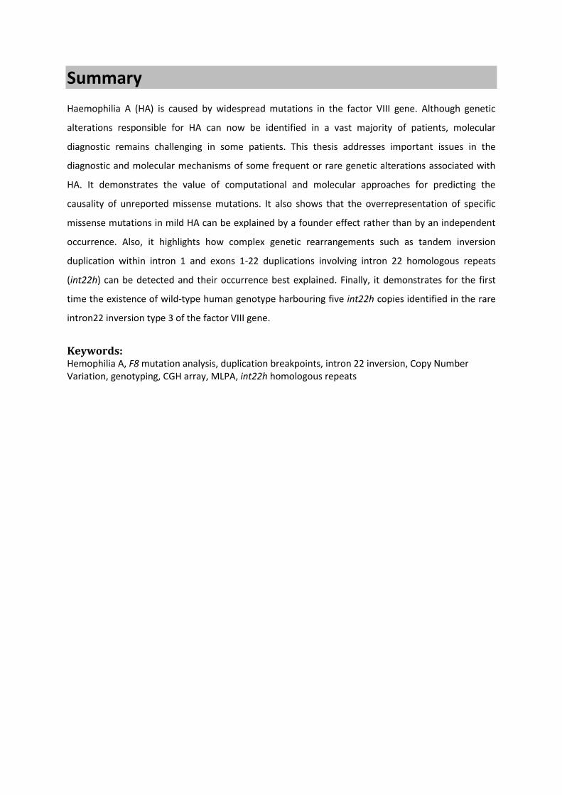