Module 4: Understanding MechanicalVentilation · 2017-10-25 · ventilation (HFOV) and airway...
52
Module 4: Understanding MechanicalVentilation Jennifer Zanni, PT, DScPT Johns Hopkins Hospital
Transcript of Module 4: Understanding MechanicalVentilation · 2017-10-25 · ventilation (HFOV) and airway...
Module 4:Understanding MechanicalVentilationJennifer Zanni, PT, DScPT Johns Hopkins Hospital
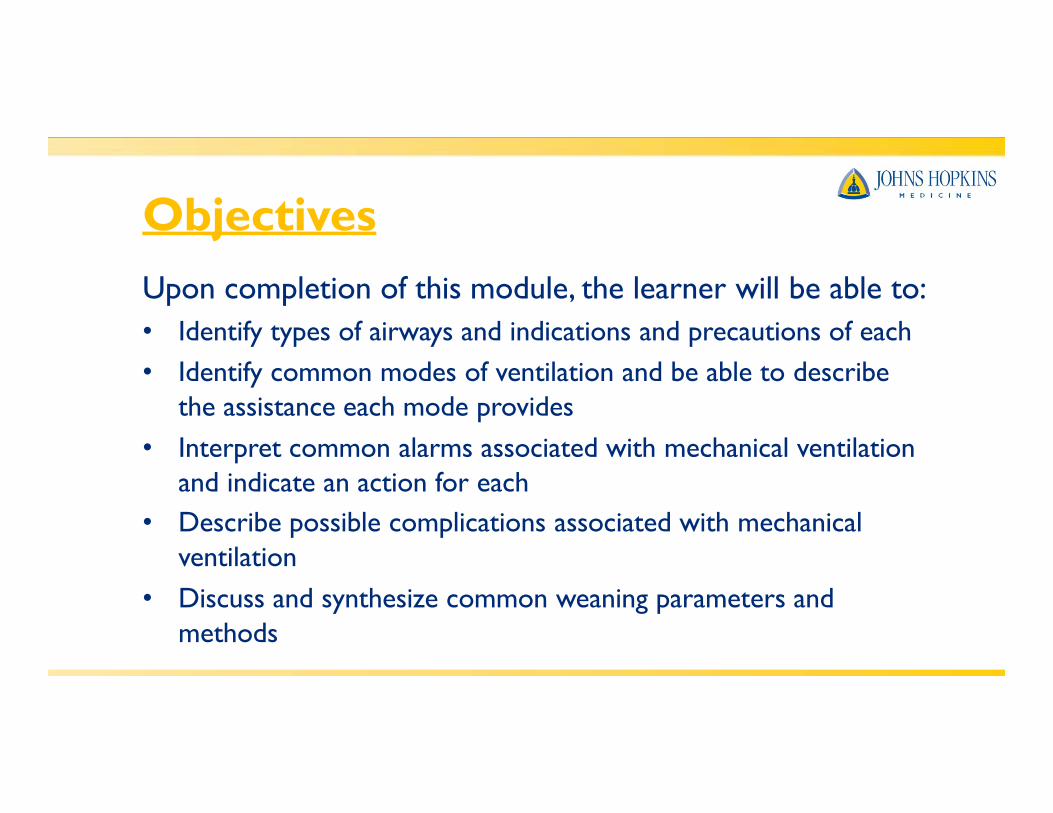
ObjectivesUpon completion of this module, the learner will be able to:• Identify types of airways and indications and precautions of each• Identify common modes of ventilation and be able to describe
the assistance each mode provides• Interpret common alarms associated with mechanical ventilation
and indicate an action for each• Describe possible complications associated with mechanical
ventilation• Discuss and synthesize common weaning parameters and
methods
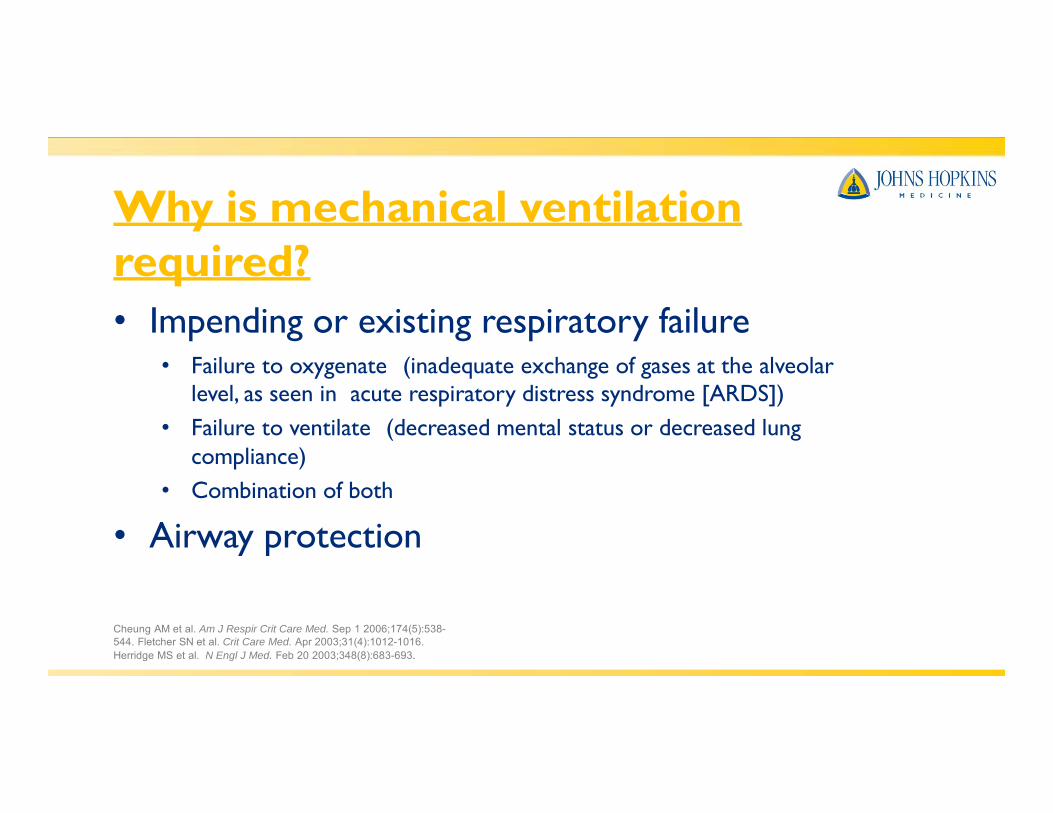
Why is mechanical ventilationrequired?• Impending or existing respiratory failure
• Failure to oxygenate (inadequate exchange of gases at the alveolarlevel, as seen in acute respiratory distress syndrome [ARDS])
• Failure to ventilate (decreased mental status or decreased lungcompliance)
• Combination of both
• Airway protection
Cheung AM et al. Am J Respir Crit Care Med. Sep 1 2006;174(5):538-544. Fletcher SN et al. Crit Care Med. Apr 2003;31(4):1012-1016.Herridge MS et al. N Engl J Med. Feb 20 2003;348(8):683-693.

Other things to consider…• What type of airway is in place?• What are the ventilator settings and what do they all
mean?• What do I do if the vent alarms?• What to I do if the vent fails?• How do I plan treatment in conjunction with vent
weaning?

Start with the ABC’s…
Airway
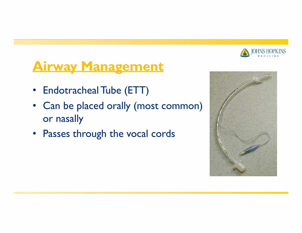
Airway Management
• Endotracheal Tube (ETT)• Can be placed orally (most common)
or nasally• Passes through the vocal cords
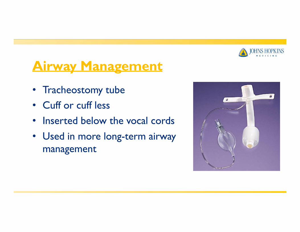
Airway Management
• Tracheostomy tube• Cuff or cuff less• Inserted below the vocal cords• Used in more long-term airway
management
Airway Management
• Airway cuffs:• Assist with holding the airway in place• Allow positive pressure ventilation without loss of tidal volume• May reduce risk of aspiration of oral and gastric secretions• If patient can talk or is losing tidal volume, the cuff may not be
fully inflated
Ventilator Settings• TidalVolume• PEEP• Mode (type of assist given by vent)• Rate (Breaths per minute.Adjusted based on patient’s
own respiratory rate)• FiO2 (amount of O2 being delivered)

Type of respiration
ModeSet respiratory rate
Actual respiratory rate
FiO2 PEEP

PEEP• Positive End-Expiratory Pressure
• Pressure given in expiratory phase to prevent closure of the alveoli and allow increased time for O2 exchange
• Used in pts who haven’t responded to treatment and are requiring high amount of FiO2
• PEEP will lower O2 requirements by recruiting more surface area
• Normal PEEP is approximately 5cmH20. Can be as high as 20cmH20
Oxygen Therapy• Prolonged exposure to high levels of oxygen can
be toxic to the lungs• High FiO2 (>.5) can lead to atelactasis• Balancing act between FiO2 and PEEP
Modes ofVentilation

Modes ofVentilation• ControlledVentilation
• Vent initiates all breaths at a pre-set rate and tidal volume
• Vent will block any spontaneous breaths• Used mainly in the OR for paralyzed and sedated
patients.

Modes ofVentilation• Assist Control (A/C)
• Vent will allow a patient to initiate a breath and then vent will deliver a pre-set tidal volume
• Machine set at a minimum rate so apnea will not occur if the patient does not initiate a breath
• Disadvantages:• Hyperventilation if patient has increased respiratory rate (can
lead to respiratory alkalosis)• Vent dysynchrony, breath-stacking
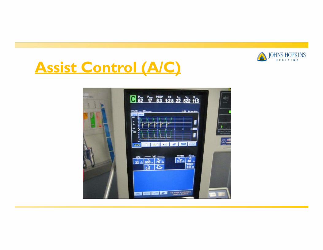
Assist Control (A/C)

“Breath-Stacking”

Modes ofVentilation• Synchronized Intermittent Ventilation (SIMV)
• Similar to A/C, but patients can take own breaths with their own TV between mechanically assisted breaths
• Can be used as a primary mode or a weaning mode• May lead to a low respiratory rate in a patient who does
not initiate breaths if set rate is low
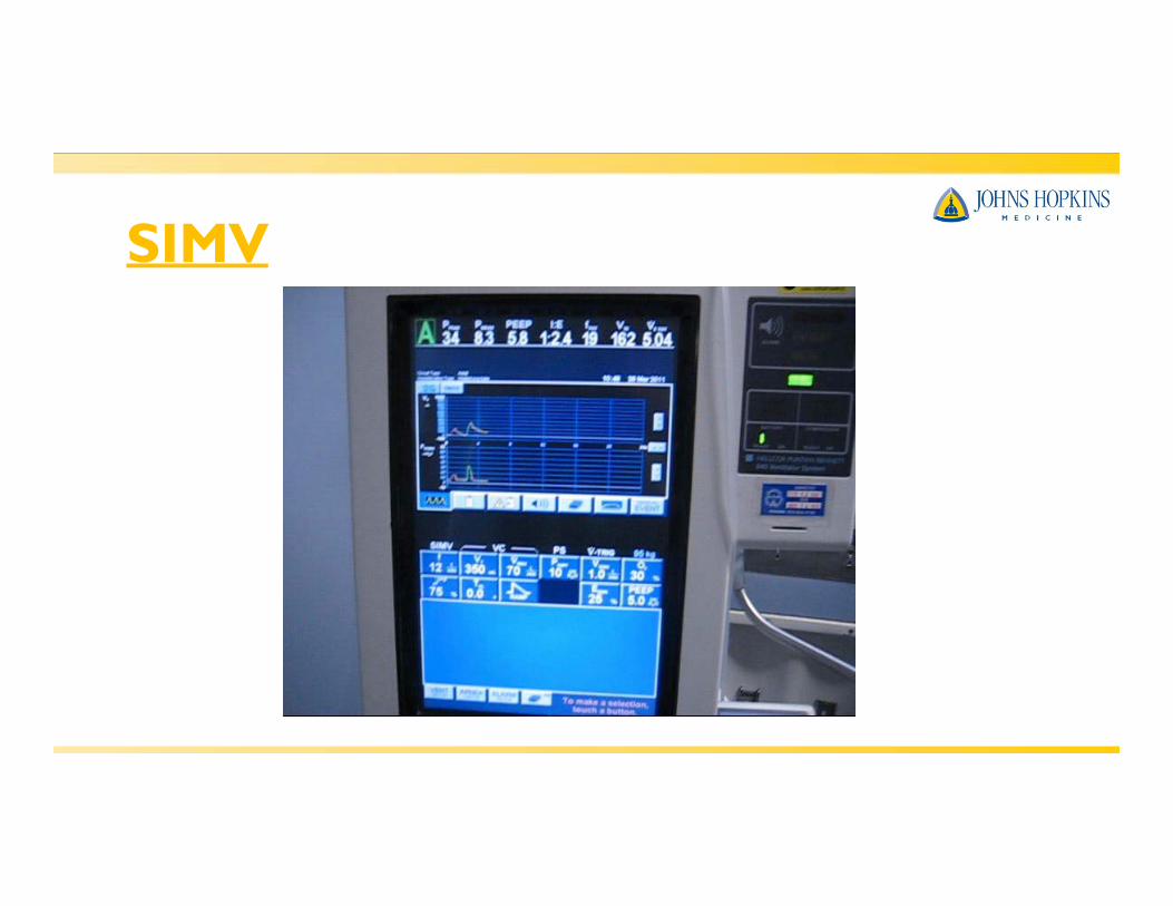
SIMV
Modes ofVentilation• Pressure Support Ventilation (PSV)
• Also called “spontaneous mode”• Pt initiates breath & vent delivers a pre-set inspiratory
pressure to help overcome airway resistance and keeps airways open
• Patient controls the rate, tidal volume, and minute ventilation• Tidal volume is variable• Can be used in conjunction with SIMV or CPAP settings

Pressure Support (PS)
Modes ofVentilation• Continuous Positive Airway Pressure (CPAP)
• Positive airway pressure provided during both inspiration andexpiration
• Vent provides O2 and alarms, but no respirations• Improves gas exchange and oxygenation in patients able to
breathe on their own
• Can also be used non-invasively via a face or nasal mask for patients with sleep apnea

Modes ofVentilation• Airway Pressure ReleaseVentilation (APRV)
• Differs from conventional vent• Elevation of airway pressures with brief intermittent releases
of airway pressure• Facilitates oxygenation and CO2 clearance• May be an improved way to treat ALI/ ARDS
Non-InvasiveVentilation (NIV)
• Bi-Level Airway pressure (BiPAP)• Delivered by mask, not through an airway• Similar to CPAP, but can be set at one pressure for
inhalation and another for exhalation.• Used in sleep apnea, but also has been found to be useful in
patients with CHF and respiratory failure to avoid intubation

Vent AlarmsHigh Pressure Alarm (common alarm to come and go…more serious if it continues to alarm):
• Secretions/ needs to be suctioned (common)
• Kinked tubing/ malposition of ETT
• Pt biting tube/ fighting vent
• Water in tubing(common)
• Bronchospasm• Pneumothorax• Decreased compliance
(i.e.ARDS)
Vent Alarms• Low Pressure Alarm
• Tubing disconnect from vent• Leak in cuff or tubing connections (if patient can talk around
trach, a leak in the cuff is probable)• Extubation
• Also alarms for tidal volume, rate, temperature, and O2

Vent Alarms• General principles…
• Look at the patient first!!! Then follow tubing to the vent tosearch for any disconnections.
• If can’t find the problem and the patient is in distress, disconnect the patient from the vent and bag with 100% O2 (and call for help)

Complications of Mech Vent• Asynchrony (“bucking”)• AutoPEEP
• Patient doesn’t expire full tidal volume and air becomes trapped
• Can cause increased alveolar damage
• Barotrauma• Damage to alveoli caused by increased pressure and volume
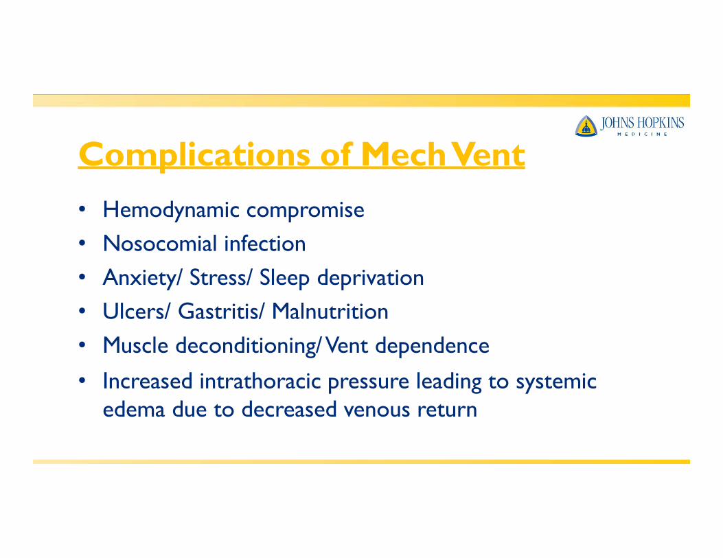
Complications of Mech Vent• Hemodynamic compromise• Nosocomial infection• Anxiety/ Stress/ Sleep deprivation• Ulcers/ Gastritis/ Malnutrition• Muscle deconditioning/Vent dependence
• Increased intrathoracic pressure leading to systemicedema due to decreased venous return
Weaning parameters
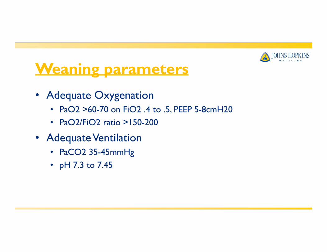
• Adequate Oxygenation• PaO2 >60-70 on FiO2 .4 to .5, PEEP 5-8cmH20• PaO2/FiO2 ratio >150-200
• AdequateVentilation• PaCO2 35-45mmHg• pH 7.3 to 7.45
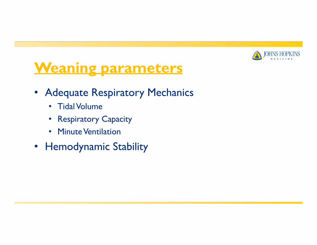
Weaning parameters• Adequate Respiratory Mechanics
• TidalVolume• Respiratory Capacity• MinuteVentilation
• Hemodynamic Stability

Spontaneous BreathingTrials(SBT)• Spontaneous breathing trials (whether single or
multiple trials) lead to extubation more quicklythan those receiving Pressure Support and IMV forweaning purposes in patients who are mechanicallyventilated for > 1 week
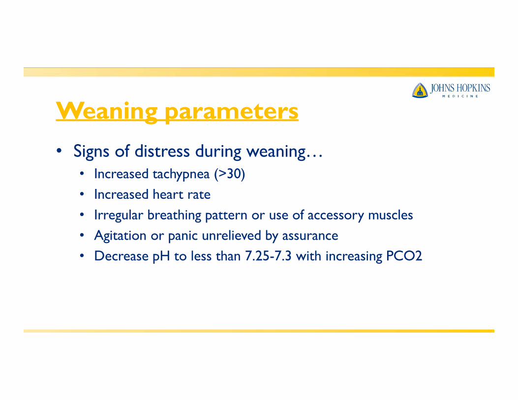
Weaning parameters• Signs of distress during weaning…
• Increased tachypnea (>30)• Increased heart rate• Irregular breathing pattern or use of accessory muscles• Agitation or panic unrelieved by assurance• Decrease pH to less than 7.25-7.3 with increasing PCO2

Treatment Guidelines• Consider the need for increased FiO2 or vent support
prior to mobilizing a patient in order to maximize theirpulmonary status
• Communicate with medical team to discuss best settings• Consider how weaning trials may affect the patient’s
ability to participate in therapy. May want to coordinate working with patients around weaning schedules, and not during
Mobilizing the Patient requiringMechanicalVentilation• Patients weaning from mechanical ventilation
• Consider each patient’s case and determine their ability totolerate both spontaneous breathing trials (SBTs) and rehab sessions
• May be optimal to treat prior to SBT or after they are rested• Consider mobility and strengthening first, then weaning

Ambulating a Patient on MechVent
• Requires teamwork with the entire medical team• Most standard ventilators do not run on battery.
Will likely need to use a portable ventilator or an ambu-bag to ventilate the patient while mobilizing
Ambulating a Patient on MechVent• ETT placement
• Make sure tape is secure prior to moving patient!!!• Look at cm mark at lip before and after treatment to assure
no movement had occurred
• May want to talk with team if patient has an FiO2of .60 or greater and/or if PEEP is -10cm H2O orgreater to ensure medical stability

Steps to Mobilization…
• Look at BaselineVitals…

Steps to Mobilization…
• Look at BaselineVent Settings…
Steps to Mobilization…
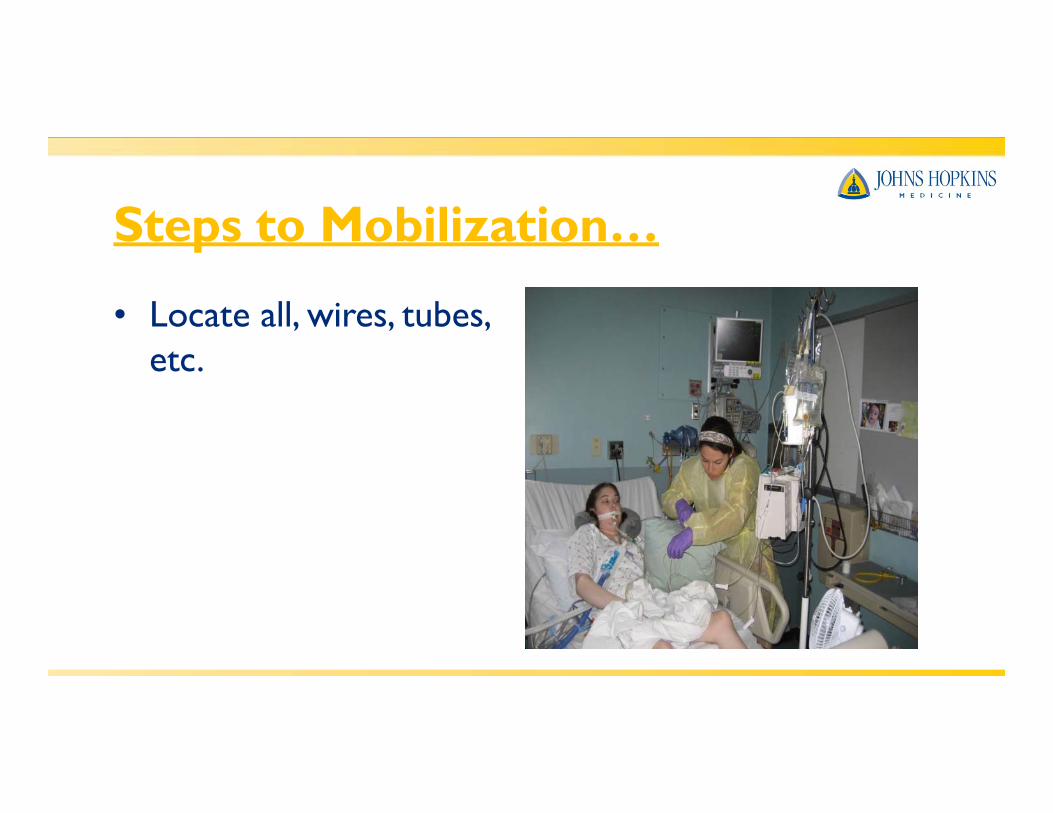
• Locate all, wires, tubes, etc.

Steps to Mobilization…
• Move IV pole and linesto the side you are getting up on (usuallytowards the vent)

Steps to Mobilization…
• Assist patient to sittingat the side of the bedtowards the vent, making sure that all lines are accounted forand have enough slack

Steps to Mobilization…
• Transfer to the chairwhile managing lines toensure clearance

Steps to Mobilization…
• Try to consolidate equipment together asbest as you can
Steps to Mobilization…
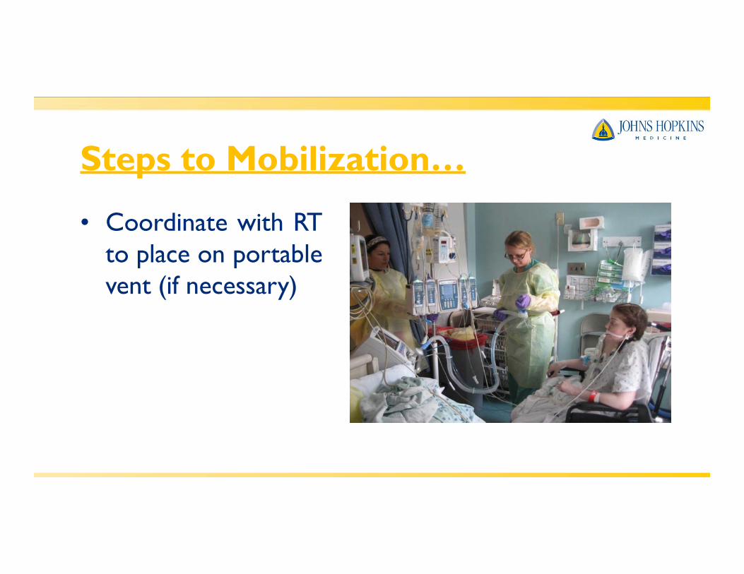
• Coordinate with RTto place on portablevent (if necessary)

Steps to Mobilization…• Organize as much as
you can to have all lines in front of the patient. Make sure nolines are on the flooror can get caught in the equipment.
Steps to Mobilization…
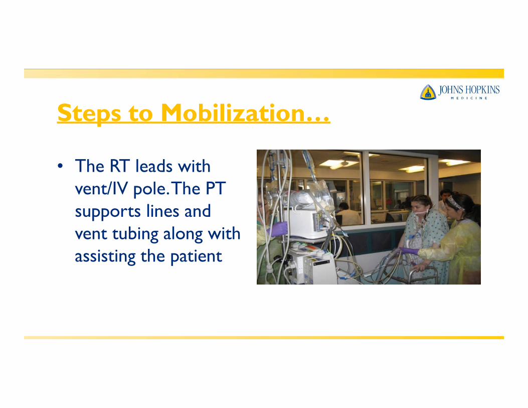
• The RT leads with vent/IV pole.The PT supports lines andvent tubing along with assisting the patient

Steps to Mobilization…
• Have a tech or nurse follow with awheelchair for safety

Steps to Mobilization…
• Always ensure that you have control of all lines and vent tubing/ETT with all transfers and movement of the patient
References• Bailey, P.P., Miller, R.R., 3rd, & Clemmer,T.P. (2009, Oct). Culture of early mobility in
mechanically ventilated patients. Crit Care Med, 37(10 Suppl):S429-35.• Clini, E & Ambrosino, N. (2005, Sep). Early physiotherapy in the respiratory
intensive care unit. Respiratory Medicine, (9):1096-104.• Ciesla, N. D. (2004). Physical therapy associated with respiratory failure. In
DeTurk, W.E and Cahalin, L.P (Eds.), Cardiovascular and Pulmonary PhysicalTherapy (pp. 541-587). NewYork: McGraw-Hill.
• Dean, E. (2008). Mobilizing patients in the ICU: Evidence and principles of practice. Acute Care Prospectives,Vol. 17(1).
• Esteban,A., Frutos, F., & Tobin, M.J. (1995, Feb 9). A comparison of fourmethods of weaning patients from mechanical ventilation. Spanish Lung Failure Collaborative Group. N Engl J Med, 332(6):345-50.
References
Practice, (4th ed.). St Louis: Mosby.
• Fessler, H.E. & Hess, D.R. (2007). Respiratory controversies in the critical care setting. Does high-frequency ventilation offer benefits over conventional ventilation in adult patients with acute respiratory distress syndrome? Respiratory Care. 2007. 52, (5), 595-605.
• Frownfelter, D. (1987). Chest Physical Therapy and Pulmonary Rehabilitation. (pp. 729-744). St. Louis: Mosby.
• Frowley PM and Habashi, NM. Airway Pressure Release Ventilation:Theoryand Practice. AACN. 2001;Vol 12(2), 234-246.
• Hopkins, R.O., & Spuhler,V.J. (2009, Jul-Sep). Strategies for promoting earlyactivity in critically ill mechanically ventilated patients. ACCN Adv Crit Care, 20(3):277-289.
• Irwin, S and Tecklin, JS. (2004). Cardiopulmonary Physical Therapy:A Guide to

References
rehabilitation in the intensive care unit. PT in Motion, 32-39.
• Perme, C., & Chandraskekar, R.K. (2008). Managing the patient on mechanical ventilation in ICU: Early mobility and walking program. Acute Care Prospectives,Vol 17(1).
• Sadowsky, H.S. Monitoring and life support equipment. In E.A. Hillegass and H.S. Sadowsky, Essentials of cardiopulmonary physical therapy. (pp. 509-533). 2001; Philadelphia: Saunders.
• Stawicki, S.P., Goval, M., & Sarani, B. (2009, Jul-Aug). Frequency oscillatory ventilation (HFOV) and airway pressure release ventilation (APRV): a practical guide. J Intensive Care Med, 24(4):215-29. Epub 2009 Jul 17.
• Yosefy, C., Hay, E., & Ben-Barak, A. (2003). BiPAP ventilation as assistance for patients presenting with respiratory distress in the department of emergency medicine. American Journal of Respiratory Medicine. 2(4), 343-7.
• Zanni, J.M., & Needham, D.M. (2010, May). Promoting early mobility and