Module 2 Pathophysiology of veno-occlusive disease.
14
Module 2 Pathophysiology of veno-occlusive disease
-
Upload
augustus-mcdaniel -
Category
Documents
-
view
238 -
download
3
Transcript of Module 2 Pathophysiology of veno-occlusive disease.

Module 2
Pathophysiology of veno-occlusive disease

Learning objectives
• To understand the cause of VOD
• To understand the sequence of steps in the pathophysiology of VOD
• To recognise the pathophysiological endpoint of VOD
VOD, veno-occlusive disease
Early complications of HSCT:vascular endothelial syndromes
• It is proposed that during HSCT, endothelial cells lining blood vessels are activated by:– The conditioning regimen– Cytokines produced by injured tissues– Microbial products translocated through mucosal barriers– The process of engraftment
• Intense and sustained activation of endothelial cells leads to cellular damage
• Alloreactivity has been postulated to play a role in this damage and activation– This explains the greater incidence of these complications after
allogeneic transplantation
Carreras E & Diaz-Ricart M. Bone Marrow Transplant 2011;46:1495–1502
Early complications of HSCT:vascular endothelial syndromes
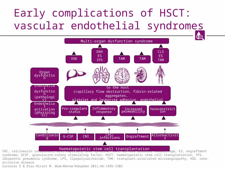
CNI, calcineurin inhibitors; CLS, capillary leak syndrome; DAH, diffuse alveolar haemorrhage; ES, engraftment syndrome; GCSF, granulocyte-colony stimulating factor; HSCT; haematopoietic stem cell transplantation; IPS, idiopathic pneumonia syndrome; LPS, lipopolysaccharide; TAM, transplant-associated microangiopathy; VOD, veno-occlusive diseaseCarreras E & Diaz-Ricart M. Bone Marrow Transplant 2011;46:1495–1502
Multi-organ dysfunction syndrome
Endothelial phenotype represents a net liability to the host(capillary flow obstruction, fibrin-related aggregates,
platelet and leukocyte adhesion, endothelial apoptosis)
Organdysfunction
Endothelialdysfunction(pathologic)
Endothelialactivation
(physiologic)
Haematopoietic stem cell transplantation
Pro-coagulantstatus
Inflammatoryresponse
Increasedpermeability Vasoconstriction
VOD
DAHESIPS TAM TAM
CLSES
TAM
Conditioning G-CSF CNI LPS/infections Engraftment Alloreactivity

Veno-occlusive disease
• VOD, also known as sinusoidal obstruction syndrome, is a potentially life-threatening complication of HSCT
• The conditioning regimens given before HSCT result in the production of toxic metabolites by the hepatocytes in the liver
• These metabolites trigger the activation, damage and inflammation of the endothelial cells that line the sinusoids– Sinusoids are small capillary-like blood vessels
found in the liver
• This ultimately leads to VOD, which is characterised by – Increased thrombosis and decreased fibrinolysis– Sinusoidal damage and narrowing– Inflammation
Richardson PG et al. Expert Opin Drug Saf 2013;12:123–136
Histological section of the liver viewed through a microscope. Blood vessels shown in red, cell nuclei in blue, cytoplasm in pink
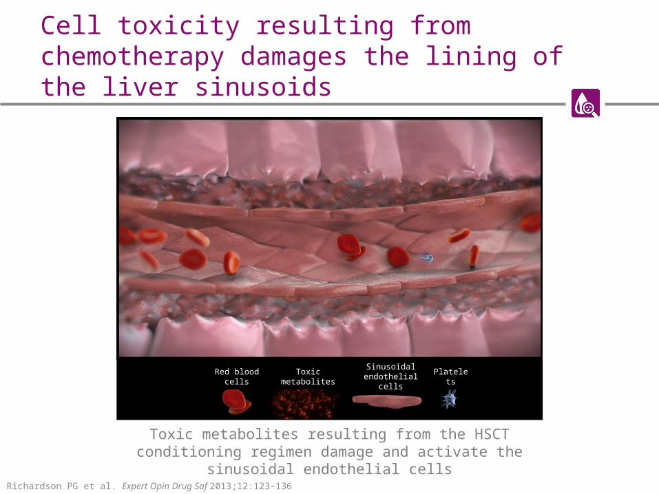
Cell toxicity resulting from chemotherapy damages the lining of the liver sinusoids
Toxic metabolites resulting from the HSCT conditioning regimen damage and activate the sinusoidal endothelial cells
Richardson PG et al. Expert Opin Drug Saf 2013;12:123–136
Red blood cells Toxic metabolitesSinusoidal
endothelial cellsPlatelets
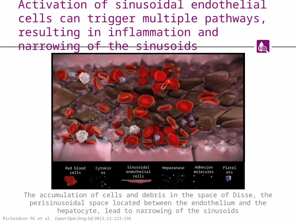
Activation of sinusoidal endothelial cells can trigger multiple pathways, resulting in inflammation and narrowing of the sinusoids
Richardson PG et al. Expert Opin Drug Saf 2013;12:123–136
The accumulation of cells and debris in the space of Disse, the perisinusoidal space located between the endothelium and the hepatocyte, lead to narrowing of the sinusoids
Red blood cells Cytokines Sinusoidal endothelial cells
Cytokines Heparanase Adhesion molecules
Platelets

VOD is characterised by increased clot formation and reduced clot breakdown
PAI-1,plasminogen activator inhibitor-1; TF, tissue factorRichardson PG et al. Expert Opin Drug Saf 2013;12:123–136
The narrowing of the sinusoids, embolised endothelial cells and increased clot formation lead to the endpoint of VOD, namely obstruction of the sinusoids
Red blood cells Cytokines Fibrin Heparanase Adhesion molecules Tissue factor PAI-1 Platelets

Pathophysiology of VOD
ICAM, intracellular adhesion molecule; IL, interleukin; TNF, tumour necrosis factor; VCAM, vascular cell adhesion proteinRichardson PG et al. Expert Opin Drug Saf 2013;12:123–136; Coppell JA et al. Blood Rev 2003;17:63–70
Triggering of multiple pathways
Inflammation Cytoskeletal structure
Sinusoidal narrowing
Endothelial cell and hepatocyte damage
VOD
• Activation and damage due to conditioning regimen-mediated injury. Damage is both directed and mediated by cytokines such as:
- TNF-α, IL-1b, IL-6
• Increased expression of adhesion molecules ICAM-1 and VCAM-1 of endothelial cell surface
• Activation of leukocytes that release additional inflammatory cytokines
• Digestion of extracellular matrix
• Portal vein hypotension• Hepatic venous outflow obstruction

Summary of VOD pathophysiology
• The conditioning regimen given prior to HSCT increases endothelial cell activation, resulting in damage to the SECs and hepatocytes
• The accumulation of cells in the space of Disse (the perisinusoidal space), increased inflammation and formation of clots lead to narrowing of the sinusoids
• This results in VOD, which is characterised by blockage of the sinusoids, portal vein hypotension and reduced hepatic venous outflow
SEC, sinusoidal endothelial cell

Self-assessment questions
1. VOD affects which major organ?
Self-assessment questions
2. What by-product of HSCT conditioning causes damage to the sinusoidal endothelial cells and hepatocytes?

Self-assessment questions
3. Which of the following is not a characteristic of VOD pathophysiology?
a) Increased clot formation
b) Decreased clot breakdown
c) Expansion of the sinusoids
d) Inflammation

Self-assessment questions
4. What causes obstruction of the sinusoids?