MODULATION OF NOTCH SIGNALING …ethesis.nitrkl.ac.in/5354/1/411LS2126.pdfMODULATION OF NOTCH...
48
MODULATION OF NOTCH SIGNALING COMPONENTS IN PRESENCE OF EPIGENETIC MODULATORS IN BREAST CANCER THESIS SUBMITTED TO NATIONAL INSTITUTE OF TECHNOLOGY, ROURKELA FOR PARTIAL FULFILLMENT OF THE MASTER OF SCIENCE DEGREE IN LIFE SCIENCE Submitted by TAPAS TRIPATHY ROLL NO – 411LS2126 Under the guidance of Dr. SAMIR KUMAR PATRA ASSOCIATE PROFESSOR AND HEAD DEPARTMENT OF LIFE SCIENCE NATIONAL INSTITUTE OF TECHNOLOGY ROURKELA-769008, ODISHA 2012-2013
Transcript of MODULATION OF NOTCH SIGNALING …ethesis.nitrkl.ac.in/5354/1/411LS2126.pdfMODULATION OF NOTCH...
MODULATION OF NOTCH SIGNALING COMPONENTS IN
PRESENCE OF EPIGENETIC MODULATORS IN
BREAST CANCER
THESIS SUBMITTED TO
NATIONAL INSTITUTE OF TECHNOLOGY, ROURKELA
FOR PARTIAL FULFILLMENT
OF THE MASTER OF SCIENCE DEGREE IN LIFE SCIENCE
Submitted by
TAPAS TRIPATHY
ROLL NO – 411LS2126
Under the guidance of
Dr. SAMIR KUMAR PATRA
ASSOCIATE PROFESSOR AND HEAD
DEPARTMENT OF LIFE SCIENCE
NATIONAL INSTITUTE OF TECHNOLOGY
ROURKELA-769008, ODISHA
2012-2013
DEPARTMENT OF LIFE SCIENCE
NATIONAL INSTITUTE OF TECHNOLOGY
ROURKELA-769008
………….....................................................................................................................................
Dr. Samir Kumar Patra Ref. No.
Associate Professor and Head Date: ............................
CERTIFICATE This is to certify that the thesis entitled “MODULATION OF NOTCH SIGNALING
COMPONENTS IN PRESENCE OF EPIGENETIC MODULATORS IN BREAST CANCER”
which is being submitted by Mr. Tapas Tripathy, Roll No. 411LS2126, for the award of the
degree of Master of Science from National Institute of Technology, Rourkela, is a record of
bonafide research work, carried out by him under my supervision. The results embodied in
this thesis are new and have not been submitted to any other university or institution for the
award of any degree or diploma.
Dr. SAMIR K. PATRA
ASSOCIATE PROFESSOR AND HEAD,
Department of Life Science
National Institute of Technology
Rourkela – 769008, Odisha, India.
..................................................................................................................................
Phone no: 0661-2462683. Email: [email protected]

ACKNOWLEDGEMENTS
A research project like this is never the work of one alone. The contributions of many
people, in different ways, have made this possible. I would like to extend my appreciation
especially to the following.
First of all I thank “The Almighty” for the wisdom and perseverance that has been
bestowed upon me during this research project, and indeed, throughout my life: "I can do
everything through him who gives me the strength.”
My deepest sense of gratitude and thanks goes to my guide Dr. Samir Kumar Patra, for
making this research possible. His support, guidance and advice throughout the research
project, as well as his pain-staking effort in proof reading the drafts, are greatly appreciated.
Indeed, without his guidance, I would not be able to put the topic together. Thank You Sir.
Thanks to my mentor Mr. Dipta Sengupta, who has been a friend philosopher cum guide,
encouraging and supporting me always. The experience of working with him has been an
interesting and rewarding one. My ultimate respect and reverence goes to Ms. Moonmoon
Deb (Moonmoon Di) for all her care and affection.
Special thanks go to the rest of my lab members including Ms. Madumita Rakhshit, Ms.
Swayamsiddha Kar, Ms. Arunima Shilpi, Ms. Shabnam Parbin and Mr. Sandip Rath .
I would also like to thank my dear friend Dibyojyoti Baruah who has been a real source of
support and sustenance during this work and Namita, Monalisa for their cheering.
Of course, this project would not have been possible without the participation of the
subjects.
Last but not least, I would like to thank my parents for their unconditional support, both
financially and emotionally throughout my work period. Especially, I would like to thank my
father who has been the role model and the ever- renewable source of inspiration for this
young boy throughout the journey of my life.
DECLARATION
I hereby declare that this project report on, “MODULATION OF NOTCH
SIGNALING COMPONENTS IN PRESENCE OF EPIGENETIC
MODULATORS IN BREAST CANCER”, is the result of the work carried
out by me .Wherever contributions of others are involved, every effort is made
to indicate this clearly, with due reference to the literature, and
acknowledgement of collaborative research and discussions. The work was
done under the guidance of Dr. Samir Kumar Patra , Associate Professor and
Head, Department of Life Science, National Institute of Technology,
Rourkela.
Date:
Place: (Tapas Tripathy)
TABLE OF CONTENTS
Sr.
No. Title Pg. No
1. Introduction 1-12
2. Review of Literature 13-19
3. Objectives 20
3. Materials and Methods 21-27
4. Results and Discussion 28-34
5. Conclusion 35
6. References 36-41
LIST OF FIGURES
Sr.
No. Title of Figure Pg. No.
1 (A) Structure of Notch receptors (1-4) and ligands (Jagged-1, 2, Dll-1, 3, 4 (B) The Canonical notch Signaling Pathway
2
2
(A) Maintenance and (B) de novo DNMTS methylate DNA. DNMT1 binds methyl groups to the hemimethylated DNA during replication, whereas DNMT3A and DNMT3B can add methyl groups to CpG dinucleotides of unmethylated DNA.
5
3 Mechanism of DNA methylation proposed by Reither et al. (2003) and S K Patra et al. (2008) Cancer and Metast Rev.
7
4 Molecular structure of S-Adenosyl Methionine (SAM)
12
5 Molecular structure of S-Adenosyl Homocysteine (SAH) 12
6 Aberrant Notch signaling in cancers showing context dependent properties
15
7 Graphical analysis of cell viability of MDA MB-231 cells by MTT assay after SAM and SAH treatment
28
8
Graphical representation of RT-PCR results for expression level of Notch1 in MDA MB-231 cells after SAM and SAH treatment
29
9
Graphical representation of RT-PCR results for expression level of HES1 in MDA MB-231 cells after SAM and SAH treatment
29
10 SEM images of MDA MB-231 cells showing changes in cellular morphology after SAM and SAH treatment
30
11 Microscopic images showing changes in the migratory property of MDA MB-231 cells after SAM and SAH treatment
31
12 Fluorescence microscopic images of MDA MB-231 cells showing condensed chromatin after SAM and SAH treatment
32
13
Fluorescence microscopic images of MDA MB-231 cells showing formation of autophagosomes after SAM and SAH treatment
33
14 Fluorescence microscopic images of MDA MB-231 cells showing DNA damage after SAM and SAH treatment
34
ABSTRACT
Notch signaling is one of the pleotropic signaling pathway that plays key
role in development as it promotes differentiation and tissue morphogenesis. In
recent years it has been observed that this pathway along with many other
developmental pathways is seen to be deregulated in different cancers. The role
that is played by this pathway is highly context dependent in cancers that is in
some cases it act as a tumor suppressor while as oncogenic in others. It is
proved with its oncogenic role in all forms of breast cancer. We tried to assess
the effects on this pathway in MDA MB-231 breast cancer cells after treatment
with epigenetic modulators S-Adenosyl Methionine (SAM) and S-Adenosyl
Homocysteine (SAH) in a time dependent manner for 0-48 hr. The expression
of important notch receptor Notch-1 and downstream effector Hes-1 were
shown to be down regulated after SAM treatment but the SAH treatment
upregulated them. This was accompanied by apoptotic induction in SAM
treated cells in a more aggravated manner as compared to the SAH treated cells.
From all these results we tentatively conclude that SAM treatment of invasive
breast cancer cell line induces cell death in a notch dependent manner.
Introduction 2013
1
INTRODUCTION
The lifetime risk of developing breast cancer is about one in nine for women with
around 1,41,000 new cases being diagnosed in the world wide each year. This has been
proved recently as the worst and fatal breed of cancers in females. However, a significant
decrease in the mortality since 1989 due to rigorous research, screening, specialization of
care, and the widespread adoption of tamoxifen along with other effective anti-cancer
therapeutics. More recently, number of Epigenetic therapeutics have emerged as new hopes
for successful therapy of many cancers along with breast cancer working by altering the
oncogenic epigenetic modifications that are one of the root causes of cancers. Despite this,
there are still just short of 13,000 deaths each year due to breast cancer in western countries,
suggesting that a much more in-depth understanding of the disease is required to improve
treatment. Here in this study we have tried to test the anti-cancer efficacy of some epigenetic
modulators like S-Adenosyl methionine (SAM), S- Adenosyl homocysteine (SAH) on breast
cancer cell lines.
Notch signaling is an important embryonic signaling for development and tissue
homeostasis and is deregulated in many human cancers. It is an important pathway for cell
fate determination, stem cell maintenance and the initiation of differentiation in many tissues
(Ye et al., 2012). Notch was discovered first in Drosophila melanogaster nearly a century ago
pioneered the way to an ever-widening understanding of notch regulated or notch influenced
cellular processes. The deregulation of such a pleiotropic pathway leads to several
pathological conditions including cancer. Deregulated Notch signaling is well established in
haematological malignancies and more recent studies have proved the importance of Notch
activity in solid tumors. As the recent findings suggests it as an oncogene in some cancers but
as a tumor suppressor in others. The role of Notch in solid tumors seems to be highly context
dependent (Ranganathan et al., 2011).
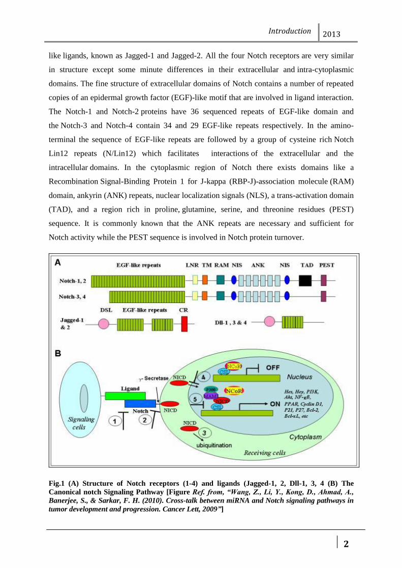
Notch receptors are a class of single-pass trans-membrane proteins encoded by the Notch
genes which can be activated by the binding of a family of compatible ligands. Four
Notch receptors have been identified till date in mammals, including human, described
as Notch-1-4. The mammalian canonical notch ligands are total five in number and divided
into two groups denoted as Delta-like (Delta-like 1, Delta-like 3, and Delta-like 4) or Serrate-
Introduction 2013
2
like ligands, known as Jagged-1 and Jagged-2. All the four Notch receptors are very similar
in structure except some minute differences in their extracellular and intra-cytoplasmic
domains. The fine structure of extracellular domains of Notch contains a number of repeated
copies of an epidermal growth factor (EGF)-like motif that are involved in ligand interaction.
The Notch-1 and Notch-2 proteins have 36 sequenced repeats of EGF-like domain and
the Notch-3 and Notch-4 contain 34 and 29 EGF-like repeats respectively. In the amino-
terminal the sequence of EGF-like repeats are followed by a group of cysteine rich Notch
Lin12 repeats (N/Lin12) which facilitates interactions of the extracellular and the
intracellular domains. In the cytoplasmic region of Notch there exists domains like a
Recombination Signal-Binding Protein 1 for J-kappa (RBP-J)-association molecule (RAM)
domain, ankyrin (ANK) repeats, nuclear localization signals (NLS), a trans-activation domain
(TAD), and a region rich in proline, glutamine, serine, and threonine residues (PEST)
sequence. It is commonly known that the ANK repeats are necessary and sufficient for
Notch activity while the PEST sequence is involved in Notch protein turnover.
Fig.1 (A) Structure of Notch receptors (1-4) and ligands (Jagged-1, 2, Dll-1, 3, 4 (B) The
Canonical notch Signaling Pathway [Figure Ref. from, “Wang, Z., Li, Y., Kong, D., Ahmad, A.,
Banerjee, S., & Sarkar, F. H. (2010). Cross-talk between miRNA and Notch signaling pathways in
tumor development and progression. Cancer Lett, 2009”]
Introduction 2013
3
The cytoplasmic region of the receptor conveys the signal to the nucleus. The
Notch ligands possess multiple EGF-like repeats in their extracellular domain and a cysteine-
rich region (CR) in serrate type while these are devoid in Delta. The Jagged-1 and Jagged-2
bears almost twice the numbers of EGF-like repeats compared to Delta. Notch signaling is
activated by the ligand dependent way where the neighboring cells secrete the ligand. A
series of proteolytic cleavages are underway by the metalloproteases, tumor necrosis factor-
α-converting enzyme (TACE) and γ-secretase complex (comprised of presenilin-1/2,
nicastrin, Pen- 2, and Aph-1) when the signaling is active. TACE sheds the first blood
making the first cleavage that leads to cleave the receptor in the extracellular domain. The
trans-endocytosis of the released extracellular domain takes place by the ligand
expressing cell. The second cut is usually caused by the γ-secretase complex releases the
Notch intracellular domain (NICD) into the cytoplasm, which can subsequently translocate
into the nucleus because of the presence of nuclear localization signals located within it.
Blocking the γ-secretase function prevents the cleavage of the Notch receptor thereby
stopping the Notch signal transduction. The γ-secretase inhibitors (GSI) could be potential
candidates for the treatment of human malignancies. When the NICD is absent the
transcription of Notch target genes is blocked with a repressor complex mediated by the CSL
(C protein binding factor 1/Suppressor of Hairless/Lag-1). When NICD comes in to the
nucleus, it forms an active transcriptional complex by displacing the histone deacetylase–
corepressor complex and recruiting the protein mastermind like1 (MAML1) and histone
acetyl transferases (HAT) to the CSL complex and leads to its conversion from a
transcriptional repressor into a transcription activator complex. A few important Notch target
genes that have been established includes Hes (Hairy enhance of split) family, Hey
(Hairy/ enhancer of spit related with YRPW motif), nuclear factor-kappa B (NF- κB),
vascular growth factor receptor (VEGF), mammalian target of rapamycin (mTOR), cyclin
D1, c-myc, p21, p27, Akt, etc. All of these candidates have been proved with their foul play
in tumor development and progression (Wang et al., 2010).
The aberrations in Notch signaling are significantly correlated to the happening and
development of many cancers along with many other developmental pathways (Kar et al.,
2012). The expression levels of genes associated with the Notch signaling pathway are
correlated with tumor pathology and the degree of differentiation. Notch1 and Notch2 gene
expression are correlated with tumor pathology type and degree of differentiation. (Jin et al.,
2012). In some tumor the tumor suppressor genes are silenced partly through deacetylation of
Introduction 2013
4
promoter regions and the treatment with HDAC inhibitors therefore contributes to re-
expression of these genes. The whole phenomenon is known to be mediated by the notch
signaling components in various solid tumors. Still an in-depth understanding of the
underlying molecular mechanisms behind the mode of action of HDAC inhibitors on tumour
cells remains obscure .In another mechanism the bHLH protein Hairy and Enhancer of Split-
1(Hes-1), that functions as a negative regulator of transcription, is one direct and important
transcriptional target of Notch signaling (Stockhausen et al., 2005).Emerging reports signifies
that Notch signaling pathway is one of the convicts for the drug-resistant tumor phenotype.
The down-regulation of Notch pathway could be an important therapeutic approach for
induction of drug sensitivity and increased inhibition of cancer cell growth, invasion, and
metastasis (Wang et al., 2010).
1.1 Epigenetics and cancer
Epigenetics can be described as a stable alteration in gene expression potential that takes
place during development and cell proliferation, without any change in gene sequence. DNA
methylation is one of the most commonly occurring epigenetic events in the mammalian
genome. This change, though heritable, is reversible, making it a therapeutic target.
Epigenetics has evolved as a rapidly developing area of research. Recent studies have shown
that epigenetics plays an important role in cancer biology, viral infections, activity of mobile
elements, somatic gene therapy, cloning, transgenic technologies, genomic imprinting,
developmental abnormalities, mental health, and X-inactivation. DNA methylation is an
important regulator of gene transcription, and its role in carcinogenesis has been a topic of
considerable interest in the last few years. Alterations in DNA methylation are common in a
variety of tumours as well as in development. Of all epigenetic modifications, hyper
methylation, which represses transcription of the promoter regions of tumours suppressor
genes leading to gene silencing, has been most extensively studied (Patra et al., 2008) .
DNA methylation is a covalent chemical modification, resulting in the addition of a
methyl (CH3) group at the carbon 5 position of the cytosine ring. Even though most cytosine
methylation occurs in the sequence context 5‟-CG-3‟ (also called the CpG dinucleotide),
some also involves CpA and CpT dinucleotides. DNA is made up of four bases, thus there are
16 possible dinucleotide combinations that can occur. Therefore the CpG-dinucleotide should
occur with a frequency of approximately 6%.However, the actual presence is only 5% to10%
Introduction 2013
5
of its predicted frequency. This CpG suppression may be related to the hyper mutability of
methylated cytosine. The human genome is not methylated uniformly and contains regions of
unmethylated segments interspersed by methylated regions. In contrast to the rest of the
genome, smaller regions of DNA, called “CpG islands”, ranging from 0.5 to 5 kb and
occurring on average every 100 kb, have distinctive properties. These regions are
unmethylated, GC rich (60% to 70%), have a ratio of CpG to GpC of at least 0.6, and thus do
not show any suppression of the frequency of the dinucleotide CpG. Approximately half of
all the genes in humans have CpG islands and these are present on both housekeeping genes
and genes with tissue specific patterns of expression. DNA methylation is brought about by a
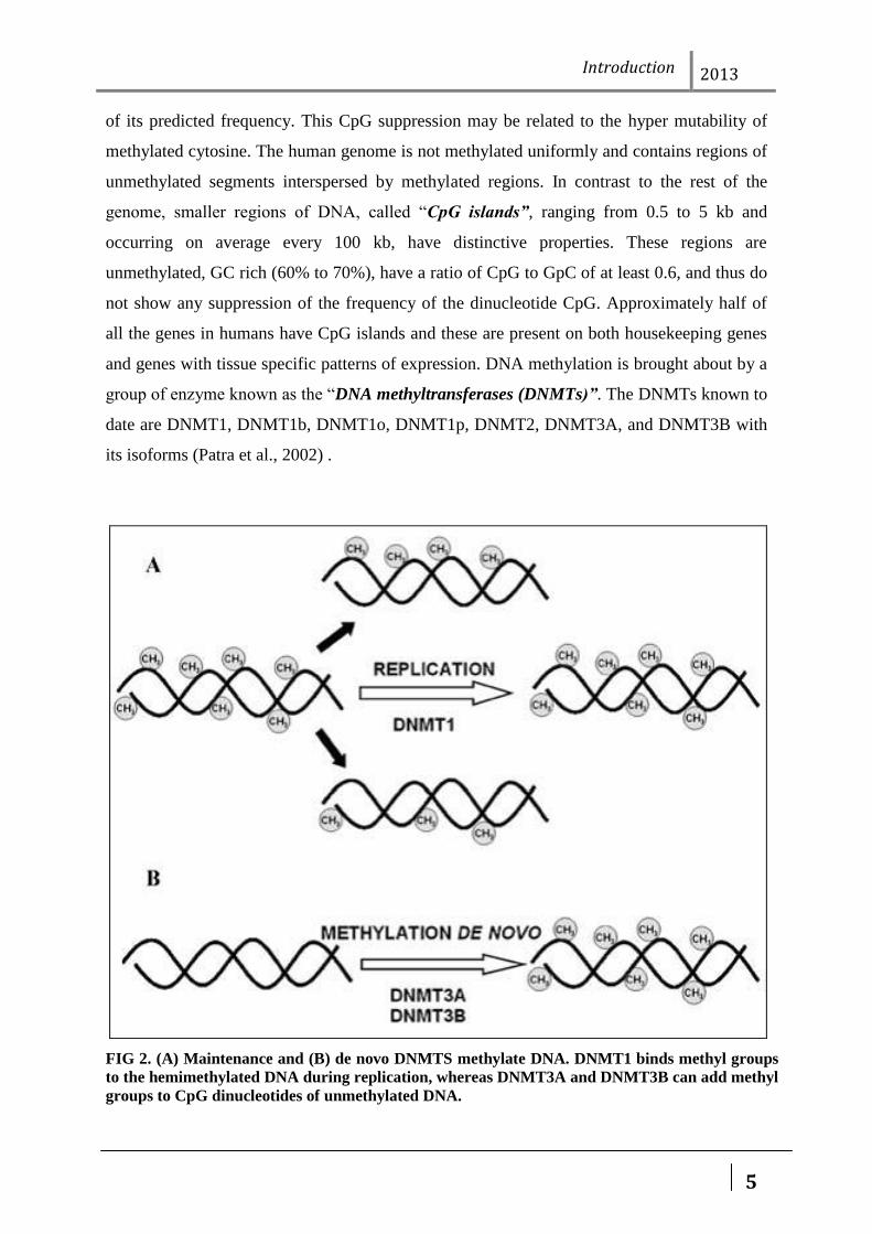
group of enzyme known as the “DNA methyltransferases (DNMTs)”. The DNMTs known to
date are DNMT1, DNMT1b, DNMT1o, DNMT1p, DNMT2, DNMT3A, and DNMT3B with
its isoforms (Patra et al., 2002) .
FIG 2. (A) Maintenance and (B) de novo DNMTS methylate DNA. DNMT1 binds methyl groups
to the hemimethylated DNA during replication, whereas DNMT3A and DNMT3B can add methyl
groups to CpG dinucleotides of unmethylated DNA.
Introduction 2013
6
1.1.1 DNA methylation and gene regulation
The regulation of eukaryotic gene expression is a complex process. Transcription
initiation is a highly controlled and integrated event that involves cis-acting and trans-acting
factors. The cis-acting elements are DNA sequences that act as the substrate for the trans-
acting factors, and the DNA in the vicinity is prepared for transcription. Increased
methylation in the promoter region of a gene leads to reduced expression, whereas
methylation in the transcribed region has a variable effect on gene expression. Several
mechanisms have been proposed to account for transcriptional repression by DNA
methylation. The first mechanism involves direct interference with the binding of specific
transcription factors to their recognition sites in their respective promoters. Several
transcription factors, including AP-2, c-Myc/Myn, the cyclic AMP-dependent activator
CREB, E2F, and NF-kB, recognize sequences that contain CpG residues, and binding of each
has been shown to be inhibited by methylation.
The second mode of repression involves a direct binding of specific transcriptional
repressors to methylated DNA. The DNA methylation signals are analyzed by the methyl-
CpG–binding proteins, the target being the 5-methylated CpG sequence. MeCP1 and MeCP2
were the first two protein complexes identified. However, several new proteins have now
been identified. They include MBD1, MBD2, MBD4, and Kaiso. MeCP1, MBD1, MBD2,
and MBD4 bind to 5mCpG through a motif called the methyl CpG binding domain (MBD).
Kaiso, however, is different in mechanism, as it binds through a zinc finger motif. MBD4 is
associated with DNA repair, whereas MBD1, MBD2, MeCP2, and Kaiso have been shown to
repress transcription both in vitro and in cell culture assays by interacting with histone
deacetylase complexes. DNA methylation can also affect histone modifications and
chromatin structure, which, in turn, can alter gene expression. The underlying patterns of
methylated cytosines are important in guiding histone deacetylation to certain residues. At
present, there are five known proteins that have the methyl-CpG–binding domain, and four of
these (MeCP2, MBD1, MBD2, and MBD3) are implicated in transcriptional repression.
Three of these (MeCP2, MBD2, and MBD3) are in complexes (MeCP-2, MeCP-1 and Mi-2,
respectively) that contain histone deacetylase. Studies of methylated transfected genes
containing binding sites for all four of these methyl-binding proteins have shown at least
partial abrogation of transcriptional repression by treatment with the histone deacetylase
Introduction 2013
7
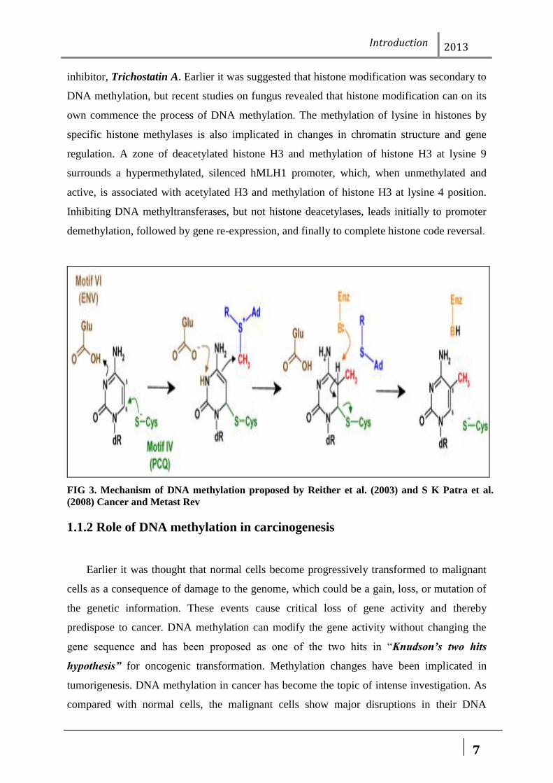
inhibitor, Trichostatin A. Earlier it was suggested that histone modification was secondary to
DNA methylation, but recent studies on fungus revealed that histone modification can on its
own commence the process of DNA methylation. The methylation of lysine in histones by
specific histone methylases is also implicated in changes in chromatin structure and gene
regulation. A zone of deacetylated histone H3 and methylation of histone H3 at lysine 9
surrounds a hypermethylated, silenced hMLH1 promoter, which, when unmethylated and
active, is associated with acetylated H3 and methylation of histone H3 at lysine 4 position.
Inhibiting DNA methyltransferases, but not histone deacetylases, leads initially to promoter
demethylation, followed by gene re-expression, and finally to complete histone code reversal.
FIG 3. Mechanism of DNA methylation proposed by Reither et al. (2003) and S K Patra et al.
(2008) Cancer and Metast Rev
1.1.2 Role of DNA methylation in carcinogenesis
Earlier it was thought that normal cells become progressively transformed to malignant
cells as a consequence of damage to the genome, which could be a gain, loss, or mutation of
the genetic information. These events cause critical loss of gene activity and thereby
predispose to cancer. DNA methylation can modify the gene activity without changing the
gene sequence and has been proposed as one of the two hits in “Knudson’s two hits
hypothesis” for oncogenic transformation. Methylation changes have been implicated in
tumorigenesis. DNA methylation in cancer has become the topic of intense investigation. As
compared with normal cells, the malignant cells show major disruptions in their DNA
Introduction 2013
8
methylation patterns. Hypomethylation usually involves repeated DNA sequences, such as
Long Interspersed Nuclear Elements (LINE), whereas Hypermethylation involves CpG
islands. Both hypo- and hypermethylation play a prominent role in carcinogenesis, and their
contribution shows scarcely defined boundaries. It has long been known that in cancer cells
both alterations co-exist: malignant tumors show global hypomethylation and regional
hypermethylation. Whether one must precede the other or whether both should start at the
same time remains to be elucidated. In terms of carcinogenesis, the first observations in fact
were done on hypomethylation (Feinberg et al., 1983); later, the discovery of regional
hypermethylation as a means to silence the tumor suppressor genes expression gained the
most attention (de Bustros et al., 1988).
1.1.3 Hyper-methylation and gene silencing
Observations that tumour suppressor genes can be inactivated not only through structural
changes (mutation, deletion) but also by lack of expression due to promoter hypermethylation
positioned tumour suppressor gene epigenetic silencing as a well-established oncogenic
process (Laird et al., 1994). The first suppressor gene known to be hypermethylated and
silenced was Retinoblastoma (RB) (Greger et al., 1989), which was followed by multiple
publications describing similar findings for a variety of tumour suppressor genes, among
them p16, MLH1, VHL, and E-cadherin (Santini et al., 2001).
To date, numerous genes have been found to undergo hypermethylation in cancer. The
genes that are susceptible are the genes involved in cell cycle regulation (p16INK4a,
p15INK4a, Rb, p14ARF) genes associated with DNA repair (BRCA1, MGMT), apoptosis
(DAPK, TMS1), drug resistance, detoxification, differentiation, angiogenesis, and metastasis.
Although certain genes such as RASSF1A and p16 are commonly methylated in a variety of
cancers, other genes are methylated in specific cancers. One example is the GSTP1 gene,
which is hypermethylated in more than 90% of prostate cancers but is largely unmethylated
in acute myeloid leukaemia (Lee et al., 1994; Melki et al., 1999). The mechanisms involved
in targeting of methylation to specific genes in cancer remain to be determined. In one report,
the leukaemia-promoting PMLRAR fusion protein induced gene hypermethylation and
silencing by recruiting DNA methyltransferases to target promoters (Di Croce et al., 2002).
Interestingly, retinoic acid treatment induced promoter demethylation, gene re-expression,
and reversion of the transformed phenotype. Many tumors show some kind of
Introduction 2013
9
hypermethylation of one or more genes. One of the most detailed studies was conducted on
lung cancer, and more than 40 genes were found to have some degree of alteration in DNA
methylation patterns. Of the various genes studied, the commonly hypermethylated ones
include RARβ2, RASSF1A, CDNK2A, CHD13, and APC (Tsou et al., 2002).
Hypermethylation results in loss of expression of a variety of genes critical in the
development of breast cancer. These include steroid receptor genes, cell adhesion genes, and
inhibitors of matrix metalloproteinases (Yang et al., 2001). Among the genes commonly
hypermethylated in breast cancer are the p16NK4A, estrogen receptor (ER) alpha, the
progesterone receptor (PR), BRCA1, GSTP1, TIMP-3, and E-cadherin. The steroid receptor
genes, ER and PR, have long been associated with breast cancer. Methylation studies of these
have shown that the ER gene has a CpG island in its promoter and first exon areas (Yang et
al., 2001). The ER gene is unmethylated in normal cells and in ER-positive cell lines but
shows a high degree of methylation in more than half of primary cancers. The BRCA1 gene,
located at chromosome 17q21, is one of the more commonly associated genes in breast
cancer, and the protein product is reduced or absent. DNA methylation has been proposed as
one of the causes of its inactivation (Catteau et al., 2002).
Whether gene promoter hypermetylation is the cause or consequence for the tumor
suppressor gene silencing is still a matter of controversy; nevertheless, these views are not
mutually exclusive. DNA methylation is causal has been shown by the ability of diverse
pharmacologic compounds and molecular techniques to reactivate gene expression upon
inhibition of DNA methylation in cancer cells (Szyf et al., 2003). On the other hand, other
findings suggest that hypermethylation-induced gene silencing could be secondary to changes
that determine gene expression, such as chromatin modification, so that methylation helps to
maintain the silenced status of the gene. Strong support for the second view came from
experiments showing that methylation of histone H3 lysine 9–that is, chromatin modification
occurred, along with re-silencing of p16 in absence of DNA methylation in cells in which p16
had previously been activated by knocked out of DNA methyltransferase (Bachman, 2003)
and by data demonstrating p16 silencing in mammary epithelial cells that had escaped
senescence and had demethylated the promoter (Clark et al., 2002).
Introduction 2013
10
1.1.4 Hypomethylation and gene activation
Tumour cells have global DNA hypomethylation that can be as high as 60% less than
their normal counterparts (Goelz et al., 1985). It is common in solid tumours such as
metastatic hepatocellular cancer, (Lin et al., 2001) in cervical cancer, (Kim et al., 1994)
prostate tumours (Bedford et al., 1987), and also in hematologic malignancies such as B-cell
chronic lymphoblastic leukaemia (Ehrlich, 2002). The global hypomethylation seen in a
number of cancers, such as breast, cervical, and brain, show a progressive increase with the
grade of malignancy (Ehrlich, 2002). This hypomethylation occurs mainly in the body of
genes (coding regions and introns), as well as in pericentromeric regions of chromosomes
rich in repetitive DNA sequences (Ehrlich, 2002). Interestingly, hypomethylation is
progressive from premalignant conditions to fully developed malignancies (Dunn, 2003). The
main mechanisms put forward in attempting to explain cancer causation by hypomethylation
include chromosome instability and reactivation of transposable elements and/or
inappropriate gene activation (Gamma-Sosa et al., 1983). (Oncogenes such as cMYC and H-
RAS75). There are two pieces of convincing evidence linking hypomethylation with
chromosomal instability. The congenital disorder ICFs syndrome immunodeficiency,
chromosomal instability, and facial anomalies caused by mutations at DNMT3b demonstrates
loss of methylation in classical satellite DNA and mitogen-inducible formation of bizarre
multiracial chromosomes that contain arms from chromosomes 1 and 16 (Eden A et al.,
2003). This disorder, however, is not associated with cancer, but common somatic tumors
such as breast, ovarian, and other epithelial tumors commonly have unbalanced chromosomal
translocations with breakpoints in the pericentromeric DNA of chromosomes 1 and 16
(Hansen RS et al., 1999). In mouse models with an inactivated allele of NF1 and p53 genes,
introduction of a hypomorphic DNMT1 allele caused a 2.2-fold increase in LOH frequency
(Narayan A et al., 1998). Finally, some reports have stressed the fact that many CpG islands
are normally methylated in somatic tissues (Strichman-Almashanu LZ, 2002), and that
demethylation could lead to activation of nearby genes such as HRAS. Indeed, experimental
demonstration exists that hypomethylation leads to activation of genes important for cancer
development, including promoter CpG demethylation and overexpression of 14-3-3sigma,
maspin, heparanase, and S100A4 in several tumor types (Ogishima T et al., 2005; Sato N et
al., 2003; Akiyama Y et al., 2003).
Introduction 2013
11
The question here is whether over-expression was indeed caused by hypomethylation or
whether promoters are hypomethylated secondary to its high transcriptional activity. There
are data showing that the sole hypomethylation as achieved by pharmacologic means is not
sufficient to activate gene expression. In this context, some genes are not permisive for
expression; this means that despite the fact that methylation is relieved the necessary
ancillary factors to activate transcription are not present. Others are permissive and therefore
reactivated by demethylation, whereas for others hypomethylation does not affect their levels
of expression but can be overexpressed due to activation of signalling pathways known to
activate them (Karpf AR et al., 2004).
1.1.5 Histone Methylation and Cancer
The transfer of methyl group from its cofactor SAM (S Adenosyl Methionine) to the
amino acids basically arginine and lysine of histone protein is called as Methylation.
Methylation occurs in two levels that is in the DNA level and histone level and therefore
known as DNA Methylation and Histone Methylation. The enzymes those are involved in
DNA Methylation are DNMT (DNA Methyltransferase) and histone Methylation is HMT
(Histone Methyltransferase). Methylation works as gene silencing as a result of which they
repress the transcription process. Methylation in addition is of two types i.e. genome wide
hypomethylation and regional hypermethylation. Histone methylation takes place at lysine
and arginine residues. Methylation of lysine takes place at H3K4, H3K36, H3K79, H3K9,
H3K27 and H4K20 position. Arginine methylation of histone takes place at H3R2, H3R17
and H3R26 etc.
2. Epigenetic Modulators: SAM and SAH
2.1 S-Adenosyl Methionine (SAM)
S-Adenosyl methionine (ademetionine, AdoMet, SAM, SAMe, SAM-e) is a common co-
substrate involved in methyl group transfers. SAM was first discovered in Italy by G. L.
Cantoni in 1952. It is made from adenosine triphosphate (ATP)
and methionine by methionine adenosyltransferase (EC 2.5.1.6). Transmethylation, trans-
sulfuration, and aminopropylation are the metabolic pathways that use SAM. Although
these anabolic reactions occur throughout the body, most SAM is produced and consumed in
the liver. The methyl group (CH3) attached to the methionine sulfur atom in SAM is
chemically reactive. This allows donation of this group to an acceptor substrate in trans

Introduction 2013
12
methylation reactions. More than 40 metabolic reactions involve the transfer of a methyl
group from SAM to various substrates, such as nucleic acids, proteins, lipids and secondary
metabolites. In bacteria, SAM is bound by the SAM riboswitch which regulates genes
involved in methionine or cysteine biosynthesis.
Fig 4. Molecular structure of S-Adenosyl Methionine (SAM)
2.2 S-Adenosyl Homocysteine
S-Adenosyl-L-homocysteine (SAH) is an amino acid derivative used in several metabolic
pathways in most organisms. It is an intermediate in the synthesis of cysteine and adenosine.
SAH is formed by the demethylation of S-adenosyl-L-methionine (SAM).
Fig 5. Molecular structure of S-Adenosyl Homocysteine (SAH)
Review of Literature 2013
13
REVIEW OF LITERATURE
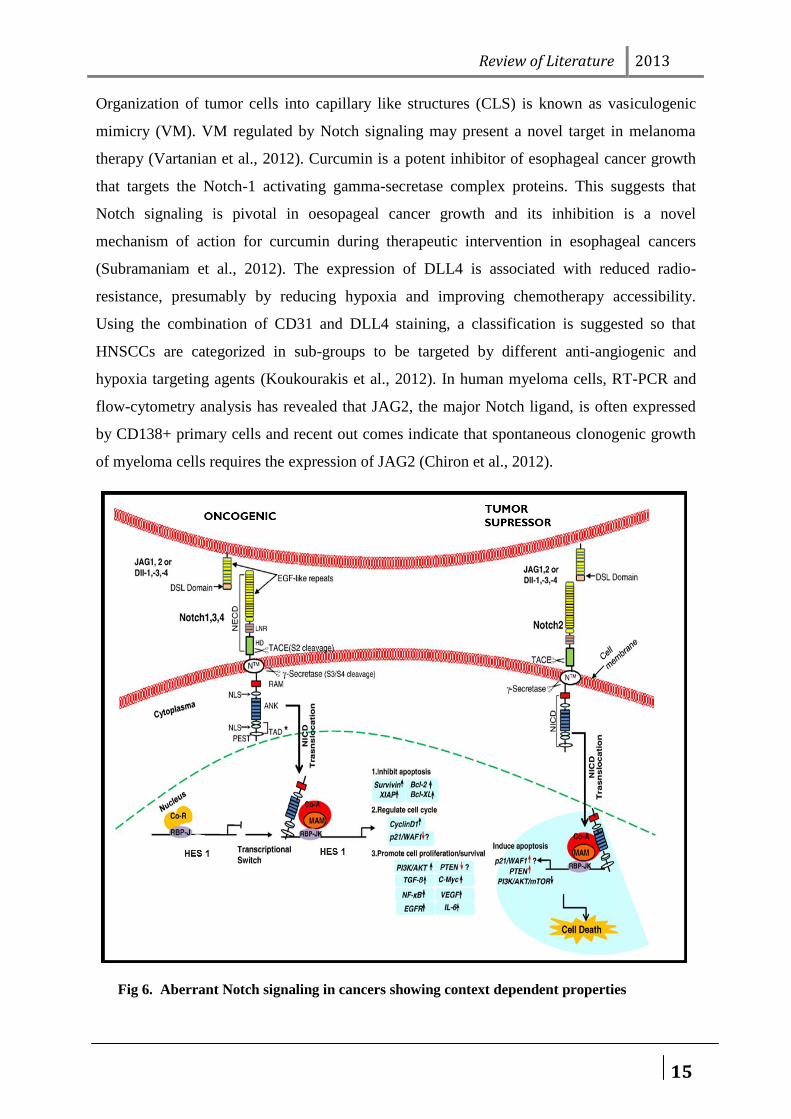
1. Notch signaling in cancers
The expression of the first two Notch receptors Notch1 and Notch2 have important role in
tumor pathology, type casting and degree of differentiation. In the tumors expressing high
levels of Notch2 and JAG1 are correlated with a significantly better prognosis in comparison
to patients expressing stunted levels of Notch2 and JAG1 (Jin et al., 2012).Involvement of
Notch signaling is variedly reported in different cancers.
Notch expression is not yet fully determined in hepatoblastoma as most of the genes
closed to Notch signaling pathway show abridged expression. But in another study of
hepatoblastoma, NOTCH1 along with DLL1, CD44, FZD2, GLI1, IL17B, LMO2, LOR,
PAX5, PT-CRA, SH2D1A and WISP1 were shown to be upregulated and contributed to
invasiveness but DLL1, HEY1, DTX1, HDAC1, NOTCH2 and JAG1 were not found to be
methylated there (Aktas et al., 2010). Within Kaposisarcoma (KS) lesions, the KSHV-
infected spindle cells showed features compatible with KSHV-induced EndMT and involves
a complex phenotype of endothelial and mesenchymal properties with Notch activity, and
nuclear ZEB1 expression. This proves Notch signaling inhibitors could be effective in KS
treatment (Gasperini et al., 2012). The active role of Notch in HPV-mediated transformations
in different gynecological cancers is dose dependent and correlated to a variation in activator
protein, AP-1 expression (Henken et al., 2012). Delta-like ligand 4 (DLL4) is one of the
major notch inducing ligand and its expression is vividly associated with poor prognosis for
surgically resected Pancreatic ductal adeno-carcinoma (PDAC), advanced tumor stage and
lymph node metastasis (Chen et al., 2012). In oral squamous cell carcinoma tumor growth is
promoted by Notch-1 along with β-catenin (component of Wnt signaling), ΔNp63
(proliferation marker and trans-membrane receptor) expression. The same study also aims to
investigate the interaction between β-catenin and ΔNp63 in oral cancer(Ravindran and
Devaraj, 2012). The down regulated expression of Notch 1 was related to invasion and
differentiation in oral carcinoma cells. In some lingual squamous carcinoma cell Notch1
down-regulation could be correlated with the expression of matrix metallo-proteinase
(MMP)-2 and MMP-9 to promote inhibition of invasion and metastasis (Yu et al., 2012a).
Review of Literature 2013
14
ER-Notch signaling along with the GPR30-PI3K/AKT signaling mediates regulation of
proliferation in nuclear ER-positive endometrial cancer cells (Wei et al., 2012). The down-
regulation of Notch3 sufficiently forces Rhabdomyosarcoma (RMS) cells to complete a
correct full myogenic program thus provides evidence that it contributes to their malignant
phenotype partially through HES1 sustained expression, (Raimondi et al., 2012).
Deregulation of NUMB-mediated suppression of NOTCH1 by TNFα/IKKα- associated
FOXA2 inhibition likely contributes to inflammation-mediated cancer pathogenesis in
hepatocellular carcinoma (Liu et al., 2012). Notch2 and JAG1 expression levels are
correlated with survival in colorectal cancers (Jin et al., 2012). Kaposi's sarcoma-associated
herpes-virus (KSHV) activated Notch-induced transcription factors Slug and ZEB1, and
canonical Notch signaling is pivotal for KSHV-induced Endothelial-mesenchymal Transition
(EndMT). So Notch inhibitors together with the currently used chemotherapeutic drugs to
improve the outcome of Osteosarcoma (OS) treatment (Ma et al., 2013). The link between the
anti-apoptotic pathways of Notch, Wnt and Hedgehog and bone marrow stromal cells in
chronic lymphoblastic leukemia (CLL) has been pointed out only recently. A number of anti-
cancer drugs, including γ-secretase inhibitors, Cyclopamine and Quercetin, were reported to
block Notch along with Wnt, and Hedgehog anti-apoptotic signaling pathways respectively in
these cancers (Seke Etet et al., 2012). Overexpressed DLL4 on neoplastic cells is shown to
enhance chemo resistance via angiogenesis-dependent/independent mechanisms in pancreatic
ductal adeno-carcinoma PDAC(Kang et al., 2013). The matrix cellular protein, secreted
protein acidic and rich in cysteine (SPARC), is known to inhibit proliferation and migration
of endothelial cells stimulated by growth factors. In a recent report pSPARCCM- suppressed
expression of growth factors was shown to be mediated by inhibition of the Notch signaling
pathway, and cells cultured on conditioned medium from tumor cells overexpress both Notch
intracellular domain (NICD-CM) and SPARC resumed the pSPARCCM- suppressed
capillary tube formation and growth factor expression in vitro (Gorantla et al., 2013).
Notch signaling aggravates the efficacy of some drugs in some cancers and also is
activated by some drugs and in many ways can contribute to targeted cancer therapy. Notch2
activation impedes inhibitory effect of Benzene isothiocyanate (BITC) on cell migration in
human breast cancer cells (Kim et al., 2012). Withaferin A (WA) one small-molecule
constituent of the ayurvedic medicine plant Withania somnifera with efficacy against cultured
and xenografted human breast cancer cells treatment activates Notch2 and Notch4, which
impede inhibitory effect of WA on breast cancer cell migration (Lee et al., 2012).
Review of Literature 2013
15
Organization of tumor cells into capillary like structures (CLS) is known as vasiculogenic
mimicry (VM). VM regulated by Notch signaling may present a novel target in melanoma
therapy (Vartanian et al., 2012). Curcumin is a potent inhibitor of esophageal cancer growth
that targets the Notch-1 activating gamma-secretase complex proteins. This suggests that
Notch signaling is pivotal in oesopageal cancer growth and its inhibition is a novel
mechanism of action for curcumin during therapeutic intervention in esophageal cancers
(Subramaniam et al., 2012). The expression of DLL4 is associated with reduced radio-
resistance, presumably by reducing hypoxia and improving chemotherapy accessibility.
Using the combination of CD31 and DLL4 staining, a classification is suggested so that
HNSCCs are categorized in sub-groups to be targeted by different anti-angiogenic and
hypoxia targeting agents (Koukourakis et al., 2012). In human myeloma cells, RT-PCR and
flow-cytometry analysis has revealed that JAG2, the major Notch ligand, is often expressed
by CD138+ primary cells and recent out comes indicate that spontaneous clonogenic growth
of myeloma cells requires the expression of JAG2 (Chiron et al., 2012).
Fig 6. Aberrant Notch signaling in cancers showing context dependent properties
Review of Literature 2013
16
Correlation of Notch1, 2, 3, Jagged1, cMET, and pMAPK, has shown their correlation to
clinicopathologic characteristics and prognostic utility for prediction of patient outcome
(Krikelis et al., 2012).The Notch repressor Numbl negatively regulates glioma cell migration
and invasion by abrogating TNF receptor-associated factor 5, TRAF5-induced activation of
NF-κB this suggests that Notch inhibitors may be best used in combination with other agents
or therapy. (Yu et al., 2012b). Down-regulated Notch1 is shown to be an effective approach
to inactivating Snail/E-cadherin by regulating the COX-2 enzyme responsible for
inflammation and pain results in inhibition of the invasion and migration of Hepatocellular
carcinoma cells (HCC) cells. Here the inhibitory effects of down regulated Notch1 on cell
invasion and migration were found to be independent of apoptosis (Zhou et al., 2012). In
Nasopharyngeal carcinoma (NPC) patients high expression level of DLL4 ligand serves as an
independent predictor of poor prognosis (Zhang et al., 2013). Notch has an important role in
lung cancer and treatment with GSI after radiation can significantly enhance radiation
mediated tumor cytotoxicity (Mizugaki et al., 2012).
Aberrant Notch-Hey1 signaling contributes to Embryonal rhabdomyosarcoma (ERMS) by
reducing differentiation and promoting proliferation. The efficacy of Notch pathway
inhibition in vivo supports the development of Notch-Hey1 axis inhibitors in the treatment of
ERMS (Belyea et al., 2011). The NOTCH Modulator LNX2 overexpression and
chromosome 13 amplification therefore constitutively activates the WNT pathway and offers
evidence of an aberrant NOTCH-WNT axis in Colorectal cancer (CRC) progression (Camps
et al., 2013). Midkine (MK), a heparin-binding growth factor that is actively overexpressed in
chemoresistant Pancreatic ductal adenocarcinoma (PDAC) interacts with Notch-2 and is
shown to activate Notch signaling, induced EMT, upregulated NF-kβ, and increased chemo
resistance (Gungor et al., 2011). Gain-of-function mutations in Notch-1 are very common in
T-cell lymphoblastic leukemia (T-ALL). The proliferation inhibitory and apoptotic effects
resulting from down-regulation of Notch-1 was expected to be mediated through regulating
the expression of cell cycle regulatory proteins cyclin D1, CDK2 and p21 and the activity of
Akt signaling in T-cell lymphoblastic leukemia (T-ALL) (Guo et al., 2009). JAG2, a notch
ligand, overexpression may be an early event in the pathogenesis of multiple myeloma
involving IL-6 production (Houde et al., 2004). In ovarian cancer Dll4 plays a functionally
important role in both the tumor and endothelial compartments and targeting Dll4 in
combination with anti-VEGF treatment can promote outcomes of treatment (Hu et al., 2011).
Review of Literature 2013
17
In another study activating point mutations in NOTCH1 in more than 50% of T-cell acute
lymphoblastic leukemia (T-ALL) shows that NOTCH1 cascade as a central player of T-ALL
pathogenesis (Koch and Radtke, 2011). NOTCH signaling is now shown to be pivotal in the
pathogenesis of a subset of intracranial ependymomas (Pfister and Witt, 2009). Neuralized
(Neurl) is a highly conserved E3 ubiquitin ligase, which in Drosophila acts upon Notch
ligands to regulate Notch pathway signaling. Human Neuralized1 (NEURL1) was
investigated as a potential tumor suppressor in medulloblastoma (MB). NEURL1 is a
candidate tumor suppressor in MB, at least in part through its effects on the Notch pathway
(Teider et al., 2010). Jagged1 may be involved in the differentiation and proliferation of
tongue cancer. Targeting Jagged1 RNA interference by lentiviral vector can effectively lower
Jagged1 mRNA and protein expression levels in Tca8113 tongue cancer cell lines, thereby
preventing the proliferation of Squamous cell carcinoma of the tongue (TSCC) cells (Zhang
et al., 2012). Skeletrophin (mindbomb homolog 2 (MIB2)) is a RING (Really Interesting
New Gene) finger-dependent ubiquitin ligase, which targets the intracellular region of Notch
ligands. Exogenously expressed skeletrophin, but not its RING mutant, increased
transcription of Hes1 gene, a downstream effector of Notch pathway in melanoma cells
(Stockhausen et al., 2005). Ectopic Myc expression plays a key role in human tumorigenesis
and Notch signaling downstream of Myc enables cells to adapt in the tumor hypoxic
microenvironment (Yustein et al., 2010) .
2. Notch, Epigenetics and Cancer
Cancer is both a genetic and an epigenetic disease. Inactivation of tumor-suppressor genes
by epigenetic changes is frequently observed in human cancers, particularly as a result of the
modifications of histones and DNA methylation (Ferres-Marco et al., 2006). Epigenetic
silencers and Notch collaborate to promote malignant tumors by Retinoblastoma (Rb)
silencing (Ferres-Marco et al., 2006). Two Polycomb group epigenetic silencers, Pipsqueak
and Lola that participate in this process tumorigenesis were studied in the drosophila eye.
When coupled with overexpression of Delta, relating to the Notch-Delta pathway,
deregulation of the expression of Pipsqueak and Lola induces the formation of metastatic
tumors. This phenotype depends on the histone-modifying enzymes Rpd3 (a histone
deacetylase), Su(var)3-9 and E(z), as well as on the chromodomain protein Polycomb.
Expression of the gene Retinoblastoma-family protein (Rbf) is downregulated in these tumors
and, indeed, this downregulation is associated with DNA hypermethylation (Ferres-Marco et
Review of Literature 2013
18
al., 2006). Neuroblastoma cell lines express multiple Notch receptors, which are inactive at
baseline. Activation of the Notch pathway via ligand binding consistently resulted in growth
arrest. Hes gene expression here appears to be regulated epigenetically and could be induced
with decitabine. These findings support a tumor suppressor role for Notch signaling in
neuroblastoma and also prove its context dependent behavior. In many childhood acute
leukemias, transcription factors are altered through chromosomal translocations that change
their functional properties resulting in repressed activity or inappropriate activation. Histone
deacetylase inhibitors, drugs targeting the NOTCH pathway, and short interfering RNAs have
shown encouraging results in pre-clinical studies and are likely to enter the clinical arena in
the near future. Through an improved understanding of the pathways and mechanisms
underlying the malignant transformation induced by altered transcription factors, new
targeted epigenetic therapies will be designed that should greatly enhance current available
treatments (Berman and Look, 2007).
The Groucho homologue Transducin-like Enhancer of Split 1 (TLE1) is a transcriptional
corepressor which acts ill in the acute myelogenous leukemia through Wnt, and Notch
signaling pathways. The epigenetic inactivation TLE1 contributes significantly to the
development of the hematologic malignancies by disrupting important pathway of
differentiation and growth-suppression (Fraga et al., 2008). Dynamic regulation of histone
modifications is critical during development, and aberrant activity of chromatin-modifying
enzymes has been associated with diseases such as cancer. The functional interplay between
histone demethylases in vivo, providing insights into the epigenetic regulation of
heterochromatin/euchromatin boundaries by Lid and dLsd1 and shows their involvement in
Notch pathway-specific control of gene expression in euchromatin (Di Stefano et al., 2011).
Correlation between constitutive acetylation of the JAG2 core promoter in the MM cell lines
reduced levels of the SMRT corepressor that recruits HDAC to promoter regions. SMRT
function restoration induced JAG2 down-regulation as well as Multiple myeloma (MM) cell
apoptosis (Ghoshal et al., 2009).The miR-181c may be silenced through methylation and play
important roles in gastric carcinogenesis through its target genes, such as NOTCH4 and
KRAS (Hashimoto et al., 2010). Notch pathway is important in regulating osteosarcoma
metastasis and may be useful as a epigenetic therapeutic target of valproic acid and other
HDAC inhibitors (Hughes, 2009). Epigenetic alterations like DNA methylation and miRNAs
mediated control of gene expression of multiple Notch target genes and pathway interacting
genes (PPARG, CCND1, and RUNX1) may relate to activation of this pathway and poor
Review of Literature 2013
19
survival of patients with high-grade serous ovarian cancer (HGS-OvCa) (Ivan et al., 2013).
Epigenetic deregulation of NOTCH4 signaling in OSCC was also observed, as part of a
possible methylation signature for recurrence, with parallels to recently discovered NOTCH
mutations in HNSCC (Jithesh et al., 2013).
Notch3 is acetylated and deacetylated at lysines 1692 and 1731 by p300 and HDAC1,
respectively, a balance impaired by HDAC inhibitors (HDACi) that favor hyperacetylation in
leukemias. Notch signaling control in which Notch3 acetylation/deacetylation process
represents a key regulatory switch and also a suitable druggable target for Notch3-sustained
T-cell acute lymphoblastic leukemia therapy (Palermo et al., 2012). Notch1 activity in gastric
cancer is controlled by the epigenetic silencing of the ligand DLL1, and that Notch1
inhibition is associated with the diffuse type of gastric cancer (Piazzi et al., 2011). In
colorectal cancer CRC cell lines, unlike gastric cancer, DLL1 expression is not regulated by
promoter methylation (Piazzi et al., 2012). Histone deacetylase inhibitor valproic acid is seen
to be effective by acting on Notch signaling in human neuroblastoma cells.
Objectives 2013
20
OBJECTIVES
The Objectives of our study were:
1. Determination of the effects of S-Adenosyl Methionine (SAM) and S-
Adenosyl Homocysteine (SAH) on the cellular morphology of breast
cancer cell line.
2. Role of SAM and SAH in modulation of Notch signaling pathway
components (Notch-1 and HES-1) in breast cancer.
3. Correlation of Notch signaling components (Notch-1 and HES-1)
expression and its role in promoting breast cancer cell death.
Materials and Methods 2013
21
MATERIALS AND METHODS
1. Cell lines and culture
We obtained the MDA MB-231 cell line from the National Centre for Cell Science
(NCCS), Pune, India. The cells are known to be of epithelial breast adenocarcinoma origin
and are triple negative. These were maintained in Dulbecco's modified Eagle's medium
(DMEM) with 10% fetal bovine serum, 2 mM L-glutamine, and 100 units/ml penicillin-
streptomycin sulfate (Invitrogen). A treatment was carried out with S-Adenosyl methionine
(SAM) and S-Adenosyl-L-homocysteine (SAH), all purchased from Sigma and added to the
regular growth media under sterile conditions for 3 days. The media was changed every 24
well culture plates.
2. MTT Assay
To determine the proliferative activity, MDA MB-231 cells were seeded in two 96-well
plates at a density (5000-40,000 cells or 106 cells) based on the doubling time, with 200 μl
growth media (10% FBS) and incubate for 24 hrs in incubator with 5% CO2 concentration at
37oC. Cell seeding must be uniform in order to obtain a dose response effect of the drug. The
drug of interests S-Adenosyl Methionine (SAM) and S-Adenosyl Homocysteine (SAH) were
diluted at 10 different concentrations in the growth media. In parallel the cells with the
solvent control was also treated to assess its effect on cells. After 24 hours existing media
was removed and replaced with media with various concentration of drugs and was incubated
for 24 or 48 hours at 37o C. To detect the cell viability MTT working solution was prepared
by diluting the stock solution (stock 5mg/ml PBS, PH 7.2) in growth medium without FBS
to the final concentration of 0.8mg/ml. 100 μl of MTT working solution was added to each
well and incubated for 4 hours in CO2 incubator. After incubation, the media was removed
carefully without disturbing formazan precipitate and dissolved in 100 μl of 100% DMSO.
An incubation of 15 minutes was carried out in dark and the colorimetric estimation of
formazan product was performed at 570nm in a microplate reader. The data was plotted
against drug concentration and non-linear regression curve fitting was performed using graph
pad prism software to calculate the optimal growth inhibitory concentration (LC50) of the
drugs.
Materials and Methods 2013
22
3. Isolation of Total Cellular RNA
The total cellular RNA was extracted using TRI reagent (Sigma), following the
manufacturer's instructions. On the culture dish 1 ml of the TRI Reagent per 10 cm2 of glass
culture plate surface area was added. After addition of the reagent, the cell lysate was passed
several times through a pipette to form a homogenous lysate. TRI Reagent is not compatible
with plastic culture plates. To ensure complete dissociation of nucleoprotein complexes,
samples were allowed to stand for 5 minutes at room temperature. 0.1 ml of 1-bromo-3-
chloropropane or 0.2 ml of chloroform was added for per ml of TRI Reagent used. Samples
were covered tightly, shaken vigorously for 15 seconds, and were allowed to stand for 2–15
minutes at room temperature. The resulting mixture was centrifuged at 12,000 g for 15
minutes at 2–8 °C. Centrifugation separates the mixture into 3 phases: a red organic phase
(containing protein), an interphase (containing DNA), and a colorless upper aqueous phase
(containing RNA).The aqueous phase was transferred to a fresh tube and 0.5 ml of
isopropanol was added per ml of TRI Reagent used in Sample Preparation, step 1 and mixed.
The sample was allowed to stand for 5–10 minutes at room temperature and centrifuged at
12,000 g for 10 minutes at 2–8 °C. The RNA precipitate formed a pellet on the side and
bottom of the tube. The supernatant was removed and RNA pellets were washed by adding a
minimum of 1 ml of 75% ethanol per 1 ml of TRI Reagent used in Sample Preparation. The
sample was vortexed and then centrifuged at 7,500 ´ g for 5 minutes at 2–8 °C. The RNA
pellets were briefly dried for 5–10 minutes by air drying. An appropriate volume of nuclease
free water was added and mixed by repeated tapping at 25 °C for 10–15 minutes.
4. Quantification of the total cellular RNA
Final preparation of RNA was analyzed using a nano-drop UV spectrophotometric
analyzer. It was likely that a standard preparation of RNA should have a 260/280 ratio of 1.7
and a 260/230 ratio of <1.65 which indicates the preparation to be free from proteins and
oligo-peptides contamination. Ethidium bromide (EtBr) staining of RNA in agarose gels
visualizes two predominant bands of small (2 kb) and large (5 kb) ribosomal RNA,
low molecular mass (0.1–0.3 kb) RNA, and discrete bands of high molecular mass (7–15 kb)
RNA.
Materials and Methods 2013
23
5. cDNA Synthesis and Evaluation
In a 1.5 ml tube 5 µl of Deionized RNAse free water, 1 µl of T18- oligo (1 µg), 6 µl of
dNTPs (10mM), 6 µl of Total RNA (3 µg) were added to make a total volume of 18 µl . The
tube with the contents was incubated at 65°C for 3 minutes. The tube was snap cooled on ice
and 6 µl of Reverse Transcriptase buffer (5X ) ,3 µl of DTT, 1 µl of Reverse Transcriptase,
1 µl of RNase inhibitor and 1 µl of RNase free water the following added to make a total
volume of the reaction mix 12 µl .The tube was then snap spinned and incubated at 45 °C for
45 minutes. Again an incubation at 90 °C for 5 minutes followed up. After incubation a snap
spin was carried out and 30 µl of deionized water was added. The cDNA prepared can now
be used immediately or can be stored at -30 °C. The synthesized cDNA was evaluated by
performing PCR for one of the house keeping genes such as β-actin. PCR reaction mix was
prepared with 5.0 µl of 10X Taq polymerase buffer, 2.5 µl of 10mM dNTP mix, 1.5 µl of 1.5
mM MgCl2, 2.0 µl of Oligo (mActin- Forward) 10 µM, 2.0 µl of Oligo (mActin- Reverse) 10
µM, 1.0 µl of Taq DNA polymerase and 26.0 µl of deionized water to make a total volume
of 40.0 µl. Two aliquots of 20 µl of PCR mix was transferred into 2 PCR tubes. 5 µl of
cDNA was added in one tube and 5 µl deionized water was added in another. The tubes were
snap spinned to collect the contents in the bottom of the tubes and were subject to PCR with
the following cycling conditions of ; 92° C for 3 minutes, 30 cycles of 92 °C 30 sec, 50 °C
for 30 sec, 72 °C for 30 sec, 72 °C for 5 minutes and 4 °C for forever. The contents were
loaded in 1.5% agarose gel and electrophoresed along with a molecular weight marker. The
gel was visualized under UV light and results were documented in a gel documentation
system.
6. RT-PCR Analysis
We used total RNA (2 μg.) isolated from the cancerous cell lines for reverse transcription
and amplification. The primers used for RT-PCR were designed so that there is an intron
between the amplified regions to recognize any DNA contamination. We used three sets of
primers to amplify NOTCH1, HES-1 and β-actin genes having sequence as detailed in Table
no. for each RNA sample. The PCR were carried out using standard protocols and the DNA
was amplified under the following conditions: 95 °C for 3 min, 30 cycles of 95 °C for 30 s,
60 °C for 30 s, and 72 °C for 30 s, and the final extension of 72 °C for 5 min. We then
analyzed the PCR products on a 1% agarose gel.
Materials and Methods 2013
24
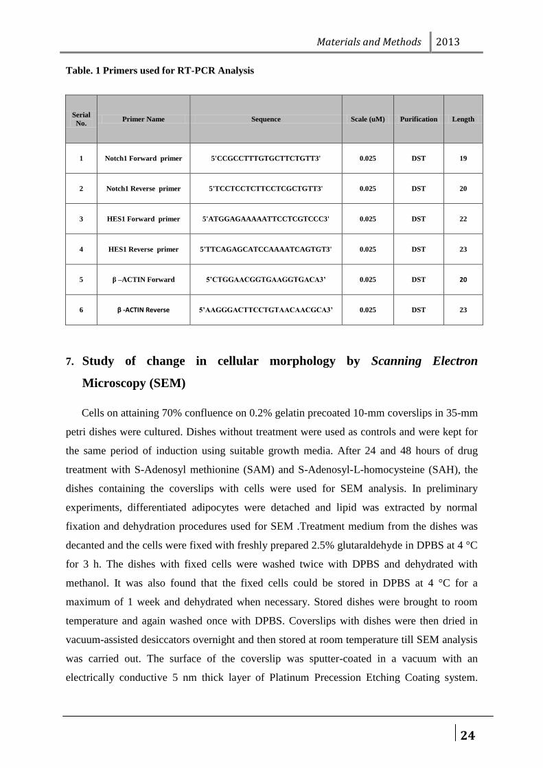
Table. 1 Primers used for RT-PCR Analysis
Serial
No. Primer Name Sequence Scale (uM) Purification Length
1 Notch1 Forward primer 5'CCGCCTTTGTGCTTCTGTT3' 0.025 DST 19
2 Notch1 Reverse primer 5'TCCTCCTCTTCCTCGCTGTT3' 0.025 DST 20
3 HES1 Forward primer 5'ATGGAGAAAAATTCCTCGTCCC3' 0.025 DST 22
4 HES1 Reverse primer 5'TTCAGAGCATCCAAAATCAGTGT3' 0.025 DST 23
5 β –ACTIN Forward 5’CTGGAACGGTGAAGGTGACA3’ 0.025 DST 20
6 β -ACTIN Reverse 5’AAGGGACTTCCTGTAACAACGCA3’ 0.025 DST 23
7. Study of change in cellular morphology by Scanning Electron
Microscopy (SEM)
Cells on attaining 70% confluence on 0.2% gelatin precoated 10-mm coverslips in 35-mm
petri dishes were cultured. Dishes without treatment were used as controls and were kept for
the same period of induction using suitable growth media. After 24 and 48 hours of drug
treatment with S-Adenosyl methionine (SAM) and S-Adenosyl-L-homocysteine (SAH), the
dishes containing the coverslips with cells were used for SEM analysis. In preliminary
experiments, differentiated adipocytes were detached and lipid was extracted by normal
fixation and dehydration procedures used for SEM .Treatment medium from the dishes was
decanted and the cells were fixed with freshly prepared 2.5% glutaraldehyde in DPBS at 4 °C
for 3 h. The dishes with fixed cells were washed twice with DPBS and dehydrated with
methanol. It was also found that the fixed cells could be stored in DPBS at 4 °C for a
maximum of 1 week and dehydrated when necessary. Stored dishes were brought to room
temperature and again washed once with DPBS. Coverslips with dishes were then dried in
vacuum-assisted desiccators overnight and then stored at room temperature till SEM analysis
was carried out. The surface of the coverslip was sputter-coated in a vacuum with an
electrically conductive 5 nm thick layer of Platinum Precession Etching Coating system.
Materials and Methods 2013
25
SEM images were then recorded with a scanning electron microscope at a lower voltage (20
kV) and low vacuum mode with a tilt of 30°.
8. Cell migration assay (Scratch assay)
60-mm dishes were coated with proper ECM substrates for the cell type to be studied by
incubating the dishes overnight at 4 °C or for 2 h at 37 °C without rotation or shaking. The
unbound ECM substrate was removed and dishes were blocked and coated with 3 ml of 2 mg
ml-1 bovine serum albumin for 1 h at 37°C. Then the dishes were once washed with PBS and
refilled with 3–5 ml of media before plating the cells. For the particular cell type used, the
appropriate amount of serum in the medium during the in vitro scratch assay is required to be
determined. It is always recommended to use a lower percentage of serum than that used in
the growth media to minimize cell proliferation, but just sufficient to prevent apoptosis and/or
cell detachment. Sub-confluent growing cells were re-suspended in a tissue culture dish by
washing cells twice with PBS, adding versene containing trypsin, and then mixing cells with
medium containing serum. The solution was gently pipetted and the dish was rocked to
disperse the cells equally. An aliquot from the cell suspension was taken and the cell count
was determined using a hemocytometer. Cells were plated onto the prepared 60-mm dish to
create a confluent monolayer and incubated properly for approximately 6 hours at 37°C,
allowing cells to adhere and spread on the substrate completely. The required number of cells
for a confluent monolayer depends on both the particular cell type and the size of dishes and
need to be adjusted appropriately. The cell monolayer was scraped in a straight line to create
a „„scratch‟‟ with a p200 pipet tip.
The debris was removed and the edge of the scratch was smoothed by washing the cells
once with 1 ml of the growth medium and then replaced with 5 ml of medium specific for the
in vitro scratch assay. To obtain the same field during the image acquisition markings were
created to be used as reference points close to the scratch. The reference points can be made
by etching the dish lightly with a razor blade on the outer bottom of the dish or with an
ultrafine tip marker. After the reference points were made, the dish was placed under a phase-
contrast microscope, and reference mark was left outside the capture image field but within
the eye-piece field of view. The first image of the scratch was taken. The dish was placed in a
tissue culture incubator at 37°C for 8–18 hours. After the incubation dish was placed under a
Materials and Methods 2013
26
phase-contrast microscope, the reference point was matched; the photographed region was
aligned to acquire a second image.
9. Analysis of chromatin condensation by Hoechst 33342 stain
After treatment with drugs, the cells were stained with Hoechst 33342 stain (1 mg/ml) and
incubated for 10 min at 37°C and images were taken under UV filter using Epi-fluorescent
Microscope (Nikon TE 2000E). Condensed nucleus was counted against total number of
nucleus in the field, and the percentages of apoptotic nuclei were analyzed.
10. Cytochemical staining of for detecting autophagosomes
An optimal number of cells were seeded based on doubling time in a 96 well plate and kept
for 24 hours in incubator with 5% CO2 at 37°C. After the incubation period, the cells were
challenged with the suspected autophagy modulating factor for 12 hours. Then after
incubation, the existing media was removed and 100µl of fresh media containing 1µg/ml of
acridine orange was added. The cells were incubated for 15min at normal culture
conditions. The media was discarded and washed with PBS and fresh media was added to the
cells. The cells were analyzed under a fluorescent microscope using blue filter (495nm) to
view the green fluorescence (510-530nm) from free Acridine Orange and red fluorescence (>
650nm) from acidic vesicles (autophagosomes).
11. Comet assay to measure the DNA damage
Two water baths were equilibrated at 40 °C and 100 °C respectively. Than 1% low-gelling-
temperature agarose was prepared by mixing powdered agarose with distilled water in a glass
beaker or bottle. The bottle was placed in the 100 °C water bath for several minutes and was
transferred into a 40 °C water bath. Agarose-precoated slides were prepared by dipping the
slides into molten 1% agarose and wiping one side clean. It is best to work in a low-humidity
environment to ensure agarose adhesion. Agarose was allowed to air-dry to a thin film. Slides
can be prepared ahead of time and stored with desiccant. A single-cell suspension was
prepared using enzyme disaggregation or mechanical dissociation. The cells were kept in ice-
cold medium or phosphate-buffered saline to minimize cell aggregation and inhibit DNA
repair. Using a hemocytometer or particle counter, cell density was adjusted to about 2 × 10⁴
Materials and Methods 2013
27
cells/ml in phosphate-buffered saline lacking divalent cations. Slides were labeled on frosted
end using a pencil. 0.4 ml of cells into a 5 ml plastic disposable tube.1.2 ml 1% low-gelling-
temperature agarose at was added at 40 °C.1.2 ml of cell suspension onto the agarose-covered
surface of a pre-coated slide was mixed by vigorous pipetting. Agarose was allowed to be gel
for about 2 min. After agarose has gelled, slides were submerged in a covered dish containing
A1 lysis solution. Samples were lysed overnight (18−20 h) at 4 °C in the dark. After
overnight lysis, slides were removed carefully and submerge in A2 rinse solution for 20 min
at room temperature (18−25 °C). The process was repeated two times to ensure removal of
salt and detergent. Care was taken for not allowing DNA to renature even briefly (i.e., by
lowering pH below 12.3) until after electrophoresis, as this will result in DNA tangling and
reduced migration. After these three rinses, slides were submerged in fresh A2 solution in an
electrophoresis chamber. The chamber was filled with a consistent volume of buffer that is
about 1–2 mm above the top of the agarose. It was ensured that the chamber is level using a
bubble leveling device. Electrophoresis was conducted in solution A2 for 25 min at a voltage
of 0.6 V/cm. The current was about 40 mA using 20 V. The distance in centimeters was
measured between the negative and positive electrodes in the electrophoresis chamber. Slides
were removed from electrophoresis chamber and were rinsed and neutralized in 400 ml of
distilled water. Slides were placed in staining solution containing 2.5 μg/ml of propidium
iodide in distilled water for 20 min. Finally the slides were rinsed with 400 ml distilled water
to remove excess stain. Analysis of cells was done by examining at least 50 comet images
from each slide. Analyzing doublets or comets at slide edges should be avoided.
Results & Discussion 2013
28
RESULTS AND DISCUSSION
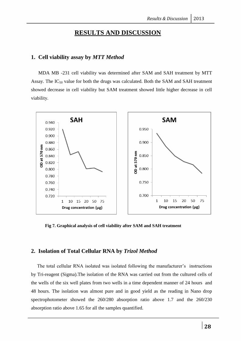
1. Cell viability assay by MTT Method
MDA MB -231 cell viability was determined after SAM and SAH treatment by MTT
Assay. The IC50 value for both the drugs was calculated. Both the SAM and SAH treatment
showed decrease in cell viability but SAM treatment showed little higher decrease in cell
viability.
Fig 7. Graphical analysis of cell viability after SAM and SAH treatment
2. Isolation of Total Cellular RNA by Trizol Method
The total cellular RNA isolated was isolated following the manufacturer‟s instructions
by Tri-reagent (Sigma).The isolation of the RNA was carried out from the cultured cells of
the wells of the six well plates from two wells in a time dependent manner of 24 hours and
48 hours. The isolation was almost pure and in good yield as the reading in Nano drop
spectrophotometer showed the 260/280 absorption ratio above 1.7 and the 260/230
absorption ratio above 1.65 for all the samples quantified.
Results & Discussion 2013
29
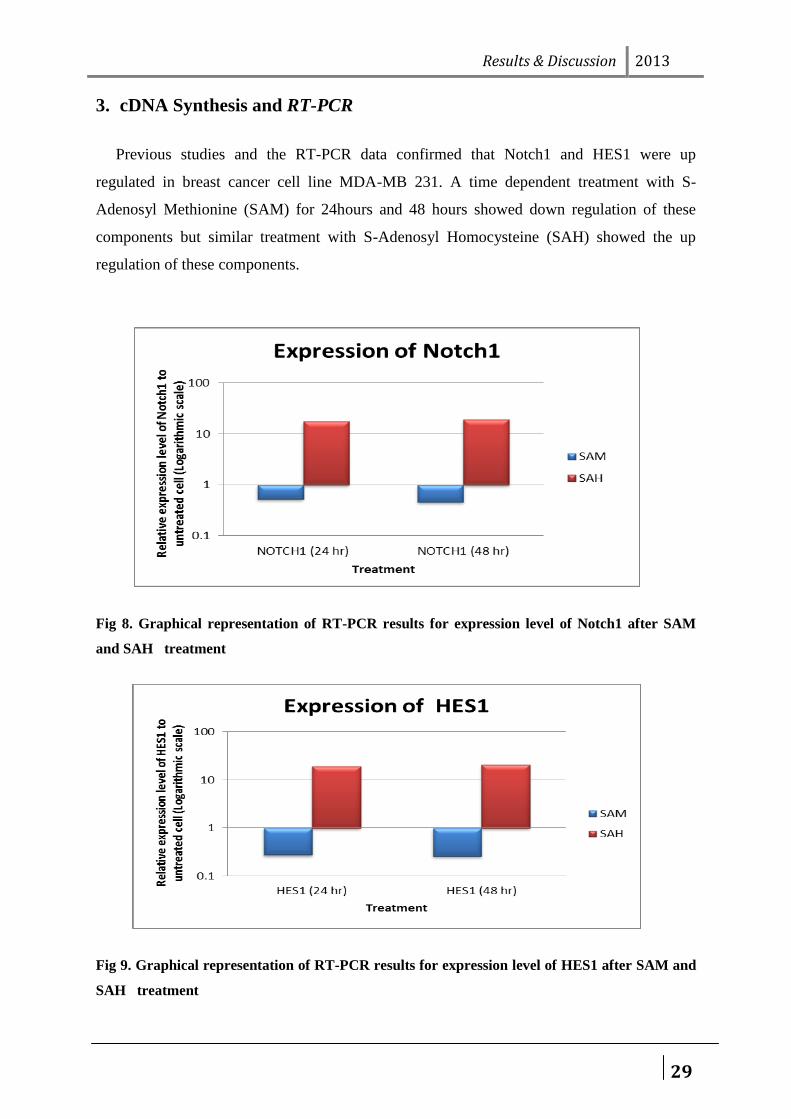
3. cDNA Synthesis and RT-PCR
Previous studies and the RT-PCR data confirmed that Notch1 and HES1 were up
regulated in breast cancer cell line MDA-MB 231. A time dependent treatment with S-
Adenosyl Methionine (SAM) for 24hours and 48 hours showed down regulation of these
components but similar treatment with S-Adenosyl Homocysteine (SAH) showed the up
regulation of these components.
Fig 8. Graphical representation of RT-PCR results for expression level of Notch1 after SAM
and SAH treatment
Fig 9. Graphical representation of RT-PCR results for expression level of HES1 after SAM and
SAH treatment
Results & Discussion 2013
30
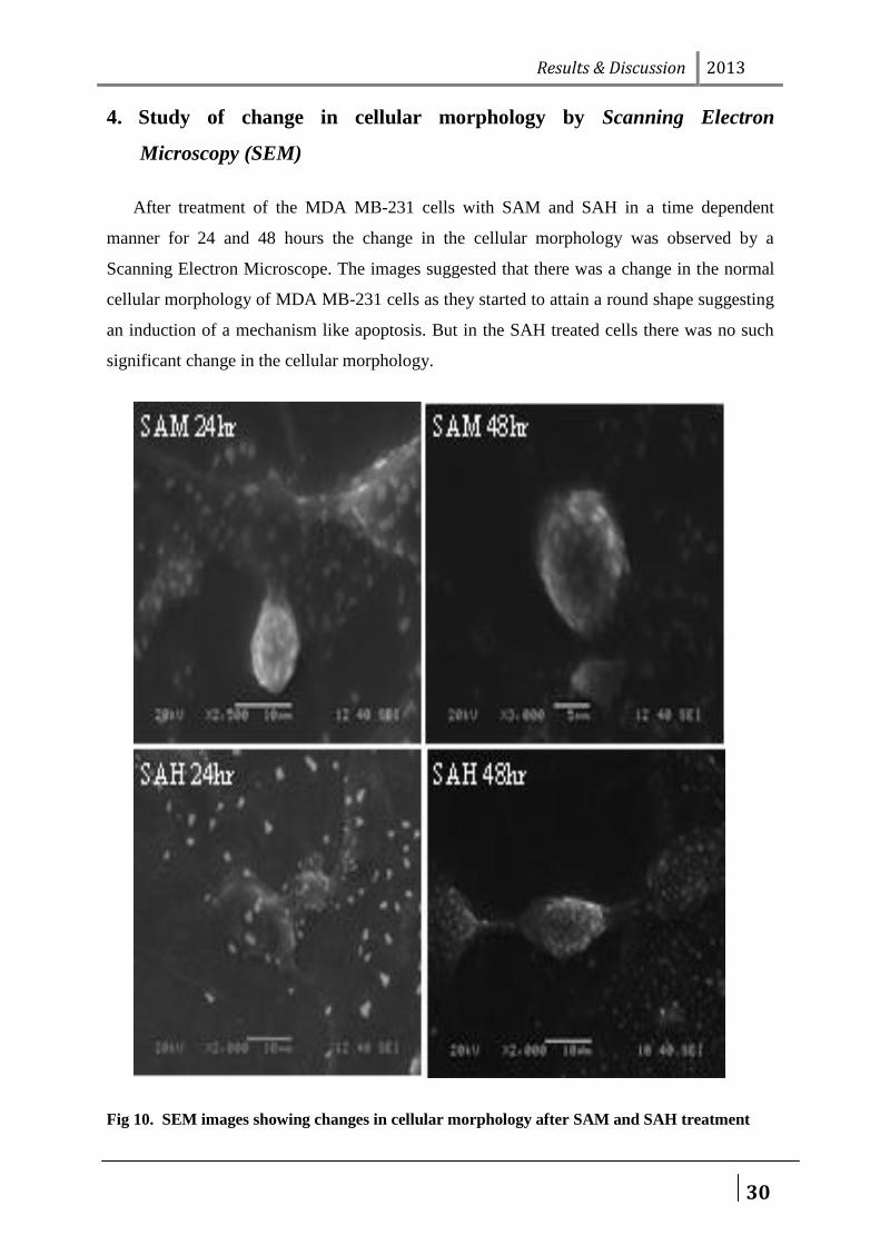
4. Study of change in cellular morphology by Scanning Electron
Microscopy (SEM)
After treatment of the MDA MB-231 cells with SAM and SAH in a time dependent
manner for 24 and 48 hours the change in the cellular morphology was observed by a
Scanning Electron Microscope. The images suggested that there was a change in the normal
cellular morphology of MDA MB-231 cells as they started to attain a round shape suggesting
an induction of a mechanism like apoptosis. But in the SAH treated cells there was no such
significant change in the cellular morphology.
Fig 10. SEM images showing changes in cellular morphology after SAM and SAH treatment
Results & Discussion 2013
31
5. Cell migration study by Scratch assay
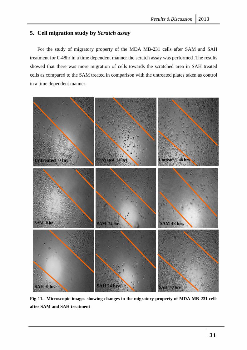
For the study of migratory property of the MDA MB-231 cells after SAM and SAH
treatment for 0-48hr in a time dependent manner the scratch assay was performed .The results
showed that there was more migration of cells towards the scratched area in SAH treated
cells as compared to the SAM treated in comparison with the untreated plates taken as control
in a time dependent manner.
Fig 11. Microscopic images showing changes in the migratory property of MDA MB-231 cells
after SAM and SAH treatment
Untreated 0 hr. Untreated 24 hrs. Untreated 48 hrs.
SAH 0 hr. SAH 24 hrs. SAH 48 hrs.
SAM 0 hr.
hrs. SAM 24 hrs. SAM 48 hrs.
Results & Discussion 2013
32
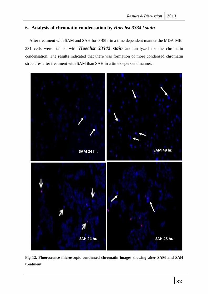
6. Analysis of chromatin condensation by Hoechst 33342 stain
After treatment with SAM and SAH for 0-48hr in a time dependent manner the MDA-MB-
231 cells were stained with Hoechst 33342 stain and analyzed for the chromatin
condensation. The results indicated that there was formation of more condensed chromatin
structures after treatment with SAM than SAH in a time dependent manner.
Fig 12. Fluorescence microscopic condensed chromatin images showing after SAM and SAH
treatment
SAM 24 hr. SAM 48 hr.
SAH 24 hr. SAH 48 hr.
Results & Discussion 2013
33
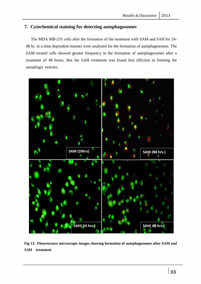
7. Cytochemical staining for detecting autophagosomes
The MDA MB-231 cells after the formation of the treatment with SAM and SAH for 24-
48 hr. in a time dependent manner were analyzed for the formation of autophagosomes. The
SAM treated cells showed greater frequency in the formation of autophagosomes after a
treatment of 48 hours. But the SAH treatment was found less efficient in forming the
autophagic vesicles.
Fig 13. Fluorescence microscopic images showing formation of autophagosomes after SAM and
SAH treatment
SAM (24hrs)
SAH( 48 hrs.)
SAM (48 hrs.)
SAH( 24 hrs.)
Results & Discussion 2013
34
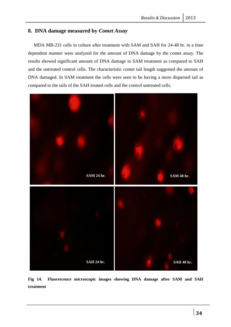
8. DNA damage measured by Comet Assay
MDA MB-231 cells in culture after treatment with SAM and SAH for 24-48 hr. in a time
dependent manner were analyzed for the amount of DNA damage by the comet assay. The
results showed significant amount of DNA damage in SAM treatment as compared to SAH
and the untreated control cells. The characteristic comet tail length suggested the amount of
DNA damaged. In SAM treatment the cells were seen to be having a more dispersed tail as
compared to the tails of the SAH treated cells and the control untreated cells.
Fig 14. Fluorescence microscopic images showing DNA damage after SAM and SAH
treatment
SAM 24 hr. SAM 48 hr.
SAH 48 hr. SAH 24 hr.
Conclusion 2013
35
CONCLUSION
Work over the last decade has shown that Notch signaling is aberrantly activated in breast
cancer and that it regulates many of the cellular properties associated with transformation.
This has led to significant interest in the use of Notch pathway inhibitors for breast cancer
treatment, especially as they are expected to have effects in bulk tumor cells, cancer stem
cells and the surrounding tumor stroma. The question now is how best to use these inhibitors
in clinical trials. Current preclinical work indicates that Notch pathway inhibitors are unlikely
to be effective on their own, but that they should significantly increase the efficacy of current
therapies. This said, there is still a need to identify patients that are likely to respond to Notch
pathway inhibitors (He et al. 2011; Watters et al. 2009). From our study we found that after
treating the MDA MB-231 metastatic breast cancer cell line with epigenetic modulators SAM
and SAH , the Notch signaling components Notch1 and Hes1 are down regulated after SAM
treatment and were significantly upregulated after SAH treatment. This was accompanied by
changes in cellular phenotypes of the cells as the SAM treatment changed the cells to more
round shape, Reduced migratory potential , Induced autophagosome formation , Aggravated
chromatin condensation , Caused DNA damage as compared to SAH treated cells.
From these above experimental results we can tentatively conclude that SAM treatment of
invasive breast cancer cell line induced cell death in a notch dependent manner. This
experimental data suggests that SAM can be a potent therapeutic agent for treatment of breast
cancer.
References 2013
36
REFERNCES
Aktas, S., Zadeoglulari, Z., Ercetin, P., and Olgun, N. (2010). The effect of differentiating and apoptotic agents on notch signalling pathway in hepatoblastoma. Hepatogastroenterology 57, 891-8.
Belyea, B. C., Naini, S., Bentley, R. C., and Linardic, C. M. (2011). Inhibition of the Notch-Hey1 axis blocks embryonal rhabdomyosarcoma tumorigenesis. Clin Cancer Res 17, 7324-36.
Berman, J. N., and Look, A. T. (2007). Targeting transcription factors in acute leukemia in children. Curr Drug Targets 8, 727-37.
Camps, J., Pitt, J. J., Emons, G., Hummon, A. B., Case, C. M., Grade, M., Jones, T. L., Nguyen, Q. T., Ghadimi, B. M., Beissbarth, T., Difilippantonio, M. J., Caplen, N. J., and Ried, T. (2013). Genetic Amplification of the NOTCH Modulator LNX2 Upregulates the WNT/beta-Catenin Pathway in Colorectal Cancer. Cancer Res 73, 2003-13.
Chen, H. T., Cai, Q. C., Zheng, J. M., Man, X. H., Jiang, H., Song, B., Jin, G., Zhu, W., and Li, Z. S. (2012). High expression of delta-like ligand 4 predicts poor prognosis after curative resection for pancreatic cancer. Ann Surg Oncol 19 Suppl 3, S464-74.
Chiron, D., Maiga, S., Descamps, G., Moreau, P., Le Gouill, S., Marionneau, S., Ouiller, T., Moreaux, J., Klein, B., Bataille, R., Amiot, M., and Pellat-Deceunynck, C. (2012). Critical role of the NOTCH ligand JAG2 in self-renewal of myeloma cells. Blood Cells Mol Dis 48, 247-53.
Di Stefano, L., Walker, J. A., Burgio, G., Corona, D. F., Mulligan, P., Naar, A. M., and Dyson, N. J. (2011). Functional antagonism between histone H3K4 demethylases in vivo. Genes Dev 25, 17-28.
Ferres-Marco, D., Gutierrez-Garcia, I., Vallejo, D. M., Bolivar, J., Gutierrez-Avino, F. J., and Dominguez, M. (2006). Epigenetic silencers and Notch collaborate to promote malignant tumours by Rb silencing. Nature 439, 430-6.
Fraga, M. F., Berdasco, M., Ballestar, E., Ropero, S., Lopez-Nieva, P., Lopez-Serra, L., Martin-Subero, J. I., Calasanz, M. J., Lopez de Silanes, I., Setien, F., Casado, S., Fernandez, A. F., Siebert, R., Stifani, S., and Esteller, M. (2008). Epigenetic inactivation of the Groucho homologue gene TLE1 in hematologic malignancies. Cancer Res 68, 4116-22.
Gasperini, P., Espigol-Frigole, G., McCormick, P. J., Salvucci, O., Maric, D., Uldrick, T. S., Polizzotto, M. N., Yarchoan, R., and Tosato, G. (2012). Kaposi sarcoma herpesvirus promotes endothelial-to-mesenchymal transition through Notch-dependent signaling. Cancer Res 72, 1157-69.
References 2013
37
Ghoshal, P., Nganga, A. J., Moran-Giuati, J., Szafranek, A., Johnson, T. R., Bigelow, A. J., Houde, C. M., Avet-Loiseau, H., Smiraglia, D. J., Ersing, N., Chanan-Khan, A. A., and Coignet, L. J. (2009). Loss of the SMRT/NCoR2 corepressor correlates with JAG2 overexpression in multiple myeloma. Cancer Res 69, 4380-7.
Gorantla, B., Bhoopathi, P., Chetty, C., Gogineni, V. R., Sailaja, G. S., Gondi, C. S., and Rao, J. S. (2013). Notch signaling regulates tumor-induced angiogenesis in SPARC-overexpressed neuroblastoma. Angiogenesis 16, 85-100.
Gungor, C., Zander, H., Effenberger, K. E., Vashist, Y. K., Kalinina, T., Izbicki, J. R., Yekebas, E., and Bockhorn, M. (2011). Notch signaling activated by replication stress-induced expression of midkine drives epithelial-mesenchymal transition and chemoresistance in pancreatic cancer. Cancer Res 71, 5009-19.
Guo, D., Ye, J., Dai, J., Li, L., Chen, F., Ma, D., and Ji, C. (2009). Notch-1 regulates Akt signaling pathway and the expression of cell cycle regulatory proteins cyclin D1, CDK2 and p21 in T-ALL cell lines. Leuk Res 33, 678-85.
Hashimoto, Y., Akiyama, Y., Otsubo, T., Shimada, S., and Yuasa, Y. (2010). Involvement of epigenetically silenced microRNA-181c in gastric carcinogenesis. Carcinogenesis 31, 777-84.
Henken, F. E., De-Castro Arce, J., Rosl, F., Bosch, L., Meijer, C. J., Snijders, P. J., and Steenbergen, R. D. (2012). The functional role of Notch signaling in HPV-mediated transformation is dose-dependent and linked to AP-1 alterations. Cell Oncol (Dordr) 35, 77-84.
Houde, C., Li, Y., Song, L., Barton, K., Zhang, Q., Godwin, J., Nand, S., Toor, A., Alkan, S., Smadja, N. V., Avet-Loiseau, H., Lima, C. S., Miele, L., and Coignet, L. J. (2004). Overexpression of the NOTCH ligand JAG2 in malignant plasma cells from multiple myeloma patients and cell lines. Blood 104, 3697-704.
Hu, W., Lu, C., Dong, H. H., Huang, J., Shen, D. Y., Stone, R. L., Nick, A. M., Shahzad, M. M., Mora, E., Jennings, N. B., Lee, S. J., Roh, J. W., Matsuo, K., Nishimura, M., Goodman, B. W., Jaffe, R. B., Langley, R. R., Deavers, M. T., Lopez-Berestein, G., Coleman, R. L., and Sood, A. K. (2011). Biological roles of the Delta family Notch ligand Dll4 in tumor and endothelial cells in ovarian cancer. Cancer Res 71, 6030-9.
Hughes, D. P. (2009). How the NOTCH pathway contributes to the ability of osteosarcoma cells to metastasize. Cancer Treat Res 152, 479-96.
Ivan, C., Hu, W., Bottsford-Miller, J., Zand, B., Dalton, H. J., Liu, T., Huang, J., Nick, A. M., Lopez-Berestein, G., Coleman, R. L., Baggerly, K. A., and
References 2013
38
Sood, A. K. (2013). Epigenetic analysis of the Notch superfamily in high-grade serous ovarian cancer. Gynecol Oncol 128, 506-11.
Jin, H. Y., Zhang, H. Y., Wang, X., Xu, J., and Ding, Y. (2012). Expression and clinical significance of Notch signaling genes in colorectal cancer. Tumour Biol 33, 817-24.
Jithesh, P. V., Risk, J. M., Schache, A. G., Dhanda, J., Lane, B., Liloglou, T., and Shaw, R. J. (2013). The epigenetic landscape of oral squamous cell carcinoma. Br J Cancer 108, 370-9.
Kang, M., Jiang, B., Xu, B., Lu, W., Guo, Q., Xie, Q., Zhang, B., Dong, X., Chen, D., and Wu, Y. (2013). Delta like ligand 4 induces impaired chemo-drug delivery and enhanced chemoresistance in pancreatic cancer. Cancer Lett 330, 11-21.
Kar, S., Deb, M., Sengupta, D., Shilpi, A., Bhutia, S. K., and Patra, S. K. (2012). Intricacies of hedgehog signaling pathways: a perspective in tumorigenesis. Exp Cell Res 318, 1959-72.
Kim, S. H., Sehrawat, A., and Singh, S. V. (2012). Notch2 activation by benzyl isothiocyanate impedes its inhibitory effect on breast cancer cell migration. Breast Cancer Res Treat 134, 1067-79.
Koch, U., and Radtke, F. (2011). Notch in T-ALL: new players in a complex disease. Trends Immunol 32, 434-42.
Koukourakis, M. I., Giatromanolaki, A., Sivridis, E., Gatter, K. C., and Harris, A. L. (2012). High DLL4 expression in tumour-associated vessels predicts for favorable radiotherapy outcome in locally advanced squamous cell head-neck cancer (HNSCC). Angiogenesis.
Krikelis, D., Pentheroudakis, G., Goussia, A., Siozopoulou, V., Bobos, M., Petrakis, D., Stoyianni, A., Golfinopoulos, V., Cervantes, A., Ciuleanu, T., Fountzilas, G., Malamou-Mitsi, V., and Pavlidis, N. (2012). Profiling immunohistochemical expression of NOTCH1-3, JAGGED1, cMET, and phospho-MAPK in 100 carcinomas of unknown primary. Clin Exp Metastasis 29, 603-14.
Lee, J., Sehrawat, A., and Singh, S. (2012). Withaferin A causes activation of Notch2 and Notch4 in human breast cancer cells. Breast Cancer Research and Treatment 136, 45-56.
Liu, M., Lee, D. F., Chen, C. T., Yen, C. J., Li, L. Y., Lee, H. J., Chang, C. J., Chang, W. C., Hsu, J. M., Kuo, H. P., Xia, W., Wei, Y., Chiu, P. C., Chou, C. K., Du, Y., Dhar, D., Karin, M., Chen, C. H., and Hung, M. C. (2012). IKKalpha activation of NOTCH links tumorigenesis via FOXA2 suppression. Mol Cell 45, 171-84.
Ma, Y., Ren, Y., Han, E. Q., Li, H., Chen, D., Jacobs, J. J., Gitelis, S., O'Keefe, R. J., Konttinen, Y. T., Yin, G., and Li, T. F. (2013). Inhibition of the Wnt-beta-
References 2013
39
catenin and Notch signaling pathways sensitizes osteosarcoma cells to chemotherapy. Biochem Biophys Res Commun 431, 274-9.
Mizugaki, H., Sakakibara-Konishi, J., Ikezawa, Y., Kikuchi, J., Kikuchi, E., Oizumi, S., Dang, T. P., and Nishimura, M. (2012). gamma-Secretase inhibitor enhances antitumour effect of radiation in Notch-expressing lung cancer. Br J Cancer 106, 1953-9.
Palermo, R., Checquolo, S., Giovenco, A., Grazioli, P., Kumar, V., Campese, A. F., Giorgi, A., Napolitano, M., Canettieri, G., Ferrara, G., Schinina, M. E., Maroder, M., Frati, L., Gulino, A., Vacca, A., and Screpanti, I. (2012). Acetylation controls Notch3 stability and function in T-cell leukemia. Oncogene 31, 3807-17.
Patra, S. K., Patra, A., Rizzi, F., Ghosh, T. C., and Bettuzzi, S. (2008). Demethylation of (Cytosine-5-C-methyl) DNA and regulation of transcription in the epigenetic pathways of cancer development. Cancer Metastasis Rev 27, 315-34.
Patra, S. K., Patra, A., Zhao, H., and Dahiya, R. (2002). DNA methyltransferase and demethylase in human prostate cancer. Mol Carcinog 33, 163-71.
Pfister, S., and Witt, O. (2009). Pediatric gliomas. Recent Results Cancer Res 171, 67-81.
Piazzi, G., Bazzoli, F., and Ricciardiello, L. (2012). Epigenetic silencing of Notch signaling in gastrointestinal cancers. Cell Cycle 11, 4323-7.
Piazzi, G., Fini, L., Selgrad, M., Garcia, M., Daoud, Y., Wex, T., Malfertheiner, P., Gasbarrini, A., Romano, M., Meyer, R. L., Genta, R. M., Fox, J. G., Boland, C. R., Bazzoli, F., and Ricciardiello, L. (2011). Epigenetic regulation of Delta-Like1 controls Notch1 activation in gastric cancer. Oncotarget 2, 1291-301.
Raimondi, L., Ciarapica, R., De Salvo, M., Verginelli, F., Gueguen, M., Martini, C., De Sio, L., Cortese, G., Locatelli, M., Dang, T. P., Carlesso, N., Miele, L., Stifani, S., Limon, I., Locatelli, F., and Rota, R. (2012). Inhibition of Notch3 signalling induces rhabdomyosarcoma cell differentiation promoting p38 phosphorylation and p21(Cip1) expression and hampers tumour cell growth in vitro and in vivo. Cell Death Differ 19, 871-81.
Ranganathan, P., Weaver, K. L., and Capobianco, A. J. (2011). Notch signalling in solid tumours: a little bit of everything but not all the time. Nat Rev Cancer 11, 338-51.
Ravindran, G., and Devaraj, H. (2012). Aberrant expression of beta-catenin and its association with DeltaNp63, Notch-1, and clinicopathological factors in oral squamous cell carcinoma. Clin Oral Investig 16, 1275-88.
Seke Etet, P. F., Vecchio, L., and Nwabo Kamdje, A. H. (2012). Interactions between bone marrow stromal microenvironment and B-chronic
References 2013
40
lymphocytic leukemia cells: any role for Notch, Wnt and Hh signaling pathways? Cell Signal 24, 1433-43.
Stockhausen, M. T., Sjolund, J., Manetopoulos, C., and Axelson, H. (2005). Effects of the histone deacetylase inhibitor valproic acid on Notch signalling in human neuroblastoma cells. Br J Cancer 92, 751-759.
Subramaniam, D., Ponnurangam, S., Ramamoorthy, P., Standing, D., Battafarano, R. J., Anant, S., and Sharma, P. (2012). Curcumin induces cell death in esophageal cancer cells through modulating Notch signaling. PLoS One 7, e30590.
Teider, N., Scott, D. K., Neiss, A., Weeraratne, S. D., Amani, V. M., Wang, Y., Marquez, V. E., Cho, Y. J., and Pomeroy, S. L. (2010). Neuralized1 causes apoptosis and downregulates Notch target genes in medulloblastoma. Neuro Oncol 12, 1244-56.
Vartanian, A., Gatsina, G., Grigorieva, I., Solomko, E., Dombrovsky, V., Baryshnikov, A., and Stepanova, E. (2012). The involvement of Notch signaling in melanoma vasculogenic mimicry. Clin Exp Med.
Wang, Z., Li, Y., Ahmad, A., Azmi, A. S., Banerjee, S., Kong, D., and Sarkar, F. H. (2010). Targeting Notch signaling pathway to overcome drug resistance for cancer therapy. Biochim Biophys Acta 1806, 258-67.
Wei, Y., Zhang, Z., Liao, H., Wu, L., Wu, X., Zhou, D., Xi, X., Zhu, Y., and Feng, Y. (2012). Nuclear estrogen receptor-mediated Notch signaling and GPR30-mediated PI3K/AKT signaling in the regulation of endometrial cancer cell proliferation. Oncol Rep 27, 504-10.
Ye, Q. F., Zhang, Y. C., Peng, X. Q., Long, Z., Ming, Y. Z., and He, L. Y. (2012). siRNA-mediated silencing of Notch-1 enhances docetaxel induced mitotic arrest and apoptosis in prostate cancer cells. Asian Pac J Cancer Prev 13, 2485-9.
Yu, B., Wei, J., Qian, X., Lei, D., Ma, Q., and Liu, Y. (2012a). Notch1 signaling pathway participates in cancer invasion by regulating MMPs in lingual squamous cell carcinoma. Oncol Rep 27, 547-52.
Yu, S. D., Liu, F. Y., and Wang, Q. R. (2012b). Notch inhibitor: a promising carcinoma radiosensitizer. Asian Pac J Cancer Prev 13, 5345-51.
Yustein, J. T., Liu, Y. C., Gao, P., Jie, C., Le, A., Vuica-Ross, M., Chng, W. J., Eberhart, C. G., Bergsagel, P. L., and Dang, C. V. (2010). Induction of ectopic Myc target gene JAG2 augments hypoxic growth and tumorigenesis in a human B-cell model. Proc Natl Acad Sci U S A 107, 3534-9.
Zhang, J. X., Cai, M. B., Wang, X. P., Duan, L. P., Shao, Q., Tong, Z. T., Liao, D. Z., Li, Y. Y., Huang, M. Y., Zeng, Y. X., and Shao, J. Y. (2013). Elevated DLL4
References 2013
41
expression is correlated with VEGF and predicts poor prognosis of nasopharyngeal carcinoma. Med Oncol 30, 390.
Zhang, T. H., Liu, H. C., Liang, Y. J., Liang, L. Z., Zheng, G. S., Huang, H. Z., Wu, J. N., and Liao, G. Q. (2012). Suppression of tongue squamous cell carcinoma growth by inhibition of Jagged1 in vitro and in vivo. J Oral Pathol Med.
Zhou, L., Wang, D. S., Li, Q. J., Sun, W., Zhang, Y., and Dou, K. F. (2012). The Down-Regulation of Notch1 Inhibits the Invasion and Migration of Hepatocellular Carcinoma Cells by Inactivating the Cyclooxygenase-2/Snail/E-cadherin Pathway In Vitro. Dig Dis Sci.