Modulation of host innate immunity by health-promoting ... · Abstract 9 ABSTRACT Modulation of...
139
FACOLTÀ DI SCIENZE AGRARIE E ALIMENTARI Department of Food, Environmental and Nutritional Sciences (DeFENS) Graduate School in Molecular Sciences and Plant, Food and Environmental Biotechnology PhD programme in Food Science, Technology and Biotechnology XXV cycle Modulation of host innate immunity by health-promoting bacteria and dietary compounds Scientific field AGR/16 VALENTINA TAVERNITI Tutor: Dr. Simone Guglielmetti Co-tutor: Prof. Marisa Porrini PhD Coordinator: Prof. Maria Grazia Fortina 2011/2012
Transcript of Modulation of host innate immunity by health-promoting ... · Abstract 9 ABSTRACT Modulation of...
FACOLTÀ DI SCIENZE AGRARIE E ALIMENTARI
Department of Food, Environmental and Nutritional Sciences
(DeFENS)
Graduate School in Molecular Sciences and Plant, Food and
Environmental Biotechnology
PhD programme in Food Science, Technology and Biotechnology
XXV cycle
Modulation of host innate immunity by health-promoting
bacteria and dietary compounds
Scientific field AGR/16
VALENTINA TAVERNITI
Tutor: Dr. Simone Guglielmetti
Co-tutor: Prof. Marisa Porrini
PhD Coordinator: Prof. Maria Grazia Fortina
2011/2012


A Manuela

Contents
5
ABSTRACT ................................................................................................................................. 9
RIASSUNTO .............................................................................................................................. 11
PREFACE ................................................................................................................................... 13 REFERENCES ............................................................................................................. .........15
1 COMMENSALS AND FOOD-DERIVED BACTERIA AS POTENTIAL PROBIOTICS
FOR THE ORO-PHARYNGEAL MUCOSA ............................................................................ 17 1.1 STATE OF THE ART .................................................................................................. 18
1.1.1 History of probiotics ........................................................................................... 18 1.1.2 Probiotic definition and regulation ..................................................................... 20 1.1.3 Safety of probiotics ............................................................................................. 21 1.1.4 The probiotic approach ....................................................................................... 22 1.1.5 Probiotics in infectious diseases ......................................................................... 23 1.1.6 Upper respiratory tract infections and probiotics for the oro-pharyngeal
mucosa……………………………………………………………………………………23 1.1.7 The oro-pharyngeal environment ........................................................................ 24 1.1.8 Probiotics mechanisms of action: the modulation of the immune response........ 24 1.1.9 The first line of defense of the immune system: the innate immunity ................ 25
1.2 AIMS OF THE STUDY ............................................................................................... 28
1.3 MATERIALS AND METHODS ................................................................................. 29 1.3.1 Isolation of bacteria from pharyngeal mucosa and culture conditions ................ 29 1.3.2 Identification and molecular characterization of bacterial isolates ..................... 29 1.3.3 Antibacterial activity against Streptococcus pyogenes and PCR detection of
bacteriocin encoding genes of pharyngeal isolates ................................................................ 31 1.3.4 Preparation of bioluminescent Streptococcus pyogenes ..................................... 31 1.3.5 Antagonistic activity against Streptococcus pyogenes ........................................ 32 1.3.6 Bacterial adhesion to FaDu cell layer ................................................................. 32 1.3.7 Antibiotic susceptibility of selected bacteria ...................................................... 33 1.3.8 Determination of urease activity and PCR detection of ureC gene ..................... 33 1.3.9 Stimulation of FaDu monolayers and enzyme-linked immunosorbent assay
(ELISA) measurement of cytokine production ................................................................. 33 1.3.10 Construction of stable NF-κB reporting FaDu cells............................................ 34 1.3.11 Study of NF-κB activation .................................................................................. 34 1.3.12 Study of the activation of the U937 human macrophage cell line. Cell culture,
growth conditions, and stimulation protocol. .................................................................... 35 1.3.13 Inhibition assay with Toll-like Receptor neutralizing antibodies on U937 cells. 35 1.3.14 Preparation of RNA and reverse transcription. ................................................... 35 1.3.15 Growth experiments. ........................................................................................... 36
1.4 RESULTS AND DISCUSSION ................................................................................... 37 1.4.1 Identification and molecular characterization of bacterial isolates ..................... 37 1.4.2 Oral, dairy and probiotic strains differently adhered to FaDu human pharyngeal
cell line ..............................................................................................................................39 1.4.3 The antagonistic activity against Streptococcus pyogenes on human epithelial
cell lines is strain-dependent ............................................................................................. 41 1.4.4 Inhibition of S. pyogenes and PCR detection of bacteriocin encoding genes ........ 44 1.4.5 Immunomoduatory properties of selected LAB strains ...................................... 44 1.4.6 Cytokine induction profile elicited by selected bacterial strains on FaDu cells. . 44 1.4.7 Modulation of NF-κB activation by tested bacteria in transfected FaDu cells. .. 46

Contents
6
1.4.8 The selected LAB strains drive different immune responses in vitro. ................ 46 1.4.9 Safety assessment of selected bacteria by antibiotic susceptibility test ................. 47 1.4.10 ST3 is a natural urease negative S. salivarius strain ........................................... 49 1.4.11 ST3/MIMLh5 co-suspension did not affect adhesion and antagonistic properties
of the individual strains on the FaDu cell layer. ................................................................ 49 1.4.12 The effect of strain MIMLh5 on NF-κB activation in FaDu epithelial cells is
predominant over that of strain ST3. ................................................................................. 50 1.4.13 Effect of the strains MIMLh5 and ST3 on activation of U937 human
macrophages. ..................................................................................................................... 51 1.4.14 The strains MIMLh5 and ST3 induce cyclooxygenase (COX)-2 expression in
U937 cells and in BMDCs. ............................................................................................... 52 1.4.15 TLR-2 participates in the recognition of the strains MIMLh5 and ST3 by U937
cells............................................................................................................................ .........54 1.4.16 Strains ST3 and MIMLh5 can be used in combination to ferment bovine milk. 55
1.5 CONCLUSIONS .......................................................................................................... 57
1.6 REFERENCES ............................................................................................................. 58
2 THE ROLE OF BACTERIAL CELL COMPONENTS IN THE INTERACTION WITH
HOST IMMUNE SYSTEM: S-LAYER PROTEIN FROM LACTOBACILLUS HELVETICUS
MIMLH5 AS MEDIATIOR OF THE STIMULATING ACTIVITY ON INNATE
IMMUNITY.................................................................................................................................69 2.1 STATE OF THE ART .................................................................................................. 70
2.1.1 Beneficial effects reported for L. helveticus strains ............................................ 70 2.1.2 The impact of bacterial cell viability and the role of bacterial cell molecules: the
paraprobiotic approach ...................................................................................................... 70 2.1.3 Immunomodulatory properties of bacterial cell components .............................. 71 2.1.4 Surface layer proteins ......................................................................................... 72 2.1.5 Functional roles of Surface layer proteins .......................................................... 74
2.2 AIMS OF THE STUDY ............................................................................................... 75
2.3 MATERIALS AND METHODS ................................................................................. 76 2.3.1 Bacterial strains, isolation, and growth conditions ............................................. 76 2.3.2 Extraction, purification and chemical characterization of the S-layer protein from
L. helveticus MIMLh5 ....................................................................................................... 76 2.3.3 Experiments with Caco-2 cell layers .................................................................. 77 2.3.4 Study of the activation of U937 human macrophage cell line ............................ 77 2.3.5 Isolation and differentiation of mouse bone marrow-derived macrophages
(BMDMs) .......................................................................................................................... 78 2.3.6 Isolation of mouse peritoneal cavity macrophages (PCMs) ................................ 78 2.3.7 Ethics statement .................................................................................................. 79 2.3.8 Preparation of RNA and reverse transcription .................................................... 79 2.3.9 Statistical analysis ............................................................................................... 80
2.4 RESULTS AND DISCUSSION ................................................................................... 81 2.4.1 Extraction, purification and analysis of MIMLh5 S-layer protein ...................... 81 2.4.2 L. helveticus MIMLh5 reduces NF-κB activation in transfected Caco-2 cells ... 82 2.4.3 S-layer protein from L. helveticus MIMLh5 reduces the activation of NF-κB in
recombinant Caco-2 cells .................................................................................................. 83 2.4.4 MIMLh5 strain and its S-layer protein elicit pro-inflammatory responses in
human U937 macrophages ................................................................................................ 84
Contents
7
2.4.5 Bone marrow-derived macrophages (BMDMs) display a pro-inflammatory
profile upon stimulation with MIMLh5 and its S-layer protein ........................................ 84 2.4.6 The S-layer protein from the strain MIMLh5 induces pro-inflammatory activity
in murine peritoneal cavity macrophages (PCMs) ............................................................ 87 2.4.7 Toll-like receptor (TLR)-2 is involved in the recognition of the S-layer protein
from strain MIMLh5 in human U937 cells ....................................................................... 88 2.4.8 S-layer protein modulates the pro-inflammatory response triggered by LPS in
human U937 macrophages ................................................................................................ 88 2.5 CONCLUSIONS .......................................................................................................... 91
2.6 REFERENCES ............................................................................................................. 92
3 THE IMMUNOMODULATORY ACTIVITY OF ANTHOCYANINS FROM WILD
BLUEBERRY ORIGIN .............................................................................................................. 99 3.1 STATE OF THE ART ................................................................................................ 100
3.1.1 Anthocyanins: chemical structure, sources and bioavailability ........................ 100 3.1.2 Biological activities of ACNs ........................................................................... 102
3.2 AIMS OF THE STUDY ............................................................................................. 104
3.3 MATERIALS AND METHODS ............................................................................... 105 3.3.1 Extraction and characterization of different fractions from Wild Blueberry
(Vaccinium angustifolium) Powder ................................................................................. 105 3.3.2 Experiments with Caco-2 cell layers ................................................................ 106 3.3.3 Study of the immunomodulatory activity of WB ACNs ................................... 106 3.3.4 Study of the WB ACNs effect on U937 human macrophage cell line .............. 107 3.3.5 Preparation of RNA and reverse transcription .................................................. 107 3.3.6 Statistical analysis ............................................................................................. 108
3.4 RESULTS AND DISCUSSION ................................................................................. 109 3.4.1 Modulation of NF-κB activation by WB soluble, phenolic and anthocyanin
fractions in Caco-2 cells .................................................................................................. 109 3.4.2 WB ACNs fraction displays in vitro protective effects towards LPS-dependent
inflammatory response on human U937 macrophages .................................................... 112 3.5 CONCLUSIONS ........................................................................................................ 114
3.6 REFERENCES ........................................................................................................... 115
APPENDIX 1. COPIES OF ABSTRACTS OF PAPERS, ORAL COMMUNICATIONS AND
POSTERS ................................................................................................................................. 119
APPENDIX 2. INDEX OF TABLES ....................................................................................... 133
APPENDIX 3. INDEX OF FIGURES...................................................................................... 134
ACKNOWLEDGEMENTS.......................................................................................................139

Abstract
9
ABSTRACT
Modulation of host innate immunity by health-promoting bacteria and
dietary compounds
In its widest meaning, the probiotic approach consists in exogenous administration of microbial
cells (or cell components) aimed at benefiting the host‟s health, both in terms of maintenance of
homeostasis and also as alternative strategy for the prevention and/or treatment of infectious
diseases. More recently, it has been demonstrated that an important way through which
probiotics can exert their beneficial effects is the ability to interact with host‟s immune system,
both at local and systemic level, thus having efficacy also in body niches different from the gut.
Starting from these observations, in the first part of the PhD research activity we screened
several bacterial strains for their potential use as probiotics for the pharyngeal mucosa. We
tested the ability of bacteria employed in food industry and newly isolated from the pharynx of
healthy volunteers to adhere to the human pharyngeal epithelium, and to antagonize the oro-
pharyngeal pathogen S. pyogenes on FaDu cells and HaCat keratinocytes. Two bacterial strains,
Streptococcus salivarius ST3, and the dairy starter Lactobacillus helveticus MIMLh5 were
selected and compared with the oral commercial probiotic S. salivarius strain K12. These
strains resulted sensitive to a variety of antibiotics routinely used for the control of upper
respiratory tract infections. The in vitro immunological characterization performed on FaDu
cells revealed that ST3 and MIMLh5 were both able to significantly reduce the activation of the
nuclear-factor (NF)-κB in presence of the pro-inflammatory stimulus interleukin (IL)-1β,
whereas presenting different modulatory abilities at baseline. Moreover these strain showed
different cytokine profile under the above mentioned conditions. We subsequently decided to
characterize the effects of the combined use of strain ST3 and MIMLh5. We found that strains
MIMLh5 and ST3 activated innate immunity by inducing in U937 human macrophages the
expression of cyclooxygenase (COX)-2, a balanced IL10/Tumor-Necrosis Factor (TNF)-α ratio,
and we demonstrated that Toll-like receptor 2 (TLR-2) participates in the recognition of both
strains. We also observed that these microorganisms grow efficiently when co-coltured in milk,
suggesting that the preparation of a milk-based fermented product containing both strains can
be a practical solution for the administration of these bacteria.
Considered the ability of L. helveticus MIMLh5 to trigger immune responses also on murine
bone marrow-derived dendritic cells, in the second part of the research we focused our attention
on the possible molecular determinants involved in the immunostimulating activity of this
strain. We studied MIMLh5 surface layer protein (SlpA) and we found that the bacterium and
its SlpA exerted anti-inflammatory effects on the intestinal epithelial Caco-2 cell line by
reducing the activation of NF-κB. On the contrary, MIMLh5 and SlpA acted as stimulators of
the innate immune system by triggering the expression of the pro-inflammatory factors TNF-α
and COX-2 in the human macrophage cell line U937 via recognition through TLR-2, whereas
having slighter effect on the anti-inflammatory IL10, particularly for SlpA. When we tested
MIMLh5 bacterial cells depleted from the protein, we observed a reduced pro-inflammatory
activity, suggesting that SlpA plays a major role in mediating the immunostimulatory attitude of
the bacterium, which could help to induce host‟s defenses against and responses towards
infections. Most of these results were confirmed when we tested the bacterium and the protein
on murine macrophages isolated from bone marrow and from peritoneal cavity.
In the third part of the research we analyzed the effects on immune system of food compounds
from vegetal origin. To this aim, we evaluated the immunomodulatory potential of different

Abstract
10
fractions extracted from wild blueberries (WB) powder. We observed that only the anthocyanin
(ACN) fraction was effective in reducing the activation of NF-κB on Caco-2 cells, whereas both
the soluble and the phenolic fractions had no significant effects. Consequently, we used only the
anthocyanin fraction for the subsequent characterization on U937 macrophages. We found that
the presence of ACNs decreased the induction of TNF-α triggered by lipopolysaccharide (LPS)
from Escherichia coli on U937, particularly when the cells were pretretated with ACNs and
afterwards treated with LPS. These data suggest that ACNs from WB might have a protective
role towards inflammation and that, probably, the described anti-oxidant features of these
compound might be partially mediated by direct effects on immune system.
In conclusion, this PhD work evidenced the noticeable abilities of bacteria and dietary
compounds to modulate host immune system responses. Particularly, this study suggests that
the use of selected food-grade bacteria, bacterial components or dietary compounds has a
promising potential for the maintenance of host health and the prevention of diseases.

Riassunto
11
RIASSUNTO
Modulazione dell’immunità innata dell’ospite da parte di batteri probiotici
e di componenti della dieta.
Nel suo più ampio significato, l‟approccio probiotico consiste nella somministrazione di batteri
(o di componenti batteriche) finalizzate a un beneficio per la salute dell‟ospite, sia in termini di
mantenimento dell‟omeostasi sia come strategia alternativa per la prevenzione di malattie
infettive. Recentemente, è stato dimostrato che un‟importante via di azione dei probiotici è la
capacità di interagire con il sistema immunitario dell‟ospite, sia a livello locale sia sistemico,
agendo così anche in distretti corporei diversi dall‟intestino. Partendo da queste considerazioni,
nella prima parte del progetto di dottorato sono stati selezionati ceppi batterici come potenziali
probiotici per la mucosa faringea. Sono stati utilizzati ceppi batterici alimentari e ceppi isolati
dalla faringe di soggetti sani per valutare sulle linee cellulari FaDu e su cheratinociti HaCat la
loro capacità di aderire all‟epitelio della mucosa faringea umana, e di inibire Streptococcus
pyogenes, il principale patogeno batterico della cavità oro-faringea. Un batterio di origine orale,
Sterptocossus salivarius ST3, e lo starter caseario Lactobacillus helveticus MIMLh5 sono stati
selezionati e confrontati con il probiotico orale commerciale S. salivarius K12. Tali ceppi sono
risultati sensibili a diversi antibiotici usati comunemente per il controllo delle infezioni delle
alte vie respiratorie. La caratterizzazione immunologica in vitro effettuata su cellule FaDu ha
dimostrato che ST3 e MIMLh5 erano in grado di ridurre l‟attivazione del nuclear-factor (NF)-
κB in modo significativo in presenza dello stimolo pro-infiammatorio interleuchina (IL)-1β;
presentando un‟attività modulatoria diversa a livello basale. Inoltre, tali ceppi hanno mostrato
un profilo di espressione di citochine diverso nelle medesime condizioni sopra indicate.
Conseguentemente, abbiamo caratterizzato gli effetti dell‟uso combinato dei 2 ceppi. È stato
dimostrato che ST3 e MIMLh5 attivavano l‟immunità innata inducendo nella linea cellulare
U937 di macrofagi di origine umana l‟espressione di ciclooxigenase (COX)-2 e un rapporto
bilanciato IL10/Tumor-Necrosis Factor (TNF)-α. È stato inoltre dimostrato che il recettore
Toll-like 2 (TLR-2) partecipa al riconoscimento di entrambi i ceppi. Abbiamo infine osservato
che tali microorganismi crescono efficientemente in co-coltura in latte, suggerendo come
possibile via di somministrazione per questi batteri un prodotto a base di latte fermentato.
Considerata la capacità di L. helveticus MIMLh5 di indurre risposte immunitarie anche in
cellule dendritiche di origine murina estratte da midollo osseo, nella seconda parte della ricerca
abbiamo focalizzato l‟attenzione sui possibili determinanti molecolari coinvolti nell‟attività
immunostimolatoria di questo ceppo. È stata studiata la proteina di superficie S-layer (SlpA) di
MIMLh5 ed è stato evidenziato che il batterio e la sua SlpA esercitavano effetti anti-
infiammatori sulla linea cellulare epiteliale intestinale Caco-2 mediante la riduzione
dell‟attivazione del NF-κB. Al contrario, MIMLh5 e SlpA agivano da stimolatori del sistema
immunitario innato attivando l‟espressione dei fattori pro-infiammatori TNF-α e COX-2 in
macrofagi U937, attraverso il riconoscimento da parte del TLR-2, e, particolarmente per SlpA,
una più bassa espressione della citochina anti-infiammatoria IL-10,. Quando le cellule
batteriche di MIMLh5 sono state private della proteina, è stata osservata una ridotta attività pro-
infiammatoria.. Questo dato suggerisce come SlpA abbia un ruolo importante nel mediare
l‟attitudine immunostimolatoria del batterio, che può risultare positiva nell‟aiutare l‟ospite ad
attivare meccanismi di difesa e di risposta contro le infezioni. Molti di questi dati si sono
riconfermati su macrofagi di origine murina estratti da midollo osseo e da cavità peritoneale.

Riassunto
12
Nella terza parte del progetto di ricerca sono stati valutati gli effetti di componenti alimentari di
origine vegetale sul sistema immunitario. A tale scopo, abbiamo valutato il potenziale
immunomodulatorio di diverse frazioni estratte da polvere di mirtillo selvatico (WB). La sola
frazione contenente antociani (ACN) è risultata efficace nel ridurre l‟attivazione del NF-κB
nelle cellule Caco-2, mentre la frazione solubile e quella fenolica non hanno dato variazioni
significative. Di conseguenza, abbiamo utilizzato unicamente la frazione antocianica per la
caratterizzazione immunologica sui macrofagi U937. Tali esperimenti hanno evidenziato come
l‟induzione di TNF-α indotta dal lipopolisaccaride (LPS) di Escherichia coli nelle cellule U937
diminuisse in presenza degli ACN. Questo effetto è stato particolarmente evidente quando le
cellule sono state pretrattate con ACN e in seguito con LPS. Questi dati indicano come gli ACN
derivati da WB possano avere un ruolo protettivo contro i processi infiammatori. Pertanto le
proprietà anti-ossidanti descritte in letteratura per questi composti possono probabilmente in
parte derivare da effetti diretti sul sistema immunitario.
Concludendo, il presente lavoro di dottorato di ricerca ha evidenziato le notevoli capacità dei
batteri e di componenti bioattivi della dieta di modulare le risposte del sistema immunitario
dell‟ospite. In particolare, questo studio suggerisce l‟impiego di batteri “food-grade”, di
componenti batteriche e di componenti alimentari come potenziale e promettente strategia per il
mantenimento della salute dell‟ospite e per la prevenzione di malattie.

Preface
13
PREFACE
There are increasing evidences of the importance that a proper and well-balanced functioning of
the immune system plays in host‟s health maintenance (Wichers, 2009). Different endogenous
and exogenous factors can take part in the modulation of the type and the magnitude of immune
responses. Diet and nutrition have been demonstrated to affect diverse immune parameters
(López-Varela et al., 2002; Veldhoen & Brucklacher-Waldert, 2012), and several studies
revealed the pivotal role that microbiota has in shaping host‟s immune system (Macpherson &
Harris, 2004; Round & Mazmanian, 2009). Moreover, the strong interrelationship between
these factors has been shown both in terms of influence that diet can have on gut microbiota
composition (Turnbaugh et al., 2009; Maslowski & Mackay, 2011) and impact that commensal
and/or ingested bacteria have on host‟s nutritional status, depending on their metabolic
activities (Sekirov et al., 2010; Tremaroli & Bäckhed, 2012). All these observations opened the
way to an expanding field of studies targeted to the evaluation of the immunomodulatory
potential of food-derived compounds, and food-associated and commensal/probiotic
microorganisms. According to FAO/WHO, probiotics are defined as „live microorganisms
which when administered in adequate amounts confer a health benefit on the host‟ (FAO/WHO
2002). Increasing evidences support the idea that one of the main mechanisms through which
beneficial microbes can positively affect the host‟s health involves their ability to interact with
the host‟s immune system by eliciting responses at both local and systemic level (Borchers et
al., 2009; Lebeer et al., 2010; Taverniti & Guglielmetti, 2011). Thus far probiotics have been
most predominantly investigated for and applied to the intestinal tract; however, more recently
it has been demonstrated that commensals and probiotic bacteria can act on immune functions
beyond the gut (Noverr & Huffnagle, 2004; Smith et al., 2007), and that they can influence both
innate and adaptive immunity (Chervonsky, 2010). But, even though commensal/beneficial
microorganisms are important components of host‟s defence in many body sites, only a few
applications beyond the gut suggested the potential positive role of probiotics, for instance for
the stomach (Johnson-Henry et al., 2004), vaginal mucosa (Reid et al., 2009; Rose et al., 2012),
urinary tract (Borchert et al., 2008), skin (Krutmann, 2009) and oral cavity (Tagg & Dierksen,
2003; Guglielmetti et al., 2010a,b). Moreover, elucidations of the mechanisms involved in the
cross-talk between microorganisms and host‟s cells, in terms of identification of bacterial
molecules involved and immune signaling pathways activated, are still needed. Several studies
demonstrate that even non-viable bacterial cells or single cell components are able to drive
immune responses, supporting the potentiality of a “paraprobiotic” approach (Taverniti &
Guglielmetti, 2011). Identifying and characterizing unique bacterial components that act as
effectors of the immune system is crucial for the elucidation of host-microbial interplay. In
addition, a deeper understanding of the molecular mechanisms underlying the dialogue between
bacteria and the host organism‟s system is of great importance for several reasons: to better
define both the benefits and the potential risks associated with the administration of probiotic
therapies (Besselink et al., 2008), and to open to new perspectives and alternative uses and
applications of probiotics.
Considered all the previous observations concerning diet and microbes beneficial potential, the
subsequent topics have been investigated during the PhD research activity:
1. Identification and characterization of bacterial strains isolated from the oro-pharyngeal
cavity; evaluation of the probiotic abilities of food-derived bacteria and oral isolates
for a potential application in the prevention and/or treatment of upper-respiratory tract
infections; characterization of the immumodulatory properties derived from the
combined use of a dairy strain of Lactobacillus helveticus with a selected strain of the

Preface
14
oral commensal Streptococcus salivarius, targeted to the improvement of host‟s
surveillance at level of innate immunity.
2. Immunological evaluation of the previous characterized L. helveticus strain and study
of its surface layer protein involvement in mediating the bacterial immunostimulating
activity. Comparison with the immunological behavior of a commercial probiotic
strain of L. acidophilus, and evaluation of the kind of immune responses and signaling
pathways activated by our selected bacterium and its purified protein.
3. Evaluation of the immunomodulatory potential of food-associated compounds by
characterizing the anthocyanin (ACN) fraction isolated from wild blueberries
(Vaccinium angustifolium) powder, by analyzing the potential anti-inflammatory
activities of ACNs in presence of pro-inflammatory stimuli.

References
15
REFERENCES
Besselink MG et al., 2008, Dutch Acute Pancreatitis Study Group. Probiotic
prophylaxis in predicted severe acute pancreatitis: a randomised, double-blind,
placebo-controlled trial. Lancet 371:651-659.
Borchers AT et al., 2009, Probiotics and immunity. J Gastroenterol 44:26-46.
Borchert D et al., 2008, Prevention and treatment of urinary tract infection with
probiotics: review and research perspective. Indian J Urol 24:139-144.
Chervonsky AV, 2010, Influence of microbial environment on autoimmunity. Nat
Immunol 11:28-35.
Guglielmetti S et al., 2010a, Oral bacteria as potential probiotics for the pharyngeal
mucosa. Appl Environ Microbiol 76:3948-3958.
Guglielmetti S et al., 2010b, A dairy bacterium displays in vitro probiotic properties
for the pharyngeal mucosa by antagonizing group A streptococci and modulating the
immune response. Infect Immun 78:4734-4743.
Johnson-Henry KC et al., 2004, Probiotics reduce bacterial colonization and gastric
inflammation in H. pylori-infected mice. Dig Dis Sci 49:1095-1102.
Krutmann, J. 2009, Pre- and probiotics for human skin. J Dermatol Sci 54:1-5.
Lebeer S et al., 2010, Host interactions of probiotic bacterial surface molecules:
comparison with commensals and pathogens. Nat Rev Microbiol 8:171-184.
López-Varela S et al., 2002, Functional foods and the immune system: a review. Eur J
Clin Nutr 56 Suppl 3:S29-33.
Macpherson AJ, Harris NL, 2004, Interactions between commensal intestinal bacteria
and the immune system. Nat Rev Immunol 4:478-485.
Maslowski KM, Mackay CR, 2011, Diet, gut microbiota and immune responses. Nat
Immunol 12:5-9.
Noverr MC, Huffnagle GB, 2004, Does the microbiota regulate immune responses
outside the gut? Trends Microbiol 12:562-568.
Reid G et al., 2009, Targeting the vaginal microbiota with probiotics as a means to
counteract infections. Curr Opin Clin Nutr Metab Care 12:583-587.
Rose WA et al., 2012, Commensal bacteria modulate innate immune responses of
vaginal epithelial cell multilayer cultures. PLoS One 7:e32728.

References
16
Round JL, Mazmanian SK, 2009, The gut microbiota shapes intestinal immune
responses during health and disease. Nat Rev Immunol 9:313-323.
Sekirov I et al., 2010, Gut microbiota in health and disease. Physiol Rev 90:859-904.
Smith K et al., 2007, Use of axenic animals in studying the adaptation of mammals to
their commensal intestinal microbiota. Semin Immunol 19:59-69.
Tagg JR., Dierksen KP, 2003, Bacterial replacement therapy: adapting „germ warfare‟
to infection prevention. Trends Biotechnol 21: 217-223.
Taverniti V, Guglielmetti S, 2011, The immunomodulatory properties of probiotic
microorganisms beyond their viability (ghost probiotics: proposal of paraprobiotic
concept). Genes Nutr 6:261-274.
Tremaroli V, Bäckhed F, 2012, Functional interactions between the gut microbiota and
host metabolism. Nature 489:242-249.
Turnbaugh PJ et al., 2009, The effect of diet on the human gut microbiome: a
metagenomic analysis in humanized gnotobiotic mice. Sci Transl Med 1:6ra14.
Veldhoen M, Brucklacher-Waldert V, 2012, Dietary influences on intestinal immunity.
Nat Rev Immunol 12:696-708.
Wichers H, 2009, Immunomodulation by food: promising concept for mitigating
allergic disease? Anal Bioanal Chem 395:37-45.

17
1 COMMENSALS AND FOOD-DERIVED BACTERIA AS
POTENTIAL PROBIOTICS FOR THE ORO-PHARYNGEAL
MUCOSA
1.1 State of the art
18
1.1 STATE OF THE ART
1.1.1 History of probiotics
The term “probiotic” (derived from the ancient Greek meaning “for life”) dates back to 1956,
when it was probably used for the first time by the food scientist Werner Kollath to indicate
microbial cells or molecules able to promote or improve the survival of live organisms (in
contrast to the terms “antibiotics”). Although a relatively new word, the beneficial effects of
certain fermented foods containing live bacteria have been recognized for centuries. The
Russian physiologist Elie Metchnikoff (1845-1916) proposed, in fact, that shortening of life is a
consequence of chronic poisoning due to intestinal putrefactions, and that the modification of
microbiota composition through the consumption of viable microbes might help to improve
health and longevity. Following Metchnikoff, in the early 20th
century other investigators
started to suggest that gut flora could be altered with beneficial bacteria replacing harmful
microbes, leading to the concept of probiotics (Williams, 2010). Interestingly, the use of this
word in scientific literature was negligible until the 1990s, but it reached 100 papers/y at the
turn of the millennium, and is present today in about 900 papers/y. In 2001, the year of the Food
and Agriculture Organization and World Health Oganization (FAO/WHO) Expert Consultation
on “Health and Nutritional Properties on Probiotics in Food”, there were 274 papers listed in
PubMed under the keyword “probiotics,” whereas in 2011 this number was as high as 975 (data
from http://www.gopubmed.org; Morelli & Capurso, 2012).
For what it concerns the market of probiotics, the first probiotic product, including a bacterium
belonging to the species Lactobacillus paracasei, has been isolated in 1930 by Prof. Shirota
from human intestine, and later sold in yogurt drink “Yakult” in Japan in the 50‟s. In Europe,
the first probiotic has been introduced in 1980, and nowadays the continuously growing interest
in the field of probiotic microorganisms and products supports a global market expected to be
worth $19.6 billion in 2013 (BCC Research 2008).
1.1 State of the art
19
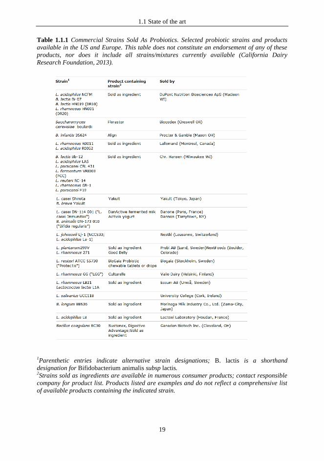
Table 1.1.1 Commercial Strains Sold As Probiotics. Selected probiotic strains and products
available in the US and Europe. This table does not constitute an endorsement of any of these
products, nor does it include all strains/mixtures currently available (California Dairy
Research Foundation, 2013).
1Parenthetic entries indicate alternative strain designations; B. lactis is a shorthand
designation for Bifidobacterium animalis subsp lactis. 2Strains sold as ingredients are available in numerous consumer products; contact responsible
company for product list. Products listed are examples and do not reflect a comprehensive list
of available products containing the indicated strain.
1.1 State of the art
20
1.1.2 Probiotic definition and regulation
According to the definition proposed by the FAO/WHO workshop of 2002, probiotics are „live
microorganisms which when administered in adequate amounts confer a health benefit on the
host‟ (“Guidelines for the Evaluation of Probiotics in Food”, FAO/WHO, 2002). The
FAO/WHO consultation had as major outcomes the providing of an official definition for
probiotics, together with guidelines regarding safety evaluation of the microorganisms possibly
employed as probiotics, indications about the strategies for the assessment of nutritional and
beneficial properties, and an overview of the rules needed for the elaboration of the health
claims. An aspect of particular importance stated in these recommendations is that the probiotic
properties must be strictly correlated to a single bacterial strain, meaning that benefits obtained
from the ingestion of a specific bacterial strain cannot be extended to other strains, even
belonging to the same species.
Nowadays, the situation of probiotics products regulation, especially in European Union (EU),
is still a controversial issue. Probiotic products which have been introduced on market after
1997 must follow the indications regarding the Novel Foods reported in the Regulation (EC) of
the European Parliament and of the Council No 258/1997, as well as the general guidelines
reported in the Regulation (EC) No 1924/2006 on nutrition and health claims made on food,
which thus encompass also the health claims allowed for probiotic products. Moreover,
subsequent regulations on labeling, presentation and advertising of foodstuffs were introduced
starting from 2007, aimed to provide a common regulation in all members states of EU, and to
guarantee consumers safety and protection against misleading labels. To these specific aims, in
2002 the European Food Safety Authority (EFSA) was established by EU politicians in order to
ensure an high level of consumer protection and to restore confidence in food, in particular after
several food scares. According to current rules, food industry before introduce a new probiotic
product on market must receive an EU authorization based on EFSA‟s assessment of safety.
In 2007 EFSA introduced the concept of “Qualified Presumption of Safety” (QPS), an
evaluation tool to assess the safety of a microorganism for a deliberate introduction into food
and feed. These guidelines establish that, to obtain the QPS status (akin to the concept of
“Generally recognized as safe”, the American Food and Drug Administration designation that a
chemical or substance added to food must be considered safe by experts) for a microorganism it
is required the deposition of the strain and its characterization to molecular level, together with
data on antibiotic resistances, genetic stability, toxins and virulence factors, and a body of
knowledge regarding history of safe use and established uses of the bacterium, accompanied by
scientific literature on absence of clinical cases and evidences of beneficial effects from in vitro
and in vivo studies. Thus, industry must submit to EFSA each health claim for scientific
evaluation prior to its approval and use in EU. In particular, the European Commission in 2008
produced guidance that indicated that the Authority would have rejected any health claim not
including human clinical data (Vogel, 2010). Thus far EFSA has rejected every submitted
dossiers regarding health claims for probiotics.
Currently, in Italy the only label allowed from the Health Ministry for a probiotic product is “it
supports gut flora balance”, even though in the Opinion reported in “EFSA Journal 2009;
7(9):1232”, the NDA (nutrition, dietetic and allergy) panel of EFSA states: “Increasing the
number of any group of bacteria is not considered in itself as beneficial”.
Even in the United States market, which is showing a growing interest for these products,
probiotics regulation is very complex. Probiotics sold as dietary supplements do not require
1.1 State of the art
21
Food and Drug Administration (FDA) approval before introduction on market. Labels allowed
for probiotics sold as dietary supplements without FDA consent may refer only to effects on
structure/function of the body (like claims for the strengthening of body‟s defense, caring for
the digestive system..), but they cannot refer to reduction of risk of diseases without FDA
approval. Thus far, FDA has not approved any health claims for probiotics (Saldanha, 2008).
Similarly, in Australia probiotics sold for specific health benefits require premarket review by
the Therapeutic Goods Administration and are regulated as complementary medicines, and in
Japan a premarket review by Health Ministry is required (Boyle et al., 2006). Nonetheless,
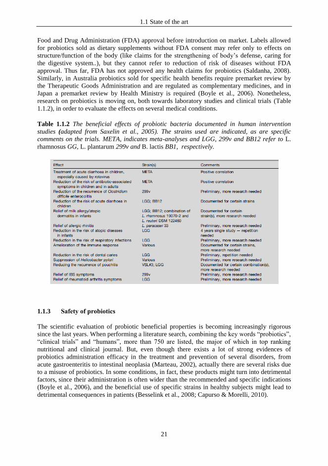
research on probiotics is moving on, both towards laboratory studies and clinical trials (Table
1.1.2), in order to evaluate the effects on several medical conditions.
Table 1.1.2 The beneficial effects of probiotic bacteria documented in human intervention
studies (adapted from Saxelin et al., 2005). The strains used are indicated, as are specific
comments on the trials. META, indicates meta-analyses and LGG, 299v and BB12 refer to L.
rhamnosus GG, L. plantarum 299v and B. lactis BB1, respectively.
1.1.3 Safety of probiotics
The scientific evaluation of probiotic beneficial properties is becoming increasingly rigorous
since the last years. When performing a literature search, combining the key words “probiotics”,
“clinical trials” and “humans”, more than 750 are listed, the major of which in top ranking
nutritional and clinical journal. But, even though there exists a lot of strong evidences of
probiotics administration efficacy in the treatment and prevention of several disorders, from
acute gastroenteritis to intestinal neoplasia (Marteau, 2002), actually there are several risks due
to a misuse of probiotics. In some conditions, in fact, these products might turn into detrimental
factors, since their administration is often wider than the recommended and specific indications
(Boyle et al., 2006), and the beneficial use of specific strains in healthy subjects might lead to
detrimental consequences in patients (Besselink et al., 2008; Capurso & Morelli, 2010).

1.1 State of the art
22
Safety evaluation of probiotics is controversial. Probiotics have been used for years in food
industry, and there are also several studies confirming that the wider use of probiotic products
has not caused any increase of bacteremia cases (Saxelin et al., 1996; Salminen et al., 2002).
Moreover, some studies reported the safe use of probiotics even in high risk populations
(Stansbridge et al., 1993; Wolf et al., 1998; Salminen et al., 2004). However, cases of sepsis are
reported in literature (Ha et al., 1999; Kunz et al., 2004; Land et al., 2005). Along with
translocation potential, other possible risks which can derive from the introduction of
microorganisms include antibiotic resistance-transfers, gastrointestinal toxicity (caused by
deleterious metabolic activities and production of dangerous metabolites), excessive immune
stimulation (Snydman, 2008). The major obstacle in a safe definition of the possible
applications of probiotic therapy is the deficiency in the knowledge of probiotic mechanism of
action, in particular at immunological level. In general, the main aspect that still creates concern
and confusion about probiotics use is the target consumer. FAO/WHO guidelines are in fact
only related to food and the use of beneficial bacteria in healthy subject, thus excluding the use
in pathologic conditions, which is properly a “therapeutic matter”. Probiotics can be considered
safe for the use in healthy people, but the observed beneficial properties cannot be extended to
other populations, and administration should be evaluated with caution in medical conditions
and in specific categories of person, like immunocompromised subjects, preterm infants or with
underlying diseases, elderly patients”. So each probiotic strain characterization activity should
have a study-by-study and population-based approach (Snydman, 2008).
1.1.4 The probiotic approach
The probiotic approach could be an alternative and mild strategy for the prevention and
treatment of either inflammatory or allergic diseases. It is estimated that the human microbiota
contains as many as 1014 bacterial cells, a number that is 10 times greater than the number of
human cells present in our bodies, starting from the skin surface to the genitourinary tract, oral
cavity, respiratory tract, ear, and the gastrointestinal tract (Ley et al., 2006). Metagenomics and
functional molecular immunology substantiate the interpretation of humans as holobionts, in the
sense of functional superorganisms, combining self and microbes acting in concert to produce
phenomena governed by the collective (Kelly, 1994; Zilber-Rosenberg & Rosenberg, 2008).
The association between host and symbionts affects the fitness of the holobiont within its
environment and it often governs the physiological homeostasis on the narrow balance between
host wellbeing and dysfunction (Eberl & Lochner, 2009; Proal et al., 2009). The mechanisms
underlying the cross-talk between a human host and microbes are only marginally understood.
However, several studies have shown that different bacterial strains can exert their probiotic
abilities by influencing the host‟s immune system, thereby modulating immune responses, at
both the local level and the systemic level (Borchers et al., 2009; Lebeer et al., 2010; Taverniti
& Guglielmetti, 2011). Thus, the elucidation of probiotic effects at molecular level could supply
the theoretical bases to develop strategies for preventing or treating several human
dysfunctions, through the reconstitution of a proper human-microbe mutualism. The probiotic
approach, in its widest sense, falls into this context, since it consists of the modification of a
human microbiota by exogenous administration of microbial cells or cell components, aimed at
benefiting the host's health. The majority of probiotic bacteria belong to the genera
Lactobacillus and Bifidobacterium (Table 1.1.1), which are Gram-positive bacteria that
constitute a major part of the normal intestinal microflora in animals and humans (Kotzampassi
& Giamarellos-Bourboulis, 2012). However, there are also evidences of probiotic roles of
other microbes such as yeasts (Saccharomyces boulardii and Saccharomyces cerevisiae) and
1.1 State of the art
23
some non-pathogenic strains of Escherichia coli and Bacillus spp. not normally found in the
gastrointestinal tract (de Vrese & Schrezenmeir, 2008).
1.1.5 Probiotics in infectious diseases
Infectious diseases represent a perpetual issue to deal with. According to the World Health
Organization (WHO)‟s 2004 World Health Report, infectious diseases are the second cause of
death (following cardiovascular diseases) responsible for about the 26 % of the deaths occurred
worldwide in 2002. One of the major concerns is represented by the acquiring of antimicrobial
resistance by pathogens, and thus to the spread of resistant microorganisms. This event is
mainly caused by the misuse of antibiotics that often are prescribed unnecessarily (Wise et al.,
1998). In this context, the use of probiotic microorganisms able to exert an antagonistic activity
towards pathogens can represent an alternative intervention to prevent infections, and might
also reduce excessive antibiotics administration. Regarding their role for the prevention and
treatment of infectious diseases, there is an increasing evidence coming from randomized
clinical trials (RCTs) for what it concerns antibiotic-associated diarrhea (AAD), Clostridium
difficile infection (CDI), acute gastroenteritis and infectious complications following admission
to the Intensive Care Unit (ICU).
Traditionally, probiotics have been associated with gastrointestinal tract (GIT) and used for the
prevention and treatment of gastrointestinal disorders. However, more recently, probiotics
effectiveness has been proposed also for a broader use. In fact, several probiotics have been
designed for the vaginal mucosa (Reid et al., 2009), the urinary tract (Borchert et al., 2008), the
skin (Krutmann, 2009), and the oro-pharyngeal cavity (Tagg & Dierksen, 2003). Particularly for
the latter, related studies report that the mechanisms of action of probiotics at oro-pharyngeal
level is analogous of that described for the gut (Rao et al., 2012).
1.1.6 Upper respiratory tract infections and probiotics for the oro-pharyngeal mucosa
The use of probiotics for the oro-pharyngeal tract (OPT) is particularly promising. OPT
dysfunctions are often related to the presence of microbial pathogens (for instance,
Streptococcus mutans, group A streptococci, or Porphyromonas gingivalis) or to microbial
dysbiosis. Furthermore, compared with the distal GIT, the OPT is a more accessible site for
microorganisms of exogenous origin. For these reasons, the OPT is a potential target for new,
specifically designed probiotic products. Upper respiratory tract infections (URTIs) represent
the most common acute illness in the patient outsetting, and they account for 9% of all
consultations in general practice (Bourke, 2007). URTIs include rhinitis, rhinosinusitis,
rhinopharyngitis, also called the common cold, pharyngitis, epiglottitis and laryngitis (Popova
et al., 2012). There are reported different studies demonstrating probiotics beneficial effects in
several pediatric infectious diseases (Weichert et al, 2012). In studies performed thus far on
probiotic effects for URTIs, half of the species employed belong to Lactobacillus genus (L.
rhamnosus, L. acidophilus, L. debrueckii, L. paracasei, L. plantarum) and Bifidobacterium
genus (B. animalis, B. bifidum, B. longum); the rest are represented by Streptococcus genus (S.
salivarius, S. mitis, S. oralis, S. sanguinis). Among these studies, an interesting example
demonstrating the potential of the probiotic approach for the OPT and URTIs is represented by
the research activity of Prof. J. R. Tagg and coworkers, who isolated the Streptococcus

1.1 State of the art
24
salivarius strain K12 (Wescombe et al., 2006). Strain K12 has a marked ability to inhibit
pathogenic bacteria, mainly due to the production of three different bacteriocins, salivaricin A2,
B and 9 (Power et al., 2008). Strain K12 was also demonstrated to be able to colonize the upper
respiratory tract (Horz et al., 2007) and to down-regulate the innate immune responses of
human epithelial cells (Cosseau et al., 2008). The scientific results from the Tagg lab studies
supported the creation of a set of probiotic pharmaceutical products (lozenges, powders and
chewing gum), commercialized under the name of BLIS, that were specifically designed for the
prevention or treatment of dysfunctions such as cavities, periodontitis, halitosis and pharyngitis
(http://blis.co.nz/international/). In July 2011, the oral probiotic products BLIS K12 were
granted GRAS status by FDA, enabling this probiotic to be included as an ingredient in food
products within the United States.
1.1.7 The oro-pharyngeal environment
In any potential probiotic application it is important consider the peculiarity of the target body
site. In the oro-pharyngeal cavity is present an organized secondary lymphoid tissue, namely the
naso-pharynx associated lymphoid tissue (NALT), a specialized component of the family of
mucosa-associated lymphoid tissue (MALTs), that all together constitute the mucosal immune
system (Fukuyama et al., 2009). In humans NALT is mainly represented by the Waldeyer's
ring, that comprise palatine tonsils, nasopharyngeal tonsil (adenoid) and lingual tonsil as major
constituents (Hellings et al., 2000). Defense responses at level of mucous membranes are very
critical, since these sites are continuously exposed to a large amount of different antigens,
meaning that their role is not only to provide an efficient barrier to potentially harmful agents,
but also to induce protective immune reactions (Debertin et al., 2003). In particular, the oro-
pharyngeal tract is a way of entrance of both airborne and alimentary antigens. Characteristics
and functions of the immune system at this level are organized in order to act either with local
mucosal response or systemic immunological effects (Brandtzaeg, 2011). Considering the
enormous number of microorganisms with which the mucosal immune system has constantly to
interface, the ability to discriminate between “friends and foe”, and thus to recognize and
eliminate pathogens while tolerating harmless commensals, represents the basis for the
maintenance of homeostasis (Cutler & Jotwani, 2006).
1.1.8 Probiotics mechanisms of action: the modulation of the immune response
The modes of action by which probiotics are suggested to affect human health fall include three
main categories (Lebeer et al., 2008). First, some probiotics can inhibit pathogens though
various antagonism mechanisms. This is the most studied probiotic mechanism and has been
exhaustively reviewed (Servin, 2004). A second way is the enhancement of the functions of
mucosal barriers, particularly at intestinal level, and the modulation of various signalling
pathways that lead to, for instance, the induction of mucus, and defensin production,
enhancement of tight junction functioning, and prevention of apoptosis (Lebeer et al., 2010).
The third mechanism is the modulation of host‟s immune responses, both at local and systemic
level (Smits et al., 2005). Increasing evidence supports this hypothesis (Borchers et al., 2009;
Lebeer et al., 2010; Adams, 2010; Taverniti & Guglielmetti, 2011). Clinical applications of the
immunomodulatory properties of bacteria have been performed particularly for lactobacilli and
bifidobacteria. These studies refer to prevention and treatment of allergic diseases, in particular
1.1 State of the art
25
atopic dermatitis in children, inflammatory bowel diseases, and prevention of virus infection
and use as an adjuvant in vaccination (Borchers et al., 2009). In spite of this, the mechanisms
determining the microbial ability to interact with the immune system of mammals are largely
unexplored. Thus, studies targeted to elucidate the mechanisms involved in bacteria-host‟s cells
cross-talk are required.
1.1.9 The first line of defense of the immune system: the innate immunity
The role of the immune system is to defend against diseases and to maintain homeostasis.
Host‟s cells are continuously exposed to both endogenous and exogenous bacteria, which
encompass harmless and potential detrimental microorganisms. Thus, one of the most important
function of a balanced immune system is a proper recognition of self and non-self, that results
in the mounting of non-inflammatory or sub-inflammatory responses against commensals, and
activation of immune responses against pathogens. The immune system consists of two
components, innate and acquired immunity, that induce both the systemic and the mucosal
immune responses. The innate immunity is the first line of defense and is preposed to provide
an early and non-specific response to antigens, targeted to a rapid elimination of the harmful
agents. Innate immune responses can be initiated at level of epithelial cells and, more
specifically, by phagocytes like neutrophils, dendritic cells, monocytes and macrophages, that
play a crucial role in host defense (Galdeano & Perdigón, 2006).
Macrophages, once activated by microbial products, acquire microbicidal competence
(Mackaness, 1964). Polarized macrophages have been classified into two main groups, M1 and
M2, differing for their phenotype and effector functions: M1 subset, activated by bacterial
products and inflammatory cytokines, produces cytokines like IL6, IL12, COX-2, IL1β and
TNFα, and possesses microbicidal and inflammatory properties, while M2 are poorly
microbicidal and with immunomodulating characteristics (Benoit et al., 2008). Macrophages are
able to produce cytokines recruiting other inflammatory cells such as neutrophils. Phagocytic
cells are attracted to the infection site by chemotaxis.
Natural Killer (NK) cells also participate in the innate immune response. NK cells rapidly react
to the presence of virus infected cells in the early stages of infection by killing the infected
target cells (Delcenserie et al., 2008).
Lymphocytes (B and T) are the main actors in the adaptive immune response. The adaptive
immune system can provide a more effective protection against pathogens through the ability to
recognize and remember an impressive number of antigens. (Delcenserie et al., 2008). Helper T
cells are mainly found in two distinct cell types, Th1 and Th2, distinguished by the cytokines
they produce and respond to, and the immune responses they are involved in. Th1 cells produce
pro-inflammatory cytokines which stimulate the phagocytosis and destruction of microbial
pathogens, while Th2 cytokines such as IL-4 generally stimulate the production of antibodies
directed toward large extracellular parasites. Even though a true balance between Th1 and Th2
profiles is difficult to maintain, there are categories of immune cells, i. e. regulatory T cells
(Treg), which are preposed to this crucial function. The balance between Th1 and Th2
responses determines, in fact, the direction and outcome of an immune response; moreover,
prolonged or exaggerated responses in each direction can lead to the development of Th1-
driven (chronic-inflammatory diseases) or Th2-driven (allergic diseases) pathologies
(Delcenserie et al., 2008).
1.1 State of the art
26
Dendritic cells (DCs), as well as macrophages and monocytes, are the link between the innate
and adaptive immune systems, since they act as professional “antigen-presenting cells” (APCs).
This function is crucial in initiating the adaptive immune response, as T cells do not respond to
free-antigen but only to antigen that is presented by APCs. Cells of the innate immune system
also influence the adaptive immune response through the production of chemical mediators
called “cytokines”: these are proteins which are involved in the communication among cells,
and are essential in regulating cell priming, acquiring of a specific phenotype, and thus
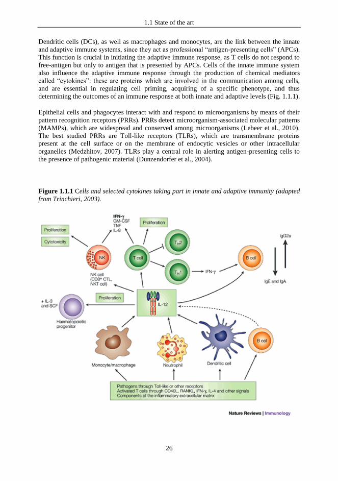
determining the outcomes of an immune response at both innate and adaptive levels (Fig. 1.1.1).
Epithelial cells and phagocytes interact with and respond to microorganisms by means of their
pattern recognition receptors (PRRs). PRRs detect microorganism-associated molecular patterns
(MAMPs), which are widespread and conserved among microorganisms (Lebeer et al., 2010).
The best studied PRRs are Toll-like receptors (TLRs), which are transmembrane proteins
present at the cell surface or on the membrane of endocytic vesicles or other intracellular
organelles (Medzhitov, 2007). TLRs play a central role in alerting antigen-presenting cells to
the presence of pathogenic material (Dunzendorfer et al., 2004).
Figure 1.1.1 Cells and selected cytokines taking part in innate and adaptive immunity (adapted
from Trinchieri, 2003).

1.1 State of the art
27
The modality through which bacteria can elicit a specific immune response, both in terms of
cells signaling pathways activated and mechanisms involved in the host-microbes interplays, is
still poorly understood. Thus, in order to identify the ways of action of health-promoting
bacteria, and to find new applications in the prevention and treatment of several disorders, as
well as for the evaluation of potential risks, studies at mechanistic level are required. Moreover,
it is important to stress that bacteria interact with host cells in a strain-specific way. That leads
to the need to identify which component in each bacterial strain is responsible for an
immunomodulatory activity.
1.2 Aims of the study
28
1.2 AIMS OF THE STUDY
Starting from the above-mentioned promising studies regarding the beneficial properties of
food-grade bacteria in infectious diseases and on host innate immunity, we screened bacterial
strains both from oral and food origin for their potential use as probiotics for the oro-pharyngeal
mucosa. We tested the ability of bacteria, newly isolated from the pharynges of healthy
volunteers or from dairy origin, to adhere to a human pharyngeal cell layer and to antagonize S.
pyogenes on two different epithelial cell lines. As a result of these studies, we selected the oral
isolate Streptococcus salivarius ST3 and the dairy strain Lactobacillus helveticus MIMLh5 as
new probiotic candidates for the pharyngeal mucosa. These strains were further investigated for
their in vitro probiotic properties when employed in combination, and also analyzed from an
immunological point of view. Specifically, we examined their ability to adhere to FaDu human
pharyngeal cells, to antagonize Streptococcus pyogenes and to modulate the immune response
on U937 macrophages. Moreover, to explore the potential of combining strains ST3 and
MIMLh5 in a fermented food product, we also studied the growth of S. salivarius ST3 in the
presence of different sugars and in milk in co-culture with strain MIMLh5.
Our results support the possibility of producing a novel milk-based fermented food product to
be used as probiotic for the oro-pharyngeal mucosa.
1.3 Materials and Methods
29
1.3 MATERIALS AND METHODS
1.3.1 Isolation of bacteria from pharyngeal mucosa and culture conditions
To isolate bacteria from pharynx, specimens were collected using polyester fiber-tipped
applicator swabs (VWR, Milano, Italy) from 4 healthy donors (3 females, 58, 32 and 29 years
old, and a 32 years old male). After serial dilutions in 0.1 % peptonated saline, speciments were
plated on MRS agar (Fluka Feinchemikalien GmbH, Neu-Ulm, Germany) supplemented with
0,05 % cysteine-HCl (cMRS), M17 (Fluka Feinchemikalien GmbH) containing 2 % lactose (L-
M17) and 2 % glucose TSA (Difco, Detroit, MI, USA). Around 50 colonies were randomly
picked up and spread on plate with a loop. This procedure was repeated at least four time, in
order to obtain pure cultures. Tables 1.3.1 and 1.3.2 list the bacterial strains used in this study.
If not differently specified, oral streptococci, Streptococcus thermophilus and lactococci were
routinely grown overnight at 37°C in L-M17. Lactobacilli were cultivated overnight in cMRS at
37°C. Bifidobacterium animalis subsp. lactis Bb12 was grown anaerobically at 37°C in pre-
reduced cMRS, while Streptococcus pyogenes was grown overnight at 37°C in BHI (Difco)
supplemented with 0.3 % yeast extract.
1.3.2 Identification and molecular characterization of bacterial isolates
The isolates from each subject have been clustered by means of BOX-PCR assay (Table 1.3.1),
which was performed with primer BoxA1 (5′-CTACGGCAAGGCGACGCTGACG- 3′) in a
PCR reaction mix consisting of of 20 mM Tris–HCl, 50 mM KCl, 200 μM of each
deoxynucleoside triphosphate, 1 μM of the primer, 1.5 mM MgCl2 and 1.5 U of Taq
polymerase (Fermentas, Vilnius, Lithuania). The 16S rRNA gene was amplified from at least
one representative isolate from each BOX-genotypic group (Table 1.3.1) by PCR using the
primers P0 (5′-GAAGAGTTTGATCCTGGCTCAG-3′) and P6 (5′-
CTACGGCTACCTTGTTACGA-3′). Each PCR mixture (50 μl) was as for BOX-PCR analysis.
Each PCR cycling profile consisted of an initial denaturation time of 2 min at 95°C followed by
an amplification for 35 cycles of denaturation (30 s at 95°C), annealing (45 s at 55°C) and
extension steps (2 min at 72°C). The PCR was completed with an elongation period (7 min at
72°C). The resulting amplicons were then sequenced by using an ABI Prism BigDye™
terminator technology in an ABI Prism™ 310 DNA sequencer (Applied Biosystems, Foster
City, CA, USA). Streptococcus salivarius isolates were further characterized by RAPD
analysis, performed with primers OPI17 (5‟-CGAGGGTGGTGATC -3‟), OPI02-mod (5‟-
GCTCGGAGGAGAGG-3‟), M13 (5‟-GTAAAACGACGGCCAGT-3‟) and PedAF (5‟-
ATACTACGGTAATGGGGT-3‟). Reaction mix was as for BOX analysis, and the temperature
profile was as follows: primary DNA denaturation step at 94°C for 2 min followed by 5 cycles
of 45 s at 94°C, 45 s at 31°C and 2 min at 72°C; 35 additional cycles were carried out
increasing the annealing temperature to 40°C. For all the amplification cycles the final
extension was continued for 7 min at 72°C.
Similarity dendrogram was built using NTSYSpc v. 2.01 (Applied Biostatistic Inc., New York,
NY).

1.3 Materials and Methods
30
Table 1.3.1 Bacteria isolated from pharyngeal mucosa.
Human
source‟s
designation
BOX-PCR
genotype1
Denomination of
the isolates2
Isolation
medium Taxonomic identification3
Additional
identification
method4
IS
IS-A1 IS1 cMRS
S. salivarius IS8, IS10, IS12 L-M17
IS-A2 IS5, IS6, L-M17
S. salivarius IS7 gTSA
IS-B IS3, IS4 cMRS S. sanguinis
IS-C IS9 L-M17
Staphylococcus aureus IS15 gTSA
IS-D IS11, IS13 L-M17
Rothia mucillaginosa IS14 gTSA
RS
RS-A1
RS1, RS8, RS10 L-M17
S. salivarius RS3, RS6 cMRS
RS13, RS14 gTSA
RS-A2 RS9 L-M17 S. salivarius
RS-B RS4 cMRS Alloscardovia omnicolens
RS-C RS5 cMRS Lactococcus lactis subsp. Lactis his
RS-D RS11 L-M17 Micrococcus sp. (luteus)
RS-E RS12 gTSA Rothia mucillaginosa
RS-F RS15 gTSA Bacillus subtilis
SM
SM-A1
SM1, SM2,
SM4, SM5 cMRS S. salivarius
SM6, SM8, SM9 L-M17 S. salivarius
SM11, SM14,
SM15 gTSA S. salivarius
SM-A2 SM12 gTSA S. salivarius
SM-B SM3 cMRS S. oralis gdh
SM-C SM7 L-M17 Rothia mucillaginosa
SM-D SM13 gTSA Neisseria sp. (subflava)
ST
ST-A1 ST3 cMRS S. salivarius
ST-A2 ST12 L-M17 S. salivarius
ST-B ST2, ST5p cMRS S. oralis gdh
ST-C ST5g cMRS S. sanguinis
ST-D ST4 cMRS
S. oralis gdh ST15 gTSA
ST-E ST7, ST11 L-M17 S. infantis gdh
ST-F ST1, ST8 L-M17 Bacillus sp. (cereus)
ST-G ST9 L-M17 S. oralis gdh
ST-H ST6 cMRS Staphylococcus epidermidis
ST-I ST13, ST14 gTSA Rothia mucillaginosa 1 Similar (but not identical) BOX-PCR electrophoretic patters are indicated with the same letter and a
different number.
2 Representative strains chosen for 16S rDNA sequence analyses are in bold.
3 S. salivarius stands for Streptococcus salivarius. Species designation between brackets refers to 16S
rDNA sequence similarity below 95%. 4 gdh: taxonomic identification based on the sequence of an approximately 500-bp internal fragment of the
glucose-6-phosphate dehydrogenase gene (gdh) (Bek-Thomsen et al., 2008); his: taxonomic identification
based on the length polymorphism of a PCR amplified fragment from histidine biosynthesis operon
(Beimfohor et al., 1997).
1.3 Materials and Methods
31
Table 1.3.2 Streptococcus salivarius K12 and non-oral bacteria included in the study.
Species Strain Source Reference
Bifidobacterium animalis subsp. lactis Bb12 Probiotic commercial strain Juntunen et al., 2001
Lactobacillus acidophilus LA5 Probiotic commercial strain Juntunen et al., 2001
Lactobacillus paracasei Shirota Probiotic commercial strain Juntunen et al., 2001
Lactobacillus helveticus MIMLh5 Dairy natural starter Guglielmetti et al., 2008
Lactobacillus rhamnosus GG Probiotic commercial strain Juntunen et al., 2001
Lactococcus lactis subsp. cremoris Viili Finnish fermented milk Kahala et al., 2008
Streptococcus salivarius K12 BLIS throat guard Burton et al., 2006
Streptococcus thermophilus DSM20617T DSMZ type strain, yogurt /
1.3.3 Antibacterial activity against Streptococcus pyogenes and PCR detection of
bacteriocin encoding genes of pharyngeal isolates
In a first set of experiments, tester bacterial strains were spread with a loop on agar plate
containing LM17 medium and incubated overnight at 37°C. Then, 15 ml of soft yeBHI agar
containing about 106 cells of the indicator strain (S. pyogenes C11) were poured over the plates.
The plates were checked for inhibition zones after incubation at 37°C for 24 and 48 h. The
production of antimicrobial substances was also tested through disk-diffusion. Briefly, tester
strains were grown until stationary growth phase in LM17 medium. Culture supernatants were
neutralized to pH 7, filter-sterilized and spotted (0.1 ml) on a filter paper disk, which was
previously placed on yeBHI softagar plates inoculated with about 105 S. pyogenes cells. The
presence of an inhibition halo was checked after 24 and 48 h.The PCR reactions to detect
previously characterized bacteriocin structural genes (salivaricin A, salivaricin B, streptin, and
peptide SA-FF22) were performed as described by Wescombe et al. (2006).
1.3.4 Preparation of bioluminescent Streptococcus pyogenes
Reporter vector pCSS810, carrying a phage T5 promoter–lac operator upstream of the insect
luciferase gene lucFF (Lampinen et al., 1992), was used to obtain the luminescent phenotype in
Streptococcus pyogenes C11. In brief, electrocompetent S. pyogenes cells were prepared as
follows. Fresh BHI broth (25 ml) supplemented with 0.3% yeast extract (yeBHI) was inoculated
with 0.25 ml of an overnight culture of the bacterium and incubated at 37°C until an optical
density (at 600 nm) of about 0.2 was reached. The cells were chilled on ice and concentrated in
further washing steps of 25 and 10 ml (washing buffer EB: 0.3 M sucrose and 1 mM MgCl2 in 5
mM phosphate buffer, pH 6.9). After washing, the pellet was resuspended in 2 ml of the same
buffer, dispensed in 80 μl-aliquotes and maintained at -80°C until used for electroporation with
an Eppendorf Multiporator 2510 (Eppendorf, Milano, Italy), at 12.5 kV/cm, in 2-mm cuvettes.
The yeBHI supplemented with sucrose (final concentration 0.3 mM) was used as outgrowth
medium. After 2 h of incubation at 37°C in the outgrowth medium, transformants were selected
1.3 Materials and Methods
32
on yeBHI agar plates with 5 μg ml-1
of chloramphenicol. The selected luminescent S. pyogenes
clone was named C11LucFF
.
1.3.5 Antagonistic activity against Streptococcus pyogenes
FaDu (human pharynx carcinoma cell line; ATCC HTB-43) and HaCat cells (human
keratinocytes from a spontaneous immortalized, non-tumorigenic cell line) were routinely
grown in 24-well tissue culture plates in Dulbecco‟s Modified Eagle‟s Medium (DMEM),
supplemented with 10 % (v/v) heat-inactivated (30 min at 56°C) fetal calf serum, 100 U ml–1
penicillin, 100 mg ml–1
streptomycin, 0.1 mM non-essential amino acids, 2 mM L-glutamine,
and incubated at 37°C in a water-jacketed incubator in an atmosphere of 95 % air and 5 %
carbon dioxide, until a confluent monolayer was formed. The bacterial cell concentration of an
overnight culture was determined microscopically with Neubauer Improved counting chamber
(Marienfeld GmbH, Lauda-Königshofen, Germany). The antagonism was studied through
exclusion and competition assays. Exclusion consisted of a pre-incubation of the FaDu layer
with 1 ml of a tester strain suspension (5 x 10⁸ cells ml
-1), followed by a washing step with
phosphate-buffered saline (PBS) and the incubation with 1 ml of the indicator strain (S.
pyogenes C11LucFF
) suspension (2 x 10⁸ cells ml
-1). The concentration of 5 x 10
8 tester cells ml
-1
was chosen because corresponded to the plateau of a dose-response curve which was prepared
during the set-up of the experiment by measuring the antagonistic activity as a function of tester
cell concentration (Data not shown).
Competition consisted of co-incubation of the same number of tester and indicator strains (2 x
108 cells ml
-1). After incubation, FaDu layers were quickly washed twice with 1 ml PBS (pH
7.3) and D-luciferin (Sigma-Aldrich, Steinheim, Germany) was added at the concentration of
12.5 μM in citrate buffer, pH 5. Immediately, the luminescence signal was measured with a
Victor 3 luminometer (PerkinElmer, Monza, Italy). Each tester strain was analyzed in triplicate
in at least two independent experiments. Unpaired Student‟s t-test was run for statistically
significant differences.
1.3.6 Bacterial adhesion to FaDu cell layer
FaDu cells were grown in 3-cm Petri plates on microscopy cover glasses as described above.
Cell monolayers were carefully washed with PBS pH 7.3 before bacterial cells were added. The
bacterial cell concentration of an overnight culture was determined microscopically with
Neubauer Improved counting chamber (Marienfeld GmbH). Approximately 2×10⁸ cells of each
strain resuspended in PBS (pH 7.3) were incubated with a monolayer of FaDu cells. After 1 h at
37°C, all monolayers were washed 3 times with PBS to release unbound bacteria. Cells were
then fixed with 3 ml of methanol and incubated for 8 min at room temperature. After methanol
was removed, cells were stained with 3 ml of Giemsa stain solution (1:20) (Carlo Erba, Milan,
Italy) and left for 30 min at room temperature. Wells were then washed until no color was
observed in the washing solution and dried in an incubator for 1 h. Microscopy cover glasses
were then removed from the Petri plate and examined microscopically (magnification × 100)
immersed in oil. Adherent bacteria in 20 randomly selected microscopic fields were counted
and averaged.

1.3 Materials and Methods
33
1.3.7 Antibiotic susceptibility of selected bacteria
The inhibitory concentrations of the antimicrobial agents were determined by broth
microdilution in commercial 96-well microtiter plates, for the following concentration ranges:
ampicillin, chloramphenicol, erythromycin, oxytetracyclin and vancomycin 1-16 μg ml-1
;
gentamicin 8-64 μg ml-1
; kanamycin and streptomycin 16-128 μg ml-1
. Antibiotics (Sigma-
Aldrich) were serially 1:2 diluted in culture medium starting from a concentrated stock solution
conserved at –20°C. In brief, bacterial cells from an overnight culture were washed once with
saline and resuspended in fresh medium at a concentration corresponding to an optical density
of 1 at 600 nm. Afterward, bacterial suspensions were 1:100 diluted in the liquid medium
containing the appropriated antibiotic concentration, and loaded in duplicate in microtiter wells
(200 μl well-1
). Three different liquid media were used in this experiment: L-M17, MRS and
BHI (Table 3). The inoculated plates were subsequently incubated aerobically at 37°C for 48 h,
after which the inhibitory concentrations of antibiotics were read as the lowest concentration.
1.3.8 Determination of urease activity and PCR detection of ureC gene
Urease activity was tested by evaluating the release of ammonia by means of the phenol red
assay (Lanyi, 1987). Specifically, cells from 0.5 ml of an overnight culture were resuspended in
a solution containing one volume of solution A (urea, 2 g dissolved in 2 ml of ethanol and 4 ml
of sterilized water) and 19 volumes of solution B (KH2PO4, 1 g l-1
; K2HPO4, 1 g l-1
; NaCl, 5 g l-
1; phenol red, 20 μg ml
-1). The suspension was incubated at 37°C for 1-4 h and the development
of a red-violet colour indicated positive urease activity. The amplification of the gene coding
for the main subunit of the urease complex (ureC) was carried out as previously described
(Mora et al., 2004), using primers ureC-F (5‟-CTGTTCATGATCCTATTCAG-3‟) and ureC-R
(5‟-CGAATACCGAATTCATTCCCATG-3‟).
1.3.9 Stimulation of FaDu monolayers and enzyme-linked immunosorbent assay
(ELISA) measurement of cytokine production
Human pharyngeal carcinoma cells (FaDu) were seeded into 24-well plates and grown as
previously described. Bacterial cells were added to monolayers of FaDu cells in 0.5 ml of fresh
antibiotic-free Eagle's Minimum Essential Medium (EMEM) containing 25 mM HEPES (pH
7.4) and incubated overnight at 37°C. Each bacterial strain was used at a multiplicity of
infection (MOI) of about 1000, while EMEM/HEPES medium without bacterial cells was used
as control. After overnight incubation, the supernatants were collected by pipetting, centrifuged
to remove cells, and kept at -80°C. Finally, 17 different cytokines in the supernatants were
determined on a Bio-plex Array Reader (LUMINEX 100, Bio-RadLaboratories, Hercules, CA)
using Bio-Plex Human Cytokine 17-plex panel (Bio-Rad), according to the human cytokine
Bio-Plex panel assay Bio-protocol (Bio-Rad). The list of tested cytokines and the corresponding
detection limits were as follows: interleukin (IL)-1β, 0,3 pg ml-1
; IL-2, 0.2 pg ml-1
; IL-4, 0.1 pg
ml-1
; IL-5, 0.3 pg ml-1
; IL-6, 0.2 pg ml-1
; IL-7, 0.3 pg ml-1
; IL-8, 0.3 pg ml-1
; IL-10, 0.2 pg ml-1
;
IL-12 (p70), 0.4 pg ml-1
; IL-13, 0.3 pg ml-1
; IL-17, 0.5 pg ml-1
; granulocyte colony-stimulating
factor (G-CSF), 0.2 pg ml-1
; granulocyte macrophage colony-stimulating factor (GM-CSF), 1.1
pg ml-1
; interferon (IFN)-γ, 2.6 pg ml-1
; monocyte chemotactic protein (MCP)-1, 0.8 pg ml-1
;
1.3 Materials and Methods
34
macrophage inflammatory protein (MIP)-1β, 0.6 pg ml-1
; tumor necrosis factor (TNF)-α, 0.6 pg
ml-1
.
1.3.10 Construction of stable NF-κB reporting FaDu cells
Stable transfectants of FaDu cell line were obtained after transfection with the plasmid pNiFty2
(InvivoGen, LaboGen, Rho, Italy; Fig. 1.3.1). This plasmid combines five NF-κB sites with the
insect luciferase reporter gene luc. The presence of active NF-κB molecules in the cell activates
the promoter, resulting in the expression of the luciferase gene. Transfection was performed by
means of StoS Transfection Kit (GeneSpin, Milano, Italy) in accordance with the
manufacturer's protocol. Afterward, cells were resuspended in fresh DMEM medium and
seeded in 24-well plates for 48 h, in order to obtain the expression of the antibiotic resistance.
Finally, stable recombinant clones were selected by adding into the culture medium 50 μg ml-1
of zeocin.
Figure 1.3.1. Genetic map of a plasmid pNiFty2-Luc.
This plasmid contains promoter combining five NF-
B-binding sites and the firefly luciferase reporter
gene luc. The presence of NF-B activating molecules
in the cell activates this transcription factor, which
binds to the promoter, resulting in the expression of
the luciferase gene.
1.3.11 Study of NF-κB activation
Recombinant FaDu cells were cultured using the same protocol as for non-transfected FaDu, in
presence of 50 μg ml-1
of zeocin. After growth, FaDu layer was resuspended in EMEM at a
concentration of 250.000 cells ml-1
in presence of 100 mM HEPES (pH 7.4). After that, 50 μl of
tester bacterial suspension, containing 2 x 10⁹, 2 x 10
8 or 2 x 10
7 cells ml
-1, were added to 450
μl of FaDu suspension, resulting in a MOI of about 1000, 100 or 10, respectively. After
incubation at 37°C for 4 h, samples were kept in ice and sonicated at maximum speed for 5 s
(Bandelin sonicator, Bandelin electronic GmbH & Co., Berlin, Germany). Bacterial cells and
insoluble particles were removed by centrifugation and the supernatants were moved into a new
tube. At this point, 200 μl of supernatants were aliquoted in duplicate into the wells of a 96-
wells white microtiter plate (PerkinElmer); then 12.5 μl of a 10 mM ATP solution and 12.5 μl
of 0.1 mM D-luciferin were added and the emitted bioluminescence was immediately recorded
every 90 s with a Victor 3 luminometer (PerkinElmer). The maximum of the light-production
curve was considered for comparing the results. In a different set of experiments, recombinant
FaDu cells were simultaneously stimulated with IL-1β (2 ng ml-1
). All strains were analyzed in
duplicate in at least three independent experiments for each MOI considered. Unpaired
Student‟s t-test was run for statistically significant differences.
pNiFty2-Luc4501 bp
Luc
SV40 pAnbetaGlo pAn
NF-kB 5xELAM
EM7
hEF1/HTLV prom
Ori
Zeo
1.3 Materials and Methods
35
1.3.12 Study of the activation of the U937 human macrophage cell line. Cell culture,
growth conditions, and stimulation protocol.
The cell line U937 (ATCC CRL-1593.2™) was derived from a human histiocytic lymphoma
(Sundstrom et al., 1976). These cells are maintained as replicative, non-adherent cells and have
many of the biochemical and morphological characteristics of blood monocytes (Harris and
Ralph, 1985). When treated with phorbol myristate acetate (PMA), U937 cells differentiate to
become adherent, non-replicative cells with characteristics similar to tissue macrophages,
including similar isoenzyme patterns (Radzun et al., 1983) and other phenotypic markers
(Harris and Ralph, 1985). The normal growth medium for the U937 cells consisted of RPMI
1640 medium (Lonza, Basel, Swiss) supplemented with 10 % (v/v) fetal bovine serum (FBS)
(Gibco-BRL, Life Technologies, Milan. Italy), 2 mM L-glutamine, 100 units ml-1
penicillin and
100 μg ml-1
streptomycin (Sigma-Aldrich). U937 cells were seeded at a density of 5×10⁵ cells
well-1
in 12-well plates and incubated at 37°C in a humidified atmosphere of 95 % air and 5 %
CO2. Differentiation was induced by the addition of PMA (Sigma-Aldrich) into the cellular
medium at a final concentration of 100 nM and was allowed to proceed for 48 h. Afterwards,
cells were washed once with sterile PBS buffer to remove all non-adherent cells. One hour
before the bacteria were added to the cells, the culture media was replaced with RPMI 1640
medium supplemented with 1 % (v/v) FBS to allow the cells to adapt. Bacteria were used at
MOIs of 100 and 1000, and lipopolysaccharide (LPS, final concentration of 1 μg ml-1
) from
Escherichia coli 0127:B8 (Sigma-Aldrich) was used as positive control for pro-inflammatory
stimulus in U937 cells. An untreated sample, i.e., only RPMI 1640 medium with 1 % (v/v) FBS,
was used as control.
1.3.13 Inhibition assay with Toll-like Receptor neutralizing antibodies on U937 cells.
Human anti-Toll-like receptor (TLR) 2 antibody (Invivogen) was added to U937 cells 1 hour
before the stimulation with bacterial cells. A human immunoglobulin-A (IgA) 2 isotype control
(Invivogen) was used as control to exclude non-specific binding and blocking activity of the
antibody. Both the anti-TLR2 and IgA2 isotype control were used at 5 μg ml-1
. This
concentration was determined by examining the neutralizing efficacy of anti-TLR2 with
zymosan from Saccharomyces cerevisiae (Invivogen) as ligand; we observed that 5 μg ml-1
resulted in a 5-fold reduction in the expression level of tumor necrosis factor (TNF)-α, a
cytokine known to be induced in the downstream pathway activated by TLR-2.
1.3.14 Preparation of RNA and reverse transcription.
After incubating U937 cells at 37 °C four 4 hours, the supernatant was carefully removed from
each well and the total cellular RNA was isolated from the adhered U937 cells with an RNeasy
Mini Kit (Qiagen Inc., Valencia, CA). Afterwards, RNA concentration and purity was
determined with a Nanodrop Spectrophotometer (ND-1000, Thermo Fischer Scientific) and
reverse transcription to cDNA was performed with the iScript Select cDNA Synthesis Kit
(Bio-Rad Laboratories, Hercules, CA) using the following thermal cycle: 5 min at 25°C for, 30
min at 42°C, and 5 min at 85°C. Real-time Quantitative PCR. The mRNA expression levels of
cytokines were analyzed with SYBR Green technology in real-time quantitative PCR using
SsoFast EvaGreen Supermix (Bio-Rad) on a Bio-Rad CFX96 system according to the
1.3 Materials and Methods
36
manufacturer's instructions. The primers used are as follow (5‟3‟): 18srRNA forward
ATCCCTGAAAAGTTCCAGCA; 18srRNA reverse CCCTCTTGGTGAGGTCAATG; IL10
forward AGCAGAGTGAAGACTTTCTTTC; IL10 reverse CATCTCAGACAAGGCTTGG;
TNF-α forward TCAGCTCCACGCCATT; TNF-α reverse CCCAGGCAGTCAGATCAT;
COX2 forward CCCTTGGGTGTCAAAGGTAA; COX2 reverse
TGAAAAGGCGCAGTTTACG. All primers were selected using the Primer3Plus software
(http://www.bioinformatics.nl/cgi-bin/primer3plus/primer3plus.cgi), and the specificity of the
primers was tested with melting curves during amplification and by 1 % agarose gels.
Quantitative PCR was carried out according to the following cycle: initial hold at 96°C for 30 s
and then 40 cycles at 96°C for 2 s and 60°C for 5 s. Gene expression was normalized to the
housekeeping gene 18s rDNA. The amount of template cDNA used for each sample was 12.5
ng. All results regarding cytokine mRNA expression levels are reported as the fold of induction
(FOI) respective to the control (namely unstimulated U937), to which we attributed an FOI of 1.
1.3.15 Growth experiments.
The growth curves of strain ST3 were determined in 384-well microtiter plates using a BioTek
Synergy HT Multi-Mode Microplate Reader (AHSI S.p.A., Bernareggio Italy) to measure
growth in M17 broth supplemented with 2 %, 1 %, 5 ‰, 2.5 ‰, 1.25 ‰ or 0.625 ‰ of one of
the following sugars: fructose, galactose, glucose, inulin, lactose, lactulose, sucrose or
fructooligosaccharides (FOS). All sugars were purchased from Sigma-Aldrich (Steinheim,
Germany) with the exception of FOS, which were from Actilight® (Giulio Gross S.p.A.,
Trezzano sul Naviglio, Italy). The preparation of 384-well microtiter plates was conducted with
an epMotion Automated Pipetting System (Eppendorf, Milan, Italy). Acidification curves were
determined by inoculating skim milk (Difco) (1 % inoculum) with an overnight culture of
MIMLh5 and ST3 alone or in co-culture and recording the pH every hour for 24 hours with a
pH-meter recorder (XS instruments pH 2100, Opto-Lab, Concordia, Italy).
1.4 Results and Discussion
37
1.4 RESULTS AND DISCUSSION
1.4.1 Identification and molecular characterization of bacterial isolates
In this study we aimed to select oral bacteria with potential probiotic features for the pharyngeal
mucosa. We included bacteria newly isolated from the pharynges of healthy donors. Potentially,
pharyngeal isolates could, in fact, display better performances in the colonization of the oral
ecosystem than traditional dairy or intestinal probiotic bacteria. A total of 56 isolates were
obtained from pharyngeal swab samples taken from four healthy donors by using three different
culture media (Table 1.3.1). After a preliminary grouping of all isolated bacteria through BOX-
PCR fingerprinting (Data not shown) 16S rRNA genes sequence analysis revealed that 39 of the
isolates belonged to the Streptococcus genus. Streptococcus salivarius was the most represented
species (28 isolates). We also isolated Streptococcus oralis (6 isolates) and Streptococcus
infantis (2 isolates), which were distinguished by glucose-6-phosphate dehydrogenase gene
(gdh) sequence analysis (Bek-Thomsen et al., 2008) and 1 strain of Lactoccus lactis sub. Lactis.
The only species isolated from all four pharyngeal samples were S. salivarius and Rothia
mucilaginosa (7 isolates). All the other bacteria are listed in Table 1.3.1. Streptococcus
salivarius isolates were further characterized at the intraspecies level by means of BOX-PCR
and RAPD-PCR analyses (Data not shown). A computer evaluation of similarities and
clustering resulted in a total of 11 unique S. salivarius genotypes out of 28 pharyngeal isolates
and the K12 commercial probiotic strain (Fig. 1.4.1). The isolates that were included in a single
genotypic group originated from the same pharyngeal sample, suggesting that they were
probably multiple isolates. In this research, half of the pharyngeal isolates were ascribed to the
species Streptococcus salivarius (28 out of 56 isolates), in accordance with previous studies
showing these bacteria to be the dominant cultivable species in the oro-pharyngeal tract (Kang
et al., 2006). Nonpathogenic streptococci are the bacteria most largely present at the oro-
pharyngeal level, and they have been proposed to exert a key role in the protection against
pathogenic agents, which cause inflammation and infections (Tagg & Dierksen, 2003). In
particular, Streptococcus salivarius already becomes a stable colonizer of the oral microbiota a
few days after birth and represents, in adults, the major species at the levels of the pharyngeal
mucosa and dorsal tongue.

1.4 Results and Discussion
38
Figure 1.4.1 Unweighted-pair group method using average linkages (UPGMA) dendrogram
derived from similarity coefficients calculated by the Jaccard method (simple Jaccard [Sj]
coefficients; shown on the scale at the bottom), showing the relationship among Streptococcus
salivarius pharyngeal isolates, analyzed by BOX-PCR and RAPD analysis using primers M13,
OPI02mod, OPI17mod, and PedAF. Samples with a similarity coefficient higher than 0.9 have
been included in the same genotype. Selected bacterial isolates included in antagonism
experiments are indicated in boldface.
1.4 Results and Discussion
39
1.4.2 Oral, dairy and probiotic strains differently adhered to FaDu human pharyngeal
cell line
Thirty bacterial strains were studied for their ability to adhere to FaDu epithelial cell layer.
Along with bacteria isolated from human pharyngeal mucosa we tested bacterial strains of dairy
and probiotic origin already present on the market. In detail, strains MIMLh5, Viili and DSM
20617T are used as starters in the production of Grana Padano cheese, Finnish fermented milk
and yogurt, respectively. The strains Bifidobacterium animalis subsp. lactis Bb12, Lactobacillus
acidophilus La-5, Lactobacillus rhamnosus GG and Lactobacillus paracasei Shirota are among
the most widespread probiotic strains in the Western Countries. Certain properties of these
strains have been compared to Streptococcus salivarius K12, the only probiotic bacterium
specifically commercialized as probiotic for the pharyngeal mucosa up to date (BLIS K12
Throat Guard TM
, Tagg et al., 2006). The first probiotic property that we considered was the
adhesion to the host‟s target epithelium. In fact, the ability of commensals and probiotics to
bind human mucosa has a pivotal importance because it can promote the colonization and
sustain host epithelium-bacteria cross-talk (Lee et al., 1999). After extensive washings with
PBS, a significant proportion of cells of most bacterial strains remained attached to the FaDu
monolayer, providing evidence that the adhesion was not only non-specific physical
entrapment. In particular, 5 strains displayed a strong adhesive phenotype, coinciding with an
adhesion index (bacterial cells per 100 FaDu cells) of more than 2500 (Fig. 1.4.2). The 5 most
adhesive strains included L. helveticus MIMLh5, S. salivarius ST3, S. salivarius K12 and two
Lactococcus lactis strains (RS5 and Viili). A good adhesion ability was also displayed by 9
other strains, among which L. rhamnosus GG and 8 S. salivarius strains (Fig.1.4.2). Two strains
in particular, L. helveticus MIMLh5 and S. salivarius ST3, showed to adhere to FaDu
pharyngeal epithelial cell line comparably to the oral probiotic bacterium S. salivarius K12 that
we used as reference (Horz et al., 2007).
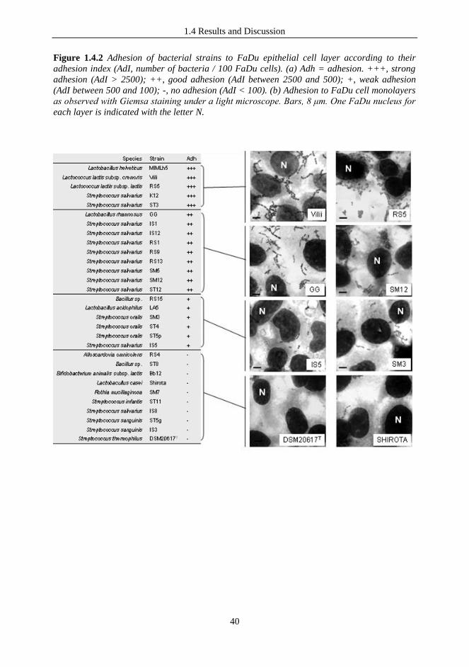
1.4 Results and Discussion
40
Figure 1.4.2 Adhesion of bacterial strains to FaDu epithelial cell layer according to their
adhesion index (AdI, number of bacteria / 100 FaDu cells). (a) Adh = adhesion. +++, strong
adhesion (AdI > 2500); ++, good adhesion (AdI between 2500 and 500); +, weak adhesion
(AdI between 500 and 100); -, no adhesion (AdI < 100). (b) Adhesion to FaDu cell monolayers
as observed with Giemsa staining under a light microscope. Bars, 8 μm. One FaDu nucleus for
each layer is indicated with the letter N.

1.4 Results and Discussion
41
Figure 1.4.3 Adhesion to FaDu epithelial cells of selected bacterial strains as observed with
Giemsa staining under a light microscope. Bars, 8 μm. A FaDu nucleus for each layer is
indicated with the letter N.
1.4.3 The antagonistic activity against Streptococcus pyogenes on human epithelial cell
lines is strain-dependent
Streptococcus pyogenes (group A streptococci, GAS) causes approximately 15 to 30 % of
pediatric sore throat (Bisno, 1996) and it is the etiological agent of skin and soft tissue
infections, glomerulonephritis and acute rheumatic fever. The oral administration of probiotic
bacteria could be a prophylactic strategy effective to reduce the transmission of GAS in the
community (Wagenvoort et al., 2005). For this reason, we tested the ability of probiotic bacteria
to antagonize GAS on in vitro epithelial layers. Since the oral cavity contains several types of
surface, including keratinized and non-keratinized epithelia, besides FaDu, we also included in
the experimentation the HaCat cell line, which resembles many characteristics of human
keratinocytes (Boukamp et al., 1998).Twenty-five bacterial strains were tested for their ability
to adhere to the FaDu epithelial cell layer. We studied 13 S. salivarius strains, including at least
one representative isolate from each genotypic cluster, and the commercial probiotic strain K12.
We also tested 5 intestinal probiotic and dairy lactic acid bacteria (Table 1.3.2) and 6 other
pharyngeal isolates that have been arbitrarily selected. In this experiment we used a three-
component system consisting of the epithelial cell layer, the S. pyogenes C11LucFF
indicator
bioluminescent strain, and the tester bacterium. We measured the reduction of bioluminescence
produced by S. pyogenes C11LucFF
as an indication of the antagonistic activity exerted by the
tester strains. Antagonism through exclusion was tested on the layers of two different human
epithelial cell lines, FaDu (Fig. 1.4.4A). and HaCaT (Fig. 1.4.4B). The results showed that
antagonistic exclusion against S. pyogenes C11LucFF
was generally stronger on FaDu
hypopharyngeal carcinoma cells than HaCaT keratinocytes.
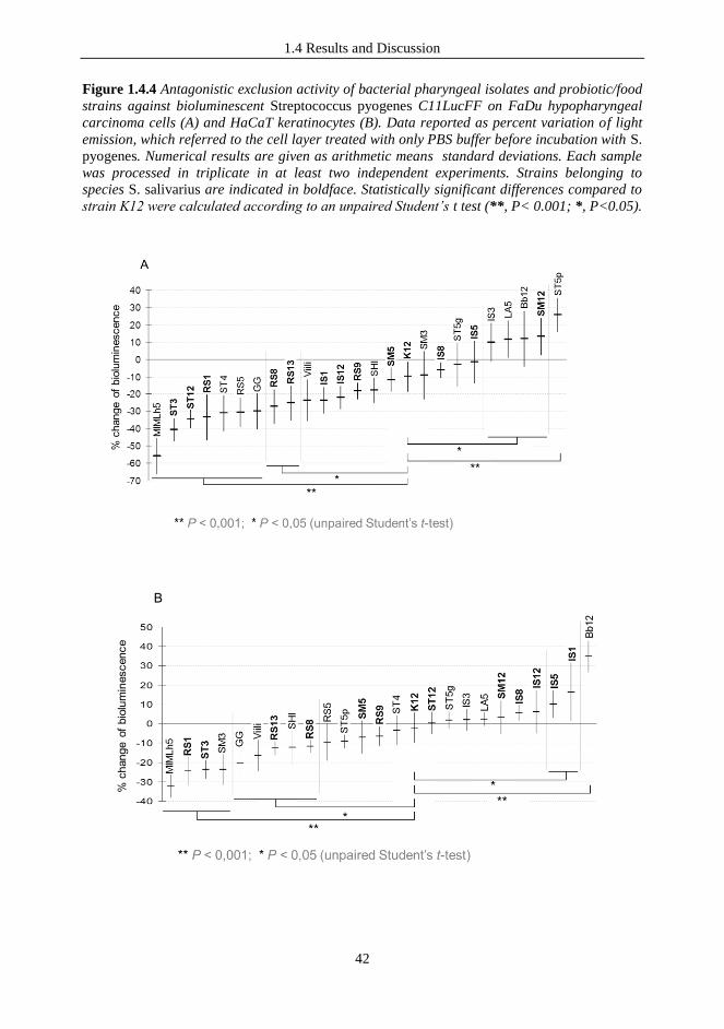
1.4 Results and Discussion
42
Figure 1.4.4 Antagonistic exclusion activity of bacterial pharyngeal isolates and probiotic/food
strains against bioluminescent Streptococcus pyogenes C11LucFF on FaDu hypopharyngeal
carcinoma cells (A) and HaCaT keratinocytes (B). Data reported as percent variation of light
emission, which referred to the cell layer treated with only PBS buffer before incubation with S.
pyogenes. Numerical results are given as arithmetic means standard deviations. Each sample
was processed in triplicate in at least two independent experiments. Strains belonging to
species S. salivarius are indicated in boldface. Statistically significant differences compared to
strain K12 were calculated according to an unpaired Student’s t test (**, P< 0.001; *, P<0.05).

1.4 Results and Discussion
43
Adhesion experiments performed on the FaDu cell layer can give a partial explanation of the
strong antagonizing activity displayed by these strains. (Fig. 1.4.3). In fact, it can be
hypothesized that the competition for adhesion sites is a major mechanism through which these
bacteria antagonize S. pyogenes on FaDu cells.
The results also demonstrated that the exclusion activity is strain specific, since significant
differences were observed also among strains of the same species. We found a correlation
between the results obtained with the two cell lines. In fact, with only a few exceptions, those
strains that significantly reduced the light production in HaCaT cells also did so in the FaDu
cell layer. Particularly, the following two strains were the most active in both cell lines: L.
helveticus MIMLh5 (with an average reduction of bioluminescence of -55 % on FaDu and -32
% on HaCaT) and S. salivarius ST3 (with average reductions of bioluminescence of 40% in
FaDu and 24% in HaCaT). In addition to MIMLh5 and ST3, the probiotic strains L.
rhamonosus GG and L. paracasei Shirota and the dairy strain L. lactis subsp. cremoris Viili
also displayed an antagonistic exclusion significantly stronger than the reference oral probiotic
strain S. salivarius K12.
The antagonism by competition was tested only on the FaDu cell layer for the two selected
strains S. salivarius ST3 and L. helveticus MIMLh5, the reference oral probiotic K12, three oral
strains (S. salivarius RS13 and RS1, S. oralis ST5p) and the commercial probiotic B. animalis
lactis Bb12, which were selected in order to have at least one representative strain for each of
the four typologies of bacterial adhesion on the FaDu layer (Fig. 1.4.2). In this experiment, all
tested strains markedly inhibited S. pyogenes bioluminescence (> 50 % reduction) and
particularly L. helveticus MIMLh5 (78 %) and S. salivarius strains (ca. 80 % reduction) We also
noticed a partial dependence of both exclusion and competition on adhesion ability, since the
antagonism efficacy was slightly weaker for poorly adhesive strains like ST5p than strongly
adhesive bacteria (e.g., K12 and ST3; P values of 0.001, according to an unpaired Student‟s t
test) (Fig. 1.4.2 and Fig. 1.4.5).
Fig. 1.4.5 Antagonistic competition activity of bacterial pharyngeal isolates against
bioluminescent Streptococcus pyogenes C11lucFF on FaDu hypopharyngeal carcinoma cells.
Data reported as percentage variation of light emission as referred to the cell layer treated with
only S. pyogenes cells. Numerical results are given in the arithmetic means standard
deviations. All samples resulted significantly different compared to control (P < 0.001,
according to unpaired Student’s t-test).

1.4 Results and Discussion
44
1.4.4 Inhibition of S. pyogenes and PCR detection of bacteriocin encoding genes
The potential oral probiotic activity of pharyngeal streptococcal isolates were characterized by
PCR for known lantibiotic structural genes coding for salivaricin A, salivaricin B, streptin and
SA-FF22, which are known to be active against S. pyogenes. According to literature, strain K12
gave an amplicon of the expected length for salivaricin A and B. Among the other isolates, all
SM S. salivarius isolates and S. salivarius IS7 resulted positive for the only salivaricin A.
Sequence analyses revelaed that all salivaricin A amplicons, including K12 amplicon, belonged
to the variant A2 (Wescombe et al., 2006). None of the streptococcal isolates resulted positive
for streptin and SA-FF22 (Data not shown). When we performed inhibition test of S. pyogenes
after 24 h incubation at 37°C we observed clear inhibition zones corresponding to the colonies of
the strains K12 and ST3 (Data not shown). On the contrary, when cell-free neutralized broths were
used in disk-diffusion tests, inhibition zones were observed only for the reference strain S.
salivarius K12. Accordingly, PCR experiments suggested that ST3 could not possess bacteriocin
genes which are known to be encoded in strain K12 on a 190-kilobase transmissible plasmid
(Hyink et al., 2007). In contrast, ST3 is a plasmid-free strain. Therefore, it appears plausible that
the pharyngeal isolate ST3 inhibited the growth of S. pyogenes in plate probably partially due to
its acid production. For what it concerns L. helveticus MIMLh5, this strain was not able to prevent
S. pyogenes growth.
1.4.5 Immunomoduatory properties of selected LAB strains
One mechanism of action of probiotics is suggested to be their modulation of host immune
responses. In a recent study, Cosseau and collaborators showed that the oral probiotic S.
salivarius K12 can induce in vitro anti-inflammatory responses in epithelial cells, indicating a
potential promotion of cellular health and homeostasis (Cosseau et al., 2008). In that study, after
co-culture of human bronchial epithelial cells (16HBE14O-cells) with strain K12, they observed
an inhibition of baseline secretion of the chemokine interleukin (IL)-8, in coincidence with the
inhibition of activation of the NF-κB pathway (Cosseau et al., 2008). In our study, the
immunomodulatory properties of S. salivarius K12, ST3 and L. helveticus MIMLh5 were tested
on a FaDu layer by means of ELISA quantification of 17 secreted cytokines. Subsequently, in
order to elucidate the possible mechanisms involved in the effects on cytokine production, we
studied the modulation of NF-κB activation. Both experiments were performed in absence and
presence of IL-1β, a prototypical proinflammatory cytokine that plays a central role in the inflammation
amplification cascade.
1.4.6 Cytokine induction profile elicited by selected bacterial strains on FaDu cells.
Overnight incubation of a FaDu monolayer with selected bacteria resulted in a modulation of
the cytokine production profile, as determined by the Bio-Plex human cytokine 17-plex array
system. We observed different responses of FaDu cell layer to the three bacterial strains
included in the experiment (Fig. 1.4.6). Streptococcus salivarius strain ST3 abolished the
secretion of IL-1β and TNF-α and enhanced the production of IL-8 (from 1721 to 2331 pg ml-1
)
and GM-CSF (from 0.2 to 1.9 pg ml-1
). The commercial oral probiotic Streptococcus salivarius
K12 decreased IL-6 and IL-8 (respectively, from 78.1 to 24.2 pg ml-1
and from 1721 to 579 pg
ml-1
). Furthermore, strain K12 induced a reduction of the secretion of TNF-α (from 1.9 to 0.9
pg ml-1
) and G-CSF (from 3.5 to 1.7 pg ml-1
) and increased GM-CSF (from 0.2 to 1.6 pg ml-1
).
Finally, Lactobacillus helveticus MIMLh5 displayed the strongest ability to inhibit the secretion
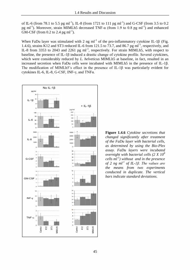
1.4 Results and Discussion
45
of IL-6 (from 78.1 to 5.5 pg ml-1
), IL-8 (from 1721 to 111 pg ml-1
) and G-CSF (from 3.5 to 0.2
pg ml-1
). Moreover, strain MIMLh5 decreased TNF-α (from 1.9 to 0.8 pg ml-1
) and enhanced
GM-CSF (from 0.2 to 2.4 pg ml-1
).
When FaDu layer was stimulated with 2 ng ml-1
of the pro-inflammatory cytokine IL-1 (Fig.
1.4.6), strains K12 and ST3 reduced IL-6 from 121.5 to 73.7, and 86.7 pg ml-1
, respectively, and
IL-8 from 3353 to 2043 and 2261 pg ml-1
, respectively. For strain MIMLh5, with respect to
baseline, the presence of IL-1β induced a drastic change of cytokine profile. Several cytokines,
which were considerably reduced by L. helveticus MIMLh5 at baseline, in fact, resulted in an
increased secretion when FaDu cells were incubated with MIMLh5 in the presence of IL-1β.
The modification of MIMLh5‟s effect in the presence of IL-1β was particularly evident for
cytokines IL-6, IL-8, G-CSF, INF-γ, and TNFα.
Figure 1.4.6 Cytokine secretions that
changed significantly after treatment
of the FaDu layer with bacterial cells,
as determined by using the Bio-Plex
assay. FaDu layers were incubated
overnight with bacterial cells (2 X 10⁸
cells ml-1
) without and in the presence
of 2 ng ml-1
of IL-1β. The values are
the means from two experiments
conducted in duplicate. The vertical
bars indicate standard deviations.

1.4 Results and Discussion
46
1.4.7 Modulation of NF-κB activation by tested bacteria in transfected FaDu cells.
To further explore the immunomodulatory properties of selected lactic acid bacteria, we tested
the effect of the microorganisms on NF-κB activation using a reporter cell line, obtained by
transfecting FaDu cells with a luciferase reporter vector induced by NF-κB. In FaDu cells, L.
helveticus MIMLh5 was the only strain that decreased the NF-κB-dependent production of
bioluminescence (Fig. 1.4.7A). On the contrary, strain ST3 showed a stimulatory effect. NF-κB
activation was particularly evident for strain ST3, which doubled the bioluminescence produced
by FaDu cells, when we employed a MOI of about 1000. S. salivarius K12 did not affect
significantly NF-κB (Fig. 1.4.7A). In a following series of experiments, the effect of bacterial
strains on NF-κB was assessed during stimulation of FaDu cells with the pro-inflammatory
cytokine IL-1β (Fig. 1.4.7B). The addition of 2 ng ml-1
of IL-1β to FaDu culture broth caused
nearly a two-fold increase of NF-κB activity after 4 h incubation. In presence of IL-1, we
observed that, at a MOI of 1000, strains K12 and ST3 reduced the NF-B activation in a statistically
significant manner, while the other conditions tested had no significant effect. In contrast, strain
L. helveticus MIMLh5 strongly inhibited NF-κB activation by IL-1β in a concentration
dependent manner (–84 % at MOI 1000 and –61 %; Fig. 1.4.7B).
Figure 1.4.7 Effects of selected bacterial strains on FaDu cells stably transfected with an NF-
B/luciferase reporter vector, without (A) or with (B) stimulation with IL-1 (2 ng ml-1).
Luciferase activity is expressed as percent change in relative luminescence units (RLU),
assuming the control as 100 %. Control, FaDu layers incubated without bacterial cells. The
values are the means (± standard deviations) of at least three independent experiments
conducted in duplicate. Asterisks (*) indicate statistically significant differences compared to
the control. MOI stands for multiplicity of infection (bacterial cells per FaDu cell).
1.4.8 The selected LAB strains drive different immune responses in vitro.
In these experiments, none of the tested strains exhibited a potential pro-inflammatory effect,
suggesting that they could be well tolerated by human epithelial cells in vivo. This statement is
consistent with the induced reduction of baseline TNF-α secretion, which was observed with all
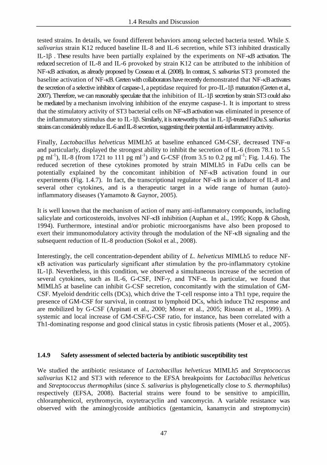
1.4 Results and Discussion
47
tested strains. In details, we found different behaviors among selected bacteria tested. While S.
salivarius strain K12 reduced baseline IL-8 and IL-6 secretion, while ST3 inhibited drastically
IL-1 . These results have been partially explained by the experiments on NF-B activation. The
reduced secretion of IL-8 and IL-6 provoked by strain K12 can be attributed to the inhibition of
NF-B activation, as already proposed by Cosseau et al. (2008). In contrast, S. salivarius ST3 promoted the
baseline activation of NF-B. Greten with collaborators have recently demonstrated that NF-B activates
the secretion of a selective inhibitor of caspase-1, a peptidase required for pro-IL-1 maturation (Greten et al.,
2007). Therefore, we can reasonably speculate that the inhibition of IL-1 secretion by strain ST3 could also
be mediated by a mechanism involving inhibition of the enzyme caspase-1. It is important to stress
that the stimulatory activity of ST3 bacterial cells on NF-B activation was eliminated in presence of
the inflammatory stimulus due to IL-1. Similarly, it is noteworthy that in IL-1-treated FaDu S. salivarius
strains can considerably reduce IL-6 and IL-8 secretion, suggesting their potential anti-inflammatory activity.
Finally, Lactobacillus helveticus MIMLh5 at baseline enhanced GM-CSF, decreased TNF-α
and particularly, displayed the strongest ability to inhibit the secretion of IL-6 (from 78.1 to 5.5
pg ml-1
), IL-8 (from 1721 to 111 pg ml-1
) and G-CSF (from 3.5 to 0.2 pg ml-1
; Fig. 1.4.6). The
reduced secretion of these cytokines promoted by strain MIMLh5 in FaDu cells can be
potentially explained by the concomitant inhibition of NF-κB activation found in our
experiments (Fig. 1.4.7). In fact, the transcriptional regulator NF-κB is an inducer of IL-8 and
several other cytokines, and is a therapeutic target in a wide range of human (auto)-
inflammatory diseases (Yamamoto & Gaynor, 2005).
It is well known that the mechanism of action of many anti-inflammatory compounds, including
salicylate and corticosteroids, involves NF-κB inhibition (Auphan et al., 1995; Kopp & Ghosh,
1994). Furthermore, intestinal and/or probiotic microorganisms have also been proposed to
exert their immunomodulatory activity through the modulation of the NF-κB signaling and the
subsequent reduction of IL-8 production (Sokol et al., 2008).
Interestingly, the cell concentration-dependent ability of L. helveticus MIMLh5 to reduce NF-
κB activation was particularly significant after stimulation by the pro-inflammatory cytokine
IL-1β. Nevertheless, in this condition, we observed a simultaneous increase of the secretion of
several cytokines, such as IL-6, G-CSF, INF-γ, and TNF-α. In particular, we found that
MIMLh5 at baseline can inhibit G-CSF secretion, concomitantly with the stimulation of GM-
CSF. Myeloid dendritic cells (DCs), which drive the T-cell response into a Th1 type, require the
presence of GM-CSF for survival, in contrast to lymphoid DCs, which induce Th2 response and
are mobilized by G-CSF (Arpinati et al., 2000; Moser et al., 2005; Rissoan et al., 1999). A
systemic and local increase of GM-CSF/G-CSF ratio, for instance, has been correlated with a
Th1-dominating response and good clinical status in cystic fibrosis patients (Moser et al., 2005).
1.4.9 Safety assessment of selected bacteria by antibiotic susceptibility test
We studied the antibiotic resistance of Lactobacillus helveticus MIMLh5 and Streptococcus
salivarius K12 and ST3 with reference to the EFSA breakpoints for Lactobacillus helveticus
and Streptococcus thermophilus (since S. salivarius is phylogenetically close to S. thermophilus)
respectively (EFSA, 2008). Bacterial strains were found to be sensitive to ampicillin,
chloramphenicol, erythromycin, oxytetracyclin and vancomycin. A variable resistance was
observed with the aminoglycoside antibiotics (gentamicin, kanamycin and streptomycin)

1.4 Results and Discussion
48
depending on the strain and the growth medium. All strains were assessed to be resistant to
kanamycin with the only exception of S. salivarius ST3, when it was grown in LM17 medium
(Table 1.4.1). Strain K12 was resistant to gentamicin in LM17 medium and to streptomycin in
BHI medium. Finally, S. salivarius ST3 was resistant to gentamicin in MRS medium and to
streptomycin in BHI and MRS media (Table 1.4.1). For what it concerns L. helveticus
MIMLh5, it was found to be sensitive to all tested antibiotics except kanamycin (Table 1.4.1).
However, it appears reasonable to consider the resistance to kanamycin as intrinsic and not
related to a specific gene. It is known, in fact, that several lactobacilli have a high natural
resistance to kanamycin and other aminoglycosidic antibiotics (Bernardeu et al., 2008).
Table.1.4.1 Antibiotic sensitivities of selected bacterial strains. Minimal inhibitory
concentrations (MICs).
1 Possible interference of the growth medium is reported (EFSA, 2008).
2 Microbiological breakpoints categorizing bacteria as resistant (μg ml-1
) according to EFSA
guidelines (EFSA, 2008); S.t. and L.h. stands for Streptococcus thermophilus and Lactobacillus
helveticus, respectively.
3 M17 containing 2 % lactose.
Recently, the European Food Safety Authority (EFSA) assigned a "Qualified Presumption of
Safety" (QPS) status (EFSA, 2004) to several lactic acid bacterial species, including
Lactobacillus helveticus, Streptococcus thermophilus, but not Streptococcus salivarius. In
Europe, S. salivarius belongs to the risk group 2 (such as S. pyogenes or S. pneumoniae), while
the very closely related species S. thermophilus, S. uberis and S. vestibularis (Mora et al., 2003)
belong to risk group 1. Presumably, S. salivarius is considered an opportunistic pathogen
because, as with many food grade lactobacilli, there have been sporadic reports of infections,
generally in subjects under adverse medical conditions (Ahmed et al., 2003; Afek et al., 2004;
Corredoira et al., 2005). Quite the opposite, in other parts of the world, S. salivarius has already
acquired the status of safe microorganism and has been commercialized for several years as
1.4 Results and Discussion
49
probiotic without any reported adverse consequences (Burton et al., 2006). In the light of the
above mentioned facts, the optimal strategy to assess the safety of S. salivarius, would be
considering every specific strain independently, in accordance with FAO/WHO guidelines on
probiotics (FAO/WHO, 2002), as has been done for the strain S. salivarius K12 (Burton et al.,
2006; Cosseau et al., 2008). From this perspective, the absence of transmissible antibiotic
resistances is considered a key safety prerequisite for the selection of a probiotic microorganism
(EFSA, 2004; FAO/WHO, 2002). In this study, S. salivarius strain ST3, according to the EFSA
breakpoints suggested for S. thermophilus (2008), resulted sensitive to a variety of antibiotics
that are routinely used for the control of URTIs. Differently, they showed resistance exclusively
to the aminoglycosidic antibiotics gentamicin, kanamycin and streptomycin, for which an
intrinsic resistance is known for several lactic acid bacteria (Corredoira et al., 2005; Hummel et
al., 2007; EFSA, 2008; Maragkoudakis et al., 2009).
1.4.10 ST3 is a natural urease negative S. salivarius strain
All Streptococcus salivarius isolates obtained from pharyngeal mucosa and the probiotic strain
K12 were tested for their ability to hydrolyze urea. All tested strains resulted urealytic when
grown in a medium containing nickel cations, with the only exception of strain ST3. According
to the phenotypic assay, we failed to amplify by PCR from strain ST3 ureC gene, which
encodes the alpha subunit protein containing the active site and conserved nickel binding
ligands of the urease complex. On the contrary, a PCR fragment of the expected length was
obtained using primers targeting ureC from all the other S. salivarius strains (Data not shown).
The urease activity is a bacterial feature that exerts an important role in the interaction with the
human host is. Ammonia production from ureolysis in saliva and crevicular fluids is, in fact, a
primary source of amino nitrogen and is thought to inhibit the initiation and progression of
dental caries by reducing acidity (Kleinberg, 1967; Peterson et al., 1985). At the same time, a
high ammonia concentration can have deleterious effects on host cells (Helgeland, 1985), such
as fibroblasts and polymorphonuclear leukocytes, and may therefore contribute to tissue
damage (Helgeland, 1985). Among the species of oral bacteria that have been identified as
ureolytic, S. salivarius is known to produce high levels of urease (Sissons & Cutress, 1987).
Unexpectedly, strain S. salivarius ST3, selected in this work for its potential probiotic
properties, was unable to hydrolyze urea. In the consideration of its use as pharynx's probiotic,
this bacterium would interact directly with oro-pharyngeal mucosa and tonsil crypts. Therefore,
the inability to hydrolyze urea could be of potential benefit in preserving the host's mucosal
cells from ammonia injury.
1.4.11 ST3/MIMLh5 co-suspension did not affect adhesion and antagonistic properties
of the individual strains on the FaDu cell layer.
In the following series of experiments, we evaluated in vitro the potential probiotic properties of
strains ST3 and MIMLh5 used in combination. We first studied the interaction with FaDu
human pharyngeal cell line in terms of adhesion and ability to antagonize Streptococcus
pyogenes. We found that the adhesion on pharyngeal epithelium of each strain was not affected
when the other one was also present, indicating that there is no competition for adhesion sites
between MIMLh5 and ST3 in our test conditions (MOI of 1000 for each bacterium; Fig. 1.4.8).

1.4 Results and Discussion
50
Afterwards, we measured the bioluminescence produced by S. pyogenes C11LucFF
as an
indication of the exclusion antagonism exerted by strains ST3, MIMLh5 and their mixed
suspension. L. helveticus MIMLh5 showed a better antagonism against S. pyogenes (72 %
reduction of luminescence), compared to S. salivarius strain ST3 (53 % luminescence
reduction). When the tester bacteria were used in combination (MOI 1000 each), the
antagonistic activity was maintained at the highest level (72 % reduction), indicating that the
two strains can coexist without reducing their in vitro exclusion ability (Fig. 1.4.9).
1.4.12 The effect of strain MIMLh5 on NF-κB activation in FaDu epithelial cells is
predominant over that of strain ST3.
In this study, we explored the effect of strains ST3 and MIMLh5 in combination on NF-κB
activation in FaDu cells using the same luciferase reporter system previously described. As
observed in previous investigations (Guglielmetti et al. 2010a;b), L. helveticus MIMLh5
decreased the NF-κB-dependent production of bioluminescence, while ST3 showed a
stimulatory effect (Fig. 1.4.10). When the two bacterial strains were used in combination at the
same multiplicity of infection (MOI 1000 each), activation of NF-κB was reduced to an extent
not significantly dissimilar to that determined by MIMLh5 alone (Fig. 1.4.10), indicating that
the inhibitory effect of this bacterium on NF-κB-mediated responses is predominant over the
outcome induced by strain ST3.
Figure 1.4.8 Bacterial adhesion as
observed with Giemsa staining
under a light microscope. FaDu
nuclei appear in red.
Figure 1.4.9 Antagonistic exclusion
activity against bioluminescent S.
pyogenes C11lucFF; control is a cell
layer treated with only PBS before
incubation with S. pyogenes.
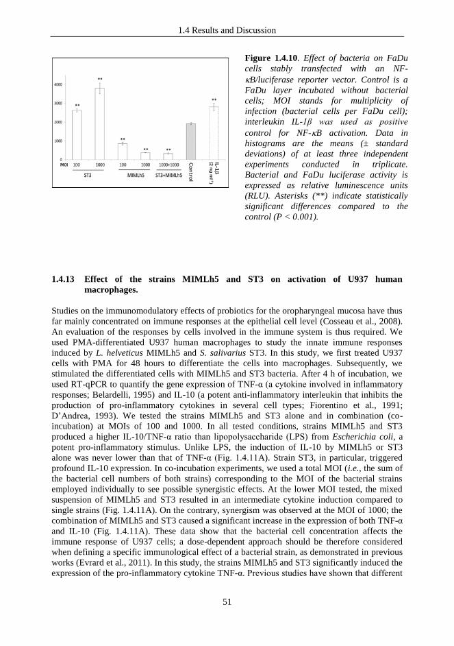
1.4 Results and Discussion
51
1.4.13 Effect of the strains MIMLh5 and ST3 on activation of U937 human
macrophages.
Studies on the immunomodulatory effects of probiotics for the oropharyngeal mucosa have thus
far mainly concentrated on immune responses at the epithelial cell level (Cosseau et al., 2008).
An evaluation of the responses by cells involved in the immune system is thus required. We
used PMA-differentiated U937 human macrophages to study the innate immune responses
induced by L. helveticus MIMLh5 and S. salivarius ST3. In this study, we first treated U937
cells with PMA for 48 hours to differentiate the cells into macrophages. Subsequently, we
stimulated the differentiated cells with MIMLh5 and ST3 bacteria. After 4 h of incubation, we
used RT-qPCR to quantify the gene expression of TNF-α (a cytokine involved in inflammatory
responses; Belardelli, 1995) and IL-10 (a potent anti-inflammatory interleukin that inhibits the
production of pro-inflammatory cytokines in several cell types; Fiorentino et al., 1991;
D‟Andrea, 1993). We tested the strains MIMLh5 and ST3 alone and in combination (co-
incubation) at MOIs of 100 and 1000. In all tested conditions, strains MIMLh5 and ST3
produced a higher IL-10/TNF-α ratio than lipopolysaccharide (LPS) from Escherichia coli, a
potent pro-inflammatory stimulus. Unlike LPS, the induction of IL-10 by MIMLh5 or ST3
alone was never lower than that of TNF-α (Fig. 1.4.11A). Strain ST3, in particular, triggered
profound IL-10 expression. In co-incubation experiments, we used a total MOI (i.e., the sum of
the bacterial cell numbers of both strains) corresponding to the MOI of the bacterial strains
employed individually to see possible synergistic effects. At the lower MOI tested, the mixed
suspension of MIMLh5 and ST3 resulted in an intermediate cytokine induction compared to
single strains (Fig. 1.4.11A). On the contrary, synergism was observed at the MOI of 1000; the
combination of MIMLh5 and ST3 caused a significant increase in the expression of both TNF-α
and IL-10 (Fig. 1.4.11A). These data show that the bacterial cell concentration affects the
immune response of U937 cells; a dose-dependent approach should be therefore considered
when defining a specific immunological effect of a bacterial strain, as demonstrated in previous
works (Evrard et al., 2011). In this study, the strains MIMLh5 and ST3 significantly induced the
expression of the pro-inflammatory cytokine TNF-α. Previous studies have shown that different
Figure 1.4.10. Effect of bacteria on FaDu
cells stably transfected with an NF-
B/luciferase reporter vector. Control is a
FaDu layer incubated without bacterial
cells; MOI stands for multiplicity of
infection (bacterial cells per FaDu cell);
interleukin IL-1β was used as positive
control for NF-B activation. Data in
histograms are the means (± standard
deviations) of at least three independent
experiments conducted in triplicate.
Bacterial and FaDu luciferase activity is
expressed as relative luminescence units
(RLU). Asterisks (**) indicate statistically
significant differences compared to the
control (P < 0.001).
1.4 Results and Discussion
52
Lactobacillus species can trigger pronounced pro-inflammatory activity in DCs (Christensen et
al., 2002; Weiss et al., 2010) and in macrophages (Morita et al., 2002). Macrophages are
professional phagocytes that reside in the secondary lymphoid organs as well as in almost all
tissues, including the tonsils (Yamamoto et al., 1988), serving as sentinels to detect microbial
invaders (Adherem & Underhill, 1999). Upon the recognition of pathogen-associated molecular
patterns (PAMPs; Janeway & Medzhitov, 2002), macrophages produce inflammatory
mediators, such as cytokines and chemokines, that alert the immune system to the infection of
injury. A pro-inflammatory behavior that results in the activation of cell-mediated (Th1 type)
immunity, like that observed for MIMLh5 and ST3, could thus be crucial in combating
intracellular pathogen attacks and viral infections and alerting the host‟s immune system. Other
studies have reported that most lactic acid bacterial strains that have been tested could induce a
Th1 response, even though this capability is strictly dependent on the specific strain and dose
used (Perdigòn et al., 2002; Ongol et al., 2008). Notably, in the same studies, some strains of S.
thermophilus, a species phylogenetically close to S. salivarius, induced a high level of IL-10
(Ongol et al., 2008). In our experiments, S. salivarius ST3 induced a higher level of IL-10 than
TNF-α. Furthermore, the increased production of the anti-inflammatory cytokine was more
pronounced in ST3 than in MIMLh5 at the highest MOI tested (Fig. 1.4.11A). Therefore, we
speculate that the presence of both bacteria, even at high doses (MOI 1000), might not result in
detrimental effects. The regulatory effect of the significant IL-10 expression could protect
against secondary outcomes that can occur after streptococcal infections, namely, reactive
inflammatory conditions such as rheumatic disease (Puliti et al., 2002).
1.4.14 The strains MIMLh5 and ST3 induce cyclooxygenase (COX)-2 expression in
U937 cells and in BMDCs.
Cyclooxygenase (COX), or prostaglandin synthase H (PGH), is a homodimer enzyme involved
in the synthesis of prostaglandins (PGs) from arachidonic acid (Funk, 2001). PGs are involved
in several physiological processes and contribute to the protection of the gastrointestinal
mucosa (Williams et al., 1999; Morteau et al., 2000). Two isoforms of the COX enzyme have
been identified and described (Kujubu et al., 1991). COX-1 is constitutively expressed in a wide
range of tissues, whereas COX-2 is constitutively expressed in very few tissues but is induced
by several stimuli, including bacterial components (Herschmann, 1996). We investigated the
effect of the strains MIMLh5 and ST3 on COX-2 activation in the U937 cell model. The
association of the two bacteria resulted in an enhanced transcription of the gene (Fig. 1.4.11B).
In similar experiments, we also observed a strong increase in COX-2 gene expression in murine
bone marrow-derived dendritic cells (Data not shown). Although other studies have suggested
that lactic acid bacteria could affect COX-2 secretion, there is no clear consensus in the
literature. For instance, several probiotic preparations were found to reduce the amount of
prostaglandins and levels COX-2 expression at the intestinal level in murine models (Urbanska
et al., 2010; Amdekar et al., 2011;) and in vitro (Lee et al., 2008). In contrast, other papers
reported a significant up-regulation of COX-2 expression levels by probiotic bacteria in vitro
(Otte et al., 2009; Khailova et al., 2010) and in vivo (Khailova et al., 2010; Kwon et al., 2010).
Lactobacillus acidophilus, a species phylogenetically related to L. helveticus, was reported to
significantly increase COX-2 expression and PGE2 secretion in the human colon cancer cell line
Colo320 (Otte et al, 2009). Differences in tested strains and model systems likely explain the
lack of consensus found in the literature. The ability of the strains MIMLh5 and ST3 to induce
COX-2 expression suggests that these bacteria could directly affect inflammatory processes not
only by modulating cytokine secretion but also inducing prostaglandin production.

1.4 Results and Discussion
53
Figure 1.4.11 Transcription analysis of cytokine genes in U937 cells stimulated with
Lactobacillus helveticus MIMLh5 and Streptococcus salivarius ST3 after 4 h of incubation with
bacterial strains used alone or in association at two different MOIs. Lipopolysaccharide (LPS)
was used as positive control at a concentration of 1 μg ml-1
. The values are the means (±
standard deviations) for a result representative of three independent experiments, expressed as
the fold change in induction respective to the control (U937 unstimulated cells) set at a value of
1. Asterisks indicate statistically significant differences compared to the corresponding control
(*, P < 0.05). A, Expression levels of IL-10 and TNF-α. B, Expression levels of COX-2. C,
Expression levels of IL10 and TNF-α in the presence of a neutralizing antibody against TLR-2
(anti-TLR2). Anti-TLR2 was added to U937 cells 1 h before stimulation with bacteria.
Immunoglobulin-A2 isotype (IgA2) was used as control for non-specific blocking activity.
1.4 Results and Discussion
54
COX-2 is involved in the induction of oral tolerance through the action of PGs in guiding T
cells towards an immunosuppressive phenotype (Newberry et al., 1999) and in resolving
inflammation (Wallace, 2006). The activity observed for MIMLh5 and ST3 on COX-2 could
thus be of immediate benefit for host‟s mucosa, and in light of the fact that, rapid COX-2 up-
regulation in response to injury or inflammation has been reported to restore mucosal integrity
(Tan et al., 2000).
1.4.15 TLR-2 participates in the recognition of the strains MIMLh5 and ST3 by U937
cells.
To gain information on possible signaling pathways involved in the immune responses induced
by L. helveticus MIMLh5 and S. salivarius ST3, we investigated which Toll-like receptor
(TLR) recognizes these bacteria. TLRs are a class of transmembrane proteins involved in innate
immunity (Akira and Hemmi, 2003); in mammals, they work as pattern recognition receptors
(PRRs) and participate in detecting various conserved microbial molecules, such as
lipopolysaccharide (LPS), lipoteichoic acid (LTA), peptidoglycan (PGN), bacterial lipoproteins,
lipoarabinomannan, and zymosan (Means et al., 2000). More specifically, TLR-2 forms
heterodimers with TLR-1 or TLR-6 and primarily interacts with Gram-positive bacteria by
recognizing PGN, LTA, and lipoproteins (Takeda et al., 2003).
In this study, we performed experiments in U937 cells with a neutralizing antibody against
TLR-2 (antiTLR2-Ab) to partially block its ability to bind ligands. An immunoglobulin-A (IgA)
isotype control was also used to exclude non-specific binding to U937 cell receptors. After 1
hour pre-incubation with an anti-TLR2 neutralizing Ab (5 μg ml-1
), we stimulated the U937
cells for 4 hours with bacteria. We then evaluated the expression of the genes that encode TNF-
α and IL-10 using RT-qPCR. When TLR-2 was blocked with anti-TLR2, we observed
significantly less TNF-α mRNA, both when the strains MIMLh5 and ST3 were used
individually and in combination (Fig. 1.4.11C). These results indicate that TLR-2 is involved in
mediating the immunostimulatory activity of both strains. These data are consistent with
previous studies that showed that the immunomodulatory activity elicited by lactobacilli
involves TLR-2 in macrophages (Shida et al., 2009) and in dendritic cells (Zeuthen et al., 2008;
Weiss et al., 2010). However, pre-incubation with anti-TLR2 resulted in a reduction of IL-10
expression only with strain ST3, whereas MIMLh5-mediated expression of IL-10 was not
affected (Fig. 1.4.11C), indicating that receptors independent from TLR-2 may be involved in
the induction of this cytokine. The interference of anti-TLR2-Ab with IL-10 induction that we
observed in our study with strain ST3 is in agreement with the literature. Molecules able to
induce IL-10 in a TLR2-dependent manner have been already described (Re & Strominger,
2004; Yamazaki et al., 2011). It is plausible that a similar response mechanism is triggered by S.
salivarius ST3 in U937 cells.
When the strains were combined, the reduction of the anti-inflammatory cytokine TNF-α was
similar to that of ST3 alone (1.43- and 1.34-fold reduction, respectively). Even though anti-
TLR2 generally reduced TNF-α induction, the relative increase of TNF-α gene expression
caused by the bacteria in combination compared to bacteria employed singularly was markedly
higher when TLR-2 was blocked with anti-TLR2 than in control conditions (i.e., in presence of
IgA2, Fig. 1.4.11C). In other words, the synergistic effect of using both strains was intensified
by the presence of the anti-TLR2 antibody. As a consequence, when TLR-2 was blocked, the
IL-10/TNF-α ratio increased only slightly (approximately 18 %) with bacteria in combination,
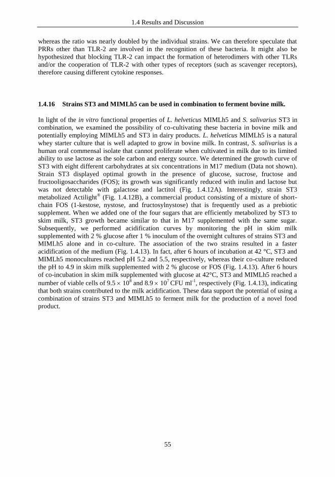
1.4 Results and Discussion
55
whereas the ratio was nearly doubled by the individual strains. We can therefore speculate that
PRRs other than TLR-2 are involved in the recognition of these bacteria. It might also be
hypothesized that blocking TLR-2 can impact the formation of heterodimers with other TLRs
and/or the cooperation of TLR-2 with other types of receptors (such as scavenger receptors),
therefore causing different cytokine responses.
1.4.16 Strains ST3 and MIMLh5 can be used in combination to ferment bovine milk.
In light of the in vitro functional properties of L. helveticus MIMLh5 and S. salivarius ST3 in
combination, we examined the possibility of co-cultivating these bacteria in bovine milk and
potentially employing MIMLh5 and ST3 in dairy products. L. helveticus MIMLh5 is a natural
whey starter culture that is well adapted to grow in bovine milk. In contrast, S. salivarius is a
human oral commensal isolate that cannot proliferate when cultivated in milk due to its limited
ability to use lactose as the sole carbon and energy source. We determined the growth curve of
ST3 with eight different carbohydrates at six concentrations in M17 medium (Data not shown).
Strain ST3 displayed optimal growth in the presence of glucose, sucrose, fructose and
fructooligosaccharides (FOS); its growth was significantly reduced with inulin and lactose but
was not detectable with galactose and lactitol (Fig. 1.4.12A). Interestingly, strain ST3
metabolized Actilight® (Fig. 1.4.12B), a commercial product consisting of a mixture of short-
chain FOS (1-kestose, nystose, and fructosylnystose) that is frequently used as a prebiotic
supplement. When we added one of the four sugars that are efficiently metabolized by ST3 to
skim milk, ST3 growth became similar to that in M17 supplemented with the same sugar.
Subsequently, we performed acidification curves by monitoring the pH in skim milk
supplemented with 2 % glucose after 1 % inoculum of the overnight cultures of strains ST3 and
MIMLh5 alone and in co-culture. The association of the two strains resulted in a faster
acidification of the medium (Fig. 1.4.13). In fact, after 6 hours of incubation at 42 °C, ST3 and
MIMLh5 monocultures reached pH 5.2 and 5.5, respectively, whereas their co-culture reduced
the pH to 4.9 in skim milk supplemented with 2 % glucose or FOS (Fig. 1.4.13). After 6 hours
of co-incubation in skim milk supplemented with glucose at 42°C, ST3 and MIMLh5 reached a
number of viable cells of 9.5 108 and 8.9 10
7 CFU ml
-1, respectively (Fig. 1.4.13), indicating
that both strains contributed to the milk acidification. These data support the potential of using a
combination of strains ST3 and MIMLh5 to ferment milk for the production of a novel food
product.
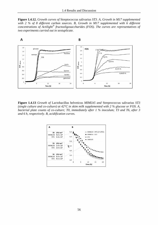
1.4 Results and Discussion
56
Figure 1.4.12. Growth curves of Streptococcus salivarius ST3. A, Growth in M17 supplemented
with 2 % of 8 different carbon sources. B, Growth in M17 supplemented with 6 different
concentrations of Actilight®
fructooligosaccharides (FOS). The curves are representatives of
two experiments carried out in sextuplicate.
Figure 1.4.13 Growth of Lactobacillus helveticus MIMLh5 and Streprococcus salivarius ST3
(single culture and co-culture) at 42°C in skim milk supplemented with 2 % glucose or FOS. A,
bacterial plate counts of co-culture; T0, immediately after 1 % inoculum; T3 and T6, after 3
and 6 h, respectively. B, acidification curves.

1.5 Conclusions
57
1.5 CONCLUSIONS
There is an increasing interest in prophylactic strategies effective for upper respiratory tract
infections, that represent a major cause of medical prescription for antibiotics, especially in
children. The identification of bacterial strains that could be used as probiotics for the
pharyngeal mucosa is one potential strategy.
During this first part of the research, an in vitro rational process led us to select two potential
probiotic bacterial strains for the pharyngeal mucosa, the commensal S. salivarius ST3 and the
dairy bacterium L. helveticus MIMLh5. These bacteria efficiently adhered to human epithelial
pharyngeal cell lines, antagonizing the adhesion of Streptococcus pyogenes. Furthermore,
immunological tests suggested a good degree of adaptation of these selected strains to the host
and potential immunomodulatory and anti-inflammatory abilities.
Therefore we further characterized the two selected strains, and as first we demonstrated that
strains MIMLh5 and ST3, when used together, maintain their in vitro probiotic abilities.
Subsequently, we gave particular attention to bacterial immunomodulatory properties because
modulation of the host‟s immunity is one of the most commonly purported benefits of the
consumption of probiotics and is supported by an increasing number of in vitro and in vivo
studies (Corthesy et al., 2007). We found that the combination of MIMLh5 and ST3 resulted in
a synergistic effect, according to cytokine induction, that might help the host immune system to
react to potential pathogens while maintaining a balance between pro- and anti-inflammatory
cytokines, thus preventing possible exaggerated responses. Finally, we observed that these
microorganisms grow efficiently when co-cultured in milk, suggesting that the preparation of a
milk-based fermented product containing both MIMLh5 and ST3 can be a practicable solution
for the administration of these bacteria.
In conclusion, we propose the combined use of Lactobacillus helveticus MIMLh5 and
Streptococcus salivarius ST3 for the preparation of novel products that can display probiotic
properties for the pharyngeal mucosa (Taverniti et al., 2012).
1.6 References
58
1.6 REFERENCES
Adams CA, 2010, The probiotic paradox: live and dead cells are biological response
modifiers. Nutr Res Rev 23:37-46.
Aderem A, Underhill DM, 1999, Mechanisms of phagocytosis in macrophages. Annu
Rev Immunol 17:593-623.
Afek S et al., 2004, Carcinoma of the colon presenting as Streptococcus salivarius
sepsis. J Clin Gastroenterol 38:86-87.
Ahmed R et al., 2003, Viridans streptococcus bacteremia in children on chemotherapy
for cancer: an underestimated problem. Pediatr Hematol Oncol 20:439-444.
Akira S, Hemmi H, 2003, Recognition of pathogen-associated molecular patterns by
TLR family. Immunol Lett 85:85-95.
Amdekar S et al., 2011, Lactobacillus casei reduces the inflammatory joint damage
associated with collagen-induced arthritis (CIA) by reducing the pro-inflammatory
cytokines: Lactobacillus casei: COX-2 inhibitor. J Clin Immunol 31:147-154.
Arpinati M et al., 2000, Granulocyte-colony stimulating factor mobilizes T helper 2-
inducing dendritic cells. Blood 95:2484-2490.
Auphan N et al., 1995, Immunosuppression by glucocorticoids: inhibition of NF-kB
activity through induction of I k B synthesis. Science. 270:286-290.
BCC Research, 2008, The probiotic market: ingredients, supplements, foods
(FOD035B). BCC Research web site.
http://www.bccresearch.com/pressroom/FOD035B.html. Accessed 13 March 2011.
Beimfohr C et al., 1997, Rapid genotypic differentiation of Lactococcus lactis
subspecies and biovar. Syst. Appl Microbiol 20:216-221.
Bek-Thomsen M et al., 2008, Population diversity and dynamics of Streptococcus
mitis, Streptococcus oralis, and Streptococcus infantis in the upper respiratory tracts of
adults, determined by a nonculture strategy. Infect Immun 76:1889-1896.
Belardelli F. 1995, Role of interferons and other cytokines in the regulation of the
immune response. APMIS 103:161-179.
Benoit M et al., 2008, Macrophage polarization in bacterial infections. J Immunol
181:3733-3739.
Bernardeau M et al., 2008, Safety assessment of dairy microorganisms: the
Lactobacillus genus. Int J Food Microbiol 126:278-285.

1.6 References
59
Besselink MG et al., 2008, Dutch Acute Pancreatitis Study Group. Probiotic
prophylaxis in predicted severe acute pancreatitis: a randomised, double-blind,
placebo-controlled trial. Lancet 371:651-659.
Bisno A, 1996, Acute pharyngitis: etiology and diagnosis. Pediatrics 97:949-954.
Boyle RJ et al., 2006, Probiotic use in clinical practice: what are the risks? Am J Clin
Nutr 83:1256-64.
Borchers AT et al., 2009, Probiotics and immunity. J Gastroenterol 44, 26-46.
Borchert D et al, 2008, Prevention and treatment of urinary tract infection with
probiotics: Review and research perspective. Indian J Urol. 24:139-144.
Boukamp P et al., 1988, Normal keratinization in a spontaneously immortalized
aneuploid human keratinocyte cell line. J Cell Biol 106:761-771.
Bourke SJ, 2007, Lecture Notes: Respiratory Medicine. Oxford, UK: Blackwell
Publishing.
Brandtzaeg P, 2011, Immune functions of nasopharyngeal lymphoid tissue. Adv
Otorhinolaryngol 72:20-24.
Burton JP et al., 2006, Safety assessment of the oral cavity probiotic Streptococcus
salivarius K12. Appl. Environ Microbiol 72:3050-3053.
California Dairy Research Foundation, www.cdrf.org/home/checkoff-
investments/usprobiotics/products-with-probiotics/ (accessed: 05/01/2013).
Christensen HR et al., 2002, Lactobacilli differentially modulate expression of
cytokines and maturation surface markers in murine dendritic cells. J Immunol
168:171-178.
Corredoira JC et al., 2005, Clinical characteristics and significance of Streptococcus
salivarius bacteremia and Streptococcus bovis bacteremia: a prospective 16-year study.
Eur J Clin Microbiol Infect Dis 24:250-255.
Corthesy B et al., 2007, Cross-talk between probiotic bacteria and the host immune
system. J Nutr 137:781S–790S.
Cosseau C et al., 2008, The commensal Streptococcus salivarius K12 downregulates
the innate immune responses of human epithelial cells and promotes host-microbe
homeostasis. Infect Immun 76:4163-4175.
Cutler CW, Jotwani R, 2006, Dendritic cells at the oral mucosal interface. J Dent Res
85:678-689.

1.6 References
60
D‟Andrea A et al., 1993, Interleukin 10 (IL-10) inhibits human lymphocyte interferon
gamma-production by suppressing natural killer cell stimulatory factor/IL-12 synthesis
in accessory cells. J Exp Med 178:1041-1048.
Debertin AS et al., 2003, Nasal-associated lymphoid tissue (NALT): frequency and
localization in young children. Clin Exp Immunol 134:503-507.
Delcenserie V, 2008, Immunomodulatory effects of probiotics in the intestinal tract.
Curr Issues Mol Biol 10:37-54.
de Vrese M, Schrezenmeir J, 2008, Probiotics, prebiotics, and synbiotics. Adv
Biochem Eng Biotechnol. 111:1-66.
Dunzendorfer S et al., 2004, TLR4 is the signaling but not the lipopolysaccharide
uptake receptor. J Immunol 173:1166-1170.
Eberl G, Lochner M, 2009, The development of intestinal lymphoid tissues at the
interface of self and microbiota. Mucosal Immunol 2:478-485.
EFSA, 2004, Scientific Colloquium Summary Report. QPS: Qualified Presumption of
Safety of Micro-organisms in Food and Feed. 13-14 December 2004, Brussels,
Belgium.
EFSA, 2008, Update of the criteria used in the assessment of bacterial resistance to
antibiotics of human or veterinary importance. Prepared by the Panel on Additives and
Products or Substances used in Animal Feed. The EFSA Journal 2008; 732:1-15.
EFSA, 2009, Scientific Opinion on the substantiation of health claims related
toBifidobacterium animalis Lafti B94 (CBS118.529) and decreasing potentially
pathogenic intestinal microorganisms (ID 867) pursuant toArticle 13(1) of Regulation
(EC) No 1924/2006. The EFSA Journal 2009; 7(9):1232.
Evrard B et al., 2011, Dose-dependent immunomodulation of human dendritic cells by
the probiotic Lactobacillus rhamnosus Lcr35. PLoS One 6:e18735.
FAO/WHO, 2002, Report of a joint FAO/WHO expert consultation on guidelines for
the evaluation of probiotics in food. World Health Organization and Food and
Agriculture Organization of the United Nations, London Ontario, Canada.
Fiorentino DF et al., 1991, IL-10 inhibits cytokine production by activated
macrophages. J Immunol 147:3815-3822.
Fukuyama S et al., 2009, Mucosa-Associated Lymphoid Tissue and Dynamics of
Lymphoid Cells in the Five Different Compartments in Allergic Diseases. Allergy
frontiers 2:563-601.
Funk CD, 2001, Prostaglandins and leukotrienes: advances in eicosanoid biology.
Science 294:1871-1875.
1.6 References
61
Galdeano CM, Perdigón G, 2006, The probiotic bacterium Lactobacillus casei induces
activation of the gut mucosal immune system through innate immunity. Clin Vaccine
Immunol 13:219-226.
Greten FR et al., 2007, NF-kB is a negative regulator of IL-1 secretion as revealed by
genetic and pharmacological inhibition of IKK. Cell 130:918-931.
Guglielmetti S et al, 2008, Bacterial cinnamoyl esterase activity screening for the
production of a novel functional food product. Appl Environ Microbiol 74:1284-1288.
Guglielmetti S et al., 2010a, Oral bacteria as potential probiotics for the pharyngeal
mucosa. Appl Environ Microbiol 76:3948-3958.
Guglielmetti S et al., 2010b, A dairy bacterium displays in vitro probiotic properties
for the pharyngeal mucosa by antagonizing group A streptococci and modulating the
immune response. Infect Immun 78:4734-4743.
Ha GY et al., 1999, Case of sepsis caused by Bifidobacterium longum. J Clin
Microbiol 37:1227-1228.
Harris P, Ralph P, 1985, Human leukemic models of myelomonocytic development: a
review of the HL-60 and U937 cell lines. J Leukoc Biol 37:407-422.
Helgeland K, 1985, pH and the effect of NH4Cl on human gingival fibroblasts. Scand J
Dent Res 93:39-45.
Hellings P et al., 2000, The Waldeyer's ring. Acta Otorhinolaryngol Belg 54:237-241.
Herschman HR, 1996, Prostaglandin synthase 2. Biochim Biophys Acta 1299:125-140.
Horz HP et al., 2007, Distribution and persistence of probiotic Streptococcus salivarius
K12 in the human oral cavity as determined by real-time quantitative polymerase chain
reaction. Oral Microbiol Immunol 22:126-130.
Hyink O et al., 2007, Salivaricin A2 and the novel lantibiotic salivaricin B are encoded
at adjacent loci on a 190-kilobase transmissible megaplasmid in the oral probiotic
strain Streptococcus salivarius K12. Appl Environ Microbiol 73:1107-1113.
Janeway CA,., Medzhitov R, 2002, Innate immune recognition. Annu Rev Immunol
20:197-216.
Juntunen M et al., 2001, Adherence of probiotic bacteria to human intestinal mucus in
healthy infants and during rotavirus infection. Clin Diagn Lab Immunol 8:293-296.
Kahala M et al., 2008, Characterization of starter lactic acid bacteria from the Finnish
fermented milk product viili. J Appl Microbiol 105:1929-1938.

1.6 References
62
Kang JG et al., 2006, Bacterial diversity in the human saliva from different ages. J
Microbiol 44:572-576.
Kelly K, et al., 1994, Out of control: the new biology of machines, social systems and
the economic world. Boston: Addison-Wesley. p. 98.
Khailova L et al., 2010, Bifidobacterium bifidum reduces apoptosis in the intestinal
epithelium in necrotizing enterocolitis. Am J Physiol Gastrointest Liver Physiol
299:G1118-G1127.
Kleinberg I, 1967, Effect of urea concentration on human plaque pH levels in situ.
Arch Oral Biol 12:1475-1484.
Kopp E, Ghosh S, 1994, Inhibition of NF-kappa B by sodium salicylate and aspirin.
Science 265:956-959.
Kotzampassi K, Giamarellos-Bourboulis EJ, 2012, Probiotics for infectious diseases:
more drugs, less dietary supplementation. Int J Antimicrob Agents 40:288-296.
Krutmann J, 2008, Pre- and probiotics for human skin. J Dermatol Sci 54:1-5.
Kujubu DA et al., 1991, TIS10, a phorbol ester tumor promoter-inducible mRNA from
Swiss 3T3 cells, encodes a novel prostaglandin synthase/cyclooxygenase homologue. J
Biol Chem 266:12866-12872.
Kunz AN et al., 2004, Two cases of Lactobacillus bacteremia during probiotic
treatment of short gut syndrome. J Pediatr Gastroenterol Nutr 38:457–458.
Kwon HK et al., 2010, Generation of regulatory dendritic cells and CD4+Foxp3+ T
cells by probiotics administration suppresses immune disorders. Proc Natl Acad Sci
USA 107:2159-2164.
Lampinen J et al., 1992, Expression of luciferase genes from different origins in
Bacillus subtilis. Mol Gen Genet 232:498-504.
Land MH et al., 2005, Lactobacillus sepsis associated with probiotic therapy.
Pediatrics 115:178–181.
Lanyi B, 1987, Classical and rapid identification: methods for medically important
bacteria. In Methods in Microbiology, Vol. 19 ed. Colwell, R.R. and Grigorova, R. pp.
1-67. New York: Academic Press.
Lebeer S et al., 2008, Genes and molecules of lactobacilli supporting probiotic action.
Microbiol Mol Biol Rev 72:728-764.
Lebeer S et al., 2010, Host interactions of probiotic bacterial surface molecules:
comparison with commensals and pathogens. Nat Rev Microbiol 8:171-184.

1.6 References
63
Lee YK et al., 1999, Introduction. In Handbook of Probiotics. New York: John Wiley
& Sons, Inc.
Lee JM et al., 2008, Antiinflammatory effect of lactic acid bacteria: inhibition of
cyclooxygenase-2 by suppressing nuclear factor-kappaB in Raw264.7 macrophage
cells. J Microbiol Biotechnol 18:1683-1688.
Ley RE et al., 2006, Ecological and evolutionary forces shaping microbial diversity in
the human intestine. Cell 124:837–848.
Mackaness GB, 1964, The Immunological basis of acquired cellular resistance. J Exp
Med 120:105-120.
Maragkoudakis PA et al., 2009, In vitro and in vivo safety evaluation of the bacteriocin
producer Streptococcus macedonicus ACA-DC 198. Int J Food Microbiol 133:141-
147.
Marteau PR, 2002, Probiotics in clinical conditions. Clin Rev Allergy Immunol
22:255-273.
Means TK et al., 2000, The biology of Toll-like receptors. Cytokine Growth Factor
Rev 11:219-232.
Medzhitov R, 2007, Recognition of microorganisms and activation of the immune
response. Nature 449, 819–826.
Mora D et al, 2004, Characterization of urease genes cluster of Streptococcus
thermophilus. J Appl Microbiol 96:209-219.
Mora D et al., 2003, 16S-23S rRNA intergenic spacer region sequence variation in
Streptococcus thermophilus and related dairy streptococci and development of a
multiplex ITS-SSCP analysis for their identification. Microbiology 149:807-813.
Morelli L, Capurso L, 2012, FAO/WHO Guidelines on Probiotics: 10 Years Later. J
Clin Gastroenterol 46 Suppl:S1-2.
Morita H et al., 2002, Cytokine production by the murine macrophage cell line J774.1
after exposure to lactobacilli. Biosci Biotechnol Biochem 66:1963-1966.
Morteau O et al., 2000, Impaired mucosal defense to acute colonic injury in mice
lacking cyclooxygenase-1 or cyclooxygenase-2. J Clin Invest 105:469-478.
Moser C et al., 2005, Serum concentrations of GM-CSF and G-CSF correlate with the
Th1/Th2 cytokine response in cystic fibrosis patients with chronic Pseudomonas
aeruginosa lung infection. APMIS. 113:400-409.
1.6 References
64
Newberry RD et al., 1999, Cyclooxygenase-2-dependent arachidonic acid metabolites
are essential modulators of the intestinal immune response to dietary antigen. Nat Med
5:900-906.
Ongol MP et al., 2008, Potential of selected strains of lactic acid bacteria to induce a
Th1 immune profile. Biosci Biotechnol Biochem 72:2847-2857.
Otte JM et al., 2009, Probiotics regulate the expression of COX-2 in intestinal
epithelial cells. Nutr Cancer 61:103-113.
Perdigon G et al., 2002, Interaction of lactic acid bacteria with the gut immune system.
Eur J Clin Nutr 56 Suppl 4:S21-S26.
Peterson S et al., 1985, Caries resistance in children with chronic renal failure: plaque
pH, salivary pH, and salivary composition. Pediatr Res 19:796-799.
Popova M et al., 2012, Beneficial effects of probiotics in upper respiratory tract
infections and their mechanical actions to antagonize pathogens. J Appl Microbiol
1136:1305-1318.
Power DA et al.,2008, Preliminary investigations of the colonisation of upper
respiratory tract tissues of infants using a paediatric formulation of the oral probiotic
Streptococcus salivarius K12. Eur J Clin Microbiol Infect Dis 27:1261-1263.
Proal AD et al., 2009, Autoimmune disease in the era of the metagenome. Autoimmun
Rev 8:677-681.
Puliti M et al., 2002, Regulatory role of interleukin-10 in experimental group B
streptococcal arthritis. Infect Immun 70:2862-2868.
Radzun HJ et al., 1983, Monocytic origin of the human hematopoietic cell line U-937
and its convertibility to macrophages evidenced by isoenzyme mapping. Int J Cancer
31:181-186.
Rao Y et al., 2012, Probiotics in oral health - a review. J N J Dent Assoc Spring 83:28-
32.
Re F, Strominger JL, 2004, IL-10 released by concomitant TLR2 stimulation blocks
the induction of a subset of Th1 cytokines that are specifically induced by TLR4 or
TLR3 in human dendritic cells. J Immunol 173:7548-7555.
Reid G et al., 2009, Targeting the vaginal microbiota with probiotics as a means to
counteract infections. Curr Opin Clin Nutr Metab Care 12:583-587.
Rissoa MC et al., 1999, Reciprocal control of T helper cell and dendritic cell
differentiation. Science 283:1183-1186.
1.6 References
65
Saldanha LG, 2008, US Food and Drug Administration regulations governing label
claims for food products, including probiotics. Clin Infect Dis 1:46.
Salminen MK et al., 2002, Lactobacillus bacteremia during a rapid increase in
probiotic use of Lactobacillus rhamnosus GG in Finland. Clin Infect Dis 35:1155-
1160.
Salminen MK et al., 2004, The efficacy and safety of probiotic Lactobacillus
rhamnosus GG on prolonged, noninfectious diarrhea in HIV patients on antiretroviral
therapy: a randomized, placebo-controlled, crossover study. HIV Clin Trials 5:183-
191.
Saxelin M et al., 1996, Lactobacilli and bacteremia in southern Finland, 1989–1992.
Clin Infect Dis 22:564–566.
Saxelin M et al., 2005, Probiotic and other functional microbes: from markets to
mechanisms. Curr Opin Biotechnol 162:204-211.
Servin AL, 2004, Antagonistic activities of lactobacilli and bifidobacteria against
microbial pathogens. FEMS Microbiol Rev 284:405-440.
Shida K et al., 2009, Peptidoglycan from lactobacilli inhibits interleukin-12 production
by macrophages induced by Lactobacillus casei through Toll-like receptor 2-
dependent and independent mechanisms. Immunology 128:e858-e869.
Sissons CH, Cutress TW, 1987, In-vitro urea-dependent pH-changes by human
salivary bacteria and dispersed, artificial-mouth, bacterial plaques. Arch Oral Biol
32:181-189.
Smits HH et al., 2005, Selective probiotic bacteria induce IL-10-producing regulatory
T cells in vitro by modulating dendritic cell function through dendritic cell-specific
intercellular adhesion molecule 3-grabbing nonintegrin. J Allergy Clin Immunol
115:1260-1267.
Snydman DR, 2008, The safety of probiotics. Clin Infect Dis 1;46 Suppl 2:S104-111;
discussion S144-S151.
Sokol H et al., 2008, Faecalibacterium prausnitzii is an anti-inflammatory commensal
bacterium identified by gut microbiota analysis of Crohn disease patients. Proc Natl
Acad Sci USA 105:16731-16736.
Stansbridge EM et al., 1998, Effects of feeding premature infants with Lactobacillus
GG on gut fermentation. Arch Dis Child 69:488–492.
Sundstrom C, Nilsson K, 1976, Establishment and characterization of a human
histiocytic lymphoma cell line (U-937). Int J Cancer 17:565-577.
Tagg JR, Dierksen KP, 2003, Bacterial replacement therapy: adapting 'germ warfare'
to infection prevention. Trends Biotechnol 21:217-223.

1.6 References
66
Tagg J et al., 2006, Oral streptococcal BLIS: Heterogeneity of the effector molecules
and potential role in the prevention of streptococcal infections. International Congress
Series 1289:347-350.
Takeda K., et al., 2003, Toll-like receptors. Annu Rev Immunol 21:335-376.
Tan XD et al., 2000, Prostanoids mediate the protective effect of trefoil factor 3 in
oxidant-induced intestinal epithelial cell injury: role of cyclooxygenase-2. J Cell Sci
113:2149-2155.
Taverniti V, Guglielmetti S, 2011, The immunomodulatory properties of probiotic
microorganisms beyond their viability (ghost probiotics: proposal of paraprobiotic
concept). Genes Nutr 6:261-274.
Taverniti V et al., 2012, In vitro functional and immunomodulatory properties of the
Lactobacillus helveticus MIMLh5-Streptococcus salivarius ST3 association that are
relevant to the development of a pharyngeal probiotic product. Appl Environ
Microbiol 78:4209-4216.
Trinchieri G, 2003, Interleukin-12 and the regulation of innate resistance and adaptive
immunity. Nat Rev Immunol 3:133-146.
Urbanska AM et al., 2010, Suppression of tumorigenesis: modulation of inflammatory
cytokines by oral administration of microencapsulated probiotic yogurt formulation.
Int J Inflam 2010:894972. doi:10.4061/2010/894972.
Vogel L, 2010, European probiotics industry fears regulations will scuttle market for
health-promoting or disease-preventing foods. CMAJ 10;182:E493-E494.
Wagenvoort JH et al., 2005, Similar environmental survival patterns of Streptococcus
pyogenes strains of different epidemiologic backgrounds and clinical severity. Eur J
Clin Microbiol Infect Dis 24:65-67.
Wallace JL, 2006, COX-2: a pivotal enzyme in mucosal protection and resolution of
inflammation. Scientific World Journal 6:577-588.
Weichert S et al., 2012, The role of prebiotics and probiotics in prevention and
treatment of childhood infectious diseases. Pediatr Infect Dis J 318:856-862.
Weiss G et al., 2010, Lactobacillus acidophilus induces virus immune defence genes
in murine dendritic cells by a Toll-like receptor-2-dependent mechanism. Immunology
131:268-281.
Wescombe PA et al., 2006, Megaplasmids encode differing combinations of
lantibiotics in Streptococcus salivarius. Antonie Van Leeuwenhoek 90:269-280.
Williams CS et al., 1999, The role of cyclooxygenases in inflammation, cancer, and
development. Oncogene 18:7908-7916.
1.6 References
67
Williams NT, 2010, Probiotics. Am J Health Syst Pharm 67:449-458.
Wise R et al., 1998, Antimicrobial resistance. Is a major threat to public health. BMJ,
317:609-610.
Wolf BW et al., 1998, Safety and tolerance of Lactobacillus reuteri supplementation to
a population infected with the human immunodeficiency virus. Food Chem Toxicol
36:1085–1094.
Yamamoto Y, Gaynor RB, 2001, Therapeutic potential of inhibition of the NF-kB
pathway in the treatment of inflammation and cancer. J. Clin Invest 107:135-142.
Yamazaki S et al., 2011, TLR2-dependent induction of IL-10 and Foxp3+ CD25+
CD4+ regulatory T cells prevents effective anti-tumor immunity induced by Pam2
lipopeptides in vivo. PLoS One 6:e18833.
Zeuthen LH et al., 2008, Toll-like receptor 2 and nucleotide-binding oligomerization
domain-2 play divergent roles in the recognition of gut-derived lactobacilli and
bifidobacteria in dendritic cells. Immunology 124:489-502.
Zilber-Rosenberg I, Rosenberg E, 2008, Role of microorganisms in the evolution of
animals and plants: the hologenome theory of evolution. FEMS Microbiol Rev 32:723-
735.

69
2 THE ROLE OF BACTERIAL CELL COMPONENTS IN THE
INTERACTION WITH HOST IMMUNE SYSTEM: S-LAYER
PROTEIN FROM LACTOBACILLUS HELVETICUS MIMLh5 AS
MEDIATIOR OF THE STIMULATING ACTIVITY ON INNATE
IMMUNITY

2.1 State of the art
70
2.1 STATE OF THE ART
2.1.1 Beneficial effects reported for L. helveticus strains
Lactobacillus helveticus is a non-spore forming Gram positive thermophilic homofermentative
lactic acid bacterium, which has a long history of use in the production of cheese types where
high cooking temperatures are required. Besides its well-known technological importance in
cheese making, a growing number of studies is demonstrating that L. helveticus strains can also
exhibit significant health-promoting properties. There are, in fact, more than 50 scientific
publications on the probiotic properties of L. helveticus, including in vitro characterizations, in
vivo animal studies and interventional/clinical trials. These studies demonstrate that members of
the species L. helveticus can potentially affect human health through direct or indirect
mechanisms, such as the inhibition of pathogens, modification of gut microbiota and modulation
of the host immune system. It has been shown the ability of strains belonging to the L. helveticus
species to exert immunostimulatory effects, both when used alone or in combination with other
bacterial strains, and at local and systemic level (Taverniti & Guglielmetti, 2012).
It is important to note that within the L. delbrueckii group, the species L. acidophilus and L.
helveticus are phylogenetically very closely related (the 16S rDNA sequences of these bacteria
differ by just 1.6%; Callanan et al., 2008); however, while L. helveticus is a specialist dairy
culture, L. acidophilus is a natural inhabitant of the human gut. L. acidophilus strains, moreover,
are commonly used as probiotics. If we compare these two species, more than 250 scientific
studies have been published concerning the probiotic potential of L. acidophilus in less than 3
years (from January 2010 to August 2012, according to a PubMed search), whereas L. helveticus
has been included in less than 20 studies on probiotics in the same period. Although limited
respect to L. acidophilus, the literature on the potential beneficial effects of L. helveticus includes
significant scientific facts that highlight the ability of L. helveticus strains to interact with the host
and influence health. Thus, all these studies reporting that L. helveticus cells and cell
components, or the enzymatic activities of this bacterium, are linked to specific effects on host
physiology, support that the species L. helveticus includes strains that can be properly considered
to be probiotic.
2.1.2 The impact of bacterial cell viability and the role of bacterial cell molecules: the
paraprobiotic approach
The mechanisms underlying probiotic effects are generally attributed to the interaction of
probiotics with other microorganisms (members of the microbiota or pathogens) or to the cross-
talk of probiotics with host cells. The former type of interaction is typically (though not
exclusively) dependent on the viability of probiotic cells, since it is exerted by competitive
exclusion (competition for nutrients or adhesion sites), direct inhibition of certain
microorganisms (production of antimicrobial molecules) or increased growth of healthy
components of the microbiota (nutritional or environmental proto-cooperation). In contrast, direct
interaction with the host can be mediated by bacterial cells independently from their viability and
is based on the capacity of human cells to recognize specific bacterial components or products,
giving rise to responses that commonly involve the mucosa associated lymphoid tissue (MALT)
and, therefore, the immune system (Adams, 2010).
The FAO/WHO official definition of probiotics specifies that probiotic microorganisms must be
“live”, and this stipulation is supported by an extensive number of studies suggesting that to
2.1 State of the art
71
provide health benefits, probiotic microorganisms must be viable (Gobbetti et al. 2010).
However, the administration of viable bacterial cells to certain categories, like individuals with
weaker immune systems, enhanced inflammatory responses and/or compromised mucosal barrier
functions, could turn “generally recognised as safe” harmless probiotic bacteria into detrimental
microorganisms (Besselink et al., 2008). The emerging concern regarding safety problems arising
from the extensive use of live microbial cells is enhancing the interest in non-viable
microorganisms or microbial cell extracts and single bacterial components, as they could
eliminate shelf-life problems and reduce the risks of microbial translocation and infection. There
exist and expanding scientific literature concerning studies in which non-viable microbial cells or
crude microbial cell fractions have been investigated as health-promoting agents (Kataria et al.
2009; Taverniti & Guglielmetti, 2011). The scientific literature suggests that loss of viability of
probiotic microorganisms can induce complex effects in terms of immunomodulation.
Products intentionally containing non-viable microbial cells are already present on the market
(e.g., Lactéol Fort from PUMC Pharmaceutical Co., Ltd and Fermenti Lattici Tindalizzati from
Frau, AF United S.p.a.). Therefore, the use of killed/inactivated bacteria and/or bacterial
components, as long as the beneficial effects are retained, would represent an advantage because
it is possible to make these bacteria potentially harmless through a tailored inactivation treatment,
or through the employment of a specific and characterized compound extracted from bacterial
cell.
In the last fifteen years several definitions of probiotic have been proposed, some of them even
comprising non-viable microbial cells. For instance, Prof. S. Salminen (1999) spoke about
probiotics as “microbial cell preparations or components of microbial cells that have a beneficial
effect on the health and well-being of the host”. Therefore, since the FAO/WHO official
definition of probiotics might results, in some cases, restrictive, a new terminology which could
unambiguously define the use of nonviable microorganisms or microbial fractions to positively
affect health would be required. To this aim, has also been recently proposed the term
“paraprobiotic” (or “ghost probiotics”), to be defined as “nonviable microbial cells (intact or
broken) or crude cell extracts (i.e., with complex chemical composition), which, when
administered (orally or topically) in adequate amounts, confer a benefit on the human or animal
consumer” (Taverniti & Guglielmetti, 2011). Purified molecules of microbial origin or pure
microbial cell products are omitted from the concept of paraprobiotic, since their use should be
included in conventional pharmaceutical methodologies. However, this new approach and all the
emerging studies regarding the bioactive properties of bacterial components open the way to new
perspectives and possible use of probiotic and their single molecules.
2.1.3 Immunomodulatory properties of bacterial cell components
Several mechanistic studies have demonstrated that specific chemical compounds isolated from
bacteria can induce specific immune responses. These investigations provide the scientific basis
for a molecular explanation of the immunological effects observed in vivo after administration of
inactivated probiotic bacteria or probiotic cell extracts. Excluding extracellular bacterial
products, a major role in immunomodulatory activity should be mediated by the structural
components of the cell, particularly the cell envelope, the outermost structure that immune
system cells come into contact with first, which includes cell wall constituents or, if they are
present, S-layer proteins, capsule and pellicle (Chapot-Chartier et al., 2009).
2.1 State of the art
72
The immunological effects of bacterial cell envelope components are not surprising, considering
data present in literature and the immunomodulatory properties attributed to specific molecular
cell wall components.
Peptidoglycan (PGN) and lipopolysaccharide (LPS, also known as bacterial endotoxin) for
instance, are well-known potent activators of immune responses. PGN is the main constituent of
Gram-positive bacterial cell walls, accounting for up to 90% of their weight, whereas it
constitutes only 15-20% of the cell wall in Gram-negative bacteria (Warshakoon, 2009). A thick
PGN layer is generally the outermost structure covering Gram-positive cells, whereas in Gram-
negative bacteria, there is an outer biological membrane that contains around 13% LPS and
exposes the LPS-core polysaccharides and LPS-O-antigens to the external environment. PGN
and LPS have traditionally been considered molecules that promote adverse events, such as
fever, arthritis and auto-immune diseases (Marteau &Shanahan, 2003; Nahra &Dellinger, 2008;
Opal, 2010). Nonetheless, the composition of PGN and LPS differs largely from one bacterium
to another, and even small chemical modifications can yield significantly different
immunological consequences (Lebeer et al., 2010).
In Gram-positive bacterial cell walls, there are molecules protruding from the external surface of
the PGN layer known as teichoic acids (TAs). TAs are phosphodiester polymers of glycerol or
ribitol, and they can be covalently linked to either peptidoglycan (wall teichoic acids, WTAs) or
the cytoplasmic membrane (lipoteichoic acids, LTAs). Several studies have demonstrated the
involvement of TAs in immune responses. With respect to microorganisms of probiotic interest,
TAs from lactobacilli have been proposed to elicit proinflammatory responses through Toll-like
receptor (TLR)-2 (Matsuguchi et al. 2003; Vidal et al., 2003).
Proteins associated with probiotic bacteria have been shown to elicit immune responses. For
example, the heat shock protein GroEL (Hsp60 class), of L. johnsonii La1 (NCC 533), which is
also present at the cell surface, was expressed in E. coli, and its purified recombinant form
(rGroEL) was shown to bind to mucins and epithelial cells, stimulating IL-8 secretion in
macrophages and HT29 cells in a CD14-dependent manner (Bergonzelli et al. 2006). Another
example is BopA, a cell surface-associated lipoprotein of Bifidobacterium bifidum MIMBb75
that mediates adhesion to human Caco-2 intestinal epithelial cells. Upon purification from strain
MIMBb75, BopA has been demonstrated to induce the production of IL-8 by Caco-2 cells in a
dose-dependent manner (Guglielmetti et al., 2008).
2.1.4 Surface layer proteins
Some microorganisms may also engage in cross-talk with the host immune system by means of
their surface layer (S-layer) proteins. Surface (S-) layers are crystalline arrays of proteinaceous
subunits present as the outermost component of cell wall in several species of the genus
Lactobacillus, as well as in many other bacteria and Archaea (Sára & Sleytr, 2000). The presence
of S-layer has been described in many species of lactobacilli such as Lactobacillus acidophilus,
L. helveticus, L. casei, L. brevis, L. buchneri, L. fermentum, L. bulgaricus, L. plantarum , L.
crispatus, and more recently in L. kefir and L. parakefir isolated from kefir grains (Mobili et al.,
2010). S-layer is composed from one single protein or glycoprotein, forming crystalline structure
of 5–15 nm with identical pores, which have diameter of 2–8 nm (Sleytr, 1997). The pores
comprise up to 70 % of the lattice surface area (Sleytr & Beveridge, 1999). S-layer proteins have
the ability to self-assemble into monomolecular crystalline arrays. The S-layer lattice can exhibit
oblique, square, or hexagonal symmetry. From the Lactobacillus S-layer proteins, only oblique
and hexagonal lattice types have been characterised (Figure 2.1.1), but in most cases, only a

2.1 State of the art
73
periodic structure, without further characterisation, has been observed using electron microscopy
analysis. Problems related to electron microscopy of Lactobacillus S-layers have been reported,
and these problems may be one of the reasons why the lattice type and lattice constants have not
been determined for most of the S-layer proteins of these bacteria (Åvall-Jääskeläinen & Palva,
2005).
Figure 2.1.1. Scanning electron micrograph of a freeze-etched preparation of a bacterial cell
with an Slayer with oblique (p2; A) and hexagonal (p6; B) lattice symmetry. Scale bar = 100 nm.
Adapted from Sleytr & Beveridge (1999).
S-layer proteins are normally the most abundant bacterial proteins, constituting even up to 15 %
of total bacterial protein content (Åvall-Jääskeläinen & Palva, 2005). In the case of L. helveticus,
it forms around 45 % of cell wall dry weight (Messner et al., 1997). Most of the S-layer proteins
of the genus Lactobacillus appear to be non-glycosylated, and up to date all known L. helveticus
S-layer proteins are known not to have this modification. The Slayer monomers are non-
covalently linked to each other as well as to the supporting cell wall. The overall lack of identity
between the S-layer proteins of different species is a common characteristic of all bacterial S-
layer proteins. Lactobacilli make no exception in this respect (Åvall-Jääskeläinen & Palva, 2005;
Gatti et al., 2005). The highest similarity can be found in the C-terminal region. The N-terminal
regions are more variable except the signal peptides (Nterminal secretion signals) that share a
high level of sequence similarity. Typical characteristics of Lactobacillus spp. S-layer proteins,
distinguishing them from other Slayer proteins, is small size, ranging from 25 to 71 kDa and thus
being among the smallest known for the S-layer proteins, which can be up to 200 kDa (Åvall-
Jääskeläinen & Palva, 2005). S-layer proteins of lactobacilli have high content of positively
charged residues, which makes them highly basic proteins: predicted isoelectric point (pI) values
range from 9.35 to 10.4 (Mobili et al., 2010). A characteristic feature of Lactobacillus spp. S-
layer proteins is a high percentage (23–33 %) of amino acid residues with hydroxyl groups and a
high content of hydrophobic amino acid residues, ranging from 31.9 % to 38.7 %, which is a
typical feature for all S-layer proteins (Åvall-Jääskeläinen & Palva, 2005).

2.1 State of the art
74
2.1.5 Functional roles of Surface layer proteins
Even though S-layer protein is so abundant in carrying bacteria, its general role still remains
elusive. Several specific functions have been assigned to the S-layer from different
microorganisms (protective coats, molecular sieves, molecule and ion traps, cell adhesion and
surface recognition, virulence factors). A protection role against hostile factors has been
proposed for these superficial structures, considering that purified S-layers are stable toward non-
physiological pH, radiation, temperature, some kind of proteolysis, high pressures and detergent
treatments (Mobili et al., 2010). Moreover, when the S-layer is extracted with chaotropic agents
such as 5M lithium chloride, microorganisms become more sensitive toward aggressive
environments such as the gastro intestinal juices (Frece et al., 2005).
Several studies demonstrated participation of lactobacilli S-layer protein in mechanisms of
aggregation (Kos et al., 2003; Chen et al., 2007), adhesion to epithelial cells (Åvall-Jääskeläinen
et al., 2003; Tallon et al., 2007), activity against pathogens (Horie et al., 2002; Golowczyc et al.,
2007), adhesion to mucus and extracellular matrix (ECM) protein (Antikainen et al., 2002; De
Leeuw et al., 2006), development of complex microbial communities (Angulo et al., 1993;
Garrote et al., 2004).
S-layer protein has also been proposed to play a role in the interaction with host immune system.
The commercial probiotic Lactobacillus acidophilus NCFM strain possesses an S-layer primarily
composed of a 45 kDa protein named SlpA. Konstantinov and collaborators (2008) demonstrated
that NCFM SlpA was recognized and bound by DC-SIGN (Dendritic Cell-Specific Intercellular
adhesion molecule-3-Grabbing Nonintegrin, also known as CD209), a C-type lectin receptor
present on both macrophages and dendritic cells. Notably, they found that NCFM-expressing
SlpA was captured by DC-SIGN on DCs, resulting in an interaction that appeared to be crucial
for the activation of IL-4-producing T cells; in contrast, a knockout mutant of L. acidophilus
NCFM lacking SlpA demonstrated significantly impaired binding to DC-SIGN (Konstantinov et
al., 2008). These data were confirmed by experiments performed with purified SlpA protein,
which ligated to DC-SIGN and induced IL-10 expression by DCs in the presence of LPS
(Konstantinov et al., 2008).
In particular for lactobacilli, different approaches have been used to study and comprehend the
role of S-layer protein. Indirect approaches were targeted to test the impact of S-layer protein on
specific properties of bacteria (like aggregation, adhesion, haemagglutination) before and after S-
layer removal. Direct approaches mainly focused on the study of the isolated protein in its
interaction with other bacteria, eukaryotic cells or different kind of matrices and inert surfaces.
Both approaches seem to suggest that the role of S-layer is not only species-specific, but also
strain-dependent. Thus, a general prediction of its role in carrying lactobacilli is not possible, and
should be evaluated case by case for each specific strain (Mobili et al., 2010). Even small
differences in the aminoacidic sequences and, consequentially, in the structural characteristic of
S-layer proteins can result in diverse surface and/or probiotic properties of whole bacteria
(Mobili et al., 2009). In this perspective we wanted to characterize S-layer protein of our strain
MIMLh5.
2.2 Aims of the study
75
2.2 AIMS OF THE STUDY
Considered the L. helveticus ability to support the cross-talk with host‟s immune system, and on
the basis of our results obtained for strain MIMLh5 as potential probiotic for the oro-pharyngeal
mucosa, we decided to deeper characterize its immunomodulatory potential. To this aim we
tested MIMLh5 effects on different cell lines, including intestinal epithelial cells and different
type of macrophages, by means of both in vitro and ex vivo experiments. Moreover, we tried to
identify the bacterial cell components that could mediate the bacterial interaction with host‟s
cells. Thus, using the same models above mentioned, we compared the immunomodulatory
properties of the whole bacterium with the ones of its purified Surface-layer protein, which has
been already demonstrated to play a role in the interaction with host immune system in other
bacterial strains belonging, or closely related to L. helveticus species. We, moreover, tried to
identify which host cell receptors can be involved in the recognition of both the whole bacterium
and the protein, in order to possibly define which kind of signaling pathways could be activated.

2.3 Materials and Methods
76
2.3 MATERIALS AND METHODS
2.3.1 Bacterial strains, isolation, and growth conditions
L. helveticus MIMLh5 was grown in de Man-Rogosa-Sharpe (MRS) broth (Difco Laboratories
Inc., Detroit, MI, USA) at 42°C, whereas L. acidophilus NCFM was grown in the same medium
at 37°C. Lactobacillus strains were inoculated from frozen glycerol stocks and sub-cultured twice
in MRS using 1:100 inoculum. For immunological experiments, bacterial cells from an overnight
culture were collected, washed twice with sterile phosphate-buffered saline (PBS) and then
resuspended in the same medium used to culture human or murine cells. Bacteria were tested at a
multiplicity of infection (MOI) of 100 and 1000.
2.3.2 Extraction, purification and chemical characterization of the S-layer protein from
L. helveticus MIMLh5
Extraction of the S-layer protein from L. helveticus MIMLh5 was performed with LiCl as
described previously (Lortal et al., 1992; Smit et al., 2001; Johnson-Henry et al., 2007; Agave
Biosystems, 2012). Briefly, 500 ml of an overnight culture of MIMLh5 was harvested by
centrifugation at 10000 g for 20 min at 4°C and washed with 1 volume of cold sterile MilliQ
water. The cell pellet was extracted with 0.1 volume of 1 M LiCl solution for 30 min at room
temperature in the presence of Protease Inhibitor Cocktail (Sigma-Aldrich, St. Louis, MO, USA)
with slight agitation and centrifuged. The cell pellet was then extracted with 0.1 volumes of 5 M
LiCl solution for 1 h at room temperature in the presence of Protease Inhibitor Cocktail and
centrifuged. The supernatant was filtered through a 0.2 μm filter and exhaustively dialyzed for 36
h at 4°C against distilled water using 12000 kDa cut-off membranes (Sigma-Aldrich), which
were prepared for dialysis by boiling in 2% of NaHCO3 and 1 mM EDTA solution. At each water
change, 0.001% of Protease Inhibitor Cocktail was added. The dialysate was collected and
centrifuged at 20000 g for 20 min at 4°C. The supernatant was removed, and the pellet
resuspended in sterile MilliQ water and freeze dried. We calculated an approximately 4-fold
underestimation in the quantification of S-layer protein with Bradford microassay method using
bovine serum albumin (BSA) as a standard (probably due to the difference in aminoacidic
residue composition between S-layer and BSA proteins). For this reason, for all the experiments
in this study, the amount of S-layer was determined by measuring the weight of freeze dried
dialyzed protein on an electronic analytical balance. Protein purity was determined by sodium
dodecylsulphate–polyacrylamide gel electrophoresis (SDS–PAGE) and RP–HPLC analysis.
SDS–PAGE. S-layer protein and total bacterial lysates were resuspended in SDS–PAGE
(Laemmli) sample buffer, boiled for 5 min, and separated on 10% polyacrylamide gel in TRIS–
glycine–SDS buffer on Mini-PROTEAN 3 system (Bio-Rad Italia, Milan, Italy). Gels were
stained with Coomassie brilliant blue G-250 (Sigma-Aldrich) or silver staining. RP–HPLC/ESI–
MS analysis of the S-layer protein. RP–HPLC/ESI–MS analysis was performed on Waters
Alliance 2695 instrument (Waters, Vimodrone, Italy) connected to a Q-Tof micro mass
spectrometer (Micromass/Waters) equipped with an orthogonal electrospray source (Z-spray). S-
layer protein was dissolved in 8 M urea and separated at 40 C on PLRP-S column (2.1 mm i.d.,
5 m, 300 Å, Polymer Laboratories Ltd, Church Stretton, UK). The eluents used for the
separation were: solvent A, 0.1 % TFA in MilliQ-treated water, and solvent B, 0.1 % TFA in
acetonitrile. The linear elution gradient expressed as the solvent B proportion was as follows: 0
min, 25 %; 0 to 5 min, 25 %; 5 to 35 min, 25 to 55 %; 35 to 36 min, 55 to 95 %; 36 to 38 min, 95
%; 38 to 39 min, 95 to 25 % (run–to–run time, 40 min). Protein was eluted at a flow rate of 0.2
ml min-1
and monitored at 210 nm with Waters 2996 Photodiode Array Detector (Waters). Mass
2.3 Materials and Methods
77
spectra acquisition was performed in positive ion mode. The optimized mass spectrometry
conditions were: capillary voltage 3200 V, source temperature 100 C, and cone voltage 45 V.
The electrospray ionization (ESI) gas was nitrogen. The Tof analyzer collected data between m/z
650 and 2000. The acquired MS spectrum was analyzed with the MassLynx 4.1 software
(Waters). MS data were processed using MaxEnt 1 algorithm.
2.3.3 Experiments with Caco-2 cell layers
In vitro cultivation. Caco-2 cells (human epithelial colorectal adenocarcinoma cell line; ATCC
HTB-37) were routinely grown in Eagle‟s minimum essential medium (EMEM) supplemented
with 10% (v/v) heat-inactivated (30 min at 56°C) fetal calf serum (FCS), 100 U/ml penicillin,
100 mg ml-1
streptomycin, 0.1 mM nonessential amino acids (NEAA), and 2 mM L-glutamine
and were incubated at 37°C in a water-jacketed incubator in an atmosphere of 95% air and 5%
carbon dioxide. Study of NF-κB activation. Stable recombinant Caco-2 cell line was generated by
transfecting cells with the plasmid pNiFty2-Luc (Invivogen, Labogen, Rho, Italy) as described by
Guglielmetti et al. (2010). This plasmid contains a promoter with five NF-B-binding sites
followed by the firefly luciferase reporter gene luc. Stimuli that activate NF-B promote its
binding to the vector promoter, resulting in the expression of the luciferase gene. After growth in
the presence of 50 g ml-1
zeocin, cell monolayers (approximately 3×10⁵ cells well
-1) were
carefully washed with 0.1 M Tris-HCl buffer (pH 8.0). Subsequently, 50 µl of a tester bacterial
suspension containing 2.5 × 10⁸ cells (or the purified S-layer protein) were added to 0.45 ml of
fresh EMEM medium, containing 100 mM HEPES (pH 7.4). The resulting 0.5 ml were finally
pipetted in the microtiter well containing Caco-2 layer, resulting in a MOI of approximately
1000. Stimulation was conducted both in presence and absence of 2 ng ml-1
of interleukin (IL)-
1β. After incubation at 37°C for 4 h, the samples were treated and the bioluminescence was
measured as described by Stuknyte et al. (2011). All conditions were analyzed in triplicate in at
least two independent experiments.
2.3.4 Study of the activation of U937 human macrophage cell line
Cell culture, growth conditions, and stimulation protocol. The cell line U937 (ATCC CRL-
1593.2) was derived from a human histiocytic lymphoma (Sundstrom & Nilsson, 1976). These
cells are maintained as replicative, non-adherent cells and have many of the biochemical and
morphological characteristics of blood monocytes (Harris & Ralph, 1985). When treated with
phorbol myristate acetate (PMA), U937 cells differentiate to become adherent, non-replicative
cells with characteristics similar to tissue macrophages, including similar isoenzyme patterns
(Radzun et al., 1983) and other phenotypic markers (Harris & Ralph, 1985). The normal growth
medium for the U937 cells consisted of RPMI 1640 medium (Lonza, Basel, Lonza, Basel,
Switzerland) supplemented with 10% (v/v) fetal bovine serum (FBS) (Gibco-BRL, Life
Technologies, Milan. Italy), 2 mM L-glutamine, 100 U ml-1
penicillin and 100 μg ml-1
streptomycin (Sigma-Aldrich). U937 cells were seeded at a density of 5×10⁵ cells well
-1 in 12-
well plates and incubated at 37°C in a humidified atmosphere of 95% air and 5% CO2.
Differentiation was induced by the addition of PMA (Sigma-Aldrich) into the cellular medium at
a final concentration of 100 nM and was allowed to proceed for 48 h. Afterwards, cells were
washed once with sterile PBS buffer to remove all non-adherent cells. One h before the bacteria
were added to the cells, the culture medium was replaced with RPMI 1640 supplemented with
1% (v/v) FBS to allow the cells to adapt. Bacteria were used at MOIs of 100 and 1000.
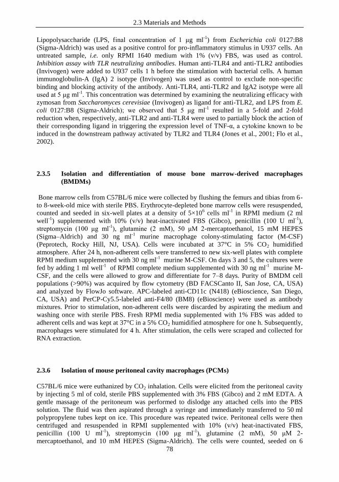
2.3 Materials and Methods
78
Lipopolysaccharide (LPS, final concentration of 1 μg ml-1
) from Escherichia coli 0127:B8
(Sigma-Aldrich) was used as a positive control for pro-inflammatory stimulus in U937 cells. An
untreated sample, i.e. only RPMI 1640 medium with 1% (v/v) FBS, was used as control.
Inhibition assay with TLR neutralizing antibodies. Human anti-TLR4 and anti-TLR2 antibodies
(Invivogen) were added to U937 cells 1 h before the stimulation with bacterial cells. A human
immunoglobulin-A (IgA) 2 isotype (Invivogen) was used as control to exclude non-specific
binding and blocking activity of the antibody. Anti-TLR4, anti-TLR2 and IgA2 isotype were all
used at 5 μg ml-1
. This concentration was determined by examining the neutralizing efficacy with
zymosan from Saccharomyces cerevisiae (Invivogen) as ligand for anti-TLR2, and LPS from E.
coli 0127:B8 (Sigma-Aldrich); we observed that 5 μg ml-1
resulted in a 5-fold and 2-fold
reduction when, respectively, anti-TLR2 and anti-TLR4 were used to partially block the action of
their corresponding ligand in triggering the expression level of TNF-α, a cytokine known to be
induced in the downstream pathway activated by TLR2 and TLR4 (Jones et al., 2001; Flo et al.,
2002).
2.3.5 Isolation and differentiation of mouse bone marrow-derived macrophages
(BMDMs)
Bone marrow cells from C57BL/6 mice were collected by flushing the femurs and tibias from 6-
to 8-week-old mice with sterile PBS. Erythrocyte-depleted bone marrow cells were resuspended,
counted and seeded in six-well plates at a density of 5×10⁶ cells ml
-1 in RPMI medium (2 ml
well-1
) supplemented with 10% (v/v) heat-inactivated FBS (Gibco), penicillin (100 U ml-1
),
streptomycin (100 μg ml-1
), glutamine (2 mM), 50 μM 2-mercaptoethanol, 15 mM HEPES
(Sigma–Aldrich) and 30 ng ml-1
murine macrophage colony-stimulating factor (M-CSF)
(Peprotech, Rocky Hill, NJ, USA). Cells were incubated at 37°C in 5% CO2 humidified
atmosphere. After 24 h, non-adherent cells were transferred to new six-well plates with complete
RPMI medium supplemented with 30 ng ml-1
murine M-CSF. On days 3 and 5, the cultures were
fed by adding 1 ml well-1
of RPMI complete medium supplemented with 30 ng ml-1
murine M-
CSF, and the cells were allowed to grow and differentiate for 7–8 days. Purity of BMDM cell
populations (>90%) was acquired by flow cytometry (BD FACSCanto II, San Jose, CA, USA)
and analyzed by FlowJo software. APC-labeled anti-CD11c (N418) (eBioscience, San Diego,
CA, USA) and PerCP-Cy5.5-labeled anti-F4/80 (BM8) (eBioscience) were used as antibody
mixtures. Prior to stimulation, non-adherent cells were discarded by aspirating the medium and
washing once with sterile PBS. Fresh RPMI media supplemented with 1% FBS was added to
adherent cells and was kept at 37°C in a 5% CO2 humidified atmosphere for one h. Subsequently,
macrophages were stimulated for 4 h. After stimulation, the cells were scraped and collected for
RNA extraction.
2.3.6 Isolation of mouse peritoneal cavity macrophages (PCMs)
C57BL/6 mice were euthanized by CO2 inhalation. Cells were elicited from the peritoneal cavity
by injecting 5 ml of cold, sterile PBS supplemented with 3% FBS (Gibco) and 2 mM EDTA. A
gentle massage of the peritoneum was performed to dislodge any attached cells into the PBS
solution. The fluid was then aspirated through a syringe and immediately transferred to 50 ml
polypropylene tubes kept on ice. This procedure was repeated twice. Peritoneal cells were then
centrifuged and resuspended in RPMI supplemented with 10% (v/v) heat-inactivated FBS,
penicillin (100 U ml-1
), streptomycin (100 μg ml-1
), glutamine (2 mM), 50 μM 2-
mercaptoethanol, and 10 mM HEPES (Sigma-Aldrich). The cells were counted, seeded on 6
2.3 Materials and Methods
79
well-plates at a density of 1×10⁶ cells ml
-1, and cultured for 24 h at 37°C in a 5% CO2 humidified
atmosphere. After 24 h, cells were washed three times with PBS to remove non-adherent cells,
and fresh RPMI media supplemented with 1% FBS was added to adherent cells. After 1 h
incubation at 37°C in a 5% CO2 humidified atmosphere, the macrophages were stimulated for 4
h. Subsequently, the cells were scraped and collected for RNA extraction.
2.3.7 Ethics statement
All experimental methods have been accepted by the National Animal Experiment Board
(Finland).
2.3.8 Preparation of RNA and reverse transcription
For both in vitro and ex vivo experiments, after incubating macrophages at 37°C for 4 h, the
supernatant was carefully removed from each well and the total cellular RNA was isolated from
the adhered cells with an RNeasy Mini Kit (Qiagen Inc., Valencia, CA, USA). Afterwards, RNA
concentration and purity was determined with a Nanodrop Spectrophotometer (ND-1000,
Thermo Fischer Scientific, Middletown, VA, USA) and reverse transcription to cDNA was
performed with the iScript Select cDNA Synthesis Kit (Bio-Rad Laboratories, Hercules, CA)
using the following thermal cycle: 5 min at 25°C, 30 min at 42°C, and 5 min at 85°C. Reverse
transcription quantitative PCR (RT-qPCR). The mRNA expression levels of cytokines were
analyzed with SYBR Green technology in RT-qPCR using SsoFast EvaGreen Supermix (Bio-
Rad Italia, Segrate, Italy) on a Bio-Rad CFX96 system according to the manufacturer's
instructions. The primers used are as follow (5‟3‟): for human macrophages, 18S rRNA
forward ATCCCTGAAAAGTTCCAGCA; 18S rRNA reverse CCCTCTTGGTGAGGTCAATG;
IL10 forward AGCAGAGTGAAGACTTTCTTTC; IL10 reverse
CATCTCAGACAAGGCTTGG; TNF-α forward TCAGCTCCACGCCATT; TNF-α reverse
CCCAGGCAGTCAGATCAT; COX2 forward CCCTTGGGTGTCAAAGGTAA; COX2 reverse
TGAAAAGGCGCAGTTTACG. For murine macrophages, 18S rRNA forward
GTGATCCCTGAGAAGTTCCAG; 18S rRNA reverse TCGATGTCTGCTTTCCTCAAC; IL10
forward GCCCAGAAATCAAGGAGCAT; IL10 reverse TGTAGACACCTTGGTCTTGGAG;
TNF-α forward CTTCTGTCTACTGAACTTCGGG; TNF-α reverse
CAGGCTTGTCACTCGAATTTTG; COX-2 forward TGTGCTGACATCCAGATCAT; COX-2
reverse GGCAAAGAATGCAAACATCA. All primers were designed using the Primer3Plus
software (http://www.bioinformatics.nl/cgi-bin/primer3plus/primer3plus.cgi), and the specificity
of the primers was tested with melting curves during amplification and by 1% TAE agarose gel
electrophoresis. Quantitative PCR was carried out according to the following cycle: initial hold at
96°C for 30 s and then 40 cycles at 96°C for 2 s and 60°C for 5 s. Gene expression was
normalized to the housekeeping gene coding for the18S rRNA. The amount of template cDNA
used for each sample was 12.5 ng. All results regarding cytokine mRNA expression levels are
reported as the fold of induction (FOI) in comparison to the control (namely unstimulated
macrophages), to which we attributed a FOI of 1.

2.3 Materials and Methods
80
2.3.9 Statistical analysis
The significance of the results was analyzed by unpaired heteroscedastic Student‟s t test with
two-tailed distribution. Differences of P < 0.05 were considered to be significant.
2.4 Results and Discussion
81
2.4 RESULTS AND DISCUSSION
2.4.1 Extraction, purification and analysis of MIMLh5 S-layer protein
The S-layer protein (SlpA) of L. helveticus MIMLh5 was extracted with LiCl and purified as
described in the methods section. We verified that the protocol based on LiCl-washes efficiently
removed most of the S-layer protein from the surface of L. helveticus cells. In fact, when we
loaded the LiCl-treated and LiCl-untreated bacterial cells resuspended in SDS-PAGE loading
buffer on a polyacrylamide gel, the electrophoretic profiles displayed a nearly complete
disappearance of the band corresponding to SlpA (Fig.2.4.1A). SDS-PAGE and RP-HPLC
analyses revealed that the protein was purified to apparent homogeneity (Fig. 2.4.1B, C and D).
Furthermore, we did not consider as possible the contamination of the S-layer preparation with
lipoteichoic acids (LTA), because it was previously demonstrate that LiCl treatments do not
remove LTA from L. helveticus cells (Mozes & Lortal, 1995). Mass spectrometry indicated a
molecular mass of 43853 Da, which is consistent with the average molecular mass calculated
from the amino acid sequence deduced from the slpA gene sequence (Fig. 2.4.1E).
Figure 2.4.1 Biochemical characterization of L. helveticus
MIMLh5 S-layer protein. A, SDS-PAGE profile of MIMLh5
cells before (lanes 1-3) and after (3-6) treatment with LiCl
solutions. B (Coomassie-blue staining) and C (silver
staining), SDS-PAGE profile of purified S-layer protein; 50
ng to 3 µg of protein have been loaded per well on the gel.
D, HPLC profile of purified S-layer protein. E, ESI-MS
spectrum of the S-layer protein and reconstructed mass
spectrum, indicating an average mass value of 43853 Da.

2.4 Results and Discussion
82
2.4.2 L. helveticus MIMLh5 reduces NF-κB activation in transfected Caco-2 cells
In this study, we investigated L. helveticus, a bacterium specialized for colonizing dairy
environments and traditionally used in the manufacture of Swiss-type and long-ripened Italian
cheeses. Phylogenetically, L. helveticus is closely related to L. acidophilus (the 16S rDNA
sequences of these bacteria differ by just 1.6%; Callanan et al., 2008), an intestinal bacterium
regularly used as a probiotic and thoroughly investigated for its ability to modulate host immune
responses (Konstantinov et al., 2008; Weiss et al., 2010; Maroof et al., 2012; Taverniti &
Guglielmetti, 2012). Studying the interaction of the dairy bacterium L. helveticus with the host
immune system could provide important insights into the health-modulating potential of a
bacterium that, in contrast to the intestinal/probiotic L. acidophilus, had its adaptive evolution in
dairy environments and, therefore, outside of a human/animal host. We performed a preliminary
immunological characterization of L. helveticus MIMLh5 using a reporter cell line obtained by
transfecting Caco-2 cells with a luciferase reporter vector induced by active NF-κB. Therefore,
we compared MIMLh5 with the well-described commercial probiotic strain, L. acidophilus
NCFM, which we included as a reference. In these experiments, L. helveticus MIMLh5 and L.
acidophilus NCFM cells from an overnight culture were incubated with the transfected Caco-2
layer for 4 h at 37°C at an MOI of 1000. Similar to what we observed in our previous studies on
the pharyngeal epithelial FaDu cell-line, L. helveticus MIMLh5 significantly reduced NF-κB
activation both at baseline and in the presence of IL-1β (Fig. 2.4.2); bioluminescence was, in fact,
always significantly lower when bacterial cells were added to the Caco-2 layer compared to the
unstimulated control. In both tested conditions, strain MIMLh5 was significantly more effective
in the immunomodulatory activity than L. acidophilus NCFM (Fig. 2.4.2).
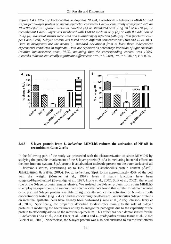
2.4 Results and Discussion
83
Figure 2.4.2 Effect of Lactobacillus acidophilus NCFM, Lactobacillus helveticus MIMLh5 and
its purified S-layer protein on human epithelial colorectal Caco-2 cells stably transfected with an
NF-κB/luciferase reporter vector at baseline (A) or stimulated with 2 ng ml-1
of IL-1β (B). A
recombinant Caco-2 layer was incubated with EMEM medium only (A) or with the addition of
IL-1β (B). Bacterial strains were used at a multiplicity of infection (MOI) of 1000 (bacterial cells
per Caco-2 cell). S-layer protein was tested at two different concentrations (100 and 10 μg ml-1
).
Data in histograms are the means (+ standard deviations) from at least three independent
experiments conducted in triplicate. Data are reported as percentage variation of light emission
(relative luminescence units, RLU), assuming that the corresponding control was 100%.
Asterisks indicate statistically significant differences: ***, P < 0.001; **, P < 0.01; *, P < 0.05.
2.4.3 S-layer protein from L. helveticus MIMLh5 reduces the activation of NF-κB in
recombinant Caco-2 cells
In the following part of the study we proceeded with the characterization of strain MIMLh5 by
studying the possible involvement of the S-layer protein (SlpA) in mediating bacterial effects on
the host immune system. SlpA protein is an abundant molecule present on the outer surface of all
L. helveticus strains, constituting up to 15% of total Lactobacillus protein content (Åvall-
Jääskeläinen & Palva, 2005). For L. helveticus, SlpA forms approximately 45% of the cell
wall dry weight (Messner et al., 1997). Even if many functions have been
suggested/hypothesized (Beveridge et al., 1997; Horie et al., 2002; Smit et al., 2002), the actual
role of the S-layer protein remains elusive. We isolated the S-layer protein from strain MIMLh5
to employ in experiments on recombinant Caco-2 cells. We found that similar to whole bacterial
cells, purified S-layer protein was able to significantly reduce the activation of NF-κB at both
concentrations tested (Fig. 2.4.2). Studies concerning the effects of Lactobacillus S-layer proteins
on intestinal epithelial cells have already been performed (Frece et al., 2005; Johnson-Henry et
al., 2007). Specifically, the properties described to date refer mainly to the role of S-layer
proteins in mediating the bacterium‟s ability to antagonize pathogens due to the capability of the
protein to efficiently adhere to the intestinal epithelium. This effect has been demonstrated for the
L. helveticus (Kos et al., 2003; Frece et al., 2005) and L. acidophilus strains (Smit et al., 2002;
Buck et al., 2005). Nonetheless, the S-layer protein was also demonstrated to exert direct effects
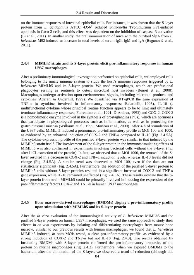
2.4 Results and Discussion
84
on the immune responses of intestinal epithelial cells. For instance, it was shown that the S-layer
protein from L. acidophilus ATCC 4356T reduced Salmonella Typhimurium FP1-induced
apoptosis in Caco-2 cells, and this effect was dependent on the inhibition of caspase-3 activation
(Li et al., 2011). In another study, the oral immunization of mice with the purified SlpA from L.
helveticus M92 induced an increase in total levels of serum IgG, IgM and IgA (Beganović et al.,
2011).
2.4.4 MIMLh5 strain and its S-layer protein elicit pro-inflammatory responses in human
U937 macrophages
After a preliminary immunological investigation performed on epithelial cells, we employed cells
belonging to the innate immune system to study the host‟s immune responses triggered by L.
helveticus MIMLh5 and its S-layer protein. We used macrophages, which are professional
phagocytes serving as sentinels to detect microbial host invaders (Benoit et al., 2008).
Macrophages undergo activation upon environmental signals, including microbial products and
cytokines (Aderem & Underhill, 1999). We quantified via RT-qPCR the gene expression of
TNF-α (a cytokine involved in inflammatory responses; Belardelli, 1995), IL-10 (a
multifunctional cytokine whose principal routine function appears to be to limit and ultimately
terminate inflammatory responses; Fiorentino et al., 1991; D‟Andrea, 1993) and COX-2. COX-2
is a homodimeric enzyme involved in the synthesis of prostaglandins (PGs), which are hormones
that participate in physiological processes such as inflammation, as well as in protecting the
gastrointestinal mucosa (Williams et al., 1999; Morteau et al., 2000). After 4 h of stimulation of
the U937 cells, MIMLh5 induced a pronounced pro-inflammatory profile at MOI 100 and 1000,
as evidenced by an enhanced induction of COX-2 and TNF-α compared to IL-10 (Fig. 2.4.5A).
The cytokine-expression profile of the purified S-layer protein was similar to that induced by the
MIMLh5 strain itself. The involvement of the S-layer protein in the immunostimulating effects of
MIMLh5 was also confirmed in experiments involving bacterial cells without the S-layer (i.e.,
after LiCl-extraction of the protein). In fact, we observed that at MOI 1000, the removal of the S-
layer resulted in a decrease in COX-2 and TNF-α induction levels, whereas IL-10 levels did not
change (Fig. 2.4.5A). A similar trend was observed at MOI 100, even if the data are not
statistically significant (Fig. 2.4.5A). Furthermore, the addition of the purified S-layer protein to
MIMLh5 cells without S-layer proteins resulted in a significant increase of COX-2 and TNF-α
gene expression, while IL-10 remained unaffected (Fig. 2.4.5A). These results indicate that the S-
layer protein from strain MIMLh5 could be primarily involved in inducing the expression of the
pro-inflammatory factors COX-2 and TNF-α in human U937 macrophages.
2.4.5 Bone marrow-derived macrophages (BMDMs) display a pro-inflammatory profile
upon stimulation with MIMLh5 and its S-layer protein
After the in vitro evaluation of the immunological activity of L. helveticus MIMLh5 and the
purified S-layer protein on human U937 macrophages, we used the same approach to study their
effects in ex vivo experiments by isolating and differentiating macrophages from mouse bone
marrow. Similar to our previous results with human macrophages, we found that L. helveticus
MIMLh5 induced, at both MOIs tested, a clear pro-inflammatory profile, as evidenced by a
strong induction of COX-2 and TNF-α but not IL-10 (Fig. 2.4.3). The results obtained by
incubating BMDMs with S-layer protein confirmed the pro-inflammatory properties of the
protein on murine macrophages (Fig. 2.4.3). Furthermore, when we exposed BMDMs to the
bacterium after the elimination of the S-layer, we observed a trend of reduction (although this

2.4 Results and Discussion
85
result was not statistically significant) of TNF-α levels for MIMLh5 (particularly at the highest
MOI tested), whereas the absence of the protein led to a slight increase of IL-10 levels (Fig.
2.4.3).
Thus, both in experiments on human U937 macrophages and on murine BMDMs, MIMLh5
exerted pro-inflammatory effects by inducing high levels of TNF-α and COX-2 and lower levels
of IL-10. Interestingly, the purified SlpA extracted from L. helveticus MIMLh5, according to the
expression levels of TNF-a and COX2, seems to be responsible for the pro-inflammatory
behavior of the whole bacterium, as also confirmed by the experiments performed with SlpA-
depleted MIMLh5 cells (Fig. 2.4.3 and Fig. 2.4.5A).
The ability to induce pro-inflammatory cytokines has already been shown in lactic acid bacteria
(LAB) (Christensen et al., 2002; Morita et al., 2002; Peridigón et al., 2002; Ongol et al., 2008).
Nonetheless, such ability of LAB should not be considered detrimental. In fact, even though
cytokines belonging to the TNF-α superfamily are connected to the occurrence of inflammatory
diseases (Kwon et al., 2010), they have also been shown to participate in the rejection of tumors
and infections (Yasutake et al., 1999; Dinarello, 2003; Wajant et al., 2003). Furthermore, the
induction of TNF-α could be important for the initiation of cross-talk among immune cells
without causing any inflammation or detrimental effects (Galdeano et al., 2007). In addition, a
transient inflammatory state could aid host defense. As recently suggested, differences among
pathogenic, probiotic and commensal microorganisms lie in the magnitude of the immune
response evoked, which can be defined as strong, intermediate or homeostatic (Lebeer et al.,
2010).
Similarly, the up-regulation of COX-2 induced by LAB has been demonstrated both in vitro
(Otte et al., 2009; Khailova et al., 2010) and in vivo (Khailova et al., 2010; Kwon et al., 2010).
The pro-inflammatory role of COX-2, however, has recently been questioned. In fact, it has been
proposed that COX-2 can induce the resolution of inflammation (Wallace, 2006) and an
immunosuppressive phenotype in T-cells (Newberry et al., 1999) through the activity of PGs. In
addition, it has also been shown that a rapid COX-2 up-regulation in response to injury or
inflammation helps to restore mucosal integrity (Tan et al., 2000). In this context, the effects of
MIMLh5 and SlpA on COX-2 expression could potentially be beneficial for the host‟s mucosa.
The ability of specific cell wall components to drive the immune response elicited by LAB has
been rarely described (de Ambrosini et al., 1996; Tejada-Simon & Pestka, 1999). Concerning S-
layer proteins, Konstantinov and colleagues (2008) demonstrated that SlpA from L. acidophilus
NCFM mediates the interaction of the bacterium with human monocyte-derived dendritic cells
(DCs) by specifically binding to the ligand of the dendritic cell-specific intercellular adhesion
molecule (ICAM)-3-grabbing non-integrin (DC-SIGN). Even if the purified SlpA protein of
NCFM did not affect DCs maturation, it was responsible for the anti-inflammatory cytokine
profile observed for L. acidophilus NCFM because the protein induced higher levels of IL-10 in
the presence of LPS when compared to SlpA or LPS alone.

2.4 Results and Discussion
86
Figure 2.4.3 Quantitative analysis of cytokine gene expression in murine bone marrow-derived
macrophages (BMDMs) after 4 h stimulation with L. helveticus MIMLh5 and its S-layer protein.
Expression levels of TNF-α, IL-10 and COX-2 are indicated as the fold change of induction
(FOI) relative to the control (unstimulated BMDMs), which was set at a value of 1. LPS was used
as a positive control at a concentration of 1 μg ml-1
. S-layer protein was tested at a concentration
of 10 μg ml-1
. MIMLh5 was used at MOIs 1000 and 100. MIMLh5 w/o Slay: MIMLh5 cells after
removal of the S-layer protein by LiCl-extraction. Presented data are the means of measurements
(+ standard deviations) for a result representative of three independent experiments. Asterisks
indicate statistically significant differences compared to results for the corresponding control (*:
P < 0.05).
In comparison to the SlpA of L. acidophilus NCFM, our data suggest that the S-layer protein
isolated from L. helveticus MIMLh5 exhibits a more pronounced pro-inflammatory behavior.
Although the S-layer proteins of L. acidophilus NCFM and L. helveticus MIMLh5 are highly
similar (73% identity, 83% positivity; Data not shown), it has been suggested that even small
differences in surface proteins could alter the type of immune responses elicited by LAB
(Vinderola et al., 2004; Lebeer et al., 2010). Nonetheless, the most plausible explanation for the
different immunological behavior observed between MIMLh5 and NCFM SlpAs (Konstantinov
et al., 2008) could reside in the different cell models utilized.
When we tested strain L. helveticus MIMLh5 and its SlpA on macrophages, we observed an
immunological effect (stimulation of the pro-inflammatory factors) that was substantially
different from what was noted for epithelial cells (i.e., inhibition of NF-κB activation). These
results are not surprising, considering that macrophages are immune cells that can express class
and number of receptors on their cell surface considerably different from epithelial cells. This
fact might result in recognition through diverse mechanisms and activation of alternative
signaling pathways. Moreover, it is plausible (and desirable) that a potential probiotic candidate
and its cell components do not trigger any inflammatory response at the host epithelium. This
type of non-inflammatory interaction can be assimilated to a mechanism of oral tolerance, which,
in vivo, is due to the natural cohabitation of epithelial cells with commensal and/or food-
associated bacteria. In contrast, when bacteria come in direct contact with immune cells, for
instance, when they cross the epithelial barrier following loss of epithelial integrity, the immune
system is typically alerted through pro-inflammatory signaling.
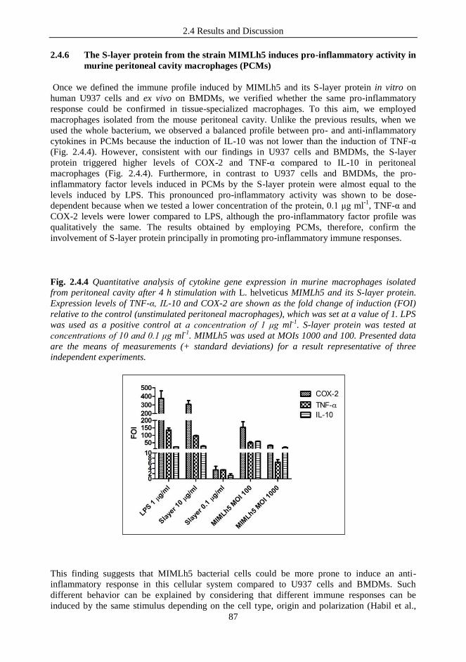
2.4 Results and Discussion
87
2.4.6 The S-layer protein from the strain MIMLh5 induces pro-inflammatory activity in
murine peritoneal cavity macrophages (PCMs)
Once we defined the immune profile induced by MIMLh5 and its S-layer protein in vitro on
human U937 cells and ex vivo on BMDMs, we verified whether the same pro-inflammatory
response could be confirmed in tissue-specialized macrophages. To this aim, we employed
macrophages isolated from the mouse peritoneal cavity. Unlike the previous results, when we
used the whole bacterium, we observed a balanced profile between pro- and anti-inflammatory
cytokines in PCMs because the induction of IL-10 was not lower than the induction of TNF-α
(Fig. 2.4.4). However, consistent with our findings in U937 cells and BMDMs, the S-layer
protein triggered higher levels of COX-2 and TNF-α compared to IL-10 in peritoneal
macrophages (Fig. 2.4.4). Furthermore, in contrast to U937 cells and BMDMs, the pro-
inflammatory factor levels induced in PCMs by the S-layer protein were almost equal to the
levels induced by LPS. This pronounced pro-inflammatory activity was shown to be dose-
dependent because when we tested a lower concentration of the protein, 0.1 μg ml-1
, TNF-α and
COX-2 levels were lower compared to LPS, although the pro-inflammatory factor profile was
qualitatively the same. The results obtained by employing PCMs, therefore, confirm the
involvement of S-layer protein principally in promoting pro-inflammatory immune responses.
Fig. 2.4.4 Quantitative analysis of cytokine gene expression in murine macrophages isolated
from peritoneal cavity after 4 h stimulation with L. helveticus MIMLh5 and its S-layer protein.
Expression levels of TNF-α, IL-10 and COX-2 are shown as the fold change of induction (FOI)
relative to the control (unstimulated peritoneal macrophages), which was set at a value of 1. LPS
was used as a positive control at a concentration of 1 μg ml-1
. S-layer protein was tested at
concentrations of 10 and 0.1 μg ml-1
. MIMLh5 was used at MOIs 1000 and 100. Presented data
are the means of measurements (+ standard deviations) for a result representative of three
independent experiments.
This finding suggests that MIMLh5 bacterial cells could be more prone to induce an anti-
inflammatory response in this cellular system compared to U937 cells and BMDMs. Such
different behavior can be explained by considering that different immune responses can be
induced by the same stimulus depending on the cell type, origin and polarization (Habil et al.,
2.4 Results and Discussion
88
2011). Moreover, because BMDMs and U937 cells have been maturated in vitro, it is plausible
that they are less physiological than PCMs, and that they could not possess all the features of
matured macrophages. It could also be speculated that macrophages from the peritoneal cavity
can present different phenotypes compared to other macrophage populations due to the activity
exerted on PCMs by the intestinal microbiota, which can promote a more tolerogenic activity
based on differential expression of pattern-recognition receptors (PRRs). Accordingly, the
induction of an anti-inflammatory cytokine profile by lactobacilli has been observed in murine
PCMs (Marcinkiewicz et al., 2007). Nonetheless, in comparison to whole bacterial cells, the pro-
inflammatory effect of S-layer protein was particularly evident in PCMs. In fact, for MIMLh5
cells, the TNF-α/IL-10 ratios were 0.84 at MOI 100, 0.35 at MOI 1000, and approximately 3.7
when purified SlpA protein was used as a stimulus.
2.4.7 Toll-like receptor (TLR)-2 is involved in the recognition of the S-layer protein from
strain MIMLh5 in human U937 cells
To gain information about the possible signaling pathways underlying the immune response
activated by the S-layer protein isolated from L. helveticus MIMLh5, we evaluated whether
TLR2 and TLR4 could be involved in the recognition of this protein. To this aim, we used
neutralizing antibodies (Abs) on U937 cells to partially block the ability of TLR2 and TLR4 to
bind ligands prior to the addition of the S-layer protein. We found that TLR2 and, to a lesser
extent, TLR4 are involved in mediating the pro-inflammatory response elicited by S-layer protein
because the treatments with Abs significantly reduced the S-layer-induced activation of TNF-α
and COX-2, but not IL-10 (Fig. 2.4.5B). TLR2 is a receptor that was previously shown to be
involved in mediating the immunological effects of LAB (Konstantivov et al., 2008; Zeuthen et
al., 2008; Vizoso et al., 2009; Weiss et al., 2010). The observed differences when anti-TLR2 was
used, either alone or in combination with anti-TLR4, were statistically significant compared to
U937 cells stimulated with S-layer protein in the presence of Immunoglobulin-A2 isotype
(IgA2), which was used as a control for nonspecific blocking activity. From these data, we can
hypothesize that the S-layer, which is the outermost part of the bacterial cell wall, might come in
contact first with host cells, mediate bacterial recognition, and therefore play a key role in
triggering the immune response.
2.4.8 S-layer protein modulates the pro-inflammatory response triggered by LPS in
human U937 macrophages
In a final set of experiments, we tested the effects of the S-layer protein on U937 cells in
presence of LPS. Interestingly, we observed that the presence of the S-layer protein induced a
clear reduction in the pro-inflammatory cytokine TNF-α, compared with the levels induced by
LPS alone (Fig. 2.4.5C). These results suggest that although the S-layer protein and LPS
presented similar pro-inflammatory effects, the simultaneous presence of both stimuli did not
result in an additive or synergistic effect on the pro-inflammatory response. Rather, the addition
of the S-layer protein induced a reduction of the stimulating activity of LPS. In our experiments
on macrophages, the SlpA of MIMLh5 induced a cytokine profile qualitatively similar to that
evoked by LPS; nonetheless, the cytokine induction levels on human U937 cells and murine
BMDMs were lower for the S-layer protein than for LPS. In peritoneal macrophages, the S-layer
protein triggered a cytokine induction similar to LPS only when ten-times more S-layer protein
(10 µg) was present compared to LPS (1 µg).
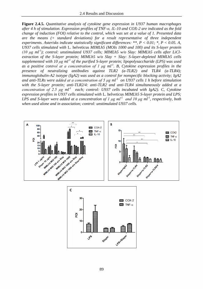
2.4 Results and Discussion
89
Figure 2.4.5. Quantitative analysis of cytokine gene expression in U937 human macrophages
after 4 h of stimulation. Expression profiles of TNF-α, IL-10 and COX-2 are indicated as the fold
change of induction (FOI) relative to the control, which was set at a value of 1. Presented data
are the means (+ standard deviations) for a result representative of three independent
experiments. Asterisks indicate statistically significant differences: **, P < 0.01; *, P < 0.05. A,
U937 cells stimulated with L. helveticus MIMLh5 (MOIs 1000 and 100) and its S-layer protein
(10 μg ml-1
); control: unstimulated U937 cells; MIMLh5 w/o Slay: MIMLh5 cells after LiCl-
extraction of the S-layer protein; MIMLh5 w/o Slay + Slay: S-layer-depleted MIMLh5 cells
supplemented with 10 μg ml-1
of the purified S-layer protein; lipopolysaccharide (LPS) was used
as a positive control at a concentration of 1 μg ml-1
. B, Cytokine expression profiles in the
presence of neutralizing antibodies against TLR2 (a-TLR2) and TLR4 (a-TLR4);
immunoglobulin-A2 isotype (IgA2) was used as a control for nonspecific blocking activity; IgA2
and anti-TLRs were added at a concentration of 5 μg ml-1
on U937 cells 1 h before stimulation
with the S-layer protein; anti-TLR2/4: anti-TLR2 and anti-TLR4 simultaneously added at a
concentration of 2.5 μg ml-1
each; control: U937 cells incubated with IgA2). C, Cytokine
expression profiles in U937 cells stimulated with L. helveticus MIMLh5 S-layer protein and LPS;
LPS and S-layer were added at a concentration of 1 μg ml-1
and 10 μg ml-1
, respectively, both
when used alone and in association; control: unstimulated U937 cells.

2.4 Results and Discussion
90
These data indicate that the SlpA of MIMLh5 is not a pro-inflammatory stimulus as potent as
LPS. We could hypothesize that the S-layer alone, in basal conditions, may exert a mild
stimulatory effect on the immune system. However, in the presence of an inflammatory stimulus,
such as LPS, the MIMLh5 S-layer could act as an immune modulator, representing a potential
protective element against the septic shock caused by LPS, as already observed in murine models
for certain probiotic bacteria (Arribas et al., 2009a;b).
Since S-layer protein and LPS interact with different TLRs, the observed modulatory effect of
the S-layer protein should not be attributed to a competition for the same host cells‟ receptors
but, more likely, to an activation of different immune response pathways.
2.5 Conclusions
91
2.5 CONCLUSIONS
The cross-talk between host and intestinal/probiotic bacteria principally relies on the capacity of
host cells to recognize specific bacterial components or products, thus giving rise to responses
that most frequently involve the mucosa-associated lymphoid tissue (MALT) and, therefore, the
immune system (Adams, 2010; Taverniti & Guglielmetti, 2011). Specifically, cell surface
components of commensal or food/probiotic bacteria, known as microbe-associated molecular
patterns (MAMPs), can be identified by pattern recognition receptors (PRRs) on host‟s cells
constituting the innate immune system, resulting in the activation of immune responses. The
molecular mechanisms of such immune modulations are largely unknown. Therefore, the
immunological characterization of single bacterial components, according to the strategy
undertaken during this study, represents a reductionist approach of key importance for the
elucidation of host–microbial interplays and bacterial modes of action that result in the immune
modulation. In this study, we characterized the dairy strain Lactobacillus helveticus MIMLh5 and
its surface layer protein (SlpA) using in vitro and ex vivo analyses. We found that MIMLh5 and
SlpA exert anti-inflammatory effects by reducing the activation of NF-κB on the intestinal
epithelial Caco-2 cell line. On the contrary, MIMLh5 and SlpA act as stimulators of the innate
immune system by triggering the expression of pro-inflammatory factors (Taverniti et al., 2012).
These results must not be surprising, since several studies reveal that probiotics exert stimulatory
effects rather than suppressory effects on innate immunity. This mechanism is similar to a
“physiologic inflammation”, like the one induced by the commensal flora, that can be useful for
immune system development and for combating pathogens (Pagnini et al., 2010).
Furthermore, our study supports the concept that the viability of bacterial cells is not always
essential to exert immunomodulatory effects (Adams, 2010). In fact, dead or inactivated bacterial
cells, or even their single molecular components, might also be effective in exerting beneficial
immunostimulating properties, thus permitting the development of safer therapies for the
treatment of specific diseases. This approach has recently been defined as paraprobiotic
intervention (Taverniti & Guglielmetti, 2011). In addition, a deeper understanding of the
molecular mechanisms of cross-talk between bacteria and the host organism‟s system is of great
importance to better define the benefits and potential risks associated with the administration of
probiotic therapies (Besselink et al., 2008).
Moreover, the possible medical and clinical uses of the S-layer protein are particularly intriguing,
especially in light of the physicochemical properties of this molecule. The S-layer proteins are
crystalline arrays of subunits forming a highly regular crystalline structure that has the capacity
to reassemble spontaneously in suspension at the liquid-air interface, at solid surfaces, at floating
lipid monolayers, on liposomes, and on nanocapsules (Sleytr et al., 2003; Sleytr et al., 2007).
These features could support the use of the S-layer protein as a bioactive coating material, as a
matrix for the immobilization and delivery of different molecules, and as a template for the
formation of regularly arranged bioactive nanoparticles (Pum & Sleytr, 1999). Future efforts will
explore the feasibility of such applications.

2.6 References
92
2.6 REFERENCES
Adams CA., 2010, The probiotic paradox: live and dead cells are biological response
modifiers. Nutr Res Rev 23:37-46.
Aderem A, Underhill DM, 1999, Mechanisms of phagocytosis in macrophages. Annu
Rev Immunol 17:593-623.
Agave Biosystems, 2012, S-layer catalog. Agave Biosystems, Ithaca, NY
Angulo L et al., 1993, Microflora present in kefir grains of the Galician region (north-
west of Spain). J Dairy Res 60:263-267.
Antikainen J et al., 2002, Domains in the S-layer protein CbsA of Lactobacillus
crispatus involved in adherence to collagens, laminin and lipoteichoic acids and in self-
assembly. Mol Microbiol 46:381-394.
Arribas B et al., 2009a, A probiotic strain of Escherichia coli, Nissle 1917, given orally
exerts local and systemic anti-inflammatory effects in lipopolysaccharide-induced sepsis
in mice. Br J Pharmacol 157:1024-1033.
Arribas B et al., 2009b, Evaluation of the preventative effects exerted by Lactobacillus
fermentum in an experimental model of septic shock induced in mice. Br J Nutr 101:51-
58.
Åvall-Jääskeläinen S et al., 2003, Surface display of the receptor-binding region of the
Lactobacillus brevis S-layer protein in Lactococcus lactis provides nonadhesive
lactococci with the ability to adhere to intestinal epithelial cells. Appl Environ Microbiol
69: 2230-2236.
Åvall-Jääskeläinen S, Palva A, 2005, Lactobacillus surface layers and their applications.
FEMS Microbiol Rev 29:511-529.
Beganovic´ J et al., 2011, Functionality of the S-layer protein from the probiotic strain
Lactobacillus helveticus M92. Antonie Van Leeuwenhoek 100:43-53.
Belardelli F. 1995, Role of interferons and other cytokines in the regulation of the
immune response. APMIS 103:161-179.
Benoit M et al., 2008, Macrophage polarization in bacterial infections. J Immunol
181:3733-3739.
Besselink MG et al., 2008, Probiotic prophylaxis in predicted severe acute pancreatitis:
a randomised, doubleblind, placebo-controlled trial. Dutch Acute Pancreatitis Study
Group. Lancet 371:651- 659.
Beveridge TJ et al., 1997, Functions of S-layers. FEMS Microbiol Rev 20:99-149.
Buck BL et al., 2005, Functional analysis of putative adhesion factors in Lactobacillus
acidophilus NCFM. Appl Environ Microbiol 71:8344–8351.
Callanan M et al., 2008, Genome sequence of Lactobacillus helveticus, an organism
distinguished by selective gene loss and insertion sequence element expansion. J
Bacteriol 190:727-735.
Chapot-Chartier MP et al., 2010, Cell surface of Lactococcus lactis is covered by a
protective polysaccharide pellicle. J Biol Chem 285:10464-10471.

2.6 References
93
Chen X et al., 2007, The S-layer proteins of Lactobacillus crispatus strain ZJ001 is
responsible for competitive exclusion against Escherichia coli O157:H7 and Salmonella
typhimurium. Int J Food Microbiol 115: 307-312.
Christensen HR et al., 2002, Lactobacilli differentially modulate expression of cytokines
and maturation surface markers in murine dendritic cells. J Immunol 168:171-178.
D‟Andrea A et al., 1993, Interleukin 10 (IL-10) inhibits human lymphocyte interferon
gamma-production by suppressing natural killer cell stimulatory factor/IL-12 synthesis
in accessory cells. J Exp Med 178:1041-1048.
De Ambrosini VM et al., 1996, Chemical composition of the cell wall of lactic acid
bacteria and related species. Chem Pharm Bull 44:2263–2267.
De Leeuw E et al., 2006, Binding characteristics of the Lactobacillus brevis ATCC 8287
surface layer to extracellular matrix proteins. FEMS Microbiol Lett 260:210-215.
Dinarello CA. 2003, Anti-cytokine therapeutics and infections. Vaccine 21:S24-S34.
Fiorentino DF et al., 1991, IL-10 inhibits cytokine production by activated
macrophages. J Immunol 147:3815-3822.
Flo TH et al., 2002, Involvement of Toll-like receptor (TLR) 2 and TLR4 in cell
activation by mannuronic acid polymers. J Biol Chem 277:35489-35495.
Frece J et al., 2005, Importance of S-layer proteins in probiotic activity of Lactobacillus
acidophilus M92. J Appl Microbiol 98:285-292.
Galdeano CM et al., 2007, Proposed model: mechanisms of immunomodulation induced
by probiotic bacteria. Clin Vaccine Immunol 14:485-492.
Garrote GL et al., 2004, Lactobacilli isolated from kefir grains: Evidence of the
presence of S-layer proteins. J Dairy Res 71:222-230.
Gatti M et al., 2005, Heterogeneity of putative surface layer proteins in Lactobacillus
helveticus. Appl Environ Microbiol 71:7582-7588.
Gobbetti M et al., 2010, Functional microorganisms for functional food quality. Crit
Rev Food Sci Nutr 50:716-727.
Golowczyc MA et al., 2007, Protective action of Lactobacillus kefir carrying S-layer
protein against Salmonella enterica serovar Enteritidis. Int J Food Microbiol 118:264-
273.
Guglielmetti S et al., 2008, Implication of an outer surface lipoprotein in adhesion of
Bifidobacterium bifidum to Caco-2 cells. Appl Environ Microbiol 74:4695-4702.
Guglielmetti S et al., 2010, Oral bacteria as potential probiotics for the pharyngeal
mucosa. Appl Environ Microbiol 76:3948 –3958.
Habil N et al., 2011, Probiotic bacterial strains differentially modulate macrophage
cytokine production in a strain-dependent and cell subset-specific manner. Benef
Microbes 2:283-293.
Harris P, Ralph P, 1985, Human leukemic models of myelomonocytic development: a
review of the HL-60 and U937 cell lines. J Leukoc Biol 37:407-422.
2.6 References
94
Horie M et al., 2002, Inhibition of the adherence of Escherichia coli strains to basement
membrane by Lactobacillus crispatus expressing an S-layer. J Appl Microbiol 92:396-
403.
Johnson-Henry KC et al., 2007, Surface-layer protein extracts from Lactobacillus
helveticus inhibit enterohaemorrhagic Escherichia coli O157:H7 adhesion to epithelial
cells. Cell Microbiol 9:356-367.
Jones BW et al., 2001, Differential roles of Toll-like receptors in the elicitation of
proinflammatory responses by macrophages. Ann Rheum Dis 60:iii6-iii12.
Kataria J et al., 2009, Probiotic microbes: do they need to be alive to be beneficial? Nutr
Rev 67:546-550.
Khailova L et al., 2010, Bifidobacterium bifidum reduces apoptosis in the intestinal
epithelium in necrotizing enterocolitis. Am J Physiol Gastrointest Liver Physiol
299:G1118-G1127.
Konstantinov SR et al., 2008, S layer protein A of Lactobacillus acidophilus NCFM
regulates immature dendritic cell and T cell functions. Proc Natl Acad Sci USA
105:19474-19479.
Kos B et al., 2003, Adhesion and aggregation ability of probiotic strain Lactobacillus
acidophilus M92. J Appl Microbiol 94:981-987.
Kwon B et al., 2003, Involvement of tumor necrosis factor receptor superfamily
(TNFRSF) members in the pathogenesis of inflammatory diseases. Exp Mol Med 35:8-
16.
Kwon HK et al., 2010, Generation of regulatory dendritic cells and CD4_Foxp3_T cells
by probiotics administration suppresses immune disorders. Proc Natl Acad Sci USA
107:2159-2164.
Lebeer S et al., 2010, Host interactions of probiotic bacterial surface molecules:
comparison with commensals and pathogens. Nat Rev Microbiol 8:171-184.
Li P et al., 2011, Lactobacillus acidophilus S-layer protein-mediated inhibition of
Salmonella-induced apoptosis in Caco-2 cells. Biochem Biophys Res Commun
409:142-147.
Lortal S et al., 1992, S-layer of Lactobacillus helveticus ATCC 12046: isolation,
chemical characterization and re-formation after extraction with lithium chloride. J Gen
Microbiol 138:611-618.
Marcinkiewicz J et al., 2007, Differential inflammatory mediator response in vitro from
murine macrophages to lactobacilli and pathogenic intestinal bacteria. Int J Exp Pathol
88:155-164.
Maroof H et al., 2012. Lactobacillus acidophilus could modulate the immune response
against breast cancer in murine model. J Clin Immunol doi:10.1007/s10875-012-9708-x.
Marteau P, Shanahan F, 2003, Basic aspects and pharmacology of probiotics: an
overview of pharmacokinetics, mechanisms of action and side-effects. Best Pract Res
Clin Gastroenterol 17:725-740.
Matsuguchi T et al., 2003, Lipoteichoic acids from Lactobacillus strains elicit strong
tumor necrosis factor alpha-inducing activities in macrophages through Toll-like
receptor 2. Clin Diagn Lab Immunol 10:259-266.

2.6 References
95
Messner P et al., 1997, Biochemistry of S-layers. FEMS Microbiol Rev 20:25-46.
Mobili P et al., S-layers in lactobacilli: structural characteristics and putative role in
surface and probiotic properties of whole bacteria. In Current Research, Technology and
Education Topics in Applied Microbiology and Microbial Biotechnology, Méndez-Vilas
A, ed., Formatex Research Center 2010, pp. 1224-1234.
Morita H et al., 2002, Cytokine production by the murine macrophage cell line J774.1
after exposure to lactobacilli. Biosci Biotechnol Biochem 66:1963-1966.
Morteau O et al., 2000, Impaired mucosal defense to acute colonic injury in mice
lacking cyclooxygenase-1 or cyclooxygenase-2. J Clin Invest 105:469-478.
Mozes N, Lortal S, 1995, X-ray photoelectron spectroscopy and biochemical analysis of
the surface of Lactobacillus helveticus ATCC 12046. Microbiology 141:11-19.
Nahra R, Dellinger RP, 2008, Targeting the lipopolysaccharides: still a matter of
debate? Curr Opin Anaesthesiol 21:98-104.
Newberry RD et al., 1999, Cyclooxygenase-2-dependent arachidonic acid metabolites
are essential modulators of the intestinal immune response to dietary antigen. Nat Med
5:900-906.
Ongol MP et al., 2008, Potential of selected strains of lactic acid bacteria to induce a
Th1immune profile. Biosci Biotechnol Biochem 72:2847-2857.
Opal SM, 2010, Endotoxins and other sepsis triggers. Contrib Nephrol 167:14-24.
Otte JM et al., 2009, Probiotics regulate the expression of COX-2 in intestinal epithelial
cells. Nutr Cancer 61:103-113.
Pagnini C et al., 2010, Probiotics promote gut health through stimulation of epithelial
innate immunity. Proc Natl Acad Sci USA 107:454-459.
Perdigón G et al., 2002, Interaction of lactic acid bacteria with the gut immune system.
Eur J Clin Nutr 56:S21–S26.
Pum D, Sleytr UB, 1999, The application of bacterial S-layers in molecular
nanotechnology. Trends Biotechnol 17:8-12.
Radzun HJ et al., 1983, Monocytic origin of the human hematopoietic cell line U-937
and its convertibility to macrophages evidenced by isoenzyme mapping. Int J Cancer
31:181-186.
Salminen, S et al., 1999, Probiotics: how should they be defined. Trends Food Sci
Technol 10:107-110.
Sára M, Sleytr UB, 2000, S-layer proteins. J Bacteriol 182:859-868.
Sleytr UB, 1997, Basic and applied S-layer research: An overview. FEMS Microbiol
Rev 20:5-12.
Sleytr UB, Beveridge TJ, 1999, Bacterial S-layers. Trends Microbiol 7:253-260.
Sleytr UB et al., 2003, Crystallization of S-layer protein lattices on surfaces and
interfaces. Prog Org Coat 47:279-287.
Sleytr UB et al., 2007, S-layers as a basic building block in a molecular construction kit.
FEBS J 274:323-334.

2.6 References
96
Smit E et al., 2001, The S-layer protein of Lactobacillus acidophilus ATCC 4356:
identification and characterisation of domains responsible for S-protein assembly and
cell wall binding. J Mol Biol 305:245-257.
Smit E et al., 2002, Structural and functional analysis of the S-layer protein
crystallisation domain of Lactobacillus acidophilus ATCC 4356: evidence for protein-
protein interaction of two subdomains. J Mol Biol 324:953-964.
Stuknyte M et al., 2011, Potential immunomodulatory activity of bovine casein
hydrolysates produced after digestion with proteinases of lactic acid bacteria. Int Dairy J
21:763-769.
Sundstrom C, Nilsson K, 1976, Establishment and characterization of a human
histiocytic lymphoma cell line (U-937). Int J Cancer 17:565-577.
Tallon R et al., 2007, Strain- and matrix-dependent adhesion of Lactobacillus plantarum
is mediated by proteinaceous bacterial compounds. J Appl Microbiol 102:442-451.
Tan XD et al., 2000, Prostanoids mediate the protective effect of trefoil factor 3 in
oxidant-induced intestinal epithelial cell injury: role of cyclooxygenase-2. J Cell Sci
113:2149–2155.
Taverniti V, Guglielmetti S, 2011, The immunomodulatory properties of probiotic
microorganisms beyond their viability (ghost probiotics: proposal of paraprobiotic
concept). Genes Nutr 6:261-274.
Taverniti V, Guglielmetti S, 2012, Health-Promoting Properties of Lactobacillus
helveticus. Front Microbiol 3:392.
Taverniti V, et al., 2012, S-layer protein mediates the stimulatory effect of Lactobacillus
helveticus MIMLh5 on innate immunity. Appl Environ Microbiol
doi:10.1128/AEM.03056-12.
Tejada-Simon MV, Pestka JJ, 1999, Proinflammatory cytokine and nitric oxide
induction in murine macrophages by cell wall and cytoplasmic extracts of lactic acid
bacteria. J Food Prot 62:1435-1444.
Vidal K et al., 2002, Lipoteichoic acids from Lactobacillus johnsonii strain La1 and
Lactobacillus acidophilus strain La10 antagonize the responsiveness of human intestinal
epithelial HT29 cells to lipopolysaccharide and Gram-negative bacteria. Infect Immun
70:2057-2064.
Vinderola CG et al., 2004, Relationship between interaction sites in the gut,
hydrophobicity, mucosal immunomodulating capacities and cell wall protein profiles in
indigenous and exogenous bacteria. J Appl Microbiol 96:230-243.
Vizoso Pinto MG et al., 2009, Lactobacilli stimulate the innate immune response and
modulate the TLR expression of HT29 intestinal epithelial cells in vitro. Int J Food
Microbiol 133:86-93.
Wajant H et al., 2003, Tumor necrosis factor signaling. Cell Death Differ 10:45-65.
Wallace JL., 2006, COX-2: a pivotal enzyme in mucosal protection and resolution of
inflammation. Scientific World J 6:577-588.
Warshakoon HJ et al., 2009, Potential adjuvantic properties of innate immune stimuli.
Hum Vaccin 5:381-394.

2.6 References
97
Weiss G et al., 2010, Lactobacillus acidophilus induces virus immune defence genes in
murine dendritic cells by a Toll-like receptor-2-dependent mechanism. Immunology
131:268-281.
Williams CS et al., 1999, The role of cyclooxygenases in inflammation, cancer, and
development. Oncogene 18:7908-7916.
Yasutake N et al., 1999, The role of tumor necrosis factor (TNF)-alpha in the antitumor
effect of intrapleural injection of Lactobacillus casei strain Shirota in mice. Med
Microbiol Immunol 188:9-14.
Zeuthen LH et al, 2008, Toll-like receptor 2 and nucleotide-binding oligomerization
domain-2 play divergent roles in the recognition of gut-derived lactobacilli and
bifidobacteria in dendritic cells. Immunology 124:489-502.


99
3 THE IMMUNOMODULATORY ACTIVITY OF ANTHOCYANINS
FROM WILD BLUEBERRY ORIGIN

3.1 State of the art
100
3.1 STATE OF THE ART
3.1.1 Anthocyanins: chemical structure, sources and bioavailability
Berries such as cranberries, blueberries, strawberries, blackcurrant and raspberries contain
significant amounts of non-nutritive phytochemicals including polyphenols and other
antioxidants. Polyphenols are defined chemically as substances that have an aromatic ring with
an hydroxyl substituents, including esters and glycosides. Polyphenols are grouped in different
classes (Fig. 3.1.1) on the basis of the number of phenolic rings and of the type and number of
elements bound to the rings (Manach et al., 2004). The classes include: simple phenolic acids;
stilbenes; more complex chalcones; flavonoids, divided into seven subclasses that include
flavonols, flavanols, flavones, flavanones, flavanonols, isoflavones and anthocyanins (Nicholson
et al., 2008).
Figure 3.1.1. Polyphenol classes in fruit and vegetables.
Anthocyanins (ACNs) compounds, of the flavonoid class, are natural pigments that provide
colours ranging from dark blue to purple to fruits and vegetables, including edible berries. They
can be found in nature in different structural arrangements (Fig. 3.1.2) depending on the number
of glycosilating sugars, their position in the aglycone (anthocyanidin) and the degree and nature
of esterification of the sugar with aliphatic or aromatic acids (Giordano et al., 2007).
Flavonoids in general exist in plant predominantly as glycoside conjugates. The absorption of
some (but not all) components into the circulatory system occurs in the small intestine
(Silberberg et al., 2006). Flavonoids and their metabolites which have not been absorbed in the
small intestine can be absorbed in the large intestine, upon colonic microflora action, which will
lead to the production of phenolic acids and hydroxycinnamates. These molecules can also be
absorbed and ultimately excreted in urine system (Manach et al., 2005).

3.1 State of the art
101
Fig. 3.1.2 Chemical structures of selected anthocyanidins (Adapeted from Nicholson et al.,
2008.)
Wild blueberry (WB, Vaccinium augustifolium) is one of the richest sources in ACNs and is
composed of different anthocyanidins such as delphinidin, malvidin, petunidin, cyanidin and
peonidin. (Bushway et al., 1983).
In a recent study it was found that following the intake of a blueberry product, ACNs were
rapidly absorbed, and their plasma concentration increased 1 h after consumption, achieving their
maximum concentration at 1.5 h. In particular, the main compounds found at plasma levels were
Cyanidin-3-glc, Malvidin-3-glc, and Delphinidin-3-glc (Del Bo‟ et al., 2012).
Moreover the excretion of hippuric acid increased within 24−48 h demonstrating that ACNs and
other phenolic compounds were absorbed and metabolized.
Interestingly metabolites of ACNs in the plasma, urine, feces, and tissues were also found in an
animal model, the Sprague-Dawley (SD) rats fed with a wild blueberry-enriched diet (8%) for 4
and 8 weeks (Del Bo‟ et al., 2010). However it was not possible to detect their native forms in
liver, brain tissues or in the plasma since ACN metabolites are rapidly metabolized and the
animals were sacrificed 3-4 h after the last meal, as also confirmed in previous studies (Felgines
et al., 2002). ACNs content in the urine significantly increased at 8 weeks compared to that at 4
weeks. The percentage of intake that is absorbed can varys with structure and the food matrix.
The exact yields and proportions of metabolites from any substrate can be also dependent on the
individual's genetic profile and on the composition and competence of the individual's intestinal
microbiota. In fact since most of the ACNs introduced with foods reach the colon, they can be
extensively metabolized by intestinal microbiota, which contribute to the bioavailability and
bioefficacy of ACNs and phenolic compounds in the systemic circulation. Moreover, it has been
recently documented that ACNs and phenolic compounds from wild blueberry can positively
modulate the composition of intestinal microbiota and promote the colonization of the
gastrointestinal tract by beneficial bacteria (i.e., Bifidobacterium spp. and Lactobacillus spp.) at
the expense of unhealthy bacteria (Vendrame et al., 2011).

3.1 State of the art
102
Figure 3.1.3 Absortption and excretion of ACNs and metabolites. (A) Total plasma ACN, (B) Mv-
3-glc, and (C) Cy-3-glc concentrations after the consumption of 300 g of unblanched (NB-P) and
blanched (BL-P) blueberry pur es. Data are mean SD. (A) BL-P was significantly different
with respect to NB-P at 1.5 and 2 h; p ≤ 0.05. (B) BL-P at 2 h was significantly different with
respect to BL-P at 1 h; p ≤ 0.05. D) Total hippuric acid concentration in urine before and 24 and
48 h after the consumption of the unblanched (NB-P) and blanched (BL-P) blueberry pur es.
Data are mean ± SD. Data with different letters are significantly different at p ≤ 0.05.
3.1.2 Biological activities of ACNs
Different health-promoting properties have been attributed to anthocyanins.
The most documented and important property is the antioxidant activity (capacity to scavenge
free-radicals) since it is involved in the development of several chronic degenerative diseases
(Espin et al., 2007). Several studies, moreover, have shown that ACNs can play a critical role in
promoting host health through diverse mechanisms: amelioration of lipid profile and vasomotor
tone, modulation of detoxifying enzyme, reduction of blood pressure (Mazza et al., 2002; Norton
3.1 State of the art
103
et al., 2005; Del Bo‟ et al., 2010), and by exerting anti-inflammatory and antitumoral properties
(Bagchi et al., 2004).
Particularly, anti-inflammatory abilities have been demonstrated to be partially due to a direct
action on host immune system (Karlsen et al., 2007). Many phenolic compounds have been
shown to exert their effects through the modulation of pro-inflammatory cytokines, which are the
chemical mediators of the immune system, involved in the cross-talk and priming.of the immune
cells. Phenolic compounds in fact can be recognized by specific receptors of the intestinal
mucosa triggering specific immune responses. Consequently, it has been proposed that they may
decrease the risk of individuals to develop pathologies related to chronic exposures to pro-
inflammatory cytokines (Clarke & Mullin, 2008). As an example, in fact, some pure polyphenols,
such as quercetin, have been demonstrated to inhibit nuclear factor κB (NF-κB) activation in
intestinal models of inflammation (Romier et al., 2008). The transcriptional regulator NF-κB is a
transcriptional inducer of several inflammatory cytokines and is a therapeutic target in a wide
range of human (auto)inflammatory diseases (Yamamoto & Gaynor, 2001).
In particular the effects described for ACNs seem to be dependent on the inhibition of NF-kB
activation and down-stream signals, and possibly on modulating effects on other pro-
inflammatory pathways (Calixto et al., 2004). Nevertheless, no single or definitive mechanism
can explain all the effects of polyphenol compounds that appear to be signal specific and cell
type dependent, with no general rule governing structure/activity relationships.
Further investigations are therefore necessary to validate a potential beneficial role of singles or
mixtures of polyphenols contained in foods.

3.2 Aims of the study
104
3.2 AIMS OF THE STUDY
Wild blueberry has been hypothesized to exert immunomodulatory potential due to its high
content of ACNs and other bioactives. Considered the abilities, already reported in literature, of
bioactive molecules of food-origins to exert anti-inflammatory effects on the host (like ACNs
and other polyphenols), we wanted to better investigate the single contribution of different
fractions isolated from wild blueberries. To this aim we extracted and characterized three
fractions from WB and tested their immunomodulatory potential both on intestinal epithelial cells
and on cells properly belonging to the innate immune system.

3.3 Materials and Methods
105
3.3 MATERIALS AND METHODS
3.3.1 Extraction and characterization of different fractions from Wild Blueberry
(Vaccinium angustifolium) Powder
Extraction of bioactives from WB. Soluble (sugars and organic acids), phenolic (Phe) and
anthocyanin (ACN) fractions were obtained from a freeze-dried Wild Blueberry (WB) powder
provided by Future-Ceuticals Company (Momence, IL, USA). Extraction was performed
following a method described by Wrolstad (2005) with few modifications. Briefly, the powder
was suspended in a 1% TFA aqueous solution, sonicated for 10 min, and centrifuged at 3000 × g
for 10 min. Fractions separation from the supernatant was obtained through solid-phase
extraction (SPE)-cartridge (Strata-X 3 ml, Phenomenex, Torrence, CA) preactivated with
methanol (5 ml) and then washed with water (5 ml). The elution of soluble, Phe and ACN
fractions were carried out respectively with HCl 0.01 N (5 ml), ethyl acetate (10 ml) and
methanol (5 ml) containing 0.1% HCl,. The fractions were dried under vacuum with rotavapor
(RC Jouan 10, Jouan, Winchester, VA) at 20°C for ACN, 40°C for Phe and up to 60°C for the
soluble fraction. The residues were dissolved in methanol acidified with HCl (0.05 mM) for the
ACNs, methanol for the Phe, and water for the soluble fractions. The solutions were analyzed for
the content of ACNs, Phe, sugars and organic acids, and stored at -20°C until use.
Analysis of Anthocyanin and phenol fractions. The analysis was performed with a liquid
chromatographic system, which consisted of an Alliance mod. 2695 (Water, Milford, MA)
equipped with a mod. 2998 photodiode array detector (Waters). The separation was carried out
through a C18 Kinetex column (150 x 4.6 mm, 2.6 µm, Phenomenex, Torrence, CA) at 45°C and
1.7 ml/min as flow rate. The eluents were (A) 1% H3PO4 and (B) acetonitrile/water (35:65, v/v).
The elution gradient was linear as indicated: 0-15 min 14% B; 15-25 min from 14 to 20% B; 25-
35 min from 20 to 32% B; 35-45 min from 32 to 50% B; 45-48 min from 50 to 90% B; 90 % for
3 minutes. Chromatographic data were acquired from 200 to 700 nm and integrated at 520
(anthocyanins) and 320 nm (phenolic acids). Calibration curves ranged from 2 to 50 mg l-1
were
obtained for cyanidin (Cy-), delphinidin (Dp-), petunidin (Pt-), peonidin (Pe-) and malvidin (Mv-
) 3-O-glc, Cy- and Pt-3-O-gal and Pt-3-O-ara and chlorogenic acid. For the anthocyanins, the
working solution was diluted from the stock solution with methanol acidified with 0.1% TFA.
Each analysis, was carried out in duplicate. The identification of single ACNs was confirmed by
LC coupled to ESI-MS (electro spray ionization - mass spectrometry) as already described by
Del Bo‟ et al. (2010). Briefly, the mass spectrometer operated in positive full-scan mode in the
range 200-800 Da. The capillary voltage was set to 3.5 kV, the cone voltage to 20 V, the source
temperature to 130 °C, and the desolvating temperature to 350°C. Data were acquired by
Masslinx 4.0 software (Micromass, Beverly, MA).
Analysis of sugars. Glucose and fructose were quantified in WB powder and fractions by UPLC
(Acquity, Waters) coupled with a triple quadrupole mass spectrometer mod. Quattro micro
(Micromass, Beverly, MA).
The separation was carried out on BEH (Ethylene Bridged Hybrid) Amide column (150 x 2.1
mm, 1.7 µm, Waters) at 35°C. Solvents were (A) triethylamine 0.2% and (B) triethylamine 0.2%
in acetonitrile. Flow-rate was 0.40 ml min-1
. The calibration curve was obtained from 1 to 50 mg
l-1
for both sugars. The mass spectrometer operated in ESI negative mode monitoring the ions
with m/z 179 (glucose, fructose). The capillary voltage was set to 3.0 kV, the cone voltage to 20
V, the source temperature to 120 °C, and the desolvating temperature to 250°C.
Analysis of organic acids. Malic and citric acid were analyzed by UPLC (Acquity, Waters)
coupled to a triple quadrupole mass spectrometer mod. Quattro micro (Micromass, Beverly,
MA). The separation was performed on C18 Atlantis T3 column (150 x 2.1 mm, 1.7 µm, Waters)
3.3 Materials and Methods
106
at 45°C and the flow rate was 0.5 ml min-1
. The eluent was formic acid 0.05%. The calibration
curves were obtained at concentration ranged from 2 to 25 mg l-1
for both malic and citric acid.
The capillary voltage was set to 3.5 kV, the cone voltage and the collision energy was specific
for each compound. The source temperature was 120°C, the desolvating temperature was 300°C
and argon was used at 1.3x10⁻³
mbar to improve fragmentation in the collision cell. Masslinx 4.0
acquired data with Quan-Optimize option for fragmentation study. The fragmentation transitions
for the multiple reaction monitoring (MRM) were (m/z)-
133115, 13371 for malic and (m/z)-
191111, 19187 for citric acid, with a dwell time of 0.2 s per transition.
3.3.2 Experiments with Caco-2 cell layers
In vitro cultivation. Caco-2 cells (human epithelial colorectal adenocarcinoma cell line; ATCC
HTB-37) were routinely grown in Eagle‟s minimum essential medium (EMEM) supplemented
with 10% (v/v) heat-inactivated (30 min at 56°c) fetal calf serum (FCS), 100 U/ml penicillin, 100
mg/ml streptomycin, 0.1 mM nonessential amino acids (NEAA), and 2 mM L-glutamine and
were incubated at 37°c in a water-jacketed incubator in an atmosphere of 95% air and 5% carbon
dioxide. Study of NF-κB activation. Stable recombinant Caco-2 cell line was generated by
transfecting cells with the plasmid pNiFty2-Luc (Invivogen, Labogen, Rho, Italy) as described by
Guglielmetti et al. (2010). This plasmid contains a promoter with five NF-B-binding sites
followed by the firefly luciferase reporter gene luc. Stimuli that activate NF-B promote its
binding to the vector promoter, resulting in the expression of the luciferase gene. Briefly, Caco-2
were transfected by means of the StoS transfection kit (GeneSpin, Milan, Italy), in accordance
with the manufacturer‟s protocol. Afterwards, cells were resuspended in fresh EMEM, seeded in
24-well plates, and incubated for 48 h, in order to obtain the expression of the antibiotic
resistance. Finally, stable recombinant clones were selected by adding into the culture medium
50 μg ml-1
of zeocin (InvivoGen).
3.3.3 Study of the immunomodulatory activity of WB ACNs
After growth in the presence of 50 g/ml zeocin, cell monolayers (approximately 3×10⁵ cells
well-1
) were carefully washed with 0.1 M Tris-HCl buffer (pH 8.0). Subsequently, fresh EMEM
medium, containing 100 mM HEPES (pH 7.4) was added to Caco-2 cells. The soluble, Phe and
ACNs fractions obtained by WB powder have been tested. Concentrations used were 25, 50 and
100 μg ml-1
for the water soluble fraction (calculated considering the sugars and organic acid
concentration), and 5, 25, 50 and 100 μg ml-1
for the phenolic (calculated considering the
chlorogenic acid concentration) and the anthocyanin fractions (calculated considering the total
ACNs concentration). Recombinant Caco-2 cells were simultaneously stimulated with IL-1β (2
ng ml-1
), used as pro-inflammatory stimulus. After incubation at 37°C for 4 h, 24-well plates
were put on ice for 15 min; recombinant Caco-2 cells were detached mechanically from the
bottom of a well, samples were transferred into an eppendorf tube and subjected to sonication at
maximum power for 5 s using a Bandelin SONOPLUS Ultrasonic Homogenizer (Bandelin
electronic GmbH & Co., Berlin, Germany). Insoluble particles were removed by centrifugation
and the supernatants were transferred into a new tube. One hundred μl of supernatants were
aliquoted in duplicate into the wells of a 96-well white microtiter plate (PerkinElmer, Monza,
Italy) by means of epMotion Automated Pipetting System (Eppendorf, Milan, Italy). Then 12.5 μl
of a 10 mM ATP solution (i.e. up to the final concentration of 1 mM) and 12.5 μl of 0.1 mM D-
luciferin were added and the emitted bioluminescence was immediately measured every 120 s
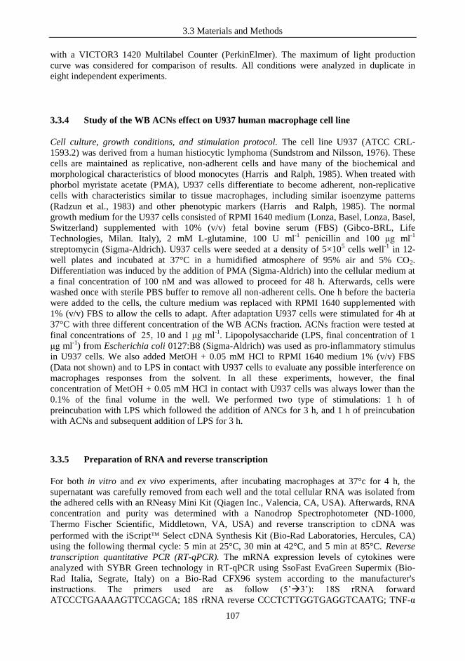
3.3 Materials and Methods
107
with a VICTOR3 1420 Multilabel Counter (PerkinElmer). The maximum of light production
curve was considered for comparison of results. All conditions were analyzed in duplicate in
eight independent experiments.
3.3.4 Study of the WB ACNs effect on U937 human macrophage cell line
Cell culture, growth conditions, and stimulation protocol. The cell line U937 (ATCC CRL-
1593.2) was derived from a human histiocytic lymphoma (Sundstrom and Nilsson, 1976). These
cells are maintained as replicative, non-adherent cells and have many of the biochemical and
morphological characteristics of blood monocytes (Harris and Ralph, 1985). When treated with
phorbol myristate acetate (PMA), U937 cells differentiate to become adherent, non-replicative
cells with characteristics similar to tissue macrophages, including similar isoenzyme patterns
(Radzun et al., 1983) and other phenotypic markers (Harris and Ralph, 1985). The normal
growth medium for the U937 cells consisted of RPMI 1640 medium (Lonza, Basel, Lonza, Basel,
Switzerland) supplemented with 10% (v/v) fetal bovine serum (FBS) (Gibco-BRL, Life
Technologies, Milan. Italy), 2 mM L-glutamine, 100 U ml-1
penicillin and 100 μg ml-1
streptomycin (Sigma-Aldrich). U937 cells were seeded at a density of 5×10⁵ cells well
-1 in 12-
well plates and incubated at 37°C in a humidified atmosphere of 95% air and 5% CO2.
Differentiation was induced by the addition of PMA (Sigma-Aldrich) into the cellular medium at
a final concentration of 100 nM and was allowed to proceed for 48 h. Afterwards, cells were
washed once with sterile PBS buffer to remove all non-adherent cells. One h before the bacteria
were added to the cells, the culture medium was replaced with RPMI 1640 supplemented with
1% (v/v) FBS to allow the cells to adapt. After adaptation U937 cells were stimulated for 4h at
37°C with three different concentration of the WB ACNs fraction. ACNs fraction were tested at
final concentrations of 25, 10 and 1 μg ml-1
. Lipopolysaccharide (LPS, final concentration of 1
μg ml-1
) from Escherichia coli 0127:B8 (Sigma-Aldrich) was used as pro-inflammatory stimulus
in U937 cells. We also added MetOH + 0.05 mM HCl to RPMI 1640 medium 1% (v/v) FBS
(Data not shown) and to LPS in contact with U937 cells to evaluate any possible interference on
macrophages responses from the solvent. In all these experiments, however, the final
concentration of MetOH + 0.05 mM HCl in contact with U937 cells was always lower than the
0.1% of the final volume in the well. We performed two type of stimulations: 1 h of
preincubation with LPS which followed the addition of ANCs for 3 h, and 1 h of preincubation
with ACNs and subsequent addition of LPS for 3 h.
3.3.5 Preparation of RNA and reverse transcription
For both in vitro and ex vivo experiments, after incubating macrophages at 37°c for 4 h, the
supernatant was carefully removed from each well and the total cellular RNA was isolated from
the adhered cells with an RNeasy Mini Kit (Qiagen Inc., Valencia, CA, USA). Afterwards, RNA
concentration and purity was determined with a Nanodrop Spectrophotometer (ND-1000,
Thermo Fischer Scientific, Middletown, VA, USA) and reverse transcription to cDNA was
performed with the iScript Select cDNA Synthesis Kit (Bio-Rad Laboratories, Hercules, CA)
using the following thermal cycle: 5 min at 25°C, 30 min at 42°C, and 5 min at 85°C. Reverse
transcription quantitative PCR (RT-qPCR). The mRNA expression levels of cytokines were
analyzed with SYBR Green technology in RT-qPCR using SsoFast EvaGreen Supermix (Bio-
Rad Italia, Segrate, Italy) on a Bio-Rad CFX96 system according to the manufacturer's
instructions. The primers used are as follow (5‟3‟): 18S rRNA forward
ATCCCTGAAAAGTTCCAGCA; 18S rRNA reverse CCCTCTTGGTGAGGTCAATG; TNF-α

3.3 Materials and Methods
108
forward TCAGCTCCACGCCATT; TNF-α reverse CCCAGGCAGTCAGATCAT. Primers were
designed using the Primer3Plus software (http://www.bioinformatics.nl/cgi-
bin/primer3plus/primer3plus.cgi), and the specificity of the primers was tested with melting
curves during amplification and by 1% TAE agarose gel electrophoresis. Quantitative PCR was
carried out according to the following cycle: initial hold at 96°C for 30 s and then 40 cycles at
96°C for 2 s and 70°C for 5 s. Gene expression was normalized to the housekeeping gene coding
for the18S rRNA. The amount of template cDNA used for each sample was 50 ng. All results
regarding cytokine mRNA expression levels are reported as the fold of induction (FOI) in
comparison to the control (namely unstimulated U937 macrophages), to which we attributed a
FOI of 1.
3.3.6 Statistical analysis
The statistical analysis was performed by means of STATISTICA software (Statsoft Inc., Tulsa,
OK, US). Analysis of variance (ANOVA) with type of treatment as the dependent factor was
used to evaluate the immunomodulatory activity. Post-hoc analysis of differences between
treatments was assessed by the Least Significant Difference (LSD) test with p ≤ 0.05 as level of
statistical significance.

3.4 Results and Discussion
109
3.4 RESULTS AND DISCUSSION
3.4.1 Modulation of NF-κB activation by WB soluble, phenolic and anthocyanin
fractions in Caco-2 cells
To evaluate the presence of potential bioactive molecules in our WB powder preparation, as
preliminary immunological investigation, we tested the effects of the three different fractions
isolated from WB on NF-κB, a transcriptional factor responsible for the induction of pro-
inflammatory cytokines in immune responses. To this aim we used a reporter cell line obtained
by transfecting Caco-2 cells with a luciferase reporter vector induced by NF-κB. In Table 3.4.1
the chemical composition of the different WB fractions is reported.
Table 3.4.1 Different WB fractions characterization and concentration tested in in vitro
immunological assay on NF-κB-transfected Caco-2 cells. A: water soluble fraction; B: phenolic
fraction; C: anthocyanin (ACN) fraction.
A
Compound Extracted
CaCo cell addition
μg/ml
25 μg/ml 50 μg/ml 100 μg/ml
Glucose 2284,4
13,97 27,9 55,9
Fructose 1836,4
11,3 22,6 45,2
Malic acid 19,9
0,12 0,24 0,48
Citric acid 47,2
0,29 0,58 1,16
B
Compound Extracted
CaCo cell addition
μg/ml
μg/ml μg/ml μg/ml μg/ml
Chlorogenic acid 719,2
5 25 50 100

3.4 Results and Discussion
110
C
Compound
Extracted CaCo cell addition
μg/ml % μg/ml μg/ml μg/ml μg/ml
Delphinidin 3-gal 166 10,6 0,53 2,7 5,4 10,8
Delphinidin 3-glc 224 14,3 0,72 3,6 7,2 14,4
Cyanidin 3-gal 69 4,4 0,22 1,1 2,2 4,4
Delphinidin 3-ara 102 6,5 0,33 1,7 3,4 6,8
Cyanidin 3-glc 99 6,3 0,32 1,6 3,2 6,4
Petunidin 3-gal 107 6,8 0,35 1,8 3,6 7,2
Cyanidin 3-ara +
Petunidin 3-glc 167 10,7
0,54 2,7 5,4 10,8
Peonidin 3-gal 38 2,4 0,12 0,6 1,2 2,4
Petunidin 3-ara +
Malvidin 3-gal 247 15,8
0,96 3,8 7,6 15,2
Peonidin 3-glc +
Malvidin 3-glc 279 17,9
0,89 4,4 8,8 17,6
Malvidin 3-ara 65 4,2 0,20 1,0 2,0 4,0
Total 1563 100 5 25 50 100
These experiments were performed in the presence of the pro-inflammatory stimulus IL-1β. All
WB fractions, namely ACNs, Phe and water soluble, were tested at different concentrations (Fig
3.4.1). We found no effects for the Phe and the water soluble fractions on Caco-2 cells, while the
ACNs fraction had a significant effect in reducing the activation of NF-κB, and in a dose-
dependent manner. In fact, the addition of ACNs at the concentration of 50 and 100 μg/ml
reduced the bioluminescence, and therefore the NF-κB activation, up to 70% and 85% (p≤0.05),
respectively.
Roles of ACNs in modulating the activation of NF-κB, and the down-stream signals, have been
already reported in literature (Epinat & Gilmore, 1999; Karlsen et al., 2007). Interestingly,
ACNs isolated from bilberries and blackcurrant have been demonstrated to modulate LPS-
3.4 Results and Discussion
111
induced activation of NF-κB in monocytes, and supplementation with ACNs in a parallel-
designed, controlled clinical trial reduced the levels of circulating NF-κB-dependent
inflammatory mediators (Karlsen et al., 2007).
Figure 3.4.1 Modulation of light emission expressed by Caco-2 cells stably transfected with a
NF-κB/luciferase reporter vector and incubated in presence of Interleukin-1β with different
fractions extracted from wild blueberry powder. Data are reported as percent variation of light
emission, referred to the control. Control: Caco-2 cell layers incubated only with media
supplemented with MetOH + 0.05 mM HCl at a concentration correspondent to the one present
in highest concentration of fractions tested. ACN: anthocyanin fraction; PHE: phenolic fraction;
S.F.: soluble fraction. Fraction concentrations reported (5-25-50-100) are referred to μg ml-1
.
The values are the means (± standard deviations) for eight independent experiments conducted in
duplicate. Asterisks indicate statistically significant differences compared to results for the
control (*, P<0.05.)
The transcription factor NF-kB is essential in orchestrating the inflammatory responses to a wide
range of insults, by leading to the induction of genes involved in innate immune response and in
the secretion of inflammatory responses mediators, like chemokines and cytokines (Pahl, 1999).
High levels and/or prolonged presence of pro-inflammatory mediators is connected to increased
risks of chronic-degenerative diseases onset (Libby, 2002; Boos and Lip, 2006). Therefore, the
use of natural bioactive molecules, like WB ACNs which possess the ability to modulate NF-κB
activation as we also tested and described in our study, might represent a mild an effective
strategy in dampening inflammatory processes.
However, when the same ACNs fraction was tested on bone marrow-derived dendritic cells
(BMDCs; Data not shown) we found no effect in inducing Th1-related nor Treg cytokines. Our
results suggest that food molecules, like ACNs extracted from WB, may not have an effect on
immune system per se, but rather they modulate the immune responses in presence of other
stimuli, as demonstrated in our experiments on epithelial Caco-2 cells in presence of IL-1β, and
as also confirmed in the experiments on U937 macrophages in presence of LPS.
3.4 Results and Discussion
112
3.4.2 WB ACNs fraction displays in vitro protective effects towards LPS-dependent
inflammatory response on human U937 macrophages
In the subsequent immunological experiments we moved from epithelial cells to cells properly
belonging to the immune system, and we decided to focus on ACNs, since it was the only
fraction which showed immunomodulatory properties in our first characterization on Caco-2
cells. To study the host‟s responses elicited by ACN at level of innate immunity, we employed
macrophages cells, which are professional phagocytes which undergo activation upon
environmental signals (Benoit et al., 2008). We used PMA-differentiated U937 human
macrophages, and we first treated U937 cells with PMA for 48 hours to differentiate the cells
into macrophages. Subsequently, we stimulated the differentiated cells with WB ACNs at three
different concentrations, namely 25, 10 and 1 μg ml-1
. We decided to use concentrations lower
than the ones tested on Caco-2 cells, since it is plausible that in vivo epithelial cells are exposed
to higher amounts of dietary molecules with respect to immune cells. After 4 h of incubation, we
used RT-qPCR to quantify the gene expression of TNF-α, a cytokine involved in inflammatory
responses (Belardelli, 1995). In these experiments we used LPS to induce a pro-inflammatory
response on U937 macrophages.
Since ACNs fraction has been resuspended in MetOH + 0.05 mM HCl, as first we wanted to
verify any possible interference of the solvent on cell permeability and/or on cell responses.
When we incubated U937 cells with RPMI 1% (v/v) FBS media supplemented with MetOH +
0.05 mM HCl (at a concentration correspondent to that present in the highest concentration of
ACNs used), we found that TNF-α expression levels were similar to the control (U937 cells
incubated only with RPMI media 1% (v/v) FBS; Data not shown); moreover, the presence of
MetOH + 0.05 mM HCl had only a slight impact on TNF-α expression level induced by LPS
(Fig. 3.4.2).
When we stimulated U937 cells only with the ACNs extracts we found no difference in the
induction of TNF-α compared to control, thus confirming the lack of immune response towards
these molecules, as previously observed on BMDCs (Data not shown). Consequently, we tested
the ACNs fraction on U937 cells together with LPS, by using two different experiment settings: 1
h of preincubation of U937 cells with LPS and then addition of ACNs, and 1h of preincubation of
macrophages with ACNs and subsequent addition of LPS for 3 h. The aims were to mimic and
evaluate, respectively, a possible anti-inflammatory activity of the ACNs in presence of an
ongoing inflammatory status, or a protective role towards an incoming inflammatory agent. For
the first hypothesis we found that only the highest concentration tested of ACNs had a significant
effect in decreasing the induction of TNF-α with respect to U937 cells stimulated only with LPS.
In the other experiment we observed, instead, a clear dose-response in the anti-inflammatory
activity of ACNs. Moreover, two ACNs concentrations (25 and 10 μg ml-1
) resulted significantly
effective in reducing the inflammatory response induced by LPS on U937 cells.
Summarizing our immunological experiments, we observed that ACNs fraction extracted from
WB powder displayed anti-inflammatory properties, both on epithelial cells by decreasing the
activation NF-κB in presence of IL-1β, and by exerting a protective role towards LPS-induced
TNF-α in immune cells. These abilities might be particularly useful in several inflammatory
disorders. Obesity, for instance, is a low-grade chronic inflammatory disease, characterized by
macrophages infiltration in the white adipose tissue (WAT) (Trayhurn & Wood, 2004). The
production of pro-inflammatory factors by activated macrophages can result in further
inflammation of the WAT, contributing to the pathogenesis of obesity-related diseases, like
hypertension, type 2 diabetes and atherosclerosis (Laine et al., 2007). It has been reported that
obese subjects present high serum levels of pro-inflammatory factors, like TNF-α (Fantuzzi,
2004; Wellen and Hotamisligil, 2005). Diet-derived obesity has been also shown to lead to high
level of serum LPS; moreover, endotoxemia and the presence of activated macrophages have
been demonstrated to be connected with high plasma levels of LPS (Cani et al., 2008).
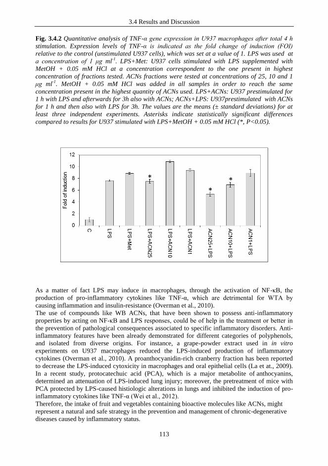
3.4 Results and Discussion
113
Fig. 3.4.2 Quantitative analysis of TNF-α gene expression in U937 macrophages after total 4 h
stimulation. Expression levels of TNF-α is indicated as the fold change of induction (FOI)
relative to the control (unstimulated U937 cells), which was set at a value of 1. LPS was used at
a concentration of 1 μg ml-1
. LPS+Met: U937 cells stimulated with LPS supplemented with
MetOH + 0.05 mM HCl at a concentration correspondent to the one present in highest
concentration of fractions tested. ACNs fractions were tested at concentrations of 25, 10 and 1
μg ml-1
. MetOH + 0.05 mM HCl was added in all samples in order to reach the same
concentration present in the highest quantity of ACNs used. LPS+ACNs: U937 prestimulated for
1 h with LPS and afterwards for 3h also with ACNs; ACNs+LPS: U937prestimulated with ACNs
for 1 h and then also with LPS for 3h. The values are the means (± standard deviations) for at
least three independent experiments. Asterisks indicate statistically significant differences
compared to results for U937 stimulated with LPS+MetOH + 0.05 mM HCl (*, P<0.05).
As a matter of fact LPS may induce in macrophages, through the activation of NF-κB, the
production of pro-inflammatory cytokines like TNF-α, which are detrimental for WTA by
causing inflammation and insulin-resistance (Overman et al., 2010).
The use of compounds like WB ACNs, that have been shown to possess anti-inflammatory
properties by acting on NF-κB and LPS responses, could be of help in the treatment or better in
the prevention of pathological consequences associated to specific inflammatory disorders. Anti-
inflammatory features have been already demonstrated for different categories of polyphenols,
and isolated from diverse origins. For instance, a grape-powder extract used in in vitro
experiments on U937 macrophages reduced the LPS-induced production of inflammatory
cytokines (Overman et al., 2010). A proanthocyanidin-rich cranberry fraction has been reported
to decrease the LPS-induced cytoxicity in macrophages and oral epithelial cells (La et at., 2009).
In a recent study, protocatechuic acid (PCA), which is a major metabolite of anthocyanins,
determined an attenuation of LPS-induced lung injury; moreover, the pretreatment of mice with
PCA protected by LPS-caused histiologic alterations in lungs and inhibited the induction of pro-
inflammatory cytokines like TNF-α (Wei et al., 2012).
Therefore, the intake of fruit and vegetables containing bioactive molecules like ACNs, might
represent a natural and safe strategy in the prevention and management of chronic-degenerative
diseases caused by inflammatory status.

3.5 Conclusions
114
3.5 CONCLUSIONS
Inflammation consists of a complex series of reactions and processes activated by the host in
order to prevent ongoing tissue damage, and to repair processes and defense mechanisms against
infectious diseases. However, if prolonged, inflammation may contribute to the pathogenesis of
chronic diseases such as diabetes, neurodegenerative diseases, cancers, and cardiovascular
disease (Nathan, 2002; Blomhoff , 2005; Karlsen et al., 2007). Thus, an attenuation of such
processes might potentially help in retarding or preventing detrimental consequences.
Several studies report that anthocyanins display anti-inflammatory features. The anti-
inflammatory activity of anthocyanins have mainly been attributed to a direct antioxidant action,
since ACNs are well-known anti-oxidant factors able to control lipid peroxidation and to dampen
detrimental effects of reactive-oxygen species.
However, more recently it has been proposed, and also demonstrated, that the anti-inflammatory
attitude of such molecules is also mediated by a direct regulatory effects on the expression of
genes involved in the inflammatory and immune responses. Several studied have shown ACNs
immunomodulatory activity in cell cultures and animal models (Wang & Mazza, 2002; Herath et
al., 2003; Cimino et al., 2006).
Accordingly, in our experiments on ACNs isolated from WB we demonstrated that these
molecules can have immunomodulatory properties, both on epithelial cells and on immune cells,
in particular in presence of pro-inflammatory stimuli.
In conclusion, exploring the role of food and, more specifically, the effect of bioactive
compounds such as ACNs on the metabolic processes involved in diseases is crucial, also for
designing public health directives for population approaches to prevention, and the results
obtained in the present study add important knowledge and insights on the properties of ACN-
rich foods and their exploitation for the prevention and treatment of chronic inflammatory
diseases.

3.6 References
115
3.6 REFERENCES
Bagchi D et al., 2004, Anti-agiogenic, antioxidant, anti-carcinogenic properties of a
novel anthocyanin rich berry extract formula. Biochemistry 69:75-80.
Belardelli F. 1995, Role of interferons and other cytokines in the regulation of the
immune response. APMIS 103:161-179.
Benoit M et al., 2008, Macrophage polarization in bacterial infections. J Immunol
181:3733-3739.
Blomhoff R. 2005, Dietary antioxidants and cardiovascular disease. Curr Opin Lipidol
16:47-54
Boos CJ, Lip GY, 2006, Is hypertension an inflammatory process? Curr Pharm Des
12:1623-1635.
Bushway RB et al., 1983, Mineral and vitamin content of lowbush blueberries
(Vaccinium angustifolium). J Food Sci 48:1878-1880.
Calixto JB et al., 2004, Anti-inflammatory compounds of plant origin. Part II.
modulation of pro-inflammatory cytokines, chemokines and adhesion molecules. Planta
Med 70:93-103.
Cani PD et al., 2008, Changes in gut microbiota control metabolic endotoxemia-
induced inflammation in high-fat fed mice-induced obesity and diabetes. Diabetes
57:1470-1481.
Cimino F et al., 2006, Effect of cyanidin-3-Oglucoside on UVB-induced response in
human keratinocytes. J Agric Food Chem 54:4041-4047.
Clarke JO, Mullin GE, 2008, A review of complementary and alternative approaches to
immunomodulation. Nutr Clin Pract 23:49-62.
Del Bo‟ C et al., 2010, Anthocyanins adsorption, metabolism, and distribution from a
wild-blueberry-enriched diet (Vaccinium angustifolium) is affected by diet duration in
the Sprague-Dawley rat. J Agric Food Chem 58:2494-2497.
Del Bo' C et al., 2012, Blanching improves anthocyanin absorption from highbush
blueberry (Vaccinium corymbosum L.) purée in healthy human volunteers: a pilot study.
J Agric Food Chem 60:9298-9304.
Epinat JC, Gilmore TD, 1999, Diverse agents act at multiple levels to inhibit the
Rel/NF-kB signal transduction pathway. Oncogene 18:6896-6909.
Espin JC et al., 2007, Nutraceuticals: facts and fiction. Phytochemistry 68:2986-3008.
Fantuzzi G. 2004, Adipose tissue, adipokines, and inflammation. J Allergy Clin
Immunol 115:911-919.
Felgines C et al., 2002, Blackberry anthocyanins are slightly bioavailable in rats. J Nutr
132:1249-1253.
Giordano L et al., 2007, Development and validation of an LC-MS/MS analysis for
simultaneous determination of delphinidin-3-glucoside, cyanidin-3-glucoside and
cyanidin-3-(6-malonylglucoside) in human plasma and urine after blood orange juice
administration. J Sep Sci 30:3127-3136.

3.6 References
116
Guglielmetti S et al., 2010, Oral bacteria as potential probiotics for the pharyngeal
mucosa. Appl Environ Microbiol 76:3948-3958.
Harris P, Ralph P, 1985, Human leukemic models of myelomonocytic development: a
review of the HL-60 and U937 cell lines. J Leukoc Biol 37:407-422.
Herath HM et al., 2003, Inhibitory effect of some flavonoids on tumor necrosis factor-
alpha production in lipopolysaccharide stimulated mouse macrophage cell line J774.1. J
Med Food 6:365-370.
Karlsen A et al., 2007, Anthocyanins inhibit nuclear factor-kappaB activation in
monocytes and reduce plasma concentrations of pro-inflammatory mediators in healthy
adults. J Nutr Aug 137:1951-1954.
La VD et al., 2009, Cytoprotective effect of proanthocyanidin-rich cranberry fraction
against bacterial cell wall-mediated toxicity in macrophages and epithelial cells.
Phytother Res 23:1449-1452.
Laine PS et al., 2007, Palmitic acid induces IP-10 expression in human macrophages
via NF-kappaB activation. Biochem Biophys Res Commun 358:150-155.
Libby P, 2002, Inflammation in atherosclerosis. Nature 420:868-874.
Manach C et al., 2004, Polyphenols: food sources and bioavailability. Am J Clin Nutr
79:727-747.
Manach C et al., 2005, Bioavailability and bioefficacy of polyphenols in humans. I.
Review of 97 bioavailability studies. Am J Clin Nutr 81:230S-242S.
Mazza G et al, 2002, Absorption of anthocyanins from blueberries and serum
antioxidant status in human subjects. J Agric Food Chem 50:7731–7737.
Milbury PE et al., 2010, Anthocyanins are bioavailable in humans following an acute
dose of cranberry juice. J Nutr 140:1099-1104.
Murkovic M et al., 2001, Detection of anthocyanins from elderberry juice in human
urine. J Sci Food Agric 81:934-937.
Nielsen et al., 2003, Absorption and excretion of black currant anthocyanins in humans
and Watanabe heritable hyperlipidemic rabbits. J Agric Food Chem 51:2813-2820.
Nathan C, 2002, Points of control in inflammation. Nature 420: 846–852.
Nicholson SK et al., 2008, Effects of dietary polyphenols on gene expression in human
vascular endothelial cells. Proc Nutr Soc 67:42-47.
Norton C et al., 2005, Wild blueberry-rich diets affect the contractile machinery of the
vascular smooth muscle in the Sprague-Dawley rat. J Med Food 8:8-13.
Overman A et al., 2010, Polyphenol-rich grape powder extract (GPE) attenuates
inflammation in human macrophages and in human adipocytes exposed to macrophage-
conditioned media. Int J Obes 34:800-808.
Pahl HL. 1999, Activators and target genes of Rel/NF-kB transcription factors.
Oncogene 18:6853-6866.
Passamonti S et al., 2002, The interaction of anthocyanins with bilitranslocase. Biochem
Biophys Res Commun 296:631-636.
3.6 References
117
Radzun HJ et al., 1983, Monocytic origin of the human hematopoietic cell line U-937
and its convertibility to macrophages evidenced by isoenzyme mapping. Int J Cancer
31:181-186.
Romier B et al., 2008, Modulation of signalling nuclear factor-κB activation pathway by
polyphenols in human intestinal Caco-2 cells. Br J Nutr 100:542-551.
Silberberg M et al., 2006, The bioavailability of polyphenols is highly governed by the
capacity of the intestine and of the liver to secrete conjugated metabolites. Eur J Nutr
45:88-96.
Sundstrom C, Nilsson K, 1976, Establishment and characterization of a human
histiocytic lymphoma cell line (U-937). Int J Cancer 17:565-577.
Trayhurn P, Wood IS, 2004, Adipokines: inflammation and the pleiotropic role of white
adipose tissue. Br J Nutr 92:347-355.
Vendrame S et al., 2011, Six-week consumption of a wild blueberry powder drink
increases bifidobacteria in the human gut. J Agric Food Chem 59:12815-12820.
Wang J, Mazza G, 2002, Effects of anthocyanins and other phenolic compounds on the
production of tumor necrosis factor alpha in LPS/ IFN-gamma-activated RAW 264.7
macrophages. J Agric Food Chem 50:4183-4189.
Wei M et al., 2012, Protocatechuic acid attenuates lipolysaccharide-induced acute lung
injury. Inflammation 35:1169-1178.
Wellen KE, Hotamisligil GS, 2005, Inflammation, stress, and diabetes. J Clin Invest
115:1111-1119.
Wrolstad R. Handbook of food analytical chemistry. Volume I, 2005, pp. 473-475.
Yamamoto Y, Gaynor RB, 2001, Therapeutic potential of inhibition of the NF-kappaB
pathway in the treatment of inflammation and cancer. J Clin Invest Jan 107:135-142.
Yang M et al., 2011, Food matrix affecting anthocyanin bioavailability: Review Curr
Med Chem 18:291-300.


Appendix 1
119
APPENDIX 1. COPIES OF ABSTRACTS OF PAPERS, ORAL
COMMUNICATIONS AND POSTERS
Guglielmetti S, Taverniti V, Minuzzo M, Arioli S, Stuknyte M, Karp M, Mora D,
2010, Oral bacteria as potential probiotics for the pharyngeal mucosa. Appl Environ
Microbiol 76:3948-3958.
Guglielmetti S, Taverniti V, Minuzzo M, Arioli S, Zanoni I, Stuknyte M, Granucci F,
Karp M, Mora D, 2010, A dairy bacterium displays in vitro probiotic properties for the
pharyngeal mucosa by antagonizing group A streptococci and modulating the immune
response. Infect Immun 78: 4734-4743.
Taverniti V, Guglielmetti S, 2011, The immunomodulatory properties of probiotic
microorganisms beyond their viability (ghost probiotics: proposal of paraprobiotic
concept). Genes Nutr 6:261-274.
Taverniti V et al., 2012, In vitro functional and immunomodulatory properties of the
Lactobacillus helveticus MIMLh5-Streptococcus salivarius ST3 association that are
relevant to the development of a pharyngeal probiotic product. Appl Environ
Microbiol 78:4209-4216.
Taverniti V, Guglielmetti S, 2012, Health-Promoting Properties of Lactobacillus
helveticus. Front Microbiol 3:392.
Taverniti V, et al., 2012, S-layer protein mediates the stimulatory effect of
Lactobacillus helveticus MIMLh5 on innate immunity. Appl Environ Microbiol
doi:10.1128/AEM.03056-12.
Taverniti V, The role of probiotic microorganisms and dietary compounds in the
modulation of immune response: a mechanistic approach. In Proceedings of the 15th
Workshop on the Development in the Italian PhD Research on Food Science
Technology and Biotechnology, Portici, Italy, 15-17 September, 2010.
Guglielmetti S, Taverniti V, Minuzzo M, Arioli S, Stuknyte M, Zanoni I, Granucci F,
Karp M, Mora D, Lactic acid bacteria as potential probiotics for the pharyngeal mucosa. In
Abstracts of the 10th Symposium on Lactic Acid Bacteria, Egmond aan Zee, The
Netherlands, 28 August – 1 September, 2011.
Taverniti V, The role of probiotic microorganisms and dietary compounds in the
modulation of immune response: a mechanistic approach. In Proceedings of the 16th
Workshop on the Developments in the Italian PhD Research on Food Science
Technology and Biotechnology, Lodi, Italy, 21-23 September, 2011.
Taverniti V, Stuknyte M, Minuzzo M, Arioli S, Mora D, Karp M, Pesu M, Guglielmetti S,
Immunomodulatory characterization of S-layer protein from the dairy probiotic strain
Lactobacillus helveticus MIMLh5In Book of Abstracts of The 4th Interdepartmental
Twinning Symposium, Tammela, Finland, 28 September – 2 October, 2011.

Appendix 1
120
Fracasetti D, Del Bo‟C, Guglielmetti S, Riso P, Taverniti V, Klimis-Zacas D, Porrini
M. Immunomodulatory properties of Wild Blueberries (Vaccinium Angustifolium) in
human intestinal epithelial Caco-2 Cells: preliminary data. In Abstracts of The Oxygen
Club Of California - Oxidants And Antioxidants In Biology - Cell Signaling And
Nutrient-Gene Interaction, 20-23 june, 2012 (Alba, Italy).
Taverniti V, The role of food and probiotic microorganisms, and dietary compounds
in the modulation of the immune response. In Proceedings of the 17th
Workshop on the
Developments in the Italian PhD Research on Food Science Technology and
Biotechnology, Cesena, Italy, 19-21 September, 2012.

Appendix 1
121

Appendix 1
122

Appendix 1
123
Appendix 1
124
Appendix 1
125
Appendix 1
126
Appendix 1
127
15th
Workshop on the Development in the Italian PhD Research on Food Science
Technology and Biotechnology, 15-17 September, 2010 (Portici, Italy)
The role of probiotic microorganisms and dietary compounds in the modulation of
immune response: a mechanistic approach
Valentina Taverniti
The research project of this PhD thesis deals with probiotic bacteria and dietary components
and their role in immune responses, especially at intestinal level. The main goal of the project is
the identification of bacterial and dietary molecules determining the immunomodulating
activity. To this aim, we will employ in vitro experimental approaches such as luciferase
reporter gene systems, real time quantitative PCR and ELISA tests, to evaluate the expression of
immunologic key mediators on different cell lines.
.
Appendix 1
128
10th
Symposium on Lactic Acid Bacteria, 28 August – 1 September, 2011 (Egmond aan
Zee, The Netherlands)
Lactic acid bacteria as potential probiotics for the pharyngeal mucosa
Guglielmetti S, Taverniti V, Minuzzo M, Arioli S, Stuknyte M, Zanoni I, Granucci F, Karp M,
Mora D
The probiotic approach represents an alternative strategy in the prevention and treatment of
infections in those body sites where the microbiota plays a role in the maintenance of
physiological homeostasis. In this context, we evaluated in vitro the potential abilities of oral,
probiotic and dairy bacteria in controlling Streptococcus pyogenes infections at the pharyngeal
level. Initially, we analysed the bacterial adhesion on FaDu hypopharyngeal carcinoma cells
and the ability to antagonize S. pyogenes on FaDu layer and HaCat keratinocytes. Due to their
promising adhesive and antagonistic features, we studied the oral isolate Streptococcus
salivarius ST3 and the dairy strain Lactobacillus helveticus MIMLh5 also through
immunological in vitro experiments. We performed the quantification of several cytokines and
the measurement of NF-κB activation in FaDu cells. Strain ST3 significantly increased NF-κB
activation at baseline, while it had an opposite effect when FaDu cells were co-stimulated with
IL-1β. In addition, ST3 biased cytokines production at baseline (reduction of IL-1β and TNFα,
increase of MIP-1β and GM-CSF) and after IL-1β-induction (reduction on IL-6 and IL-8).
MIMLh5 efficiently reduced the induction of IL-6, IL-8 and TNF in a dose-dependent
manner. After stimulating with IL-1β, active NF-κB was still markedly lowered. Nevertheless,
we observed an increased secretion of IL-6, IFN-γ and GM-CSF under these conditions. In
addition, MIMLh5 increased the GM-CSF/G-CSF ratio. This is compatible with a switch of the
immune response towards a TH1 pathway, as supported by our observation that MIMLh5, once
in contact with bone marrow derived dendritic cells, triggered the secretion of TNFα and IL-2.
In conclusion, we propose ST3 and MIMLh5 as a potential probiotic bacteria for the human
pharynx, with promising antagonistic and immunomodulatory properties.

Appendix 1
129
16th
Workshop on the Developments in the Italian PhD Research on Food Science
Technology and Biotechnology, 21-23 September, 2011 (Lodi, Italy)
The role of probiotic microorganisms and dietary compounds in the modulation of
immune response: a mechanistic approach
Valentina Taverniti
To investigate the immunomodulatory properties of probiotic bacteria, two strains of
Lactobacillus helveticus have been selected through a screening assay based on a NF-
κB/luciferase reporter gene system in Caco-2 human intestinal epithelial cell line. The setup of
a purification protocol of their Surface (S)-layer proteins allowed testing the role of these
molecules in the immune response. In vitro and ex vivo experiments, respectively performed on
U937cell line and murine bone marrow-derived macrophages, demonstrated the involvement of
S-layer protein in a transient activation of the immune system.
Appendix 1
130
The 4th
Interdepartmental Twinning Symposium, 28 September- 2 October, 2011
(Tammela, Finland)
Immunomodulatory characterization of S-layer protein from the dairy probiotic
strain Lactobacillus helveticus MIMLh5
Taverniti V, Stuknyte M, Minuzzo M, Arioli S, Mora D, Karp M, Pesu M, Guglielmetti S
This work is targeted to a mechanistic investigation on the immunomodulatory properties of the
dairy bacterium Lactobacillus helveticus MIMLh5, a strain isolated from Grana Padano cheese
natural whey starter culture, that already showed to possess probiotic abilities and to modulate
the immune response at pharyngeal level. Thus we decided to test the activity of this strain at
gut level, using as proper model the intestinal epithelial Caco-2 cell line, and we observed that
MIMLh5 was able to efficiently decrease the activation of the transcriptional factor NF-κB. To
individuate which bacterial components could be responsible for the immunological activity,
attention was given to Surface (S)-layer protein, which represents up to 14% of total protein
content in L. helveticus. We improved an extraction protocol that allowed us to obtain the
protein with a high level of purity, and thus suitable for immunological experiments. When we
tested in Caco-2 model the purified protein isolated from L. helveticus MIMLh5, we saw that it
was able to decrease the activation of NF-κB, similarly to the whole bacterium, both at baseline
and in presence of IL-1β. We decided to study the effects on cells of the immune system; in in
vitro experiments on U937 cell line, we observed by quantitative Real Time-PCR (RT-qPCR)
that both the bacterium and the purified protein were more prone to induce the activation of pro-
inflammatory cytokines such as COX2 and TNF-α, respect to the anti-inflammatory/regulatory
IL10. These results were even confirmed when we performed ex vivo experiments by isolating
macrophages from mouse peritoneal cavity and bone marrow (BMDMs), and moreover in bone
marrow-derived dendritic cells. We found confirmation of the involvement of S-layer protein in
the activation of the immune system elicited by the bacterium since, when we tested the
bacterial cells without S-layer (after LiCl treatment for the removal of the protein), the
propensity to induce pro-inflammatory cytokines decreased, while the level of IL10 did not
change significantly. Moreover, when the protein was added to LiCl-washed bacterial cells the
pro-inflammatory attitude raised again. We performed also preliminary in vivo experiments by
injecting S-layer protein in Zebrafish embryos; even in this case we observed an increased level
of some pro-inflammatory cytokines (like IFN-γ) respect to control embryos. As first step to
individuate the signal pathways involved in the immune response elicited by MIMLh5 and its
protein, we found that Toll-like Receptor (TLR)2 is involved in the recognition of both the
whole bacterium and the purified protein. Its block resulted in a less production of TNF-α and
COX2. Indeed the blocking effect was more efficient in LiCl-treated cells, probably due to the
absence of the protein. In conclusion from all these data it seems that S-layer protein is a key
molecular determinant for the immunomodulatory properties observed in L. helveticus
MIMLh5. The pro-inflammatory activity elicited by the whole bacterium and the purified
protein, as already demonstrated for other probiotics , could be transient and thus resulting in a
“positive” activation of immune system that can support human-host defence and homeostasis.

Appendix 1
131
The Oxygen Club Of California - Oxidants And Antioxidants In Biology - Cell Signaling
And Nutrient-Gene Interaction, 20-23 june, 2012 (Alba, Italy)
Immunomodulatory properties of Wild Blueberries (Vaccinium Angustifolium) in human
intestinal epithelial Caco-2 Cells: preliminary data
Fracasetti D, Del Bo‟C, Guglielmetti S, Riso P, Taverniti V, Klimis-Zacas D, Porrini M
Wild blueberries (WB) are a rich source of polyphenols, particularly anthocyanins, which may
be beneficial for human health because of their antioxidant, antiinflammatory and
immunomodulatory potential. This study aims to investigate the immunomodulatory potential
of three WB fractions (anthocyanin, phenolic and soluble fraction) in an inflammatory state
model, based on human intestinal Caco-2 cells. A reporter gene system was prepared by
transfecting Caco-2 cells with the pNiFty-SEAP reporter- construct containing an engineered
ELAM promoter with 5 NF-kB binding sites and an insect luciferase reporter gene. The
transfected Caco-2 cells were stimulated with interleukin (IL)-1β to mimic an inflammatory
state and then supplemented with 5 mg/L, 25 mg/L, 50 mg/L and 100 mg/L of the anthocyanin,
phenolic and soluble fractions. Immunomodulatory activity for each fraction was monitored in
real time by quantification of bioluminescence with a luminometer. The addition of
anthocyanins at the concentration of 50 and 100 mg/L reduced the bioluminescence, and
therefore NF-κB activation, up to 70% and 85% (p≤0.05), respectively. No dose-response was
observed after supplementing Caco-2 cells with both phenolic and soluble fractions. These data
suggest that only the anthocyanin fraction can modulate Caco-2 cells inflammatory state in the
experimental conditions adopted. Future experiments will address supplementation of Caco-2
cells with anthocyanin standards to elucidate the compound(s) responsible for the observed
immunomodulatory effect.

Appendix 1
132
17th
Workshop on the Developments in the Italian PhD Research on Food Science
Technology and Biotechnology, 19-21 September, 2012 (Cesena, Italy)
The role of food and probiotic microorganisms, and dietary compounds in the modulation
of the immune response
Valentina Taverniti
The aim of this PhD research project was to investigate the interaction between the immune
system and (i) bacterial cells and (ii) their components, and (iii) food molecules. Specifically,
our experiments were designed to study by means of in vitro and ex vivo analyses the possible
signalling pathways involved in the cross-talk between epithelial and immune cells, and a dairy
bacterium, its purified Surface (S)-layer protein and an anthocyanin fraction extracted from
wild blueberry (Vaccinium Angustifolium).

Appendix 2. Index of Tables
133
APPENDIX 2. INDEX OF TABLES
Table 1.1.1 Commercial Strains Sold As Probiotics……………………..…………………….19
Table 1.1.2 The beneficial effects of probiotic bacteria documented in human intervention
studies (adapted from Saxelin et al., 2005) …………………………….………………………21
Table 1.3.1 Bacteria isolated from pharyngeal mucosa.............................................................30
Table 1.3.2 Streptococcus salivarius K12 and non-oral bacteria included in the study……….31
Table.1.4.1 Antibiotic sensitivities of selected bacterial strains. .............................................. .48
Table 3.4.1 Different WB fractions characterization and concentration tested in in vitro
immunological assay on NF-κB-transfected Caco-2 cells. ....................................................... 109
Appendix 3. Index of Figures
134
APPENDIX 3. INDEX OF FIGURES
Figure 1.1.1 Cells and selected cytokines taking part in innate and adaptive immunity……….26
Figure. 1.3.1. Genetic map of a plasmid pNiFty2-Luc………………………………………………34
Figure 1.4.1 Unweighted-pair group method using average linkages (UPGMA) dendrogram
derived from similarity coefficients calculated by the Jaccard method (simple Jaccard [Sj]
coefficients; shown on the scale at the bottom), showing the relationship among Streptococcus
salivarius pharyngeal isolates, analyzed by BOX-PCR and RAPD analysis using primers M13,
OPI02mod, OPI17mod, and PedAF……………………………………………………………………38
Figure 1.4.2 Adhesion of bacterial strains to FaDu epithelial cell layer according to their
adhesion index (AdI, number of bacteria / 100 FaDu cells)………………………………………..40
Figure 1.4.3 Adhesion to FaDu epithelial cells of selected bacterial strains as observed with
Giemsa staining under a light microscope…………………………………………………………….41
Figure 1.4.4 Antagonistic exclusion activity of bacterial pharyngeal isolates and probiotic/food
strains against bioluminescent Streptococcus pyogenes C11lucFF on FaDu hypopharyngeal
carcinoma cells(A) and HaCat keratinocytes (B)………………………………………………....... 44
Figure 1.4.5 Antagonistic competition activity of bacterial pharyngeal isolates against
bioluminescent Streptococcus pyogenes C11lucFF on FaDu hypopharyngeal carcinoma
cells………………………………………………………………………………………………………... 43
Figure 1.4.6 Cytokine secretions that changed significantly after treatment of the FaDu layer
with bacterial cells, as determined by using the Bio-Plex assay……………………………………45
Figure 1.4.7 Effects of selected bacterial strains on FaDu cells stably transfected with an NF-
B/luciferase reporter vector, without (A) or with (B) stimulation with IL-1 (2 ng ml-1
)……..46
Figure 1.4.8 Bacterial adhesion as observed with Giemsa staining under a light microscope;
FaDu nuclei appear in red……………………………………………………………………...50
Figure 1.4.9 Antagonistic exclusion activity against bioluminescent S. pyogenes C11lucFF;
control is a cell layer treated with only PBS before incubation with S. pyogenes……………..50
Figure 1.4.10. Effect of bacteria on FaDu cells stably transfected with an NF-B/luciferase
reporter vector…………………………………………………………………………………………….51
Figure 1.4.11 Transcription analysis of cytokine genes in U937 cells stimulated with
Lactobacillus helveticus MIMLh5 and Streptococcus salivarius ST3 after 4 h of incubation with
bacterial strains used alone or in association at two different MOIs……………………………...53
Figure 1.4.12. Growth curves of Streptococcus salivarius ST3…………………………………….56
Appendix 3. Index of Figures
135
Figure 1.4.13 Growth of Lactobacillus helveticus MIMLh5 and Streprococcus salivarius ST3
(single culture and co-culture) at 42°C in skim milk supplemented with 2 % glucose or
FOS………………………………………………………………………………………………………....56
Figure 2.1.1. Scanning electron micrograph of a freeze-etched preparation of a bacterial cell
with an Slayer with oblique (p2; A) and hexagonal (p6; B) lattice symmetry……………………73
Figure 2.4.1 Biochemical analyses of L. helveticus MIMLh5 S-layer protein…………………...81
Figure 2.4.2 Effect of Lactobacillus acidophilus NCFM, Lactobacillus helveticus MIMLh5 and
its purified S-layer protein on human epithelial colorectal Caco-2 cells stably transfected with
an NF-κB/luciferase reporter vector at baseline (A) or stimulated with 2 ng ml-1
of IL-1β
(B)…………………………………………………………………………. ………………………….......83
Figure 2.4.3 Quantitative analysis of cytokine gene expression in murine bone marrow-derived
macrophages (BMDMs) after 4 h stimulation with L. helveticus MIMLh5 and its S-layer
protein..........................................................................................................................................86
Figure 2.4.4 Quantitative analysis of cytokine gene expression in murine macrophages isolated
from peritoneal cavity after 4 h stimulation with L. helveticus MIMLh5 and its S-layer
protein............................................................................................................................ ..............87
Figure 2.4.5. Quantitative analysis of cytokine gene expression in U937 human macrophages
after 4 h of stimulation.................................................................................................... .............89
Figure 3.1.1. Polyphenol classes in fruit and vegetables..........................................................100
Figure 3.1.2 Chemical structures of selected anthocyanidins (Adapeted from Nicholson et al.,
2008.)............................................................................................................................. ............101
Figure 3.1.3 Absortption and excretion of ACNs and metabolites............................................102
Figure 3.4.1 Modulation of light emission expressed by Caco-2 cells stably transfected with a
NF-κB/luciferase reporter vector and incubated in presence of Interleukin-1β with different
fractions extracted from wild blueberry powder........................................................................111
Figure 3.4.2 Quantitative analysis of TNF-α gene expression in U937 macrophages after total
4 h stimulation.............................................................................................................. .............113


Itaca
Sempre devi avere in mente Itaca -
raggiungerla sia il pensiero costante.
Soprattutto, non affrettare il viaggio;
fa che duri a lungo, per anni, e che da vecchio
metta piede sull’isola, tu, ricco
dei tesori accumulati per strada
senza aspettarti ricchezze da Itaca.
Itaca ti ha dato il bel viaggio,
senza di lei mai ti saresti messo
sulla strada: che cos’altro ti aspetti?
E se la trovi povera, non per questo Itaca ti avrà deluso.
Fatto ormai savio, con tutta la tua esperienza addosso
già tu avrai capito ciò che Itaca vuole significare.
Ithaka
Keep Ithaka always in your mind.
Arriving there is what you are destined for.
But do not hurry the journey at all.
Better if it lasts for years,
so you are old by the time you reach the island,
wealthy with all you have gained on the way,
not expecting Ithaka to make you rich.
Ithaka gave you the marvelous journey.
Without her you would not have set out.
She has nothing left to give you now.
And if you find her poor, Ithaka won’t have deceived you.
Wise as you will have become, so full of experience,
you will have understood by then what Ithaka means.
(1911 - Konstantinos P. Kavafis)


Acknowledgements
139
ACKNOWLEDGEMENTS
I want to sincerily thank all the persons that were significant for me for many reasons in these
three years of PhD research activity.
First of all I want to thank my tutor Dr. Simone Guglielmetti, my co-tutor Prof. Marisa Porrini,
Dr. Patrizia Riso and Prof. Diego Mora, for all the things I have learned, for all the support, and
for all the great opportunities I had thanks to them during my PhD activity. I want to thank also
Prof. Francesco Bonomi and Prof. Stefania Iametti for the possibility to spend a very fruitful
exchange period at University of Copenhagen.
I want to thank particularly some colleagues and friends from University of Milan: Chiara
Ferrario, Christian Scabiosi, Cristian del Bò, Giovanni Ricci, Mario Minuzzo, Matteo Miriani,
Milda Stuknyte, and in particular for all the help, assistance and pleasant presence especially in
the last period: Elisa Gualtieri, Maria Chiara Remagni, Mauro Marengo, Silvia Balzaretti,
Valentina Ambrogi and William Remelli. A thanks also to the great "new entries" Ornella and
Valeria.
A very important and hearty thanks to a special and irreplaceable person for me: thank you
Stefania…..
A warm thanks to people I had the fortune to met and work with in Tampere: Dr. Marko Pesu,
Prof. Matti Karp, Dr. Hannu Turpeinen, Annemari Latvala, Anna Oksanen, Dr. Kati Pulkkinen,
Dr. Ilkka Junttila, Sanna Hämäläinen, Zsuzsi Ortutay, Alessandro Ciranna, Alessandra Radici,
Alfonso Urbanucci, Giuseppe Cannino, Mauro Scaravilli, Roberto Munoz, Serena Previtali,
Carmelenzo, and particularly to my dearest Zuzita….
I want to thank as well all the person with which I spent a beautiful period in the lab and in the
everyday life in Copenhagen: a special thanks to Prof. Hanne Frøkiaer, Anni Melsen, Marianne
Petersen, Dr. Stine Metzdorff, Dr. Kristina Maaetoft-Udsen, Dr. Matilde Bylov Kristensen, Dr.
Gudrun Weiss, Lisbeth Drodz Lund, Louise Henningsen, Eva Fuslang, Daniela Silva, Ronja
Mathiesen, Matteo Gentilini, Daniele Silvestro, Alice de Porcellinis, Sara Favalli, Antonino
Azzarello, Stefano Manzo, Alice Murciano and in particular to Andrea Bellucci.
I want to thank all my friends, and in a special way: Elisa, Silvia, Marcella, Peppe, Maddalena,
Anna, Chiara, Rosa Maria, Annalucia and Roberta.
A warm thanks to all my family.