Modulation of Epileptic Network by Responsive … program Director, Kuwait Institute for Medical...
22
Dr Faisal T. Sayer, B.Med.Sc, M.D., M.Sc., SBNS Consultant and Chairman, Department of Neurosurgery, Ibn Sina Hospital. Residency program Director, Kuwait Institute for Medical Specialization. Vice-chairman, The Kuwait Society of Neurological Surgeons email: [email protected] Modulation of Epileptic Network by Responsive Technology
Transcript of Modulation of Epileptic Network by Responsive … program Director, Kuwait Institute for Medical...

Dr Faisal T. Sayer, B.Med.Sc, M.D., M.Sc., SBNS
Consultant and Chairman, Department of Neurosurgery, Ibn Sina Hospital.
Residency program Director, Kuwait Institute for Medical Specialization.
Vice-chairman, The Kuwait Society of Neurological Surgeons
email: [email protected]
Modulation of Epileptic Network by Responsive Technology
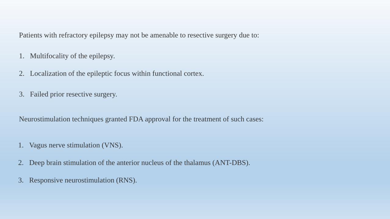
Patients with refractory epilepsy may not be amenable to resective surgery due to:
1. Multifocality of the epilepsy.
2. Localization of the epileptic focus within functional cortex.
3. Failed prior resective surgery.
Neurostimulation techniques granted FDA approval for the treatment of such cases:
1. Vagus nerve stimulation (VNS).
2. Deep brain stimulation of the anterior nucleus of the thalamus (ANT-DBS).
3. Responsive neurostimulation (RNS).
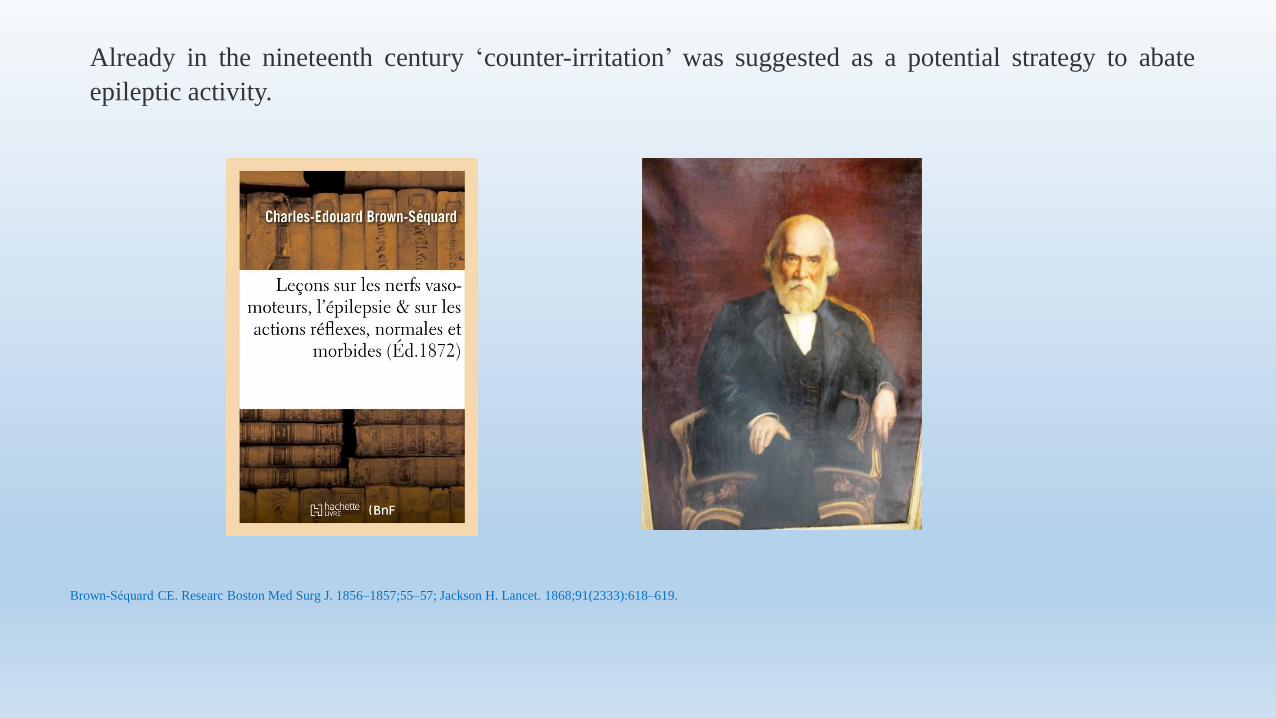
Already in the nineteenth century ‘counter-irritation’ was suggested as a potential strategy to abate
epileptic activity.
Brown-Séquard CE. Researc Boston Med Surg J. 1856–1857;55–57; Jackson H. Lancet. 1868;91(2333):618–619.
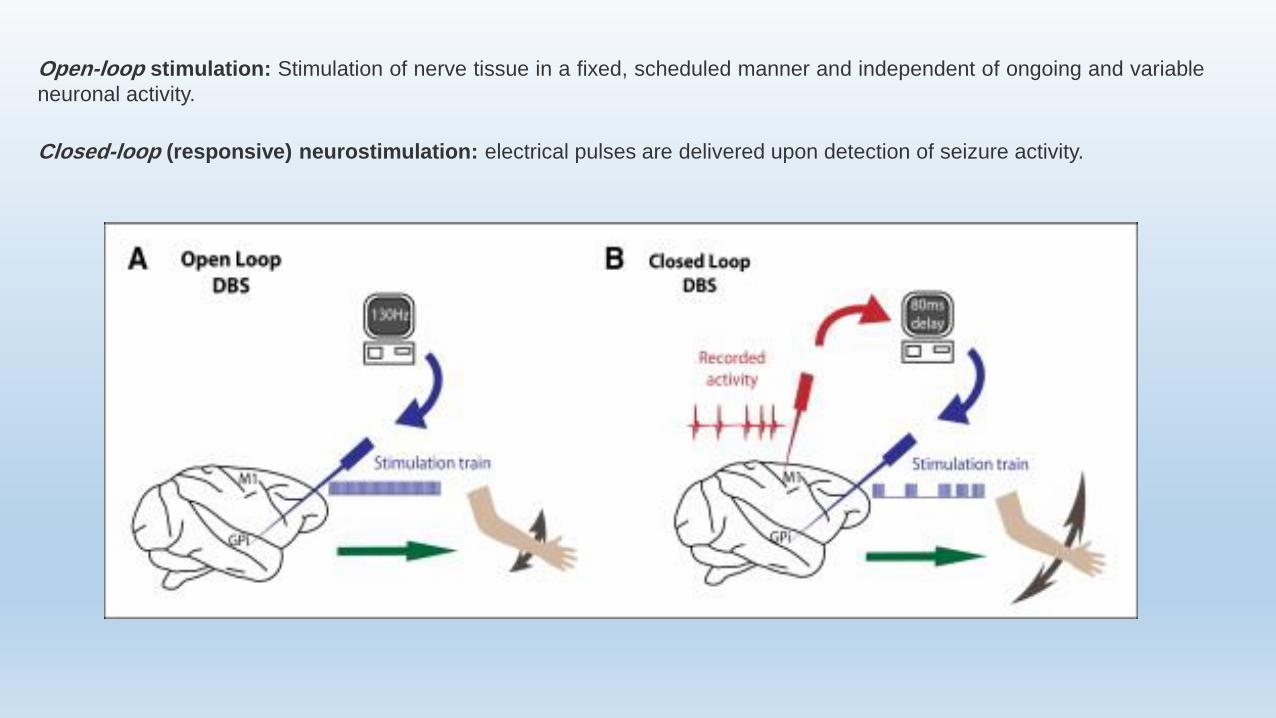
Open-loop stimulation: Stimulation of nerve tissue in a fixed, scheduled manner and independent of ongoing and variable
neuronal activity.
Closed-loop (responsive) neurostimulation: electrical pulses are delivered upon detection of seizure activity.
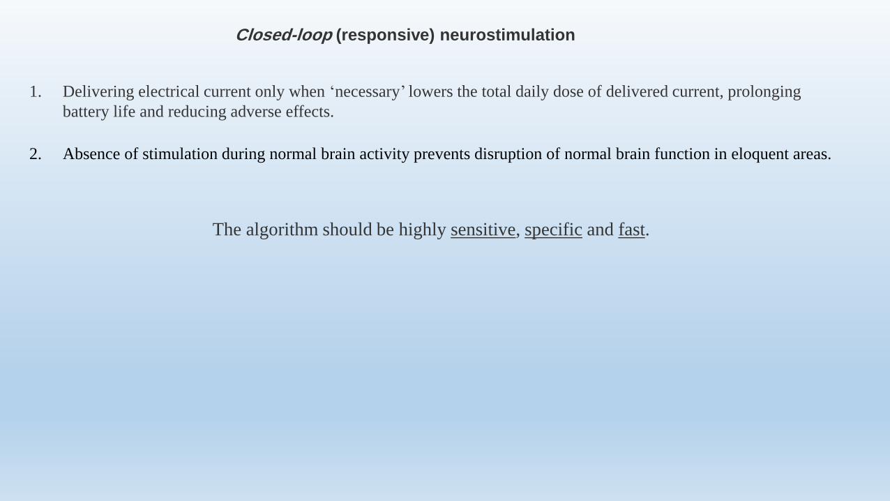
1. Delivering electrical current only when ‘necessary’ lowers the total daily dose of delivered current, prolonging
battery life and reducing adverse effects.
2. Absence of stimulation during normal brain activity prevents disruption of normal brain function in eloquent areas.
The algorithm should be highly sensitive, specific and fast.
Closed-loop (responsive) neurostimulation

Early detection of ictal EEG activity requires intracranial electrodes exactly at the site of seizure onset.
Seizure activity may also be recorded outside the brain, which allows a more accessible read-out:
1. Abnormal behavior.
2. Seizure-related muscle activity (EMG).
3. Seizure-related cardiac changes (ECG).
In an optimal scenario ‘sensing and pacing’: clinically seizures are not only aborted, but even prevented..
Closed-loop (responsive) neurostimulation

1. The RNS system (Neuropace, Inc.) FDA approval (November 2013).
2. The AspireSR VNS system in which a cardiac-based seizure-detection (CBSD) algorithm was incorporated for
additional on-demand stimulation (FDA approval May 2015).
Closed-loop (responsive) neurostimulation
The time window between ictal EEG and clinical seizure onset may favor intracranial closed-loop stimulation systems
compared with closed-loop treatments that measure ictal activity outside the brain.

It is not a detection system for clinical seizures.
As soon as relevant EEG changes are identified stimulation is initiated
Closed-loop stimulation occurred 600–2000 times a day
cumulative total of <5 min of stimulation over 24 h (battery longevity).
The RNS system
Sun FT, Morrell MJ. Neurotherapeutics.2014;11 (3):553–563; Morrell MJ, Group RNSSiES. Neurology. 2011;77(13):1295–1304..
The contacts serve a dual function:
Continuous monitoring of electrophysiological activity.
Delivery of electrical pulses.
Connected to one or two electrodes:
• 4-contact depth lead.
• 4-contact cortical strip.
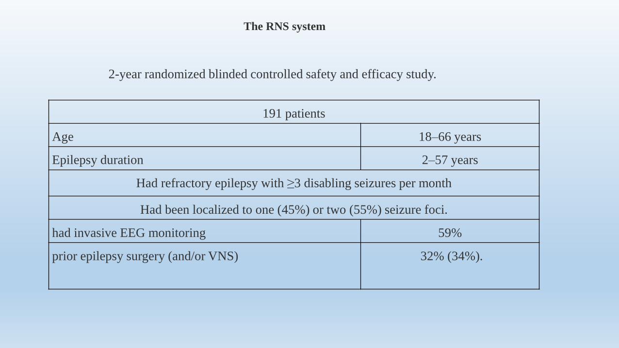
191 patients
Age 18–66 years
Epilepsy duration 2–57 years
Had refractory epilepsy with ≥3 disabling seizures per month
Had been localized to one (45%) or two (55%) seizure foci.
had invasive EEG monitoring 59%
prior epilepsy surgery (and/or VNS) 32% (34%).
2-year randomized blinded controlled safety and efficacy study.
The RNS system

12-week blinded evaluation period
Stimulation group −37.9%
Control group −17.3%
implantation effect
Trend towards increased efficacy over time
Stimulation group Control group
after 1 month 34.2% 25.2%
2 months 38.1% 17.2%
3 months 41.5% 9.4%
Fisher R, Salanova V, Witt T, et al. Epilepsia. 2010;51(5):899–908.
Morrell MJ, Group RNSSiES. Neurology. 2011;77(13):1295–1304.
Heck CN, King-Stephens D, Massey AD, et al. Epilepsia. 2014;55 (3):432–441.
The RNS system

Long-term open-label extension study providing an additional 7 years of efficacy and safety data.
Bergey GK, Morrell MJ, Mizrahi EM, et al. Neurology. 2015;84 (8):810–817.
open-label follow-up period
1 year 44%
2 years 53%
year 3 60%
year 6 66%
Heck CN, King-Stephens D, Massey AD, et al. Epilepsia. 2014;55 (3):432–441.Bergey GK, Morrell MJ, Mizrahi EM, et al. Neurology. 2015;84 (8):810–817.
some improvement 84%
≥50% reduction in seizure frequency 60%
seizure-free 16%
The RNS system
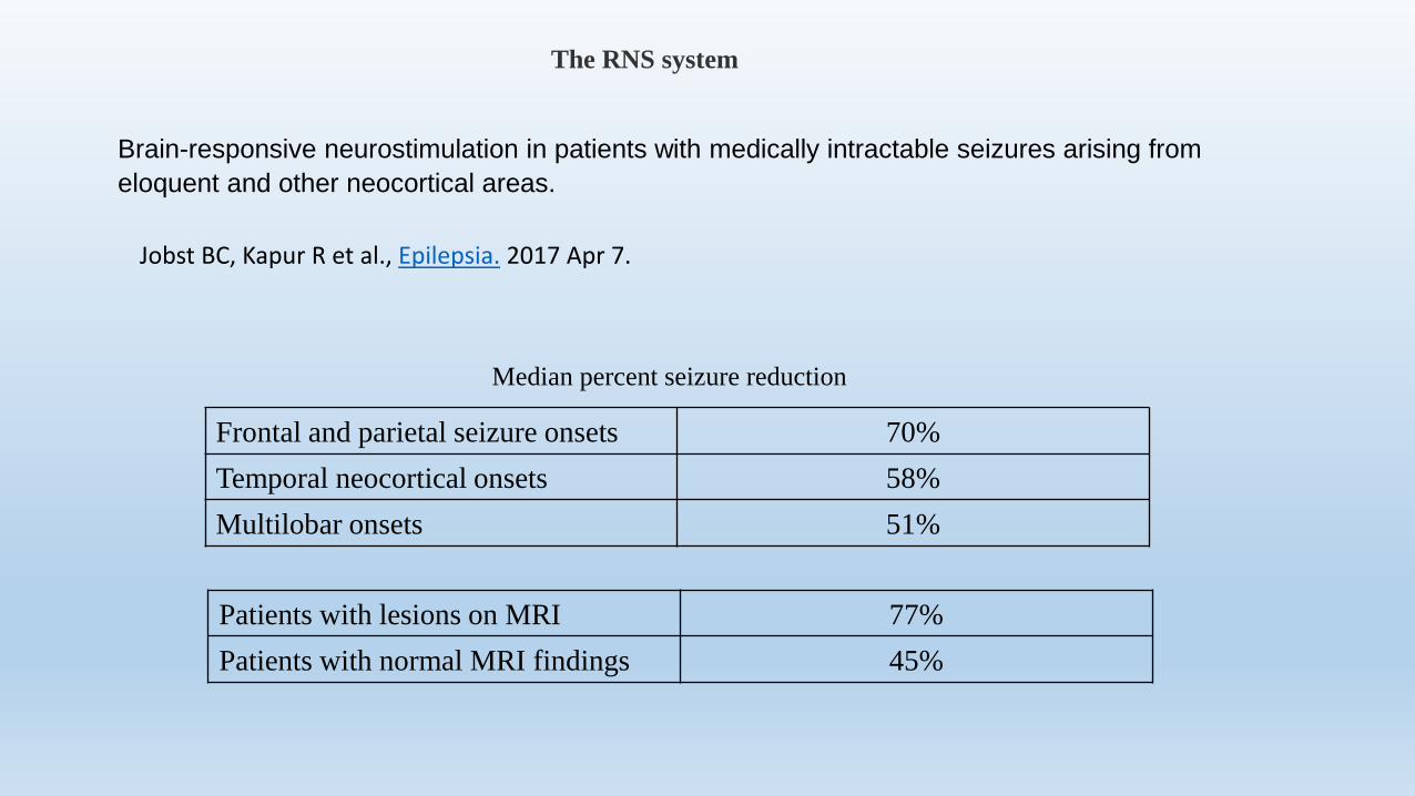
Jobst BC, Kapur R et al., Epilepsia. 2017 Apr 7.
Brain-responsive neurostimulation in patients with medically intractable seizures arising from
eloquent and other neocortical areas.
Frontal and parietal seizure onsets 70%
Temporal neocortical onsets 58%
Multilobar onsets 51%
Median percent seizure reduction
Patients with lesions on MRI 77%
Patients with normal MRI findings 45%
The RNS system

Closed-loop VNS
Recently a new VNS device has been developed (AspireSR) allowing automated on-demand VNS triggered by
CBSD. The closed-loop algorithm functions in addition to conventional duty cycle VNS in one device
Ictal tachycardia occur in >80% of adult epilepsy patients.
Cardiac changes may precede electrical/clinical onset with several seconds, resulting in a
considerable amount of seizures that may be detected and prematurely aborted.

Modulation of Epileptic Network by Responsive Technology

Epileptogenesis
Disruption of these mechanisms create a imbalance between excitation and inhibition.
Neural network hypothesis: Epilepsy become refractory due to seizure-induced alterations of brain plasticity:
Formation of abnormal neural network
Loss of the inhibitory effect of endogenous AE system Prevent the AEDs from entering their targets
DRE
Axonal sprouting Synaptic reorganization Neurogenesis Gliosis

No single investigation delineate the epileptgenic zone.
Current investigations, have different levels of spatial and
temporal resolutions.
Different modalities of investigations provide different
snapshots of the epileptogenesis at different time periods and at
variable magnifications.

The epileptogenic focus is not a “fixed” zone but a dynamic, constantly changing group of networks.
Janszky et al., demonstrated that the seizure freedom was inversely proportional to the duration of epilepsy:
Janszky J, Janszky I, Schulz R, Hoppe M, Behne F, Pannek HW, et al. Brain. 2005;128:395–404.
Duration of epilepsy between 1 and 10 90% Class I Engel
Duration of epilepsy being >30 years 30% Class I Engel
The “network” hypothesis can explain this by the fact that more neurons become recruited into the “epileptogenic
network” over a period of time if epilepsy is not controlled.

The idea of directly stimulating the cortex is appealing, as seizures are thought to arise from cortical grey matter.
Different deep grey matter nuclei have been targeted based on experience with basal ganglia stimulation in movement disorders.

1. Lower current is required to modulate neural activity.
2. Its stimulation paradigm is more efficient, where stimulating a small number of axons in white matter allow the
current to propagate to the neuronal cell bodies, either ortho- or antidromically, to impact large cortical areas.
3. White matter tracts stimulation is thought to synchronize its electrical output, and subsequently ‘overdrive’ the
electrical activity of the downstream cortical epileptogenic zone, effectively reducing seizures.
Why white matter stimulation?
However, targeting white matter fibers may represent an alternative to stimulation of neural cell bodies.
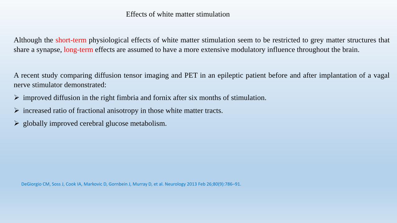
Although the short-term physiological effects of white matter stimulation seem to be restricted to grey matter structures that
share a synapse, long-term effects are assumed to have a more extensive modulatory influence throughout the brain.
A recent study comparing diffusion tensor imaging and PET in an epileptic patient before and after implantation of a vagal
nerve stimulator demonstrated:
improved diffusion in the right fimbria and fornix after six months of stimulation.
increased ratio of fractional anisotropy in those white matter tracts.
globally improved cerebral glucose metabolism.
Effects of white matter stimulation
DeGiorgio CM, Soss J, Cook IA, Markovic D, Gornbein J, Murray D, et al. Neurology 2013 Feb 26;80(9):786–91.
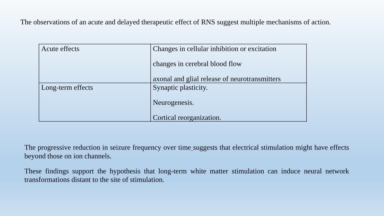
The observations of an acute and delayed therapeutic effect of RNS suggest multiple mechanisms of action.
Acute effects Changes in cellular inhibition or excitation
changes in cerebral blood flow
axonal and glial release of neurotransmitters
Long-term effects Synaptic plasticity.
Neurogenesis.
Cortical reorganization.
The progressive reduction in seizure frequency over time suggests that electrical stimulation might have effects
beyond those on ion channels.
These findings support the hypothesis that long-term white matter stimulation can induce neural network
transformations distant to the site of stimulation.

There are multiple mechanisms mediating the effects of electrical brain stimulation on seizures.
The RNS technology reflects the culmination of decades of research in electrical stimulation for seizures and
offers an alternative to traditional epilepsy surgery for patients with drug-resistant partial-onset seizures.
The decrease in seizure frequency over time suggests that electrical stimulation might alter gene expression or
perhaps brain network architecture and connectivity.
The RNS represents a milestone in the treatment of medically resistant epilepsy, providing an alternative to
surgery that is both adjustable and reversible.
Conclusion
Further research and clinical experience will provide a more clear understanding of the mechanisms underlying
its effect on seizures, as well as further refinement of indications and applications.