Modification of nanocellulose for blood...
14
TVE 16 043 juni Examensarbete 15 hp Juni 2016 Modification of nanocellulose for blood purification Hampus Hahne
-
Upload
duongduong -
Category
Documents
-
view
236 -
download
0
Transcript of Modification of nanocellulose for blood...

TVE 16 043 juni
Examensarbete 15 hpJuni 2016
Modification of nanocellulose for blood purification
Hampus Hahne

Teknisk- naturvetenskaplig fakultet UTH-enheten Besöksadress: Ångströmlaboratoriet Lägerhyddsvägen 1 Hus 4, Plan 0 Postadress: Box 536 751 21 Uppsala Telefon: 018 – 471 30 03 Telefax: 018 – 471 30 00 Hemsida: http://www.teknat.uu.se/student
Abstract
Modification of nanocellulose for blood purification
Hampus Hahne
This reports treats chemicalmodification of nanocellulose with aimto employ it in dialysis. Themodification steps consists of oxidizingand sulfonation reactions. Infra-redspectroscopy, BET surface area analysisand zeta potential was used tocharacterize the modified nanocellulose.The results shows that the modifiednanocellulose achieve the desiredproperties.
ISSN: 1401-5757, TVE 16 043 juniExaminator: Martin Sjödin, Maria StrömmeÄmnesgranskare: Jonas LindhHandledare: Igor Rocha

Contents
1 Popularvetenskaplig sammanfattning avprojektet pa svenska 4
2 Introduction 42.1 Aim . . . . . . . . . . . . . . . . . . . . . . . . . . . . . . . . 5
3 Experimental modification 5
4 Material characterization 74.1 IR spectroscopy . . . . . . . . . . . . . . . . . . . . . . . . . . 74.2 BET surface area analysis . . . . . . . . . . . . . . . . . . . . 74.3 Zeta potential . . . . . . . . . . . . . . . . . . . . . . . . . . . 9
5 Result 105.1 Results from IR . . . . . . . . . . . . . . . . . . . . . . . . . . 105.2 Results from BET surface analysis . . . . . . . . . . . . . . . 115.3 Results from zeta potential analysis . . . . . . . . . . . . . . . 11
6 Discussion 11
7 Conclusions 13
3

1 Popularvetenskaplig sammanfattning av
projektet pa svenska
For att rengora blod fran gifter och andra skadliga amnen anvands dialys,denna process ar vanligt bland patienter som har njursjukdomar. I dagslagetmaste patienterna ta ett lakemedel som heter heparin for att undvika kougala-tion och aktivering av blodet under dialysen. Malet med detta projekt aratt tillverka ett material som teoretisk sett kan anvandas i en dialys. Ma-terialet modifieras med olika reaktioner for att uppna den onskade kemiskastrukturen som kan inneha dem onskade egenskaperna. I detta fall ar egen-skaperna att materialet skall verka antikoagulerande och rengorande. Slut-produkten kommer att analyseras med olika analysmetoder. I detta projektanvands nanocellulosa fran Cladophora,en gronalg, som genom tva kemiskareaktioner, oxidering och sulfonering uppnar de onskade egenskaperna. Detmodifierade materialet karaktariserades med hjalp av infrarod (IR) spek-troskopi, BET ytarea analys och zeta potential for att verifiera rekationernaoch studera slutresultatet.
2 Introduction
Cellulose is an polymer that exists in different forms and in various lifeformsin nature, it can be found in plants, animals and in bacteria for example. Oneof the forms is nanocellulose which can be extracted from Cladophora greenalgae, figure 1, and this the raw material used in this project. Nanocel-lulose is described as products or extracts from native cellulose composedof nanoscaled structure material.[6] The nanocellulose is the subject for anexperimental project at the Department of Engineering Sciences, Nanotech-nology and Functional Materials at Uppsala University. The project involvesmanipulation of the material to give it desired properties that can be used forblood purification in dialysis. Dialysis is often used for patients with kidneyfailure. The technique consists of passing blood through an extra corporealdevice filled with a porous material containing antibodies that retain specifictoxins. During the dialysis, the blood is activated and coagulate because ofthe material inside the dialysis machine. In order to avoid activation andcoagulation of the blood, heparin is administrated to patients. Heparin is asulfonated polysaccharide that cause many side effects such as intense bleed-ing and osteoporosis. Therefore the goal is to obtain a material for bloodpurification with improved anticoagulant properties so that a decrease inheparin intake for the patients would be possible.
4

Figure 1: Chemical structure of Cladophora.
2.1 Aim
This project aims to understand and correlate the properties of modifiednanocellulose with various analytical methods. The analysis methods in thisproject are zeta potential, IR spectroscopy and BET surface area analysis.
3 Experimental modification
This section explain the required methods to modify the cellulose so itachieves the desired properties. The modification steps consist of oxida-tion and sulfonation, figure 2. The oxidation process modifies the hydroxylgroup (R − OH) to an aldehyde group (R = O), the oxidizing agent in thiscase is sodium periodate NaIO4. This process makes the hydroxyl grouplose the hydrogen atom and the remaning oxygen atom forms a double bondwith the carbon chain. The main goal with the oxidation is to form thealdehyde group which is sufficient for further modifications. In this project,the oxidation was carried out using 1800 ml buffer (0.98 ml HOAc, 13.35mg NaOAc, 1800 ml H2O), 79 g of NaIO4, 12 g of Cladophora celluloseand 180 ml of 1-propanol that were put in a round bottom flask covered byaluminum foil for light protection, together with a magnetic stirrer for 240hours. Further in this report, oxidized Cladophora is reffered to DAC beads,DAC is a shortening for DiAldehyde Cellulose.One of the main goals of this project is to make a material with anticoagulantproperties similar to heparin. Figure 1 represent the chemical structure of thenanocellulose Cladophora and figure 3 shows the chemical structure of hep-arin. The functional groups that gives heparin the anticoagulant propertiesare the sulphite groups (S0−
3 ) in figure 3. The sulfonation reaction substitutes
5

the aldehyde group with a sulphite group (SO−3 ). Herein, the sulfonation was
carried out using two using two different concentrations of sodium bisulfite(Na2SO3). In the first reaction, 0.55 g of DAC beads was mixed with 0.77ml of Na2SO3, this reaction represent high concentration sulfonated DACbeads. In the second reaction, 0.32 g of DAC beads was mixed with with0.44 ml of Na2SO3, this reaction represent low concentration sulfonated DACbeads. For both reactions, the DAC beads and Na2SO3 were put in a con-tainer with 150 ml of destillated water and a magnetic stirrer for 72 hours.The aim was to obtain two materials with different concentration of sulphitegroups present on the surface. However, even if the sulfonation reaction havebeen successful, the whole surface will not consist entirely of sulphite func-tional groups. It will be shared by the remaining aldehyde groups that nothave been effected by the reaction. [9]
Figure 2: Illustration of the oxidation and sulfonation reactions.[9]
Figure 3: Chemical structure of Heparin.
6

4 Material characterization
In order to verify that a reaction have taken place, it is necessary to have avarious of analytical methods to study the product. The methods that wereused in this project are infra-red spectroscopy (IR), Brunauer/Emmett/Teller(BET) surface analysis and zeta potential.
4.1 IR spectroscopy
One way to analyse the bonds within a molecule is using IR spectroscopy.When a matter is exposed to electromagnetic radiation such as IR, it causesthe chemical bonds inside to vibrate and rotate three dimensionally withcertain frequencies that can be translated to wavelength. The vibrational in-tensity is measured at different time points and thereafter the Fourier trans-formation is applied to transform the time data to frequencies data. Thisinformation is required to characterize the bonds within the substance. Inthis project, IR is used to verify the oxidation reaction.[3] The x axis in theIR plot is the wavenumber of the spectrum. To convert to the correspondingwavelength, equation 1 is used.
v =1
λ(1)
where v is the wavenumber and λ is the wavelength.[7]
4.2 BET surface area analysis
Since the aim with the modified material are to adsorb toxins in the dialysisprocess, it is necessary to evaluate the surface area (BET surface area). Theway to measure it is to expose the sample of interest to a non-reactive gasand see how much the material adsorbs, this gives the material isotherm fromwhich the BET area can be determined. The BET equation is described byequation 2
1
v(
PP0− 1
) =c− 1
vm
(P
P0
)+
1
vmc(2)
where P is the varying pressure, P0 the saturation pressure, v the molarvolume of the adsorbed gas, vm the monolayer molar volume adsorbed gasand c the BET constant. Plotting the left hand side of equation 1 againstPP0
holds a linear equation where the slope and intersect at y-axis determinesthe BET constant and adsorbed amount of gas. The linear relationship holds
7

for the region 0.05 < PP0< 0.35.[3] The value of vm is used to determine the
specific area SBET . Equation 3 describes the relationship.
SBET = vmNψ (3)
where N is Avrogados number for the adsorbed gas and ψ is the average areaoccupied by each molecule in the monolayer. To determine ψ, Emmett andBrunauer postulate that it can be calculated by the formula
ψ = f
(M
ρN
) 23
(4)
Where f is the packing factor, M the molar mass of the adsorptive and ρ theabsolute density of the liquid adsorptive at the operational temperature. Forthe special case of nitrogen adsorption at 77 K, ψ is usually by convention0.162 nm2. [8]
8
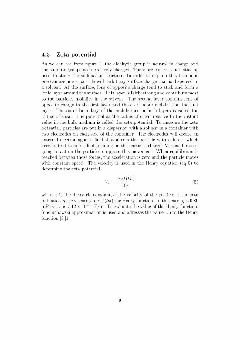
4.3 Zeta potential
As we can see from figure 1, the aldehyde group is neutral in charge andthe sulphite groups are negatively charged. Therefore can zeta potential beused to study the sulfonation reaction. In order to explain this techniqueone can assume a particle with arbitrary surface charge that is dispersed ina solvent. At the surface, ions of opposite charge tend to stick and form aionic layer around the surface. This layer is fairly strong and contribute mostto the particles mobility in the solvent. The second layer contains ions ofopposite charge to the first layer and these are more mobile than the firstlayer. The outer boundary of the mobile ions in both layers is called theradius of shear. The potential at the radius of shear relative to the distantvalue in the bulk medium is called the zeta potential. To measure the zetapotential, particles are put in a dispersion with a solvent in a container withtwo electrodes on each side of the container. The electrodes will create anexternal electromagnetic field that affects the particle with a forces whichaccelerate it to one side depending on the particles charge. Viscous forces isgoing to act on the particle to oppose this movement. When equilibrium isreached between those forces, the acceleration is zero and the particle moveswith constant speed. The velocity is used in the Henry equation (eq 5) todetermine the zeta potential.
Ve =2εzf(ka)
3η(5)
where ε is the dielectric constant,Ve the velocity of the particle, z the zetapotential, η the viscosity and f(ka) the Henry function. In this case, η is 0.89mPa×s, ε is 7.12× 10⁻¹⁰ F/m. To evaluate the value of the Henry function,Smoluchowski approximation is used and adresses the value 1.5 to the Henryfunction.[3][1]
9

5 Result
5.1 Results from IR
As described in section 1.3.1, the IR analysis was used to verify the oxidation.Figure 4 shows the result from the analysis.
Figure 4: Result from IR of the oxidation process.
10

5.2 Results from BET surface analysis
Table 1 shows the result from the BET surface analysis.1A is the BET surfacearea, c is the BET constant and vm the monolayer molar volume adsorbedgas. From equation 2, a linear model is used between 0.05 < P
P0< 0.35, k is
the slope of the line and m is the intercept value of 1
v(
PP0
−1)
Table 1 A [m2g−1] k m vm cDAC beads 56.91 (11) 0.07542 (15) 0.001067 (28) 13.07 71.68
HC Sulf. DAC beads 106.76 (63) 0.04051 (24) 0.000254 (44) 24.53 160.53
5.3 Results from zeta potential analysis
Table 2 shows the result from the zeta potential measurements2. Three zetapotential measurements was performed and ζi is the zeta potential measure-ments with number index i, ζmean the mean value and ζstd is the standarddeviation of the measurements.
Table 2 ζ1[mV] ζ2[mV] ζ3 [mV] ζmean [mV] ζstd [mV]LC-sulf-DAC-beads -20.8 -23.1 -22.4 -22.1 1.18HC-sulf-DAC-beads -36.1 -35.7 -36.3 -36.0 0.306
6 Discussion
From figure 4 it is clear that after 240 hours of oxidation, new peaks havebeen formed at 1730 cm−1 (λ1 = 5.78 cm) and at 885 cm−1 (λ2 = 11.29 cm)in comparison with the initial state of non oxidized Cladophora cellulose.This shows that the oxidation reaction has been succesful since 1730 cm−1
and 885 cm−1 correspond to the aldehyde group.[5]From the BET analysis, table 2, it can be seen that the sulfonated DACbeads have a larger surface area than the non-sulfonated DAC beads. Thisimply that the surface reaction capacity will be improved for the sulfonatedDAC beads since there exist more area for a reaction to occur. However,
1It should have been measurements on low concentration DAC beads. unfortunatelythe sample disappeared and the project time is limited so a remeasuring was not possiblewithin the time frame for this project.
2There should have been measurements on the the DAC beads but unfortunately didthe oxidation process fail because the dried DAC beads were unable to disperse. The pHvalue for the different dispersed solvents was forgotten to be measured as well.
11

the surface of the sulfonated DAC beads will not entirely consist of the sul-phite groups, it will be shared by the remaining aldehyde groups that is leftafter the sulfonation reaction. Another benefit with a larger surface area isthat the amount of sulphite and aldehyde functional group increases. Thepurification process works so that the antibodies attach specific toxins, theidentification between them is possible through the antigen mechanism. Thismechanism means that if a specific toxin/virus or other particles that areharmful to the human body enters inside the body, the antibodies creates anantigen that works as a identification key to the specific toxin/virus so theantibodies can bond with this harmful object.[2] Further on, the antibodiesis attached to aldehyde groups on the materials surface. This means that thenon purified blood runs through dialysis machine, enters a chamber with theanticoagulent material which is acting like a filter so that the toxins attachesto the antibodies on the materials surface and stay there.[4] The incrementof surface area should increase the amount of retained toxins due to morealdehyde groups, however this was not measured and therefore not verifiedin this project.The zeta potential analysis was used to verify the sulfonation reaction. Sincethe aldehyde groups in the DAC beads is neutrally charged and sulphitegroups in the sulfonated DAC beads is negative charged, the materials over-all charge is going to be different before and after the sulfonation reaction.Therefore the sulfonation reaction could be studied using the zeta potential.The results shows that for the zeta potential mean value, the low concentra-tion sulfonated DAC beads got -22.1 mV and the high concentration DACbeads got -36 mV. The higher the concentration of sulfonation agent, themore negatively zeta potential. Regarding the absolute value of the zetapotential, it becomes higher with higher sulfonation concentrations, the signof the potential is dependent on how the reference direction is defined. Asdescribed in section 1.3.3, the zeta potential gives information about thedispersed particles stability in a solvent. From the result of the analysis,the increase of sulfonation agent entails the zeta potential and the kineticstability increases since the radius of shear decreases to keep the potentialenergy that comes from repulsion and attraction in balance against eachother. The potential energy from repulsion is defined as the energy thatrepulsive interactions between charges in the electric double layer produces.The attractive potential energy comes from the van der Waals interactionsbetween the molecules in the particle. However, the reliability of the resultsis effected negative since the pH value was not measured and it play a rolesince high and low pH have different affects on the samples charge. [3].It was observed that the sulfonated DAC beads, regardless of the concentra-tion, were repelled by a metallic spoon when dried. The unsulfonated DAC
12

beads did not show the same repelling properties. This can be understoodsince the sulfonated DAC beads got negatively charged sulfonated functionalgroups on the surface and metallic object is generally surrounded by freeelectrons.
7 Conclusions
The oxidizing reaction of the Cladophora cellulose were successful since theIR spectrum showed that new functional group with new bonds have beenformed. From the BET analysis, it was clear that using more sulfonationagent resulted in a higher surface area. This is of importance since oneof the main functions is to purify blood from toxins and a larger surfacearea may increase the amount of retained toxins. This project is a part ofmaking the nanocellulose Cladophora to a functional material which purposeis to purify blood and act anticouagalent in the dialysis process, other partsuch as analysing the toxicity, testing the anticoagulant properties was notinvolved in this project. With this results, it could be verified that oxidizingand sulfonation reactions was successful and future studies will determine ifit posses anticoagulant and purification properties, suitable for dialysis.
References
[1] Zetasizer nano series user manual. Malvern Instruments Ltd, 4.0 edition,2008.
[2] Bruce Alberts and Alberts-Bray-Hopkin-Johnson-Lewis-Raff-Roberts-Walter, editors. Essential cell biology. Garland, New York, 2. ed edition,2004.
[3] Peter W. Atkins. Physical chemistry. Oxford Univ. Press, Oxford, 5. ed.,reprint edition, 1994.
[4] Ganesh C. Ingavle, Les W.J. Baillie, Yishan Zheng, Elzbieta K. Lis,Irina N. Savina, Carol A. Howell, Sergey V. Mikhalovsky, and Susan R.Sandeman. Affinity binding of antibodies to supermacroporous cryogeladsorbents with immobilized protein A for removal of anthrax toxin pro-tective antigen. Biomaterials, 50:140–153, May 2015.
[5] Hongli Li, Bo Wu, Changdao Mu, and Wei Lin. Concomitant degra-dation in periodate oxidation of carboxymethyl cellulose. CarbohydratePolymers, 84(3):881–886, March 2011.
13

[6] Ning Lin and Alain Dufresne. Nanocellulose in biomedicine: Current sta-tus and future prospect. European Polymer Journal, 59:302–325, October2014.
[7] Carl*1931 Nordling and Jonny Osterman. Physics handbook for scienceand engineering. Studentlitteratur, Lund, 8. ed edition, 2013.
[8] FranA§oise Rouquerol, J. Rouquerol, K. S. W. Sing, P. L. Llewellyn,and G. Maurin. Adsorption by powders and porous solids: principles,methodology and applications. Elsevier/AP, Amsterdam, second editionedition, 2014.
[9] Jianguo Zhang, Nan Jiang, Zheng Dang, Thomas J. Elder, and Arthur J.Ragauskas. Oxidation and sulfonation of cellulosics. Cellulose, 15(3):489–496, June 2008.
14