Modification of flower color pigments and color ... et al.pdf · had no significant effect on the...
11
Academia Journal of Agricultural Research 5(6): 000-000, June 2017 DOI: 10.15413/ajar.2017.0137 ISSN: 2315-7739 ©2017 Academia Publishing Research Paper Modification of flower color pigments and color composition with hormonal treatments and sucrose in Tulipa gesneriana 'Kingsblood' Accepted 7 th June, 2017 ABSTRACT An experiment was conducted to investigate the interplay between three phytohormones and sucrose for the change of flower color composition and the plant secondary metabolites in Tulipa. The variations of the physiological and morphological characteristics with abscisic acid (ABA) at 5 and 10 mg/L, gibberellic acid (GA3) at 300 and 500 mg/L, jasmonic acid (JA) at 50 and 100 μm and their interactions with sucrose at 1 and 2 g/L were analyzed. By reviewing the HPLC charts and UV-Vis spectra, it was found that the production of plant secondary metabolites, total flavonoids and anthocyanins composition pigments was influenced by the foliar application of the plant hormones. The sucrose alone had no significant effect on the quantification of different phytochemicals, but the interaction with JA and ABA showed a considerable variation in the anthocyanin accumulation and total flavonoids. Both JA and ABA hormones, in spite of enhanced anthocyanin accumulation and increased cyanidin and pelargonidin pigments’ percentage were associated with reduced vegetative growth parameters as well as, post-harvest performance compared to the control plants. However, GA3 at 500 mg/L without sucrose played a key role in the accumulation of anthocyanins, post-harvest performance and increase of the three major anthocyanin pigments. Moreover, the data provide evidence of interference between the sucrose and GA3 in the regulation of the anthocyanin accumulation. Key words: Anthocyanins pigments, abscisic acid, jasmonic acid, gibberellic acid, total flavonoids. INTRODUCTION Anthocyanin pigments play a major role in the color composition of the tulip flowers. Therefore, enhancement of flower color by altering anthocyanin production and the composition of pigments for the purpose of improving the quality of the flowers can be of great importance in the flower industry. The variation of flower color is influenced by many endogenous and exogenous factors, but the plant growth regulators and sucrose have the greatest impact (Zhao et al., 2015). Recent studies have shown that the sucrose as the main form of transport of carbon in addition to its contribution to the carbon skeleton of the plant cells and as a source of energy, acts as a molecular signal. The role of sucrose as inducer molecule or molecular signal is certainly proven in many of the plants (Bolouri‐Moghaddam et al., 2010). Moreover, sucrose can activate the expression of the genes involved in the biosynthesis of anthocyanins and flavonoids in different in vivo and in vitro cultures (Teng et al., 2005). The ability of sucrose to induce the biosynthesis of anthocyanin pigments in various species of plants such as Arabidopsis (Solfanelli, 2006), grapevine (Pirie, 1976) and radish (Hara et al., 2003) has been proven. The activation process of anthocyanin biosynthesis pathway in the petals and other organs is the result of a Hojjati Yaghoub, Tehranifar Ali, Abedi Bahram and Shoor Mahmood* Department of Horticulture, Faculty of Agriculture, Ferdowsi University of Mashhad, P.O. Box 91775-1163, Mashhad, Iran. *Corresponding author. E-mail: [email protected]
Transcript of Modification of flower color pigments and color ... et al.pdf · had no significant effect on the...

Academia Journal of Agricultural Research 5(6): 000-000, June 2017 DOI: 10.15413/ajar.2017.0137 ISSN: 2315-7739 ©2017 Academia Publishing
Research Paper
Modification of flower color pigments and color composition with hormonal treatments and sucrose in Tulipa gesneriana 'Kingsblood'
Accepted 7th June, 2017 ABSTRACT An experiment was conducted to investigate the interplay between three phytohormones and sucrose for the change of flower color composition and the plant secondary metabolites in Tulipa. The variations of the physiological and morphological characteristics with abscisic acid (ABA) at 5 and 10 mg/L, gibberellic acid (GA3) at 300 and 500 mg/L, jasmonic acid (JA) at 50 and 100 µm and their interactions with sucrose at 1 and 2 g/L were analyzed. By reviewing the HPLC charts and UV-Vis spectra, it was found that the production of plant secondary metabolites, total flavonoids and anthocyanins composition pigments was influenced by the foliar application of the plant hormones. The sucrose alone had no significant effect on the quantification of different phytochemicals, but the interaction with JA and ABA showed a considerable variation in the anthocyanin accumulation and total flavonoids. Both JA and ABA hormones, in spite of enhanced anthocyanin accumulation and increased cyanidin and pelargonidin pigments’ percentage were associated with reduced vegetative growth parameters as well as, post-harvest performance compared to the control plants. However, GA3 at 500 mg/L without sucrose played a key role in the accumulation of anthocyanins, post-harvest performance and increase of the three major anthocyanin pigments. Moreover, the data provide evidence of interference between the sucrose and GA3 in the regulation of the anthocyanin accumulation. Key words: Anthocyanins pigments, abscisic acid, jasmonic acid, gibberellic acid, total flavonoids.
INTRODUCTION Anthocyanin pigments play a major role in the color composition of the tulip flowers. Therefore, enhancement of flower color by altering anthocyanin production and the composition of pigments for the purpose of improving the quality of the flowers can be of great importance in the flower industry. The variation of flower color is influenced by many endogenous and exogenous factors, but the plant growth regulators and sucrose have the greatest impact (Zhao et al., 2015).
Recent studies have shown that the sucrose as the main form of transport of carbon in addition to its contribution to the carbon skeleton of the plant cells and as a source of
energy, acts as a molecular signal. The role of sucrose as inducer molecule or molecular signal is certainly proven in many of the plants (Bolouri‐Moghaddam et al., 2010). Moreover, sucrose can activate the expression of the genes involved in the biosynthesis of anthocyanins and flavonoids in different in vivo and in vitro cultures (Teng et al., 2005). The ability of sucrose to induce the biosynthesis of anthocyanin pigments in various species of plants such as Arabidopsis (Solfanelli, 2006), grapevine (Pirie, 1976) and radish (Hara et al., 2003) has been proven.
The activation process of anthocyanin biosynthesis pathway in the petals and other organs is the result of a
Hojjati Yaghoub, Tehranifar Ali, Abedi Bahram and Shoor Mahmood* Department of Horticulture, Faculty of Agriculture, Ferdowsi University of Mashhad, P.O. Box 91775-1163, Mashhad, Iran. *Corresponding author. E-mail: [email protected]

complex interaction between plant growth regulators and sucrose (Weiss, 2000). In previous studies, it was proven that the three main groups of the phytohormones contain gibberellic acid (GA3), jasmonic acid (JA) and abscisic acid which are involved in the biosynthesis of anthocyanin pigments (Medina-Puche et al., 2014). Each of these hormones has a different situation process to change and regulate anthocyanin biosynthetic pathway and the synthesis of secondary metabolites. However, all of these hormones, whether directly or indirectly, act by affecting the expression or suppression of related genes involved in the biosynthesis of anthocyanin and flavonoids (Loreti et al., 2008).
The effect of GA3 on anthocyanin synthesis was reported in many different plants. Ohlsson and Berglund (2001) studied the influence of GA3 on anthocyanin metabolism in cell culture of Catharanthus roseus and observed increased anthocyanin accumulation. The influence of GA3 in vivo and in vitro cultures of periwinkle (Piovan and Filippini, 2007), gerbera (Danaee et al., 2011) and Hyacinthus (Hosokawa, 1999) was observed. The involvement of JA on anthocyanin synthesis is demonstrated by some experimental evidence in various plant species and tissues (Steyn et al., 2002). The effects of JA in Arabidopsis thaliana (Qi et al., 2011), Tulipa (Saniewski et al., 1998a), grapevine cell cultures (Belhadj et al., 2008) and in apple fruit (Kondo et al., 2001) were also reported. The results obtained from the exogenous applications of ABA showed that depending on the concentration and the plant species, ABA stimulated the synthesis of anthocyanin (Jaakola, 2015).
Furthermore, some studies show that ABA can induce biosynthesis pigment in Fragaria ananassa (Medina-Puche et al., 2014), Litchi chinensis (Lai et al., 2014) and sweet cherry (Shen et al., 2014). The aim of this research was to study the interaction between sucrose and plant hormones on the variation of flavonoids, anthocyanin and color composition pigments, as well as, to evaluate the physiological and morphological changes of tulip plants. MATERIALS AND METHODS Plant materials and treatments The tulip plants (T. gesneriana 'Kingsblood') used for this study were grown in the fields of the Netherlands, with a cover of 11 to 12 cm in circumference and were purchased from a commercial grower. The bulbs were grown in individual pots filled with three growth beds: peat, coco peat and perlite in equal ratio. After planting, these pots were stored at 5°C for 14 weeks and thereafter transferred to the greenhouse to be grown at 20°C/15°C (day/night). During the growing phase, the plants were irrigated with the normal Hoagland nutrient solution. The plants were separately sprayed at three stages of the inflorescence development with ABA at 5 and 10 mg/L, GA3 at 300 and
500 mg/L and 50 and 100 µm of JA and their interaction with 1 and 2 g/L sucrose. In all the treatments, the spray solutions contained 0.2% Tween-20 as a surfactant. At flowering, the measured characteristics in this experiment included days to bud appearance, total plant weight, total plant length, flower stalk length, weight of fresh flower, weight of fresh flower, vase life, water content of leaf and water content of flower. The petal samples for total anthocyanin, flavonoid and HPLC detection were collected from the tulip plants in a fully developed stage when the petals were fully pigmented. The samples were collected, weighed, immediately frozen in liquid nitrogen and stored at −80°C until it was ready for use.
Estimation of proline content The free proline content was determined according to the method described by Bates et al. (1973) by measuring the quantity of the colored reaction product of proline with ninhydrin acid. The absorbance was read at 520 nm. The amount of proline was calculated from the previously plotted standard curve and expressed in μmol/gfw.
Total flavonoid content Aluminum chloride colorimetric method of Woisky and Salatino (1998) was used in the determination of total flavonoids. The plant extract (0.5 ml of 1:10 g/ml) was mixed with 1.5 ml of methanol, 0.1 ml of 10% aluminum chloride, 0.1 ml of 1M potassium acetate and 2.8 ml of distilled water. It remained at room temperature for 30 min. The absorbance of the reaction mixture was measured at 415 nm in triplicate. The calibration curve was prepared with quercetin solutions at concentrations 10 to 100 μg/ml in methanol.
Isolation of anthocyanin and estimation of total anthocyanin content Petal slices (0.5 g of each treatment) were macerated in 1 ml acidic ethanol solution (1% HCL in MeOH) in a test tube and allowed to equilibrate overnight at 4°C. These samples were centrifuged at 12000 g for 10 min at 4°C, leaving the anthocyanin in the supernatant. Thereafter, the total anthocyanin of the petals extracted was measured using the pH differential method as described by (Lee et al., 2005). Quantities of anthocyanin were determined in cyanidin-3-glucoside (MW=449.2, molar absorbance, ε=26.900) for tulip materials. Identification and quantification of anthocyanin pigments in the perianthes by HPLC The flower colors of perianthes were evaluated by
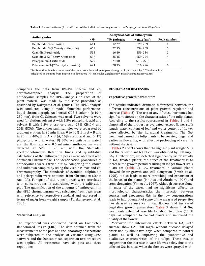
Table 1: Retention times (Rt) and max of the individual anthocyanins in the Tulipa gesneriana 'Kingsblood'.
Anthocyanins Analytical data of anthocyanin
x M+ Y Rt (min)ODS z max (nm) Peak number
Delphinidin 3-rutinoside 611 12.27 529, 269 1
Delphinidin 3-(2'''-acetylrutinoside) 653 22.55 534, 269 5
Cyanidin 3-rutinoside 595 16.40 559, 254 2
Cyanidin 3-(2'''-acetylrutinoside) 637 25.45 559, 254 4
Pelargonidin 3-rutinoside 579 24.00 516, 276 3
Pelargonidin 3-(2'''-acetylrutinoside) 621 28.35 516, 276 6
YRt: Retention time is a measure of the time taken for a salute to pass through a chromatography ODS column. It is calculated as the time from injection to detection; xM+: Molecular weight and z max: Maximum absorbance.
comparing the data from UV–Vis spectra and co-chromatographed analysis. The preparation of anthocyanin samples for HPLC analysis on each of the plant material was made by the same procedure as described by Nakayama et al. (2004). The HPLC analysis was conducted using a model Shimadzu performance liquid chromatograph. An Inertsil ODS-2 column (φ10 × 250 mm), from GL Sciences was used. Two solvents were used for elution: solvent A with 1.5% phosphoric acid and solvent B with 1.5% phosphoric acid, 25% CH3CN, and 20% HCO2H. The anthocyanin samples were separated by gradient elution: in 20 min linear 0 to 40% B in A + B and in 20 min 40% B in A + B (A, 10% acetic acid and 0 .1% phosphoric acid in water; B) 50% acetonitrile in water) and the flow rate was 0.6 ml min-1. Anthocyanins were detected at 520 ± 20 nm with the Shimadzu spectrophotometer. Retention times and quantitative calculations of the anthocyanin peaks were obtained with Shimadzu Chromatopac. The identification procedures of anthocyanins were carried out by comparing the known and unknown samples by using the visible max and co-chromatography. The standards of cyanidin, delphinidin and pelargonidin were obtained from Chromadex (Santa Ana, CA). For quantification, peak areas were correlated with concentrations in accordance with the calibration plot. The quantification of the amounts of anthocyanin in the HPLC chromatograms was calculated from peak areas with reference to respective standard and expressed in terms of mg/g fresh weight sample (Torskangerpoll et al., 2005). Statistical analysis The experiment was conducted based on Completely Randomized Design (CRD). The data obtained from the measurements of the pots and the laboratory observations were subjected to the analysis of variance using SPSS software and the Duncan mean separation test procedure was applied. All treatments have six pots and three repetitions.
RESULTS AND DISCUSSION Vegetative growth parameters The results indicated dramatic differences between the different concentrations of plant growth regulator and sucrose (Table 2). The use of any of these hormones has significant effects on the characteristics of the tulip plants. According to the results represented in Tables 2 and 3, almost all of the properties evaluated, except flower stalk length, water content of leaf and water content of flower were affected by the hormonal treatments. The GA3
treatment caused the tulip plants to be heavier, longer and earlier in flowering, with effective prolonging of vase life without abscission.
Tables 2 and 3 shows that the highest plant weight 65 g and the tallest plant 63.21 cm was obtained by 500 mg/L GA3. Furthermore, we detected significantly faster growth in GA3 treated plants; the effect of the treatment is to increase the growth period resulting in longer flower stalk 40.08 cm (Table 2). GA3 treatment in various plants showed faster growth and cell elongation (Smith et al., 1996). It also leads to more stretching and expansion of the leaves of the plants (Pinthus and Abraham, 1996) and stem elongation (Yim et al., 1997). Although sucrose alone, in most of the cases, had no significant effects on morphological characteristics, the interaction between sources and exogenous GA3 in the low concentrations leads to improvement of some of the measured properties like delayed senescence in cut flowers and increased vegetative growth parameters. Table 3 shows that GA3 treatments extended vase life for about two days (11.58 days) as compared to control plants and improved the quality of the flower.
Moreover, the interaction effects between GA3 with sucrose show GA3 500 mg/L without sucrose delayed abscission by about two days when compared to control plants, as well as, improving the quantitative and qualitative characteristics of the tulips. These results suggest that the increase in vase life was solely due to the effect of GA3 because when the flowers were sprayed with
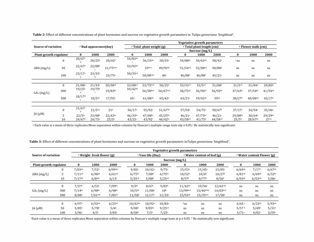
Table 2: Effect of different concentrations of plant hormones and sucrose on vegetative growth parameters in Tulipa gesneriana 'kingblood'.
Source of variation y Bud appearance(day)
Vegetative growth parameters y Total plant weight (g) y Total plant length (cm) y Flower stalk (cm)
Sucrose (mg/L)
Plant growth regulator 0 1000 2000
0 1000 2000
0 1000 2000
0 1000 2000
ABA (mg/L)
0 20/67c
d 20/25d 20/42d
54/83ab
c 56/33ab 58/33a 54/88bc 56/63ab 58/42a z ns ns ns
50 22/67a
b 22/08b
c 21/75bcd
53/92ab
c 53bcd 49/92de 51/54cd 52/08cd 50/88d ns ns ns
100 23/17a
b 23/33a
b 23/75a
50/33cd
e 50/08cde 46e 46/88e 46/88e 45/21e ns ns ns
GA3 (mg/L)
0 21/08a 21/33a 20/58ab
53/08cd 53/75bcd 50/25d
53/33cd 52/5cd 51/08d
31/5cd 31/04d 29/83d
300 19/25c
d 19/75b
c 19/83bc
59/42ab
c 56/58bcd 56/67bcd 58/75ab 56/96bc 56/92bc 37/63bc 37/54bc 41/54ab
500 18/17d
e 18/5de 17/92e 65a 61/08ab 65/42a 63/21a 59/42ab 59ab 38/27b 40/08ab 45/17a
JA (µM) 0
21/67c
d 21/5cd 21d
56/17a 55/92a 51/67ab
57/54a 54/75a 50/67b
37/17a 34/54a 35/46a
5 22/5bc 23/08b 22/42bc 46/33bc 47/08bc 45/25bc 46/21c 47/75bc 46/21c 29/88bc 30/64b 29/29bc 10 24/67a 24/75a 25/5a 43/25 c 43/92c 46/42bc 43/58cd 41/75d 44/58cd 25/5d 28/67bc 27cd
y Each value is a mean of three replicates.Mean separation within columns by Duncan’s multiple range tests atp ≤ 0.05.z Be statistically non-significant.
Table 3: Effect of different concentrations of plant hormones and sucrose on vegetative growth parameters inTulipa gesneriana 'kingblood'.
Source of variation
Vegetative growth parameters
z Weight fresh flower (g) z Vase life (Day) z Water content of leaf (g) z Water content Flower (g)
Sucrose (mg/L)
Plant growth regulator 0 1000 2000
0 1000 2000
0 1000 2000
0 1000 2000
ABA (mg/L)
0 7/03ab 7/52a 6/99abc 9/83a 10/42a 9/75a 15/53a 15/45a 15/05a 6/69ab 7/17a 6/67ab
5 7/11ab 6/98ab 6/61ab 6/75bc 7/08b 6/75bc 10/52b 10/6b 10/27b 6/81ab 6/69ab 6/32bc
10 7/17ab 6/8abc 6/13c 5/25cd 5/08d 5/25cd 8/57b 8/77b 8/56b 6/93ab 6/53abc 5/86c
GA3 (mg/L)
0 7/2abc 6/53c 7/09bc
9/5bc 8/67c 9/83b
11/42de 10/56e 12/62cde
ns ns ns
300 7/14bc 6/98bc 6/98bc 10/5ab 11/08a 10b 13/99bcd 13/46bcde 14/03bcd ns ns ns
500 8/08a 7/41abc 7/85ab 11/58a 11/17a 11/33a 15/93ab 15/35abc 17/58a ns ns ns
JA (µM)
0 6/97a 6/55ab 6/25ab
10/42ab 10/92a 10/83a
Yns ns ns
6/65 a 6/23ab 5/93ab
50 5/85b 5/78b 5/6b 9/08d 9/83bc 9/25cd ns ns ns 5/57 b 5/49b 5/31b
100 3/96c 4/3c 3/83c 8/58d 7/5e 7/25e ns ns ns 3/71 c 4/02c 3/59c
zEach value is a mean of three replicates.Mean separation within columns by Duncan’s multiple range tests at p ≤ 0.05. Y Be statistically non-significant.

GA3 combined with sucrose, the vase life did not show any significant difference from when the flowers were sprayed with only GA3. These results correspond to the observations by Imsabai et al. (2013). Generally, GA3 may synergistically improve post-production quality of Kingsbloodtulips. Therefore, we conclude that sprays containing both GA3 might be of commercial value in enhancing the post-production quality of tulip flowers, but in the case of JA, the treatments showed a contradictory effect.
According to Tables 2 and 3, most of the vegetative characteristics of treated plants showed a considerably significant decrease, that is, height, weight and vase life went down and the treated flowers exhibited tepals abscission after harvest with slight wilting of the leaves. On the other hand, comparison of the data obtained from ABA treatments showed delayed flowering (three more days later) and reduced vegetative growth parameters like plant length (45.21 cm) is about 10 cm smaller than the control plants. The results of contemporary studies show the inhibitory effect of ABA on shoot growth and flowering induced by endogenous GA3 in tulips. Our results indicate ABA 10 mg/L and JA of over 100 µm could lead to unwanted early senescence of matured tepals of cut tulips. The lowest vase life of cut flowers is related to 10 mg/L ABA (5.08 days) and JA of over 100 µm (7.5 days). Hence, we concluded that ABA and JA play a pivotal role in tepals senescence, leaf chlorosis, and gummosis in Kingsblood tulips during the post-production evaluation. The senescence of tulip flowers seems to be co-ordinately mediated by the interactions of plant hormones in a way that ethylene plays a minor role (Kim and Miller, 2008). Although ethylene seems not be directly involved in the senescence of tulip tepals, we observed that exogenously applied high concentrations of ABA and JA may trigger a higher rate of ethylene production, resulting in the unusual senescence and abscission of the tepals (Van Doorn, 2002).
Generally, there was a positive correlation between vase life and water content and dry weight of tepals and leaves after treatment with plant growth regulators. The relative water content of tepals and leaf were significantly affected by GA3 and JA treatments (Table 3). The relative water content of tepals and leaves were reduced by ABA and JA, as compared to control plants. The least amount of flower water content (3.71 g) was related to JA treatments and the highest amount of water content in leaf was recorded in GA3 (17.58 g) treatment (Table 3).
The application of GA3 in combination with sucrose not only improved the water content of leaves, but also increased the water content of flower by more than 25% in treated tulips (Table 3). It has been scientifically proven that exogenous application of GA3 serves to improve petal water relations by increasing the level of osmotic solutes and it delays cell death. All these effects of GA3 treatment manifest its great potential for improving the quality and yield of 'Kingsblood’ tulips grown for the cut flower. Therefore, we concluded that sprays containing GA3 plus
sucrose might be of commercial value in enhancing post-production quality of tulip flowers. Though some characteristics were non-significant, generally, there was a positive correlation between treatment with the plant growth regulators and vase life, water content and dry weight of the tepals and leaves. Proline content Due to protein degradation in plant elements, the accumulation of free amino acids, especially proline content increased and could not be transported out to other plant organs. Though this was expected in protease activity, it is a sign of senescence (Silveira et al., 2003). Alongside increased proline levels, a widely-distributed multi-functional osmoprotectant (Szabados and Savoure, 2010) is also often associated with increased sucrose levels, demonstrating that proline synthesis also depends on sucrose-specific signaling events. These increased the levels of endogenous sucrose to increase the biosynthesis of pigments (Van den Ende and El-Esawe, 2014). The increase in the levels of proline on one hand signifies active genes involved in anthocyanin biosynthesis and on the other hand, is a sign of senescence. A previous study showed that application of some of the plant growth regulator can promote the biosynthesis of proline in water limited environments (Bhaskara et al., 2015). Based on our results, treatments that increased the proline content (JA and ABA) showed the shortest vase life and poor vegetative growth parameters (Tables 3 and 4).
In this study, the amounts of proline were found to be significantly increased in JA treatments as compared to the control leaves (Table 4). Although different values were determined for proline content with use of GA3, there was no significant difference from the control leaves. The lowest amount of proline was found in GA3(0/69 μmol/gfw) treatments leaves and the highest free proline accumulation resulted from 100 µm of JA treatment (8.10 μmol/gfw), while exogenous foliar application of ABA led to a slight increase in the proline content (1.24 μmol/gfw) (Table 4). Therefore, it is evident that JA did accelerate protein degradation and increased the free level of proline content, resulting from proteolysis which indicates typical processes of senescence. In general terms, tulips that are sprayed with JA demonstrate this phenomenon best. The results proved that senescence in tulip tepals is closely associated with free proline accumulation; similar to what is found in rice (Yang et al., 2000). The effect of the treatments on total flavonoids content The total flavonoid content (TFC) in treated tulip flower was found to have a significantly wide variation (Table 4). The highest TFC was found in the perianthes of the tulips

Table 4: Effect of different concentrations of plant hormones and sucrose on physiological parameters in Tulipa gesneriana 'kingbloods'.
Source of variation
Physiological parameters
z Flavonoid content (mg/gfw) z Anthocyanin content (mg/L) z Proline content (μmol/gfw) z Dry weight of flower ( mg)
Sucrose (mg/L)
Plant growth regulator 0 1000 2000
0 1000 2000
0 1000 2000
0 1000 2000
ABA(mg/L)
0 2/244c 2/306c 2/32c 1/588c 1/476 c 1/662c 0/77c 0/70d 1/24a 335/75a 341/83a 321/58a
5 2/616b 2/624b 1/098a 1/352cd 2/256b 2/764a 0/87b 0/73dc 1/24a 294b 311/5bc 288/42bc
10 2/488bc 2/382c 1/666d 1/588c 1/176d 1/214d 0/70d 0/87b 0/73d 239/67d 280/92c 270/17c
GA3(mg/L)
0 2/124d 2/22d 2/314cd
1/582d 1/552d 1/678d
0/93b 1/04a 0/72d
311/75bc 287/42d 281/75d
300 2/536c 2/732bc 2/644bc 2/102b 1/944c 2/104b 0/81c 1/00a 0/69d 309/17abc 311/5bc 302/75cd
500 2/912a 2/34cd 2/252d 2/406a 2/112b 2/124b 0/87b 0/89b 0/81c 346/42a 331/08ab 326/33ab
JA(µm)
0 2/33c 2/334c 1/984c
1/586c 1/596c 1/712bc
0/70f 0/72f 1/17e
316/67a 315/67a 326/33a
50 2/346b 2/856a 1/68d 1/52c 2/256a 1/838b 3/24d 5/24c 7/22b 283/67b 292/33b 270/17b
100 1/276e 1/464d 1/986c 1/792b 1/052d 1/152d 3/54d 7/35b 8/10a 248/75c 280/75b 245/83c
zEach value is a mean of three replicates.Mean separation within columns by Duncan’smultiple range tests at p ≤ 0.05.
sprayed with 500 mg/L GA3 (2.91 mg/gfw) and the lowest concentration recorded in the control plants and those treated with 1 g/L sucrose (2.12 mg/gfw) (Table 4). The results indicate that both sucrose and GA3 increased the flavonoid content in lower concentrations, but in higher concentration was reduced to 2/25 mg/gfwTFC. The two compounds act to suppress each other's effect in higher concentrations. These results support the findings of Sarrou et al. (2015) who reported that flavonoid levels of plants were significantly affected by GA3 and GA3 also significantly promotes secondary metabolites.
The results obtained after the use of ABA and JA showed significant impact on TFC, both hormones increased TFC, but in higher concentrations showed negative effects (Table 4). ABA at 10 mg/L with sucrose at 2 g/L had decreasing effect on TFC and was reduced to 1/66 mg/gfw. The interaction of these hormones with sucrose in low concentrations showed a synergetic effect and the highest range of
TFC obtained with ABA at 5 mg/L and sucrose at 1 g/L (2.62 mg/gfw). In the case of Orthosiphon aristatus, the use of ABA can enhance the production of primary and secondary metabolites, which was confirmed by our results (Ibrahim and Jaafar, 2013). Our study showed that increased application of ABA improved the production of total phenolics and flavonoids. It has long been proven that ABA naturally accumulates in grape skins at the onset of ripening, a time when anthocyanin and other phenolic compounds also increase (Koyama et al., 2010).
Furthermore, flavonoids production is enhanced by 50 µm JA to 2.85 mg/gfw, but increasing the level of sucrose to 2 g/L leads to decreased range of TFC to 1.08 mg/gfw. It is already known that low concentrations of jasmonate induce the expression of the genes encoding enzymes of flavonoid biosynthesis (Wasternack and Parthier, 1997). Our data showed increasing the levels of JA and sucrose leads to the decrease of TFC. Table 4 showed the
minimum range in 100 µm of JA and 0 g/L of sucrose (1.27 mg/gfw). The low concentrations of jasmonate have been shown to induce the gene expression of the enzymes involved in flavonoid biosynthesis (Walker et al., 2002). Moreover, this conclusion is in accordance with the results obtained for TFC; there were similar effects on TFC with the use of ABA and JA. However, in low concentrations both ABA and JA had the effect of increasing TFC, but in increased concentrations led to a decreased range of TFC. Perianthes coloration and accumulation of anthocyanins Endogenous anthocyanins of perianthes in dark red tulip were analyzed by HPLC and UV-Vis spectra. Six anthocyanins Pelargonidin 3-rutinoside (Peak 3), Pelargonidin 3-(2'''-acetylrutinoside) (Peak 6), Cyanidin 3-rutinoside (Peak 2), Cyanidin 3-(2'''-acetylrutinoside) (Peak 4), Delphinidin 3-rutinoside

Table 5: Effect of different concentrations of plant hormones and sucrose on anthocyanin compostion pigmentsin Tulipa gesneriana 'kingbloods'.
Source of variation Distribution and amounts of individual anthocyanins x
Delphinidin (%) x Cyanidin (%) x Pelargonidin (%) y TRA(mg/gfw)
Sucrose (mg/L)
Plant growth regulator 0 1000 2000
0 1000 2000
0 1000 2000
0 1000 2000
ABA(mg/L)
0 66/1506
4 61/71463 65/84426
30/28594
34/47843
30/3061 3/563418 3/80693
4 3/84963
5 0/3524014 0/368319
0/4005826
5 71/4982
6 70/06309 69/1177
25/48503
28/6902 29/9446
5 3/016715
1/246702
0/937648
0/3258214 0/386456
6 0/467420
4
10 68/7629
1 73/49828 63/92256
28/96806
24/72523
34/17429
2/269029 1/77649
5 1/90315 0/3922752
0/2822552
0/2129336
GA3(mg/L)
0 67/9308
2 61/20779 65/75432
28/78385
34/90762
30/96713
3/285329 3/88459 3/27854
4
0/3470114 0/624721
4 0/326908
4
300 59/371 63/26614 64/80391 35/7056
2 33/1607
4 30/7762
7 4/923388
3/573122
4/419825
0/5527942 0/334837
8 0/482648
8
500 59/8546
8 62/98213 63/71821
36/10605
33/34836
32/74241
4/039275 3/66951
3 3/53937
5 0/6876854 0/438159
0/3841826
JA(µm)
0 67/7709
1 52/36654 54/60278
28/70659
43/20551
41/00811
3/522498 4/42795
5 4/38910
7
0/3491376 0/394664 0/525188
2
50 61/6975
7 64/49078 71/49667
35/15972
32/68034
26/26401
3/142713 2/82887
3 2/23931
9 0/3821896
0/4371642
0/6029166
100 74/4804 81/62155 77/90853 24/0859
3 16/5197
19/28372
1/43367 1/85875
5 2/80774
5 0/4029992
0/2409726
0/2030494
x The area of each pigment in the HPLC-chromatogram relative to the total area of three pigments for this sample detected at 520±20 nm y TRA: (Total relative amount of anthocyanin); Data represent
relative amount of anthocyanin composition from total absorbed peaks areas that detected at 520±20 nm by HPLC-chromatogram. Each value is a mean of three replicates. Assignments are supported by published data (Nakayama et al., 2004; Torskangerpoll et al., 2005).
(Peak 1) and Delphinidin 3-(2'''-acetylrutinoside) (Peak 5) were identified by comparing the known samples using the visible max (Table 1). The retention times (Rt) and absorption spectra ( max) of anthocyanin 5 and 3 coincided with those of pelargonidin 3-rutinoside and cyanidin 3-rutinoside, respectively. Anthocyanins 2 and 4 were extracted from tulip perianthes and purified using chromatography methods. The molecular weights (M+) were 621 and 637, suggesting that these anthocyanins are the acetyl ester of pelargonidin 3-
rutinoside and cyanidin 3-rutinoside (Table 1). Tables 4 and 5 show the distribution and amounts of individual anthocyanins. The effect of jasmonic acid on perianthes coloration and accumulation of anthocyanins The amount of anthocyanin content in perianthes was enhanced by the use of JA treatments and sucrose (Table 4). The highest range of total
anthocyanin was quantified in flowers that received 50 µm JA and 2 g/L sucrose (1.83 mg/L) and the HPLC chart shows that higher amounts of anthocyanin accumulated in the petals that received this treatment. On the other hand, the highest amount of absorbed peak was recorded for 50 µm JA treatment and 2 g/L sucrose (0.60 mg/L).
There are many research studies that proved that jasmonates induce the formation or the accumulation of anthocyanin, for example in peach shoots (Saniewski et al., 1998b), tulip stems
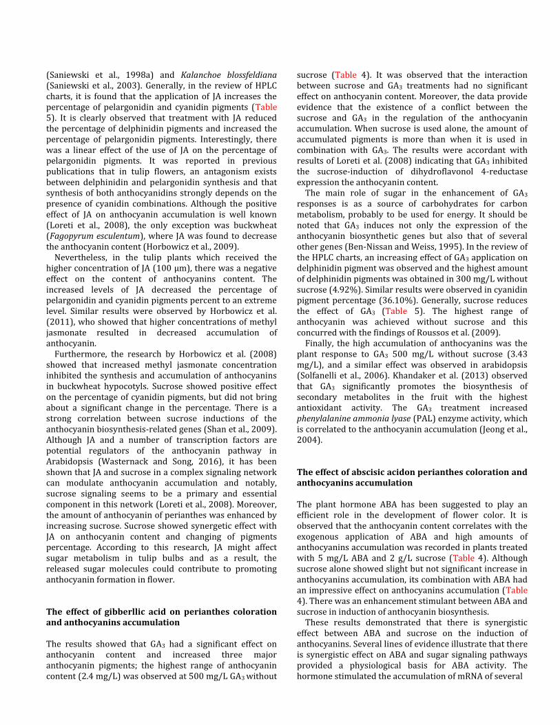
(Saniewski et al., 1998a) and Kalanchoe blossfeldiana (Saniewski et al., 2003). Generally, in the review of HPLC charts, it is found that the application of JA increases the percentage of pelargonidin and cyanidin pigments (Table 5). It is clearly observed that treatment with JA reduced the percentage of delphinidin pigments and increased the percentage of pelargonidin pigments. Interestingly, there was a linear effect of the use of JA on the percentage of pelargonidin pigments. It was reported in previous publications that in tulip flowers, an antagonism exists between delphinidin and pelargonidin synthesis and that synthesis of both anthocyanidins strongly depends on the presence of cyanidin combinations. Although the positive effect of JA on anthocyanin accumulation is well known (Loreti et al., 2008), the only exception was buckwheat (Fagopyrum esculentum), where JA was found to decrease the anthocyanin content (Horbowicz et al., 2009).
Nevertheless, in the tulip plants which received the higher concentration of JA (100 µm), there was a negative effect on the content of anthocyanins content. The increased levels of JA decreased the percentage of pelargonidin and cyanidin pigments percent to an extreme level. Similar results were observed by Horbowicz et al. (2011), who showed that higher concentrations of methyl jasmonate resulted in decreased accumulation of anthocyanin.
Furthermore, the research by Horbowicz et al. (2008) showed that increased methyl jasmonate concentration inhibited the synthesis and accumulation of anthocyanins in buckwheat hypocotyls. Sucrose showed positive effect on the percentage of cyanidin pigments, but did not bring about a significant change in the percentage. There is a strong correlation between sucrose inductions of the anthocyanin biosynthesis-related genes (Shan et al., 2009). Although JA and a number of transcription factors are potential regulators of the anthocyanin pathway in Arabidopsis (Wasternack and Song, 2016), it has been shown that JA and sucrose in a complex signaling network can modulate anthocyanin accumulation and notably, sucrose signaling seems to be a primary and essential component in this network (Loreti et al., 2008). Moreover, the amount of anthocyanin of perianthes was enhanced by increasing sucrose. Sucrose showed synergetic effect with JA on anthocyanin content and changing of pigments percentage. According to this research, JA might affect sugar metabolism in tulip bulbs and as a result, the released sugar molecules could contribute to promoting anthocyanin formation in flower. The effect of gibberllic acid on perianthes coloration and anthocyanins accumulation The results showed that GA3 had a significant effect on anthocyanin content and increased three major anthocyanin pigments; the highest range of anthocyanin content (2.4 mg/L) was observed at 500 mg/L GA3 without
sucrose (Table 4). It was observed that the interaction between sucrose and GA3 treatments had no significant effect on anthocyanin content. Moreover, the data provide evidence that the existence of a conflict between the sucrose and GA3 in the regulation of the anthocyanin accumulation. When sucrose is used alone, the amount of accumulated pigments is more than when it is used in combination with GA3. The results were accordant with results of Loreti et al. (2008) indicating that GA3 inhibited the sucrose-induction of dihydroflavonol 4-reductase expression the anthocyanin content.
The main role of sugar in the enhancement of GA3 responses is as a source of carbohydrates for carbon metabolism, probably to be used for energy. It should be noted that GA3 induces not only the expression of the anthocyanin biosynthetic genes but also that of several other genes (Ben-Nissan and Weiss, 1995). In the review of the HPLC charts, an increasing effect of GA3 application on delphinidin pigment was observed and the highest amount of delphinidin pigments was obtained in 300 mg/L without sucrose (4.92%). Similar results were observed in cyanidin pigment percentage (36.10%). Generally, sucrose reduces the effect of GA3 (Table 5). The highest range of anthocyanin was achieved without sucrose and this concurred with the findings of Roussos et al. (2009).
Finally, the high accumulation of anthocyanins was the plant response to GA3 500 mg/L without sucrose (3.43 mg/L), and a similar effect was observed in arabidopsis (Solfanelli et al., 2006). Khandaker et al. (2013) observed that GA3 significantly promotes the biosynthesis of secondary metabolites in the fruit with the highest antioxidant activity. The GA3 treatment increased phenylalanine ammonia lyase (PAL) enzyme activity, which is correlated to the anthocyanin accumulation (Jeong et al., 2004). The effect of abscisic acidon perianthes coloration and anthocyanins accumulation The plant hormone ABA has been suggested to play an efficient role in the development of flower color. It is observed that the anthocyanin content correlates with the exogenous application of ABA and high amounts of anthocyanins accumulation was recorded in plants treated with 5 mg/L ABA and 2 g/L sucrose (Table 4). Although sucrose alone showed slight but not significant increase in anthocyanins accumulation, its combination with ABA had an impressive effect on anthocyanins accumulation (Table 4). There was an enhancement stimulant between ABA and sucrose in induction of anthocyanin biosynthesis.
These results demonstrated that there is synergistic effect between ABA and sucrose on the induction of anthocyanins. Several lines of evidence illustrate that there is synergistic effect on ABA and sugar signaling pathways provided a physiological basis for ABA activity. The hormone stimulated the accumulation of mRNA of several

genes involved in anthocyanin biosynthesis and the anthocyanin concentration was increased after the application of ABA (Jaakola, 2013). It was also demonstrated that ABA provokes the expression of anthocyanin biosynthesis-related genes such as CHS, CHI, DFR and UFGT, as well as, the regulatory factor VvmybA1 in grape skin (Jeong et al., 2004).
A similar effect was observed in Arabidopsis by Solfanelli et al. (2006). With low concentrations of ABA there was increase in total anthocyanin content, but with increased concentrations there was a decrease in anthocyanin content. It is probable that a lower dose of ABA could have a sufficient effect on color development. The Table 5 shows the representative HPLC chromatograms for ABA treatment. Among these types, analysis of data indicates that sucrose alone increased delphinidin and cyanidin pigments, but the interaction of sucrose with ABA reduces both pigments. The application of ABA showed a substantial increase on the amount of pelargonidin pigments and the highest level of the total peak areas in the HPLC-chromatogram were detected in combination of ABA 5 mg/L and 2 g/L sucrose.
Table 5 shows that increasing the level of ABA concentration had positive effect on pelargonidin pigment percentage and the highest percentage of pelargonidin in 10 mg/L ABA with 1 g/L sucrose was recorded (73.49 %), but this treatment lead to reduced amount of total anthocyanin content and two other pigments. As mentioned, anthocyanin synthesis is under the control of a sucrose specific pathway in Arabidopsis, but extensive cross-interaction with hormonal signaling pathways can be expected, especially with ABA. ABA causes a hypersensitive response leading to anthocyanin accumulation in rice (Zhou et al., 2009). In other experiments, it was observed that the exogenous application of ABA promotes anthocyanin accumulation, like in grapes (Koyama et al., 2010). In this study, we observed that ABA promotes anthocyanin accumulation in tulip flower. Conclusion In conclusion, each of the three plants hormones improves flower color pigmentation in T. gesneriana 'Kingsblood', but each had a different effect on the physiological and morphological characteristics of tulip plants. This study found that the exogenous application of foliar ABA and JA can be useful tools that enhance secondary metabolites properties of this plant and improves the production of phytochemicals (total anthocyanins content and flavonoids).
Furthermore, the application of both hormones reduce vegetative growth parameters, act as senescence enhancer in vase life and delayed flowering, but improved flower color and color composition pigments coefficient as compared to control plants. More importantly, ABA and JA
in higher concentrations have negative effect on stimulation of the anthocyanins biosynthetic pathway. Additionally, it was observed showed that exogenous sucrose can significantly modulate anthocyanin accumulation and enhances the stimulating effect of ABA and JA on anthocyanin accumulation. It is conclusively proved that the application of GA3 results in improved growth, flowering, prolongs flower life and improves bulb attributes of tulip. It can be concluded that GA3, particularly, 500 mg/L, could stimulate tulip growth and pigmentation, as well as, the vase life of tulips. The treatment with GA3 accelerated bud appearance, flavonoids and anthocyanin accumulation. Interestingly, it has been shown that the interaction between GA3 and sucrose in a complex signaling network can modulate anthocyanin accumulation. Finally, the data provide evidence that GA3 treatments may be used for extending the flowering stage and improve the quality of flower. The results obtained in the present study may be useful in providing important information for the cut-flower industry. ACKNOWLEDGMENTS The authors would like to gratefully thank all the members of the Ferdowsi University of Mashhad, Faculty of Agriculture and the Department of Horticulture for their support. We extend special thanks to Mahde-laleha Company for providing the facilities to carry out this work and supporting tulips materials and standards. REFERENCES Bates L, Waldren R, Teare I (1973). Rapid determination of free proline for water-stress studies. Plant Soil. 39(1):205-207. Belhadj A, Telef N, Saigne C, Cluzet S, Barrieu F, Hamdi S, Mérillon JM
(2008). Effect of methyl jasmonate in combination with carbohydrates on gene expression of PR proteins, stilbene and anthocyanin accumulation in grapevine cell cultures. Plant Physiol. Biochem. 46(4):493-499.
Ben-Nissan G, Weiss D (1995). Developmental and hormonal regulation of a triosephosphate isomerase gene in petunia corollas. J. Plant Physiol. 147(1):58-62.
Bhaskara GB, Yang TH, Verslues PE (2015). Dynamic proline metabolism: Importance and regulation in water-limited environments. Front Plant Sci. 6:484.
Bolouri‐Moghaddam MR, Le Roy K, Xiang L, Rolland F, Van den Ende W (2010). Sugar signalling and antioxidant network connections in plant cells. FEBS J. 277(9):2022-2037.
Danaee E, Mostofi Y, Moradi P (2011). Effect of GA3 and BA on postharvest quality and vase life ofgerbera (Gerbera jamesonii. cv. Good Timing) cut flowers. Hortic. Environ. Biote. 52(2):140-144.
Hara M, Oki K, Hoshino K, Kuboi T (2003). Enhancement of anthocyanin biosynthesis by sugar in radish (Raphanus sativus) hypocotyl. Plant Sci. 164(2):259-265.
Horbowicz M, Grzesiuk ANNA, Dębski HENRYK, Koczkodaj, DANUTA, Saniewski M (2008). Methyl jasmonate inhibits anthocyanins synthesis in seedlings of common buckwheat (Fagopyrum esculentum Moench). Acta Biol. Crac. Ser. Bot. 50:71-78.
Horbowicz M, Kosson R, Wiczkowski W, Koczkodaj D, Mitrus J (2011). The effect of methyl jasmonate on accumulation of 2-phenylethylamine

and putrescine in seedlings of common buckwheat (Fagopyrum esculentum). Acta. Physiol. Plant. 33(3):897-903.
Horbowicz M, Mioduszewska H, Koczkodaj D, Saniewski M (2009). The effect of cis-jasmone, jasmonic acid and methyl jasmonate on accumulation of anthocyanins and procyanidins in seedlings of common buckwheat (Fagopyrum esculentum Moench). Acta Soc. Bot. Pol.78(4):271-277.
Hosokawa K (1999). Cell Layer-Specific accumulation of Anthocyanins in Responss to Gibberellic Acid in Tepals of Hyacinthus orientalis. Biosci. Biotechnol. Biochem. 63(5):930-931.
Ibrahim MH, Jaafar HZ (2013). Abscisic acid induced changes in production of primary and secondary metabolites, photosynthetic capacity, antioxidant capability, antioxidant enzymes and lipoxygenase inhibitory activity of Orthosiphon stamineus Benth. Molecules.18(7):7957-7976.
Imsabai W, Leethiti P, Netlak P, van Doorn WG (2013). Petal blackening and lack of bud opening in cut lotus flowers (Nelumbo nucifera): Role of adverse water relations. Postharvest Biol. Technol.79:32-38.
Jaakola L (2013). New insights into the regulation of anthocyanin biosynthesis in fruits. Trends Plant Sci. 18(9):477-483.
Jeong ST, Goto-Yamamoto N, Kobayashi S, Esaka M (2004). Effects of plant hormones and shadingon the accumulation of anthocyanins and the expression of anthocyanin biosynthetic genes in grape berry skins. Plant Sci.167(2):247-252.
Khandaker MM, Boyce AN, Osman N, Golam F, Rahman MM, Sofian-Azirun, M, (2013). Fruit development, pigmentation and biochemical properties of wax apple as affected by localized Application of GA3 under field conditions.Braz. Arch. Biol. Technol. 56(1):11-20.
Kim H-J, Miller WB (2008). Effects of GA 4+ 7 and benzyladenine application on postproduction quality of ‘Seadov’pot tulip flowers. Postharvest Biol. Technol.47(3):416-421.
Kondo S, Tsukada N, Niimi Y, Seto H (2001). Interactions between jasmonates and abscisic acid in apple fruit, and stimulative effect of jasmonates on anthocyanin accumulation. J. Jpn. Soc. Hortic. Sci. 70(5):546-552.
Koyama K, Sadamatsu K, Goto-Yamamoto N (2010). Abscisic acid stimulated ripening and gene expression in berry skins of the Cabernet Sauvignon grape. Funct. Integr. Genomics.10(3):367-381.
Lai B, Li X-J, Hu B, Qin Y-H, Huang X-M, Wang H-C, Hu G-B (2014). LcMYB1 is a key determinant of differential anthocyanin accumulation among genotypes, tissues, developmental phases and ABA and light stimuli in Litchi chinensis. PLoS One.9(1):e86293.
Lee J, Durst RW, Wrolstad RE (2005). Determination of total monomeric anthocyanin pigment content of fruit juices, beverages, natural colorants, and wines by the pH differential method: collaborative study. J. AOAC Int. 88(5):1269-1278.
Loreti E, Povero G, Novi G, Solfanelli C, Alpi A, Perata P (2008). Gibberellins, jasmonate and abscisic acid modulate the sucrose‐induced expression of anthocyanin biosynthetic genes in Arabidopsis. New Phytol.179(4):1004-1016.
Medina-Puche L, Cumplido-Laso G, Amil-Ruiz F, Hoffmann T, Ring L, Rodrيguez-Franco A, Caballero JL, Schwab W, Mu ٌ oz-Blanco J, et al (2014). MYB10 plays a major role in the regulation of flavonoid/phenylpropanoid metabolism during ripening of Fragaria× ananassa fruits. J. Exp. Bot. 65(2)401-417.
Nakayama M, Okada M, Taya-kizu M, Urashima O, Kan Y, Fukui Y, Koshioka M (2004). Coloration and anthocyanin profile in tulip flowers. Jpn. Agric. Res. Quart. 38(3):185-190.
Ohlsson AB, Berglund T (2001). Gibberellic acid-induced changes in glutathione metabolism and anthocyanin content in plant tissue. Plant Cell Tissue Organ Cult. 64(1):77-80.
Pinthus MJ, Abraham M (1996). Effects of light, temperature, gibberellin (GA3) and their interaction on coleoptile and leaf elongation of tall, semi-dwarf and dwarf wheat. Plant Growth Regul. 18(3):239-247.
Piovan A, Filippini R (2007). Anthocyanins in Catharanthus roseus in vivo and in vitro: a review. Phytochem. Rev. 6(2-3):235-242.
Pirie A, Mullins MG (1976). Changes in anthocyanin and phenolics content of grapevine leaf and fruit tissues treated with sucrose, nitrate, and abscisic acid. Plant Physiol. 58(4):468-472.
Qi T, Song S, Ren Q, Wu D, Huang H, Chen Y, Fan M, Peng W, Ren C, et al (2011). The Jasmonate-ZIM-domain proteins interact withthe WD-
Repeat/bHLH/MYB complexes to regulate Jasmonate-mediated anthocyanin accumulation and trichome initiation in Arabidopsis thaliana. Plant Cell. 23(5):1795-1814.
Roussos P, Denaxa N, Damvakaris T (2009). Strawberry fruit quality attributes after application of plant growth stimulating compounds. Sci. Hort.119(2):138-146.
Saniewski M, Horbowicz M, Puchalski J, Ueda J (2003). Methyl jasmonate stimulates the formation and the accumulation of anthocyanin in Kalanchoe blossfeldiana. Acta Physiol. Plant. 25(2):143-149.
Saniewski M, Miszczak A, Kawa-Miszczak L, Wegrzynowicz-Lesiak E, Miyamoto K, Ueda J (1998a). Effects of methyl jasmonate on anthocyanin accumulation, ethylene production, and CO2 evolution in uncooled and cooled tulip bulbs. J. Plant Growth Regul. 17(1):33-37.
Saniewski M, Miyamoto K, Ueda J (1998b). Methyl jasmonate induces gums and stimulates anthocyanin accumulation in peach shoots. J. Plant Growth Regul.17(3):121-124.
Sarrou E, Chatzopoulou P, Dimassi-Theriou K, Therios I, Koularmani A (2015). Effect of melatonin, salicylic acid and gibberellic acid on leaf essential oil and other secondary metabolites of bitter orange young seedlings. J. Essent. Oil Res. 27(6):487-496.
Shan X, Zhang Y, Peng W, Wang Z, Xie D (2009). Molecular mechanism for jasmonate-induction of anthocyanin accumulation in Arabidopsis. J. Exp. Bot. 60(13):3849-3860.
Shen X, Zhao K, Liu L, Zhang K, Yuan H, Liao X, Wang Q, Guo X, Li F, et al (2014). A role for PacMYBA in ABA-regulated anthocyanin biosynthesis in red-colored sweet cherry cv. Hong Deng (Prunus avium L.). Plant Cell Physiol. 55(5):862-880.
Silveira JAG, Viegas RA, Rocha IMA, Moreira ACOM, Moreira RA, Oliveira JTA (2003). Proline accumulation and glutamine synthetase activity are increased by salt-induced proteolysis in cashew leaves. J. Plant Physiol. 160(2):115-123.
Smith RC, Matthews PR, Schünmnn PH, Chandler PM (1996). The regulation of leaf elongation and xyloglucan endotransglycosylase by gibberellin in ‘Himalaya’barley (Hordeum vulgare L.). J. Exp. Bot. 47(9):1395-1404.
Solfanelli C, Poggi A, Loreti E, Alpi A, Perata P (2006). Sucrose-specific induction of the anthocyanin biosynthetic pathway in Arabidopsis. Plant Physiol.179(4):1004-1016.
Steyn W, Wand S, Holcroft D, Jacobs G (2002). Anthocyanins in vegetative tissues: a proposed unified function in photoprotection. New Phytologist. 155(3):349-361.
Szabados L, Savoure A (2010). Proline: a multifunctional amino acid. Trends Plant Sci.15(2):89-97.
Teng S, Keurentjes J, Bentsink L, Koornneef M, Smeekens S (2005). Sucrose-specific induction of anthocyanin biosynthesis in Arabidopsis requires the MYB75/PAP1 gene. Plant Physiol.139(4):1840-1852.
Torskangerpoll K, N ٌ rbوk R, Nodland E, طvstedal DO, Andersen طM (2005). Anthocyanin content of Tulipa species and cultivars and its impact on tepal colours. Biochem. Syst. Ecol. 33(5):499-510.
Van den Ende W, El-Esawe SK (2014). Sucrose signaling pathways leading to fructan and anthocyanin accumulation: A dual function in abiotic and bioticstress responses. Environ. Exp. Bot. 108:4-13.
Van Doorn WG (2002). Effect of ethylene on flower abscission: a survey. Ann. Bot. 89(6):689-693.
Walker TS, Bais HP, Vivanco JM (2002). Jasmonic acid-induced hypericin production in cell suspension cultures of Hypericum perforatum L. (St. John's wort). Phytochem. 60(3):289-293.
Wasternack C, Parthier B (1997). Jasmonate-signaled plant gene expression. Trends Plant Sci. 2(8):302-307.
Wasternack C, Song S (2016). Jasmonates: biosynthesis, metabolism, and signaling by proteins activating and repressing transcription. J. Exp. Bot. 68 (6):1303-1321.
Weiss D (2000). Regulation of flower pigmentation and growth: multiple signaling pathways control anthocyanin synthesis in expanding petals. Physiol. Plant.110(2):152-157
Woisky R, Salatino A (1998). Analysis of propolis some parameters and procedures for chemical quality control. J. Apic. Res. 37(2):99-105.
Yang C-W, Lin C, Kao C (2000). Proline, ornithine, arginine and glutamic acidcontents in detached rice leaves. Biol. Plant.43(2):305-307.
Yim K-O, Kwon Y, Bayer D (1997). Growth responses and allocation of assimilates of rice seedlings by paclobutrazol and gibberellin treatment.

Plant Growth Regul.16(1):35-41.
Zhao D, Tao J (2015). Recent advances on the development and regulation of flower color in ornamental plants. Front Plant Sci. 6-261.
Zhou X, Hua D, Chen Z, Zhou Z, Gong Z (2009). Elongator mediates ABA responses, oxidative stress resistance and anthocyanin biosynthesis in Arabidopsis. Plant J. 60(1):79-90.