Modes of Spread Metastasis Steps - Columbia University · 1 Modes of Spread 1. Lymphatic spread –...
8
1 Modes of Spread 1. Lymphatic spread – colon carcinoma 2. Hematogenous spread – renal cell carcinoma Tumor Progression to Metastasis Multiple Genetic Changes Lead To: • Loss of regulated growth or death • Invasion • Increased angiogenesis • Cell adhesive changes • Metastasis Metastasis Steps • Invasion – necessary by not sufficient • Increased protease activity necessary to degrade basement membrane and other tissue compartments. • Basal cell carcinoma – an example of an invasive but not metastatic tumor.
Transcript of Modes of Spread Metastasis Steps - Columbia University · 1 Modes of Spread 1. Lymphatic spread –...
1
Modes of Spread
1. Lymphatic spread – colon carcinoma
2. Hematogenous spread – renal cell carcinoma
Tumor Progression to Metastasis
Multiple Genetic Changes Lead To:• Loss of regulated growth or death• Invasion• Increased angiogenesis• Cell adhesive changes• Metastasis
Metastasis Steps
• Invasion – necessary by not sufficient• Increased protease activity necessary
to degrade basement membrane and other tissue compartments.
• Basal cell carcinoma – an example of an invasive but not metastatic tumor.

2

3
Metastasis Concepts
1. Only a small subset of tumor cells can metastasize.
2. The increased genetic instability of tumors leads to heterogeneity and the generation of metastatic variants.
3. Remember that often by the time a tumor is identified clinically there may be cells which have acquired metastatic ability

4
Metastasis Steps
• After the cells enter the circulation (or lymphatics) they must be able to survive.
• One mechanism is platelet-tumor emboli.
• Another is lack of immune recognition.
Metastasis Steps
• Efficient metastatic spread requires cells to cross endothelium and basement membranes to reach tissues.
• Melanoma – a very deadly tumor, mimicks lymphocytes and monocytes in crossing endothelium.
• These tumor cells have α4β1 integrin and make interleukins to activate endothelium to express VCAM.

5
Metastasis Steps
Why do some tumors prefer selected tissues?• Accessibility due to differences in tissue
endothelium• Enhanced growth or survival in specific
tissues, due to growth factors or extracellular matrix.
• A striking example is bone growth in breast and prostate cancers.

6
Angiogenesis and Metastasis
• Highly metastatic tumors may produce angiogenic stimuli, such as fibroblast growth factor (FGF).
• Non-metastatic tumors may produce inhibitors of angiogenesis (angiostatins).
• Blockade of tumor angiogenesis may shrink metastases
Tumor Angiogenesis and the Magic Bullet
• Blockade of endothelial integrins (particularly αvβ3) may prevent tumor neovascularization.
• This integrin is expressed on activated, migrating endothelium, but not resting.
• Anti-αvβ3 peptides cause tumor associated endothelium to die, leading to necrosis of tumors.
7
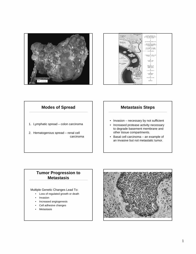
Metastasis Genes•Despite years of searching, there is no one geneor even a common small subset of genes that leadto metastasis.
•However, for some tumors, there appears to be a metastasis signature, a group of genes whose expressioncorrelates with metastasis.
•This signature appears to be the result of a number of activated signaling pathways, rather then a single one that could be inhibited.
Non metastatic metastatic
Regulators of Metastasis
1. Many genetics screens have been used to identify metastasis suppressors or required genes.
2. These screens have evolved as the technologies have changed, but the result is usually the same.
3. There is not one key regulator or master switch which turns on the metastatic program.
4. For many carcinomas, activation of RTK’s such as EGFR, HER2 or Met may be a critical regulator of such a program.
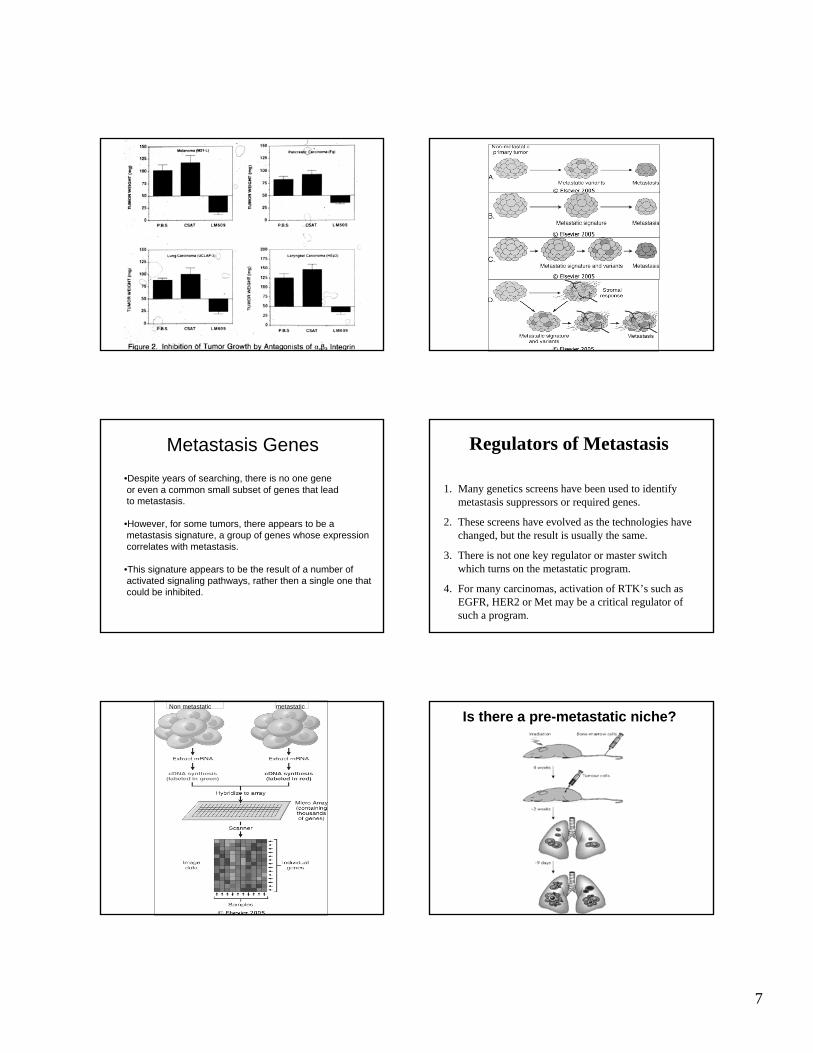
Is there a pre-metastatic niche?
8
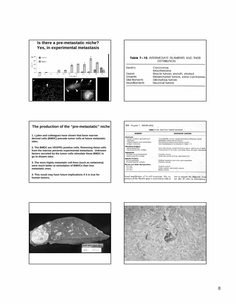
Is there a pre-metastatic niche?Yes, in experimental metastasis
The production of the “pre-metastatic” niche
1. Lyden and colleagues have shown that bone marrow derived cells (BMDC) precede tumor cells at future metastaticsites.
2. The BMDC are VEGFR1 positive cells. Removing these cells from the marrow prevents experimental metastasis. Unknown factors secreted by the tumor cells stimulate these BMDC to go to distant sites.
3. The more highly metastatic cell lines (such as melanoma) were much better at stimulation of BMDCs than less metastatic ones.
4. This result may have future implications if it is true for human tumors.
![Inflammation and cancer: How hot is the link? · carcinoma [30], colon carcinoma, lung carcinoma, squamous cell carcinoma, pancreatic cancer [31,32], ovarian carcinoma biochemical](https://static.fdocuments.in/doc/165x107/5fcdd6c81c76a34db570e7e6/iniammation-and-cancer-how-hot-is-the-link-carcinoma-30-colon-carcinoma.jpg)


![CARCINOMA ERYSIPELOIDES MIMICKING RADIATION … · Carcinoma erysipeloides (CE) is an uncommon cutaneous metastasis arising from visceral carcinoma [1]. It is the result of spread](https://static.fdocuments.in/doc/165x107/5feca92ebacb9554d41ee682/carcinoma-erysipeloides-mimicking-radiation-carcinoma-erysipeloides-ce-is-an-uncommon.jpg)