Moderate Physical Activity Mediates the Association between White ...
15
RESEARCH ARTICLE Moderate Physical Activity Mediates the Association between White Matter Lesion Volume and Memory Recall in Breast Cancer Survivors Gillian E. Cooke 1 *, Nathan C. Wetter 1,6 , Sarah E. Banducci 1,5 , Michael J. Mackenzie 2 , Krystle E. Zuniga 3 , Elizabeth A. Awick 4 , Sarah A. Roberts 4 , Brad P. Sutton 1,6 , Edward McAuley 1,4 , Arthur F. Kramer 1,5 1 Beckman Institute for Advanced Science and Technology, University of Illinois at Urbana-Champaign, Urbana, IL, United States of America, 2 Department of Behavioral Health and Nutrition, College of Health Sciences, University of Delaware, Newark, DE, United States of America, 3 School of Family and Consumer Sciences, Texas State University, San Marcos, TX, United States of America, 4 Department of Kinesiology & Community Health, University of Illinois at Urbana-Champaign, Urbana, IL, United States of America, 5 Department of Psychology, University of Illinois at Urbana-Champaign, Urbana, IL, United States of America, 6 Department of Bioengineering, University of Illinois at Urbana-Champaign, Urbana, IL, United States of America * [email protected] Abstract Increased survival rates among breast cancer patients have drawn significant attention to consequences of both the presence of cancer, and the subsequent treatment-related impact on the brain. The incidence of breast cancer and the effects of treatment often result in alterations in the microstructure of white matter and impaired cognitive functioning. How- ever, physical activity is proving to be a successful modifiable lifestyle factor in many studies that could prove beneficial to breast cancer survivors. This study investigates the link between white matter lesion volume, moderate physical activity, and cognition in breast cancer survivors following treatment compared to non-cancer age-matched controls. Results revealed that brain structure significantly predicted cognitive function via mediation of physical activity in breast cancer survivors. Overall, the study provided preliminary evi- dence suggesting moderate physical activity may help reduce the treatment related risks associated with breast cancer, including changes to WM integrity and cognitive impairment. Introduction Breast cancer is the leading cancer type in women; in 2015, 29% of new cancer cases in the United States are estimated to be breast cancer related [1]. The risk for developing breast can- cer increases with age [2], with 1 in 8 women developing invasive breast cancer. While the median age for diagnosis is 61, long-term effects of breast cancer treatment are emerging [3]. Although breast cancer is among the principal causes of cancer-related deaths [1,4,5], increased knowledge of breast cancer in the general population, earlier detection, and improvements in treatments offered has greatly impacted survival rates. The consequence is that breast cancer PLOS ONE | DOI:10.1371/journal.pone.0149552 February 25, 2016 1 / 15 OPEN ACCESS Citation: Cooke GE, Wetter NC, Banducci SE, Mackenzie MJ, Zuniga KE, Awick EA, et al. (2016) Moderate Physical Activity Mediates the Association between White Matter Lesion Volume and Memory Recall in Breast Cancer Survivors. PLoS ONE 11(2): e0149552. doi:10.1371/journal.pone.0149552 Editor: Sonia Brucki, University Of São Paulo, BRAZIL Received: June 24, 2015 Accepted: February 2, 2016 Published: February 25, 2016 Copyright: © 2016 Cooke et al. This is an open access article distributed under the terms of the Creative Commons Attribution License, which permits unrestricted use, distribution, and reproduction in any medium, provided the original author and source are credited. Data Availability Statement: All relevant data are within the paper and its Supporting Information files. Funding: The investigators are supported by the National Institute on Aging at the National Institutes of Health - grant numbers R01AG020118 (EMA) and R37AG025667 (AFK). The funders had no role in study design, data collection and analysis, decision to publish, or preparation of the manuscript. Competing Interests: The authors have declared that no competing interests exist.
Transcript of Moderate Physical Activity Mediates the Association between White ...
RESEARCH ARTICLE
Moderate Physical Activity Mediates theAssociation between White Matter LesionVolume and Memory Recall in Breast CancerSurvivorsGillian E. Cooke1*, Nathan C. Wetter1,6, Sarah E. Banducci1,5, Michael J. Mackenzie2,Krystle E. Zuniga3, Elizabeth A. Awick4, Sarah A. Roberts4, Brad P. Sutton1,6,Edward McAuley1,4, Arthur F. Kramer1,5
1 Beckman Institute for Advanced Science and Technology, University of Illinois at Urbana-Champaign,Urbana, IL, United States of America, 2 Department of Behavioral Health and Nutrition, College of HealthSciences, University of Delaware, Newark, DE, United States of America, 3 School of Family and ConsumerSciences, Texas State University, San Marcos, TX, United States of America, 4 Department of Kinesiology &Community Health, University of Illinois at Urbana-Champaign, Urbana, IL, United States of America,5 Department of Psychology, University of Illinois at Urbana-Champaign, Urbana, IL, United States ofAmerica, 6 Department of Bioengineering, University of Illinois at Urbana-Champaign, Urbana, IL, UnitedStates of America
AbstractIncreased survival rates among breast cancer patients have drawn significant attention to
consequences of both the presence of cancer, and the subsequent treatment-related
impact on the brain. The incidence of breast cancer and the effects of treatment often result
in alterations in the microstructure of white matter and impaired cognitive functioning. How-
ever, physical activity is proving to be a successful modifiable lifestyle factor in many studies
that could prove beneficial to breast cancer survivors. This study investigates the link
between white matter lesion volume, moderate physical activity, and cognition in breast
cancer survivors following treatment compared to non-cancer age-matched controls.
Results revealed that brain structure significantly predicted cognitive function via mediation
of physical activity in breast cancer survivors. Overall, the study provided preliminary evi-
dence suggesting moderate physical activity may help reduce the treatment related risks
associated with breast cancer, including changes to WM integrity and cognitive impairment.
IntroductionBreast cancer is the leading cancer type in women; in 2015, 29% of new cancer cases in theUnited States are estimated to be breast cancer related [1]. The risk for developing breast can-cer increases with age [2], with 1 in 8 women developing invasive breast cancer. While themedian age for diagnosis is 61, long-term effects of breast cancer treatment are emerging [3].Although breast cancer is among the principal causes of cancer-related deaths [1,4,5], increasedknowledge of breast cancer in the general population, earlier detection, and improvements intreatments offered has greatly impacted survival rates. The consequence is that breast cancer
PLOSONE | DOI:10.1371/journal.pone.0149552 February 25, 2016 1 / 15
OPEN ACCESS
Citation: Cooke GE, Wetter NC, Banducci SE,Mackenzie MJ, Zuniga KE, Awick EA, et al. (2016)Moderate Physical Activity Mediates the Associationbetween White Matter Lesion Volume and MemoryRecall in Breast Cancer Survivors. PLoS ONE 11(2):e0149552. doi:10.1371/journal.pone.0149552
Editor: Sonia Brucki, University Of São Paulo,BRAZIL
Received: June 24, 2015
Accepted: February 2, 2016
Published: February 25, 2016
Copyright: © 2016 Cooke et al. This is an openaccess article distributed under the terms of theCreative Commons Attribution License, which permitsunrestricted use, distribution, and reproduction in anymedium, provided the original author and source arecredited.
Data Availability Statement: All relevant data arewithin the paper and its Supporting Information files.
Funding: The investigators are supported by theNational Institute on Aging at the National Institutes ofHealth - grant numbers R01AG020118 (EMA) andR37AG025667 (AFK). The funders had no role instudy design, data collection and analysis, decision topublish, or preparation of the manuscript.
Competing Interests: The authors have declaredthat no competing interests exist.
survivor’s (BCS) often have to deal with cognitive impairments long after the cessation of treat-ment, at great cost to both themselves and society. The disease and treatment greatly impactmany facets of the BCS life, in particular, there are well-documented reports of impairments incognitive function both at diagnosis, and after treatment [6–8]. This may reflect a pattern ofaccelerated aging in the brain, with reported difficulties in executive function, processingspeed, memory and attention [9,10]. Accordingly, researchers are beginning to investigatewhether treatment for breast cancer augments age-related changes in the function and struc-ture of the brain [11]. Recent momentum to examine the structural changes in the brainaccompanying these impairments has revealed alterations in both gray and white matter, pro-posing a possible link between structural changes in the brain and functional deficits in cogni-tion [12–15]. There are several factors linked to the deficits associated with breast cancertreatment, including time since diagnosis, age at onset of disease, and lifestyle factors.
Although some studies find no differences following breast cancer treatment [16], manyreport significant changes in the structure and function of the brain in BCS for years or evendecades following treatment [10,17–20]. Comparing patients treated with chemotherapy andsurgery (C+) versus surgery only (C-) and non-cancer controls (NCC), Deprez and colleaguesreported significant cognitive and structural changes associated with chemotherapy [10,21].Patients were assessed after surgery but before chemotherapy–and then tested 3–5 monthsafter cessation of chemotherapy. Within the C+ group, cognitive performance declined signifi-cantly with treatment, but no such differences were evident in the C- group. There was also asignificant interaction between group and performance in memory and processing speed tasks,with the C+ group performing significantly worse than the C- and NCC groups. Additionally,within the C+ group there was a significant relationship between change in memory perfor-mance and change in white matter (WM) integrity; greater declines in WM integrity wererelated to greater cognitive decline. Interestingly, there were no significant differences in eitherpatient group at baseline (versus NCC), suggesting no impact of cancer diagnosis alone. Thesefindings suggest that alterations in the microstructure of WM are evident following chemother-apy treatment for breast cancer. Importantly, these alterations in WM integrity may impact thetransmission of information between regions of grey matter that support cognitive function[22]. Deprez and colleagues suggest that damage to these WM regions involved in transfer ofinformation around the brain, reduces the efficiency of interaction among different neural sys-tems [23]. While they propose that the treatment related cognitive impairments in cancer arerelated to alterations in the myelin structure of WM, they indicate that further investigation isneeded to confirm this hypothesis.
De Ruiter and colleagues reported cognitive impairments, reduced WM integrity, increasedaxonal injury and reduced gray matter (GM) volume in a small group of C+ patients whencompared to C- group up to 10 years after treatment [24]. They suggest this reflects long-termdamaging effects that chemotherapy has on the white and gray matter of the brain. This groupalso extended the length of time since treatment cessation to over 20 years in 180 C+ women;both total brain volume and GM volume were significantly lower in the C+ group [17,25].While they did not find differences in WM integrity between NCC and C+, within the C+group they did find a negative relationship between time since cessation of chemotherapy andWM integrity–suggesting that WM integrity in chemotherapy treated BCS deteriorates withtime since treatment. Long-term follow-ups are necessary to determine if WM integrity recov-ers with time.
There are well-documented changes in brain structure and function with aging [26–34].Given that recent research has suggested that treatment for breast cancer may resemble anaccelerated pattern of aging in the brain [35,36], the impact of age at diagnosis could differacross the lifespan [9,37,38]. Mandelblatt and colleagues examined the shared underlying
Physical Activity, White Matter and Cognition in Breast Cancer
PLOS ONE | DOI:10.1371/journal.pone.0149552 February 25, 2016 2 / 15
pathways of cancer-related cognitive impairment and aging, including changes in hormonelevels, inflammation and decreased blood flow [9]. In particular, they discuss the impact of lowphysical activity (PA) and decreased cognitive function. The link between PA and adult hippo-campal neurogenesis has received a lot of attention; increased PA seems to stimulate precursorcells, the cells from which adult neurogenesis derives [39]. The ‘neurogenic reserve hypothesis’proposed by Kempermann and colleagues suggest that an absence of PA early in life can onlybe somewhat counteracted later. They believe that it is imperative that we maintain a continu-ally active lifestyle, sustaining the capacity for adult neurogenesis. Furthermore, they discuss atwo-pronged issue for people with low levels of PA–not only do they have less plasticity, buttheir ability to achieve long-lasting benefits from this plasticity is diminished [39]. Conse-quently, PA is proving to be a successful modifiable lifestyle factor across the lifespan in bothhealthy and clinical populations [32,33,40–46]. A large aging study that predicted changes inbrain structure based on earlier PA (3 years prior), reported that higher levels of self-reportedPA predicted less atrophy in both GM andWM, and lower WM lesion volume [47]. Extendingthis work to include objective measures of PA, Burzynska and colleagues showed that lowerWM lesion volume in older adults was related to greater moderate-vigorous PA [44].
While breast cancer may result in accelerated aging of the brain, and aging brains benefitgreatly from increased PA, considerably less is understood about the link between PA, cogni-tion and breast cancer. PA levels often change after a diagnosis of breast cancer; with up to an11% decrease in PA within a year of diagnosis, though these decreases are considerably higherin women treated with radiation and chemotherapy [48]. The danger is that BCS may notreturn to their pre-diagnosis levels of PA, increasing their risk for weight gain and associatedcardiovascular problems. BCS often report exercise intolerance following treatment for breastcancer [49], with many spending only 2% of their awake time in moderate-vigorous PA, and80% of their time sedentary [50]. A novel study highlighting the importance of PA reportedthat 30 days of bed rest resulted in a greater reductions in fitness then 30 years of aging [51,52].In response to the growing need to encourage continued and even increased PA among cancersurvivors, the American College of Sports Medicine published guidelines [53] that recommendBCS progress up to 150 minutes of moderate PA per week. While several studies havehighlighted the benefits of physical activity in BCS [40,54,55], only two studies have explicitlyexamined the association between PA/fitness and cognition in BCS [55,56]. Both studiesreported a significant relationship between physical activity and cardiorespiratory fitness(CRF) and cognition in BCS. Although there is a lack of RCTs examining the effects of PA oncognitive function in BCS, these studies do highlight some of the potential benefits.
We were interested in examining the role that PA plays in the structure and function of thebrain following treatment for breast cancer. Our primary objectives were: (a) to determinewhether BCS and non-cancer age-matched controls differed in memory recall, WM lesion vol-ume and PA and (b) to examine the relations among these measures and the extent to whichPA mediated the relations between WM lesion volume and memory recall.
Methods
ParticipantsBCS were primarily recruited through an oncology clinic, though some were recruited, alongwith the non-cancer age-matched controls, through local advertisements. After expressing ini-tial interest, women were contacted by phone and provided a full study description. During theinitial contact, interested individuals completed a demographics questionnaire and a personalmedical history questionnaire, including self-reported information on breast cancer diagnosisand treatment history (BCS only). Of the 141 total contacts, 73 consented, and 11 women
Physical Activity, White Matter and Cognition in Breast Cancer
PLOS ONE | DOI:10.1371/journal.pone.0149552 February 25, 2016 3 / 15

withdrew after consenting owing to schedule conflicts (n = 2), no longer interested (n = 3), orunable to contact (n = 6). Furthermore, some women only underwent surgery (n = 3) and wereexcluded from these analyses, and an additional participant was identified as an outlier (seebelow). This resulted in a final sample of 58. Participants were female BCS who had undergonesurgery for breast cancer and had completed primary treatment (chemotherapy, radiation ther-apy or both) within the past 3 years (n = 30), or age-matched controls with no prior diagnosisof cancer (n = 28).
Given the likely interaction between time since cessation of therapy and age at onset of dis-ease, we recruited BCS within 3 years of completion of treatment and aged between 18–70 years.Inclusion criteria for all included English speaking, normal/corrected to normal vision, no cur-rent use of brain training games (e.g. Lumosity1, BrainHQ1), no history of stroke, transientischemic attack, or surgery that involved removal of brain tissue, not currently pregnant, a scoreof� 26 on the modified Mini-Mental Status Exam– 2nd Edition (MMSE-2), and able to walk ona treadmill unaided. All participants provided written informed consent and were required toprovide written consent from a primary care physician or oncologist indicating they were clearedto participate in the cardiorespiratory fitness, magnetic resonance imaging (MRI) and cognitivetesting sessions. Participants completed an initial visit for cardiorespiratory fitness assessmentand 7 days of accelerometer monitoring. During a second visit participants completed cognitivetesting followed by a third visit to complete a series of MRI scans. The study was approved by theUniversity of Illinois Urbana Champaign Institutional Review Board.
Physical ActivityPhysical activity (PA) was determined using 7-day accelerometer monitoring (model GT3X,Actigraph: Pensacola, FL). Participants were asked to wear the accelerometer during wakinghours and wear time was recorded on an accelerometer log. Data were downloaded and digi-tally converted to “activity counts” per minute (i.e., one epoch), and processed using MeterPlus4.2 software (Santech Health: San Diego, CA). Only days with at least 10 valid hours of weartime were included in the analysis, and hours with greater than 60 min of consecutive zeroswere considered invalid (i.e., non-wearing). Activity counts were summed and averaged acrossthe total number of valid days for a total daily activity score. Average daily moderate PA wascalculated by summing the total valid moderate activity counts, then dividing by the number ofvalid days.
Body Mass Index (BMI)Height and weight were measured using a Seca electronic scale and stadiometer (Model 7631321139, Chino, CA). Participants were measured while wearing light clothing and withoutshoes. BMI was calculated using the standard formula of weight (kg) / height (m)2.
Mini-Mental Status Exam – 2nd Edition (MMSE-2)The standard version of the MMSE-2 which allows for a brief standardized assessment of cog-nitive status was used to screen for signs of cognitive impairment [57]. It examines orientation,attention, memory, confrontation naming, language, comprehension and motor function. Par-ticipants scoring< 26 out of a possible 30 were excluded.
Story MemoryThe expanded version of the MMSE-2 includes an immediate story recall–participants wereread a short story aloud and asked to recall the story using the same words. There are 25
Physical Activity, White Matter and Cognition in Breast Cancer
PLOS ONE | DOI:10.1371/journal.pone.0149552 February 25, 2016 4 / 15

elements to the story, and a credit was given for each one successfully recalled. The story mem-ory was designed to evaluate verbal explicit learning and verbal free recall [57].
MRI ProtocolAll images were acquired during a single session on a 3 T Siemens Trio Tim system (Siemens,Erlangen, Germany). High-resolution structural MR scans were acquired using a 3D magneti-zation prepared rapid acquisition gradient echo (MPRAGE) T1-weighted sequence (TR = 1900ms; TE = 2.32 ms; TI: 900 ms; flip angle = 9°; matrix = 256 × 256; FOV = 230mm; 192 slices;resolution = 0.9 × 0.9 × 0.9 mm; GRAPPA acceleration factor 2). While the lesion volumeswere acquired using a 3D, variable flip angle turbo spin echo sequence with fluid-attenuatedinversion recovery (FLAIR) (TE 388 ms, TR 6000 ms, TI 2200 ms, FOV 250x250x160 mm, 1mm isotropic sampling; GRAPPA acceleration factor 2).
White Matter Lesion ProtocolWe used previously developed open-source software based on the image processing toolkit FSL(functional magnetic resonance imaging of the brain (FMRIB) Software library) to performautomated computation of lesion masks and volumes [58,59]. We accomplished this by usingBET (Brain Extraction Tool) [60] to extract the brain, then used FAST (FMRIB's AutomatedSegmentation Tool) [61] on the result, with two tissue classes, brain and non-brain. Hyperintense regions were classified as non-brain tissue and we examined the histogram of the non-brain tissue to identify the lesions. We then used standard space masking with nonlinear regis-tration via FNIRT (FMRIB's Nonlinear Image Registration Tool) [62] to eliminate false posi-tives that consistently appear in certain parts of the brain, such as the septum pellucidum. Thefinal output was total lesion volume, expressed as a percentage of total brain volume [59].
Data AnalysisAll analyses were performed using IBM SPSS version 22 (IBM, 2013), and data is provided inS1 Dataset. Frequency distributions were examined to check for missing information and out-of-range values. Tests of normality were run on the whole brain WM data (lesion volume cor-rected for percent of whole brain volume), story memory recall and moderate PA data–as aresult of these tests, lesion volume was transformed using log transform. We excluded subjectswith extreme values on any of these measures (3 SD beyond mean). One subject had extrememoderate PA activity in the non-cancer age-matched control group and was dropped from allanalyses.
Given the small sample size, analyses were conducted across all BCS in comparison to non-cancer age-matched controls. To investigate these differences multivariate ANCOVAs wereconducted on background demographics, lesion volume, moderate PA and story memoryrecall. Effects sizes, partial Eta squared, were calculated and reported. We explored associationsbetween lesion volume, PA and story memory using partial correlations, controlling for ageand years of education. Further analyses in the BCS group also included time since cessation oftreatment.
Where significant relationships were found, a simple mediation analysis was conductedusing ordinary least squares path analysis [63], controlling for covariates (PROCESS toolbox inSPSS). This analysis examined whether moderate PA mediated the relationship between lesionvolume and story memory recall (see Fig 1). While there is value in examining the relationshipbetween an independent and dependent variable, this relationship can be overemphasizedbefore controlling for a mediator, leading to misinformed results [64].
Physical Activity, White Matter and Cognition in Breast Cancer
PLOS ONE | DOI:10.1371/journal.pone.0149552 February 25, 2016 5 / 15
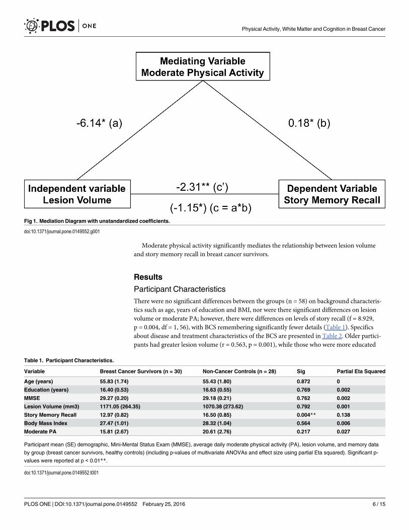
Moderate physical activity significantly mediates the relationship between lesion volumeand story memory recall in breast cancer survivors.
Results
Participant CharacteristicsThere were no significant differences between the groups (n = 58) on background characteris-tics such as age, years of education and BMI, nor were there significant differences on lesionvolume or moderate PA; however, there were differences on levels of story recall (f = 8.929,p = 0.004, df = 1, 56), with BCS remembering significantly fewer details (Table 1). Specificsabout disease and treatment characteristics of the BCS are presented in Table 2. Older partici-pants had greater lesion volume (r = 0.563, p = 0.001), while those who were more educated
Fig 1. Mediation Diagramwith unstandardized coefficients.
doi:10.1371/journal.pone.0149552.g001
Table 1. Participant Characteristics.
Variable Breast Cancer Survivors (n = 30) Non-Cancer Controls (n = 28) Sig Partial Eta Squared
Age (years) 55.83 (1.74) 55.43 (1.80) 0.872 0
Education (years) 16.40 (0.53) 16.63 (0.55) 0.769 0.002
MMSE 29.27 (0.20) 29.18 (0.21) 0.762 0.002
Lesion Volume (mm3) 1171.05 (264.35) 1070.38 (273.62) 0.792 0.001
Story Memory Recall 12.97 (0.82) 16.50 (0.85) 0.004** 0.138
Body Mass Index 27.47 (1.01) 28.32 (1.04) 0.564 0.006
Moderate PA 15.81 (2.67) 20.61 (2.76) 0.217 0.027
Participant mean (SE) demographic, Mini-Mental Status Exam (MMSE), average daily moderate physical activity (PA), lesion volume, and memory data
by group (breast cancer survivors, healthy controls) (including p-values of multivariate ANOVAs and effect size using partial Eta squared). Significant p-
values were reported at p < 0.01**.
doi:10.1371/journal.pone.0149552.t001
Physical Activity, White Matter and Cognition in Breast Cancer
PLOS ONE | DOI:10.1371/journal.pone.0149552 February 25, 2016 6 / 15
were also likely to engage in greater levels of moderate PA (r = 0.432, p = 0.001), accordingly,both variables were included as covariates throughout all analyses.
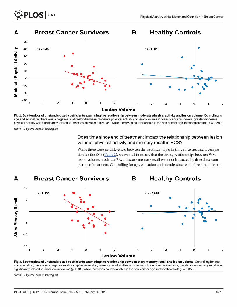
Relationship between lesion volume, physical activity and memory recallIn BCS, those who were more engaged in moderate PA had smaller WM lesion volumes(r = -0.430, p = 0.011); however, there was no relationship evident in the non-cancer age-matched controls (r = -0.120, p = 0.280; Fig 2). Furthermore, lower WM lesion volume inBCS was associated with greater story memory recall (r = -0.553, p = 0.001), while againthere was also no relationship in the non-cancer age-matched controls (r = -0.075, p = 0.358;Fig 3). Lastly, BCS with greater levels of moderate PA performed better on the story memoryrecall (r = 0.587, p = 0.001), although this was not evident in non-cancer age-matched con-trols (r = -0.243, p = 0.116; Fig 4).
Does moderate PA mediate the relationship between lesion volume andmemory recall?Given the relationships between lesion volume, moderate PA and story memory recall in BCS,and the lack of a relationship in non-cancer age-matched controls, the mediation analysis wasconducted only in the BCS group. Our aim was to examine the potential mediation of moderatePA on the relationship between lesion volume and story memory recall.
Mediation analysis controlling for age and years of education indicated that lesion volumeinfluenced story memory recall independent of its effect on moderate PA (c’ = -2.31). However,lesion volume also indirectly influenced story memory recall through its effect on moderatePA. BCS with lower lesion volume had higher levels of moderate PA (a = -6.14), and those withhigher levels of moderate PA had greater recall of the story (b = 0.19). A bias-corrected boot-strap confidence interval for the indirect effect based on 10,000 bootstrap samples was entirelyabove zero (-0.0966 to -2.7069), verifying that moderate PA significantly mediated the impactof WM lesion volume on story memory recall in BCS (Fig 4).
Table 2. Disease and treatment characteristics of breast cancer survivors (n = 30).
Stage DCIS 5 (16.7)
Stage I 8 (26.7)
Stage II 11 (36.7)
Stage III 3 (10)
Unknown 3 (10)
Estrogen Receptor Positive N (%) Yes 23 (76.7)
No 6 (20)
Unknown 1 (3.3)
Adjuvant Treatment N (%) Surgery 30 (100)
Radiotherapy Only 11 (36.7)
Chemotherapy Only 8 (26.7)
Chemotherapy Plus Radiation 11 (36.7)
Months Since End of Treatment Radiotherapy Only 13.14 (2–32)
Mean (Range) Chemotherapy Only 20.38 (7–33)
Chemotherapy Plus Radiation 15.86 (3.5–31)
DCIS, ductal carcinoma in situ.
doi:10.1371/journal.pone.0149552.t002
Physical Activity, White Matter and Cognition in Breast Cancer
PLOS ONE | DOI:10.1371/journal.pone.0149552 February 25, 2016 7 / 15
Does time since end of treatment impact the relationship between lesionvolume, physical activity and memory recall in BCS?While there were no differences between the treatment types in time since treatment comple-tion for the BCS (Table 2), we wanted to ensure that the strong relationships between WMlesion volume, moderate PA, and story memory recall were not impacted by time since com-pletion of treatment. Controlling for age, education and months since end of treatment, lesion
Fig 2. Scatterplots of unstandardized coefficients examining the relationship betweenmoderate physical activity and lesion volume.Controlling forage and education, there was a negative relationship between moderate physical activity and lesion volume in breast cancer survivors; greater moderatephysical activity was significantly related to lower lesion volume (p<0.05), while there was no relationship in the non-cancer age-matched controls (p = 0.280).
doi:10.1371/journal.pone.0149552.g002
Fig 3. Scatterplots of unstandardized coefficients examining the relationship between story memory recall and lesion volume. Controlling for ageand education, there was a negative relationship between story memory recall and lesion volume in breast cancer survivors; greater story memory recall wassignificantly related to lower lesion volume (p<0.01), while there was no relationship in the non-cancer age-matched controls (p = 0.358).
doi:10.1371/journal.pone.0149552.g003
Physical Activity, White Matter and Cognition in Breast Cancer
PLOS ONE | DOI:10.1371/journal.pone.0149552 February 25, 2016 8 / 15

volume was still negatively correlated with moderate PA (r = -0.455, p = 0.017) and story mem-ory recall (r = -0.537, p = 0.004), and finally, story memory recall and moderate PA were posi-tively related (r = 0.594, p = 0.001).
After re-running the mediation analysis controlling for age, years of education and monthssince end of treatment, we again confirmed that moderate PA significantly mediated the rela-tionship between lesion volume and story memory recall in BCS. Lesion volume influencedstory memory recall independent of its effect on moderate PA (c’ = -2.21). Lesion volume alsoindirectly influenced story memory recall through its effect on moderate PA. BCS with lowerlesion volume had higher levels of moderate PA (a = -6.89), and those with higher levels ofmoderate PA had greater recall of the story (b = 0.19). A bias-corrected bootstrap confidenceinterval for the indirect effect based on 10,000 bootstrap samples was entirely above zero(-0.1821 to -3.2297). Despite the range in time since treatment (2–33 months), moderate PAsignificantly mediated the influence of WM lesion on story memory recall.
DiscussionPrevious studies have suggested that diagnosis and treatment for breast cancer results in cogni-tive impairments and structural changes in the brain, which may reflect a pattern of acceleratedaging [3,9,12,38]. Given the association between PA and changes in brain structure and func-tion in the aging literature [32–34,44,45], PA shows great potential for improving the long-term health of BCS, yet the link between PA, cognitive function and brain structure in BCS hasreceived no attention. In this novel cross-sectional study, we examined the role that PA playsin the structure-function relationship in a group of middle-aged women within 3 years of com-pletion of treatment, and non-cancer age-matched controls. Considering the small sample size,we compared across breast cancer as a whole rather then focusing on specific treatment types,investigating the link between memory recall, WM lesion volume and moderate PA. We con-trolled for both age and education throughout, as they are known to confound memory
Fig 4. Scatterplots of unstandardized coefficients examining the relationship between story memory recall andmoderate physical activity.Controlling for age and education, there was a positive relationship between story memory recall and moderate physical activity in the breast cancersurvivors; greater story memory recall was significantly related to greater moderate physical activity (p<0.01), while there was no relationship in the non-cancer age-matched controls (p = 0.116).
doi:10.1371/journal.pone.0149552.g004
Physical Activity, White Matter and Cognition in Breast Cancer
PLOS ONE | DOI:10.1371/journal.pone.0149552 February 25, 2016 9 / 15
performance and structural integrity. Additional analyses also controlled for time since cessa-tion of treatment in the BCS group only.
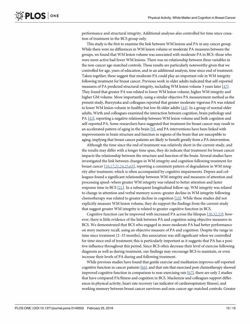
This study is the first to examine the link betweenWM lesions and PA in any cancer group.While there were no differences inWM lesion volume or moderate PA measures between thegroups, we found that WM lesion volume was associated with moderate PA in BCS–those whowere more active had fewer WM lesions. There was no relationship between these variables inthe non-cancer age-matched controls. These results are particularly noteworthy given that wecontrolled for age, years of education, and in an additional analysis, time since end of treatment.Taken together, these suggest that moderate PA could play an important role inWM integrityfollowing treatment for breast cancer. Previous work in older adults indicated that self-reportedmeasures of PA predicted structural integrity, includingWM lesion volume 3 years later [47].They found that greater PA was related to lowerWM lesion volume, higherWM integrity andhigher GM volume. More importantly, using a similar objective PA measurement method as thepresent study, Burzynska and colleagues reported that greater moderate-vigorous PA was relatedto lowerWM lesion volume in healthy but low-fit older adults [44]. In a group of normal olderadults, Wirth and colleagues examined the interaction between cognition, brain pathology andPA [45], reporting a negative relationship betweenWM lesion volume and both cognition andself-reported PA. Some researchers have suggested that treatment for breast cancer may result inan accelerated pattern of aging in the brain [9], and PA interventions have been linked withimprovements in brain structure and function in regions of the brain that are susceptible toaging, implying that breast cancer patients are likely to benefit greatly from a PA intervention.
Although the time since the end of treatment was relatively short in the current study, andthe results may differ with a longer time span, they do indicate that treatment for breast cancerimpacts the relationship between the structure and function of the brain. Several studies haveinvestigated the link between changes in WM integrity and cognition following treatment forbreast cancer [10,17,21,24,25,65], reporting a consistent pattern of degradation in WM integ-rity after treatment, which is often accompanied by cognitive impairments. Deprez and col-leagues found a significant relationship between WM integrity and measures of attention andprocessing speed–where greater WM integrity was related to better attention and fasterresponse time in BCS [21]. In a subsequent longitudinal follow-up, WM integrity was relatedto change in attention and verbal memory scores–greater decline in WM integrity followingchemotherapy was related to greater decline in cognition [10]. While these studies did notexplicitly measure WM lesion volume, they do support the findings from the current studythat suggest greater WM integrity is related to greater cognitive function in BCS.
Cognitive function can be improved with increased PA across the lifespan [30,32,33]; how-ever, there is little evidence of the link between PA and cognition using objective measures inBCS. We demonstrated that BCS who engaged in more moderate PA had better performanceon story memory recall, using an objective measure of PA and cognition. Despite the range intime since treatment (2–33 months), this association was still significant when we controlledfor time since end of treatment; this is particularly important as it suggests that PA has a posi-tive influence throughout this period. Since BCS often decrease their level of exercise followingdiagnosis as well as during treatment, our findings may encourage BCS to maintain, or evenincrease their levels of PA during and following treatment.
While previous studies have found that gentle exercise and meditation improves self-reportedcognitive function in cancer patients [66], and that rats that exercised post chemotherapy showedimproved cognitive function in comparison to non-exercising rats [67], there are only 2 studiesthat have compared PA/fitness and cognition in BCS. Mackenzie and colleagues support differ-ences in physical activity, heart rate recovery (an indicator of cardiorespiratory fitness), andworking memory between breast cancer survivors and non-cancer age-matched controls. Greater
Physical Activity, White Matter and Cognition in Breast Cancer
PLOS ONE | DOI:10.1371/journal.pone.0149552 February 25, 2016 10 / 15
cardiorespiratory fitness, heart rate recovery, and physical activity were positively associated withbetter working memory performance [56]. Furthermore, Crowgey and colleagues found thatalthough fitness levels were lower in BCS, there was a positive relationship between fitness andvisual memory [55]. Although the evidence of a significant link between PA and cognition inBCS is limited, previous studies, along with our findings, support an association between thesevariables, warranting further investigation in a large-scale longitudinal study.
One of the strengths of the current study is the use of objective measurements of PA, WMlesion volume and memory recall. However, the cross sectional nature of our study did notallow us to determine the impact of change in one or all of these variables; for example, wouldincreasing PA by taking part in an exercise intervention, result in decreased WM lesions andincreased memory recall. Yet, previous studies in healthy adults offer great promise–followinga one-year walking intervention, Voss and colleagues reported that increased fitness in olderadults who had completed an aerobic exercise program was significantly related to improvedWM integrity [32]. This was largely apparent in regions of the brain that are known to showloss due to aging, and typically overlap with regions of the brain effected by treatment forbreast cancer. Combined with our findings of a significant relationship between PA and WMlesions, this proposes a potentially modifiable lifestyle factor that could increase WM integrityin breast cancer patients undergoing treatment, as well as throughout their long-term survival.
While several studies have indicated that there are differences following diagnosis of breastcancer but before treatment [68,69], our design did not allow us to examine these. Additionally,given the novelty of our study, we did not compare across treatment types. Recently, studiesassessing the effects of breast cancer treatment type compared high-dose (HCh+) and conven-tional-dose chemotherapy (CC+), radiation (RT) only and non-cancer age-matched controls[70,71]. They reported cognitive decline in the HCh+ group, with diminished impairment evi-dent in the CC+ and RT groups; gray matter (GM) volume reductions for both the HCh+ andCC+ groups when compared to RT only; reductions in WM integrity only in the HCh+ group;and hypoactivation in task-related performance in both chemotherapy groups, though this wasmore pronounced in the HCh+ group. These findings suggest that there are treatment depen-dent differences in GM volume, WM integrity and cognitive function following therapy forbreast cancer that deserves further investigation.
ConclusionThere are modifiable risk factors associated with treatment related changes in brain structureand function, and while there is evidence indicating change in cognitive function, WM integ-rity and PA in BCS, there are no studies that have directly compared all three variables in thisgroup. Our novel findings suggest that continued physical activity might be particularly impor-tant in BCS as brain structure significantly predicted cognitive function via mediation of physi-cal activity. Continually active lifestyles may help reduce the treatment related vulnerabilitiesassociated with breast cancer, including reduced WM integrity and cognitive impairment. Col-lectively, these findings suggest that PA plays an important role in both brain structure andfunction, warranting further investigation in larger PA intervention studies.
Supporting InformationS1 Dataset. Dataset from all participants. Demographic information, moderate physicalactivity, lesion volume, and story memory recall data for each subject included in the analysis(n = 58). Data set also includes disease and treatment characteristics of breast cancer survivors(n = 30).(XLSX)
Physical Activity, White Matter and Cognition in Breast Cancer
PLOS ONE | DOI:10.1371/journal.pone.0149552 February 25, 2016 11 / 15

Author ContributionsConceived and designed the experiments: GEC SEB MJM KEZ EM AFK. Performed the exper-iments: GEC MJM KEZ EAA SAR. Analyzed the data: GEC NCW SEBMJM KEZ. Contributedreagents/materials/analysis tools: NCW BPS. Wrote the paper: GEC MJM KEZ BPS EM AFK.
References1. Siegel RL, Miller KD, Jemal A (2015) Cancer statistics, 2015. CA Cancer J Clin 65: 5–29. doi: 10.3322/
caac.21254 PMID: 25559415
2. Society AC (2015) "What are the key statistics about breast cancer?".
3. Nguyen CM, Yamada TH, Beglinger LJ, Cavanaugh JE, Denburg NL, Schultz SK (2013) Cognitive Fea-tures Ten or More Years After Successful Breast Cancer Survival: Comparisons Across Types of Can-cer Interventions. Psychooncology 22: 862–868. doi: 10.1002/pon.3086 PMID: 22585465
4. Siegel R, Ma J, Zou Z, Jemal A (2014) Cancer statistics, 2014. CA Cancer J Clin 64: 9–29. doi: 10.3322/caac.21208 PMID: 24399786
5. Siegel R, Naishadham D, Jemal A (2013) Cancer statistics, 2013. CA Cancer J Clin 63: 11–30. doi: 10.3322/caac.21166 PMID: 23335087
6. Ahles TA, Root JC, Ryan EL (2012) Cancer- and cancer treatment-associated cognitive change: anupdate on the state of the science. J Clin Oncol 30: 3675–3686. doi: 10.1200/JCO.2012.43.0116PMID: 23008308
7. Ahles TA, Saykin AJ, McDonald BC, Furstenberg CT, Cole BF, Hanscom BS, et al. (2008) Cognitivefunction in breast cancer patients prior to adjuvant treatment. Breast Cancer Res Treat 110: 143–152.PMID: 17674194
8. Wefel JS, Lenzi R, Theriault R, Buzdar AU, Cruickshank S, Meyers CA (2004) 'Chemobrain' in breastcarcinoma?: a prologue. Cancer 101: 466–475. PMID: 15274059
9. Mandelblatt JS, Hurria A, McDonald BC, Saykin AJ, Stern RA, VanMeter JW, et al. (2013) Cognitiveeffects of cancer and its treatments at the intersection of aging: what do we know; what do we need toknow? Semin Oncol 40: 709–725. doi: 10.1053/j.seminoncol.2013.09.006 PMID: 24331192
10. Deprez S, Amant F, Smeets A, Peeters R, Leemans A, Van HeckeW, et al. (2012) Longitudinal assess-ment of chemotherapy-induced structural changes in cerebral white matter and its correlation withimpaired cognitive functioning. J Clin Oncol 30: 274–281. doi: 10.1200/JCO.2011.36.8571 PMID:22184379
11. Yamada TH, Denburg NL, Beglinger LJ, Schultz SK (2010) Neuropsychological outcomes of olderbreast cancer survivors: cognitive features ten or more years after chemotherapy. J NeuropsychiatryClin Neurosci 22: 48–54. doi: 10.1176/appi.neuropsych.22.1.48 PMID: 20160209
12. Wefel JS, Kesler SR, Noll KR, Schagen SB (2015) Clinical characteristics, pathophysiology, and man-agement of noncentral nervous system cancer-related cognitive impairment in adults. CA Cancer J Clin65: 123–138. doi: 10.3322/caac.21258 PMID: 25483452
13. Kaiser J, Bledowski C, Dietrich J (2014) Neural correlates of chemotherapy-related cognitiveimpairment. Cortex 54: 33–50. doi: 10.1016/j.cortex.2014.01.010 PMID: 24632463
14. Simo M, Rifa-Ros X, Rodriguez-Fornells A, Bruna J (2013) Chemobrain: a systematic review of struc-tural and functional neuroimaging studies. Neurosci Biobehav Rev 37: 1311–1321. doi: 10.1016/j.neubiorev.2013.04.015 PMID: 23660455
15. Saykin AJ, de Ruiter MB, McDonald BC, Deprez S, Silverman DH (2013) Neuroimaging biomarkersand cognitive function in non-CNS cancer and its treatment: current status and recommendations forfuture research. Brain Imaging Behav 7: 363–373. doi: 10.1007/s11682-013-9283-7 PMID: 24327327
16. Yoshikawa E, Matsuoka Y, Inagaki M, Nakano T, Akechi T, Kobayakawa M, et al. (2005) No adverseeffects of adjuvant chemotherapy on hippocampal volume in Japanese breast cancer survivors. BreastCancer Res Treat 92: 81–84. PMID: 15980995
17. Koppelmans V, de Groot M, de Ruiter MB, BoogerdW, Seynaeve C, Vernooij MW, et al. (2014) Globaland focal white matter integrity in breast cancer survivors 20 years after adjuvant chemotherapy. HumBrain Mapp 35: 889–899. doi: 10.1002/hbm.22221 PMID: 23281152
18. Koppelmans V, Vernooij MW, BoogerdW, Seynaeve C, IkramMA, Breteler MM, et al. (2015) Preva-lence of cerebral small-vessel disease in long-term breast cancer survivors exposed to both adjuvantradiotherapy and chemotherapy. J Clin Oncol 33: 588–593. doi: 10.1200/JCO.2014.56.8345 PMID:25559803
19. Kesler S, Janelsins M, Koovakkattu D, Palesh O, Mustian K, Morrow G, et al. (2013) Reduced hippo-campal volume and verbal memory performance associated with interleukin-6 and tumor necrosis
Physical Activity, White Matter and Cognition in Breast Cancer
PLOS ONE | DOI:10.1371/journal.pone.0149552 February 25, 2016 12 / 15
factor-alpha levels in chemotherapy-treated breast cancer survivors. Brain Behav Immun 30 Suppl:S109–116. doi: 10.1016/j.bbi.2012.05.017 PMID: 22698992
20. McDonald BC, Saykin AJ (2013) Alterations in brain structure related to breast cancer and its treatment:chemotherapy and other considerations. Brain Imaging Behav 7: 374–387. doi: 10.1007/s11682-013-9256-x PMID: 23996156
21. Deprez S, Amant F, Yigit R, Porke K, Verhoeven J, Van den Stock J, et al. (2011) Chemotherapy-induced structural changes in cerebral white matter and its correlation with impaired cognitive function-ing in breast cancer patients. Hum Brain Mapp 32: 480–493. doi: 10.1002/hbm.21033 PMID:20725909
22. Fields RD (2010) Change in the Brain’sWhite Matter: The role of the brain’s white matter in active learn-ing and memory may be underestimated. Science (New York, NY) 330: 768–769.
23. Deprez S, Billiet T Fau—Sunaert S, Sunaert S Fau—Leemans A, Leemans A Diffusion tensor MRI ofchemotherapy-induced cognitive impairment in non-CNS cancer patients: a review.
24. de Ruiter MB, Reneman L, BoogerdW, Veltman DJ, Caan M, Douaud G, et al. (2012) Late effects ofhigh-dose adjuvant chemotherapy on white and gray matter in breast cancer survivors: convergingresults frommultimodal magnetic resonance imaging. Hum Brain Mapp 33: 2971–2983. doi: 10.1002/hbm.21422 PMID: 22095746
25. Koppelmans V, de Ruiter MB, van der Lijn F, BoogerdW, Seynaeve C, van der Lugt A, et al. (2012)Global and focal brain volume in long-term breast cancer survivors exposed to adjuvant chemotherapy.Breast Cancer Res Treat 132: 1099–1106. doi: 10.1007/s10549-011-1888-1 PMID: 22205140
26. Erickson KI, Prakash RS, Voss MW, Chaddock L, Heo S, McLaren M, et al. (2010) Brain-derived neuro-trophic factor is associated with age-related decline in hippocampal volume. J Neurosci 30: 5368–5375. doi: 10.1523/JNEUROSCI.6251-09.2010 PMID: 20392958
27. Erickson KI, Prakash RS, Voss MW, Chaddock L, Hu L, Morris KS, et al. (2009) Aerobic fitness is asso-ciated with hippocampal volume in elderly humans. Hippocampus 19: 1030–1039. doi: 10.1002/hipo.20547 PMID: 19123237
28. Erickson KI, Raji CA, Lopez OL, Becker JT, Rosano C, Newman AB, et al. (2010) Physical activity pre-dicts gray matter volume in late adulthood: the Cardiovascular Health Study. Neurology 75: 1415–1422. doi: 10.1212/WNL.0b013e3181f88359 PMID: 20944075
29. Colcombe SJ, Erickson KI, Raz N, Webb AG, Cohen NJ, McAuley E, et al. (2003) Aerobic fitnessreduces brain tissue loss in aging humans. J Gerontol A Biol Sci Med Sci 58: 176–180. PMID:12586857
30. Colcombe SJ, Kramer AF, McAuley E, Erickson KI, Scalf P (2004) Neurocognitive aging and cardiovas-cular fitness: recent findings and future directions. J Mol Neurosci 24: 9–14. PMID: 15314244
31. Voss MW, Erickson KI, Prakash RS, Chaddock L, Malkowski E, Alves H, et al. (2010) Functional con-nectivity: a source of variance in the association between cardiorespiratory fitness and cognition? Neu-ropsychologia 48: 1394–1406. doi: 10.1016/j.neuropsychologia.2010.01.005 PMID: 20079755
32. Voss MW, Heo S, Prakash RS, Erickson KI, Alves H, Chaddock L, et al. (2013) The influence of aerobicfitness on cerebral white matter integrity and cognitive function in older adults: results of a one-yearexercise intervention. Hum Brain Mapp 34: 2972–2985. doi: 10.1002/hbm.22119 PMID: 22674729
33. Erickson KI, Voss MW, Prakash RS, Basak C, Szabo A, Chaddock L, et al. (2011) Exercise trainingincreases size of hippocampus and improves memory. Proc Natl Acad Sci U S A 108: 3017–3022. doi:10.1073/pnas.1015950108 PMID: 21282661
34. Kramer AF, Erickson KI (2007) Capitalizing on cortical plasticity: influence of physical activity on cogni-tion and brain function. Trends Cogn Sci 11: 342–348. PMID: 17629545
35. Mandelblatt JS, Stern RA, Luta G, McGuckin M, Clapp JD, Hurria A, et al. (2014) Cognitive impairmentin older patients with breast cancer before systemic therapy: is there an interaction between cancer andcomorbidity? J Clin Oncol 32: 1909–1918. doi: 10.1200/JCO.2013.54.2050 PMID: 24841981
36. Sanoff HK, Deal AM, Krishnamurthy J, Torrice C, Dillon P, Sorrentino J, et al. (2014) Effect of cytotoxicchemotherapy on markers of molecular age in patients with breast cancer. J Natl Cancer Inst 106:dju057. doi: 10.1093/jnci/dju057 PMID: 24681605
37. Henderson TO, Ness KK, Cohen HJ (2014) Accelerated aging among cancer survivors: from pediatricsto geriatrics. Am Soc Clin Oncol Educ Book: e423–430. doi: 10.14694/EdBook_AM.2014.34.e423PMID: 24857133
38. Schmitz KH, Cappola AR, Stricker CT, Sweeney C, Norman SA (2007) The intersection of cancer andaging: establishing the need for breast cancer rehabilitation. Cancer Epidemiol Biomarkers Prev 16:866–872. PMID: 17507607
Physical Activity, White Matter and Cognition in Breast Cancer
PLOS ONE | DOI:10.1371/journal.pone.0149552 February 25, 2016 13 / 15

39. Kempermann G, Fabel K, Ehninger D, Babu H, Leal-Galicia P, Garthe A, et al. (2010) Why and howphysical activity promotes experience-induced brain plasticity. Front Neurosci 4: 189. doi: 10.3389/fnins.2010.00189 PMID: 21151782
40. Courneya KS, Segal RJ, Gelmon K, Reid RD, Mackey JR, Friedenreich CM, et al. (2007) Six-month fol-low-up of patient-rated outcomes in a randomized controlled trial of exercise training during breast can-cer chemotherapy. Cancer Epidemiol Biomarkers Prev 16: 2572–2578. PMID: 18086760
41. Courneya KS, Segal RJ, Mackey JR, Gelmon K, Reid RD, Friedenreich CM, et al. (2007) Effects of aer-obic and resistance exercise in breast cancer patients receiving adjuvant chemotherapy: a multicenterrandomized controlled trial. J Clin Oncol 25: 4396–4404. PMID: 17785708
42. Galantino ML, Greene L, Daniels L, Dooley B, Muscatello L, O'Donnell L (2012) Longitudinal impact ofyoga on chemotherapy-related cognitive impairment and quality of life in women with early stage breastcancer: a case series. Explore (NY) 8: 127–135.
43. Mustian KM, Sprod LK, Janelsins M, Peppone LJ, Mohile S (2012) Exercise Recommendations forCancer-Related Fatigue, Cognitive Impairment, Sleep problems, Depression, Pain, Anxiety, and Physi-cal Dysfunction: A Review. Oncol Hematol Rev 8: 81–88. PMID: 23667857
44. Burzynska AZ, Chaddock-Heyman L, Voss MW,Wong CN, Gothe NP, Olson EA, et al. (2014) Physicalactivity and cardiorespiratory fitness are beneficial for white matter in low-fit older adults. PLoS One 9:e107413. doi: 10.1371/journal.pone.0107413 PMID: 25229455
45. Wirth M, Haase CM, Villeneuve S, Vogel J, Jagust WJ (2014) Neuroprotective pathways: lifestyle activ-ity, brain pathology, and cognition in cognitively normal older adults. Neurobiol Aging 35: 1873–1882.doi: 10.1016/j.neurobiolaging.2014.02.015 PMID: 24656834
46. Spence RR, Heesch KC, BrownWJ (2010) Exercise and cancer rehabilitation: a systematic review.Cancer Treat Rev 36: 185–194. doi: 10.1016/j.ctrv.2009.11.003 PMID: 19962830
47. Gow AJ, Bastin ME, Munoz Maniega S, Valdes HernandezMC, Morris Z, Murray C, et al. (2012) Neuro-protective lifestyles and the aging brain: activity, atrophy, and white matter integrity. Neurology 79:1802–1808. doi: 10.1212/WNL.0b013e3182703fd2 PMID: 23091073
48. Irwin ML, Crumley D, McTiernan A, Bernstein L, Baumgartner R, Gilliland FD, et al. (2003) Physicalactivity levels before and after a diagnosis of breast carcinoma: the Health, Eating, Activity, and Life-style (HEAL) study. Cancer 97: 1746–1757. PMID: 12655532
49. Jones LW, Eves ND, Haykowsky M, Freedland SJ, Mackey JR (2009) Exercise intolerance in cancerand the role of exercise therapy to reverse dysfunction. Lancet Oncol 10: 598–605. doi: 10.1016/S1470-2045(09)70031-2 PMID: 19482248
50. Sabiston CM, Brunet J, Vallance JK, Meterissian S (2014) Prospective examination of objectivelyassessed physical activity and sedentary time after breast cancer treatment: sitting on the crest of theteachable moment. Cancer Epidemiol Biomarkers Prev 23: 1324–1330. doi: 10.1158/1055-9965.EPI-13-1179 PMID: 24753546
51. McGuire DK, Levine BD, Williamson JW, Snell PG, Blomqvist CG, Saltin B, et al. (2001) A 30-year fol-low-up of the Dallas Bedrest and Training Study: II. Effect of age on cardiovascular adaptation to exer-cise training. Circulation 104: 1358–1366. PMID: 11560850
52. McGuire DK, Levine BD, Williamson JW, Snell PG, Blomqvist CG, Saltin B, et al. (2001) A 30-year fol-low-up of the Dallas Bedrest and Training Study: I. Effect of age on the cardiovascular response to exer-cise. Circulation 104: 1350–1357. PMID: 11560849
53. Schmitz KH, Courneya KS, Matthews C, Demark-Wahnefried W, Galvao DA, Pinto BM, et al. (2010)American College of Sports Medicine roundtable on exercise guidelines for cancer survivors. Med SciSports Exerc 42: 1409–1426. doi: 10.1249/MSS.0b013e3181e0c112 PMID: 20559064
54. Chandwani KD, Perkins G, Nagendra HR, Raghuram NV, Spelman A, Nagarathna R, et al. (2014) Ran-domized, controlled trial of yoga in women with breast cancer undergoing radiotherapy. J Clin Oncol32: 1058–1065. doi: 10.1200/JCO.2012.48.2752 PMID: 24590636
55. Crowgey T, Peters KB, HornsbyWE, Lane A, McSherry F, Herndon JE 2nd, et al. (2014) Relationshipbetween exercise behavior, cardiorespiratory fitness, and cognitive function in early breast cancerpatients treated with doxorubicin-containing chemotherapy: a pilot study. Appl Physiol Nutr Metab 39:724–729. doi: 10.1139/apnm-2013-0380 PMID: 24869976
56. Mackenzie MJ, Zuniga K. E., Raine L. B., Awick E. A., Hillman C. H., Kramer A., F. M, E. (Under review)Cardiorespiratory fitness, heart rate recovery, physical activity, and working memory in breast cancersurvivors and agematched controls. Journal of Women’s Health.
57. Folstein MF, Folstein, S.E., White, T., Messer, M.A. (2010) Mini-Mental State Examination, 2nd Edition:Users Manual. Lutz, FL: Psychological Assessment Resources.
Physical Activity, White Matter and Cognition in Breast Cancer
PLOS ONE | DOI:10.1371/journal.pone.0149552 February 25, 2016 14 / 15
58. Smith SM, Jenkinson M, Woolrich MW, Beckmann CF, Behrens TEJ, Johansen-Berg H, et al. (2004)Advances in functional and structural MR image analysis and implementation as FSL. Neuroimage 23:S208–S219. PMID: 15501092
59. Wetter NC, Hubbard, E.A., Motl, R.W., Sutton B.P. (2015) T2WMHyperintensity Mapping and Quantifi-cationWith FSL. Intl Society for Magnetic Resonance in Medicine. Toronto, Canada. pp. 1408.
60. Smith SM (2002) Fast robust automated brain extraction. Human Brain Mapping 17: 143–155. PMID:12391568
61. Zhang YY, Brady M, Smith S (2001) Segmentation of brain MR images through a hidden Markov ran-dom field model and the expectation-maximization algorithm. Ieee Transactions on Medical Imaging20: 45–57. PMID: 11293691
62. Andersson JL, Jenkinson M, Smith S (2007) Non-linear registration, aka Spatial normalisation FMRIBtechnical report TR07JA2. FMRIB Analysis Group of the University of Oxford.
63. Hayes AF (2013) Introduction to Mediation, Moderation, and Conditional Process Analysis: A Regres-sion-Based Approach. New York, NY: The Guilford Press.
64. Rucker DD, Preacher KJ, Tormala ZL, Petty RE (2011) Mediation Analysis in Social Psychology: Cur-rent Practices and New Recommendations. Social and Personality Psychology Compass 5: 359–371.
65. Abraham J, Haut MW, Moran MT, Filburn S, Lemiuex S, Kuwabara H (2008) Adjuvant chemotherapyfor breast cancer: effects on cerebral white matter seen in diffusion tensor imaging. Clin Breast Cancer8: 88–91. doi: 10.3816/CBC.2008.n.007 PMID: 18501063
66. Oh B, Butow PN, Mullan BA, Clarke SJ, Beale PJ, Pavlakis N, et al. (2012) Effect of medical Qigong oncognitive function, quality of life, and a biomarker of inflammation in cancer patients: a randomized con-trolled trial. Support Care Cancer 20: 1235–1242. doi: 10.1007/s00520-011-1209-6 PMID: 21688163
67. Fardell JE, Vardy J, Shah JD, Johnston IN (2012) Cognitive impairments caused by oxaliplatin and 5-fluorouracil chemotherapy are ameliorated by physical activity. Psychopharmacology (Berl) 220: 183–193.
68. Scherling C, Collins B, Mackenzie J, Bielajew C, Smith A (2012) Prechemotherapy differences inresponse inhibition in breast cancer patients compared to controls: a functional magnetic resonanceimaging study. J Clin Exp Neuropsychol 34: 543–560. doi: 10.1080/13803395.2012.666227 PMID:22380580
69. Scherling C, Collins B, MacKenzie J, Bielajew C, Smith A (2011) Pre-chemotherapy differences invisuospatial working memory in breast cancer patients compared to controls: An fMRI study. Frontiersin Human Neuroscience 5.
70. Stouten-Kemperman MM, de Ruiter MB, Koppelmans V, BoogerdW, Reneman L, Schagen SB (2014)Neurotoxicity in breast cancer survivors >/ = 10 years post-treatment is dependent on treatment type.Brain Imaging Behav.
71. Stouten-Kemperman MM, de Ruiter MB, BoogerdW, Veltman DJ, Reneman L, Schagen SB (2014)Very Late Treatment-Related Alterations in Brain Function of Breast Cancer Survivors. J Int Neuropsy-chol Soc: 1–12.
Physical Activity, White Matter and Cognition in Breast Cancer
PLOS ONE | DOI:10.1371/journal.pone.0149552 February 25, 2016 15 / 15