
Modelling of Congo red adsorption on the hydrophobic ... · PDF fileORIGINAL PAPER Modelling...
12
ORIGINAL PAPER Modelling of Congo red adsorption on the hydrophobic surface of cellulose using molecular dynamics Karim Mazeau • Miroslaw Wyszomirski Received: 15 May 2012 / Accepted: 24 July 2012 / Published online: 5 August 2012 Ó The Author(s) 2012. This article is published with open access at Springerlink.com Abstract This study describes the interaction between crystalline cellulose and the direct dye Congo red (CR) using molecular dynamics simula- tions. A model of a microfibril corner of cellulose Ib, exposing one hydrophobic (1 0 0) and the two hydrophilic (1 1 0) and (1 -1 0) surfaces was built. The energetic and geometric features of the dye adsorption were investigated at different temperatures following different initial positions of the CR. The relative positions and orientations of the CR with respect to the cellulose surface were unambiguously characterized using three translation (shift, slide, and rise) and three rotation (roll, tilt, and twist) parame- ters. Changes of twist and roll parameters showed that there was a tendency for the once adsorbed dye molecules, to become more planar and parallel to the cellulose surface. Several stable adsorption sites, translated laterally with respect to one another were obtained and adsorbed CR molecules were laid with their long axis either parallel or inclined by 50° with respect to the cellulose chain axis. Both vacuum and explicit water environments were considered. In this latter case, there was an increase in the energy of interaction between CR and cellulose and the adsorbed dye molecule behaved more rigidly than in vacuum case. Keywords Congo red Cellulose hydrophobic surface Adsorption Molecular modelling Introduction It is commonly accepted to state that native cellulose microfibrils have a dominant hydrophilic character. At the ultrastructural level, this statement is sup- ported by recording of high-resolution details of large highly crystalline microfibrils such as those of Valonia, tunicin, Micrasterias etc. Indeed, the anal- ysis of their cross-sections by electron diffraction and diffraction contrast electron microscopy, includ- ing lattice imaging, clearly indicates that their surface corresponds to either the (1 1 0) and (1 -1 0) planes of the monoclinic cellulose Ib lattice or the (1 0 0) and (0 1 0) of its triclinic Ia counterpart (Sugiyama et al. 1991), which are rich in hydrophilic OH moieties (Sugiyama et al. 1985; Revol 1982; Helbert et al. 1998; Kim et al. 1996). Observations of Valonia surface at near atomic resolution by AFM is in agreement with the TEM results as they definitely K. Mazeau Centre de Recherches sur les Macromolecules Vegetales (CERMAV-CNRS), Universite ´ Joseph Fourier, BP 53, 38041 Grenoble Cedex 9, France e-mail: [email protected] M. Wyszomirski (&) University of Bielsko-Biala, Willowa 2, 43-309 Bielsko-Biala, Poland e-mail: [email protected] 123 Cellulose (2012) 19:1495–1506 DOI 10.1007/s10570-012-9757-6
-
Upload
duongkhanh -
Category
Documents
-
view
218 -
download
2
Transcript of Modelling of Congo red adsorption on the hydrophobic ... · PDF fileORIGINAL PAPER Modelling...

ORIGINAL PAPER
Modelling of Congo red adsorption on the hydrophobicsurface of cellulose using molecular dynamics
Karim Mazeau • Miroslaw Wyszomirski
Received: 15 May 2012 / Accepted: 24 July 2012 / Published online: 5 August 2012
� The Author(s) 2012. This article is published with open access at Springerlink.com
Abstract This study describes the interaction
between crystalline cellulose and the direct dye
Congo red (CR) using molecular dynamics simula-
tions. A model of a microfibril corner of cellulose Ib,
exposing one hydrophobic (1 0 0) and the two
hydrophilic (1 1 0) and (1 -1 0) surfaces was built.
The energetic and geometric features of the dye
adsorption were investigated at different temperatures
following different initial positions of the CR. The
relative positions and orientations of the CR with
respect to the cellulose surface were unambiguously
characterized using three translation (shift, slide, and
rise) and three rotation (roll, tilt, and twist) parame-
ters. Changes of twist and roll parameters showed that
there was a tendency for the once adsorbed dye
molecules, to become more planar and parallel to the
cellulose surface. Several stable adsorption sites,
translated laterally with respect to one another were
obtained and adsorbed CR molecules were laid with
their long axis either parallel or inclined by 50� with
respect to the cellulose chain axis. Both vacuum and
explicit water environments were considered. In this
latter case, there was an increase in the energy of
interaction between CR and cellulose and the
adsorbed dye molecule behaved more rigidly than in
vacuum case.
Keywords Congo red � Cellulose hydrophobic
surface � Adsorption � Molecular modelling
Introduction
It is commonly accepted to state that native cellulose
microfibrils have a dominant hydrophilic character.
At the ultrastructural level, this statement is sup-
ported by recording of high-resolution details of
large highly crystalline microfibrils such as those of
Valonia, tunicin, Micrasterias etc. Indeed, the anal-
ysis of their cross-sections by electron diffraction
and diffraction contrast electron microscopy, includ-
ing lattice imaging, clearly indicates that their
surface corresponds to either the (1 1 0) and (1 -1
0) planes of the monoclinic cellulose Ib lattice or the
(1 0 0) and (0 1 0) of its triclinic Ia counterpart
(Sugiyama et al. 1991), which are rich in hydrophilic
OH moieties (Sugiyama et al. 1985; Revol 1982;
Helbert et al. 1998; Kim et al. 1996). Observations of
Valonia surface at near atomic resolution by AFM is
in agreement with the TEM results as they definitely
K. Mazeau
Centre de Recherches sur les Macromolecules Vegetales
(CERMAV-CNRS), Universite Joseph Fourier,
BP 53, 38041 Grenoble Cedex 9, France
e-mail: [email protected]
M. Wyszomirski (&)
University of Bielsko-Biala, Willowa 2,
43-309 Bielsko-Biala, Poland
e-mail: [email protected]
123
Cellulose (2012) 19:1495–1506
DOI 10.1007/s10570-012-9757-6

show organized arrays of surface hydroxymethyl
groups within the Ia phase of Valonia cellulose
(Baker et al. 1997, 2000).
Despite the established dominance of the hydro-
philic surfaces of cellulose, the amphiphilic character
of cellulose, which has been underestimated in the
past, is attracting more and more attention (Med-
ronho et al. 2012). As it is believed that in some
cases, it is the occurrence of minor hydrophobic
surfaces that play a determinant role in the reactivity
of cellulose. This phenomenon is best documented in
the case of cellulose digestion, where the action of
efficient modular cellulases is strongly dependent on
the adsorption capability of the cellulose-binding
module (CBM) of the cellulases, prior to the action
of the enzymatic attack. It is now established that the
adsorption of the CBM on crystalline cellulose relies
on the affinity of a planar strip of amino acids,
phenylalanine, tyrosine or tryptophan, that is sys-
tematically found within the various CBM three-
dimensional structures (Reinikainen et al. 1995;
Linder et al. 1995; Tormo et al. 1996). In a seminal
paper, it was even proven by electron crystallography
analysis, that it was indeed at the hydrophobic (1 1 0)
surface of the Ia phase of Valonia that isolated CBM
containing the appropriate tryptophan set-up could be
selectively anchored (Lehtio et al. 2003). In theory,
this (1 1 0) surface should be negligible in cellulose,
being reduced to an acute corner consisting of only
one chain if the Valonia microfibrils had a perfect
squarish section delineated by the hydrophilic (1 0 0)
and (0 1 0) triclinic surfaces. In fact the ultrastruc-
tural observations on cross-sections have revealed
that the corners of the Valonia microfibrils were
frequently blunt (Sugiyama et al. 1985; Lehtio et al.
2003; Xu et al. 2009; Mazeau 2011) and thus the
occurrence of a significant secondary surface such as
that of the hydrophobic triclinic (1 1 0) face appears
to be far from negligible. This occurrence explains
why it is a hydrophobic type of an interaction that is
likely to account for the affinity of cellulase for
crystalline cellulose. It may be useful to recall that
this interaction can be taken as an example of the
well documented stacking of aromatic amino acid
residues at the hydrophobic surface or apolar
‘‘patches’’ of pyranose rings. The phenomenon has
been extensively studied as it is recognized as a key
factor in many sugar protein recognition systems that
are central to a wealth of biological processes
(Quiocho 1989; Weis and Drickamer 1996; Screen
et al. 2007; Diaz et al. 2008).
Without the clear-cut demonstration obtained in
the case of cellulose/cellulase interaction, further
evidences of the partial hydrophobic character of
cellulose can also be inferred from a series of other
experiments. In a recent report, the amphiphilic
character of cellulose was evidenced by observing
the stabilization of oil/water interfaces by the
addition of aqueous suspensions of cellulose nano-
crystals (Kalashnikova et al. 2012). The action of
direct dyes on cellulose is another area where
hydrophobic interactions of the van der Waals type
have been proposed to account for the affinity of the
dye molecules on cellulose (Pielesz 2007). These azo
dyes have in common an elongated flat geometry,
based on a succession of aromatic rings and the
presence of sulfonate moieties (Inglesby and Zero-
nian 2002; Bird et al. 2006). It is commonly accepted
that the direct dyes will adsorb with their long axis
parallel to the cellulose microfibrils (Morton 1946),
but it not clear whether their adsorption is essentially
based on hydrophobicity or whether the electrostatic
interactions or hydrogen bonding also play an
important role. So far, it has not been demonstrated
whether such dyes were adsorbing on the dominant
hydrophilic cellulose surfaces or on the minor
hydrophobic ones. In order to address such question,
the present paper describes an attempt to model the
adsorption of the direct Congo red (CR) dye on
model cellulose crystals. Even if the staining of
cellulose with CR is not used industrially for
toxicological and colour stability reasons, its adsorp-
tion on cellulose and other polysaccharides, which is
used for analytical purpose, is well documented
(Wood 1980, 1982; Yamaki et al. 2005). In a
molecular mechanics modelling study, Woodcock
et al. (1995), have investigated the docking of CR
molecules on the hydrophilic faces of crystalline
cellulose. These authors, however, did not consider
the possibility of adsorption on the hydrophobic face
of cellulose. In our continuing interest in modelling
the surface interactions of cellulose with hydropho-
bic and hydrophilic chemicals (Mazeau and Vergelati
2002; Mazeau and Rivet 2008; Da Silva Perez et al.
2004), this work was therefore devised to see
whether stable interactions of CR on hydrophobic
planes could also be found, using molecular dynam-
ics simulations.
1496 Cellulose (2012) 19:1495–1506
123
Methods
Computational details
All calculations were performed with the modelling
package Materials Studio 5.5 (Accelrys. Software
Inc. San Diego, USA) using the Dreiding force
field parameters (Mayo et al. 1990). The geometry
minimizations were performed using an algorithm
containing a cascade of the steepest descent, and
quasi-Newton methods. The convergence criterions
used were 0.5 (kcal/mol)/A for force and 0.001 kcal/
mol for energy. Molecular dynamics were used in the
NVT ensemble. Temperature was controlled using
Nose algorithm (1984). The standard Verlet algorithm
(1967) was used to integrate Newton’s law of motion
with the time step of 1 fs. The length of the MD
simulations was 2 ns if not stated differently. The
charges were calculated using quantum equilibration
method (Rappe and Goddard 1991).
System preparation
In this work, we have considered only the cellulose Iballomorph (Nishiyama et al. 2002), but the protocol
could be transposed to the cellulose Ia phase after
adequate transformation of the crystalline lattice
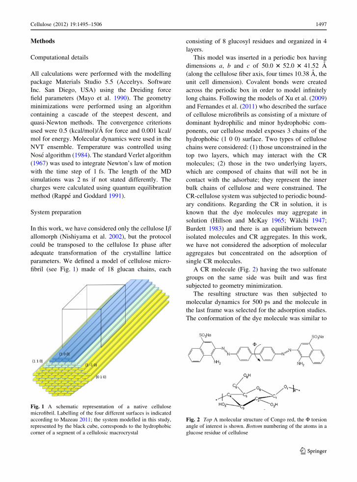
parameters. We defined a model of cellulose micro-
fibril (see Fig. 1) made of 18 glucan chains, each
consisting of 8 glucosyl residues and organized in 4
layers.
This model was inserted in a periodic box having
dimensions a, b and c of 50.0 3 52.0 3 41.52 A
(along the cellulose fiber axis, four times 10.38 A, the
unit cell dimension). Covalent bonds were created
across the periodic box in order to model infinitely
long chains. Following the models of Xu et al. (2009)
and Fernandes et al. (2011) who described the surface
of cellulose microfibrils as consisting of a mixture of
dominant hydrophilic and minor hydrophobic com-
ponents, our cellulose model exposes 3 chains of the
hydrophobic (1 0 0) surface. Two types of cellulose
chains were considered: (1) those unconstrained in the
top two layers, which may interact with the CR
molecules; (2) those in the two underlying layers,
which are composed of chains that will not be in
contact with the adsorbate; they represent the inner
bulk chains of cellulose and were constrained. The
CR-cellulose system was subjected to periodic bound-
ary conditions. Regarding the CR in solution, it is
known that the dye molecules may aggregate in
solution (Hillson and McKay 1965; Walchi 1947;
Burdett 1983) and there is an equilibrium between
isolated molecules and CR aggregates. In this work,
we have not considered the adsorption of molecular
aggregates but concentrated on the adsorption of
single CR molecules.
A CR molecule (Fig. 2) having the two sulfonate
groups on the same side was built and was first
subjected to geometry minimization.
The resulting structure was then subjected to
molecular dynamics for 500 ps and the molecule in
the last frame was selected for the adsorption studies.
The conformation of the dye molecule was similar to
Fig. 1 A schematic representation of a native cellulose
microfibril. Labelling of the four different surfaces is indicated
according to Mazeau 2011; the system modelled in this study,
represented by the black cube, corresponds to the hydrophobic
corner of a segment of a cellulosic macrocrystal
Fig. 2 Top A molecular structure of Congo red, the U torsion
angle of interest is shown. Bottom numbering of the atoms in a
glucose residue of cellulose
Cellulose (2012) 19:1495–1506 1497
123

the twisted one found in the CR crystal (Ojala et al.
1995).
In a typical simulation, a CR molecule was
positioned about 12 A away from the cellulose surface
(Fig. 3) and the whole system was subjected to
simulations of adsorption process with molecular
dynamics after a preliminary energy minimization.
Several dynamics experiments were performed
with the CR molecules initially oriented parallel and
perpendicular with respect to the cellulose fiber axis
and at different temperatures, ranging from 300 to
650 K. In order to improve the conformational
sampling, the last structure of the trajectory at 300 K
resulting from an initial parallel orientation of the CR
was extracted. The CR was then manually translated
by ±4 A along the surface, in directions parallel and
perpendicular to the fiber axis. The CR was also
rotated by 45� clockwise and anti-clockwise around an
axis normal to the cellulose surface. Each resulting
model was minimized and then subjected to a short
50 ps molecular dynamics at 300 K.
We then considered the possibility of accounting
for explicit water environment. We encountered
difficulties in performing the MD in a box full of
water molecules due to the computational demand
of MD calculations with a very large number of
atoms. To overcome such difficulty, we devised two
strategies. At first, MDs were performed with a CR
molecule placed 12 A above a cellulose surface,
both sulfonate groups were solvated with 10 water
molecules (SPC model) each. Such semi-explicit
approach was similar to that published by Fennell
et al. (2011).
Water molecules were equally distributed around
the sulfonate groups and the geometry was optimized
according to the standard procedure. MD experiments
at different temperatures and with different initial
position of CR were run and the last frames were
minimized. In the second strategy a single frame was
taken from the end of the MD trajectory in vacuum
where CR is fully adsorbed and then the system was
solvated by inserting 2,824 water molecules into the
computational box with the help of Monte Carlo
algorithm. The solvated complex was then minimized
and a 2 ns molecular dynamics simulation at 300 K
was performed.
To quantify the energetic consequences of the
adsorption process in the gas phase, the energy of
interaction between CR and cellulose surface (DEi) was
calculated. It was obtained by subtracting the energies of
the isolated dye molecule (Edye) and that of cellulose
(Ecell) from the total energy of the complete system (Etot):
DEi ¼ Etot � Edye � Ecell
In an explicit water environment, the total energy
of the CR—cellulose complex (Etot) is the sum of
contributions of: the energy of the water molecules
(Ewat), the energy of CR (ECR), the energy of cellulose
(Ecell), the energy of interaction between CR and water
(EiCR�wat), the energy of interaction between cellulose
and water (Eiwat�cell) and the energy of interaction
between CR and cellulose (EiCR�cell). Therefore, the
EiCR�cell was estimated according to:
DEiCR�cell ¼ Etot � ECR � Ecell � Ei
wat�cell
� EiCR�wat � Ewat
The global orientation of the adsorbed CR molecule
with respect to the cellulose microfibril surface can be
quantified by three relative rotation parameters: roll,
tilt, and twist and by three relative translation param-
eters: rise, shift, and slide (Dickerson 1989). Rotation
parameters are defined as specific angles between
principal axes (long, short and perpendicular) of two
Fig. 3 An initial model
showing : the periodic
limits, the (1 0 0)
hydrophobic corner the Ibcellulose elementary fibril
and CR in one of its initial
position. Left projection
perpendicular to the
cellulose chain axis, Rightprojection parallel to the
cellulose chain axis
1498 Cellulose (2012) 19:1495–1506
123

planes (Fig. 4): the first plane defining the CR
molecule and the second plane including the (1 0 0)
surface of cellulose.
Results and discussion
Adsorption process in the gas phase
First, to reveal the low energy intermolecular arrange-
ments between CR and cellulose, we performed
simulations in the gas phase, thus avoiding the
solvation effects. Trajectories were run for different
initial orientations of CR with respect to the fiber axis
and at different temperatures.
A visual inspection of the trajectories performed at
room temperature shows that CR is quickly attracted
by cellulose. It approaches the surface thanks to its
polar sulfonate groups. The electrostatic interactions
between negatively charged oxygen atoms of one of
the sulfonate group and the positively charged hydro-
gen atoms of cellulose hydroxyl groups are obviously
the driving force of the adsorption process. When the
first sulfonate group grabs the cellulose surface, the
process of adsorbing the second sulfonate group
begins. Simultaneously, both naphthalene residues of
CR come closer to the surface. Then, the entire
molecule of the dye is in contact with the cellulose
surface. Once CR is adsorbed on cellulose, a stable
system is formed and no departure from the adsorbed
conformation is observed.
A typical time evolution of the interaction energy,
DEint, between CR and cellulose for the temperature
300 K is given in Fig. 5.
As we expected, the decrease of the interaction
energy at the beginning of the simulation revealed that
the system reached a more stable state when CR left its
free state to become adsorbed. The decrease occurred
in steps: two of them are clearly visible in Fig. 5,
where DEint takes the constant values of about -32
and -72 kcal/mol. Once CR is fully adsorbed, the
interaction energy is stable at -105.4 ± 6.3 kcal/mol.
For each trajectory, the average energies of interaction
between CR and the cellulose microfibril were calcu-
lated and reported in Table 1.
Fig. 4 Description of the rotation and translation parameters
between two planes (adapted from Dickerson 1989)
Fig. 5 Evolution with time of two selected properties of the
system (initial parallel CR position) during the 2 ns MD
simulation at 300 K. Blue number of atomic close contacts
(within a 5A cutoff) between CR and the cellulose surface.
Red interaction energy. (Color figure online)
Table 1 Energies of interaction (kcal/mol) at equilibrium
between CR and cellulose calculated for different temperatures
(K) and different initial positions in vacuum
Temperature
(K)
Parallel initial
position
Perpendicular initial
position
300 -105.4 (±6.3) -91.0 (±5.8)
300-(1) -97.6 (±4.5) –
300-(2) -93.4 (±8.0) –
300-(3) -95.5 (±5.4) –
350 -99.1 (±7.2) -99.1 (±7.4)
400 -105.1 (±10.4) -90.6 (±7.8)
500 -101.1 (±9.4) -81.5 (±7.2)
600 -125.3 (±17.4) -96.2 (±8.4)
Standard deviations are given in brackets. Results from
simulations at 300 K when CR is moved or rotated are also
indicated: 300-(1) translated by 4 A along the chain direction
(z axis), 300-(2) translated by -4 A perpendicularly to the chain
direction (y axis), 300-(3) rotated by 45� anticlockwise (see
Fig. 6)
Cellulose (2012) 19:1495–1506 1499
123
The CR molecule was then translated along the y
and z axes, rotated around the x axis, and the resulting
complexes were simulated with molecular dynamics.
Energies of interaction between CR and cellulose are
also presented in Table 1. Changing the position and
orientation of CR leads to the identification of
different adsorption sites whose energies of interac-
tions were of similar magnitude, the largest difference
observed being of ?12.0 kcal/mol. The minor differ-
ence between these energies suggests that there are no
specific adsorption sites on the (1 0 0) cellulose
surface. The selected examples of adsorption sites on
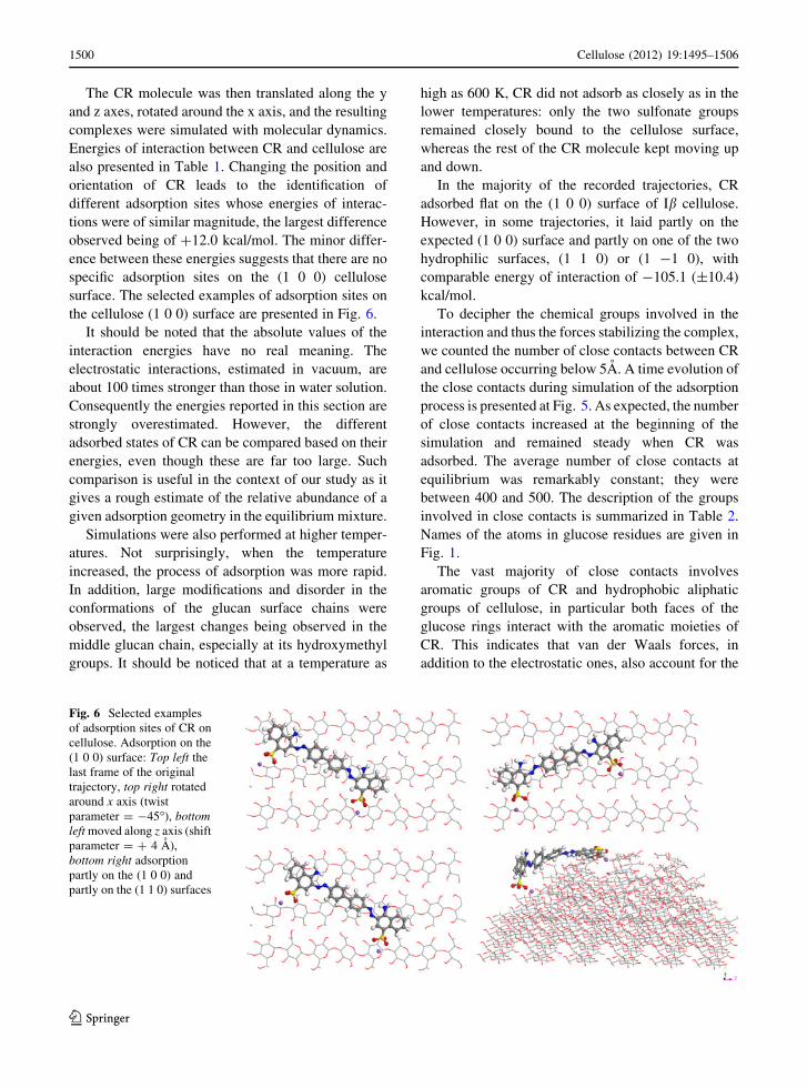
the cellulose (1 0 0) surface are presented in Fig. 6.
It should be noted that the absolute values of the
interaction energies have no real meaning. The
electrostatic interactions, estimated in vacuum, are
about 100 times stronger than those in water solution.
Consequently the energies reported in this section are
strongly overestimated. However, the different
adsorbed states of CR can be compared based on their
energies, even though these are far too large. Such
comparison is useful in the context of our study as it
gives a rough estimate of the relative abundance of a
given adsorption geometry in the equilibrium mixture.
Simulations were also performed at higher temper-
atures. Not surprisingly, when the temperature
increased, the process of adsorption was more rapid.
In addition, large modifications and disorder in the
conformations of the glucan surface chains were
observed, the largest changes being observed in the
middle glucan chain, especially at its hydroxymethyl
groups. It should be noticed that at a temperature as
high as 600 K, CR did not adsorb as closely as in the
lower temperatures: only the two sulfonate groups
remained closely bound to the cellulose surface,
whereas the rest of the CR molecule kept moving up
and down.
In the majority of the recorded trajectories, CR
adsorbed flat on the (1 0 0) surface of Ib cellulose.
However, in some trajectories, it laid partly on the
expected (1 0 0) surface and partly on one of the two
hydrophilic surfaces, (1 1 0) or (1 -1 0), with
comparable energy of interaction of -105.1 (±10.4)
kcal/mol.
To decipher the chemical groups involved in the
interaction and thus the forces stabilizing the complex,
we counted the number of close contacts between CR
and cellulose occurring below 5A. A time evolution of
the close contacts during simulation of the adsorption
process is presented at Fig. 5. As expected, the number
of close contacts increased at the beginning of the
simulation and remained steady when CR was
adsorbed. The average number of close contacts at
equilibrium was remarkably constant; they were
between 400 and 500. The description of the groups
involved in close contacts is summarized in Table 2.
Names of the atoms in glucose residues are given in
Fig. 1.
The vast majority of close contacts involves
aromatic groups of CR and hydrophobic aliphatic
groups of cellulose, in particular both faces of the
glucose rings interact with the aromatic moieties of
CR. This indicates that van der Waals forces, in
addition to the electrostatic ones, also account for the
Fig. 6 Selected examples
of adsorption sites of CR on
cellulose. Adsorption on the
(1 0 0) surface: Top left the
last frame of the original
trajectory, top right rotated
around x axis (twist
parameter = -45�), bottomleft moved along z axis (shift
parameter = ? 4 A),
bottom right adsorption
partly on the (1 0 0) and
partly on the (1 1 0) surfaces
1500 Cellulose (2012) 19:1495–1506
123

adsorption strength. In agreement with the experi-
ments, adsorption of dyes on cellulose is driven by van
der Waals forces (Diaz et al. 2008). The order of
magnitude of the strength of these van der Waals
interactions is given by density functional theory
(DFT) of L-tyrosine interacting with a methyl b-D-
glucopyranoside, a system that can be considered as a
model for the cellulose–CBM complex (Mohamed
et al. 2010). Such calculations have been performed in
vacuum, as we did, where the interaction energies
values were estimated to be between -5 and -8 kcal/
mol. Despite this main feature, the presence of short
contacts between hydrogen atoms of cellulose and
electronegative nitrogen and oxygen atoms of CR
suggests the existence of hydrogen bonds. It is
important to emphasize that most of the hydrogen
bonds occur via the hydroxyl groups associated with
the C2 and C6 carbon atoms of each glucosyl rings.
Sulfonate, amino, and azo groups of CR form hydro-
gen bonds with the hydroxymethyl groups of cellu-
lose. Even if the (1 0 0) surface has a dominant
hydrophobic character, still a significant number of
hydrogen bonds do occur between CR and cellulose.
In addition to these ‘‘classical’’ hydrogen bonds
involving polar groups, a significant number of close
contacts involving aliphatic C–H groups interacting
with an electronegative atom are also found. We found
three kinds of them: (1) C–H���O, with lengths of 2.80
and 2.65 A, also found experimentally (Bird et al.
2006), (2) C–H���N, with lengths of 2.47 and 2.95 A,
(3) N–H���O, length of 2.59 A.
The forces involved in the interaction revealed by
the present simulations are supported by the analogy
with those measured in saturation transfer difference
and line broadening NMR studies of the binding of
cellohexaose to the CBM of Clostridium thermocel-
lum (Viegas et al. 2008). In such a system, it was
observed that in addition to the specific hydrogen
bonds, the aliphatic protons of the glucosyl units were
close to the tyrosine aromatic rings of the CBM
(Viegas et al. 2008).
The description of the position and the orientation
of adsorbed CR at equilibrium with respect to the
cellulose surface, quantified by the three rotation and
three translation parameters, are presented in Table 3.
Table 2 Description of the close contacts (below 5A) between
structural groups of CR molecule and those of cellulose
Congo red
groups
Cellulose atoms Number
of close
contacts
Sulfonate
groups
C6, C5 and O6 hydrogen atoms 25
Biphenyl
moiety
C6, C5 and C3 hydrogen atoms 28
Naphthalene
moieties
C6, C4 and C1 hydrogen atoms 10
Azo-groups C6, C4 and C4 hydrogen atoms 9
Amino
groups
C6, C5 and C4 hydrogen atoms
and O5 oxygen atom
10
Table 3 Equilibrium
values of the orientation
parameters between CR
molecule and the (1 0 0)
surface of cellulose at
different temperatures and
for different initial positions
of a dye molecule in
vacuum
Roll, tilt and twist in
degrees, shift, slide and rise
in angstroms. Initial values
are indicated in italics
Temperature (K) Roll Tilt Twist Shift Slide Rise
Initial parallel 5.8 -20.9 2.2 0.34–0.81 -1.26–13.14 12.7
300 1.0 8.3 5.6 -7.41 -1.26 3.83
300 (1) -0.8 -3.0 -13.2 7.51 3.02 3.93
300 (2) -1.6 -1.0 -9.5 1.89 5.11 3.98
300 (3) -1.8 -3.5 -56.5 4.46 2.88 4.22
350 1.8 10.2 2.2 -4.14 2.73 3.98
400 10.4 2.3 56.4 -13.11 -6.86 -2.80
500 23.3 -49.0 -62.0 -17.85 13.14 -1.02
600 1.2 46.4 8.3 6.20 -3.26 4.39
Initial perpendicular 5.5 -74 ?83 1 -18.64–8.16 13.64
300 0.6 1.8 46.1 3.94 3.43 4.17
350 0.8 25.5 48.4 11.01 0.29 5.69
400 0.3 16.5 18.8 1.66 1.44 4.14
500 22.7 35.5 25.7 5.25 -18.64 -2.28
600 13.7 -10.7 47.8 1.76 8.16 -3.38
Cellulose (2012) 19:1495–1506 1501
123
At low temperatures (300 and 350 K), the roll and
tilt angle parameters are small, suggesting that both
planes of the CR molecule and of the top layer of the
cellulose chains become more coplanar. In fact, the
values of these two parameters were not rigorously
zero, since as aforementioned the CR molecule was
not fully planar and since the cellulose surface
possessed a residual roughness. As for the twist angle,
it became equilibrated to either the low values of 0–5�or the large ones of 46–48�. In addition, the shift and
slide translational parameters were equilibrated at
different values, suggesting that there were several
adsorption sites on cellulose, in agreement with a
recent study describing the adsorption of the CBM
from Cel6A on crystalline cellulose (Shiiba et al.
2012). When adsorbed on the hydrophobic surface, the
rise parameter, which describes the altitude of CR with
respect to the cellulose surface is about 4 A, a value
similar to the one found for the adsorption of the
CBM of Clostridium thermocellulm on cellohexaose
(Viegas et al. 2008). The large values of the roll and
twist angles observed for some of the high temperature
simulations together with the negative values of the
rise parameter illustrate this particular adsorption state
of CR on both hydrophobic and hydrophilic surfaces.
It occurs in this particular situation where the gravity
centre of CR is lower than that of the surface chains of
cellulose.
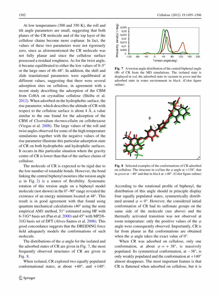
The molecule of CR is expected to be rigid due to
the low number of rotatable bonds. However, the bond
linking the central biphenyl moieties (the torsion angle
u in Fig. 2) is a source of flexibility. Systematic
rotation of this torsion angle on a biphenyl model
molecule (not shown) in the 0�–90� range revealed the
existence of an energy minimum located at 48�. This
result is in good agreement with that found using
quantum mechanical calculations (40� using the semi
empirical AM1 method, 51� estimated using HF with
6-31G* basis set (Pan et al. 2000) and 45� with MP2/6-
31G basis set of DFT (Alves-Santos et al. 2006). This
good concordance suggests that the DREIDING force
field adequately models the conformations of such
molecule.
The distributions of the u angle for the isolated and
the adsorbed states of CR are given in Fig. 7, the most
frequently observed structures of CR are given in
Fig. 8.
When isolated, CR explored two equally populated
conformational states, at about ?60�, and ?140�.
According to the rotational profile of biphenyl, the
distribution of this angle should in principle display
four equally populated states, symmetrically distrib-
uted around u = 0�. However, the considered initial
conformation of CR had its sulfonate groups on the
same side of the molecule (see above) and the
thermally activated transition was not observed at
room temperature: only the positive values of the uangle were consequently observed. Importantly, CR is
far from planar as flat conformations are obtained
when the u angle takes the exact value of 0�.
When CR was adsorbed on cellulose, only one
conformation, at about u = ? 38�, is massively
populated. Its symmetrical conformation, at -38�, is
only weakly populated and the conformation at ?140�almost disappears. The most important feature is that
CR is flattened when adsorbed on cellulose, but it is
Fig. 7 A torsion angle distribution of the central biphenyl angle
(U) of CR from the MD simulations. The isolated state is
displayed in red, the adsorbed state in vacuum in green and the
adsorbed state in water environment in black. (Color figure
online)
Fig. 8 Selected examples of the conformations of CR adsorbed
on cellulose. The structure in red has the u angle at ?138�, that
in green at -40� and that in black at ?40�. (Color figure online)
1502 Cellulose (2012) 19:1495–1506
123

still not rigorously flat. Steric interactions between the
hydrogen atoms of adjacent phenyl rings prevail over
the effect of the overlap of their p-orbitals and a fully
planar conformation is not accessible (Lye et al. 2000).
The sulfonate groups are on the same side of CR
when adsorbed. The only exception is adsorption at
600 K, when in the final adsorbed state, both sulfonate
groups are on opposite sides of the CR molecule.
Interestingly, it has been experimentally measured that
dyes in which the sulfonate groups are on one side of
the molecule have a larger binding enthalpy for
adsorption on cotton cellulose than those where the
sulfonate groups are on the opposite side of the dye
molecule (Bird et al. 2006). To check if modelling
successfully reproduces this experimental result, we
calculated the interaction energies between cellulose
surface and CR for two different conformations of the
dye molecule: one where both sulfonate groups are on
one side of the molecule and one where they are on
opposite sides. In this modelling, the dye molecule
was positioned 4 A from the cellulose surface and a
short 50 ps dynamics was run: the interaction between
the CR and the cellulose surface was stronger when the
sulfonate groups were on one side, as opposed to the
situation where they were on both sides, the difference
being of 20.7 kcal/mol.
Adsorption process in aqueous environment
Since in reality the adsorption occurs mostly in
aqueous environment, the effect of adding water
molecules was investigated. To account for both the
screening of electrostatic interactions and their
potential structural role, we undertook a two-step
strategy. In the first step, we studied the adsorption
process of CR on cellulose with both sulfonate groups
being solvated with 10 water molecules (SPC model).
This procedure, called the semi-explicit water
approach, was drawn from a recent treatment of the
hydration, in which only the important water mole-
cules were treated explicitly and the rest of the solvent
phase was treated implicitly by a continuum approach
(Fennell et al. 2011). Basically the same MD exper-
iments were run as in the vacuum case, at a 300-650 K
temperature range and in two different initial positions
of CR, i.e., parallel and perpendicular to the cellulose
fiber axis.
At room temperature, the process of adsorption
differed from that in vacuum. At 300 K, the hydrated
sulfonate groups were not the first chemical groups of
CR to touch the surface of cellulose, but it was the
aromatic rings of the CR molecule that created the first
contacts. In contrast, at higher temperatures, water left
CR quickly and thus the sulfonate groups were the first
to approach the cellulose surface. The highest tem-
perature, 650 K, was clearly not adapted in this
context since the water molecules escaped from the
sulfonate groups of CR. The water desorption tem-
perature was then assumed to be at about 650 K.
The geometrical features of the adsorption are
however preserved when water molecules are explic-
itly present in the simulated medium. At low temper-
ature, CR adsorbed entirely on the (1 0 0) surface, and
adsorption on the hydrophilic surfaces occurred at
high temperatures. The equilibrium values of the
orientation parameters between CR molecule and the
(1 0 0) surface of cellulose at different temperatures
and for different initial positions of the dye molecule,
shown in Table 4, indicates also a strong similarity
with that recorded for the vacuum trajectories.
The small values explored by the roll and tilt angles
suggest that CR adsorbed flat on the cellulose surface.
The twist angle adopts different values, revealing that
CR can accommodate different stable orientations
with respect to the cellulose fiber axis. A noticeable
difference between trajectories performed in vacuum
and when only sulfonate groups were hydrated is that
the adsorbed dye molecule is more rigid in water
compared to its relative flexibility in the vacuum phase
(see the black curve in Fig. 7). In the explicit water
environment, only the u angle at 60� is populated.
The energies of interaction between CR and
cellulose, when both sulfonate groups of CR are
hydrated with 20 water molecules, were globally
20–30 kcal/mol higher than those observed under
vacuum conditions (Table 5).
Such lowering of the interaction strength between
the substrate and the surface induced by the explicit
presence of water molecules have been recently
documented and discussed in the case of the interac-
tion between xylan and cellulose (Mazeau and Char-
lier 2012).
In a following experiment, we studied the behav-
iour of a stable pre-formed complex established in
vacuum under a full explicit water environment. At a
temperature of 300 K for a parallel initial position of
CR, the interaction energy between a dye molecule
and cellulose moiety slightly increased to -92.5
Cellulose (2012) 19:1495–1506 1503
123

(±10.2) kcal/mol, thus lowering the difference
between the energies from vacuum and hydrated
systems by a small number of water molecules.
Additionally, no noticeable modification of the geom-
etry of the complex could be observed.
These results suggest that although the interaction
energy between CR and cellulose is estimated to be
less efficient when explicit hydration is considered
(either in a partial or a full manner), the basic
associative features remain the same.
Concluding remarks
Our work has revealed the basic associative features
of the interaction of CR with the hydrophobic surface
(1 0 0) of cellulose Ib. The results clearly show that CR
indeed presents a real affinity for the hydrophobic
surface of cellulose but unlike in the case of the well
documented adsorption of aromatic amino acids, the
adsorbed aromatic parts of the CR molecules are not
laid completely flat on the cellulose substrate, but are
slightly distorted. In fact, the CR molecules consist of
two symmetric planar aromatic parts, connected by a
central biphenyl bond, which acts as a rotatable joint.
With isolated CR, the two parts become substantially
rotated with respect to one another under the repulsion
of the hydrogen atoms located in the ortho positions of
the two adjacent central phenyl rings (Takei et al.
1988). In the present modelling study, we observe that
the torsion angle along the biphenyl bond does not go
to zero, but is drastically reduced when the CR
molecules become adsorbed on the hydrophobic
surface of cellulose. This conformational adaptation
clearly indicates that in addition to the electrostatic
and hydrogen-bonding interactions, the CR adsorption
also results from a substantial van-der-Waals type
attraction of its two aromatic moieties on the corre-
sponding hydrophobic cellulose surface.
In the previous study, Woodcock et al. (1995) used
a molecular mechanics approach to model the adsorp-
tion of CR on the hydrophilic surfaces of crystalline
cellulose. In similarity with the present study, they
also obtained a series of adsorption sites of minimum
energy, indicating that possibilities existed for CR to
also adsorb on such surfaces. To explain their results,
they indicated that ‘‘the adsorption phenomenon was
dominated the electrostatic interaction between the
Table 4 Equilibrium values of the orientation parameters between CR molecule and (1 0 0) surface of cellulose at different
temperatures and for different initial positions of a dye molecule when both sulfonate groups were hydrated
Roll Tilt Twist Shift Slide Rise
Initial parallel -6.9 to -5.0 35.0–37.7 -4.4 to -5.7 0.25–0.44 -1.64 12.80
300 2.2 12.3 -16.2 3.70 2.19 4.42
350 2.5 11.0 -15.5 1.85 0.58 4.28
400 3.3 13.5 -61.2 -2.78 -2.73 3.93
500 2.1 7.9 4.0 -9.72 -7.49 3.96
600 0.4 -17.6 -24.3 3.91 -0.80 -4.23
Initial perpendicular -3.0 54.6–52.5 83.0–84.1 -1.73 to -2.20 -1.33 to 0.80 13.20
300 2.7 10.7 -15.9 8.90 10.69 2.90
350 8.7 4.2 -50.1 -1.69 12.34 2.83
400 17.3 -7.37 61.9 1.36 -8.37 2.85
500 No adsorption
600 No adsorption
Table 5 Energies of interaction (kcal/mol) between CR mol-
ecule and Ib cellulose in CR-cellulose complexes, when both
sulfonate groups are hydrated with 20 water molecules, cal-
culated for different temperatures (K) and different initial
positions
Temperature (K) Parallel Perpendicular
300 -75.9 (±4.4) -68.0 (±5.2)
350 -68.0 (±5.2) -67.2 (±5.7)
400 31.8 (±7.6) 29.6 (±12.1)
500 43.8 (±9.6) No adsorption
600 79.8 (±10.9) No adsorption
SD are given in brackets
1504 Cellulose (2012) 19:1495–1506
123
highly polar groups of the CR and the hydroxyl groups
and exposed acetal oxygen on the cellulose surfaces’’
(Woodcock et al. 1995). Our modelling, which, in
addition to the electrostatic phenomena, also takes
advantage of the van der Waals interactions, is
nevertheless different from their approach. At present
it is not possible to compare their results with ours to
decide whether CR will prefer our adsorption sites or
theirs or will cover both. Work is in progress to answer
such question.
Acknowledgments This research was partly supported by a
grant from the Polish Ministry of Science no N507081033. The
authors acknowledge the help of Dr H. Chanzy for valuable
comments during the writing of this work.
Open Access This article is distributed under the terms of the
Creative Commons Attribution License which permits any use,
distribution, and reproduction in any medium, provided the
original author(s) and the source are credited.
References
Alves-Santos M, Davila LYA, Petrilli HM, Capaz RB, Caldas
MJ (2006) Application of standard DFT theory for non-
bonded interactions in soft matter: prototype study of poly-
para-phenylene. J Comput Chem 27:217–227
Baker AA, Helbert W, Sugiyama J, Miles MJ (1997) High-
resolution atomic force microscopy of native Valoniacellulose I microcrystals. J Struct Biol 119:129–138
Baker AA, Helbert W, Sugiyama J, Miles MJ (2000) New
insight into cellulose structure by atomic force microscopy
shows the Ia crystal phase at near-atomic resolution. Bio-
phys J 79:1139–1145
Bird J, Brough N, Dixon S, Batchelor SN (2006) Understanding
adsorption phenomena: investigation of the dye-cellulose
interaction. J Phys Chem B 110:19557–19561
Burdett BC (1983) In: Whyn-Jones E, Gormally J (eds)
Aggregation processes in solution. Elsevier, Amsterdam
Da Silva Perez D, Ruggiero R, Morais LC, Machado AEH,
Mazeau K (2004) Theoretical and experimental studies on
the adsorption of aromatic compounds onto cellulose.
Langmuir 20:3151–3158
Diaz MD, Fernadez-Alonso MC, Cuevas G, Canada FJ, Jime-
nez-Barbero J (2008) On the role of aromatic-sugar inter-
actions in the molecular recognition of carbohydrates: a 3D
view by using NMR. Pure Appl Chem 80:1827–1835
Dickerson RE (1989) Definitions and nomenclature of nucleic
acid structure parameters. J Biomol Struc Dynamics 6:
627–634
Fennell CJ, Kehoe CW, Dill KA (2011) Modeling aqueous
solvation with semi-explicit assembly. Proc Natl Acad Sci
USA 108:3234–3239
Fernandes NA, Thomas LH, Altaner CM, Callow P, Forsyth VT,
Apperley DC, Kennedy CJ, Jarvis MC (2011) Nanostruc-
ture of cellulose microfibrils in spruce wood. Proc Natl
Acad Sci USA 108:E1195–E1203
Helbert W, Nishiyama Y, Okano T, Sugiyama J (1998)
Molecular imaging of Halocynthia papillosa cellulose.
J Struct Biol 124:42–50
Hillson PJ, McKay RB (1965) Aggregation of dye molecules in
aqueous solution. A polarographic study. T Faraday Soc
61:374–382
Inglesby MK, Zeronian SH (2002) Direct dyes as molecular
sensors to characterize cellulose substrates. Cellulose 9:
19–29
Kalashnikova I, Bizot H, Cathala B, Capron I (2012) Modulation
of cellulose nanocrystals amphiphilic properties to stabilize
oil/water interface. Biomacromolecules 13:267–275
Kim N-H, Herth W, Vuong R, Chanzy H (1996) The cellulose
system in the cell wall of Micrasterias. J Struct Biol 117:
195–203
Lehtio J, Sugiyama J, Gustavsson M, Fransson L, Linder M,
Teeri TT (2003) The binding specificity and affinity
determinants of family 1 and family 3 cellulose binding
modules. PNAS 100:484–489
Linder M, Mattinen ML, Kontelli M, Lindberg G, Stahlberg J,
Drakenberg T, Reinikainen T, Pettersson G, Annila A
(1995) Identification of functionally important amino acids
in the cellulose-binding domain of Trichoderma reeseicellobiohydrolase I. Protein Sci 4:1056–1064
Lye J, Freeman HS, Cox RD (2000) Molecular modeling of
Congo red analogues containing terphenyl and quarter-
phenyl moieties. Dyes Pigm 47:53–64
Mayo SL, Olafson BD, Goddard WA III (1990) DREIDING: a
generic force field for molecular simulations. J Phys Chem
94:8897–8909
Mazeau K (2011) On the external morphology of native cellu-
lose microfibrils. Carbohydr Polym 84:524–532
Mazeau K, Charlier L (2012) The molecular basis of the
adsorption of xylans on cellulose surface. Cellulose 19:
337–349
Mazeau K, Rivet A (2008) Wetting the (110) and (100) surfaces
of Ib cellulose studied by molecular dynamics. Biomac-
romolecules 9:1352–1354
Mazeau K, Vergelati C (2002) Atomistic modeling of the
adsorption of benzophenone onto cellulosic surfaces.
Langmuir 18:1919–1927
Medronho B, Romano A, Miguel MG, Stigsson L, Lindman B
(2012) Rationalizing cellulose (in)solubility: reviewing
basic physicochemical aspects and role of hydrophobic
interactions. Cellulose 19:581–587
Mohamed MNA, Watts HD, Guo J, Catchmark JM, Kubicki JD
(2010) MP2, density functional theory, and molecular
mechanical calculations of C-H���p and hydrogen bond
interactions in a cellulose-binding module-cellulose model
system. Carbohydr Res 345:1741–1751
Morton TH (1946) The dichroic behavior of substantive dyes:
molecular theory of the dyeing of cellulose. J Soc Dyers
Colour 62:272–280
Nishiyama Y, Langan P, Chanzy H (2002) Crystal structure and
hydrogen-bonding system in cellulose Ib from synchrotron
x-ray and neutron fiber diffraction. J Am Chem Soc 124:
9074–9082
Nose S (1984) A unified formulation of the constant temperature
molecular dynamics methods. J Chem Phys 81:511–519
Ojala WH, Ojala CR, Gleason WB (1995) The x-ray crystal
structure of the sulfonated azo dye Congo red, a
Cellulose (2012) 19:1495–1506 1505
123

non-peptidic inhibitor of HIV-1 protease which also binds
to reverse transcriptase and amyloid proteins. Antiv Chem
Chemoth 6:25–33
Pan JF, Chua SJ, Huang W (2000) Conformational analysis on
biphenyls with theoretical calculations: modeling torsions
in poly(para-phenylene)s with side chains. Thin Solid
Films 363:1–5
Pielesz A (2007) Spectroscopic study of interactions between
model direct dyes and cotton. J Appl Polym Sci 104:
758–766
Quiocho FA (1989) Protein-carbohydrate interactions: basic
molecular features. Pure Appl Chem 61:1293–1306
Rappe AK, Goddard WA III (1991) Charge equilibration for
molecular dynamics simulations. J Phys Chem 95:
3358–3363
Reinikainen T, Teleman O, Teeri TT (1995) Effects of pH and
high ionic strength on the adsorption and activity of native
and mutated cellobiohydrolase I from Trichoderma reesei.Proteins 22:392–403
Revol J-F (1982) On the cross-sectional shape of cellulose
crystallites in Valonia ventricosa. Carbohydr Polym 2:
123–134
Screen J, Stanca-Kaposta EC, Gamblin DP, Liu B, Macleod NA,
Snoek LC, Davis BG, Simons JP (2007) IR-spectral sig-
natures of aromatic-sugar complexes: probing carbohy-
drate-protein interactions. Angew Chem Int Ed 46:
3644–3648
Shiiba H, Hayashi S, Yui T (2012) Molecular simulation study
with complex models of the carbohydrate binding module
of Cel6A and the cellulose Ia crystal. Cellulose 19:
635–645
Sugiyama J, Harada H, Fujiyoshi Y, Uyeda N (1985) Lattice
images from ultrathin sections of cellulose microfibrils in
the cell wall of Valonia macrophysa Kutz. Planta 166:
161–168
Sugiyama J, Vuong R, Chanzy H (1991) Electron diffraction
study on the two crystalline phases occurring in native
cellulose from an algal cell wall. Macromolecules 24:
4168–4175
Takei Y, Yamaguchi T, Osamura Y, Fuke K, Kaya K (1988)
Electronic spectra and molecular structure of biphenyl and
para-substituted biphenyls in a supersonic jet. J Phys Chem
92:577–581
Tormo J, Lamed R, Chirino AJ, Morag E, Bayer EA, Shoham Y,
Steltz TA (1996) Crystal structure of a bacterial family-III
cellulose-binding domain: a general mechanism for
attachment to cellulose. EMBO J 15:5739–5751
Verlet L (1967) Computer experiments on classical fluids.
I. Thermodynamical properties of Lennard-Jones mole-
cules. Phys Rev 159:98–103
Viegas A, Bras NF, Cerqueira NMFSA, Fernandes PA, Prates
JAM, Fontes CMGA, Bruix M, Romano MJ, Carvalho AL,
Ramos MJ, Macedo AL, Cabrita EJ (2008) Molecular
determinants of ligand specificity in family 11 carbohy-
drate binding modules—an NMR, X-ray crystallography
and computational chemistry approach. FEBS J 275:
2524–2535
Walchi O (1947) Die Einlagerung von Kongorot in Zellulose.
Holzforschung 1:20–32
Weis WI, Drickamer K (1996) Structural basis of lectin-car-
bohydrate recognition. Annu Rev Biochem 65:441–473
Wood PJ (1980) The interaction of direct dyes with water sol-
uble substituted celluloses and cereal b-glucans. Ind Eng
Chem Prod Res Dev 19:19–23
Wood PJ (1982) Factors affecting precipitation and spectral
changes associated with complex-formation between dyes
and b-D-glucans. Carbohydr Res 102:283–293
Woodcock S, Henrissat B, Sugiyama J (1995) Docking of
Congo red to the surface of crystalline cellulose using
molecular mechanics. Biopolymers 36:201–210
Xu Q, Tucker MP, Arenkiel P, Ai X, Rumbles G, Sugiyama J,
Himmel ME, Ding S-Y (2009) Labeling the planar face of
crystalline cellulose using quantum dots directed by type-I
carbohydrate-binding modules. Cellulose 16:19–26
Yamaki SB, Barros DS, Garcia CM, Socoloski P, Oliveira ON,
Atvars TDZ (2005) Spectroscopic studies of the intermo-
lecular interactions of Congo red and Tinopal CBS with
modified cellulose fibers. Langmuir 21:5414–5420
1506 Cellulose (2012) 19:1495–1506
123


![Drag reduction by hydrophobic nano - particles adsorption process · -layered coating are erved[2].Correlating bio-science with mechanical engineering was extracted from this. Usage](https://static.fdocuments.in/doc/165x107/5fc798c085f2d87b161eec55/drag-reduction-by-hydrophobic-nano-particles-adsorption-process-layered-coating.jpg)


![Surface and adsorption characteristics of three elastin ... · PDF fileand ELP20-244 [ELP4] contains five hydrophobic domains flanked by four cross-linking domains [18, 19]. The](https://static.fdocuments.in/doc/165x107/5a88daa47f8b9a9f1b8e99a2/surface-and-adsorption-characteristics-of-three-elastin-elp20-244-elp4-contains.jpg)