
Hypothermia, Prevention, Recognition, Treatment_ Hypothermia Special Situations

1
Modeling the Effects of Induced Hypothermia for the
Treatment of a Cervical Spinal Cord Injury
BEE 4530: Computer-Aided Engineering
Tim Bruhn
Audrey Karabayinga
Sabrina Moroz
Sarah Song
2
Table of Contents
1. Executive Summary ................................................................................................................... 3
2. Introduction
i. Background ......................................................................................................................5
ii. Design Objectives .......................................................................................................... 7
iii. Problem Schematic ....................................................................................................... 7
iv. Mesh.............................................................................................................................. 9
3. Results and Discussion
i. Solution Analysis........................................................................................................... 11
ii. Accuracy Check............................................................................................................ 15
iii. Sensitivity Analysis .................................................................................................... 17
4. Conclusion and Design Recommendations
i. Design Recommendations............................................................................................. 23
ii. Realistic Constraints..................................................................................................... 23
5. Appendix A: Mathematical Statement of the Problem............................................................. 24
6. Appendix B: Solution Strategy ................................................................................................ 25
7. Appendix C: References .......................................................................................................... 27

3
1. Executive Summary
Spinal cord injury (SCI) is a devastating condition that affects approximately 11,000
patients each year in the United States. Moderate hypothermia has been used in a variety of
experimental and clinical situations to target several neurological disorders, including traumatic
brain injury and SCI. Studies in a large series of SCI patients, initiated in 2005, showed that early
cooling introduced by the use of endovascular catheters and continued for a 48-hour period
appeared to be safe and did not result in an increased incidence of risk factors, including cardiac
arrhythmias and severe infection. However, the procedure for inserting endovascular catheters is
invasive, which can be detrimental the patient. Prolonged systemic hypothermia in patients can
also lead to further complications, such as tissue damage due to frostbite. As a result, less
invasive local methodologies are being explored and studied to determine if the results of cooling
are comparably effective or superior to systemic hypothermic treatment. The goal of this project
was to model induced temperatures at or below 33°C (306 K) in the spinal cord through
localized surface cooling via a cooling pack in COMSOL Multiphysics.
Through the development and execution of an accurate model, we determined the amount
of time the cooling pack must be applied, the temperature the body must be cooled down to, and
the long-term cooling pack application protocol needed to maintain critical temperatures in the
spinal cord while preventing frostbite in surface tissues. A schematic of a simplified geometry of
the cross-section of the neck was created. Only half of the geometry was needed for the
schematic of the model as a result of simplification due to symmetry. It included regions for the
skin, neck tissue, vasculature, vertebral bone, and spinal cord (target) and was run in 2-D.
COMSOL was utilized to solve for transient heat transfer in cylindrical coordinates with a
conduction term and two sources of heat generation representing base metabolism and
inflammation. We expected to find that the application of the cooling pack would decrease the
core temperature from 37°C to 33°C (310 K to 306 K), which is the temperature systemic
cooling range for current therapeutic use.
Initially, the model was run with constant application of the cooling pack. We
determined that the cooling pack was capable of lowering the spinal cord temperature to 21ºC
(294 K) over the course of three hours. However, constant application of the cooling pack led to
4
tissue degradation due to frostbite. To avoid frostbite, we attempted to create a cyclic application
protocol that would maintain temperatures below 33ºC (306 K) in the spinal cord while
preventing the surrounding tissues from reaching freezing temperatures of 0ºC (273 K). We
found that the most effective protocol consisted of initially applying the cooling pack for 30
minutes at 0ºC (273 K), removing the cooling pad, heating the neck for 30 minutes at 37ºC (310
K), and then cyclically cooling for 20 minutes and heating for 30 minutes thereafter. We found
that the spinal cord temperature reached 33ºC (273 K) in less than 45 minutes and subsequently
fluctuated stably between 30ºC – 31ºC (303 K – 304 K). Also, tissue temperatures were found to
be stay above freezing, fluctuating between 2.5ºC – 37ºC (275.5 K – 310 K). An accuracy check
was executed to ensure that our solutions were comparable to the results of analogous
experiments. We performed sensitivity analysis of the temperature change in the spine to
different heat generation terms and various parameters in the skin, tissue, bone, and spine and
found that the model was relatively insensitive to these changes. The results of the sensitivity
analysis implied that the model was robust to changes in parameter values.
The significance of this data demonstrated that hypothermia could be successfully
induced with the application of a cooling pack alone, preventing the need for invasive systemic
cooling procedures. The ability to induce spinal cord cooling without insertion of cooling
catheters is a significant advancement due to the fact that significant systemic hypothermia (<
33°C, < 306 K) can result in cardiac arrhythmia, coagulopathy, and pancreatitis, among other
conditions. Therefore, by allowing the systemic temperature to remain at physiologically normal
temperatures during cooling, physicians can further prevent many of these fatal complications
through this cyclic application of a cooling pad. Finally, the development of a protocol to prevent
frostbite in the local tissues while keeping the spinal cord below the therapeutic temperature of
33°C (306 K) prevents the major complication associated with local cooling. The cooling pack
system can potentially be implemented by doctors in the future to induce localized cooling in
SCI patients with relatively rapid results without the risk of frostbite.

5
2. Introduction
i. Background
The first account of systemic hypothermia was made in November of 1938 by Dr.
Temple Fay, MD, FACS [1]. Dr. Fay’s first description of intentionally induced hypothermia
was actually an attempt to slow the progression of metastatic disease in a woman with ulcerating
breast carcinoma. This carcinoma had previously shown ‘‘remarkable response’’ to local
refrigeration of the breast and neck area. Fay et al. subsequently went on to describe the use of
hypothermia in a large number of patients with cancer and head injury [2, 3]. The idea of using
hypothermia in cardiac surgery was introduced by Bigelow et al. after a series of canine
experiments [4]. In addition, Botterell et al. began pioneering work in the use of hypothermia in
cerebral aneurysm repair in the 1950s [5]. As a result, hypothermia was defined to occur between
the temperatures, 24°C and 33°C (297 K and 306 K). Enthusiasm towards induced hypothermia
dwindled in the subsequent decades, and was largely abandoned by the 1980s. This disinterest
was largely attributed to the associated complications, which included cardiac arrhythmias,
hypotension, coagulopathies, systemic infections, and electrolyte disturbances [6]. In 1987,
Busto et al. documented that neuronal injury could be markedly attenuated by modest decreases
in body temperatures in a rat model of global ischemia. This suggested that the extent of
hypothermia required for neuroprotection was not as pronounced as initially anticipated [7].
Consequently, there was a renewed interest in hypothermia. The degree of hypothermia was
loosely defined by ranges of temperature as “mild” (33ºC – 36°C), “moderate” (28ºC – 32°C),
“severe” (16ºC – 28°C), and “profound” (15°C and below) [8]. The specifications of the levels of
hypothermia were important because of the potential for neurologic benefit with more modest
reductions in body temperatures. Due to its newly discovered applicability, mild to moderate
hypothermia has attracted more interest over the past two decades. It has been very effective in
acute traumatic brain injury, spinal cord injury, stroke, cardiac surgery, surgical repair of
cerebral aneurysms, and thoracoabdominal aortic aneurysm (TAA) [6, 9].
Localized cooling of the spinal cord can be achieved either externally by surface cooling
or internally via endovascular cooling [10]. Our study concentrated on achieving localized
hypothermia by external surface cooling, which is noninvasive and, therefore, a more appealing
procedure. Surface cooling systems, which use the circulation of cool air or water through

6
devices that are placed against the skin, have been made popular because of their ease of use.
These systems include the Bair Hugger System and Arizant Healthcare Inc. Systemic
Hypothermia. As used currently, both usually target a temperature between 32°C and 34°C (305
K and 307 K). These temperatures are less prone to severe adverse effects, such as arrhythmias,
than temperatures below 30°C (303 K). For our project, we aimed to lower the temperature of the
spinal cord to 33°C (306 K).
The goal of this project was to model localized cooling in the treatment of injuries to the
cervical spinal cord using COMSOL Multiphysics. The model was developed to demonstrate the
efficiency and effects of the application of a cooling pack to the surface of the neck on the
temperature of the spinal cord. This model involved the transfer of heat through four layers (skin,
tissue, vertebral bone, and spinal cord), which each exhibit different material properties. Our
model can ultimately create efficient and safe guidelines to inducing localized hypothermia
through the analysis of the extent of temperature changes, the time in which those changes occur,
and the sensitivity of the model to different parameters.
This report will provide the reader with the procedure of the analysis of this model
utilizing COMSOL, beginning with the governing equation and boundary conditions of this
particular model. The core vasculature was kept at a constant body temperature of 37ºC (310 K).
The model included the results of the application of an ice pack kept at a constant temperature to
the surface of the neck, as well as the results of alternating cooling and heating cycles. In order to
perform an accuracy check, we created an analogous model to a paper that studied localized
cooling of the spinal cord of rats and compared the results. Sensitivity analysis of the model was
performed to demonstrate the effects of changing the heat generation term and various
parameters of the skin, tissue, bone, and spine on the results. The analysis of the results
determined that the tissues were kept at temperatures above freezing, the temperature of the
spine was below 33 ºC (306 K), and the length of time needed for effective and successful
application of the cooling pack.

7
ii. Design Objectives
Given that our focus is to establish the conditions of induced hypothermia in neck injury patients,
our main objectives are:
Determine how long it will take for the temperature of the core region to reach 33°C (306
K) [11]
Determine what the systemic temperature needs to be to allow for a stable decrease to
33°C (306 K) in the spinal cord
Determine a cooling pack application protocol that allows the injury site temperature to
remain below 33°C (306 K) while ensuring local tissues remain above 0°C (273 K)
iii. Problem Schematic
The most common injury to the spinal cord occurs at the C3-C4 junction. This is also the
highest spinal cord injury that has outcomes that allow for positive outcomes, as most injuries
above C3 are fatal or result in required ventilator use since control of the diaphragm is lost [12].
Thus, we decided to first look for a cross-section of the neck at this location to serve as a basis of
our model.
Figure 1: Picture of a cross section of neck. The above is a photo of a cross section
of the neck including local vasculature, particularly relevant in this case, the spine.

8
After finding the above schematic, we simplified the physiology to allow for modeling in
COMSOL. Ultimately, we decided to preserve the skin/fat layer, general tissues, vertebral bone,
major vasculature, and spinal cord sections.
Geometries in the above schematic were based on measurements taken from the original
cross section schematic and the dimensional work of Gilad and Nissan [13].
Boundary Conditions:
( ) ( ), where R is at the surface.
Initial Conditions:
( )
Figure 2: Simplified schematic of a neck cross-section. The above schematic,
which was redrawn in COMSOL, is a simplified version of the cross section in
Figure 1, only including relevant vasculature.
9
Other parameters:
In order to run the model, heat transfer properties had to be determined for all the
involved tissues. Table 1 displays the values used for the various tissues.
Properties Skin1,2
Tissue3,4
Bone5,6,7
Density 1200 kg m3 1020 kg m
3 1500 kg m
3
Capacity 3600 J kg-1
K-1
3558 J kg-1
K-1
440 J kg-1
K-1
Conductivity 0.53 W m-1
K-1
0.21 W m-1
K- 0.25 W m
-1 ºC
iv. Mesh
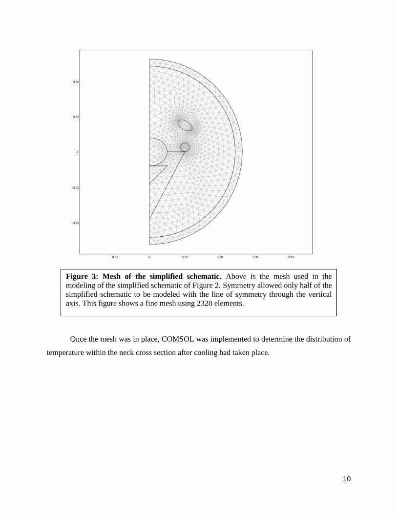
In order to utilize the simplified schematic seen in Figure 2 in COMSOL, a mesh was
created. The symmetry of the model allowed for further simplification of the schematic to only
include half of the cross section. This mesh included 2328 elements, which were used in a fine
mesh.
Table 1: The density, heat capacity, and conductivity values were found for the skin,
tissue, and bone for the implementation of the parameters in COMSOL.
10
Once the mesh was in place, COMSOL was implemented to determine the distribution of
temperature within the neck cross section after cooling had taken place.
Figure 3: Mesh of the simplified schematic. Above is the mesh used in the
modeling of the simplified schematic of Figure 2. Symmetry allowed only half of the
simplified schematic to be modeled with the line of symmetry through the vertical
axis. This figure shows a fine mesh using 2328 elements.

11
Results and Discussion
i. Solution Analysis
The temperature contour plot shows that the temperature throughout the cross section
changed from the initial temperature of 37°C (310 K). In order to further analyze this change, the
temperature change within the target area, the spine, was plotted versus time for the time span of
cooling.
Figure 4: Temperature Contour Plot of the Neck Cross Section. Above is the
temperature distribution of the neck schematic after 1 hour of ice pack application.
The data of this graph includes the generation terms of the governing equation as
applicable for 1 hour.

12
It can be seen in Figure 5 that the spine was significantly cooled during a cooling time of
3 hours. While it began at the initial temperature of 37°C (310 K), the temperature at point (0, -
0.0125), representing the center of the spine, decreased to the target temperature of 33°C (306 K)
in less than 45 minutes.
While lowering the temperature of the spinal cord to the target 33°C (306 K) in a timely
manner is very important, it is equally important to be able to safely maintain this temperature
over a longer period of time. While continuous application of the cooling pack would allow the
temperature to remain well below this critical value, over time, tissues near the interface would
be in danger of frostbite, a condition that occurs when tissues reach their freezing point. In order
to ensure the safety of these tissues then, the cooling pack must be periodically applied and
removed so as to allow for surface tissues to reheat. After several trials, we discovered that after
20
22
24
26
28
30
32
34
36
38
0 0.5 1 1.5 2 2.5 3
Tem
pe
ratu
re (
°C )
Time (Hours)
Temperature (C)
Figure 5: Temperature vs. Time Graph of Neck Schematic. The above graph
shows the change in temperature with increasing time in the spinal column at point
(0,-0.0125). The graph shows a decrease in temperature at the particular point from
37°C (310 K) to the target temperature of 33°C (306 K) in less than 45 minutes of
cooling time. The data of this graph includes the generation terms of the governing
equation.

13
application of the cooling pad for one hour initially, continuous cycles in which the pad is
removed for an hour and applied for a half hour result in a temperature profile within the tissues
that allows for sufficient cooling over a long period of time, including up to 72 hours (Figure 7).
The fact that at 72 hours the temperatures in the skin are above those inherent in frostbite
and the temperatures in the spinal cord are below the critical temperature of 33°C (306 K)
suggests that this protocol is effective over a long treatment period.
However, it is important to examine the temperatures in the aforementioned tissues over
the entire time period to ensure the safety of the protocol. Using this application protocol not
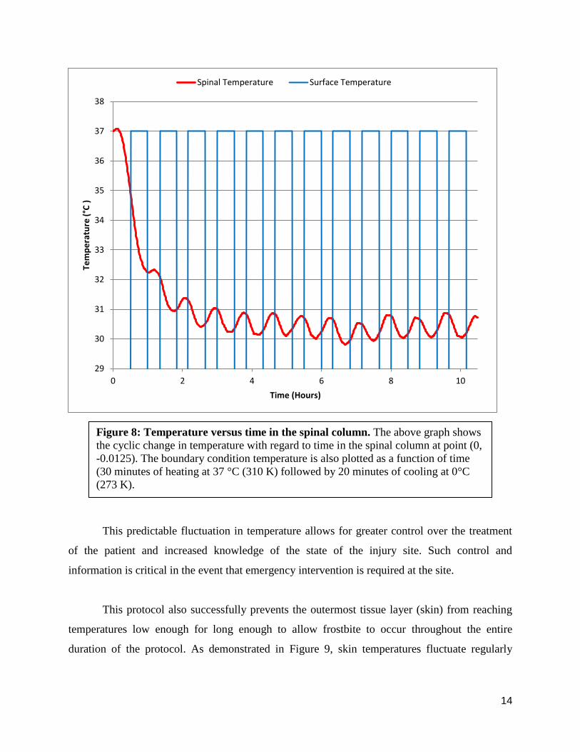
only ensures that the spinal cord stay below the target temperature of 33°C (306 K), but also
causes the temperatures within the spinal cord to fluctuate in a predictable and controlled manner
from approximately 26 C to 28 C (299 K to 301 K) over an extended time period (Figure 8).
Figure 7: Piecewise fluctuation function graph. The above figure shows the
variation in temperature when the schematic was modeled with a piecewise
fluctuation function.
14
This predictable fluctuation in temperature allows for greater control over the treatment
of the patient and increased knowledge of the state of the injury site. Such control and
information is critical in the event that emergency intervention is required at the site.
This protocol also successfully prevents the outermost tissue layer (skin) from reaching
temperatures low enough for long enough to allow frostbite to occur throughout the entire
duration of the protocol. As demonstrated in Figure 9, skin temperatures fluctuate regularly
29
30
31
32
33
34
35
36
37
38
0 2 4 6 8 10
Tem
pe
ratu
re (
°C )
Time (Hours)
Spinal Temperature Surface Temperature
Figure 8: Temperature versus time in the spinal column. The above graph shows
the cyclic change in temperature with regard to time in the spinal column at point (0,
-0.0125). The boundary condition temperature is also plotted as a function of time
(30 minutes of heating at 37 °C (310 K) followed by 20 minutes of cooling at 0°C
(273 K).

15
between approximately 2°C (275 K) and 25°C (298 K), both of which are above freezing (0°C,
273 K).
Furthermore, while the skin does eventually reach a temperature of 2°C (275 K), which is
close to 0°C (273 K), it is only at this temperature for an extremely short period of time, which
further discourages the formation of frostbite. The tissue actually spends far more time at the
comfortable condition of room temperature (25°C, 298K) than it does in this danger region.
Thus, this application protocol not only allows the spinal cord to be kept at a sufficiently low
temperature, but also allows for the protection of tissues from frostbite.
ii. Accuracy Check
Ideally, to check the accuracy of the COMSOL model, the outputs could be compared
with data obtained from an in vivo experiment where humans have their spinal cords cooled by
local cooling techniques. Unfortunately, this data is unavailable as such studies have not been
run at this time. However, while this data is not available, in vivo data for the cooling of the
0
5
10
15
20
25
30
35
40
0 2 4 6 8 10
Tem
pe
ratu
re (
°C )
Time (Hours)
Temperature (C)
Figure 9: Temperature vs time at skin surface. The figure above shows the
temperature versus time graph of the skin when doing fluctuating cooling.

16
spinal cord in animals is available. Smith (2011) used a cooling blanket to reduce the spinal cord
temperature in rats [21]. A cooling pad held at a constant 22.82°C (mild cooling condition) or
13.66°C (moderate cooling condition) was applied to the back of the rats directly above the
spinal column for a thirty minute period. The temperature in the spinal cord was monitored over
this time period through the use of a temperature probe. After a thirty minute period, the
temperature in the spinal column was reduced by 6.67 and 11.89 degrees for the mild and
moderate cooling conditions, respectively. Before the in vivo experiment, the researchers created
a model that used a perfusion model to account for the effects of heating due to blood in the
vasculature. To test the accuracy of our physiological simplifications, we created a rat model
using the same geometric simplifications used in our human model (major tissue layers are skin,
muscle, bone, and spinal tissue with normalized shapes) to compare with the experimental
results. In both our models we also accounted for the effect of the blood in the vasculature by
modeling the major arteries as constant temperature geometric figures (Figure 10).
Figure 10: Temperature Contour Plot of Rat Torso. Temperature profile for rat
torso and spinal column after thirty minutes of mild cooling (22.82°C). Note that the
temperature of the vasculature remains at physiological normal while the surrounding
tissues drop.

17
We felt this was a reasonable assumption, as core temperature should remain relatively
constant with localized cooling and this core temperature should sufficiently heat the blood as it
circulates through the body after exposure to the cooled area. This model resulted in predictions
differing from the observed drops in temperature at under 5% for both conditions, which stands
in stark contrast to the 25% and 36% error the experimenters obtained for their model of the mild
and moderate cooling (Table 2).
Cooling Temperature
(°C)
Experimental Drop
(degrees)
Smith Model Drop
(degrees)
Our Model Drop
(degrees)
22.82 6.67 4.98 6.99
13.66 11.89 7.52 11.95
While this is not the exact model used in the human case, the same approximations were
used in its creation: namely those simplifying the geometry of the neck and approximating the
effects of blood flow around the area. Thus, the success of this model lends confidence to the
human model used in this study.
iii. Sensitivity Analysis
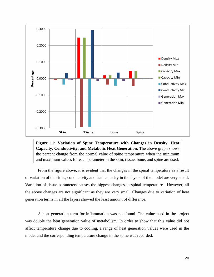
In order to observe the sensitivity of our project to variation in various tissue properties,
the different properties were varied and the resulting temperature values after an hour were
examined. The parameters changed were density, heat capacity, conductivity, and heat
generation terms. These values were varied for the skin, tissue, vertebral bone, and spinal cord
(as shown in Table 3 below) and implemented in the model. The original values (“norm”) were
reduced by 10% and increased by 10%. These were then recorded as the “min” and “max”
values, respectively, as shown in Table 3 below.
Table 2: Summary of results for experimental cooling of rat spinal column and
models of cooling of rat spinal column.

18
Property Type Skin Tissue Bone Spinal
Density (kg/m3) Min 1080 918 1350 918
Norm 1200 1020 1500 1020
Max 1320 1122 1650 1122
Heat Capacity (J/kgK) Min 3240 3202.2 396 3202.2
Norm 3600 3558 440 3558
Max 3960 3913.8 484 3913.8
Conductivity (W/mK) Min 0.477 0.189 0.225 0.189
Norm 0.53 0.21 0.25 0.21
Max 0.583 0.231 0.275 0.231
Heat Generation (W/m3) Min 328.5 328.5 328.5 630
Norm 365 365 365 700
Max 401.5 401.5 401.5 770
The above variations were used in the model. Each variation was changed while the
others were kept constant. The temperature change in the spinal column center (0, -0.0125) was
noted for each variation and recorded as shown in Table 4 below.
Table 3: Table of minimum, normal and maximum parameters (density, heat
capacity, conductivity and heat generation) of the skin, tissue, bone and spine.

19
Property
changed
Type of
change
Temperature change observed due to changes in the areas
below
Skin Tissue Bone Spinal
Density
(kg/m3)
Min 302.18 301.32 302.10 302.07
Norm 302.21 302.21 302.21 302.21
Max 302.20 302.96 302.27 302.35
Heat Capacity
(J/kgK)
Min 302.18 301.32 302.10 302.07
Norm 302.21 302.21 302.21 302.21
Max 302.20 302.96 302.27 302.35
Conductivity
(W/mK)
Min 302.31 303.10 302.32 302.20
Norm 302.21 302.21 302.21 302.21
Max 302.10 301.33 302.08 302.21
Heat Generation
(W/m3)
Min 302.19 302.17 302.17 302.20
Norm 302.21 302.21 302.21 302.21
Max 302.19 302.23 302.20 302.20
The spinal temperature changes caused by the “min” and “max” variations above were
compared to the temperature change caused by corresponding “norm” values and the percent
differences calculated and graphed for each variation and property as shown below.
Table 4: Table of spinal temperature changes accompanying changes in parameters
shown in Table 3 above.
20
From the figure above, it is evident that the changes in the spinal temperature as a result
of variation of densities, conductivity and heat capacity in the layers of the model are very small.
Variation of tissue parameters causes the biggest changes in spinal temperature. However, all
the above changes are not significant as they are very small. Changes due to variation of heat
generation terms in all the layers showed the least amount of difference.
A heat generation term for inflammation was not found. The value used in the project
was double the heat generation value of metabolism. In order to show that this value did not
affect temperature change due to cooling, a range of heat generation values were used in the
model and the corresponding temperature change in the spine was recorded.
-0.3000
-0.2000
-0.1000
0.0000
0.1000
0.2000
0.3000
Skin Tissue Bone Spine
Pe
rce
nta
ge
Density Max
Density Min
Capacity Max
Capacity Min
Conductivity Max
Conductivity Min
Generation Max
Generation Min
Figure 11: Variation of Spine Temperature with Changes in Density, Heat
Capacity, Conductivity, and Metabolic Heat Generation. The above graph shows
the percent change from the normal value of spine temperature when the minimum
and maximum values for each parameter in the skin, tissue, bone, and spine are used.

21
Heat generation values (W/m3) Observed temperature change in the Spine (°C)
630 29.05
730 29.06
770 29.05
3150 29.43
The values in Table 5 above were plotted in a graph to show how temperature in the
spine varied with changing the spinal heat generation term values.
The difference in temperature values between the lowest value (630 W/m3) and the
highest value (3150 W/m3) was found to be (29.43ºC – 29.05ºC = 0.38ºC) very small. Therefore,
increasing heat generation term (up to ~5 times the original estimated value) did not greatly
affect the change in temperature in the spine
29.05 29.06 29.05
29.43
28.8
28.9
29
29.1
29.2
29.3
29.4
29.5
630 730 770 3150
Tem
pe
ratu
re in
Sp
ine
(°C
)
Heat generation (W/m3)
Figure 12: Spinal Temperature Changes with Changes in Spinal Heat
Generation. The above graph change in spinal temperature when the heat
generationof inflammation values are varied.
Table 5: Table of observed temperature changes in the spine as a result of variation
heat generation value of the bone.

22
From the sensitivity analysis carried out above, it was concluded that the model was not
sensitive to changes in the parameter values tested (density, conductivity, heat capacity and heat
generation) in the skin, tissue, bone and spine layers where these parameters were varied.

23
Conclusions and Design Recommendations
i. Design Recommendations
Upon examination of the results of the study, several conclusions were drawn. First, the
local cooling procedure via the application of a cooling pack alone was capable of decreasing the
temperature of the spinal cord to the target temperature of 33ºC (306 K). The cooling pad was
applied for 30 minutes at 0ºC (273 K), then removed for 30 minutes of heating at 37ºC (310 K),
reapplied for 20 minutes of cooling at 0ºC (273 K), and then removed for 30 minutes of heating
at 37ºC (310 K). The target temperature was reached in less than 45 minutes, and the temperature
of the spine stabilized between 31ºC – 32ºC (304 K – 305 K). In contrast, traditional systemic
cooling procedures take an average of four hours to reach a temperature of 33ºC. An additional
benefit of local cooling is the fact that while the body must still be put in a hypothermic state to
ensure the spinal cord reach 33ºC, the core temperature can be kept a full two degrees higher
(35ºC) than it would be using current methods. The core can be kept at nearly normal
physiologically temperatures utilizing the fluctuating methodology, which indicates that tissue
damage adjacent to the local cooling site due to frostbite can be prevented. Since a major
drawback of therapeutic hypothermia use in spinal cord injury treatment is the prevalence of
complications arising from the hypothermia, mitigating the severity of this state is a major step
forward in treatment. Finally, this protocol does not require systemic cooling, which would
prevent the need for the insertion of a cooling catheter. This would decrease the risk of the
patient by avoiding invasive and potentially detrimental surgery. By means of cyclic heating and
cooling phases of the application area, the spinal cord can remain at temperatures well below 33
ºC (306 K) while maintaining the skin temperature well above freezing.
ii. Realistic Constraints
The ideal device to be used for injuries at the cervical spine would need to encircle the
neck and be in direct contact with the skin. The pack should be designed so that it is comfortable.
Also, the pack should be able to maintain cooling and heating temperatures at constant values to
limit the agitation of the patient’s injury. This could be accomplished by having two separate
inputs of fluid with a certain flow rate kept at the desired temperatures for cooling and heating
purposes.

24
5. Appendix A: Mathematical Statement of the Problem
Governing Equation for Heat Transfer:
Since we are attempting to cool the spinal cord and modeling the process over a circular cross-
section, we used the two-dimensional heat transfer equation in cylindrical coordinates with only
the transient, conduction, and heat source terms.
(
(
)
)
Since all heat transfer occurs through conduction, the convection term drops out. The tissues are
also all creating energy via metabolism in the living subject; this term must be accounted for
( ) Finally, in the injured tissues, inflammatory processes have been activated by the injury,
which results in additional heat generation, which is taken into account by the generation term
.
In the given protocol, temperature is defined at the skin-cooling pack interface. When the pack is
applied, the temperature is constant at 0°C (273 K), and if the pack is removed, the area is heated
at a constant temperature of 25°C (298 K). The entire body begins at the normal physiological
temperature of 37°C (310 K).

25
6. Appendix B: Solution Strategy
Solver
The model was solved in the Direct (UMFPACK) linear system solver in COMSOL.
Time Step
A time step of 0.01 seconds was utilized.
Tolerance
Relative tolerance was 0.1, and absolute tolerance was 0.0010.
Mesh Convergence Analysis
To determine the point at which we effectively eliminated discretization error without wasting
valuable time with an overly complex mesh, we ran a mesh convergence analysis. Through this
analysis, we determined that while lower element meshes showed significant variation in the
spinal cord temperature, after a mesh was created with 2328 elements, results became constant
(Figure 6).

26
The graph above indicates that mesh convergence occurred after 2328 elements, as the constant
spinal temperature after this point demonstrates. To avoid error and keep computation time low,
we thus used a mesh with 2328 elements.
Figure 6: Mesh convergence of modeled schematic. The above graph shows that
as the number of elements increased, the temperature did eventually converge,
staying constant at 29.03°C (302.18 K)
27
7. Appendix C
References
[1] Fay T. Observations on prolonged human refrigeration. N Y State J Med 1945;4:1351–4.
[2] Smith LW, Fay T. Observations on human beings with cancer maintained at reduced
temperature of 75–90 Fahrenheit (24–32C). Am J ClinPathol 1940;10:1–11.
[3] Fay T. Observations on generalized refrigeration in cases of severe cerebral trauma. Res
PublAssoc Res Nerv Dis 1945;4:611–9.
[4] Bigelow WG, Callaghan JC, Hopps JA. General hypothermia for experimental intracardiac
surgery; the use of electrophrenic respirations, an artificial pacemaker for cardiac standstill and
radio-frequency rewarming in general hypothermia. Ann Surg 1950;132:531–9.
[5] Botterell EH, Lougheed WM, Morley TP, Vandewater SL. Hypothermia in the surgical
treatment of ruptured in tracranial aneurysms. J Neurosurg 1958;15:4–18
[6] Arrica M, Bissonnette B. Therapeutic hypothermia. SeminCardiothoracVascAnesth
2007;11:6–15.
[7] Busto R, Dietrich WD, Globus MY, Valdes I, Scheinberg P, Ginsberg MD. Small differences
in intraischemic brain temperature critically determine the extent of ischemic neuronal injury. J
Cereb Blood Flow Metab 1987;7:729–38.
[8] Inamasu J, Ichikizaki K. Mild hypothermia in neurologic emergency: an update. Ann Emerg
Med 2002;40:220–30.
[9] Bernard SA, Buist M. Induced hypothermia in critical care medicine: a review. Crit Care
Med 2003;31:2041–51.
[10] Jordan JD, Carhuapoma JR. Hypothermia: comparing technology. J NeurolSci
2007;261:35–8

28
[11] Bernard, S. A., Gray, T. W., Buist, M. D., Jones, B. M., Silvester, W., Gutteridge, G., et al.
(2002). Treatment of comatose survivors of out-of-hospital cardiac arrest with induced
hypothermia. N Engl J Med, 346(8), 557-563.
[12] http://www.sci-info-pages.com/facts.html
[13] Gilad I, Nissan M (1985). Saggital evaluation of elemental geometrical dimensions of
human vertebrae. J Anat, 143, 115-120.
[14] Dai, T., Pikkula, B.M., Wang, L.V., and Anvari, B. (2004). Comparison of human skin
opto-thermal response to near-infrared and visible laser irradiations: a theoretical investigation.
Phys. Med. Biol. 49, 4861–4877.
[15] Beckmann, F., Heise, K., Kölsch, B., Bonse, U., Rajewsky, M. F., Bartscher, M., et al.
(1999). Three-dimensional imaging of nerve tissue by X-ray phase-contrast
microtomography.Biophysical Journal, 76(1), 98-102.
[16]http://ww.ucblueash.edu/koehler/biophys.2ed/heat.htmwl
[17]http://www.combatosteoporosis.com/treatment_for_osteoporosis.php
[18]http://www.engineeringtoolbox.com/specific-heat-solids-d_154.html
[19] http://www.sci-info-pages.com/facts.html
[20] Smith K, Zhu L (2010) Theoretical evaluation of a simple cooling pad for inducing
hypothermia in the spinal cord following traumatic injury. Med BiolEngComput 48:167–175
[21] Smith K D (2011) Experimental study and model validation of selective spinal cord and
brain hypothermia induced by a simple torso-cooling pad. Journal of Engineering in Medicine
225: 533-547