Mitosis storyboard
5
Outside of a cell. Important to distinguish which cell (the one shown is fission yeast). View of nucleus before zooming in. See more dense chromatin the centre of the nucelus and less dense chromatin around the periphery corresponding with actively transcribed genes. mRNA being transported through nuclear pores. Zoom in on the nucleus. Nuclear pores visible. Camera pans around the inside of the nucleus. Transcription factories exist around the periph- ery in the less dense regions.
-
Upload
claudia-stocker -
Category
Documents
-
view
212 -
download
1
description
Animation storyboard for mitosis
Transcript of Mitosis storyboard
Outside of a cell. Important to distinguish which cell (the one shown is fission yeast).
View of nucleus before zooming in. See more dense chromatin the centre of the nucelus and less dense chromatin around the periphery corresponding with actively transcribed genes. mRNA being transported through nuclear pores.
Zoom in on the nucleus. Nuclear pores visible.
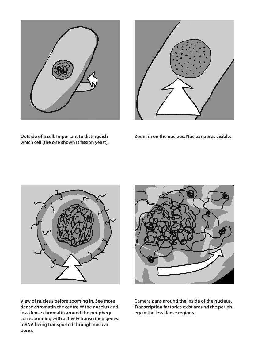
Camera pans around the inside of the nucleus. Transcription factories exist around the periph-ery in the less dense regions.

Camera pans in and out of pairs of DNA strands. These have similar patterns on them corresponding to identical base pair sequenc-es. Emphasise the fact that they are pairs of DNA strands with identical sequences to show duplication has taken place.
Assembly of histone octamer from H2A (or-ange), H2B (red), H3 (green) and H4 (blue) subunits.
Zoom in on single DNA strand. Approximately 10 base pairs per single turn. Right-hand helix structure. Camera pans around strand.
Histone octamer associates with DNA strand which wraps around it twice. H1 subunit acts like a sticking plaster to secure the DNA strand to the histone protein.

H1 subunits associate to form a solenoid struc-ture with approximately six histone proteins per turn.
Camera pans outwards to show a whole chro-mosome.
The solenoid structure coils in on itself to form a more condensed structure. Camera pans outwards.
Camera continues to pan outwards to show several chromosomes. Number of chromo-somes visible depends on which species the cell is form.
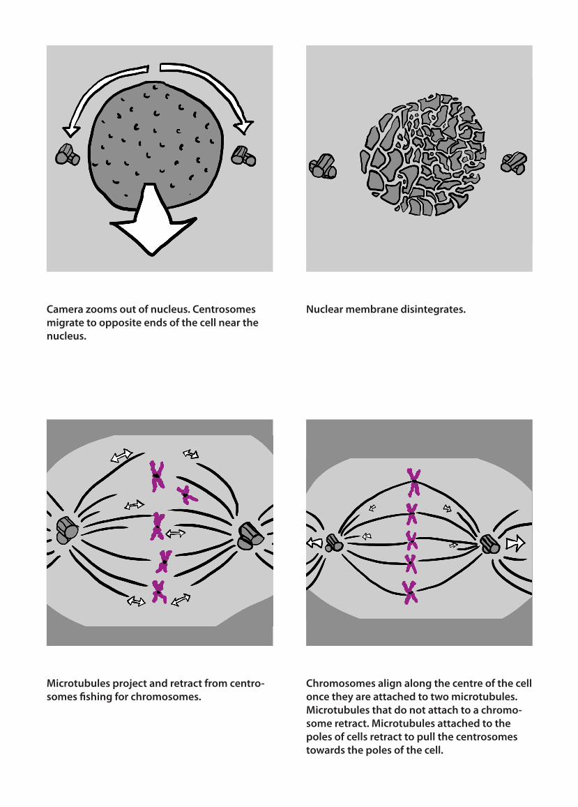
Camera zooms out of nucleus. Centrosomes migrate to opposite ends of the cell near the nucleus.
Microtubules project and retract from centro-somes fishing for chromosomes.
Nuclear membrane disintegrates.
Chromosomes align along the centre of the cell once they are attached to two microtubules. Microtubules that do not attach to a chromo-some retract. Microtubules attached to the poles of cells retract to pull the centrosomes towards the poles of the cell.

Camera pans outwards. Chromosomes are pulled apart by the tension of the microtu-bules.
FOR ANIMAL CELLS: Actin-myosin contrac-tile ring forms around the middle of the cell. The ring contracts to cleave the cell into two daughter cells
Nuclear membranes reform once the two sets of chromosomes are separated.
FOR PLANT CELLS: Septum forms to separate the two daughter cells.