“MITOCHONDRIAL RESPIRATORY - Università di...
190
Alma Mater Studiorum – Università di Bologna DOTTORATO DI RICERCA IN SCIENZE BIOCHIMICHE E BIOTECNOLOGICHE Ciclo XXVI Settore Concorsuale di afferenza: 05E1 Settore Scientifico disciplinare: B10/10 TITOLO TESI “MITOCHONDRIAL RESPIRATORY SUPERCOMPLEX ASSOCIATION LIMITS PRODUCTION OF REACTIVE OXYGEN SPECIES FROM COMPLEX I” Presentata da: Dott.ssa. Evelina Susana Beatriz Maranzana Coordinatore Dottorato Relatore Prof. Santi Mario Spampinato Prof. Carlo Guarnieri
Transcript of “MITOCHONDRIAL RESPIRATORY - Università di...

Alma Mater Studiorum – Università di Bologna
DOTTORATO DI RICERCA IN
SCIENZE BIOCHIMICHE E BIOTECNOLOGICHE
Ciclo XXVI
Settore Concorsuale di afferenza: 05E1 Settore Scientifico disciplinare: B10/10
TITOLO TESI
“MITOCHONDRIAL RESPIRATORY
SUPERCOMPLEX ASSOCIATION LIMITS
PRODUCTION OF REACTIVE OXYGEN
SPECIES FROM COMPLEX I”
Presentata da: Dott.ssa. Evelina Susana Beatriz Maranzana
Coordinatore Dottorato Relatore
Prof. Santi Mario Spampinato Prof. Carlo Guarnieri


i
Abstract
Evidence accumulated in the last ten years has demonstrated that a large proportion of the
mitochondrial respiratory chain complexes in a variety of organisms is arranged in
supramolecular assemblies called supercomplexes or respirasomes. Besides conferring a
kinetic advantage (substrate channeling) and being required for the assembly and stability
of Complex I, indirect considerations support the view that supercomplexes may also
prevent excessive formation of reactive oxygen species (ROS) from the respiratory chain.
Following this line of thought we have decided to directly investigate ROS production by
Complex I under conditions in which the complex is arranged as a component of the
supercomplex I1III2 or it is dissociated as an individual enzyme. The study has been
addressed both in bovine heart mitochondrial membranes and in reconstituted
proteoliposomes composed of complexes I and III in which the supramolecular
organization of the respiratory assemblies is impaired by: (i) treatment either of bovine
heart mitochondria or liposome-reconstituted supercomplex I-III with dodecyl maltoside; (ii)
reconstitution of Complexes I and III at high phospholipids to protein ratio.
The results of this investigation provide experimental evidence that the production of ROS
is strongly increased in either model; supporting the view that disruption or prevention of
the association between Complex I and Complex III by different means enhances the
generation of superoxide from Complex I .
This is the first demonstration that dissociation of the supercomplex I1III2 in the
mitochondrial membrane is a cause of oxidative stress from Complex I. Previous work in
our laboratory demonstrated that lipid peroxidation can dissociate the supramolecular
assemblies; thus, here we confirm that preliminary conclusion that primary causes of
oxidative stress may perpetuate reactive oxygen species (ROS) generation by a vicious
circle involving supercomplex dissociation as a major determinant.
It is easy to foresee the implications of these findings in human diseases and in aging,
where oxidative stress plays a major etiologic and pathogenic role.

ii
Contributors and Funding Sources
This work was supervised by Dr. Maria Luisa Genova and Professor Giorgio Lenaz of
Dipartimento di Scienze Biomediche e Neuromotorie (Alma Mater Studiorum, Università di
Bologna, Italia). The analysis in R4B proteoliposomes were conducted in collaboration with
Giovanna Barbero from Dipartimento di Scienze Biomediche e Neuromotorie (Università di
Bologna). All other work conducted for the dissertation was completed by the student
independently.
This work was supported by MIUR (grant number PRIN2008LSHCFC_005).
Graduate study was supported by an EADIC Erasmus Mundus External Cooperation
Windows Lot 16 (UniBO-UNQ) Fellowship from the European Commission and a
dissertation research Marco Polo fellowship from Alma Mater Studiorum, Università di
Bologna, Italia.
iii
Contents
Abstract i
Contributors and Funding Sources ii
Contents iii
List of Figures viii
List of Tables xii
Abbreviations xiii
Chapter 1 Introduction 1
Chapter 2 Materials and Methods 73
Chapter 3 Results 113
Chapter 4 Discussion 135
Chapter 5 Conclusions 141
References 144

iv
Contents
INTRODUCTION 1
THE MITOCHONDRIAL RESPIRATORY CHAIN 1
STRUCTURAL ORGANIZATION OF THE RESPIRATORY CHAIN 2
Solid-state organization 5
Liquid-state organization. Random collision model 8
Evidences for supramolecular organization 9
Structural evidences 9
Functional evidences 11
Pool behavior 12
Direct Transfer of Substrates (Channeling) 13
Flux Control Analysis 14
Respiratory supercomplexes in eukaryotes 15
Supercomplex I1III2 17
Supercomplexes III2IV1-2 17
Supercomplexes I1III2IV1-4 19
Respiratory strings 22
Respiratory supercomplexes in prokaryotes 24
Factors that affect supramolecular associations 26
Lipid content 26
Functional and Structural Consequences for Supramolecular
Association
29
Kinetic Advantage: Channeling 29
Structural Advantage: Protein stability and Assembly 29
v
Scaffold
DYNAMIC ORGANIZATION: PLASTICITY MODEL 33
CoQ compartmentalization 34
COMPLEX I 35
Structure 36
Periferal arm 42
Interface domain 45
Membrane arm 46
Evolution and modular organization of Complex I 52
The N – module 53
The Q – module 53
The P – module 53
Mammalian Complex I Assembly 56
Catalytic activity of Complex I 59
NADH oxidation and intramolecular electron transfer 59
Ubiquinone reduction and coupling mechanism 60
Complex I inhibitors 61
ROS PRODUCTION IN COMPLEX I 64
Supercomplexes and ROS production in Complex I 70
HYPOTHESIS 72
MATERIALS AND METHODS 73
MATERIALS 73
Reagents and solutions 73
METHODS 74
Preparation of bovine heart mitochondria (BHM) 74
vi
Preparation of bovine submitochondrial particles (SMP) 76
Purification of bovine Complex I-III fraction (R4B fraction) 77
Preparation of bovine Complex I-III proteoliposomes 80
Preparation of phospholipid:ubiquinone vesicles 80
Proteoliposome reconstitution 82
Determination of protein concentration 83
Ultraviolet absorbance at 280 nm (range: 0.1 – 1 mg·ml-1) 83
Biuret Method (range: 1 – 10 mg·ml-1) 84
Lowry Method (range: 0.01 – 0.1 mg·ml-1) 85
Enzyme activities 87
NADH:ubiquinone oxidoreductase. 87
NADH oxidase. 87
NADH:cytochrome c reductase. 87
Measuring Reactive Oxygen Species 88
Superoxide detection. 88
Hydrogen peroxide detection. 90
Protein electrophoresis analysis 92
First dimension: Blue-native polyacrylamide gel
electrophoresis
95
Second dimension: Sodium dodecyl sulfate polyacrylamide
gel electrophoresis
102
Protein Immunoblotting 104
Blotting 105
Protein immunodetection 109
Imaging - Analysis and documentation 111
RESULTS 113
vii
Effects of DDM-treatment over respiratory mitochondrial membranes 113
Effects of lipid dilution and DDM-treatment in reconstituted
supercomplex I1III2
120
Effects of chaotrope-treatment in SMP and reconstituted supercomplex
I1III2
128
DISCUSSION 135
Loss of enzymatic channeling between Complex I and Complex III 135
Production of reactive oxygen species from Complex I 136
Stability of Complex I 139
CONCLUSIONS 141
REFERENCES 144
viii
List of Figures
Title Page
Figure 1.1 The Mitochondrial Respiratory Chain 1
Figure 1.2 Structural Organization of Mitochondrial Respiratory
Complexes
3
Figure 1.3 Dynamic Organization of Mitochondrial Respiratory
Complexes
4
Figure 1.4 Solid-model of Chance and Williams (1955) 7
Figure 1.5 Random collision model of Hackenbrock (1986) 8
Figure 1.6 Characterization of bovine respiratory chain supercomplexes
and dimeric complex V by BN-PAGE.
10
Figure 1.7 Models of respiratory supercomplexes 18
Figure 1.8 Model for the bovine I1III2IV1 supercomplex (respirasome) 20
Figure 1.9 Cryo-EM 3D map and fitted X-ray structures of bovine
I1III2IV1 supercomplex (respirasome) and Electron transfer
pathway
21
Figure 1.10 Hypothetical models for a higher organization of respiratory
chain complexes (respiratory strings)
23
Figure 1.11 Structure of complex I from Thermus thermophilus 41
Figure 1.12 Scheme of the electron pathway in the peripheral arm of
Complex I
44
Figure 1.13 Quinone-reaction chamber of Complex I 47
Figure 1.14 Ubiquinone binding site of Complex I 48

ix
Figure 1.15 E-channel and central hydrophilic axis of Complex I 49
Figure 1.16 Putative proton-translocation channels in the antiporter-like
subunits
50
Figure 1.17 Topology model of subunits in mammalian Complex I 51
Figure 1.18 Modular organization of Complex I core subunits 54
Figure 1.19 Evolutionary modules of Complex I 55
Figure 1.20 The assembly model of mammalian Complex I biogenesis 58
Figure 1.21 Proposed coupling mechanism of Complex I 62
Figure 1.22 Overview of mitochondrial ROS production 65
Figure 1.23 Modes of mitochondrial operation that lead to O2•−
production
66
Figure 1.24 Production of O2•− by Complex I 69
Figure 1.25 Production of O2•− by Complex I in I1III2 supercomplex 71
Figure 2.1 Purification of bovine Complex I-III fraction 77
Figure 2.2 Liposomes preparation 81
Figure 2.3 Electrophoresis workflow 93
Figure 2.4 Solubilization of native oxidative phosphorylation complexes 100
Figure 2.5 Separation of supramolecular assemblies of oxidative
phosphorylation complexes by 1D-BN PAGE
101
Figure 2.6 Identification of individual constituents in supramolecular
assemblies of oxidative phosphorylation complexes by 2D
BN/SDS-PAGE
101
x
Figure 2.7 Protein blotting workflow 107
Figure 3.1 Supercomplex disassembling in bovine heart mitochondria
(BHM)
116
Figure 3.2 Functional analysis of supercomplex I1III2 and complex I in
detergent-solubilized bovine heart mitochondria (BHM):
NADH-ubiquinone oxidoreductase activity NADH-cytochrome c
oxidoreductase activity
117
Figure 3.3 Functional analysis of supercomplex I1III2 and complex I in
detergent-solubilized bovine heart mitochondria (BHM):
NADH-oxidase activity
118
Figure 3.4 Functional analysis of supercomplex I1III2 and complex I in
detergent-solubilized bovine heart mitochondria (BHM):
Production of hydrogen peroxide
119
Figure 3.5 Supramolecular organization of respiratory Complex I and
Complex III in R4B 1:1 and R4B 1:30 proteoliposomes. (A)
122
Figure 3.6 Functional analysis in R4B 1:1 and R4B 1:30
proteoliposomes.
123
Figure 3.7 ROS production mediated by Complex I in R4B 1:1 and R4B
1:30 proteoliposomes.
124
Figure 3.8 Disassembling of supercomplex I1III2 in R4B 1:1
proteoliposomes after detergent solubilization.
125
Figure 3.9 Functional analysis of supercomplex I1III2 and complex I in
detergent-solubilized R4B 1:1 proteoliposomes: NADH-
ubiquinone oxidoreductase activity and (B) NADH-cytochrome c
oxidoreductase activity
126
Figure 3.10 Functional analysis of supercomplex I1III2 and complex I in
detergent-solubilized R4B 1:1 proteoliposomes: Production of
hydrogen peroxide
127
Figure 3.11 Disassembling of supercomplex I1III2 in bovine heart
submitochondrial particles (SMP) after treatment with 0.2 M
KSCN.
129
Figure 3.12 Disassembling of supercomplex I1III2 in R4B 1:1
proteoliposomes after treatment with 0.2 M KSCN.
130

xi
Figure 3.13 Complex I activity and ROS production in bovine heart
submitochondrial particles (SMP) after treatment with KSCN.
131
Figure 3.14 Complex I activity and ROS production in R4B 1:1
proteoliposomes after treatment with KSCN.
132
Figure 3.15 Production of ROS by mitochondrial Complex I in different
situations where supercomplexes are maintained or
dissassembled.
133

xii
List of Tables
Title Page
Table 1.1 Supramolecular organization of eukaryotic respiratory
complexes (mitochondrial respiratory supercomplexes)
16
Table 1.2 Supramolecular organization of prokaryotic respiratory
complexes (Aerobic respiratory supercomplexes)
25
Table 1.3 Nomenclature for the 14 core subunits of Complex I and
prosthetic cofactors bound by the hydrophilic subunits
39
Table 1.4 Nomenclature for the supranumerary subunits of
mammalian Complex I
40
Table 1.5 Functional classification of Complex I inhibitors 63
Table 2.1 Gel buffer system formulation for BN-PAGE 98
Table 2.2 Quantity of detergent required to solubilize membrane
proteins
98
Table 2.3 Gel buffer system formulation for SDS-PAGE 104
Table 3.1 Production of Reactive Oxygen Species by mitochondrial
Complex I in different situations where supercomplexes are
maintained or disassembled
134
xiii
Abbreviations
BHM Bovine heart mitochondria
DDM Dodecyl--D-maltoside
ROS Reactive oxygen species
SDS Sodium dodecylsulfate
SMP Submitochondrial particles
R4B Mitochondrial fraction enriched in Complex I and Complex III
CoQ Ubiquinone, Coenzyme Q10
Cyt c Cytochrome c

xiv

Page 1
INTRODUCTION
THE MITOCHONDRIAL RESPIRATORY CHAIN
Reducing equivalents (hydrogen atoms) released from mitochondrial oxidations of the
tricarboxylic acid cycle, from pyruvate oxidation, fatty acid and amino acid catabolism and
other oxidative reactions, are collected by a multi enzyme system, the electron transfer
chain or respiratory chain that conveys them to molecular oxygen reducing it to water. The
free energy decrease of this electron transfer generates an electrochemical proton
gradient (H+) by proton translocation from the mitochondrial matrix, to the space existing
between the inner and outer mitochondrial membranes. The proton gradient is then used
as a source of energy to synthesize ATP from ADP and Pi by the ATPsynthase complex,
or alternatively to drive other energy-linked reactions. The electron transfer chain consists
of four major complexes designated as NADH:ubiquinone oxireductase (Complex I),
succinate-ubiquinone oxireductase (Complex II), ubiquinol- cytochrome c oxireductase
(Complex III) and cytochrome c oxidase (Complex IV) and two connecting redox-active
molecules, i.e. a lipophilic quinone, designated ubiquinone (Coenzyme Q or CoQ)
embedded in the membrane lipid bilayer ,and a hydrophilic hemeprotein, cytochrome c,
localized on the external surface of the inner membrane (Figure 1.1).
Figure 1.1: The mitochondrial respiratory chain.Textbook description of the respiratory chain. The
transmembrane protein complexes of the elec- tron transport chain generate an electrochemical gradient
over the mitochondrial inner membrane. NADH is oxidized to NAD+. Electrons are transferred from NADH via
Complex I and ubiquinone (Q) to Complex III. From there they pass through the peripheral electron carrier
cytochrome c and complex IV to the terminal acceptor, molecular oxygen, which is reduced to water.
[modified from Lehningher Principles of Biochemistry 5th edition (Nelson, D.L. and Fox, M.M)]
Page 2
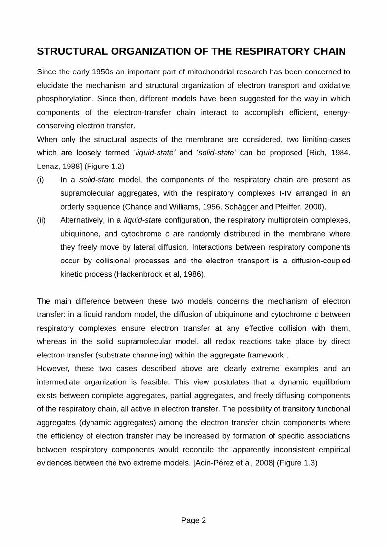
STRUCTURAL ORGANIZATION OF THE RESPIRATORY CHAIN
Since the early 1950s an important part of mitochondrial research has been concerned to
elucidate the mechanism and structural organization of electron transport and oxidative
phosphorylation. Since then, different models have been suggested for the way in which
components of the electron-transfer chain interact to accomplish efficient, energy-
conserving electron transfer.
When only the structural aspects of the membrane are considered, two limiting-cases
which are loosely termed ‘liquid-state’ and ‘solid-state’ can be proposed [Rich, 1984.
Lenaz, 1988] (Figure 1.2)
(i) In a solid-state model, the components of the respiratory chain are present as
supramolecular aggregates, with the respiratory complexes I-IV arranged in an
orderly sequence (Chance and Williams, 1956. Schägger and Pfeiffer, 2000).
(ii) Alternatively, in a liquid-state configuration, the respiratory multiprotein complexes,
ubiquinone, and cytochrome c are randomly distributed in the membrane where
they freely move by lateral diffusion. Interactions between respiratory components
occur by collisional processes and the electron transport is a diffusion-coupled
kinetic process (Hackenbrock et al, 1986).
The main difference between these two models concerns the mechanism of electron
transfer: in a liquid random model, the diffusion of ubiquinone and cytochrome c between
respiratory complexes ensure electron transfer at any effective collision with them,
whereas in the solid supramolecular model, all redox reactions take place by direct
electron transfer (substrate channeling) within the aggregate framework .
However, these two cases described above are clearly extreme examples and an
intermediate organization is feasible. This view postulates that a dynamic equilibrium
exists between complete aggregates, partial aggregates, and freely diffusing components
of the respiratory chain, all active in electron transfer. The possibility of transitory functional
aggregates (dynamic aggregates) among the electron transfer chain components where
the efficiency of electron transfer may be increased by formation of specific associations
between respiratory components would reconcile the apparently inconsistent empirical
evidences between the two extreme models. [Acín-Pérez et al, 2008] (Figure 1.3)
Page 3
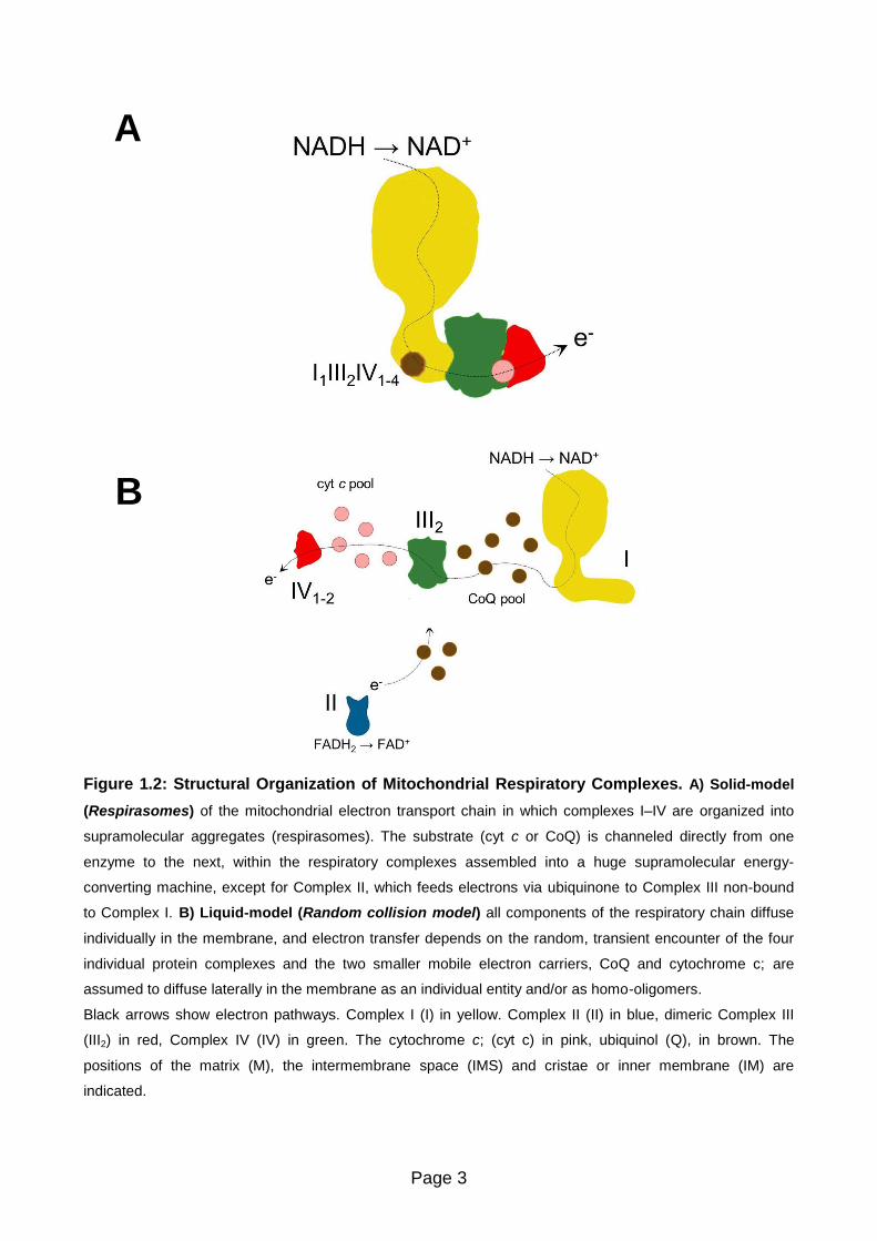
Figure 1.2: Structural Organization of Mitochondrial Respiratory Complexes. A) Solid-model
(Respirasomes) of the mitochondrial electron transport chain in which complexes I–IV are organized into
supramolecular aggregates (respirasomes). The substrate (cyt c or CoQ) is channeled directly from one
enzyme to the next, within the respiratory complexes assembled into a huge supramolecular energy-
converting machine, except for Complex II, which feeds electrons via ubiquinone to Complex III non-bound
to Complex I. B) Liquid-model (Random collision model) all components of the respiratory chain diffuse
individually in the membrane, and electron transfer depends on the random, transient encounter of the four
individual protein complexes and the two smaller mobile electron carriers, CoQ and cytochrome c; are
assumed to diffuse laterally in the membrane as an individual entity and/or as homo-oligomers.
Black arrows show electron pathways. Complex I (I) in yellow. Complex II (II) in blue, dimeric Complex III
(III2) in red, Complex IV (IV) in green. The cytochrome c; (cyt c) in pink, ubiquinol (Q), in brown. The
positions of the matrix (M), the intermembrane space (IMS) and cristae or inner membrane (IM) are
indicated.
A
B

Page 4
Figure 1.3: Dynamic Organization of Mitochondrial Respiratory Complexes. Plasticity model.
The supramolecular organization of the mitochondrial respiratory chain has been proposed to confer kinetic
advantages on electron transfer through substrate channeling, to prevent ROS production, and to aid the
assembly and stabilization of Complex I. Recently it has been reported a new functional role for the dynamic
association/dissociation of mitochondrial respiratory complexes and supercomplexes, which defines
dedicated CoQ and cyt c pools in order to organize electron flux to optimize the use of available substrates
through the respiratory chain. These dynamic rearrangements range from all-bound to all-free respiratory
complexes, and they open the possibility that different modes of organization are switched on/switched off to
regulate diverse physiological functions through, i.e., ROS signaling or turnover of respiratory enzymes.
I1III2IV1–4 ers to the respirasomes or supercomplexes formed by the association of one Complex I (I), a
homodimer Complex III (III2), and one to four copies of Complex IV (IV1–4). Intermediate supercomplex
species can be found in nature, in combination with free Complex II (II), dimers of Complex III, and Complex
IV in different stoichiometries (IV1–2). Complex I requires to be associated in supercomplexes to minimize
destabilization and ROS generation.

Page 5
Solid-state organization
In [Chance and Williams, 1955], a method based on the use of dual-beam, dual-
wavelength spectrophotometer in combination with an oxygen electrode for the study of
oxidative phosphorylation is published. The redox states of various respiratory-chain
components were determined by spectrophotometry, simultaneously with the
polarographic measurement of oxygen uptake. This method made possible the first
quantitative study of the concentrations and kinetics of electron-transport enzymes not
only in intact mitochondria but also in intact cells and tissues allowing depicting the
respiratory chain as a solid-state assembly of flavins and cytochromes in a protein matrix.
From these studies, Chance and Williams have also determined the sequence of
enzymatic steps in the mitochondrial respiratory chain and hypothesized two alternative
mechanisms for electron transfer from one protein carrier to another along the solid-state
array [Chance and Williams, 1956] (Figure 1.4).
For the first mechanism, there are restricted rotations in the protein carriers to permit
collision of the prosthetic groups. In the second, the molecules are completely fixed, and
electrons then must pass through the protein moieties to the prosthetic groups. This intra-
protein electron transfer mechanism through the insulating protein medium today is well
described as electron tunnelling transfer where the maximal distances allowing for
physiological electron transfer between the interacting centres should not exceed 13–14 Å
[Moser et al, 2005].
Contemporaneously, a comprehensive study of large-scale preparation of beef heart
mitochondria was begun in Green’s laboratory [Crane et al, 1956]. Mitochondria from beef
heart proved to possess a remarkably high degree of stability; they were capable of
withstanding preparation procedure involving disruption of the tissue by relatively harsh
mechanical means, and subsequent storage of the mitochondria in the frozen state for
long periods of time. These preparations thus became the material of choice for future
studies that aimed at a resolution and reconstitution of the respiratory chain and the
phosphorylating system.
Page 6
Shortly after the discovery of ubiquinone (coenzyme Q, CoQ) [Crane et al, 1957] and of its
participation in electron transfer [Crane et al, 1959. Hatefi et al, 1959] the resolution of the
four respiratory complexes functionally active: NADH:ubiquinone reductase (Complex I)
[Hatefi et al, 1961], succinate:ubiquinone reductase (Complex II) [Ziegler and Doeg, 1961],
ubiquinol:cytochrome c reductase (Complex III) [Hatefi et al, 1962a], and cytochrome c
oxidase (Complex IV) [Fowler et al, 1962] from bovine heart mitochondria was possible.
In 1962 Hatefi et al [Hatefi et al, 1962b] succeeded in reconstituting NADH oxidase and
succinoxidase by combining complexes I, III, and IV and complexes II, III, and IV,
respectively, in the presence of cytochrome c. In both cases, the reconstitution required
high concentrations of the complexes and resulted in a particulate preparation which did
not dissociate upon subsequent dilution.
These results gave rise to the concept that the components of the respiratory chain exist in
mitochondria as a fixed assembly (“elementary particles”) [Ernster and Schatz, 1981].
Indeed, it was found that cytochrome c can form stable complexes with complex III and
complex IV [Kuboyama et al, 1962] and that mitochondria contain the cytochromes in near
stoichiometric amounts. Thus, it was assumed that the respiratory complexes formed a
single functional unit in the mitochondria and were present in an orderly sequence which
could be disrupted by appropriate reagents [Blair, 1967].
These early ideas concerning the structure of the respiratory chain where the possibility
that electron-transport chain might exist as a structural unit (solid-model) has been
considered also in [Lehninger, 1959] and, in the following years, Chance extended this
concept to include direct communication with the ATP-synthesizing machinery [Boyer et al,
1977. Chance, 1977].
Page 7
A
B
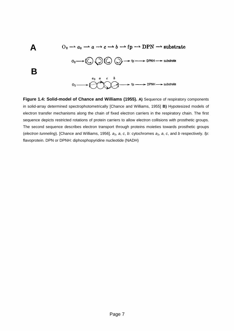
Figure 1.4: Solid-model of Chance and Williams (1955). A) Sequence of respiratory components
in solid-array determined spectrophotometrically [Chance and Williams, 1955] B) Hypotesized models of
electron transfer mechanisms along the chain of fixed electron carriers in the respiratory chain. The first
sequence depicts restricted rotations of protein carriers to allow electron collisions with prosthetic groups.
The second sequence describes electron transport through proteins moieties towards prosthetic groups
(electron tunneling). [Chance and Williams, 1956]. a3, a, c, b: cytochromes a3, a, c, and b respectively. fp:
flavoprotein. DPN or DPNH: diphosphopyridine nucleotide (NADH)

Page 8
Liquid-state organization. Random collision model
On the other hand, on the basis of the isolation of the functional individual respiratory
complexes Green and Tzagoloff [Green and Tzagoloff, 1966] postulated that the overall
respiratory activity is the result of both intracomplex electron transfer in solid-state
between redox components having fixed steric relations and, in addition, of intercomplex
electron transfer ensured by rapid diffusion of the mobile components acting as co-
substrates (i.e., ubiquinone and cytochrome c). This proposal was supported by the kinetic
analysis of Kröger and Klingenberg [Kröger and Klingenberg, 1973a. Kröger and
Klingenberg, 1973b] showing that the ubiquinone behaves kinetically as a homogeneous
pool in submitochondrial particles from beef heart.
Over the following years, this model was substantially confirmed by several lines of
evidences leading Hackenbrock et al to the postulation of the Random Diffusion Model of
Electron Transfer [Hackenbrock et al, 1986] (reviewed in [Lenaz and Genova, 2007. Lenaz
and Genova, 2009a]) (Figure 1.5).
According with this model, the respiratory complexes are randomly distributed in the plane
of the membrane, where they freely move by lateral diffusion. Ubiquinone and cytochrome
c are also mobile electron carriers, whose diffusion rates are faster than those of the
respiratory complexes. The electron-transferring reactions between all redox components
and their respective redox partners occur via a long-range diffusional process, where their
diffusion-coupled collision frequencies may be either higher or lower than any given
reaction step within the complexes. Consequently electron transfer is rate limited by the
diffusion of ubiquinone and cytochrome c.
Figure 1.5: Random collision model of Hackenbrock (1986). Structural arrangement of
mitochondrial respiratory components [Hackenbrock et al, 1986]. O.M: outer membrane, I.M.: inner
membrane, b5: cytochrome b5, c: cytochrome c, Q: ubiquinone, I, II, III, IV: complexes I, II, III, and IV,
respectively

Page 9
Evidences for supramolecular organization
Structural evidences
Despite the wide acceptance of the Random Collision Model during the following twenty
years, circumstantial evidences of supramolecular organization of the respiratory
complexes come from the pioneering isolation of bovine respiratory complexes where
NADH:cytochrome c reductase (complex I+III) [Hatefi and Rieske, 1967] and
succinate:cytochrome c reductase (complex II+III) [Tisdale, 1967] were purified, and
interaction between complexes II and III in yeast [Bruel et al, 1996] was demonstrated.
Stable supercomplexes of complexes III and IV were also isolated from some bacteria
[Berry and Trumpower, 1985. Sone et al, 1987. Keefe and Maier, 1993. Iwasaki et al,
1995] indicating that such enzymes may be perentially associated in native membrane.
Nevertheless, these reports could not challenge the prevalent view and have been
overlooked.
The paradigm of how the respiratory chain is structurally organized drastically changed in
2000 when direct evidences for the existence of higher-order stoichiometric assemblies of
respiratory complexes came from the development of the blue-native polyacrylamide gel
electrophoresis (BN-PAGE) by Hermann Schägger and colleagues [Schägger and von
Jagow, 1991. Schägger et al, 1994. Schägger, 1995].
Mitochondrial membranes solubilized with very mild non-ionic detergents like digitonin
which preserve the respiratory complexes activities as well as protein interactions are used
in BN-PAGE. This methodology is able to separate the largest stable and functional
protein complexes that can withstand solubilization.
The complex stoichiometric composition is then determined by an orthogonal second
dimension BN-PAGE (2D BN/BN-PAGE) with a relatively stronger non-ionic detergent as
dodecylmaltoside or Triton X-100, to dissociate supercomplexes, or by the subunit
composition with a denaturing second dimension (2D BN/SDS-PAGE). This approach
allowed the separation and stoichiometric characterization of high molecular weight
supramolecular aggregates of respiratory complexes first in mitochondria of bovine heart
and of the yeast Saccharomyces cerevisiae [Cruciat et al, 2000. Schägger and Pfeiffer,
2000. Schägger and Pfeiffer, 2001] which remain the best characterized species (Figure
1.6)

Page 10
The introduction of the BN-PAGE methodology marked the beginning of the study of the
higher level of structural organization for the OXPHOS system.
After the first characterizations of supercomplexes by BN-PAGE direct structural insights in
the architecture of the supercomplexes were provided more recently by the application of
electron microscopy and single particle analysis [Dudkina et al, 2008. Vonck and Schäfer,
2009].
The respiratory supercomplexes are either separated by centrifugation in sucrose density
gradients or electroeluted directly from preparative BN-gels, and then imaged by negative
stain electron microscopy and subjected to single particle analysis. Several
supercomplexes of yeast, plants and mammals have been studied.
Since then, respiratory supercomplexes have been found both in Bacteria, Achaea and in
organisms belonging to different kingdoms of eukaryotes. Despite their phylogenetic
distances from each other, all of them have in common that their respiratory chain share
similar ultrastructure in their respiratory membranes. At this stage, one could consider that
a supramolecular organization of the respiratory chain is an evolutionary-conserved trait
for which selective advantages remain to be established [Chaban et al, 2013. Magalon et
al, 2012].
Figure 1.6: Characterization of bovine respiratory chain supercomplexes and dimeric complex V by
BN-PAGE. A) BN-PAGE of bovine heart mitochondria after solubilization by digitonin. Most Complex I and Complex III
was found assembled into two major supercomplexes a and b, and two minor supercomplexes c and d. The 200 kDa
mass differences indicate the presence of varying copy numbers of monomeric Complex V [Schägger and Pfeiffer;
2000] B) Supercomplexes a-d and dimeric ATP synthase (Vdim) from the BN-PAGE were dissociated by 2D BN-PAGE
using addition of dodecylmaltoside to the cathode buffer. Direct interaction of complexes I and III was apparent from the
dissociation of supercomplex a (I1III2) into monomeric Complex I and dimeric Complex III. Superomplexes b-d
comprised Complex IV in addition [Schägger and Pfeiffer; 2000]. C) Organization of bovine heart mitochondrial
respirasomes according to Schägger [Schägger, 2001]. Supercomplexes (a–d) characterized by BN-PAGE; they all
contain a complex I monomer, a complex III dimer, and a variable copy number of monomeric complex IV, as indicated
by the 200-kDa mass differences. Only 14–16% of total complex I was found in free form in the presence of digitonin.
A B
C
Page 11
Functional evidences
When Chance and Williams have proposed their pioneering solid-state model to describe
the respiratory chain organization, they also hypothesized that electron transfer between
the protein components in the solid array occurs along predefined pathways [Chance and
Williams, 1956].
Thus, the assumption that respirasome has a major function conferring a more efficient
transfer of substrates is an inherent consequence if respiratory components are arranged
in a sequentially-ordered supramacromolecular assembly.
Considering the two extreme models, the rate of electron transfer between respiratory
components depends on their structural arrangement in the membrane:
For the liquid random model view, if two redox enzymes are connected by a mobile redox
carrier undergoing long-range diffusion in the membrane, the overall reaction rate would
be governed by the frequency of effective collisions between the mobile carrier and its two
redox partners [Gupte et al, 1984. Gupte and Hackenbrock, 1988a. Gupte and
Hackenbrock, 1988b]. According with this model, the mobile electron carriers components,
ubiquinone and cytochrome c, constitute intermediate pools diffusing into the bulk
framework of the mitochondrial inner membrane (substrate pools), then if diffusion of the
quinone and quinol species is much faster than the chemical reactions of CoQ reduction
and oxidation, the quinone behaves kinetically as a homogeneous pool (pool behavior).
On the other hand, if respiratory components are arranged in a solid-array configuration,
the frequency of effective collisions will be determined only by the proximity between the
redox components. In the case of the respiratory chain, this means direct transfer of
electrons between two active sites of two enzymes that are physically adjacent by
successive reduction and oxidation of the intermediate with restricted diffusion into the
surrounding milieu (substrate channeling) [Ovandi, 1991].
These two models in the case of the organization of the respiratory chain are kinetically
distinguishable:

Page 12
Pool behavior
Kröger and Klingenberg [Kröger and Klingenberg 1973a, 1973b], based on the assumption
that quinone behaves as a homogenous pool, postulated that the overall electron flux
observed (Vobs) between two enzymes will follow a hyperbolic relation with the rate of
ubiquinol oxidation (vox) and the rate of ubiquinone reduction (vred):
Where CoQ is the mobile electron carrier between a first enzyme reducing ubiquinone and
the second oxidizing ubiquinol. They showed this hyperbolic relation in steady-state
respiration in bovine submitochondrial particles using either NADH or succinate as
electron donors.
Further experimental evidences validated this pool behavior in a variety of mitochondrial
systems establishing that CoQ distributes electrons randomly among the dehydrogenases
and Complex III, behaving indeed as a freely diffusible intermediate. But most available
data concern succinate oxidation (Complex II) in submitochondrial particles, whereas
fewer data are available for NADH oxidation (Complex I).
Then, kinetics analysis changing Vred or Vox on inhibitor titration curves (e.g. titration of
Complex III by antimycin) allows to distinguish between CoQ pool behavior (random
model) or CoQ channeling (supercomplexes). Pool behavior is kinetically characterized by
a convex hyperbolic relationship between the integrated oxidation rate and the inhibitor
concentration, whereas a linear relationship is expected by a stoichiometric association
(supercomplexes) between the two enzymes.
Page 13
Direct Transfer of Substrates (Channeling)
Early evidences about substrate channeling in respiratory complexes came from [Ragan
and Heron, 1978]. They demonstrated that Complex I-Complex III binary complex is
formed in a 1:1 molar ratio after reconstitution of in lipid vesicles. They also showed that
this binary complex contains CoQ10, and equimolar quantities of FMN and cytochrome c1.
This study also described for the first time the stoichiometric behavior for the activity of
NADH:cytochrome c reductase, ascribable to the formation of a Complex I-Complex IIII
supercomplex. In addition, they showed that CoQ-pool behavior could be restored if
adding additional amounts of phospholipid and ubiquinone in the concentrated mixture.
Under these conditions they demonstrated that Complex I and Complex III activities were
independent of each other.
Heron and co-workers [Heron et al, 1978] have also reported that endogenous CoQ10
leaks out of the Complex I–III unit when extra phospholipid is present, causing a decrease
in activity that could be alleviated by adding more ubiquinone.
A more direct comparison of the effect of channeling with respect to CoQ-pool behavior
was performed in a simpler experimental condition in our laboratory.
A system, obtained by reconstitution of a crude mitochondrial fraction (R4B) [Hatefi et al,
1962b] enriched in Complex I and Complex III with different amounts of phospholipids and
CoQ10 [Lenaz et al, 1999] was used to discriminate whether the reconstituted protein
fraction behaves as individual enzymes (CoQ-pool behavior) or as assembled
supercomplexes depending on the experimental distances between the intramembrane
particles.
The comparison of the experimentally determined NADH:cytochrome c reductase activity
with the values expected by theoretical calculation applying the pool equation showed
overlapping results at phospholipid dilutions (w:w) from 1:10 on, i.e. for theoretical
distances > 50 nm. On the contrary, pool behavior was not effective and the observed
rates of NADH:cytochrome c reductase activity were higher than the theoretical values
[Lenaz et al, 1999. Bianchi et al, 2003. Genova et al, 2008] at a low protein:lipid dilution of
1:1 (w:w), resembling the mean nearest neighbor distance between respiratory complexes
in mitochondria [Vanderkooi, 1978].

Page 14
Flux Control Analysis
The first functional demonstration of the existence of supercomplexes was given by kinetic
analysis of the pool function of Coenzyme Q and cytochrome c in mitochondria from
Saccharomyces cerevisiae [Boumans et al, 1998]. The finding that these mitochondria did
not follow pool behaviour unless treated with chaotropic agents was considered a
peculiarity of this organism, because pool behaviour had been widely accepted after the
kinetic studies of [Kröger and Klingenberg, 1973b].
Later on, the existence of functional supercomplexes in mammalian and plant
mitochondria were confirmed flux control analysis [Bianchi et al, 2003. Bianchi et al, 2004].
In bovine heart mitochondria, Bianchi et al found that both Complex I and Complex III have
flux control coefficients approaching 1, suggesting that they behave as a single enzymatic
unit, so that electron transfer through Coenzyme Q is accomplished by channelling
between the two complexes
In addition, flux control analysis using cyanide inhibition [Bianchi et al, 2004], showed that
Complex IV appears to be randomly distributed, or in other words that a large excess of
active enzyme exists in free form in the pathway from NADH to oxygen.

Page 15
Respiratory supercomplexes in eukaryotes
In regards to eukaryotes, the first experimental evidences of supramolecular organization
came from the pioneering breakthroughs achieved by Hatefi and his collaborators in the
1960s. Starting from beef heart, mitochondria were isolated and purified in large amounts
and subjected to systematic solubilization and fractionation. The devised scheme allowed
the isolation of all five complexes from the same batch of mitochondria while at the same
time was possible to isolate NADH:cytochrome c reductase (complex I+III) and
succinate:cytochrome c reductase (complex II+III). However, even this evidence for a
supramolecular arrangement for respiratory complexes was unseen by the subsequent
studies that supported its random distribution in the inner mitochondrial membrane.
Bovine heart mitochondria have been the model for respiratory chain studies from the
earliest studies, and they were also the first mammalian source where supercomplexes
were detected and characterized by BN-PAGE [Schägger and Pfeiffer, 2000] and later
studied by direct structural methods in the 2000’s [Schäfer et al, 2006. Schäfer et al, 2007.
Althoff et al, 2011 . Dudkina et al, 2011]. (Table 1.1)
The three major complexes involved in proton translocation (complexes I, III, and IV) are
found mainly assembled in supercomplexes in fungi, plant as well as mammalian
mitochondria [Eubel et al, 2003. Muster et al, 2010].
The evidence for higher associations of other respiratory enzymes is negative or
ambiguous. Complex II is mostly found in a free, non-associated form although Acín-Pérez
et al reported Complex II associated with other respiratory complexes [Acín-Pérez et al,
2008], however this was not supported by any other structural studies [Chaban et al,
2013]. By the other side, complex V (ATP synthase) forms dimers which constitute
oligomeric chains in cristae [Arnold et al, 1998. Chaban et al, 2013]. Based on their
composition, respiratory supercomplexes can be classified into three main groups which
relative abundance varies from organism to organism:

Page 16
Table 1.1: Supramolecular organization of eukaryotic respiratory complexes (mitochondrial respiratory supercomplexes)
Organism I1III2 III2IV1-2 I1III2IV1-4 V2 References
Protista Chlamydomas reinhardtii B [Van Lis et al, 2003]
Polytomella sp. B B [Atteia et al, 2003, Dudkina et al, 2005]
Tetrahymena thermophila B B [Balaskaran et al, 2010]
Plasmodium falciparum B [Balaskaran et al, 2011]
Fungi
Saccharomyces cerevisiae* B,EM B [Arnold et al, 1998 . Schägger and Pfeiffer, 2000]
Podospora anserina B B B B [Krause et al, 2004a . Maas et al, 2009]
Neurospora crassa B B [Marques et al, 2007]
Yarrowia lipolytica B B B B [Nubel et al, 2009, Davies et al, 2011]
Plantae
Arabidopsis thaliana EM B [Eubel et al 2003]
Hordeum vulgare B [Eubel et al 2003]
Phaseolus vulgare B [Eubel et al 2003]
Solanum tuberosum B B B B [Eubel et al 2003,Eubel et al 2004]
Spinacia oleracea B B B B [Krause et al, 2004b]
Nicotiana sylvestris B [Pineau et al, 2005]
Pisum sativum B [Taylor et al, 2005]
Helianthus annuus B [Sabar et al, 2005]
Zea mays B,EM B [Dudkina et al 2008]
Asparagus officinalis B B [Dudkina et al, 2006]
Arum macalatum B B B B [Sunderhaus et al, 2010]
Animalia
Bos taurus B,EM B,EM B [Schägger and Pfeiffer, 2000. Althoff et al, 2011]
Homo sapiens B B B [Schägger et al, 2004, De los Rios Castillo et al, 2011]
Canis lupus familiaris B B B [Rosca et al, 2008]
Mus musculus B B B [Acín-Pérez et al, 2004]
Rattus norvegicus B B B [Dencher et al, 2007]
*In contrast to most eukaryotes, S.cerevisiae instead of complex I, it contains three NADH dehydrogenases that are associated with the inner mitochondrial membrane but are not involved in proton translocation. Thus the respiratory chain consists of complexes II, III, and IV. B: found by BN-PAGE or CN-PAGE. EM: identified by electron microscopy

Page 17
Supercomplex I1III2
Supercomplexes consisting of one copy of Complex I associated with dimeric Complex III
(supercomplex I1III2) have been found in mammals and plants [Schägger and Pfeiffer
2001. Eubel et al 2003 and, 2004. Krause 2004a] (Figure 1.7). Based on BN-PAGE, this
supercomplex has an estimated molecular mass 1,500 kDa.
In plants, this is the most abundant supercomplex. Structural characterization by single-
particle electron microscopy revealed that III2 is laterally attached to the membrane arm of
Complex I in its concave portion [Dudkina et al, 2005a. Dudkina et al, 2005b. Peters et al,
2008; Bultema et al 2009]. In all species studied, the complex III homodimer is associated
laterally with the membrane arm of Complex I, but the interaction between the two
complexes differs. In the bovine supercomplex, the interaction surface appears to be more
extensive and Complex III is attached to the middle of the Complex I membrane arm,
whereas in the plant Complex III attaches to the end of the membrane arm.
Supercomplexes III2IV1-2
In some organisms, dimeric Complex III was found to associate with one or two copies of
Complex IV [Heinemeyer et al, 2007. Bultema et al, 2009. Krause et al, 2004a. Dudkina et
al, 2006] (Figure 1.7). The III2+IV1 (750 kDa) and III2+IV2 (1,000 kDa) supercomplexes
are the most stable in Saccharomyces cerevisiae, which in contrast to most eukaryotes,
instead of Complex I, in the inner mitochondrial membrane contains three NADH
dehydrogenases that are not involved in proton translocation.
Single-particle analysis at 15 Å resolution obtained by electron microscopy with docking of
x-ray structures for Complex III and IV allowed to obtain a pseudo atomic model of the 3D
structure which revealed that dimeric Complex III is flanked from both sides by monomeric
Complexes IV, and that these monomeric complexes IV are attached to dimeric Complex
III at two alternate sides with their convex sides facing the Complex III2 [Heinemeyer et al,
2007]. From the recent and more detailed 3D cryo-EM map of the III2+IV2 supercomplex,
authors also concluded that the distance between cytochrome c binding sites in complexes
III and IV is about 6 nm, which supports the proposed channeling of cytochrome c between
the individual complexes. The purified yeast III2IV2 supercomplex, by the other side, also
contained bound cytochrome c and catalyzed electron transfer from reduced ubiquinone
(QH2) to oxygen [Mileykovskaya et al, 2012].

Page 18
Figure 1.7: Models of respiratory supercomplexes. (a,b) Models for the bovine I1III2
supercomplex based on data in [Schäfer et al 2006, Schäfer et al 2007]. Complex III (red) is attached
to the middlde of Complex I (yellow) membrane arm. (c) Bovine I1III2 supercomplex rotated 40° along
the Complex I membrane arm to shows dimeric Complex III attached to the end of the membrane
arm, as observed in plant I1III2 supercomplexes [Heinemeyer et al, 2007. Bultena et al 2009]. (d,e)
Model for the yeast III2IV2 supercomplex [Heinemeyer et al 2007]. Two copies of Complex IV (green)
are attached to dimeric Complex III (red) with the convex face. (a, d) The models in the plane of the
membrane (grey). (b, e) the models as seem from the mitochondrial matrix. The models are built from
bovine heart Complex III (pdb code 1BGY) and Complex IV (1IOC) and Thermus thermophilus
Complex I (3M9S). The figure was made using UCSF Chimera software (University of California, San
Francisco)

Page 19
Supercomplexes I1III2IV1-4
The terms respirasome and respiratory supercomplex are synonyms for the largest
stoichiometric associations of respiratory chain complexes which contain monomeric
Complex I, dimeric Complex III, and up to four copies of Complex IV (Figure 1.8). The
I1III2IV1 supercomplex is denominated respirasome because it is considered the minimal
unit to perform complete respiration from NADH to oxygen. The role of respirasome as
functional NADH oxidase was reported by Acín-Pérez et al [Acín-Pérez et al, 2008] for a
respirasome isolated from mammalian mitochondria.
They demonstrated that mouse repirasomes isolated by elution of protein bands separated
by BN-PAGE contained both ubiquinone and cytochrome c. These supercomplexes
showed complete NADH oxidase activity, as oxygen consumption was measured using a
Clark electrode after addition of NADH. This study demonstrated that supercomplexes not
only are real entities, but are competent in respiration.
Recently, three-dimensional (3D) density maps of the bovine heart I+III2+IV1 respirasome
(1,700 kDa) were determined by two different single-particle electron microscopy
methods: cryoelectron tomography of digitonin-solubilized respirasomes [Dudkina et al,
2011] and cryoelectron microscopy of amphipol-solubilized respirasomes [Althoff et al,
2011] (Figure 1.9).
Docking of available crystal structures of the individual complexes [Hunte et al, 2010 .
Huang et al, 2005 . Solmaz and Hunte, 2008] both 3D density maps generated very similar
pseudo-atomic models.
These 3D-structures demonstrated that the Complex III2 sits in the arc of the membrane
arm of Complex I while Complex IV is present adjacent to dimeric Complex III at the distal
tip of the membrane arm of Complex I. Additionally, the 3D structure reported by Althoff et
al. shows distances of 13 nm between ubiquinol-binding sites of complexes I and III, and
of 10-11 nm between cytochrome c binding sites of complexes III and IV (Figure 1.9). This
model indicates the pathways along which ubiquinone and cytochrome c can travel to
shuttle electrons between their respective protein partners.
Althoff and coworkers in addition reported the presence of significant amounts of bound
phospholipids in the purified respirasome and demonstrated that cardiolipin is enriched in
the surpercomplex compared with mitochondria total lipids. Moreover, analysis of lipid
extracts by HPLC indicated that each respirasome contains at least one molecule of
ubiquinol, and immunodetection analysis confirmed the presence of cytochrome c [Althoff
et al, 2011].

Page 20
Interestingly, mutual orientation of Complex IV and III in the bovine respirasome differs
significantly from their orientation in the structure of the yeast supercomplex III2IV2
[Heinemeyer et al, 2007. Mileykovskaya et al, 2012]. These differences between the
organization of the bovine and yeast respirasomes might be partly explained by the
necessity of Complex III to interact with both Complex I and Complex IV in mammalian
mitochondria. Nevertheless, considering the interaction between Complex I and Complex
III homodimer there is no spatial restriction in the bovine respirasome for the dimeric
Complex III and Complex IV to interact in the same way as in yeast [Chaban et al 2013].
Another possible explanation would be a string-like association between respirasomes via
complexes IV [Wittig et al, 2006a].
The abundance of the supercomplexes with different compositions varies from organism to
organism. Thus, I+III2 supercomplex is the most abundant in plants [Eubel et al, 2003],
III2+IV2 supercomplex in fungi [Schägger and Pfeiffer, 2000. Heinemeyer et al, 2007], and
I+III2+IV1-4 supercomplex is higher in abundance in mammals [Schägger and Pfeiffer,
2000]
Figure1.8: Model for the bovine I1III2IV1 supercomplex (respirasome) [Schäfer et al 2006,
2007]. Complex IV (green) is attached at the ende of the Complex I membrane arm (yellow) via its
concave face. (a) The model in the plane of the membrane (grey). (b) The model as seen from the
mitochondrial matrix. The models are built from bovine heart Complex III (pdb code 1BGY) and Complex
IV (1IOC) and Thermus thermophilus Complex I (3M9S). The figure was made using UCSF Chimera
software (University of California, San Francisco)

Page 21
A
B C
D
Figure 1.9: Cryo-EM 3D map and fitted X-ray structures of bovine I1III2IV1
supercomplex (respirasome) and Electron transfer pathway A). Cryo-EM 3D at 19 Å
resolution [Althoff et al 2011] with docked X-ray structure of Complex I (blue), Complex III (red),
Complex IV (green), and cytochrome c (black). MA: mitochondrial matrix, M: membrane, IM:
intermembrane space. Scale bars 10 nm. B) Ubiquinol binding sites are located between the 49
kDa and the PSST subunits near the first Fe-S cluster above the membrane in Complex I and the
cytochrome b subunit in Complex III (orange). View from the mitochondrial membrane. C)
Cytochrome c binding sites are circled and the shortest cytochrome c trajectories are marked with
arrows.Dashed circles mark the unoccupied distal cytochrome c binding site. Side view. D)
Electron transfer pathways in the respirasome. Outline of the supercomplex with factors active in
electron transport marked in blue (FMN), purple (Fe-S clusters), green (quinol), red (hemes), and
orange (copper atoms). On the left, view from the membrane.On the right, electron trajectories
marked in black. The dashed circle masks the distal cytochrome c binding site, which is
unoccupied in the supercomplex. Staight arrow on the left indicate the shortest distances from the
cytochrome c binding side on Complex III to the site of cytochrome c oxidation in Complex IV. The
shorter, proximal branch may be perred for electron transport. MA: mitochondrial matrix. M:
membrane. IM intermembrane space. UQ, ubiquinol. Cyt, c, cytochrome c. Scale bar 10 nm. ,
Page 22
Respiratory strings
It has been also postulated that the assembly of respiratory complexes into
supercomplexes is the first step in the formation of much larger supramolecular structures
called respiratory strings.
The model of respiratory strings proposed by Wittig et al [Wittig et al 2006] envisions a
linear aggregate of alternating Complex III homodimers and Complex IV tetramers, with
Complex I bound to some of these units. This model takes into account also the
occurrence of I1III2IV4, III2IV4, and IV4 supercomplexes and the ratio of respiratory
complexes I:III:IV that was determined as 1:3:6 in bovine heart mitochondria [Schägger
and Pfeiffer 2001]. The existence of these assemblies was demonstrated as the isolation
in large pore BN-PAGE of larger assemblies of Complex I, III, and IV in bovine hearth
mitochondria with apparent masses of 35 - 45 MDa [Strecker et al 2010]. These higher
assemblies are possible to split into individual Complex I, III, and IV under 2D BN/BN-
PAGE with dodecylmaltoside, allowing also the identification of tetrameric Complex IV and
showing the lack of oligomeric Complex I and Complex III. According these authors, this
evidence suggests a specific and ordered association of these complexes as core pieces
of respiratory strings and not random aggregates of respiratory supercomplexes (Figure
1.10).
By the other side, Bultema et al [Bultema et al, 2009] postulated a model based on a
single-particle electron microscopy study of potato mitochondria where among others
(I1III2, III2IV1, I1III2IV1) supercomplexes I2III2 consisting of two copies of Complex I bound to
both sides of a Complex III homodimer were found. In that model the string would have a
basic unit of Complex III dimer with a Complex I and Complex IV bound to either side, and
the contacts would be formed by Complex IV dimers (Figure 1.10).

Page 23
Figure 1.10: Hypothetical models for a higher organization of respiratory chain
complexes (respiratory strings). (a) Model for bovine heart mitochondria respiratory string according
to [Wittig et al 2006] and built from a linear combination of I1III2IV4 and III2IV4 supercomplexes, connected
via Complex IV tetramers. (b) Model for potato mitochondrial respiratory string based on Bultema et al
2009 which consists of I2III2IV2 supercomplexes, connected via Complex IV dimers. Complex I (yellow),
Complex III (red), Complex IV (green). The models are built from bovine heart Complex III (pdb code
1BGY) and Complex IV (1IOC) and Thermus thermophilus Complex I (3M9S). The figure was made using
UCSF Chimera software (University of California, San Francisco)

Page 24
Respiratory supercomplexes in prokaryotes
The organization of the respiratory chain in many prokaryotes is very complex. Respiratory
flexibility in both electron input and output can be found at its most extreme, leading to a
branched character of their respiratory chains.
Prokaryotes contain alternative terminal oxidases that, besides oxygen, enable them to
use multiple alternative electron acceptors. This metabolic characteristic allow these
microorganisms to colonize multiple and very diverse environments adapting their
oxidative phosphorylation efficiency upon environmental changing conditions [Richardson
2000].
In regards to the exceptional diversity in respiratory complexes in Archeae or Bacteria,
supramolecular organization of respiratory complexes has been reported covering only a
part of their respective existing phyla in a yet restricted number of prokaryotes (Table 1.2).
Interestingly, most of the reported supercomplexes of the aerobic respiratory chain in
prokaryotes include the highly conserved complexes III and IV. A supramolecular
organization of complexes III and IV has also been shown in eukaryotic mitochondria.
Hence, ubiquinol:oxygen oxidoreductase supercomplexes were detected in all branches of
the tree of life supporting the idea that this highly organized state is a general feature of
living organisms [Magalon et al, 2012].

Page 25
Table 1.2: Supramolecular organization of prokaryotic respiratory complexes (Aerobic respiratory supercomplexes)
Quinol:O2 OR NADH:O2 OR H2S:O2 OR Fe(II):O2 OR References
Organism bc1-aa3 bcc-aa3 bc1-ba3 bc1-cbb3 ACIII-cbb3 CI-bc1-aa3 SQR-bc1-ba3 cyc2-cyc1-RcY-aa3
Archaea
Sulfolobus sp. X [Iwasaki et al, 1995]
Bacteria (Gram-positive)
Mycobacterium smegmatis X [Megehee et al, 2006]
Corynebacterium glutamicum X [Niebisch and Bott, 2003]
Bacillus sp. PS3 X [Sone et al 1987]
Bacillus subtilis B Bc-caa3 supercomplexes
[Garcia Montes de Oca et al, 2012. Souza et al, 2013a]
Bacteria (Gram-negative)
Aquifex aeolicus B B B [Guiral et al, 2009. Gao et al, 2012. Prunetti et al, 2010]
Paracoccus denitrificans X B [Berry and Trumpower, 1985. Stroh et al, 2004]
Bradyrhizobium japonicum X [Keefe and Maier, 1993]
Rhodothermus marinus B [Refojo et al, 2010a. Refojo et al, 2010b]
Acidithiobacillus ferroxidans B [Castelle et al, 2008]
Escherichia coli B Formate-oxygen
oxidoreductase
[Souza et al, 2012. Souza et al, 2013b]
The enzymatic composition and overall activity of each supercomplex are indicated as well as in which microorganism it has been isolated. Small italic letters refer to the cytochromes, CI for the NADH dehydrogenase, ACIII for alternative complex III, SQR for sulfide quinone reductase, HYD for hydrogenase, SR for sulfur reductase. OR refers to oxidoreductase activity of the different supercomplex

Page 26
Factors that affect supramolecular associations
In the last years, research efforts have been also placed into the search for factors
responsible for gluing together the respirasome components. These aspects were
extensively analyzed in a previous reviews (Lenaz and Genova, 2007; Chaban
2013) and will be briefly summarized in the following section.
Lipid content
The role of non-bilayer-forming phospholipids, cardiolipin and
phosphatidylethanolamine in the formation and stability of supercomplexes is
generally accepted.
These phospholipids have a relative small head group and a bulky fatty acid
moiety, which results in a conical shape of the phospholipid molecule. This
structural characteristic is responsible for their tendency to form hexagonal-phase
structures and thus to increase the tension within a bilayer, which is important for
the function of membrane proteins [Osman et al 2011].
Cardiolipin (1,3-bis(sn-3’-phosphatidyl)-sn-glycerol), an anionic phospholipid,
is uniquely found in energy-transducing membranes, as the mitochondrial inner
membrane. A decrease in cardiolipin content Niebisch and Bott 2003s found to be
associated with decreased membrane potential, ATP synthesis, and overall
mitochondrial dysfunction [Santiago et al 1973].
Phosphatidylethanolamine, by the other side, is the most abundant non-bilayer-
forming phospholipid in the mitochondrial inner membrane [Bednarz-Prashad and
Mize, 1978. Böttinger et al 2012]; it also binds to respiratory chain complexes [
Shinzawa-Itho et al 2007. Palsdottir and Hunte, 2004], and in vivo data indicate an
important role of this phospholipid for mitochondrial functions [Birner et al 2001.
Kuroda et al 2011. Joshi et al 2012].
Page 27
Cardiolipin and phosphatidylethanolamine were shown to bind to the cytochrome
bc1 complex and cytochrome c oxidase, likely including the interface of both
complexes [Shinzawa-Itho et al 2007. Palsdottir and Hunte 2004 .Wenz et al 2009].
The requirement of cardiolipin for the activity of Complex I, Complex III, and
Complex IV as well as for that of several mitochondrial carriers, suggests that this
phospholipid plays a crucial role in the coupled electron transfer process of these
enzymes [Fry and Green, 1981].
Recently, some structural evidences seem also to indicate that cardiolipin
stabilizes respiratory chain supercomplexes as well as the individual complexes.
The cryo-EM maps of the yeast III2IV2 supercomplex and bovine I1III2IV1
respirasome clearly showed that the Complexes I, Complex III homodimer, and
Complex IV are at some distance within the supercomplex in the lipid bilayer and
these gaps between complexes appear as a lower density material in the electron
microscopy maps. [Mileykovskaya et al, 2012. Althoff et al, 2011. Dudkina et al,
2011]. Consistent with these observations, about 50 cardiolipin molecules per
yeast supercomplex III2IV2 were determined by mass spectrometry analysis
[Mileykovskaya et al, 2012] and about 200 cardiolipins molecules were estimated
to be present in the purified bovine respirasome I1III2IV1 [Althoff et al 2011], which
is much larger due to the presence of Complex I. Due to larger distances between
complexes, the bovine respirasome can potentially accommodate several times
more cardiolipin molecules.
Mutations in the taz1 gene which encodes and acyltransferase involved in the
metabolism of cardiolipin result in Barth syndrome, a X-linked cardiomyopathy with
neutropenia and growth retardation in humans, characterized by respiratory chain
dysfunction [Barth et al 1983]. Barth syndrome patient mitochondria showed a
cardiolipin-dependent respirasome organization with lower cardiolipin content and
polydispersity in acyl chain composition of cardiolipin molecules [Schalame and
Ren, 2006]. McKenzie et al [McKenzie et al 2006] demonstrated that cardiolipin
defect in Barth syndrome results in destabilization of the supercomplexes by
Page 28
weakening the interactions between respiratory complexes as BN-PAGE revealed
a decrease in the I1III2IV1 supercomplexes and increase of free Complex IV.
Tafazzin is found in the inner membrane in a complex including ATP synthase and
the adenine nucleotide carrier; the absence of this complex due to taz1 mutations
also induces altered cristae morphology [Claypool et al 2008].
Additionally, Gonzalvez and co-workers [Gonzalvez et al 2013] observed an
increase in mitochondrial content, which compensates for the decrease in the level
of respiratory complexes and supercomplexes. Mutants in the homologous yeast
gene [Ma et al 2004] also have reduced supercomplex and complex IV contents
[Brandner et al, 2005 . Li et al, 2007a].
Thus, evidences from yeast to humans actually support a role for cardiolipin in the
higher organization of respiratory complexes. It has been shown that reduced
formation of individual respiratory complexes and supercomplexes is also
correlated with lowered cardiolipin levels due to oxidative stress and cardiolipin
peroxidation in aging [Gomez and Hagen, 2012], neurodegenerative diseases
[Paradies et al, 2011], and cancer [Gasparre et al, 2013].
In regards to phosphatidylethanolamine, Böttinger et al [Böttinger et al 2012],
recently have demonstrated that protein transport into and across the mitochondrial
inner membrane is impaired in phosphatidylethanolamine-depleted mitochondria.
Though both phosphatidylethanolamine and cardiolipin are required for respiratory
activity and efficient generation of by mitochondria, they play opposing roles in
the stabilization of respiratory complexes. Their opposite effects on protein
complex stability may be explained by the fact that cardiolipin has a negatively
charged head group, whereas phosphatidylethanolamine is a zwitterionic
phospholipid of neutral charge.

Page 29
Functional and Structural Consequences for Supramolecular
Association
Kinetic Advantage: Channeling
The functional consequence of supercomplex assemblies in the respiratory chain is
substrate channeling between. Substrate channeling is the direct transfer of an
intermediate between the active sites of two enzymes catalyzing consecutive
reactions [Ovàdi, 1991]; in the case of electron transfer, this means direct transfer
of electrons between two consecutive enzymes by successive reduction and
reoxidation of the intermediate without its diffusion in the medium milieu. In such a
case, inter-complex electron transfer becomes indistinguishable from intra-complex
electron transfer, so that the so-called mobile intermediates, predicted to exhibit
substrate-like behavior in the classic view of the random collision model
[Hackenbrock et al 1986], would rather be buried in the interface between the two
consecutive complexes.
Structural Advantage: Protein stability and Assembly Scaffold
The interdependency of supercomplexes formation and complex stability has been
shown in several genetic models, in which low levels or respirasomes are detected
in the absence of Complex III [Schägger et al, 2004. Acin-Perez et al, 2004],
Complex IV [Diaz et al, 2006], or cytochrome c. These experimental observations
lead to the proposal that the formation of respirasomes may be essential for the
assembly / stability of Complex I.
Schägger and collaborators [Schägger et al, 2004], demonstrated that
respirasomes is required to stabilize bacterial Complex I since mutant strains of
Paracoccus denitrificans lacking Complex III or Complex IV showed complete
disassembling of Complex I from supercomplexes. Reduced stability of Complex I
in those mutant strains was also corroborated from an almost complete loss of
NADH:ubiquinone reductase activity.
Page 30
The necessity of stably assembled human Complex III for the stability of Complex I
was later demonstrated using muscle biopsies and cultured patient cells with
isolated deficiencies of single complexes. Human Complex I was almost
completely lacking in the absence of assembled Complex III. Genetic alternations
leading to a loss of Complex III prevented respirasome formation and led to
secondary loss of Complex I; seen as Complex III/Complex I defects. Conversely,
Complex III stability was not influenced by the absence of Complex I
[Schägger et al, 2004. Acin-Perez et al, 2004].
Animal models with mutations on Complex I proved to be useful to evaluate its
effects on Complex III and Complex IV and understand their role in supercomplex
assembly. Several works have demonstrated that Complex I assembly also
depends on Complex IV
[Diaz et al, 2006. Li et al, 2007b. D’Aurelio et al, 2006]. While Complex I was found
to be unstable in the absence of Complex III, lack of Complex IV totally abrogated
assembly of Complex I [Li et al, 2007b].
Suthammark et al [Suthammarak et al, 2009] have shown that Complex I activity is
dependent on the presence of Complex IV, despite no overall decrease in the
intrinsic amount of Complex I. Then, Complex III defects inhibit Complex IV activity
by several different mechanisms involving supercomplex destabilization. These
mechanisms included also inhibition of Complex I function by weakening its
interaction into the respirasome or by decreasing the amount of Complex I, or its
assembly within the respirasome [Suthammarak et al, 2010].
By the other side, mutations of Complex I showed controversial results, since in
some studies they did not affect the amount of other respiratory complexes
[Schägger et al, 2004. Pineau et al, 2005], while in others they significantly
reduced the amounts of respiratory complexes III and IV
[Acín-Pérez et al, 2009. Ugalde et al, 2004a. Grad and Lemire, 2004. Grad and
Lemire, 2006].
The reason for this discrepancy is still unknown, but one reasonable hypothesis is
that these controversial results might be related with the specificity of these
Page 31
mutations that would be affecting (or not) subunits of Complex I involved in direct
interactions with other complexes within the respirasome [Genova and Lenaz,
2013].
Because Complex I stability was found to be dependent on the assembly of
supercomplexes, it was hypothesized that respirasome assembly follows the
assembly of individual respiratory complexes. Several works addressing the
pathway for respirasome formation suggest that these supercomplexes are
assembled in stages.
Acín-Pérez et al [Acin-Perez et al, 2008] studies of pulse-chase labeling of
mitochondrial translational products indicated that there is sequential incorporation
of mtDNA encoded subunits into respective complexes followed by supercomplex
assembly. Thus there seemed to be a temporal delay in the complete assembly of
individual complexes and the following assembling into supercomplex.
However, a more extensive study showed that Complex I assembly and synthesis
was very closely linked with supercomplex formation as the formation of Complex
I/III supercomplex was observed to occur before Complex I was entirely formed
[Marques et al, 2007].
Recently, it has been shown that Complex III and IV appear to be assembled
independent of each other. In contrast, complex I is assembled in stages [Mimaki
et al, 2012].

Page 32
Moreno-Lastres et al [Moreno-Lastres et al, 2012], have shown that assembly and
not just stability of Complex I may require an association with Complex III and IV
indicating that supercomplex assembly precedes the assembly of individual
respiratory complexes as association of Complex IV and Complex III is required for
the incorporation of the remaining Complex I subunits such as NDUFS4 and
NDUFV1 and supercomplex formation occurs prior to the final stage in maturation,
namely, the addition of the NADH dehydrogenase N module. This early
supercomplex assembly intermediate lacks respirasome activity. The final addition
of the N module catalytic subunits of Complex I appears to occur on this
supercomplex intermediate.
One proposed function of the respirasome is as a platform for the sequential
assembly of Complex I [Moreno-Lastres et al, 2012]. This observation provides an
alternative interpretation of the presentation of multicomplex deficiencies in
patients with mutations within a single respiratory complex. The interpretation of
the candidate stabilizing interactions’ interconnections between the complexes
given above under composite nature of respiratory supercomplexes may not be the
major answer. Defects in Complex III or Complex IV may result in Complex I
deficiency through impaired Complex I assembly.
Supercomplex intermediates can also form in cells stalled in the maturation of
Complex III or Complex IV. The final step in Complex III biogenesis is the addition
of the Rieske Fe/S catalytic subunit, and cells impaired in Rieske maturation form
supercomplexes [Fernandez-Vizarra et al, 2007. Moreno-Lastres et al, 2012]. The
last step in the maturation of both Complex I and Complex III is the addition of a
key catalytic subunit or module. This may ensure that the supercomplex
intermediates are not activated until all components are present.

Page 33
DYNAMIC ORGANIZATION: PLASTICITY MODEL As seen in the previous sections, during the past decade significant experimental
evidence supports the organization of the mitochondrial respiratory chain into
higher order “respirasome” structures.
Although the functional significance of mitochondrial supercomplexes has been
questionated after their discovery, recent evidences indirectly support the view that
in mammalian cells I1III2IV1, respirasomes and I1III2, III2IV1 supercomplexes in
physiological conditions coexist in dynamic in equilibrium with individual respiratory
complexes randomly dispersed in the mitochondria membrane.
Acin-Perez et al. in 2008 proposed a “Plasticity model” where both random collision
and supercomplex models coexist, depending on the different mitochondrial
systems and on the particular physiological states (Figure 2).
The plasticity model well suits the information obtained by flux control analysis
suggesting that electron transfer between Complex I and Complex III is effected
only by CoQ channelling (at least in beef heart and rat liver mitochondria) whereas
that between Complex II and Complex III and between Complex III and Complex IV
seems to occur mostly by the pools of CoQ and cytochrome c, respectively
[Bianchi et al. 2004; Genova et al. 2008].

Page 34
CoQ compartmentalization
Recently, Lapuente-Braun and coworkers [Lapuente-Braun et al 2013;]
demonstrated that electron flux from Complex I to Complex III proceeds essentially
within supercomplexes, whereas electron flow from Complex II preferentially
occurs trough Complex I-unbound Complex III
By genetic modulation of the relative levels of complexes I and III, which alter the
ratios of supercomplexes and individual complexes, they were able to demonstrate
that electron transfer occurs by a mixture of direct substrate channeling of CoQ/cyt
c within supercomplexes and through random collision of CoQ/cytochrome c and
individual respiratory complexes dependent on the metabolic state of the cell and
substrate availability.
This compartametalization of CoQ molecules between NADH oxidation (Complex I-
dependent) and succinate oxidation (Complex II-dependent) implies that those
Complex III molecules physically associated with Complex I (I1III2 and I1III2IV1
supercomplexes) are also exclusively dedicated to NADH oxidation (CIIINADH) while
those Complex III molecules that are not bound to Complex I are mainly
responsible for oxidation of succinate and other substrates using the free CoQ
pool.
In addition, they concluded that Complex I has a very high affinity for Complex III,
so that this association is preferred to the free state of either Complex I or Complex
III when a partial loss of Complex III occurs. In this situation NADH oxidation is
preferentially maintained despite the risk of compromising the oxidation of FAD-
linked substrates [Lapuente-Brun et al, 2013]
These evidences would reconciliate the pool behavior of CoQ from the random
model with the structructural and functional evidences that supports the existence
of supercomplexes.

Page 35
COMPLEX I
Complex I (NADH: ubiquinone oxidoreductase, E.C. 1.6.5.3) is a main entry-point
for electrons to the respiratory chain of mitochondria and several bacteria.
Complex I catalyzes the reversible oxidation of the NADH that is produced
predominantly by the tricarboxylic acid (Krebs) cycle and the β-oxidation of fatty
acids, regenerating the NAD+ pool in the mitochondrial matrix (in eukaryotes) or
cytoplasm (in prokaryotes). [Walker, 1992. Yagi and Matsuno-Yagi, 2003. Brandt,
2006].
The exergonic transfer of two electrons from NADH to reduce ubiquinone is
coupled to the vectorial transport of four protons across the mitochondrial
membrane (in eukaryotes) or cytoplasmic membrane (in bacteria), contributing to
the proton-motive force (p) that supports ATP synthesis and the mitochondrial
import and export of metabolites and proteins:
AD i AD 2
o t
Complex I is present in many bacteria and in the mitochondria of most eukaryotes,
including animals, plants and fungi. Modified versions of the enzyme, utilizing
different electron inputs and reducing various ubiquinone analogues, have an even
broader spread, encompassing chloroplasts and archaea [Moparthi and Hagerhall,
2011].
Complex I is also considered as the main source of reactive oxygen species (ROS)
in mitochondria, which cause oxidative stress, damage to mtDNA and energetic
decline [Ozawa, 1997]. Complex I-related diseases, aging and age-related
degenerative diseases arise from both decreased Complex I activity or increased
ROS production [Lin and Beal, 2006. Balaban et al, 2005. Wallace, 2005].
Mutations in nucleus and mitochondria encoded Complex I subunits have been
associated with several neurodegenerative diseases [Schapira, 1998].

Page 36
Structure
Complex I is one of the largest known membrane proteins. The total molecular
weight is close to 1 MDa for the mitochondrial enzyme and about 550 kDa for the
prokaryotic version. The mammalian mitochondrial enzyme consists of 44 subunits
[Carroll et al, 2006a, Balsa et al, 2012], while bacterial Complex I is composed of at
least 14 subunits usually encoded in the same operon [Yagi et al, 1998]. Sequence
analysis of these 14 subunits reveals homologues in eukaryote Complex I
[Fearnley and Walker, 1992]. Thus, those 1 ‘core subunits’ are sufficient for
energy transduction and theore regarded as a minimal structure of Complex I.
[Walker, 1992. Yagi and Matsuno-Yagi, 2003].
For historic reasons, no unified nomenclature has been established for the
subunits of Complex I. Theore, regardless of the species concerned, the names
established for bovine Complex I, the first studied and best characterized enzyme
[Carroll et al, 2006a. Balsa et al, 2012], are used in this thesis for all central
subunits and those accessory subunits. Table 1.3 presents the nomenclatures of
other relevant species for convenience.
The core subunits comprise two distinct categories. The seven predominantly
hydrophilic subunits that bear all binding motifs for redox prosthetic groups (Fe-S
clusters and FMN). The remaining seven subunits are highly hydrophobic proteins
folded into several transmembrane helices embedded in the membrane [Efremov
et al, 2010].

Page 37
In addition to these core subunits, the mammalian Complex I contains 31
additional, supranumerary or ‘accessory’ subunits [Brandt, 2006] (Table 1.4).
These subunits mostly form a protective shell around the core [Brandt, 2006.
Angerer et al, 2011], although some are presumed to participate in the regulation
of the enzyme as suggested by the fact that some of these subunits can be
phosphorylated [Chen et al, 2004. Palmisano et al, 2007. Schilling et al, 2005
Ugalde et al, 2004b]. Other subunits seem to be associated with enzymatic
functions (apoptosis [Fearnley et al, 2001] or biosynthesis of lipoic acid [Cronan et
al, 2005]). Some of these accessory subunits might also play a role in the
regulation of the assembly and stability of Complex I [Gabaldon et al, 2005.
Abdrakhmanova et al, 2006].
In eukaryotes, the hydrophobic subunits are encoded by the mitochondrial genome
[Chomyn et al, 1986], whereas the hydrophilic and all the supernumerary subunits
are encoded by the nuclear genome and imported to the mitochondrion [Yagi and
Matsuno-Yagi, 2003. Carroll et al, 2003].
The 14 core subunits of the bacterial enzyme thus, are consider that represent the
minimal form that began acquiring the supernumerary subunits before the
endosymbiotic event that lead to the origin of mitochondria and creation of
eukaryotic cell [Yip et al, 2011].
Electron microscopic reconstructions of the Complex I structure established its
overall L-shaped appearance in all organisms studied consisting of a membrane-
embedded part in addition to a promontory peripheral arm protruding and pointing
toward the mitochondrial matrix side (or bacterial cytoplasm), which stands more or
less perpendicular on one end of the membrane [Sazanov, 2007 . Efremov et al,
2010. Hofhaus et al, 1991. Guenebaut et al, 1998. Grigorieff, 1998. Djafarzadeh et
al, 2000. Peng et al, 2003].

Page 38
The current knowledge about the structure of Complex I comes from the structural
studies in bacteria as crystallization of the mitochondrial Complex I, generally more
stable than bacterial enzyme, is complicated by a number of post translational
modifications and compositional heterogeneity [Hunte et al, 2010]. A molecular
structure with 3.3 Å resolution from the entire enzyme of T. thermophilus has been
recently published [Baradaran et al, 2013] (Figure 1.11).

Page 39
Table 1.3: Nomenclature for the 14 core subunits of Complex I and prosthetic cofactors bound by the hydrophilic subunits.
Domain Module B. taurus T.thermophilus H. sapiens E. coli Prosthetic groups
Peripheral
(hydrophilic)(a)
N 51 kDa Nqo1 NDUFV1 NuoF FMN and 1 tetranuclear Fe-S cluster (N3)
24 kDa Nqo2 NDUFV2 NuoE 1 binuclear Fe-S cluster (N1a)
75 kDa Nqo3 NDUFS1 NuoG 1 binuclear Fe-S cluster (N1b) and 2
tetranuclear Fe-S clusters (N4 and N5)
Q 49 kDa Nqo4 NDUFS2 NuoCD
30 kDa Nqo5 NDUFS3 NuoC
TYKY Nqo9 NDUFS8 NuoI 2 tetranuclear Fe-S clusters (N6a and N6b)
PSST Nqo6 NDUFS7 NuoB 1 tetranuclear Fe-S cluster (N2)
Membrane
(hydrophobic)(b)
(NuoH) ND1 Nqo8 ND1 NuoH
P ND2 Nqo14 ND2 NuoN
ND3 Nqo7 ND3 NuoA
ND4 Nqo13 ND4 NuoM
ND4L Nqo11 ND4L NuoK
Nqo12 ND5 ND5 NuoL
Nqo10 ND6 ND6 NuoJ
(a) nDNA-encoded subunits in eukaryotes (b) mt DNA-encoded subunits in eukaryotes

Page 40
Table 1.4: Nomenclature for the supranumerary subunits of mammalian Complex I
B. taurus H. sapiens B. taurus H. sapiens
10 kDa NDUFV3 B17.2 NDUFA12
30 kDa NDUFS3 B16.6 NDUFA13
18 kDa (AQDQ) NDUFS4 MNLL NDUFB1
15 kDa NDUFS5 AGGG NDUFB2
13 kDa NDUFS6 B12 NDUFB3
MWFE NDUFA1 B15 NDUFB4
B8 NDUFA2 SGHD NDUFB5
B9 NDUFA3 B17 NDUFB6
MLRQ*
NDUFA4* B18 NDUFB7
B13 NDUFA5 ASHI NDUFB8
B14 NDUFA6 B22 NDUFB9
B14.5a NDUFA7 PDSW NDUFB10
PGIV NDUFA8 ESSS NDUFB11
39 kDa NDUFA9 SDAP NDUFAB1
42 kDa BDUFA10 KFY1 NDUFC1
B14.7 NDUFA11 B.14.5 NDUFC2
*Recently it has been demonstrated that NDUFA4 is a subunit of complex IV [Balsa et al, 2012]

Page 41
Figure 1.11: Structure of complex I from Thermus thermophilus. The structural model of the entire 536 kDA complex I (Protein Data Bank identifier 4HEA)
at 3.3 Å resolution which comprises the 14 core subunits. B. taurus nomenclature is used for the subunits.The flavin (FMN) and FeS clusters are shown to the
side of the structure. The [4Fe-4S] cluster N7, which is particular to T. thermophilus and a few other organisms, is not shown. The B. taurus subunits
nomenclature is according to their apparent size in SDS-PAGE analysis (the 75, 51, 9, 30, and 2 kDa subunits), for the first four amino acids of the mature
protein sequences (PSST and TYKY subunits), and for the NADH dehydrogenase products of the mitochondrial DNA (ND1 to ND6 and ND4L).
Page 42
Peripheral arm. The peripheral hydrophilic domain contains the NADH-binding
site, a part of quinone binding site, and all redox centres, including a non-
covalently bound flavin mononuclotide (FMN), eight iron-sulfur (Fe-S) clusters
conserved between all enzymes, and an additional Fe-S cluster found in some
bacterial Complex I [Berrisford and Sazanov, 2009].
The 51 kDa, 24 kDa and the 75 kDa subunits form the dehydrogenase domain at
the uppermost part of the peripheral arm. By the other side, the lower part formed
by the 49 kDa, 30 kDa, TYKY, and PSST subunits, contains a part of the
ubiquinone-binding site (Q site) and forms the interface with the membrane
domain. [Sazanov and Hinchliffe, 2006].
FMN is bound by an unusual Rossman fold-like domain in the 51 kDa subunit
[Fearnley and Walker, 1992. Sazanov and Hinchliffe, 2006]. This atypical
Rossmann fold domain of Complex I, which evolved to bind both flavin and the
substrate nicotinamide by the addition of an extra glycine-rich loop has so far not
been detected in any other enzyme [Berrisford and Sazanov, 2009]. All of the
residues involved in interactions with these cofactors and most residues lining the
binding cavity are very well conserved suggesting that the mechanism of
dehydrogenation by Complex I is conserved throughout the species from bacteria
to humans [Berrisford and Sazanov, 2009].
A tetranuclear cluster (N3) is bound near the C-terminus of the 51 kDa [Ohnishi,
1998. Yano et al, 1996. Fecke et al, 1994]. The remaining clusters are the
tetranuclear N4 and N5 clusters, and the binuclear cluster N1b, all of them bound
in the 75 kDa subunit. By the other side, the tetranuclear clusters N6a and N6b,
which follow cluster N5 in the redox chain, are localized in the TYKY subunit
[Ohnishi, 1998].

Page 43
The tetranuclear N2, bound in the PSST subunit, [Duarte et al, 2002. Flemming et
al, 2003. Ahlers et al, 2000. Garofano et al, 2003], is the last redox center in the
chain. This cluster donates electrons to the bound ubiquinone substrate [Ohnishi,
1998. Vinogradov et al, 1995]. Notably, this cluster is 25-30 Å away from the
membrane surface suggesting that the ubiquinone has to move out of the
membrane to accept electrons [Baradaran et al, 2013. Hunte et al, 2010]. The
distances between neighbouring redox centres in the chain are within 14 Å, the
maximal distance of physiological electron transfer [Page et al, 1999] (Figure 1.12).
With the exception of cluster N2, all active Fe-S centers (clusters N3, N1b, N5, N4,
6a, and 6b) have a midpoint potential of about −250 mV [Ohnishi 1998.
Rasmussen et al, 2001] and are, theore, also named the isopotential Fe-S clusters
of Complex I. Cluster N2 has a more positive and pH-dependent midpoint potential
of –100 mV [Ohnishi 1998 . Ingledew and Ohnishi, 1980].
There are two additional Fe-S clusters that are not part of the main redox chain:
the prokaryotic-specific tetranuclear cluster N7 bound to the 75 kDa subunit which
is separated by more than 20 Å from the chain of redox active clusters, and hence
is not involved in electron transport. This cluster likely represents an evolutionary
remnant [Finel, 1998. Leif et al, 1995. Yano et al, 2003].
By the other side, the binuclear cluster N1a, coordinated by subunit 24 kDa, is
conserved in Complex I from all species, which suggest it has a functional role
[Yano et al, 1996]. Found in an hydrophobic environment, cluster N1a is 12.3 Å
away from FMN, has a one-electron potential of −370 mV and can thus reduce
flavosemiquinone (FSQ) efficiently [Sazanov and Hinchliffe, 2006]. It was
suggested that N1a plays an important role in reducing ROS production by
Complex I [Sazanov 2007. Sazanov and Hinchliffe, 2006].
Thus, the NADH – FMN – N3 – N1b – N4 – N5 – N6a – N6b – N2 – ubiquinone
sequence in Complex I, is likely to be the main route for electron transfer within the
enzyme [Sazanov and Hinchliffe, 2006].
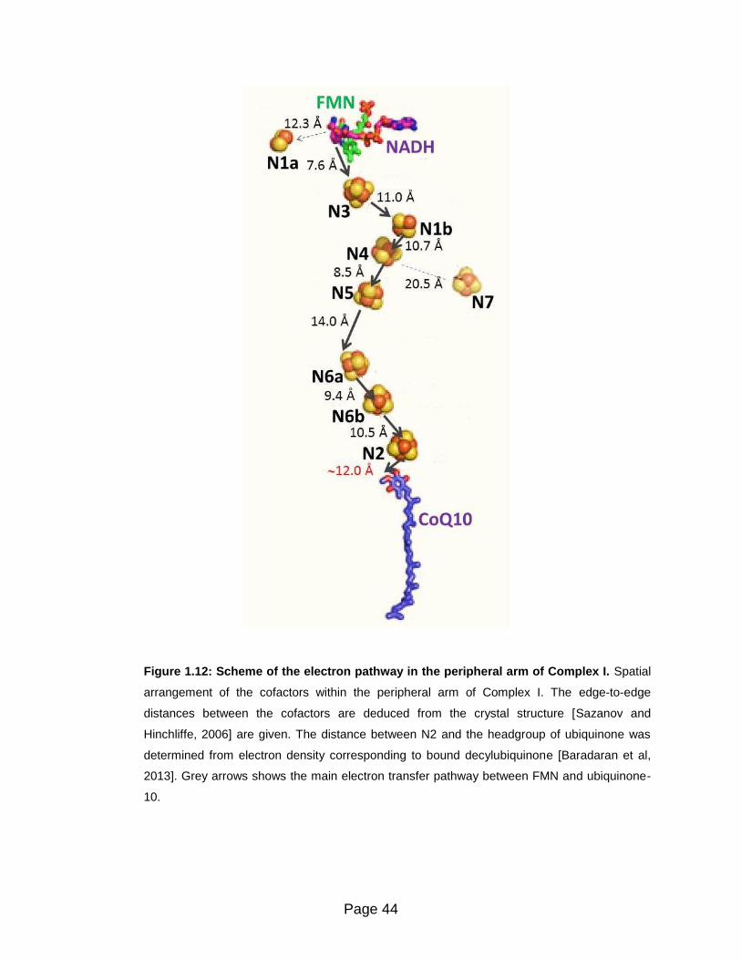
Page 44
Figure 1.12: Scheme of the electron pathway in the peripheral arm of Complex I. Spatial
arrangement of the cofactors within the peripheral arm of Complex I. The edge-to-edge
distances between the cofactors are deduced from the crystal structure [Sazanov and
Hinchliffe, 2006] are given. The distance between N2 and the headgroup of ubiquinone was
determined from electron density corresponding to bound decylubiquinone [Baradaran et al,
2013]. Grey arrows shows the main electron transfer pathway between FMN and ubiquinone-
10.

Page 45
Interface domain. Most of the interactions between the peripheral and
membrane domains involve subunit ND1, which forms many salt bridges and
hydrogen bonds to hydrophilic subunits 49 kDa, PSST, and TYKY [Baradaran et al,
2013].
ND1 subunit is the most conserved membrane subunit of Complex I and
participates in the interface with the hydrophilic domain contributing to the Q site
[Sekiguchi et al, 2009]. ND1 emerged only once during evolution, joining
hydrogenase and antiporter modules of Complex I – related proteins.
The Q site is found at the interface of 49 kDa, TYKY, PSST, and ND1 subunits
supported by the stabilizing interactions of the ND3 subunit that keeps the
peripheral and membrane domains together [Baradaran et al, 2013].
The structural studies of Complex I from T. thermophilus co-crystallized with
ubiquinone analogues [Baradaran et al, 2013] showed that the ubiquinone
headgroup interacts with conserved and essential residues. It is 12 Å from the
cluster N2 and 15 Å above the membrane surface [Baradaran et al, 2013]. Hence,
it is located at an appropriate distance for electron exchange with the Fe-S chain
[Page et al, 1999], but must move a significant distance out of the membrane
[Baradaran et al, 2013].
A 30-Å long chamber, framed by helices of ND1 and ND3 subunits, is completely
enclosed from the solvent and leads from a narrow entry point in the membrane
toward cluster N2. This quinone chamber is long and narrow enough to restrict the
quinone tail to an extended conformation (Figure 1.13 and 1.14).
It has been proposed that only the last one to three isoprenoid units from
ubiquinone-10 will protrude out of the cavity into the lipid, as the entrance of the
quinone chamber is narrower that the rest of the cavity, so that the quinone tail will
block solvent access to the cavity, sealing this reaction chamber. Consequently,
slight structural re-arragements are necessary to allow the quinone headgroup to
move in and out the cavity.
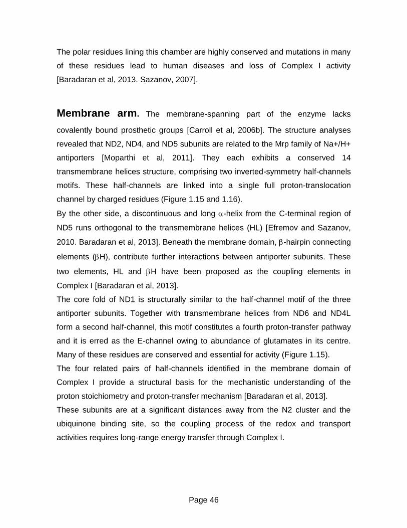
Page 46
The polar residues lining this chamber are highly conserved and mutations in many
of these residues lead to human diseases and loss of Complex I activity
[Baradaran et al, 2013. Sazanov, 2007].
Membrane arm. The membrane-spanning part of the enzyme lacks
covalently bound prosthetic groups [Carroll et al, 2006b]. The structure analyses
revealed that ND2, ND4, and ND5 subunits are related to the Mrp family of Na+/H+
antiporters [Moparthi et al, 2011]. They each exhibits a conserved 14
transmembrane helices structure, comprising two inverted-symmetry half-channels
motifs. These half-channels are linked into a single full proton-translocation
channel by charged residues (Figure 1.15 and 1.16).
By the other side, a discontinuous and long -helix from the C-terminal region of
ND5 runs orthogonal to the transmembrane helices (HL) [Efremov and Sazanov,
2010. Baradaran et al, 2013]. Beneath the membrane domain, -hairpin connecting
elements (H), contribute further interactions between antiporter subunits. These
two elements, HL and H have been proposed as the coupling elements in
Complex I [Baradaran et al, 2013].
The core fold of ND1 is structurally similar to the half-channel motif of the three
antiporter subunits. Together with transmembrane helices from ND6 and ND4L
form a second half-channel, this motif constitutes a fourth proton-transfer pathway
and it is erred as the E-channel owing to abundance of glutamates in its centre.
Many of these residues are conserved and essential for activity (Figure 1.15).
The four related pairs of half-channels identified in the membrane domain of
Complex I provide a structural basis for the mechanistic understanding of the
proton stoichiometry and proton-transfer mechanism [Baradaran et al, 2013].
These subunits are at a significant distances away from the N2 cluster and the
ubiquinone binding site, so the coupling process of the redox and transport
activities requires long-range energy transfer through Complex I.

Page 47
Figure 1.13: Quinone-reaction chamber of Complex I. Cartoon representation of the atomic model
(Protein Data Bank identifier 4HEA) at 3.3 Å resolution from T. thermophilus. A) The five subunits that
form the interface between the hydrophilic and hydrophobic domains are viewed along the membrane
domain as indicated in the inset. B. taurus nomenclature is used for the subunits. The 49 kDa, PSST,
and TYKY subunits at the bottom of the hydrophilic domain sit on top of ND1 and interact extensively
with its interhelical loops. One loop of ND3 reaches up to connect with the 49 kDa and PSST subunits
on the front of the hydrophilic domain. The cavity of the quinone-reaction chamber proposed from
structural analyses is shown in dark blue and two residues that form hydrogen bonds with the
ubiquinone headgroup are marked, residues are numbered according to the Thermus thermophilus
structure (His92 and Tyr141 in B. taurus). B) Surface (solvent-accessible) representation of the interface
between hydrophilic and hydrophobic domains. The empty crevice (C, circled) between ND6 (green),
ND1 (orange) and ND3 (blue) subunits, as well as helices framing the entry point to the quinone site (Q)
are indicated. This empty crevice may provide space for conformational changes during the catalytic
cycle, since the central helices of the ND1 half-channel tilt into this crevice. 7(TM1) = transmembrane
helix from Nqo7 subunit of T. thermophilus (ND3 in B. taurus). 8(AH1) = amphipathic helix and 8(TM1) =
transmembrane helix from Nqo8 subunit of T. thermophilus (ND1 in B. taurus). [from Baradaran et al,
2013]
A
B

Page 48
Figure 1.14: Ubiquinone binding site of Complex I. Cartoon representation of the atomic model
(Protein Data Bank identifier 4HEA) at 3.3 Å resolution from T. thermophilus. A) The internal
solvent-accessible surface colored red for negative, white for neutral and blue for positive surface
charges. Charged residues lining the cavity are shown with carbon in magenta and hydrophobic
residues in yellow. Ala63, the site of the primary Leber’s hereditary optic neuropathy disease
mutation, is labelled in red. Residues are numbered according to the Thermus thermophilus
structure [Nqo8 subunit of T.thermophilus (ND1 subunit in B. taurus): Phe28 (Val17), Arg36
(Arg25), Asp62 (Asp51), Ala63 (Ala52), Leu138 (Trp118), Arg216 (Arg195), Glu223 (Glu202),
Glu225 (Glu204), Glu248 (Glu227), Tyr249 (Tyr228), Arg294 (Arg273), Arg299 (Arg278)]; [Nqo6
subunit of T. thermophilus (PSST subunit in B. taurus): Ala47 (Ala93), Ile48 (Val94), Met50
(Met96), Met51 (Met97), Phe63 (Phe109)]; [Nqo4 subunit of T. thermophilus (49 kDa subunit in B.
taurus): His34 (His88), His38 (His92), Tyr87 (Tyr141), Asp139 (Asp193)]. B) Theoretical model of
bound ubiquinone-10. Carbon atom in cyan indicates the eighth isoprenoid unit. Residues of Nqo4
subunit (49 kDa subunit in B. taurus) interacting with the headgroup are indicated: His38 (His92),
Tyr87 (Tyr141), Asp139 (Asp193). The quinone chamber is shown with surface in brown and
helices framing its entry point are indicated. Movable hydrophobic helix 6(H1) (Nqo6 subunit in T.
thermophilus, PSST subunit in B. taurus] interacting with the amphipathic 8 (AH1) helix [Nqo8
subunit of T. thermophilus, ND1 subunit in B. taurus] are labelled. [from Baradaran et al, 2013]
A B

Page 49
Figure 1.15: E-channel and central hydrophilic axis of Complex I. Cartoon representation of
the atomic model (Protein Data Bank identifier 4HEA) at 3.3 Å resolution from T. thermophilus. A)
Central axis of charged and polar residues (charged residues have carbon in magenta, polar in
cyan). Residues shown are either central to half-channels or are forming the connection between
them. Predicted waters nearby are shown as spheres. Connecting elements are shown in solid
colors: helix HL in magenta and the H element in blue, with the C-terminal helix CH and the -
harping (-h) from each antiporter labelled. B) Charged and polar residues constituting the
channel are shown as sticks. Quinone cavity is shown with surface in brown and approximate
proton translocation path is indicated by blue arrow. Key residues are labelled, with the Glu/Asp
quartet in red. Residues are numbered according to the Thermus thermophilus structure [Nqo8
subunit of T.thermophilus (ND1 subunit in B. taurus): Glu130 (Ser110), Tyr134 (Tyr114), Arg154
(Arg134), Glu213 (Glu192), Glu223 (Glu202), Glu227 (Glu216), Glu235 (Glu224)]; [Nqo7 subunit
of T. thermophilus (ND3 subunit in B. taurus): Asp72 (Asp66)]; [Nqo10 subunit of T. thermophilus
(ND6 subunit in B. taurus): Tyr59 (Tyr60)]. [from Baradaran et al, 2013]
A
B

Page 50
Figure 1.16: Putative proton-translocation channels in the antiporter-like subunits. Cartoon
representation of the atomic model (Protein Data Bank identifier 4HEA) at 3.3 Å resolution from T.
thermophilus. Polar residues lining the channels are shown as sticks with carbon in dark blue for
the first (N-terminal) half-channel, in green for the second (C-terminal) half-channel and in orange
for connecting residues. Key residues –Glu and Lys- from the first half-channel, Lys/His from the
connection and Lys/Glu from the second half-channel, are labelled. [Nqo14 subunit of T.
thermophilus (ND2 subunit in B. taurus): Lys186 (Lys105), Lys216 (Lys135), Lys345 (Lys213)];
[Nqo13 subunit of T. thermophilus (ND4 subunit in B. taurus): Glu123 (Glu123), Lys204 (Lys206),
Lys235 (Lys237), Glu377 (Lys378)]; [Nqo12 subunit of T. thermophilus (ND5 subunit in B. taurus):
Glu132 (Glu145), Lys216 (Lys223), His241 (His248), Lys385 (Lys392)]. Approximate proton-
translocation paths are indicated by blue arrows. [from Baradaran et al, 2013]
ND
5
ND4 ND2

Page 51
The crystal structures of bacterial Complex I have given much insight into the
structure and how subunits are arranged within these complexes. In higher
eukaryotes the most detailed topological structure of subunits within complex I is
obtained from experiments using mild chaiotropic salts to break up complex I into
subcomplexes. First studies performed by Hatefi and colleagues enabled to
fractionate complex I into a flavoprotein part, a hydrogenase part and a membrane
part [Galante and Hatefi, 1978]. This approach was later ined with mildly
detergents, thus intact bovine Complex I has been resolved into various
subcomplexes (I, I, I, I fragments) that have subsequently been subjected to
extensive analysis [Carroll et al, 2003] (Figure 1.17).
Subcomplex I represents the peripheral arm of the complex, subcomplex I
consists of subcomplex I plus part of the membrane arm and subcomplex I
forms the major part of the membrane arm. Subunits that do not associate with the
I or I subcomplexes are grouped as the I ‘subcomplex’, a fraction for which it is
not clear whether it represents a true fragment of Complex I.
Figure 1.17: Topology model of subunits in mammalian Complex I.
The L-shaped Complex I can be dissected into fragments I, I, I, and I,
whose composition allows a basic arrangement of the 44 subunits currently
described for mammalian complex (H. sapiens nomenclature)

Page 52
Evolution and modular organization of Complex I
Phylogenetic analyses revealed that the two arms of Complex I are evolutionary
independent of each other representing a chimera of hydrogenases and cation-
proton antiporter [Friedrich, 2001. Moparthi and Hagerhall 2011. Efremov and
Sazanov, 2012. Friedrich and Scheide, 2000. Böhm et al, 1990. Albracht, 1993.
Friedrich and Weiss, 1997. Finel, 1998].
The intricate evolutionary history of Complex I is intimately related to energy
metabolism and the oxygen concentration levels in the early atmosphere.
Of particular interest is the modular evolution of complex I proposed by Friedrich
and Weiss (Friedrich and Weiss 1997). Based on homologies with bacterial
enzymes, this theory postulates that Complex I originates from fusion of distinct
pre-existing protein assemblies, which have combined their activities in one
complex. Thus, the initial step has been the association of a soluble heterodimeric
reversible [NiFe]-hydrogenase with membrane-bound K+/H+ or Na+ antiporters
[Friedrich and Weiss, 1997. Friedrich and Scheide, 2000]. Mathiesen and
Hägerhäll, 2002]. Subsequent acquisition of subunits with additional redox would
have transformed this ancestral enzyme in a proton-pumping hydrogen:quinone
oxidoreductase. Subsequently the multiplication and/or recruitment of antiporter
genes plus the addition of other subunits, along with loss of hydrogenase activity
and gain of diaphorase activity, would have given rise to Complex I (Figure 1.19)
The association of a soluble [NiFe]-hydrogenase with membrane-bound antiporter
subunits may have been caused by the need of highly reducing electrons. It has
been postulated that hydrogen may have been very abundant in the primordial
atmosphere [Tian et al, 2005] explaining the early appearance of soluble [NiFe]-
hydrogenases. However, this gas can easily escape to space and its atmospheric
concentration may have dropped rather rapidly at the geological scale.
Reverse electron transport coupled to CO2 reduction may have been the selective
advantage that favored the formation of the hydrogenase:antiporter complex.
The evolutionary relationships established by phylogenetic studies are consistent
with a plethora of structural and functional evidence and define three functional
modules of Complex I: the NADH oxidizing or N-module serving for electron input

Page 53
from NADH, the Q-module or output module which conducts electrons that reduces
ubiquinone, and the P-module that translocates the protons (Figure 1.18).
The N - module
The NADH dehydrogenase module (N-module) of Complex I consists of the 24
kDa, 51 kDa, and 75 kDa subunits, are homologous to the α subunit and in part
from the γ subunit of AD+-reducing hydrogenases of Alcaligenes eutrophus [Tran-
Betcke et al, 1990].
This module harbor the NADH binding site and have FMN and iron-sulfur clusters
as prosthetic groups [Yano et al, 1996].
The Q - module
The quinone module (Q-module) is composed of 49 kDa, 30 kDa, TYKY, and
PSST subunits. The 49 kDa and part of the PSST subunit are homologous to the
large and small subunits of water-soluble [NiFe] hydrogenases, respectively [Böhm
et al, 1990. Albracht, 1993]. The 30 kDa subunit has no prosthetic group and
seems to be unique for Complex I [Brandt, 2006].
The P – module
This P-module comprises the seven membrane-embedded subunits, which are
encoded by the mitochondrial genome in most eucaryotes [Brandt, 2006]. Subunits
ND2, ND4, and ND5 are homologous to each other, and one representative of this
family has been acquired together with an ND1 homologue at the level of
membrane-bound type-3 [NiFe] hydrogenases found in Escherichia coli [Fearnley
and Walker, 1992] or the archaeon Methanosarcina barkeri [Hedderich, 2004].
Remarkably, the ND2/ND4/ND5 subunits seems to have evolved from Na+/H+
antiporters [Fearnley and Walker, 1992. Mathiesen and Hägerhäll, 2002], and are
particularly homologous with proteins encoded by the mrp (multiple resistance and
pH adaptation) operon of Bacillus subtilis .This type of antiporter is built up by
seven proteins, MrpABCDEFG [Swartz et al, 2005], of which MrpA was shown to
have higher sequence similarity to ND5 whereas MrpD showed more similarity to
ND4 and ND2 [Mathiesen and Hägerhäll, 2002]. The small ND4L is homologous to
MrpC [Mathiesen and Hägerhäll, 2003].
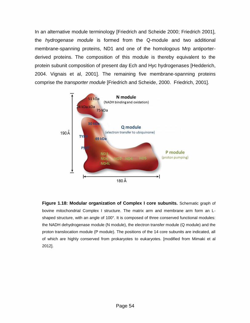
Page 54
In an alternative module terminology [Friedrich and Scheide 2000; Friedrich 2001],
the hydrogenase module is formed from the Q-module and two additional
membrane-spanning proteins, ND1 and one of the homologous Mrp antiporter-
derived proteins. The composition of this module is thereby equivalent to the
protein subunit composition of present day Ech and Hyc hydrogenases [Hedderich,
2004. Vignais et al, 2001]. The remaining five membrane-spanning proteins
comprise the transporter module [Friedrich and Scheide, 2000. Friedrich, 2001].
Figure 1.18: Modular organization of Complex I core subunits. Schematic graph of
bovine mitochondrial Complex I structure. The matrix arm and membrane arm form an L-
shaped structure, with an angle of 100°. It is composed of three conserved functional modules:
the NADH dehydrogenase module (N module), the electron transfer module (Q module) and the
proton translocation module (P module). The positions of the 14 core subunits are indicated, all
of which are highly conserved from prokaryotes to eukaryotes. [modified from Mimaki et al
2012].
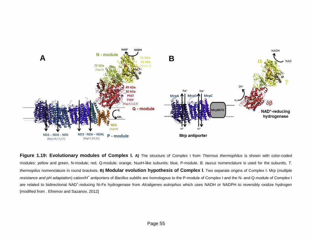
Page 55
Figure 1.19: Evolutionary modules of Complex I. A) The structure of Complex I from Thermus thermophilus is shown with color-coded
modules: yellow and green, N-module; red, Q-module; orange, NuoH-like subunits; blue, P-module. B. taurus nomenclature is used for the subunits, T.
thermopilus nomenclature in round brackets. B) Modular evolution hypothesis of Complex I. Two separate origins of Complex I: Mrp (multiple
resistance and pH adaptation) cation/H+ antiporters of Bacillus subtilis are homologous to the P-module of Complex I and the N- and Q-module of Complex I
are related to bidirectional NAD+-reducing Ni-Fe hydrogenase from Alcaligenes eutrophus which uses NADH or NADPH to reversibly oxidize hydrogen
[modified from . Efremov and Sazanov, 2012]
A B

Page 56
Mammalian Complex I Assembly
The mammalian Complex I is the most intricate structure of NADH:oxidoreductases,
as it requires the combination of 44 protein subunits, encoded by two genomes, and
at least nine cofactors. The nDNA-encoded subunits must assemble in coordination
with the hydrophobic mtDNA-encoded subunits to form the functioning mature
holocomplex.
Thus, the assembly of these subunits into a functional holoenzyme is a very
complicate process to characterize due to the large size and numerous subunits of
the enzyme, lack of a detailed crystal structure, and its dual genomic control.
It has been extensively demonstrated that Complex I assembly is disturbed if one of
the structural subunits or assembly factors is altered [Janssen et al, 2006]. Since
isolated Complex I deficiency is the main form of defective oxidative phosphorylation,
the assembly of this enzyme is being elucidated by the characterization of
intermediate subcomplexes and assembly factors from samples of patients with
mutations in one of the subunits or with cybrid cell line models (cells depleted of
mitochondrial DNA that can be repopulated with mutant DNA to study specific
mutations in a control background) [Mimaki et al, 2012].
In the last years several models for the assembly of mammalian Complex I have
been proposed [Antonicka et al, 2000. Lazarou et al, 2007. Ugalde et al, 2004b.
Vogel et al, 2007]. Although these models show some differences, the recent
consensus agrees that an early assembly intermediate is anchored to the membrane
prior to its extension with additional membrane and peripheral subunits [McKenzie
and Ryan, 2010] (Figure 1.20).
The early assembly stage starts with the preassembling of hydrogenase subcomplex,
where core subunits 49 kDa and 30 kDa (NDUFS2 and NDUFS3 human
nomenclature) form a small hydrophilic assembly complex. [Vogel et al, 2007]. This
intermediate subcomplex further expands by the incorporation of hydrophilic
subunits, PSST, TYKY, and 39 kDa (NDUFS7, NDUFS8, and NDUFA9), the
connective module between the peripheral and membrane arms [Wessels et al,
2009. Ugalde et al, 2004a. Bourges et al, 2004]. This intermediate is further
Page 57
anchored to the membrane by assembly chaperones [Saada et al, 2009]. The
mtDNA encoded ND1 subunit thus, anchors peripheral arm to the inner mitochondrial
membrane [Yano, 2002], forming an intermediate of ~400-kDa [Lazarou et al, 2007].
By the other side, independently N2, ND3, and ND4L are assembled into a ~460-kDa
membrane intermediate. The incorporation of ND6 brings together both ~460-kDa
membrane subcomplex and the~400-kDa module at a later stage and this is the last
entry point of the mitochondrially encoded subunits [Perales-Clemente et al, 2010].
The binding of these subcomplexes form the ~650-kDa intermediate under the
presence of several assembly factors [Sugiana et al, 2008.Nouws et al, 2010]. With
the association of another membrane complex containing ND4, ND5, and possibly
subunit B14.5 (NDUFC2), a ~830 kDa assembly intermediate is formed [Gershoni et
al, 2010].
Meanwhile, the hydrophilic NADH: dehydrogenase module containing 51 kDa, 24
kDa, and 75 kDa (NDUFV1, NDUFV2, and NDUFS1) subunits, is assembled
independently with several nDNA-encoded subunits that are directly or indirectly
involved in binding and oxidizing NADH [Lazarou et al, 2007].
With the addition of the NADH: dehydrogenase module and the remaining subunits,
the mature holoenzyme is assembled. In this concerted and elaborate assembly
process, several assembly factors with unknown functions and undiscovered proteins
are involved [Carilla-Latorre et al, 2010. Calvo et al, 2010].

Page 58
Figure 1.20: The assembly model of mammalian Complex I biogenesis. In the early assembly stages, the core subunits 49 kDa and 30 kDa
form a small hydrophilic assembly complex, which further expands by the incorporation of hydrophilic subunits such as TKYK, PSST and later 39 kDa. This
peripheral complex, together with a small membrane complex containing mtDNA-encoded subunit ND1, forms a ~400 kDa assembly intermediate. This ~400 kDa
complex incorporates with a ~460 kDa membrane complex containing ND3, ND6, ND2, ND4L and B17 subunits to form a ~650 kDa complex. With the association
of another membrane complex having ND4, ND5 and probably subunit B14.5, a ~830 kDa assembly intermediate is formed. Meanwhile, a hydrophilic complex,
NADH: dehydrogenase module (N module) is assembled by some nDNA-encoded subunits directly or indirectly involved in binding and oxidizing NADH. With the
addition of the N module and remaining subunits (such as the intermembrane space subunits PGIV and 15 kDa), mature complex I is assembled. The nDNA-
encoded subunits are colored with blue. The mtDNA-encoded-subunits are in green. [modified from Mimaki et al, 2012]

Page 59
Catalytic activity of Complex I
NADH oxidation and intramolecular electron transfer
The physiological activity of Complex I is the electron transfer from NADH
(Em7 = −320 mV) to ubiquinone (Em7+90 mV), this process is coupled to the
formation of an electrochemical membrane potential. Despite a difference in the
substrate redox potentials of about 400 mV, the reaction catalyzed by Complex I is
fully reversible. It was demonstrated that in the presence of a protonmotive force
mitochondria can transfer electrons from succinate onto NAD+ [Klingenberg and
Slenczka, 1959. Chance and Hollunger, 1960].
During the catalytic cycle two electrons are transferred from NADH to the non-
covalently bound FMN as a hydride ion. Upon binding, the nicotinamide ring of NADH
forms stacking interactions with the isoalloxazine ring of FMN, thus providing a
favorable geometry for efficient hydride transfer between C4N of NADH and N5 of
FMN [Berrisford and Sazanov, 2009]. The distance between these two atoms (3.2 Å)
is slightly shorter than average for flavoenzymes [Fraaije and Mattevi, 2000],
consistent with fast rates of hydride transfer in Complex I [Verkhovskaya et al, 2008].
The midpoint redox potential of the FMN/FMNH2 couple was determined to −320 mV
[Sled et al, 1994. Bungert et al, 1999. Kohlstädt et al, 2008]. Electrons are passed
from the reduced FMN though the seven active Fe-S clusters (N3, N1b, N4, N5, N6a,
N6b and N2) to the ubiquinone binding site at the interface between the two arms.
Cluster N3, represents the entrance of the electron transfer chain. The edge-to-edge
distances between the clusters of the chain vary from 8.5 to 14 Å. Thus, all distances
are compatible with the physiological electron transfer rate [Moser et al, 2006]
(Figure 1.12).
The short distances between the individual Fe-S guarantee an electron transfer rate
faster than the physiological turnover.

Page 60
Cluster N2, the last redox center in the chain, transfer electrons to ubiquinone. The
reduction of ubiquinone to ubiquinol is a two electron process, and so one-electron
reduced semiquinone intermediates may be formed during turnover.
It has been demonstrated that N2 [Ohnishi 1998. Ingledew and Ohnishi, 1980],
interacts paramagnetically with two semiquinone species that accumulate during
steady-state turnover and differ in their spin relaxation behavior [Vinogradov et al,
1995. Yano et al, 2005]. The fast-relaxing species QNf is only detected in the
presence of a membrane potential, whereas the slow-relaxing species QNs is also
observed in both the absence and presence of membrane potential. It was shown
that N2 interacts with the semiquinone radicals [Ohnishi and Salerno, 2005].
The interaction distance between N2 and QNf was determined to 12 Å, while that
between N2 and QNs might be larger than 30 Å [Ohnishi et al 2010a. Ohnishi et al
2010b].
Ubiquinone reduction and coupling mechanism
The midpoint redox potential of NADH is about -320 mV and of the Q/QH2 pair ~
+100 mV (for ubiquinone) [Efremov and Sazanov, 2012]. Theore, most of the redox
energy in Complex I is released during quinone reduction. Some of it would be also
released upon reduction of cluster N2, resulting in the observed conformational
changes [Berrisford and Sazanov, 2009].
Sazanov and co-workers, recently have proposed that the catalytic cycle of Complex
I is likely to proceed via a negatively charged ubiquinol intermediate Q2- [Efremov and
Sazanov, 2012]. Either Q2- or key charged residues nearby are likely to remain
unprotonated in order to drive conformational.
The design of the enclosed Q-site is well suited for this purpose: the binding of the
quinone at the deep end of sealed cavity, with protein tightly packed around the
headgroup, will prevent solvent access and quinone protonation by means other than
coordinating residues Tyr87 (Tyr141 in B. taurus) and His38 (His34 in B. taurus).
The charged residues can exist in this chamber because it is relatively hydrophilic
and distal from the membrane. The Q-site is linked to the centre of the E-channel by
a hydrophilic funnel (Figure 1.21).

Page 61
Thus, the negatively charged ubiquinol (or adjacent charged residues) can interact
electrostatically with these negatively charged residues driving conformational
changes in this channel.
Additional driving force is probably provided by conformational movements of 49 kDa
and PSST subunits upon N2 reduction, which directly interact with flexible parts of
ND1 [Berrisford and Sazanov, 2009.Berrisford et al, 2008].
The flexible hydrophilic axis connects the half-channels in the middle of the
membrane domain, linking membrane subunits in an overall conformational cycle
that probably involve both helix HL and H (coupling elements) [Efremov and
Sazanov, 2011].
Tight coupling observed between proton translocation and quinone chemistry
[Efremov and Sazanov, 2011] can be explained if opening of the Q-site entrance to
allow quinone in and out forms a part of this cycle.
According to this conformational switch model, the electrostatically and N2-driven E-
channel drives conformational changes first in the neighbouring antiporter ND2,
which in its turn drives distal subunits ND4 and ND5. All through the hydrophilic
central axis. The extent of the movement of the two coupling elements during the
catalytic cycle is currently unclear, but authors hypothesize that these elements
probably contribute at least to the coordination between the three antiporters. These
concerted conformational changes would lead to changes in pKa and solvent
exposure of key residues, resulting in proton translocation [Baradaran et al, 2013]
(Figure 1.21).
Complex I inhibitors
Complex I is inhibited by a variety of compounds [Friedrich et al, 1994] from
rotenone, to a number of synthetic insecticides/acaricides [Lümmen, 1998]. With the
exception of a few inhibitors that inhibit electron input into Complex I such as rhein
[Kean et al, 1971] and diphenyleneiodonium [Majander et al, 1994], all inhibitors act
at the terminal electron transfer step of this enzyme [Friedrich et al. 1994] and it is
commonly accepted that act at the ubiquinone reduction site [Fendel et al, 2008].
Complex I inhibitors have been grouped into three classes [Degli Esposti, 1998]
(Table 1.5).
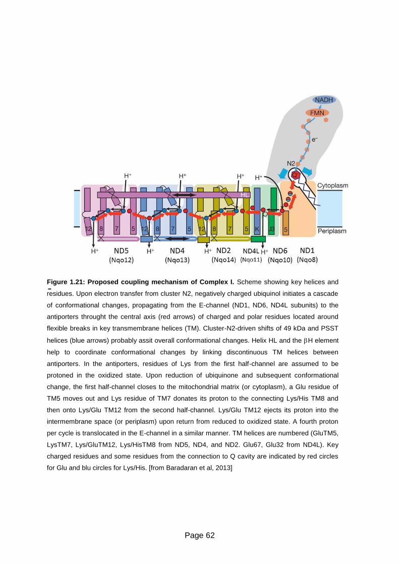
Page 62
-
Figure 1.21: Proposed coupling mechanism of Complex I. Scheme showing key helices and
residues. Upon electron transfer from cluster N2, negatively charged ubiquinol initiates a cascade
of conformational changes, propagating from the E-channel (ND1, ND6, ND4L subunits) to the
antiporters throught the central axis (red arrows) of charged and polar residues located around
flexible breaks in key transmembrane helices (TM). Cluster-N2-driven shifts of 49 kDa and PSST
helices (blue arrows) probably assit overall conformational changes. Helix HL and the H element
help to coordinate conformational changes by linking discontinuous TM helices between
antiporters. In the antiporters, residues of Lys from the first half-channel are assumed to be
protoned in the oxidized state. Upon reduction of ubiquinone and subsequent conformational
change, the first half-channel closes to the mitochondrial matrix (or cytoplasm), a Glu residue of
TM5 moves out and Lys residue of TM7 donates its proton to the connecting Lys/His TM8 and
then onto Lys/Glu TM12 from the second half-channel. Lys/Glu TM12 ejects its proton into the
intermembrane space (or periplasm) upon return from reduced to oxidized state. A fourth proton
per cycle is translocated in the E-channel in a similar manner. TM helices are numbered (GluTM5,
LysTM7, Lys/GluTM12, Lys/HisTM8 from ND5, ND4, and ND2. Glu67, Glu32 from ND4L). Key
charged residues and some residues from the connection to Q cavity are indicated by red circles
for Glu and blu circles for Lys/His. [from Baradaran et al, 2013]

Page 63
Table 1.5: Functional classification of Complex I inhibitors
Inhibitor class Representative compounds
Type A: quinone antagonists
Rolliniastin-2
Piericidin A
Idebenone
Type B: semiquinone antagonists
Rotenone
Piericidin A
Piercidin B
Amytal
4-alkyl-acridones
’-alkyl-MPP+analogues
Phenoxan
Type C: quinol antagonists
Quinol products
Reduced ubiquinone-2
Myxothiazol
Stigmatellin
Data from [Lenaz and Genova, 2010]
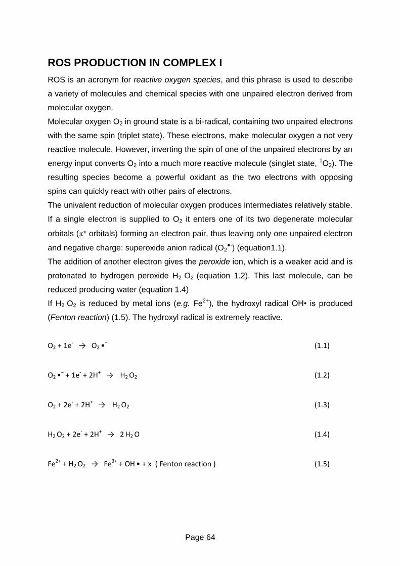
Page 64
ROS PRODUCTION IN COMPLEX I
ROS is an acronym for reactive oxygen species, and this phrase is used to describe
a variety of molecules and chemical species with one unpaired electron derived from
molecular oxygen.
Molecular oxygen O2 in ground state is a bi-radical, containing two unpaired electrons
with the same spin (triplet state). These electrons, make molecular oxygen a not very
reactive molecule. However, inverting the spin of one of the unpaired electrons by an
energy input converts O2 into a much more reactive molecule (singlet state, 1O2). The
resulting species become a powerful oxidant as the two electrons with opposing
spins can quickly react with other pairs of electrons.
The univalent reduction of molecular oxygen produces intermediates relatively stable.
If a single electron is supplied to O2 it enters one of its two degenerate molecular
orbitals (* orbitals) forming an electron pair, thus leaving only one unpaired electron
and negative charge: superoxide anion radical (O2●-) (equation1.1).
The addition of another electron gives the peroxide ion, which is a weaker acid and is
protonated to hydrogen peroxide H2 O2 (equation 1.2). This last molecule, can be
reduced producing water (equation 1.4)
If H2 O2 is reduced by metal ions (e.g. Fe2+), the hydroxyl radical O • is produced
(Fenton reaction) (1.5). The hydroxyl radical is extremely reactive.
O2 + 1e- → O2 •−
(1.1)
O2 •− + 1e- + 2H+ → H2 O2
(1.2)
O2 + 2e- + 2H+ → H2 O2
(1.3)
H2 O2 + 2e- + 2H+ → 2 H2 O
(1.4)
Fe2+ + H2 O2 → Fe3+ + OH • + x ( Fenton reaction )
(1.5)

Page 65
Mitochondria are an important source of ROS (reactive oxygen species) within most
mammalian cells with the oxygen superoxide anion (O2•−) being the primary
mitochondrial ROS that is generated mainly by Complex III and Complex I [Lenaz,
2012]
ROS contributes to mitochondrial damage of proteins, lipids and DNA (oxidative
stress) and is also important in in many signaling pathways (Figure 1.22 and 1.23).
Therefore, only excessive ROS production (or defect of protective systems) leads to
oxidative stress and pathological conditions. Whether cells will suffer from oxidative
stress, depends on the balance between the continuous production of ROS and their
removal by endogenous detoxification systems.
Figure 1:22: Overview of mitochondrial ROS production. ROS production by mitochondria can
lead to oxidative damage to mitochondrial proteins, membranes and DNA (oxidative stress), impairing the ability
of mitochondria to synthesize ATP and to carry out their wide range of metabolic functions, including the
tricarboxylic acid cycle, fatty acid oxidation, the urea cycle, amino acid metabolism, haem synthesis and FeS
centre assembly that are central to the normal operation of most cells (energetic failure). Mitochondrial oxidative
damage can also increase the tendency of mitochondria to release intermembrane space proteins such as
cytochrome c (cyt c) to the cytosol by mitochondrial outer membrane permeabilization (MOMP) and there by
activate the cell’s apoptotic machinery. In addition, mitochondrial ROS production leads to induction of the
mitochondrial permeability transition pore (PTP), which renders the inner membrane permeable to small
molecules in situations such as ischaemia/reperfusion injury. Consequently, it is unsurprising that mitochondrial
oxidative damage contributes to a wide range of pathologies. In addition, mitochondrial ROS may act as a
modulatable redox signal, reversibly affecting the activity of a range of functions in the mitochondria, cytosol and
nucleus. [From Murphy, 2009]

Page 66
Figure 1.23: Modes of mitochondrial operation that lead to O2•− production. There are
three modes of mitochondrial operation that are associated with O2•−
production. In mode 1, the NADH
pool is reduced, for example by damage to the respiratory chain, loss of cytochrome c during
apoptosis or low ATP demand. This leads to a rate of O2•−
formation at the FMN of complex I that is
determined by the extent of FMN reduction which is in turn set by the NADH/NAD+ ratio. Other sites
such as αKGD may also contribute. In mode 2, there is no ATP production and there is a high p
and a reduced CoQ pool which leads to RET through Complex I, producing large amounts of O2•−
. In
mode 3, mitochondria are actively making ATP and consequently have a lower p than in mode 2 and
a more oxidized AD pool than in mode 1. Under these conditions, the flux of O2•−
within
mitochondria is far lower than in modes 1 and 2, and the O2•−
sources are unclear. [From Murphy,
2009]

Page 67
The midpoint redox potential of mitochondrial superoxide production is between 100
– 250 mV, and a low oxygen concentrations is 68 mV. [Murphy, 2009].
Therefore, in vivo, the one-electron reduction of O2 to O2 •− is thermodynamically
favored, even by relatively oxidizing redox couples, and a wide range of electron
donors within mitochondria could potentially carry out this reaction.
However, only a small proportion of mitochondrial electron carriers with the
thermodynamic potential to reduce O2 to O2 •−.
Early experiments proved the involvement of Complex I in ROS production
[Takeshige and Minakami, 1979. More recent studies confirmed that Complex I is a
major source of superoxide production in mitochondria [Lenaz, 2012]
The superoxide production by Complex I is higher during the reverse electron
transport (RET) from succinate to NAD+, whereas during the forward electron
transport it is much lower [Turrens 2003].
In addition, it has been demonstrated that rotenone enhances ROS formation during
forward electron transfer [Herrero and Barja, 2000. Genova et al. 2001] and inhibits it
during reverse electron transfer.
The identification of the oxygen reducing site has been the subject of extensive
investigation, and debate as several prosthetic groups in the enzyme have been
suggested to be the direct reductants of oxygen (Figure 1.24).
These include FMN, ubisemiquinone, and iron sulphur cluster N2 [Fato et al, 2009].
In isolated Complex I, fully reduced FMN is considered the major electron donor to
oxygen to form superoxide anion [Galkin and Brandt, 2005. Kussmaul and Hirst,
2006]
In mitochondrial membranes, however, the identification of flavin as the site of
oxygen reduction is incompatible with the finding that two classes of inhibitors both
acting downstream of the iron sulphur clusters in the enzyme have opposite effects,
in that rotenone enhances superoxide production whereas stigmatellin inhibits it [Fato
et al, 2009]

Page 68
However, Hirst et al (208) admit the possible presence of two oxygen-reacting sites
at the two ends of the cofactor chain, ascribing the distal one to ROS generation
during reverse electron transfer. The superoxide production by Complex I is maximal
during reverse electron transfer, however the site of oxygen reduction in this instance
is even less well defined [Murphy, 2009].
The electron donor to ubiquinone in the Complex is FeS cluster N2 [Ohnishi et al,
1998]. It is likely that this centre is also the electron donor to oxygen both directly and
via one-electron reduction of several exogenous quinones.
Ohnishi et al. [Ohnishi et al, 2010c] presented a new hypothesis that the generation
of superoxide reflects a dynamic balance between the flavosemiquinone (semiflavin
or SF) and the CoQ semiquinone (SQ). In a purified preparation of Complex I, during
catalytic electron transfer from NADH to decyl ubiquinone, the superoxide generation
site was mostly shifted to the SQ. A quinone-pocket binding inhibitor (rotenone or
piericidin A) inhibits the catalytic formation of the SQ, and it enhances the formation
of SF and increases the overall superoxide generation. This suggests that if electron
transfer was inhibited under pathological conditions, superoxide generation from the
SF would be increased. The identification of SQ rather than N2 as the electron donor
to oxygen is however in contrast with the findings reported in the previous section
showing that when SQ reduction to QH2 is blocked by stigmatellin no superoxide is
produced [Fato et al, 2009].

Page 69
Figure 1.24: Production of O2•− by Complex I. The location of the FMN and the Fe-S clusters in
the hydrophilic arm are indicated, along with the putative CoQ-binding site. In mode1, there is
extensive O2•−
production from the FMN in response to a reduced NADH pool. In mode 2, a high p
and a reduced Co pool lead to RET and a high flux of O2•−
from the complex. The site of this O2•−
production is uncertain, hence the question mark, but may be associated with the CoQ-binding site.
A possible explanation is that two sites for oxygen reduction exist in the complex,
represented by flavin and an iron-sulphur cluster; the latter site would be
predominant in membrane particles whereas the former might be available after
Complex I isolation [Lenaz, 2012].
In the present study, we demonstrate a possible different interpretation involving the
supramolecular assembly of Complex I

Page 70
Supercomplexes and ROS production in Complex I
Indirect circumstantial evidence suggests that supercomplex assembly may limit the
extent of superoxide generation by the respiratory chain.
Panov et al. (2007) reason that the respirasome helps to maintain the redox
components of the complexes in the oxidized state through the facilitation of electron
flow by channeling, thus limiting ROS formation.
Similarly, Seelert et al. (2009) also suggest that facilitation of electron transfer by
channeling may limit the production of ROS.
As mentioned in the previous section, two potential sites for oxygen reduction exist in
Complex I, represented by FMN and iron–sulphur cluster N2.
Controversial results from different laboratories working either on isolated Complex I
or on mitochondrial membranes generally indicate that N2 as a source of ROS would
be predominant in membrane particles whereas FMN might become available after
Complex I isolation.
A reasonable hypothesis is that supercomplexes assembly may modify the
conformation of its component complexes confering a more shielded structure to
FMN in Complex I, and this prosthetic group becomes exposed to oxygen only when
Complex I is dissociated from Complex III (Figure 1.25)
Although the molecular structure of the individual complexes does not allow to
envisage a close apposition of the matrix arm of Complex I, where FMN is localized,
with either Complex III or IV [Dudkina et al 2005b; Schäfer et al 2007], the actual
shape of the I1III2IV1 supercomplex from bovine heart [Schäfer et al 2007] suggests a
slightly different conformation of Complex I in the supercomplex, showing a smaller
angle of the matrix arm with the membrane arm and a higher bending towards the
membrane (and presumably Complex III), in line with the notion that Complex I may
undergo important conformational changes.
Moreover, the observed destabilization of Complex I in the absence of supercomplex
may render the 51 kDa subunit containing the FMN more slightly untied from the
protein framework allowing it to interact with oxygen.

Page 71
Figure 1.25: Production of O2•− by Complex I in I1III2 supercomplex.

Page 72
HYPOTHESIS
As discussed in the previous sections, the main redox components responsible for
oxygen reduction to superoxide in mitochondria are situated in Complex I and in
Complex III. In particular, conditions limiting the electron flow within Complex I induce
large excess of superoxide production [Lenaz, 2012].
Since dissociation of the supercomplex I1III2 limits electron transfer between Complex
I and III and in addition destabilizes Complex I, a reasonable question is whether
such condition is also bound to enhance and/or modify the normal ROS production
from the Complex I.
In addition, indirect evidence suggests that supercomplex assembly may limit the
extent of ROS production by the respiratory chain where respirasomes helps
maintaining redox components of the complexes in the oxidized state through
channeling of the electron flow [Panov et al, 2007].
A reasonable hypothesis is that reducing prosthetic groups becomes exposed to
oxygen only when Complex I is dissociated from Complex III.
Following this line of thought, our study hypothesis is that disruption or prevention of
the association between Complex I and Complex III by different means enhace the
generation of reactive oxygen species from Complex I.
To demonstrate this hypothesis, we have decided to investigate ROS production by
Complex I under conditions in which the complex is arranged as a component of the
super-complex I1III2 or dissociated as an individual holoenzyme.
Hence, our experimental studies were in two experimental systems in which the
supramolecular organization of respiratory assemblies is impaired by:
(i) Treatment either of bovine heart mitochondrial membranes or
supercomplex I1III2 reconstituted in liposomes with dodecylmaltoside.
(ii) Reconstitution of Complex I and Complex III at high lipid:protein ratio

Page 73
MATERIALS AND METHODS
MATERIALS
Reagents and solutions
2’,7’-dichlorodihydrofluorescein diacetate (DCFDA) was purchased from Molecular
Probes (Invitrogen). All other chemicals were purchased from Sigma-Aldrich, unless
specifically indicated and used without further purification.
All solutions were prepared with Milli- water (18.2 MΩ cm−1) from a Milli-Q
purification system (Branstead, USA). pH is adjusted with either HCl 0.1 N or NaOH
0.1 N.
1. SUCROSE SOLUTION pH 7.6
0.25 M sucrose – 10 mM Tris base
2. BUFFER SOLUTION B pH 7.2
0.22 M D-mannitol – 70 mM sucrose – 10 mM Tris base – 1 mM EDTA
3. TSH SOLUTION pH 8.0
0.66 M sucrose – 50 mM Tris base – 1 mM L-Histidine
4. REACTION SOLUTION pH 7.4
50 mM KCl – 10 mM Tris base – 1 mM EDTA
5. BIURET REAGENT
0.15% (w:v) CuSO4.5H2O - 0.6% (w:v) potassium sodium tartrate - 10% (w:v)
NaOH
6. LOWRY STOCK SOLUTION A
2% (w:v) Na2CO3 in 0.1 N NaOH
7. LOWRY STOCK SOLUTION B
1% (w:v) CuSO4.5H2O
8. LOWRY STOCK SOLUTION C
2% (w:v) potassium sodium tartrate
Page 74
METHODS
Preparation of bovine heart mitochondria (BHM)
B. taurus heart mitochondria (BHM) are prepared by a large scale procedure
described by Smith [Smith, 1967] as follows:
One or two beef hearts are obtained from the slaughterhouse within 1 - 2 hours after
the animal is slaughtered. The hearts are placed on ice to ensure tissue cooling for
transport until use. All subsequent procedures are carried out at 2 - 4°C.
Fat and connective tissue are removed from the heart, and the tissue cut into
approximately 5 cm cubes. These meat cubes are washed twice with sucrose
solution and passed through a meat grinder which is maintained at 4°C. The plate
holes of the meat grinder are 4 - 5 mm in diameter. The resulting ground mince is
diluted in sucrose solution (final concentration: 200 grams of meat / 400 ml). Since
the pH of this mixture drops between 6 - 5.5 which may affect to mitochondria, it is
necessary to adjust the pH to 7.5 ± 0.1 continuously. This is accomplished by the
addition of drops of 1M Tris solution (pH 10.8 unneutralized).
The neutralized ground heart mince is placed in a blender. The blender is operated at
high speed for 20 seconds and the pH of the homogenate is adjusted to 7.6 with 1 M
Tris. The homogenate is centrifuged for 20 minutes at 1,200 g at 4°C (Beckman
Avanti J25 centrifuge, JA-10 rotor) to remove unruptured muscle tissue and nuclei.
The supernatant solution is caully decanted so that the loosely packed layer is not
disturbed. The pH of the suspension is readjusted to 7.6, and centrifuged for 30
minutes at 7,800 g at 4°C (Beckman Avanti J25 centrifuge, JA-10 rotor).
The resulting pellets consist of a large proportion of light, loosely packed, buff-
colored layer of broken mit0ochondria (light beef heart mitochondria, LBHM) and a
dark brown layer (heavy beef heart mitochondria, HBHM). Pellets are then dislodged
by means of a glass stirring rod, mixed with 10 ml of sucrose solution / tube, and
collected.
Page 75
This suspension is diluted at 200 ml final volume with sucrose solution and
centrifuged for 40 minutes as above. Supernatant is caully decanted and discarded.
Afterwards, pellet is suspended in approximately 10 ml of sucrose solution/centrifuge
tube and collected.
This mitochondrial suspension is hand-homogenized in a Potter-Elvehjem glass tube
with Teflon pestle while standing on ice. The volume of the homogenate is adjusted
to 100 ml with the sucrose solution, and the pH is adjusted to 7.6 with 1M Tris.
The protein concentration is determined by the biuret method and once the
concentration is adjusted by dilution with sucrose solution at 20 - 40 mg protein per
milliliter, the samples are stored at -80°C. The average yield should be approximately
1 mg protein per gram of starting mince.
Previous assaying enzymatic activities it is necessary to obtain mitochondria as
opened membranes devoid of substrate permeability barriers for NADH and other
substrates. To accomplish that, BHM are thawed and frozen 2 - 3 times. Once diluted
at 2 mg·ml-1 in buffer solution B. The suspension is subjected to ultrasonic irradiation
with 10-second periods and 50-second intervals during 5 minutes at 150 W with a
probe tip sonicator (Labsonic U2000, B. Braun Biotech International GmbBH) under a
nitrogen flux in an ice-water bath.
Page 76
Preparation of bovine submitochondrial particles (SMP)
Bovine submitochondrial particles (SMP) are prepared from bovine heart
mitochondria (BHM) by ultrasound irradiation of the frozen and thawed BHM
according to a modified version of Beyer [Beyer, 1967]; resulting in broken
membrane fragments [Fato et al, 1993]. More in detail:
Approximately 6 mg of BHM are diluted at approximately 15 mg protein·ml-1 in buffer
solution B. Thereafter, the mitochondria suspension is subjected to ultrasonic
irradiation with 30-second periods and 30-second intervals during 4 minutes, at 220-
230 W with a probe tip sonicator (Labsonic U2000, B. Braun Biotech International
GmbBH) under a nitrogen flux in an ice-water bath.
The suspension is centrifuged at 11,000 g for 20 minutes at 4°C (Beckman Avanti
J25 centrifuge, JA-20 rotor) to remove unruptured mitochondria. The supernatant
solution is caully decanted and centrifuged at 145,000 g for 40 minutes at 4°C
(Beckman L8M ultracentrifuge, Type 50.2 Ti rotor). The resulting pellet is collected,
suspended in about 20 ml of buffer solution B and centrifuged as above at 145,000 g
for 40 minutes.
The pellet is washed twice with buffer B solution to remove any traces of
mitochondrial superoxide dismutase (SOD), then suspended in 10 ml of this solution,
and hand-homogenized in a Potter-Elvehjem glass tube with a Teflon pestle while
standing on ice.
Once the protein concentration is determined by the biuret method, samples are
aliquoted and stored at -80°C.
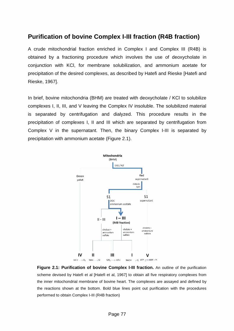
Page 77
Purification of bovine Complex I-III fraction (R4B fraction)
A crude mitochondrial fraction enriched in Complex I and Complex III (R4B) is
obtained by a fractioning procedure which involves the use of deoxycholate in
conjunction with KCl, for membrane solubilization, and ammonium acetate for
precipitation of the desired complexes, as described by Hatefi and Rieske [Hatefi and
Rieske, 1967].
In brief, bovine mitochondria (BHM) are treated with deoxycholate / KCl to solubilize
complexes I, II, III, and V leaving the Complex IV insoluble. The solubilized material
is separated by centrifugation and dialyzed. This procedure results in the
precipitation of complexes I, II and III which are separated by centrifugation from
Complex V in the supernatant. Then, the binary Complex I-III is separated by
precipitation with ammonium acetate (Figure 2.1).
Figure 2.1: Purification of bovine Complex I-III fraction. An outline of the purification
scheme devised by Hatefi et al [Hatefi et al, 1967] to obtain all five respiratory complexes from
the inner mitochondrial membrane of bovine heart. The complexes are assayed and defined by
the reactions shown at the bottom. Bold blue lines point out purification with the procedures
performed to obtain Complex I-III (R4B fraction)

Page 78
More in detail:
The frozen suspension of mitochondria is thawed and diluted to a protein
concentration of about 30 mg·ml-1 in TSH solution and then centrifuged at 32,000 g
for 30 minutes at 4°C (Beckman Avanti J25 centrifuge, JA-20 rotor). After
centrifugation, the loosely packed mitochondria pellet is suspended in TSH solution
to about 20 mg·ml-1. Sodium deoxycholate (10% w:v, pH 9.0) is added at 0.35 mg per
mg of protein followed by the addition of solid KCl to a 72 mg·ml-1 ratio.
When the KCl is completely dissolved, the suspension is centrifuged at 75,000 g for
30 minutes at 4°C (Beckman L8M ultracentrifuge, Type 50.2 Ti rotor). The green
sediment contains cytochrome c oxidase. The clear red supernatant solution is
diluted with 0.25 volumes of cold water and centrifuge as above, at 75,000 g for 30
minutes. The brown-green pellet is discarded and the supernatant, approximately
100 ml, is collected and poured into a dialysis tubing of 12-14 kDa molecular weight
cut-off, 14.3 mm diameter, and volume/length of 1.6 ml·cm-1 (Visking dialysis tubing,
Medicell International Ltd). This sample is dialyzed against 8 volumes of Tris-HCl pH
8.0 for 3 hours.
This dialysis step removes enough KCl and deoxycholate to cause the precipitation
of highly insoluble proteins, including complexes I, II, and III. Most of the cytochrome
c and other relatively soluble proteins remain in solution. The turbid dialysate is
centrifuged again at 75,000 g for 75 minutes. The supernatant is discarded and the
red pellet, hereafter erred to as S1 fraction, is suspended in approximately 5 ml of
TSH solution and stored overnight at -20°C.
Once the S1 fraction is thawed, protein concentration is determined by biuret
method. This suspension is then hand-homogenized in a glass Potter-Elvehjem tube
with a Teflon pestle and diluted with the TSH solution to a protein concentration of 10
mg·ml-1. At this point, potassium deoxycholate is added to a concentration of 0.5 mg
per milligram of protein followed by the addition of 50% w:v saturated ammonium
acetate at 16.5 ml per 100 ml of S1 protein solution. After being allowed to stand for
15 minutes on ice, the turbid sample is centrifuged as above, at 75,000 g for 30
minutes. The residue, a tightly brownish white pellet, will be discarded. The collected
supernatant is treated with 6.4 ml of 50% w:v saturated ammonium acetate solution

Page 79
per 100 ml of supernatant and incubated on ice for 15 minutes. The sample is
centrifuged at 75,000 g for 30 minutes.
The pellet, consisting of a loosely packed light brown material overlaying a small,
more dense, dark brown button, will be be discarded, whereas the supernatant is
collected. Afterwards, 50%w:v satured ammonium acetate is added to a ratio of 6 ml
per 100 ml solution. The sample is centrifuged as above for 30 minutes at 4°C. The
supernatant obtained is then treated with 3.2 ml of 50%w:v satured ammonium
acetate solution per milliliter of supernatant solution.
After 15 minutes on ice, the solution is centrifuged as before. The reddish brown
pellet designed as R4B fraction is suspended in approximately 2 ml of TSH solution.
Protein concentration is determined by the biuret assay. Samples are aliquoted and
stored at -80°C.

Page 80
Preparation of bovine Complex I-III proteoliposomes
Preparation of phospholipid:ubiquinone vesicles
Phospholipid vesicles are prepared according to a modified version of Degli Esposti
et al [Degli Esposti et al, 1981]:
Dried soybean L- -phosphatidylcholine is dissolved in petroleum ether at 0.3 g·ml-1
final concentration. Then, 20 mM Coenzyme Q10 (CoQ10) in absolute ethanol is
added to a 30 nmole CoQ10 per mg phospholipid ratio. Once the mixture is thoroughly
mixed, the organic solvent is removed, to yield a lipid film: for small volumes ( 1 ml),
the solvent is evaporated to dryness under nitrogen stream.
In aqueous solutions phospholipid molecules spontaneously form self-closed
spherical or oval structures where several phospholipid bilayers entrap part of the
solvent in their interior [Bangham and Horne, 1964. Lasic, 1988]. Thus, multilamellar
vesicles (MLVs) are prepared by the swelling of dry phospholipid:ubiquinone films
deposited in the walls of the tube. The hydration is accomplished simply by adding an
aqueous solution (50 mM KCl, 10 mM Tris, 1 mM EDTA, pH 7.4) at 30 mg
phospholipid·ml-1 under vigorous mixing and keeping on ice for 10 minutes.
Small unilamellar vesicles are formed when the MLVs dispersion is subjected to
ultrasonic irradiation (Figure 2.2)
[Saunders et al, 1962. Papahadjopoulos and Miller, 1967. Papahadjopoulos and
Watkins, 1967] at 30-second periods with 30-second intervals for 10 minutes at 150
W with a probe tip sonicator (Labsonic U2000, B. Braun Biotech International
GmbBH). In order to prevent overheating and lipid peroxidation, sonication is
performed under a nitrogen flux in an ice-water bath. Under these conditions the
phospholipid vesicles are monolamellar with a diameter of 350 - 400 Å, as controlled
by electron microscopy [Degli Esposti et al, 1981].
The vesicles (PL-vesicles) are stored at 4°C and used within 2 days from
preparation.

Page 81
Figure 2.2: Liposomes preparation. Liposomes (lipid vesicles) are formed when thin lipid films are hydrated and stacks of liquid crystalline bilayers become fluid and swell.
The hydrated lipid sheets detach during agitation and self-close to form large, multilamellar vesicles (LMV) which prevents interaction of water with the hydrocarbon core of
the bilayer at the edges. Once these particles have formed, reducing the size of the particle requires energy input in the form of sonic energy (sonication) or mechanical
energy (extrusion).
sonication

Page 82
Proteoliposome reconstitution
The most frequently used strategy for proteoliposome preparation involves the use of
detergents. In the standard procedure [Racker et al, 1979], proteins are first co-
solubilized with phospholipids in the appropriate detergent in order to form an
isotropic solution of lipid-protein-detergent and lipid-detergent micelles. Next, the
detergent is removed by diluting the reconstitution mixture resulting in the
progressive formation of bilayer vesicles with incorporated protein. Dilution lowers the
detergent concentration below its critical micelle concentration (cmc) and
proteoliposomes form spontaneously [Rigaud et al, 1995. Rigaud and Levy, 2003].
According with a modified procedure of Racker et al. [Racker et al, 1979],
proteoliposomes are prepared with a crude mitochondrial fraction enriched in
Complex I and Complex III (R4B fraction) [Hatefi and Rieske, 1967] and
phospholipid:ubiquinone vesicles (PL-vesicles).
Appropriate volumes of R4B sample and PL-vesicles are mixed in the presence of 17
mM n-octyl-D-glucopiranoside to a final concentration of 1.5% w:v at a protein to
phospholipid ratio specified in the legends of tables and figures.
After 10 minutes of incubation on ice, the samples are diluted 40-fold with 50 mM
KCl, 10 mM Tris, 1 mM EDTA, pH 7.4 to induce proteoliposome formation.
Reconstituted samples are centrifuged at 38,000 g for 1 hour at 4°C (centrifuge
Beckman Avanti J25, JA-18.1 rotor) to remove detergent traces.
R4B proteoliposomes are diluted to 0.5 mg·ml-1 in reaction solution pH 7.4, kept on
ice and immediately used.
Page 83
Determination of protein concentration
Ultraviolet absorbance at 280 nm (range: 0.1 – 1 mg·ml-1)
Spectrophotometric measurements of absorption radiation in the near ultraviolet 280
nm to determine the concentration of a protein solution is one of the oldest and most
widespread methods for quantitative analysis [Warburg and Christian, 1942 . Layne,
1957. Aitken and Learmonth, 2002].
This method is based on the inherent absorbance by the aromatic amino acid
residues tryptophan and tyrosine and provides a fairly sensitive and convenient
means for detecting and quantitating pure proteins or mixtures of pure proteins
having a concentration in the range of 0.1 to 1 mg·ml-1. This assay is used to
calculate the concentration either from its published absorbancy index (absorptivity)
at 280 nm or by comparison with a calibration curve prepared from measurements
with standard proteins.
The accurate determination of concentration of a standard erence is necessary to
know the concentration of an aqueous protein solution.
Bovine serum albumin (BSA) is the most commonly standard erence chosen for any
colorimetric method for protein determination in mitochondrial crude lysates and
proteins. Numerous values for the absorptivity of BSA have been reported in the
literature but assuming a molecular weight of 66,430 Dalton, the molar extinction
coefficient is 43,824 M-1·cm-1 at 280 nm. Theore, the absorptivity of BSA is ~ 0.67 for
a 1 mg·ml-1 solution at 280 nm (a280) [Gill and von Hippel, 1989].
For this procedure, quartz crystal or fused silica cuvettes are routinely used as plastic
and common glasses are not transparent to the ultraviolet radiation. According with
this, two cuvettes are first filled with a volume of water (blank) to zero the
spectrophotometer (V530 extended model, JASCO). After removing the blank from
the sample compartment, a clean cuvette is filled with a 1 mg·ml-1 BSA standard
solution and after insert it into the sample compartment, absorbance at 280 nm is
measured. The resulting media absorbance value at 280 nm is used after subtracting
the absorbance value of the blank to calculate the standard erence concentration as
Page 84
follows: concentration (mg·ml-1) = ba
Abs
280
280
where a280 is the absorbancy index of
the 1 mg·ml-1 BSA standard solution and b is the path-length in cm.
Biuret Method (range: 1 – 10 mg·ml-1)
The biuret reaction is the basis of this colorimetric method for quantitatively
determine total protein concentration of crude lysates [Gornall et al, 1949] whereby in
the presence of four or more peptide bonds, cupric ions (Cu2+) form a violet-colored
coordination complex in an alkaline environment [Rose, 1853].
The biuret reagent contains hydrated copper (II) sulfate and sodium hydroxide,
together with sodium potassium tartrate and reacts with peptide bonds of proteins
producing a light blue to deep purple color, with maximum absorbance (max) at 540
nm.
The intensity of the color produced is proportional to the number of peptide bonds
participating in this reaction. The working range for this assay is from 1 to 10 mg
protein·ml-1. Over this concentration range, the measured absorption is linear
according to the Lambert-Beer law [Gornall et al, 1949].
The following procedure is a modification of the original protocol of Gornall et al
[Gornall et al, 1949] which involves protein solubilization by lipid extraction of the
mitochondrial membranes with sodium deoxycholate, and rapid development of the
blue copper-protein complex on heating [Beyer, 1983] and using bovine serum
albumin (BSA) as standard.
Protein and standard samples are prepared as duplicates in test tubes containing c.a
1 mg protein (adjusting with water to 1.5 ml final volume), 0.1 ml of 10% w:v sodium
deoxycholate (pH 8.0), and1.5 ml of biuret reagent. Samples blanks are prepared in
the same way but containing suspension buffer or water instead of protein.
After vortex mixing, samples are placed in a boiling water bath for 30 seconds until
full color development, and once rapidly cooled at room temperature, absorbance is
read at 540 nm.

Page 85
The resulting average absorbance values for any sample is used after subtracting the
absorbance value of the blank and the unknown sample concentration is calculated
by the following equations:
(BSA)Abs
mass(protein)Absmass
540
[mg]BSA
540[mg] protein sample
[ml] protein sample
[mg] protein sample1-
volume
massml[mg ionconcentrat ]
Lowry Method (range: 0.01 – 0.1 mg·ml-1)
The Lowry method is a colorimetric assay to determine concentration from purified
and partially purified proteins, including highly diluted protein solutions.
This method relies on two different steps. Initially, the biuret reaction involves the
interaction of cupric ions with the imide form of the polypeptide in alkaline solution
followed by the enhancement step, the reduction of the Folin-Ciocalteau reagent
[Lowry et al, 1951. Folin and Ciocalteau, 1927] generating a characteristic blue color
with maximum absorbance (max) at 750 nm.
The mixed acid (phosphomolybdate and phosphotungstate) of the Folin-Ciocalteau
reagent is reduced by a rapid reaction with tyrosine and tryptophan amino acids
residues. Cupric ions enhances color formation by chelation with the imide form of
the peptide.
The reaction results in a strong blue color, which depends on protein sequence
variation as color depends not only to the reduced copper-amide bond complex but
also to tyrosine, tryptophan, and to a lesser extent cystine, cysteine, and histidine
residues [Peterson, 1977 . Peterson, 1979].
This assay uses bovine serum albumin (BSA) as erence standard. The calibration
curve consists in serial dilutions from 0 to 40 g in 1 ml of distilled water of 1 mg·ml-1
BSA aqueous solution whose concentration is previously determined at 280 nm.
The Lowry reagent is prepared immediately before use by mixing the three stock
solutions A, B, and C in the proportion 100:1:1 (v:v:v) respectively.
Protein samples are prepared at least by duplicate containing about 20 to 30 g
protein, adding 0.1 ml of 10% w:v sodium deoxycholate and completing volume with

Page 86
water to 1 ml. Blanks are prepared in the same way but using the sample suspension
buffer instead of protein. Straight forwards, 2 ml of the Lowry reagent is added to
each tube and after mixing, are allowed to stand for 10 minutes at room temperature
in the darkness. Subsequently, 0.20 ml of 2-fold diluted Folin-Ciocalteau reagent is
added very quickly while vortex mixing considering that this reagent is only reactive
for a short time under alkaline conditions.
After 30 minutes at room temperature in the darkness, the absorbance of samples
and blanks are read at 750 nm.
For the calibration curve, it is necessary to subtract blank absorbance values to the
standard values and the resulting mean absorbance values for each standard
samples are plotted with the vertical axis in units of absorbance at 750 nm and
horizontal axis in units of protein mass (g or mg).
The unknown protein sample concentrations are calculated from this calibration
curve considering their mean absorbance value at 750 nm after subtracting their
respective blank absorbance values.

Page 87
Enzyme activities
Complex I enzymatic activity (NADH:ubiquinone reductase), integrated Complex I+III
activity (NADH:cytochrome c reductase) and aerobic NADH oxidation are assayed at
30°C in a dual-beam, dual-wavelength spectrophotometer (V550 extended model,
JASCO) equipped with a mini-magnet cuvette stirrer.
The initial rates are obtained as first derivatives calculated from the resulting
absorbance vs. time curve with an appropriate software (Spectra Manager™,
JASCO) and expressed in moles of NADH·min-1·mg-1.
Samples are diluted to 40 g·ml-1 for BHM or SMP and 14 g·ml-1 for R4B
proteoliposomes in reaction solution, pH 7.4.
NADH:ubiquinone oxidoreductase. Complex I activity is measured
essentially as described in Lenaz et al. and Ventura et al. [Lenaz et al, 2004 .
Ventura et al, 2002]. Samples are incubated for 5 minutes with 2 M antimycin A and
1 mM KCN to inhibit electron transfer towards Complex III and IV. In the presence of
60 M decylubiquinone (DB) as electron acceptor, the rate of electron transfer is
measured after the addition of 75 M NADH. The time-course decrement of NADH
absorbance is followed at a wavelength of 340 nm minus 380 nm (absorption
coefficient 3.5 mM-1·cm-1).
NADH oxidase. Aerobic NADH oxidation is assayed in a similar way, but both
electron acceptor (DB) and inhibitors are omitted from the assay mixture.
NADH:cytochrome c reductase. Integrated Complex I+III activity is measured
according to Battino et al. [Battino et al, 1991] in the reaction medium with the
addition of 1 mM KCN to inhibit Complex IV. The time-course increase absorbance
by the reduction of 50 M cytochrome c is monitored at a wavelength of 550 nm
minus 540 nm (absorption coefficient 19.1 mM-1·cm-1) after the addition of 75 M
NADH as electron donor.

Page 88
Measuring Reactive Oxygen Species
ROS is an acronym for reactive oxygen species, a variety of intermediates relatively
stable produced by the univalent reduction of molecular oxygen.
Superoxide detection. The superoxide anion radical is produced by
autooxidation of mitochondrial redox groups such as iron-sulfur clusters and
semiquinone radicals [Raha and Robinson, 2000 Turrens, 1997. Turrens, 2003].
The determination of the rate production of O2●- is based upon the
spectrophotometric measurement of oxidation or reduction of a probe in which O2●- is
a reactant. The concentration of the spectrophotometric indicator that reacts with
superoxide anion is adjusted to compete effectively with the spontaneous dismutation
of O2●- so that nearly all superoxide anion produced can be detected. According with
this approach, native ferricytochrome c is an excellent quantitive indicator for O2●-:
2c) chrome(ferrocyto
2-
2c) chrome(ferricyto
3 O c cytFe O c cytFe
However, when exogenous ferricytochrome c is added to mitochondria membrane
preparations, it is more effectively reduced by the electron chain cytochrome c
reductase than by superoxide. To overcome this limitation and enhance specificity of
this assay for O2●-, it is known that the acetylation of some lysine residues of
ferricytochrome c decreases enzymatic direct electron transfer while preserving the
capacity of being chemically reduced [Azzi et al, 1975. Boveris, 1984].
It is theore advisable to use acetylated cytochrome c that, although not as rapidly
reduced by superoxide as native cytochrome c, is much more slowly reduced by the
reductase activity of the mitochondrial membranes and is not efficiently oxidized by
cytochrome oxidase [Azzi et al, 1975].

Page 89
In general, acetylated cytochrome c have to be in excess so that the rates of
reduction and oxidation can be treated as zero order and depend only on the rate of
superoxide production [Dröse et al, 2009]. The superoxide-dependent rate can be
deduced as the difference between the reduction rates in the absence and presence
of superoxide dismutase (SOD) as the reduction of both native and acetylated
cytochrome c are highly sensitive to this enzyme.
Nevertheless, when studying the production of superoxide mediated by Complex I is
suggested to functionally isolate Complex I from the downstream segments of the
respiratory chain by adding mucidin, an inhibitor that blocks Complex III activity and
simultaneously prevents the superoxide production from this complex and potassium
cyanide that competitively inhibits the cytochrome c oxidase activity [Lenaz et al,
2004].
One should, however, be aware that cyanide inhibits also the Cu,Zn-SOD, theore the
Mn-enzyme should be perred when cyanide is present in the reaction medium
[Boveris, 1984 . Turrens and Boveris, 1980].
According with these considerations, in the present study, the measurement of
superoxide production mediated by Complex I in the R4B proteoliposome is obtained
essentially as described by Lenaz et al [Lenaz et al, 2004]:
All samples are diluted in reaction medium to a final concentration of 14 g
protein·ml-1 in the presence of 1.8 M mucidin and 4 M rotenone.
The reduction of 50 M acetylated cytochrome c is followed spectrophometrically
with a dual-beam, dual-wavelength spectrophotometer (V550 extended model,
JASCO) at 550 - 540 nm (absorption coefficient 19.1 mM-1·cm-1) at 30°C after the
addition of 75 M NADH as electron donor.
The addition of 40 to 50 U/ml Cu,Zn-superoxide dismutase gives the SOD-sensitive
rate of acetylated cytochrome c reduction, which gives the stoichiometric rate of O2●-.
According to the method [Azzi et al, 1975. Boveris, 1984], data are shown as the
rate of SOD-sensitive and –insensitive reduction of acetylated cytochrome in
nmole·min-1·mg-1.

Page 90
Hydrogen peroxide detection. Due to the rapidly spontaneous and enzyme-
catalyzed dismutation of superoxide to hydrogen peroxide its detection is generally
more arduous than hydrogen peroxide [Murphy, 2009].
Consequently, the rate of H2O2 production can be used as an indirect measurement
of O2●- formation, recognizing that such measurements will also lect direct divalent
reduction of molecular oxygen to H2O2.
Fluorescent-based methods to detect ROS formation in mitochondria were very early
introduced in the literature [Hinkle et al, 1967. Black and Brandt, 1974], particularly
the oxidation of the leuco probe 2’,7’-dichlorodihydrofluorescein diacetate (DCFDA)
to the fluorescent compound dichlorofluorescein (DCF) was initially thought to be a
relatively specific indicator of H2O2 [Brandt and Keston, 1965 . Keston and Brandt,
1965].
DCFDA is a non-polar, reduced, colorless and non-fluorescent dye that can diffuse
through lipid membranes. This probe is rapidly cleaved by cellular esterases, which
are also present in mitochondrial preparations, resulting into the polar, non-
fluorescent compound 2’,7’- dichlorodihydrofluorescein (DCFH2).
It has been demonstrated that mitochondria and submitochondrial preparations are
able to deacetylate the probe and oxidize it by ROS [Degli Esposti, 2002].
Nevertheless, when the assay is performed to test the ROS production of purified
proteins as R4B fraction, DCFDA requires chemical hydrolysis immediately before
use (see below).
In the presence of hydrogen peroxide, DCFH2 is oxidized to 2’,7’- dichlorofluorescein
(DCF) which emits a strong fluorescence.

Page 91
In the present study, hydrogen peroxide production by Complex I is measured
according with the method of Fato et al [Fato et al, 2009]:
All samples are diluted in reaction medium to a final concentration of 0.5 mg
protein·ml-1 in the presence of 1.8 M mucidin and 4 M rotenone when indicated
and 5 M DCFDA.
Each well of a 96-well microtiter plate, optically suitable for fluorescence
measurements is filled with the reaction mixture.
The reaction is started with the addition of a large excess of electron donor (150 M
NADH) aimed to guarantee not to consume all the substrate during the 45 min of the
assay.
The fluorescence readings are obtained with an excitation / emission wavelength of
485 / 535 nm at 25°C in a fluorescence plate reader (Wallac Victor2 1420 Multilabel
counter; PerkinElmer).
The fluorescence intensity change is followed every 5 min, and the results are
calculated as the difference of fluorescence units at 45 min minus zero time and
given as mean ± S.D. of at least three independent determinations. Interference of
NADH, rotenone, mucidin, and other substrates with the background fluorescence of
DCF is evaluated in blank samples.
Preliminary laboratory tests include the direct effect of enhancing the fluorescence of
the probe by adding 5 and 50 M H2O2 to exclude the possibility that the
fluorescence signal produced by the samples are underestimated by a lack of DCF.
As mentioned before, DCFDA is spontaneously deacetylated in mitochondria
preparations [Fato et al, 2009] whereas R4B proteoliposomes require alkaline
hydrolysis of DCFDA prior to assay.
According to this, 10 l of DCFDA stock solution are mixed with 40 l of 0.01 N
NaOH for 30 min in the dark at room temperature [Cathcart et al, 1983]. The mixture
is neutralized with 200 l of 10 mM KCl, 1 mM EDTA, 25 mM Tris-HCl pH 7.4 and
kept on ice in the dark and immediately used.

Page 92
Protein electrophoresis analysis
Electrophoresis is a separation technique based on the mobility of charged
molecules in an electric field. It is used mainly for the analysis and purification of
biological macromolecules such as proteins and nucleic acids.
Polyacrylamide gel electrophoresis (PAGE) is most commonly used for separation of
proteins where polyacrylamide gel acts as an anticonvective size-selective sieve with
defining pores saturated with an electrically conductive buffered solution of salts.
The size pores of these gels are large enough to admit passage of the migrating
macromolecules as they are forced through the gel by an applied voltage, larger
molecules are retained in their migration more than smaller molecules.
In an electric field, proteins move toward the electrode of opposite charge. The rate
of migration of the macromolecules through the polyacrylamide gel depends upon a
complex relationship between the physical characteristics of both the electrophoresis
system and the macromolecules. Factors affecting protein electrophoresis include
the strength of the electric field, the porosity of the gel, the temperature of the
system, the pH, ion type, and concentration of the buffer as well as the size, shape,
and charge density of the proteins [Garfin, 1990].
In most PAGE units, the gel has a vertical orientation and is mounted between two
buffer chambers that the only electrical connection between the anode (+) and
cathode (-) chambers is through the gel. An electric potential is applied across the
buffer chambers forcing the sample proteins to migrate into and through the gel
toward the bottom [Shi and Jackowski, 1998] (Figure 2.3).

Page 93
Polyacrylamide gels are chemically inert, electrically neutral, hydrophilic and optically
transparent for detection at wavelengths greater than 250 nm. These characteristics
make polyacrylamide ideal for protein separations because the matrix does not
interarct with the solutes and has a low affinity for common protein stains [Garfin,
2009].
These gels are formed by copolymerization of acrylamide and N,- ’-methylene-bis-
acrylamide (bis-acrylamide, cross-linker).
The reaction is a vinyl addition polymerization initiated by a free radical-generating
system. Polymerization is initiated by a chemical initiator and a catalyst (e.g.
ammonium persulfate (APS) and tetramethylethylenediamine (TEMED)).
The elongating polymer chains are randomly crosslinked by bis-acrylamide, resulting
in a gel with a characteristic porosity which depends on the polymerization conditions
and monomer concentrations.
Polymerization rates depend on monomer and catalyst concentration, temperature
and purity of reagents.
Figure 2.3: Electrophoresis workflow. Electrophoresis involves the selection of the appropriate
method, instrumentation, and reagents for the intended experimental goal. Once proteins are
separated, they are available for a number of downstream applications, including enzymatic assays,
further purification, transfer to a membrane for immunological detection (immunoblotting or western
blotting), and elution and digestion for mass spectrometric analysis.

Page 94
The porosity of the gel is characterized by two parameters: total monomer
concentration (%T) and weight percentage of cross-linker (%C). By varying these two
parameters, the pore size of the gel can be optimized to yield the best separation and
resolution for the proteins of interest. %T indicates the relative pore size of the
resulting polyacrylamide gel; a higher %T ers to a larger polymer-to-water ratio and
smaller average pore sizes.
Single-percentage gels are used to separate bands that are close in molecular
weight. Since optimum separation occurs in the lower half of the gel, it is necessary
to choose a percentage in which the protein of interest migrates to the lower half of
the gel.
Gradient-gels, by the other side, are used to separate samples containing a broad
range of molecular weights. These gels allow resolution of both high- and low-
molecular weight bands on the same gel.
The pH and ionic composition of the buffer system determine the power requirements
and heavily influence the separation characteristics of a polyacrylamide gel. Buffer
systems include the buffers used to cast the gel (gel buffer), prepare the sample
(sample buffer), and fill the electrode reservoirs (running buffer).
Two types of buffer systems are commonly used: continuous buffer systems use the
same buffer, at constant pH, in the gel, sample, and electrode reservoirs [McLellan,
1982] and discontinuous buffer systems use a gel separated into two sections (a
large-pore stacking gel on top of a small-pore, the resolving gel) and different buffers
in the gels and electrode solutions [Wheeler et al, 2004].
Most common PAGE applications utilize discontinuous buffer systems, where two
ions differing in electrophoretic mobility form a moving boundary when a voltage is
applied [Niepmann, 2007].

Page 95
First dimension: Blue-native polyacrylamide gel electrophoresis
Blue-native polyacrylamide gel electrophoresis (BN-PAGE) has been developed for
the isolation of native membrane proteins in the mass range of 10 kDa to 10 MDa at
a fixed pH of 7.5 [Schägger and von Jagow, 1991. Schägger et al, 1994]. Even the
original protocol and related techniques have been improved and expanded
considerably [Schägger, 2001. Wittig et al, 2006b], the basic principles for BN-PAGE
however, are unchanged.
Mild neutral detergents are used for solubilization of biological membranes. In
principle, any neutral detergent can be used for solubilization of biological
membranes for BN-PAGE if the detergent can solubilize the desired protein and keep
it in the native form. The choice of a specific nonionic detergent depends on its
delipiding properties for a protein of interest.
The very mildest detergent digitonin has been used to isolate supramolecular
associations of multiprotein complexes, thus identifying physiological protein-protein
interactions without using chemical crosslinking.
Dodecyl--D-maltoside (dodecylmaltoside, DDM) by the other side, has stronger
delipidating properties compared to digitonin. It is a mild neutral detergent that is
suited for isolation of individual complexes [Schägger and von Jagow, 1991.
Schägger et al, 1994] (Figure 2.4)
Coomassie blue G 250 is added to the solubilized proteins prior to loading the
samples onto the gel. This dye is sufficiently soluble in water, but it can also bind to
membrane proteins because of its hydrophobic properties.
Coomassie blue is also added in the cathode buffer to provide a continuous flow of
this dye into the gel.
Membrane proteins and proteins with significant surface-exposed hydrophobic area
bind Coomassie blue G-250 nonspecifically, acquiring overall negative charges.
Consequently these proteins repel each other minimizing considerably the tendency
of protein aggregation. Furthermore, these dye-bound proteins are water-soluble, this
means that there is no need for the presence of detergent in the polyacrylamide gel,
and the risk of denaturation of detergent-sensitive membrane-proteins is also
minimized.

Page 96
Binding of a large number of anionic dye molecules shifts the isoelectric point of
proteins that causes even basic proteins to migrate in one direction towards the
positive anode at pH 7.5 [Schägger et al, 1994].
In this way, proteins will not be separated according to the charge/mass ratio but
according to size in acrylamide-gradient gel, which leads to a mass-dependent
reduction of the protein migration velocity and to an almost complete stop at a mass-
specific pore size limit during BN-PAGE. Native proteins, stained with Coomassie,
are visible as blue bands through the gel and this staining facilitates excision of
specific bands.
The potential of this technique to determine native molecular masses and oligomeric
states identifying physiological protein-protein interactions led to demonstrate in 2000
that yeast and mammalian respiratory chains exist as a network of supercomplexes,
and that detergent-sensitive multiprotein complexes could be isolated without
dissociation of detergent-labile subunits [Schägger and Pfeiffer, 2000] (Figure 2.2).
In the present study, separation of supercomplexes and single respiratory complexes
is achieved according to a modified version from Beardslee and Updyke [Beardslee
and Updyke, 2007], buffers and solutions used for BN-PAGE are listed in Table 2.1.
In detail:
Bovine mitochondria or R4B proteoliposomes samples are centrifuged 10 minutes at
20,000 g at 4°C to remove suspension buffer.
Pellets are suspended in Sample Buffer at 4 mg·ml-1. Digitonin or dodecylmaltoside is
added at detergent:protein ratios given in Table 2.2 when indicated and mixed by
pipetting up and down.
Protein extracts are incubated on ice for 20 minutes before centrifuging 10 minutes at
10,000 g at 4°C (centrifuge Beckman Avanti J25, JA-18.1 rotor).
Pellets are discarded and supernatants are aliquoted and kept on ice until use. Just
before loading onto the gel, 5% w:v Coomassie blue G-250 solution is added to the
samples so that the final concentration of G-250 is one-tenth that of the detergent
concentration.

Page 97
Prior to loading into the gel, glycerol is added to the samples at a final concentration
of 10% (v:v) in order to increase the density and facilitate sample application.
The samples are loaded onto precast 10-wells 3%-12% T gradient gels
( ativePAGE™ ovex® Bis-Tris Gel, Invitrogen) which are a 1.0 mm tick, 8 x 8 cm
mini gels that resolve proteins in the molecular weight range of 30 - 10,000 kDa.
Electrophoresis is performed at 4°C using a vertical apparatus (XCell SureLock TM
Mini-Cell Electrophoresis System, Invitrogen) and Bis-Tris Gel system buffers
( ativePAGE™ ovex® Bis-Tris Gel System, Invitrogen).
Samples are loaded up to 25 l per well. An effort is also made to minimize the
amount of time between loading sample onto the gel and beginning electrophoresis.
Five microliters of unstained native protein standards comprising IgM hexamer (1,236
kDa), IgM pentamer (1,048 kDa), Apoferritin band 1 (720 kDa), Apoferritin band 2
(480 kDa), B-phycoerythrin (242 kDa), Lactate Dehydrogenase (146 kDa), Bovine
Serum Albumin (66 kDa), and Soybean Trypsin Inhibitor (20 kDa) are used as
molecular weight markers on the gels (NativeMarkTM unstained protein standard,
Invitrogen).
The run is started with the Deep Blue cathode buffer and once the dye front migration
is one-third of the way down the gel the run is paused, this cathode buffer is removed
with a serological pipette and replaced by the Slightly Blue cathode buffer before
resuming the run.
Electrophoresis is performed at 150 V constant for 60 minutes, then the voltage is
increased to 250 V constant for the remainder of the run (30 - 90 minutes running at
4°C).

Page 98
Table 2.1: Gel buffer system formulation for BN-PAGE
Bis-Tris CHAPS/Tricine-BN PAGE (a)
Sample Buffer
50 mM NaCl, 5 mM 6-aminocaproic acid,
50 mM imidazole/HCl pH 7.0
Gel Buffer
50 mM BisTris/HCl, pH 7.0
0.3 mM CHAPS
Anode Buffer 50 mM Tricine, 50 mM Bis-Tris, pH 6.8
Deep blue cathode buffer
50 mM Tricine, 50 mM Bis-Tris, pH 6.8
0.02% w:v Coomassie blue G-250
Slightly blue cathode buffer 50 mM Tricine, 50 mM Bis-Tris, pH 6.8
0.002% w:v Coomassie blue G-250
(a) [Beardslee and Updyke, 2007]
Table2.2: Quantity of detergent required to solubilize membrane proteins
BHM (200 g)
R4B proteoliposomes (80 g)
Dodecylmaltoside (10% w:v)
5.2 l (2.6 g/g)
3.4 l (4.3 g/g)
Digitonin (10% w:v)
16 l (8 g/g)
6.4 l (8 g/g)
The volumes of added detergent and the detergent / protein ratios chosen (w:w), indicated in brackets are shown for solubilization of bovine heart mitochondria (BHM) and R4B proteoliposomes (Complex I+III proteoliposomes from BHM)

Page 99
After BN-PAGE, individual lanes are cut out and processed by an orthogonal second-
dimension electrophoresis under denaturing conditions.
The second dimension is performed by sodium dodecyl sulfate polyacrylamide gel
electrophoresis (SDS-PAGE). In this way, the mitochondrial membrane protein
complexes separated as supramolecular assemblies during the first dimension
electrophoresis are resolved in the second dimension into their individual polypeptide
subunits (2D BN/SDS-PAGE).
After immunodetection with specific antibodies, 2D BN/SDS-PAGE provides an
valuable analytical method for the determination of molecular mass and oligomeric
state of complexes, and characterization of stoichiometric associations between
protein complexes (Figure 2.5).
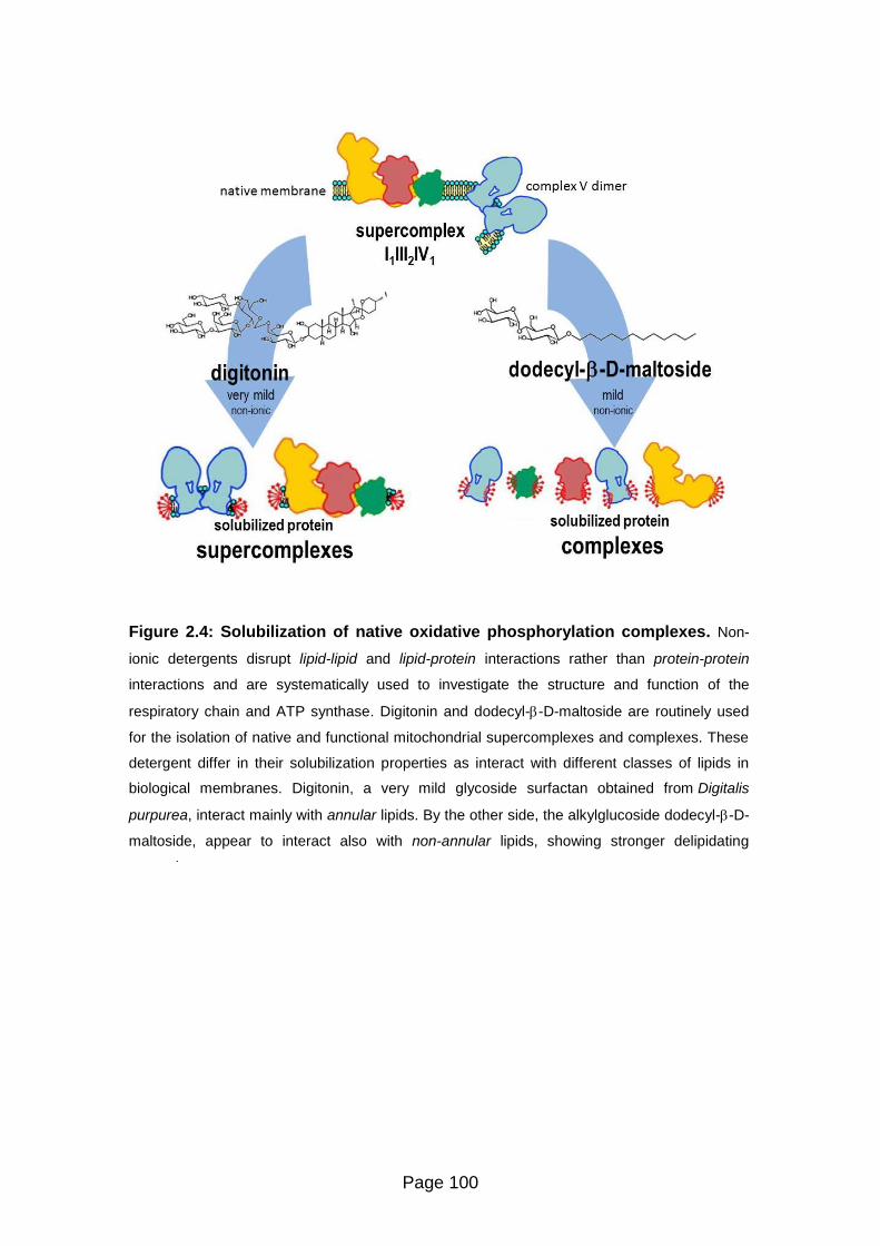
Page 100
Figure 2.4: Solubilization of native oxidative phosphorylation complexes. Non-
ionic detergents disrupt lipid-lipid and lipid-protein interactions rather than protein-protein
interactions and are systematically used to investigate the structure and function of the
respiratory chain and ATP synthase. Digitonin and dodecyl--D-maltoside are routinely used
for the isolation of native and functional mitochondrial supercomplexes and complexes. These
detergent differ in their solubilization properties as interact with different classes of lipids in
biological membranes. Digitonin, a very mild glycoside surfactan obtained from Digitalis
purpurea, interact mainly with annular lipids. By the other side, the alkylglucoside dodecyl--D-
maltoside, appear to interact also with non-annular lipids, showing stronger delipidating
properties.
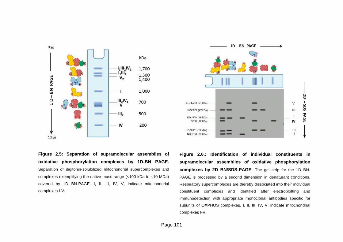
Page 101
Figure 2.5: Separation of supramolecular assemblies of
oxidative phosphorylation complexes by 1D-BN PAGE.
Separation of digitonin-solubilized mitochondrial supercomplexes and
complexes exemplifying the native mass range (<100 kDa to 10 MDa)
covered by 1D BN-PAGE. I, II. III, IV, V, indicate mitochondrial
complexes I-V.
Figure 2.6.: Identification of individual constituents in
supramolecular assemblies of oxidative phosphorylation
complexes by 2D BN/SDS-PAGE. The gel strip for the 1D BN-
PAGE is processed by a second dimension in denaturant conditions.
Respiratory supercomplexes are thereby dissociated into their individual
constituent complexes and identified after electroblotting and
immunodetection with appropriate monoclonal antibodies specific for
subunits of OXPHOS complexes. I, II. III, IV, V, indicate mitochondrial
complexes I-V.

Page 102
Second dimension: Sodium dodecyl sulfate polyacrylamide gel electrophoresis
Sodium dodecyl sulfate polyacrylamide gel electrophoresis (SDS-PAGE) is a
valuable means for separation of proteins.
The original discontinuous gel system was developed by Ornstein and Davis
[Ornstein, 1964. Davis, 1964] for the separation of serum proteins in native
conditions [Vavricka et al, 2009]. Laemmli modified this protocol by incorporating the
detergent sodium dodecyl sulfate (SDS), creating what has become the most popular
form of protein electrophoresis, SDS-PAGE [Laemmli, 1970]. When proteins are
separated in the presence of the anionic detergent SDS and reducing agents, they
become fully denatured and dissociate into their constituting subunits.
SDS is an anionic detergent that binds non-covalently to proteins in a manner that
imparts an overall negative charge on the proteins. Indeed, SDS binds at a
consistent rate of 1.4 g of SDS per 1 g protein (a stoichiometry of about one SDS
molecule per two amino acids), theore it imparts a similar charge-to-mass ratio for all
proteins in a mixture.
The electrostatic repulsion that is created causes proteins to lose their native
conformation. The disruption of non-covalent bonds leads proteins to unfold into a
rod-like shape thereby eliminating differences in shape which is an important factor
for separation of these molecules in the gel.
The addition of a reducing agent in the sample buffer, such as dithiothreitol
(DTT) or 2-mercaptoethanol (-mercaptoEtOH), additionally denatures the proteins
by reducing disulfide linkages, thus overcoming some forms of tertiary protein folding,
and breaking up quaternary protein structure (oligomeric subunits).
Consequently, the rate at which SDS-bound proteins migrate in a polyacrylamide gel
depends primarily on their size, enabling molecular weight estimation.

Page 103
The 2D SDS-PAGE procedure for mitochondrial complexes in the present study is
performed with the Bis-Tris/ MOPS buffer system (NuPAGE™ ovex® Bis-Tris Gel
System buffers, Invitrogen).
The BisTris/MOPS-SDS-PAGE was developed by Updyke and Sheldon [Updyke and
Engelhorn, 2004]. It is a discontinuous buffer system that involves chloride as the
leading ion present in the gel buffer and 3-(N-morpholino) propanesulfonate (MOPS)
as the trailing ion in the running buffer. Bis (2-hydroxyethyl) imino-tris
(hydroxymethyl) methane (Bis-Tris) cation is the common ion present in the gel and
running buffers.
The combination of the lower pH gel buffer (pH 6.4) and the running buffer (pH 7.3 -
7.7) results in a significantly lower operating pH of 7 during electrophoresis (buffers
and solutions are listed in Table 2.3). In detail:
Individual strip lanes from BN-PAGE with the protein complexes under study are
excised (0.5 cm-broad strips).
Then, these gel strips are successively incubated for 15 minutes at room
temperature, first with 50 mM DTT to reduce disulfide bonds between cystines, then
with 50 mM N,N-dimethylacrylamide to alkylate the reduced cystines, and afterward
with 5 mM DTT to quench the reaction.
The equilibrated strips are loaded onto 2-D well precast 4%-12% T gradient gels
(NuPAGE™ ovex® Bis-Tris Gel, Invitrogen) which are a 1.0 mm tick, 8 x 8 cm mini
gels that resolves proteins in the molecular weight range of 10 - 200 kDa.
Electrophoresis is performed at 4°C using a vertical apparatus (XCell SureLock TM
Mini-Cell Electrophoresis System, Invitrogen).
Five microliters of an unstained protein standards in the range of 20 - 220 kDa
comprising recombinant proteins containing repetitive units of a fusion protein which
contain an IgG binding site (MagicMark™ XP Western Protein Standard, Invitrogen).
This standard marker allows visualization of the protein standard bands using the
same reagents and protocols for the target proteins by immunodetection.
Electrophoresis is performed at 200 V constant for 50 minutes at 4°C.

Page 104
Table 2.3: Gel buffer system formulation for SDS-PAGE
Bis-Tris/MOPS-SDS PAGE (a)
Sample Buffer
25 mM Tris-HCl, 35 mM Tris base, 0.5 % w:v LDS,
2.5% v:v glycerol,0.127 mM EDTA, 0.004% w:v SERVA Blue G250, 0.0015% w:v phenol red
Reducing Solution 50 mM DTT in Sample Buffer
Alkylating Solution
50 mM N,N-dimethylacrylamide in Sample Buffer
Quenching Solution
5 mM DTT in Sample Buffer
Gel Buffer
0.375 M Bis-Tris/HCl, pH 6.5
Running Buffer
2.5 mM MOPS, 2.5 mM Bis-Tris (or Tris), pH 7.7, 0.005% w:v SDS
and 0.05 mM EDTA
(a) [Updyke and Engelhorn, 2004]
Protein Immunoblotting
On completion of the separation of proteins by 2D BN/SDS-PAGE, the next step is
detecting and identifying protein subunits with the aim to establish specific protein-
protein associations between mitochondrial complexes.
Immunoblotting (western blotting), was developed as a result of the need to probe
proteins that were inaccessible to antibodies while embedded in the polyacrylamide
matrix. This method is based on building an antibody:protein complex via specific
binding of antibodies to proteins immobilized in a membrane of nitrocellulose and
detecting the bound antibodies. Several detection methods are available [Towin et al,
1979. Brunette, 1981].
Immunoblotting involves a first step, the transfer phase which means moving the
proteins from the polyacrylamide gel onto a solid support membrane and
immobilizing them at their respective relative migration positions at the time point
when the electric current of gel was stopped (blotting).
The most commonly support membranes used are nitrocellulose or polyvinylidene
difluoride (PVDF).

Page 105
The second phase, detection, entails probing the membrane with either a primary
antibody specific to the protein/s of interest and subsequent visualization of the
labeled proteins.
Blotting
Wet electrotransfer relies on the same electromobility principles that drive the
migration of proteins during separation in PAGE: an electric field is used to eluate
proteins from gels and transfer them to membranes where they are captured in a
pattern that perfectly mirrors their migration positions of the separated protein in the
gel.
The membrane and protein-containing gel, together with filter paper, are assembled
in a sandwich. A non-conducting cassette holds the membrane in close contact with
the gel and the cassette assembly is placed between two electrodes, transverse to
the electrical field and fully immersed in a conducting transfer buffer [Towin et al,
1979. Brunette, 1981] (Figure 2.7).
A current is applied by a voltage across the electrodes in the direction of the gel to
the membrane following Ohm’s law. The electric field strength that is generated
between the electrodes is the driving force for transfer.
There are practical limits on field strength, however due to the production of heat
during transfer. The heat generated during transfer increases temperature and
decreases resistance of the transfer buffer.
Such changes in resistance may lead to inconsistent field strength and may cause
the transfer buffer to lose its buffering capacity. In addition, excessive heat may
cause the gel to deteriorate and stick to the membrane.
Additionally, most buffers become heated, increasing in temperature to a point where
proteins may be irreversibly damaged. It is theore important to start the transfer
process using cooled buffer and to maintain a low temperature.
Although the large volumes of buffer in the tank dissipate the heat generated and
provide the conducting capacity for extended transfer conditions, wet transfer
requires cooling of the tank blotter system and internal recirculation of the transfer
buffer by the presence of a stirring magnet.

Page 106
Another common procedure is to perform the entire wet transfer in a 4°C
environment, such as a cold room keeping the blotting tank on ice.
Transfer buffer should act as an electrically conducting medium in which proteins are
soluble and that does not interfere with binding of the proteins to the membrane.
Most transfer buffers contain methanol.
The addition of methanol is necessary to achieve efficient binding to the membrane,
particularly nitrocellulose membranes [Gershoni and Palade, 1982].
Buffers containing methanol may deteriorate if stored for long periods, theore
methanol is added just prior to transfer.
In addition to buffer characteristics such as pH, salt type, salt concentration, and the
presence of detergents, the degree to which molecules bind to a membrane is
influenced by the physical and chemical characteristics of the membrane itself.
Membranes are porous materials with pore sizes from 0.2 to 0.45 m in diameter.
The binding capacity of a membrane depends primarily on the pore size.
Nitrocellulose and PVDF membranes are the most common types of membranes
used for Western blotting. Nitrocellulose membranes are the most frequently used
and their main advantage is a tendency to have low background.
The exact mechanism by which biomolecules interact with the membrane is not
known, but it is assumed to be a combination of non-covalent and hydrophobic
forces.

Page 107
A B
C
Figure 2.7: Protein blotting workflow. A) Once proteins are separated by
electrophoresis, they are available to electrophoretic transfer onto a nitrocellulose
membrane (blotting) for further immunological detection. B) Gel and membrane setup
for blotting. The cassette holds the gel and membrane while fiber pads and filter
paper on both sides provide complete contact within the gel sandwich. C) Transfer
sandwich assembly. The cassete is inserted vertically in the buffer tank.

Page 108
In the present study, 2D BN/SDS-PAGE gels with separated mitochondrial
complexes are blotted as follows:
For each gel, one piece of nitrocellulose membrane (Amersham Hybond ECLTM, GE
Healthcare) and two pieces of filter paper (Whatman® 3MM Chr, GE Healthcare) are
cut to the dimensions of the gel (7.5 x 10 cm).
Membranes, cotton filter paper and fiber blotting pads are thoroughly wetted (pre-
equilibration) in Transfer Buffer (25 mM Bicine, 25 mM Bis-Tris, 1 mM EDTA pH 7.2
buffer, NuPAGE® Transfer Buffer, Invitrogen) with 20% v:v methanol prior to
assembly of the gel-membrane sandwich.
A soaked fiber blotting pad is placed on top of the black side of the cassette (Mini
Trans-Blot®, Bio-Rad), submerged in buffer, then a piece of filter paper on top of the
blotting pad and then the pre-equilibrated gel on top of the filter paper.
Caully a small roller is run across the gel to remove any air bubbles that may be
trapped underneath the gel. Removing air bubbles is essential as they can block the
transfer of proteins.
Thereafter, the pre-equilibrated membrane is placed on top of the gel and air bubbles
are removed with the roller to ensure proper contact between the gel and membrane.
The second piece of filter paper is placed on top of the membrane and afterwards, a
soaked fiber blotting pad is placed on top of the filter paper.
Once the cassette is closed and locked, it is inserted into the tank blotting cell (Mini
Trans-Blot®, Bio-Rad) with the black cassette plate facing the black electrode plate.
After transfer buffer is added to the tank and closed, the electrodes are connected to
the power supply (PowerPacTM Universal Power Supply, Bio-Rad).
The electrotransfer is performed at 100 V constant during 90 minutes with stirring
magnet in cold room keeping the blotting tank on ice.

Page 109
Protein immunodetection
Once mitochondrial complex subunits are separated and transferred onto a
membrane, the protein of interest is detected and localized using a specific antibody
(immunological detection).
Western blotting protocols, usually utilize a non-labeled primary antibody directed
against the target protein and a species-specific, labeled secondary antibody directed
against the constant region of the primary antibody [Kurien and Scofield, 2006].
The secondary antibody can be labeled or conjugated to an enzyme such as
horseradish peroxidase (HRP) for subsequent detection.
Western blotting involves the immobilization of proteins on a membrane via
hydrophobic interactions. As non-specific binding of antibodies to the membrane is
detrimental to the specificity and sensitivity of the assay, it is essential to block
spaces not already occupied by proteins.
Failure to completely block these sites can lead to high background.
Two main classes of blocking agents, proteins and non-ionic detergents, are
commonly used for Western blotting:
Antibodies are also proteins, and they can also bind non-specifically to the
membranes. To counteract this problem, membranes have to be “blocked” to prevent
non-specific antibody interactions. The most common permanent blocking agents
include bovine serum albumin (BSA) and non-fat milk.
Non-ionic detergents inhibit non-specific hydrophobic binding of proteins. They are
considered non-permanent blocking agents since they do not attach to the
membrane and can be removed in a simple washing step.
A solution of Tween-20 is commonly used in immunodetection procedures.

Page 110
Antibody incubations are generally carried out in antibody buffer containing
phosphate buffered salt (PBS) with Tween-20 and a blocking protein reagent. The
entire blot must be covered with antibody-containing solution.
The optimum antibody concentration is the greatest dilution of antibody that still
yields a strong positive signal without background or nonspecific reactions.
In detail, following transfer of mitochondrial complex proteins, immunodetection is
performed as follows: nitrocellulose membrane is washed three times with 0.05% v:v
Tween-20 in PBS (Tween/PBS) for 5 minutes and incubated with Blocking Solution
(2% w:v BSA, 2% w:v non-fat milk, 0.05% v:v Tween-20 in PBS) overnight at 4°C.
Alternatively, the membrane is incubated with blocking solution for 1 hour at room
temperature with orbital shaking and after washing with Tween/PBS, incubated with
primary antibody over night at 4°C.
Monoclonal antibodies specific for single subunits of each respiratory complexes
(MitoSciences®, abcam) are diluted according with the manufacturer’s indications
A first exposure is made with the MS111 antibody directed against the NDUFA9
subunit (39 kDa) of Complex I and is followed by double overlaid exposure either with
the MS604 cocktail of antibodies against subunits of complexes I, II, III, IV, and V or
with the MS305 antibody against the Rieske protein (22 kDa) of Complex III,
respectively as indicated in the legend of the figures in the Results section.
After washing with Tween/PBS, 5 ml of primary antibody diluted in blocking solution
is added and the membrane is incubated for 1 hour at room temperature with orbital
shaking.
Afterwards, the membrane is washed three times with Tween/PBS for 5 minutes and
incubated with goat anti-mouse IgG H+L secondary antibody conjugated with
horseradish peroxidase (Molecular Probes, Invitrogen) for 1 hour at room
temperature with constant agitation.

Page 111
Chemiluminescent detection is carried out using ECL reagents (AmershamTM ECLTM
Western Blotting Detection Reagent, GE Healthcare) according with the
manufacturer’s indications.
This enzymatic detection system is based on antibodies conjugated to horseradish
peroxidase (HRP) that catalyze the oxidation of luminol in presence of peroxide, and
results in light emission. The HRP-conjugated secondary antibody binds to the
primary antibody, specifically bound to the target protein on the membrane. After the
addition of a luminol peroxide detection reagent, the HRP enzyme catalyzes the
oxidation of luminol in a multi-step reaction [Della Ciana, 2010a. Della Ciana, 2010b].
The reaction is accompanied by the emission of light at 428 nm in a process known
as enhanced chemiluminescence (ECL). The intensity of signal is a result of the
number of reacting HRP molecules and is thus proportional to the amount of
antibody, which is related in turn to the amount of protein on the blot.
The light signal is captured in a charge-couple device (CCD) camera-based Imager
(ChemiDocTM XRS+ system, Bio-Rad).
Imaging - Analysis and documentation
The light signal from ECL-immunodetection (see section 8.3.2) can be detected by
digital imaging with a charge-coupled device (CCD) camera-based imager which can
collect and convert the emitted light to an electrical signal. The electrical signal is
then digitized for image display and analysis.
Detection of signals results in one or more visible protein bands on the membrane
image. The molecular weight of the protein can be estimated by comparison with
marker proteins and the amount of protein can be determined as this is related to
band intensity (within the limits of the detection system).
Qualitative protein analysis is performed in order to verify the presence or absence of
a specific protein of interest.
In addition, a good software package can magnify, rotate, resize, overlay, and
annotate the corresponding gel and blot images, allowing export of the images to
common documentation software.

Page 112
In the present study, after blot development, the chemiluminescence detection of the
mitochondrial proteins of interest is done with ChemiDocTM XRS+ system (Bio-Rad),
an imager controlled by Image LabTM software (Bio-Rad).
This software can be also used to determine accurate quantification and purity of
samples.
For data analysis, the 3-D view option is used to transform the gel image into a solid
three-dimensional model spinning in space with x, y, and z dimensions for better
localization of spot volumes.
Then, with the volume tools of the software, an area of interest is defined by
manually surrounding it with a shape. After remove the background pixels, the result
of the volume subtraction for a specific dot appears in the Adjusted Volume column
of the analysis table report.
This procedure is performed to compare the relative ratio of dot intensity signal
between free Complex I vs total Complex I:
The chemiluminiscence signals of NDUFB9 subunit (39 kDa) from Complex I in
western blots were quantified by densitometry.
If Complex I is a component of supercomplexes, chemiluminescent spots of NDUFB9
are found in the same vertical line together with the spots of specific subunits of
complex III and IV.
By the other side, if Complex I is separated from respirasomes, a single spot is found
in the orthogonal vertical line relative to the 1,000 kDa position in the horizontal
direction (1D-BN PAGE) (Figure 2.6).
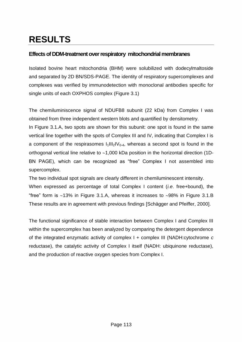
Page 113
RESULTS
Effects of DDM-treatment over respiratory mitochondrial membranes
Isolated bovine heart mitochondria (BHM) were solubilized with dodecylmaltoside
and separated by 2D BN/SDS-PAGE. The identity of respiratory supercomplexes and
complexes was verified by immunodetection with monoclonal antibodies specific for
single units of each OXPHOS complex (Figure 3.1)
The chemiluminiscence signal of NDUFB8 subunit (22 kDa) from Complex I was
obtained from three independent western blots and quantified by densitometry.
In Figure 3.1.A, two spots are shown for this subunit: one spot is found in the same
vertical line together with the spots of Complex III and IV, indicating that Complex I is
a component of the respirasomes I1III2IV0-4, whereas a second spot is found in the
orthogonal vertical line relative to 1,000 kDa position in the horizontal direction (1D-
B PAGE), which can be recognized as “free” Complex I not assembled into
supercomplex.
The two individual spot signals are clearly different in chemiluminescent intensity.
When expressed as percentage of total Complex I content (i.e. free+bound), the
“free” form is 13% in Figure 3.1.A, whereas it increases to 98% in Figure 3.1.B
These results are in agreement with previous findings [Schägger and Pfeiffer, 2000].
The functional significance of stable interaction between Complex I and Complex III
within the supercomplex has been analyzed by comparing the detergent dependence
of the integrated enzymatic activity of complex I + complex III (NADH:cytochrome c
reductase), the catalytic activity of Complex I itself (NADH: ubiquinone reductase),
and the production of reactive oxygen species from Complex I.
Page 114
As shown in Figure 3.2.A, when mitochondria are solubilized with dodecylmaltoside,
Complex I activity was decreased by 30%, indicating only marginal effect on the
activity of the enzyme whereas a 70% decrease was observed in the activity of
NADH:cytochrome c reductase (Figure 3.2.B), indicating less efficient electron
transfer between the two enzymes after DDM-treatment.
The antimycin A-insensitive residual rates of NADH:cytochrome c actvity were <10%
of the total rates shown in the figure.
On the other hand, oxidase activity (Figure 3.3.) was almost completely lost after
addition of dodecylmaltoside, probably due to the dissociation and dilution of
endogenous cytochrome c.
The functional significance of stable interaction between Complex I and Complex III
within the supercomplex has been analyzed by comparing the detergent dependence
of the integrated enzymatic activity of complex I + complex III (NADH:cytochrome c
reductase), the catalytic activity of Complex I itself (NADH: ubiquinone reductase),
and the production of reactive oxygen species from Complex I.
As shown in Figure 3.2.A, when mitochondria are solubilized with dodecylmaltoside,
Complex I activity was decreased by 30%, indicating only marginal effect on the
activity of the enzyme whereas a 70% decrease was observed in the activity of
NADH:cytochrome c reductase (Figure 3.2.B), indicating less efficient electron
transfer between the two enzymes after DDM-treatment.
The antimycin A-insensitive residual rates of NADH:cytochrome c actvity were <10%
of the total rates shown in the figure.
On the other hand, oxidase activity (Figure 3.3) was almost completely lost after
addition of dodecylmaltoside, probably due to the dissociation and dilution of
endogenous cytochrome c.

Page 115
The production of reactive oxygen species (ROS) from complex I was measured with
the probe 2’,7’- dichlorodihydrofluorescein diacetate (DCFDA) as described in
Materials and Methods.
The assays were performed both in the presence of 1.8 µM mucidin and mucidin plus
4 µM rotenone. Samples pretreated only with 1.8 µM mucidin were used as control
for all assays [Fato et al, 2009. Genova et al,2001].
Previous studies from our laboratory had shown that under these conditions ROS
production from Complex III is prevented and the addition of rotenone enhances the
generation of superoxide radicals from Complex I that represents the only source of
ROS [Fato et al, 2009]. Consequently, we can consider that mucidin functionally
isolates Complex I from downstream segments on the respiratory chain.
Fluorescence intensity data were expressed against Complex I activity to normalize
the NADH-dependent leakage of electrons by the fraction of catalytically active
molecules of the enzyme in the control and in the detergent-treated mitochondria
control samples.
As shown in Figure 3.4, after solubilization of mitochondrial membranes with
dodecylmaltoside, the production of ROS is significantly enhanced.
However, it is worth mentioning that in the DDM-treated samples rotenone had no
further effect over the conspicuous increase of ROS production above the non-
treated sample.

Page 116
Figure 3.1: Supercomplex disassembling in bovine heart mitochondria (BHM).
Respiratory supercomplexes and complexes from (A) digitonin-solubilized BHM with a detergent to
protein ratio (w:w) of 8 and from (B) DDM-solubilized BHM with a detergent to protein ratio (w:w) of
2.6 were resolved by western blotting after 2D-BN/SDS-PAGE. Arrows point to monomeric Complex I.
Monoclonal antibodies specific for single subunits of each OXPHOS complex are as follows: NDUFB8
(20 kDa) and NDUFA9 (39 kDa) of Complex I, SDHB (30 kDa) of Complex II, Core protein 2 (47 kDa)
of Complex III and COX-I (57 kDa, apparent 35 kDa) of Complex IV and alfa subunit (53 kDa) of ATP
synthase. The images shown in the picture were obtained in camera by double overlaying exposures
(not post-production computer-graphic overlay) to the antibody against the NDUFA9-subunit and, in
sequence, to a mixture of the remaining antibodies listed above. The ratio of free Complex I versus
total Complex I (free CI / total CI) were determined by densitometric analysis of the immunoblots as
described in the section “Materials and Methods”. Data are given as mean ± S.D. n shows the number
of independent samples. p-value was calculated according to the Student’s t - test.
p < 0.05

Page 117
Figure 3.2: Functional analysis of supercomplex I1III2 and complex I in detergent-
solubilized bovine heart mitochondria (BHM). Detergent dependence of (A) NADH-
ubiquinone oxidoreductase activity (B) NADH-cytochrome c oxidoreductase activity. Activity rates
are expressed in µmoles of NADH·min-1
·mg protein-1
. DDM: sample treated with 2.6 g
dodecylmaltoside / g of protein. Data are given as mean ± S.D. n shows the number of
independent samples. p-values were calculated using the Student’s t - test.

Page 118
Figure 3.3: Functional analysis of supercomplex I1III2 and complex I in detergent-
solubilized bovine heart mitochondria (BHM). Detergent dependence of NADH-oxidase
activity. Activity rates are expressed in µmoles of NADH·min-1
·mg protein-1
. DDM: sample treated with
2.6 g dodecylmaltoside / g of protein. Data are given as mean ± S.D. n shows the number of
independent samples. p-values were calculated using the Student’s t – test

Page 119
Figure 3.4: Functional analysis of supercomplex I1III2 and complex I in detergent-
solubilized bovine heart mitochondria (BHM). The production of hydrogen peroxide was
measured in the presence of 1.8 µM mucidin (dashed bars) and 1.8 µM mucidin plus 4 µM rotenone
(solid bars). Values are expressed as relative fluorescence intensity (RFU) by DCF normalized against
Complex I activity and shown as percentage values of the erence sample (i.e. control BHM in the
presence of mucidin only). DDM: sample treated with 2.6 g dodecylmaltoside/g of protein. Data are
given as mean ± S.D. n shows the number of independent samples. p-values were calculated using the
Student’s t - test.

Page 120
Effects of lipid dilution and DDM-treatment in reconstituted supercomplex I1III2
R4B proteoliposomes were reconstituted with different amounts of phospholipids.
Preliminary results from our laboratory [Lenaz and Genova, 2009b. Lenaz et al,
2010] demonstrated that the NADH: cytochrome c oxidoreductase activity,
experimentally determined, in a system at 1:30 (w:w) phospholipid dilution, overlaps
with the values expected by theoretical calculation applying the pool equation of
Kröger and Klingenberg [Kröger and Klingenberg, 1973a], whereas at 1:1
protein:lipid (w:w) ratio pool behavior is not effective anymore.
Kinetic testing according to the metabolic flux control analysis validated the
hypothesis of a random organization in the 1:30-model and of a functional
association between Complex I and Complex III in the 1:1-model [Genova et al,
2008].
Our analysis by bidimensional Blue-Native/SDS polyacrylamide gel electrophoresis
(2D BN/SDS-PAGE) confirms that the proteoliposomes at high protein to lipid ratio
are strongly enriched in the supercomplex I1III2 (Figure 3.5.A), whereas the relative
amount of bound Complex I compared to its free form appears drastically diminished
in the fraction having high phospholipid content (i.e. Complex I free/total = 0.08 vs.
0.76, respectively in the 1:1 and 1:30 samples) (Figure 3.5.B).
As shown in Figure 3.6, Complex I activity is the same at both phospholipid dilutions,
whereas the integrated activity of NADH:cytochrome c reductase is about half in the
1:30 sample with respect to the 1:1 sample.
Then, the production of ROS was evaluated both by the superoxide dismutase
(SOD)-sensitive reduction of acetylated cytochrome c for the detection of superoxide
and by the DCF assay.
Both assays showed a dramatic increase of ROS production in the R4B 1:30 with
respect to the R4B 1:1 sample (Figure 3.7).
Page 121
The fraction of SOD-insensitive NADH-acetylated cytochrome c reductase activity
that may possibly due to direct, not superoxide-mediated reduction of acetylated
cytochrome c in the R4B 1:30 sample is low (<12%), showing unchanged absolute
values compared to the SOD-insensitive activity in the 1:1 proteoliposomes (1.9 ± 0.7
and 2.8 ± 0.7 nmol·min-1·mg protein-1, respectively in the 1:30 vs. 1:1 samples).
A further proof of our interpretation of the higher values of ROS production is due to
disruption of association between Complex I and Complex III in the supercomplex
I1III2 is given by the effect of the dodecylmaltoside treatment in the R4B 1:1 sample
(Figure 3.8): The ratio of free to total Complex I increases to 0.79 after solubilization
with dodecylmaltoside, compared to a erence value of 0.08 in the control.
Conversely, the integrated NADH:cytochrome c reductase activity dramatically
decreases to less than 10% of the control, while Complex I activity is not altered
(Figure 3.9).
ROS production from Complex I is increased more than threefold when the 1:1
proteoliposomes are treated with dodecylmaltoside (Figure 3.10).

Page 122
Figure 3.5: Supramolecular organization of respiratory Complex I and Complex III in
R4B 1:1 and R4B 1:30 proteoliposomes. (A) R4B 1:1 and (B) R4B 1:30 samples were
separated by 2D BN / SDS-PAGE after solubilization with digitonin at a detergent to protein ratio of 8
(w:w) and resolved by western blotting followed by immunodetection using monoclonal antibodies
specific for single respiratory subunits. The images shown in the picture were obtained in camera by
double overlaying exposures (not postproduction computer-graphic overlay) to the antibody against
the NDUFA9 (39 kDa) subunit of Complex I and, in sequence, to the antibody against the Rieske
protein (22 kDa) of Complex III. Arrows point to monomeric Complex I. The upper panel of the figures
schematically shows the position of supercomplex I1III2 (SC), Complex I (CI) and dimeric Complex III
(CIII) in the 1D BN-gel prior to second dimension electrophoresis (2D SDS-PAGE). The ratio of free
Complex I versus total Complex I (free CI / total CI) were determined by densitometric analysis of the
immunoblots as described in the section “Materials and Methods”. Data are given as mean ± S.D. n
shows the number of independent samples. p-value was calculated according to the Student’s t - test.
p < 0.05
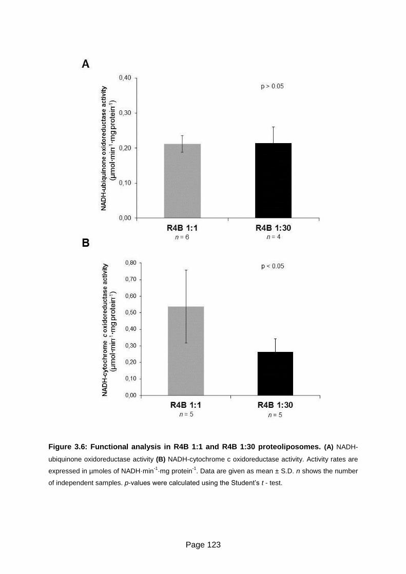
Page 123
Figure 3.6: Functional analysis in R4B 1:1 and R4B 1:30 proteoliposomes. (A) NADH-
ubiquinone oxidoreductase activity (B) NADH-cytochrome c oxidoreductase activity. Activity rates are
expressed in µmoles of NADH·min-1
·mg protein-1
. Data are given as mean ± S.D. n shows the number
of independent samples. p-values were calculated using the Student’s t - test.

Page 124
Fig
ure
3
.7:
RO
S p
rod
ucti
on
m
ed
iate
d b
y C
om
ple
x I
in R
4B
1
:1 a
nd
R
4B
1
:30
p
rote
olip
os
om
es
. (A
) T
he
ra
te o
f
su
pe
roxid
e f
orm
atio
n is d
ete
rmin
ed
as th
e s
up
ero
xid
e d
ism
uta
se
(S
OD
)-se
nsitiv
e r
ate
of a
ce
tyla
ted
cyto
ch
rom
e c
re
du
ctio
n in
th
e p
rese
nce
of
1.8
M
mu
cid
in a
nd
4
M r
ote
no
ne
. T
he
co
rre
spo
ndin
g r
ate
s o
f S
OD
-in
sen
sitiv
e a
ctivity a
re:
2.8
± 0
.7 a
nd
1.9
± 0
.7 n
mol·
min
-1·m
g p
rote
in-1
, re
sp
ectively
in
the
1:1
ve
rsus 1
:30
sa
mple
s.
(B)
Th
e p
rod
uction
of
hyd
rog
en
pe
roxid
e w
as m
easu
red
in
th
e p
rese
nce
of
1.8
M
mu
cid
in (
da
sh
ed
ba
rs)
an
d 1
.8
M
mu
cid
in p
lus 4
M
ro
ten
on
e (
so
lid b
ars
). V
alu
es a
re e
xp
resse
d a
s r
ela
tive
flu
ore
sce
nce
un
its (
RF
U)
by D
CF
no
rmaliz
ed
ag
ain
st
Com
ple
x I
activity
an
d s
ho
wn
as p
erc
enta
ge
valu
es o
f th
e e
rence
sa
mple
(i.e
. R
4B
1:1
in
th
e p
resen
ce
of
mu
cid
in o
nly
). D
ata
are
giv
en
as m
ea
n ±
S.D
. n
sh
ow
s t
he
nu
mbe
r o
f in
de
pe
nd
ent
sam
ple
s.
p-values were calculated using the Student’s t
- te
st.

Page 125
Figure 3.8: Disassembling of supercomplex I1III2 in R4B 1:1 proteoliposomes after
detergent solubilization. Respiratory complexes were separated by 2D BN/SDS-PAGE after
solubilization with (A) digitonin with a detergent to protein ratio (w:w) of 8 and with (B) DDM with a
detergent to protein ratio (w:w) of 4.3 and were resolved by western blotting using monoclonal antibodies
for single respiratory subunits. The images shown in the picture were obtained in camera by double
overlaying exposures (not postproduction computer-graphic overlay) to the antibody against the NDUFA9
(39 kDa) subunit of Complex I and, in sequence, to the antibody against the Rieske protein (22 kDa) of
Complex III. Arrows point to monomeric Complex I. The upper panel of the figures schematically shows the
position of supercomplex I1III2 (SC), Complex I (CI) and dimeric Complex III (CIII) in the 1D BN-gel prior to
second dimension electrophoresis (2D SDS-PAGE). The ratio of free Complex I versus total Complex I
(free CI / total CI) were determined by densitometric analysis of the immunoblots as described in the
section “Materials and Methods”. Data are given as mean ± S.D. n shows the number of independent
samples. p-value was calculated according to the Student’s t - test.
p < 0.05

Page 126
Figure 3.9: Functional analysis of supercomplex I1III2 and complex I in detergent-
solubilized R4B 1:1 proteoliposomes. Detergent dependence of (A) NADH-ubiquinone
oxidoreductase activity and (B) NADH-cytochrome c oxidoreductase activity. Activity rates are
expressed in µmoles of NADH·min-1
·mg·protein-1
. DDM: sample treated with 4.3 g
dodecylmaltoside / g of protein. Data are given as mean ± S.D. n shows the number of
independent samples. p-values were calculated using the Student’s t - test.

Page 127
Figure 3.10: Functional analysis of supercomplex I1III2 and complex I in detergent-
solubilized R4B 1:1 proteoliposomes. The production of hydrogen peroxide was measured in
the presence of 1.8 µM mucidin (dashed bars) and 1.8 µM mucidin plus 4 µM rotenone (solid bars).
Values are expressed as relative fluorescence intensity (RFU) by DCF normalized against Complex I
activity and shown as percentage values of the erence sample (i.e. control R4B 1:1 in the presence of
mucidin only). DDM: sample treated with 4.3 g dodecylmaltoside/g of protein. Data are given as mean
± S.D. n shows the number of independent samples. p-values were calculated using the Student’s t -
test.

Page 128
Effects of chaotrope-treatment in SMP and reconstituted supercomplex I1III2
Another experimental approach to disrupt supercomplex association is addition of
chaotropic agents [Boumans et al, 1998].
Following this line of thought, 0.2 M KSCN was tested to weaken hydrophobic
interactions between Complex I and Complex III inside supercomplexes in
submitochondrial particles from bovine heart mitochondria (SMP) and R4B (1:1)
proteoliposomes.
As shown in Figure 3.11, 2D BN/SDS-PAGE analysis reveals only partial
disassembling of the supercomplex when submitochondrial particles were previously
treated with 0.2 M KSCN (less than 40% of free Complex I) with a partial loss of
Complex I activity. Nevetherless, under these experimental conditions we can
observe enhanced ROS generation.
The same treatment with KSCN in 1:1 proteoliposomes did not produce clear
evidence of supercomplex disassembling and only 9% of Complex I was found as
free monomer compared to a rerence value of 8% in the control (Figure 3.12)
However, NADH:ubiquinone reductase activity was decreased by 45% showing a
significant increase of reactive oxygen species production from Complex I.
These last results allows to infer that chaotropic solutes seem unlikely solubilize in
and disassemble supramolecular assemblies in bovine mitochondria and 1:1
proteoliposomes.
When increasing SCN- concentration to 0.4 M is possible to partially increase
supercomplex disassembly in SMP, as 45% of free Complex I were detected in 2D
BN/SDS-PAGE (Figure 3.13A).
Although a significant increase of ROS production was observed, Complex I activity
decreased by about 50% respect control (Figure 3.13C and 3.13B). Similar results
were observed in 1:1 proteoliposomes (Figure 3.13).
Thus, rising SCN- concentration leads to a considerable increase on ROS production
from Complex I but NADH:ubiquinone activities decay in a concentration-dependent
manner in both SMP and 1:1 proteoliposomes samples.

Page 129
Figure 3.11: Disassembling of supercomplex I1III2 in bovine heart submitochondrial
particles (SMP) after treatment with 0.2 M KSCN. Respiratory supercomplexes and complexes
from (A) control SMP and from (B) KSCN-treated SMP were solubilized with digitonin with a detergent
to protein ratio (w:w) of 8 and resolved by western blotting after 2D BN/SDS-PAGE. The ratio of free
complex I vs total complex I was determined by densitometric analysis. Arrows point to monomeric
complex I. Monoclonal antibodies specific for single subunits of each OXPHOS complex are as
follows: NDUFB8 (20 kDa) of Complex I, SDHB (30 kDa) of Complex II, Core protein 2 (47 kDa) of
Complex III and COX-I (57 kDa, apparent 35 kDa) of Complex IV and alfa subunit (53 kDa) of ATP
synthase. The images shown in the picture were obtained in camera (not post-production computer-
graphic overlay). The ratio of free Complex I versus total Complex I (free CI / total CI) were
determined by densitometric analysis of the immunoblots as described in the section “Materials and
Methods”. (C) Potassium thiocyanate dependence of NADH-ubiquinone oxidoreductase activity.
Activity rates (measured after removal of KSCN by ultracentrifugation) are expressed in µmoles of
NADH·min-1
·mg protein-1
.(D) The production of hydrogen peroxide was measured, after removal of
KSCN by ultracentrifugation, in the presence of 1.8 µM mucidin (dashed bars) and 1.8 µM mucidin
plus 4 µM rotenone (solid bars). Values are expressed as relative fluorescence intensity (RFU) by
DCF normalized against Complex I activity and shown as percentage values of the erence sample (i.e.
control SMP in the presence of mucidin only). Data are given as mean ± S.D. n shows the number of
independent samples. p-values were calculated using the Student’s t - test.

Page 130
Figure 3.12: Disassembling of supercomplex I1III2 in R4B 1:1 proteoliposomes
after treatment with 0.2 M KSCN. (A) Respiratory supercomplexes and complexes from
KSCN-treated R4B 1:1 were solubilized with digitonin with a detergent to protein ratio (w:w) of 8
and resolved by western blotting after 2D BN/SDS-PAGE followed by immunodetection using
monoclonal antibodies specific for single respiratory subunits. The image shown in the picture
was obtained in camera by double overlaying exposures (not postproduction computer-graphic
overlay) to the antibody against the NDUFA9 (39 kDa) and NDUFB8 (20 kDa) subunits of
Complex I and, in sequence, to the antibodies against the Rieske protein (22 kDa) and Core
protein 2 (47 kDa) of Complex III. Arrows point to monomeric Complex I. The ratio of free
Complex I versus total Complex I (free CI / total CI) were determined by densitometric analysis of
the immunoblots as described in the section “Materials and Methods”. (B) Potassium thiocyanate
dependence of NADH-ubiquinone oxidoreductase activity, measured after removal of KSCN by
ultracentrifugation. Activity rates are expressed in µmoles of NADH·min-1
·mg protein-1
(D) The
production of hydrogen peroxide was measured, after removal of KSCN by ultracentrifugation, in
the presence of 1.8 µM mucidin (dashed bars) and 1.8 µM mucidin plus 4 µM rotenone (solid
bars). Values are expressed as relative fluorescence intensity (RFU) by DCF normalized against
Complex I activity and shown as percentage values of the erence sample (i.e. control R4B 1:1 in
the presence of mucidin only). Data are given as mean ± S.D. n shows the number of
independent samples. p-values were calculated using the Student’s t - test.

Page 131
Figure 3.13: Complex I activity and ROS production in bovine heart
submitochondrial particles (SMP) after treatment with KSCN. (A) Respiratory
supercomplexes and complexes from control SMP and from KSCN-treated SMP were solubilized with
digitonin with a detergent to protein ratio (w:w) of 8 and resolved by western blotting after 2D BN/SDS-
PAGE. The ratio of free complex I vs total complex I was determined by densitometric analysis. Arrows
point to monomeric complex I. Monoclonal antibodies specific for single subunits of each OXPHOS
complex are as follows: NDUFB8 (20 kDa) of Complex I, SDHB (30 kDa) of Complex II, Core protein 2
(47 kDa) of Complex III and COX-I (57 kDa, apparent 35 kDa) of Complex IV and alfa subunit (53 kDa) of
ATP synthase. (B) and (C). Potassium thiocyanate dependence of NADH-ubiquinone oxidoreductase
activity and ROS production (measured after removal of KSCN by ultracentrifugation), respectively.
Activity rates are expressed in µmoles of NADH·min-1
·mg protein-1
. The production of hydrogen peroxide
was measured in the presence of 1.8 µM mucidin (dashed bars) and 1.8 µM mucidin plus 4 µM rotenone
(solid bars). Values are expressed as relative fluorescence intensity (RFU) by DCF normalized against
Complex I activity and shown as percentage values of the erence sample (i.e. control SMP in the
presence of mucidin only). Data are given as mean ± S.D. n shows the number of independent samples.
p-values were calculated using the Student’s t - test.
A
B C

Page 132
Figure 3.14: Complex I activity and ROS production in R4B 1:1 proteoliposomes
after treatment with KSCN. Potassium thiocyanate dependence (measured after removal
of KSCN by ultracentrifugation) of (A) NADH-ubiquinone oxidoreductase activity and (B) ROS
production, respectively. Activity rates are expressed in µmoles of NADH·min-1
·mg protein-1
The production of hydrogen peroxide was measured in the presence of 1.8 µM mucidin
(dashed bars) and 1.8 µM mucidin plus 4 µM rotenone (solid bars). Values are expressed as
relative fluorescence intensity (RFU) by DCF normalized against Complex I activity and shown
as percentage values of the erence sample (i.e. control R4B 1:1 in the presence of mucidin
only). Data are given as mean ± S.D. n shows the number of independent samples. p-values
were calculated using the Student’s t - test.

Page 133
The following figure and table summarize the results of this study:
Figure 3.15: Production of ROS by mitochondrial Complex I in different
situations where supercomplexes are maintained or dissassembled. The
percent value of ROS production measured in all the samples listed in Table…is plotted in
the graph against the corresponding ratio of free Complex I versus total Complex I. The
statistical analysis of the data using the Pearson’s parametric test indicates a positive
correlation (r = 0.884, p<0.05) between ROS generation and relative amount of free
Complex I in the R4B and in the SMP samples (black symbols). The BHM samples (grey
symbols) were not included in the correlation analysis because the existence of
endogenous antioxidants systems operating to reduce ROS levels in the mitochondria
might have counteracted the dramatic effects of the complete dissociation of Complex I,
thus leading to a two-fold only increase of the measured ROS production. SMP,
submitochondrial particles.

Page 134
Table 3.1: Production of Reactive Oxygen Species by mitochondrial Complex I in different
situations where supercomplexes are maintained or disassembled
ROS production (%) Relative amount of Complex I (free/total)
R4B 1:1 224 83 (6) 0.08 0.07 (3) R4B 1:1 + DDM 4.3 g / g 672 257 (3) p<0.05 0.79 0.08 (3) p<0.001 R4B 1:30 851 243 (3) p<0.05 0.76 0.03 (2) p<0.001
SMP 223 56 (5) 0.23 (1)
SMP + 0.2 M KSCN 662 120 (5) p<0.05 0.39 (1)
BHM 121 20 (5) 0.13 0.07 (3) BHM + DDM 2.6 g / g 223 86 (5) p<0.05 0.98 0.02 (3) p<0.001
The samples were assayed in the presence of 1.8 M mucidin and 4 M rotenone as
described in the section “Materials and Methods”. The production of ROS is expressed as
the relative fluorescence intensity of diclorofluorescein normalized against Complex I activity.
Data are shown as percentage values of the corresponding erence sample (i.e. R4B 1:1,
SMP, and BHM, respectively assayed in the presence of 1.8 M mucidin only). The ratio of
free Complex I versus total Complex I was determined by densitometric analysis of
immunoblots after 2D BN/SDS-PAGE as described in the section “Materials and Methods”.
Values in round brackets indicate the number of independent samples. Results are
significantly different from their respective control according to the Student’s t-test.
Page 135
DISCUSSION
Disassembling of respiratory supercomplexes has several functional consequences
on mitochondrial respiration:
Loss of enzymatic channeling between Complex I and Complex III
Previous work in our laboratory have shown this effect in the CoQ region by
demonstrating that the integral NADH:cytochrome c reductase activity decreased
when Complex I and Complex III are present as individual enzymatic entities in
proteoliposomes reconstituted at high lipid to protein ratios (30:1) [Bianchi et al,
2003].
In that case, the observed rate of electron transfer between Complex I and Complex
III was found to approximate the theoretical values predicted from the CoQ-pool
equation [Kröger and Klingenberg, 1973a], indicating a random organization of these
components [Hackenbrock et al, 1986].
The diminution of the integral Complex I - Complex III activity is not a result of
enzyme inactivation or inhibition, since the individual activities of these complexes
(NADH:ubiquinone reductase and ubiquinol:cytochrome c reductase, respectively)
were not affected when comparing with proteoliposomes with lower lipid content (i.e.
lipid:protein ratio of 1) [Bianchi et al, 2003].
In the present study, we confirmed that the overall NADH:cytochrome c reductase
activity, which is ascribable to functional association between Complex I and
Complex III into supercomplexes, was dramatically decreased in proteoliposomes
30:1 (lipid:protein ratio) respect to proteoliposomes enriched on I1III2 supercomplexes
(i.e., 1:1 lipid:protein ratio), while the activity of Complex I was unaffected when
increasing the lipid content (Figure 4.6).
Moreover, we show that Complex I and Complex III are not assembled into
supercomplexes in the 30:1 (lipid to protein)-samples, as most Complex I was
detected as free holoenzyme in 2D BN/SDS-PAGE analyses (Figure 4.5).

Page 136
The functional significance of Complex I - Complex III interactions in supercomplexes
has been also demonstrated by the drastic decrease of NADH:cytochrome c
reductase, but not of NADH:ubiquinone reductase and of ubiquinol:cytochrome c
reductase in mitochondria-solubilized with dodecylmaltoside, a very mild detergent
suitable to disassemble supercomplexes [Schägger and Pfeiffer, 2000].
Similar results were obtained in the present work with both mitochondria and 1:1
proteoliposomes (Figures 3.1, 3.2 and 3.8, 3.9).
These evidences clearly demonstrate that, depending on the experimental
conditions, electron transfer between Complex I and Complex III can take place both
by CoQ channeling within the core I1III2 supercomplex and by the less efficient
collision-based pool behavior when Complex I - Complex III interactions are
disrupted by lipid-dilution or solubilized with a non-ionic detergent.
Production of reactive oxygen species from Complex I
The possibility that supercomplex assembly prevents excessive ROS generation
from Complex I has been advanced on theoretical grounds [Panov et al, 2007.
Seelert et al, 2009. Lenaz and Genova, 2012] but no direct experimental study was
previously addressed to this issue.
Indeed, in complex biological systems is difficult to ascertain a clear relationship of
cause - effect between supercomplex alteration and ROS production, since the two
phenomena may influence each other.
In this study we induce a primary condition in isolated mitochondria and Complex I-
Complex III proteoliposomes by means able to disrupt supercomplex interactions (i.e.
DDM-treatment or lipid dilution) increasing the ratios of Complex I present as free
and functionally active holoenzyme and we obtain the first direct demonstration that
loss of supercomplex organization causes an enhancement of ROS generation by
Complex I itself.

Page 137
This demonstration is clearly evident (Table 4.1 and Figure 4.15) both in
reconstituted proteoliposomes, where the dissociation of Complex I from the
respirasome is accompanied by a three/four-fold increase of ROS generation (Figure
4.7 and 4.10), and in DDM-solubilized mitochondrial membranes (Figure 4.4).
In the latter case, the existence of endogenous systems operating to reduce ROS
levels in the mitochondrial sample (i.e. mitochondrial glutathione peroxidase, Mn-
superoxide dismutase and non-enzymatic endogenous antioxidants) might have
counteracted the effects of the complete dissociation of Complex I, thus leading to a
two-fold increase of the measured ROS production.
The enhancement of ROS production from Complex I does not appear to be a result
of Complex I denaturation after the solubilization of the bovine mitochondria and 1:1
proteoliposomes with dodecylmaltoside.
As discussed above, the NADH:ubiquinone reductase activities are not affected
(Figure 4.9) are or marginally decreased (Figure 4.2), though Complex I is
functionally intact as free holoenzyme under these conditions (Figures 4.1 and 4.8).
A further demonstration that the disruption of Complex I-Complex III interactions
within supercomplex increases ROS levels from Complex I was obtained in
reconstituted Complex I/ Complex III proteoliposomes at high lipid to protein ratio
(30:1) whereas formation of the supercomplex I1III2 is prevented:
Likewise, the generation of superoxide is several fold higher than in proteoliposomes
with lower lipid content (1:1) which are rich in supercomplexes I1III2 (Figure 4.7).
In agreement with this finding, dissociation with dodecylmaltoside of the I1III2
supercomplexes in R4B 1:1 induces a strong enhancement of ROS generation
(Figure 4.9)
Moreover, the hypothetical reasoning that facilitation of electron flow by substrate
channeling within the respirasome helps maintaining the redox components of the
complexes in the oxidized state, thus limiting ROS formation, cannot be the only
explanation. In fact, in the experiments reported in this study, ROS production is
investigated in presence of inhibitors (mucidin and rotenone) that prevent electron
transfer to any possible acceptor, so that we can guess that the redox centers in

Page 138
Complex I are maximally reduced both in the situations where supercomplexes are
maintained and in the situations where Complex I is free.
On the other hand, such as chaotropic solutes that were previously found to
dissociate the supercomplex in yeast mitochondria [Boumans et al, 1998], were
partially effective in our mitochondrial membranes and proteoliposomes.
However, under these experimental conditions we observed an increase in ROS
production (Figures 4.11 and 4.12).
Unlike non-ionic detergent-treatment or lipid dilution, we observed a partial loss of
NADH:ubiquinone reductase activities after treatment of R4B 1:1 proteoliposomes
and SMP with SCN- (Figure 4.11C and 4.12B).
In addition, increasing SCN- concentration led to a considerable decay in Complex I
activities, showing at the same time higher levels of ROS production without any
significant disassembling of Complex I from supercomplexes (Figure 4.13 and 4.14).
A possible reason to explain this effect is that SCN- destabilize membrane proteins by
electrostatic interactions facilitating protein unfolding [Möglich et al, 2005.
Makhatadze and Privalov, 1992], rather than solubilizing annular and non-annular
lipids as dodecylmaltoside does [Rabilloud, 2009].
Thus SCN- treatment in mitochondrial membrane proteins would increase the
number of hydrophobic regions exposed to the solvent which could be involved in the
reduction of oxygen.

Page 139
Stability of Complex I
Another functional consequence previously described from disruption of the
supercomplex I1III2 core structure is loss of stability of Complex I.
In spite that loss of NADH:ubiquinone activity was reported in mutant cell models
lacking respiratory supercomplexes [Acín-Pérez et al, 2008. D’Aurelio et al, 2006.
Diaz et al, 2006. Schägger et al, 2004. Stroh et al, 2004], this was not observed in
the present study, since the activity of Complex I decreases only marginally upon
supercomplex disassembling in mitochondrial membranes but not in proteoliposomes
enriched of I1III2 supercomplexes (Figures 4.2, 4.6, and 4.9).
In addition, free Complex I holoenzyme stably appears in the native gels (Figures
4.1, 4.5 and 4.8).
These evidences show that monomeric Complex I persist functionally intact under
our experimental conditions.
Indeed, Complex I upon isolation and purification was shown to be sufficiently stable
to allow high turnover number [Hatefi et al, 1962c . Schägger and Pfeiffer, 2000].
The marginal decrese in NADH:ubiquinone activity observed in mitochondrial
membranes solubilized with dodecylmaltoside would be adscribable to the fact that
when Complex I is a free holoenzyme it becomes more susceptible to free radical
damage.
In regard to this, it is noteworthy that these results are compatible with very recent
observations by Diaz et al. [Diaz et al, 2012] showing that diminished stability of
supercomplexes and Complex I is correlated with increased levels of ROS in mouse
lung fibroblasts lacking the Rieske iron-sulphur protein of Complex III and hence
devoid of the supercomplexes containing Complex I.

Page 140
Although the higher ROS formation in free Complex I with respect to the
supercomplex could be a result of slower electron transport along the respiratory
chain, as this could increase the lifetime of some reactive species, we belive that in
our samples the increased production of ROS by Complex I as free holoenzyme is
due to different structural features.
In fact, when the supercomplex is dissociated, we also observe increased ROS
production after rotenone treatment, which abolishes electron flow from NADH to
ubiquinone in either experimental condition (i.e. free complex I and supercomplex I-
III).

Page 141
CONCLUSIONS
In conclusion, as summarized in Table 4.1 and in Figure 4.15 the results of the
present study provide experimental evidence that the production of reactive oxygen
species is strongly increased in either model, supporting the hypothesis that
disruption or prevention of the association between Complex I and Complex III by
different means enhances the generation of superoxide from Complex I.
Indeed, this work is the first demonstration that dissociation of the supercomplex I1III2
in the mitochondrial membrane is cause of oxidative stress from Complex I
[Maranzana et al, 2013]
Even though the results in the present work are an in vitro study, but it is supported
by several observations in cellular and animal models linking together supercomplex
dissociation and enhanced ROS production:
Enhanced ROS generation and oxidative stress were found in yeast mutants lacking
the supercomplex assembly factor Rcf1 and thus devoid of supercomplexes III-IV
[Chen et al, 2012. Strogolova et al, 2012. Vukotic et al, 2012].
Since the yeast S. cerevisiae lacks Complex I, in this case we may consider the
origin of the extra ROS being presumably free Complex III, while supposing that
other potential sources of ROS (i.e. external alternative NADH dehydrogenase) are
not affected in the Rcf1 mutants.
Numerous pathological states are accompanied by enhanced generation of ROS
[Chen and Keaney, 2012] and both mitochondria and other systems, such as plasma
membrane NADPH oxidase, have been implicated as the sources of ROS.
In an experimental model of heart failure, the decrease of oxidative phosphorylation
has been associated with a decrease of respiratory supercomplexes [Rosca and
Hoppel, 2009. Rosca and Hoppel, 2010. Rosca et al, 2008].
Lymphoblasts from patients affected by Barth Syndrome, due to genetic loss of
tafazzin, an enzyme involved in cardiolipin remodelling, have altered mitochondrial
supercomplexes [McKenzie et al 2006]; likewise, in a yeast experimental model of

Page 142
tafazzin mutation, Chen et al. [Chen et al, 2008] observed an increased oxidative
stress in response to ethanol.
Also, Baracca et al. [Baracca et al, 2010. Lenaz et al, 2010] demonstrated a strong
decrease of supercomplex assemblies in mouse fibroblasts expressing the activated
form of the k-ras oncogene associated with higher ROS generation in comparison
with wild type fibroblasts.
Aging is also accompanied by a decline of supercomplex association [Gómez et al,
2009. Frenzel et al, 2010]; despite some uncertainties and challenges, aging is
generally associated with increased ROS and oxidative damage [for a recent review
cf. [Cui et al, 2012].
Gomez and Hagen [Gómez and Hagen, 2012] reason that age-associated
destabilization of respiratory supercomplexes may be important for the development
of the mitochondrial aging phenotype by means of impaired bioenergetics and
enhanced ROS production.
In addition, Frenzel et al. [Frenzel et al, 2010] on the basis of the 3D-structures of
supercomplexes and the close spatial arrangement of the respective electron carrier
binding sites [Schäfer et al, 2007] conclude that less superoxide radical formation is
expected to occur in supercomplexes than in randomly distributed individual
complexes.
These latter studies, however, fail to show which is the causing event (i.e.
supercomplex dissociation causing ROS increase or, alternatively, ROS increase
causing supercomplex dissociation) or even if they are independent phenomena.
In this perspective we can conclude that the present study contribute to a better
understanding of the observations made in cellular and animal systems and
represent a starting point to further elucidate the intricate relationship between the
supramolecular organization of the respiratory chain and the production of ROS,
which can modulate some signalling pathways from mitochondria to the cell and also
act as damaging agents, depending on the amounts produced [Anilkumar et al, 2009.
Ide et al, 2000. Ray et al, 2012].
Page 143
In addition, this study confirms the hypothesis that primary causes of oxidative stress
may perpertuate reactive oxygen species production by a vicious circle involving
supercomplex dissociation as a major determinant. Thus, it is easy to foresee the
implications of these findings in human diseases and in aging, where oxidative stress
plays a major etiologic and pathogenic role.
Moreover, we can comment that the postulated dynamic structure of supercomplexes
[Acin Perez et al 2008. Acin Perez and Enriquez, 2014] and the modulation of their
association may represent a physiological means to control the production of ROS as
redox signals from the mitochondria to the cell.
Page 144
REFERENCES
[Abdrakhmanova et al, 2006] Abdrakhmanova, A.; Zwicker, K.; Kerscher, S.; Zickermann,
V.; Brandt, U. (2006). Tight binding of NADPH to the 39-kDa subunit of complex I is not
required for catalytic activity but stabilizes the multiprotein complex. Biochim Biophys Acta
1757:1676 – 1682
[Acin-Perez and Enriquez, 2014] Acin-Perez R, Enriquez JA. (2014). The function of the
respiratory supercomplexes: The plasticity model. Biochim Biophys Acta. 2014
Apr;1837(4):444-450.
[Acín-Pérez et al, 2004] Acín-Pérez, R.; Bayona-Bafaluy, M.P.; Fernández-Silva, P.;
Moreno-Loshuertos, R.; Pérez-Martos, A.; Bruno, C.; Moraes, C.T.; Enríquez, J.A. (2004).
Respiratory complex III is requiered to maintain complex I in mammalian mitochondria. Mol
Cell 13: 805-815
[Acín-Pérez et al, 2008] Acín-Pérez R, Fernández-Silva P, Peleato, ML, Pérez-Martos A,
Enríquez JA (2008). Respiratory active mitochondrial supercomplexes. Mol Cell 32: 529-539
[Acín-Pérez et al, 2009] Acín-Pérez, R.; Salazar, E.; Brosel, S.; Yang, H.; Schon, E.A.;
Manfredi, G. (2009). Modulation of mitochondrial protein phosphorylation by soluble adenyl
cyclase ameliorates cytochrome oxidase defects. EMBO Mol Med 1: 392 – 406
[Ahlers et al, 2000] Ahlers, P.M.; Zwicker,K.; Kerscher,S.; Brandt,U. (2000). Function of
conserved acidic residues in the PSST homologue of complex I (NADH:ubiquinone
oxidoreductase) from Yarrowia lipolytica. J Biol Chem 275: 23577 – 23582
[Aitken and Learmonth, 2002] Aitken, A.; Learmonth, M.P. (2002). Protein determination by
UV absorption. From: The protein protocols handbook 2nd edition. pp 3 - 6. Edited by
Walker, J.M. Humana Press, Inc. Totowa, NJ (USA)
[Albracht, 1993] Albracht, S.P. (1993). Intimate relationships of the large and the small
subunits of all nickel hydrogenases with two nuclear-encoded subunits of mitochondrial
NADH: ubiquinone oxidoreductase. Biochim Biophys Acta 1144: 221 - 224
[Althoff et al, 2011] Althoff, T.; Mills, D.J.; Popot, J.L.; Kühlbrandt, W. (2011). Arragement of
electron transport chain components in bovine mitochondrial supercomplex I1III2IV1. EMBO J
30: 4652-4664
[Angerer et al, 2011] Angerer,H.; Zwicker, K.; Wumaier, Z.; Sokolova, L.; Heide, H.; Steger,
M.; Kaiser, S.; Nübel, E.; Brutschy, B.; Radermacher, M. Brandt, U.; Zickermann, V. (2011).
A scaffold of accessory subunits links the peripheral arm and the distal proton-pumping
module of mitochondrial complex I. Biochem J 437: 279 – 288

Page 145
[Anilkumar et al, 2009] Anilkumar, N.; Sirker, A.; Shah, A.M. (2009). Redox sensitive
signaling pathways in cardiac remodeling, hypertrophy and failure. Front Biosci 14: 3168–
3187
[Antonicka et al, 2003] Antonicka, H.; Ogilvie, I.; Taivassalo, T.; Anitori, R. P.; Haller, R. G.;
Vissing, J.; Kennaway, N. G.; Shoubridge, E. A. (2003). Identification and characterization of
a common set of complex I assembly intermediates in mitochondria from patients with
complex I deficiency. J Biol Chem 278: 43081 – 43088
[Arnold et al, 1998] Arnold, I.; Pfeiffer, K.; Neupert, W.; Stuart, R.A.; Schägger, H. (1998).
Yeast mitochondrial F1F0-ATP synthase exists as a dimer: identification of three dimer-
specific subunits. EMBO J 17: 7170-7178
[Atteia et al, 2003] Atteia, A.; van Lis, R.; Mendoza-Hernandez, G.; Henze, K.; Martin, W.;
Riveros-Rosas, H.; Gonzalez-Halpen, D. (2003). Bifunctional aldehyde/alcohol
dehydrogenase (ADHE) in chlorophyte algal mitochondria. Plant Mol Biol 53: 175-188
[Azzi et al, 1975] Azzi, A.; Montecucco, C.; Richter, C. (1975). The use of acetylated
ferricytochrome c for the detection of superoxide radicals produced in biological membranes.
Biochem Biophys Res Commun 65: 597 - 603
[Balaban et al, 2005] Balaban, R.S.; Nemoto, S.; Finkel, T. (2005) Mitochondria, oxidants,
and aging. Cell 120: 483 – 495
[Balaskaran et al, 2010] Balaskaran Nina, R.; Dudkina, N.V.; Kane, L.A., van Eyk, J.E.;
Boekema, E.J. Mather, M.W.; Vaidya, A.B. (2010). Highly divergent mitochondrial ATP
synthase complexes in Tetrahymena thermophila. PloS Biol 8, e1000418
[Balaskaran et al, 2011] Balaskaran Nina, P.; Morrisey, J.M.; Ganesan, S.M.; Ke, H.;
Pershing, A.M.; Mather, M.W.; Vaidya, A.B. (2011). ATP synthase complex of Plasmodium
falciparum: dimeric assembly in mitochondrial membranes and resistance to genetic
disruption. J Biol Chem 286: 41312-41322
[Balsa et al, 2012] Balsa, E.; Marco, R.; Perales-Clemente, E.; Szklarczyk, R.; Calvo, E.;
Landázuri, M.O.; Enríquez, J.A. (2012). NDUFA4 is a subunit of complex IV of the
mammalian electron transport chain. Cell Metab 16: 378 – 386
[Bangham and Horne, 1964] Bangham, A.D.; Horne, R.W. (1964). Negative staining of
phospholipids and their structural modification by surface-active agents as observed in the
electron microscope. J Mol Biol 8: 660 – 668
[Baracca et al, 2010] Baracca, A.; Chiaradonna, F.; Sgarbi, G.; Solaini, G.; Alberghina, L.;
Lenaz, G. (2010). Mitochondrial Complex I decrease is responsible for bionergetic
dysfunction in K-ras transformed cells. Biochem Biophys Acta 1797: 314-323
Page 146
[Baradaran et al, 2013] Baradaran, R.; Berrisford, J.M.; Minhas, G.S.; Sazanov, L.A. (2013).
Crystal structure of the entire respiratory complex I. Nature 494: 443 - 448
[Barth et al 1983] Barth, P.G.; Scholte, H.R.; Berden, J.A.; van der Klei-van Moorsel, J.M.;
Luyt-Houwen, I.E.; van’t veer-Korthof, E.T.; van der Harten, J.J.; Sobotka-Plojhar, M.A.
(1983). A X-linked mitochondrial disease affecting cardiac muscle, skeletal muscle and
neutrophil leucocytes. J Neurol Sci 62: 327-355
[Battino et al, 1991] Battino, M.; Bertoli, E.; Formiggini, G.; Sassi, S.; Villa, R.F.; Lenaz, G.
(1991). Structural and functional aspects of the respiratory chain in synaptic and nonsynaptic
mitochondria derived from selected brain regions. J Bioenerg Biomembr 23: 345 – 363
[Beardslee and Updyke, 2007] Beardslee, T.A.; Updyke, T.V. (2007). Compositions and
methods for improving resolution of biomolecules separated on polyacrylamide gels. U.S.
patent application publication 2007/0151853. July 5, 2007
[Bednarz-Prashad and Mize, 1978] Bednarz-Prashad, A.J.; Mize, C.E. (1978).
Phospholipid, enzymatic, and polypeptide analyses of the mitochondrial membranes from
Saccharomyces carlsbergensis. Biochemistry 17: 4178-4186
[Benard et al, 2008] Benard G, Faustin B, Galinier A, Rocher C, Bellance N, Smolkova K,
Casteilla L, Rossignol R, Letellier T (2008). Functional dynamic compartmentalization of
respiratory chain intermediate substrates: implications for the control of energy production
and mitochondrial diseases, Int. J. Biochem. Cell Biol. 40 1543–1554.
[Berrisford and Sazanov, 2009] Berrisford, J.M.; Sazanov, L. A. (2009). Structural basis for
the mechanism of respiratory complex I. J Biol Chem 284: 29773 – 29783
[Berrisford et al, 2008] Berrisford, J. M., Thompson, C. J.; Sazanov, L. A. (2008). Chemical
and NADH-induced, ROS-dependent, cross-linking between subunits of complex I from
Escherichia coli and Thermus thermophilus. Biochemistry 47: 10262 - 10270
[Berry and Trumpower, 1985] Berry EA, Trumpower BL (1985). Isolation of ubiquinol
oxidase from Paracoccus denitrificans and resolution into cytochrome bc1 –aa3 complexes. J
Biol Chem 260: 2458-2467
[Beyer, 1967] Beyer, R.E. (1967). Preparation, properties, and conditions for assay of
phosphorylating electron transport particles (ETPH) and its variations. Methods Enzymol 10:
186 – 194
[Beyer, 1983] Beyer, R.E. (1983). A rapid biuret assay for protein of whole fatty tissues. Anal
Biochem 129: 483 - 485
[Bianchi et al, 2003] Bianchi C, Fato R, Genova ML, Parenti Castelli G, Lenaz G (2003).
Structural and functional organization of Complex I in the mitochondrial respiratory chain.
Biofactors 18: 3 – 9

Page 147
[Bianchi et al, 2004] Bianchi C, Genova ML, Parenti Castelli G, Lenaz G (2004). The
mitochondrial respiratory chain is partially organized in a supercomplex assembly:
kinetic evidence using flux control analysis, J. Biol. Chem. 279 36562–36569.
[Birner et al 2001] Birner, R.; Bürgermeister, M.; Schneiter, R.; Daum, G. (2001). Roles of
phosphatidylethanolamine and its several biosynthetic pathways in Saccharomyces
cerevisiae. Mol Biol Cell 12: 997-1007
[Black and Brandt, 1974] Black, M. J.; Brandt, R.B. (1974). Spectrofluorometric analysis of
hydrogen peroxide. Anal Biochem 58: 246 - 254
[Blair, 1967] Blair PV (1967) Preparation and properties of repeating units of mitochondrial
electron transfer. Methods Enzymol 10:208–212
[Böhm et al, 1990] Böhm, R.; Sauter, M.; Böck, A. (1990). Nucleotide sequence and
expression of an operon in Escherichia coli coding for formate hydrogenlyase components.
Mol Microbiol 4: 231 - 242
[Böttinger et al 2012] Böttinger, L.; Horvath, S.E.; Kleinschroth, T.; Hunte, C.; Daum, G.;
Pfanner, N.; Becker, T. (2012). Phosphatidylethanolamine and cardiolipin differentially affect
the stability of mitochondrial respiratory chain supercomplexes. J Mol Biol 423: 677-686
[Boumans et al, 1998] Boumans, H.; Grivell, L.A.; Berden, J.A. (1998). The respiratory
chain in yeast behaves as a single unit. J Biol Chem 273: 4872-4877
[Bourges et al, 2004] Bourges, I.; Ramus, C.; Mousson, C.B.;Beugnot, R.; Remacle, C.;
Cardol, P.; Hofhaus, G.; Issartel, J. P.(2004). Structural organization of mitochondrial human
complex I: role of the ND4 and ND5 mitochondria-encoded subunits and interaction with
prohibitin. Biochem J 383: 491 – 499
[Boveris, 1984] Boveris, A. (1984). Determination of production of superoxide radicals and
hydrogen peroxide in mitochondria. Methods Enzymol 105: 429 - 435
[Boyer et al, 1977] Boyer, P.D.; Chance, B.; Ernster, L.; Mitchell, P.; Racker, E.; Slater, E.C.
(1977). Oxidative phosphorylation and photophosphorylation. Annu Rev Biochem 46: 955-
966
[Brandner et al, 2005] Brandner, K.; Mick, D.U.; Frazier, A.E.; Taylor, R.D.; Meisinger, C.;
Rehling, P. (2005). Taz1, an outer mitochondrial membrane protein, affects stability and
assembly of inner membrane protein complexes: implications for Barth syndrome. Mol Biol
Cell 16: 5202-5214
[Brandt, 2006] Brandt, U. (2006). Energy converting NADH:quinone oxidoreductase
(complex I). Annu Rev Biochem 75: 69 – 92
[Brandt and Keston, 1965] Brandt, R.; Keston, A.S. (1965). Synthesis of
diacetyldichlorofluorescin: A stable reagent for fluorometric analysis. Anal Biochem 11: 6 - 9

Page 148
[Bruel et al, 1996] Bruel C, Brasseur R, Trumpower BL (1996). Subunit 8 of the
Saccharomyces cerevisiae cytochrome bc1 complex interacts with succinate-ubiquinone
reductase complex. J Bioenerg Biomembr 28: 59-68
[Brunette, 1981] Brunette, W. . (1981). “Western blotting”: electrophoretic transfer of
proteins from sodium dodecylsulfate-polyacrylamide gels to unmodified nitrocellulose and
radiographic detection with antibody and radioiodinated protein. Anal Biochem 112: 195 -
203
[Bultema et al 2009] Bultema, J.B.; Braun, H.P.; Boekema, E.J.; Kouril, R. (2009).
Megacomplex organization of the oxidative phosphorylation system by structural analysis of
respiratory supercomplexes from potato. Biochim Biophys Acta 1787: 84-93
[Bungert et al, 1999] Bungert, S.; Krafft, B.; Schlesinger, R.; Friedrich, T. (1999) One-step
purification of the NADH dehydrogenase fragment of the Escherichia coli by means of Strep-
tag affinity chromatography.FEBS Lett 460: 207 - 211
[Calvo et al, 2010] Calvo, S. E.; Tucker, E. J.; Compton, A. G.; Kirby, D. M.; Crawford, G.;
Burtt, N. P.; Rivas, R.; Guiducci, C.; Bruno, D. L.; Goldberger, O.A.; Redman, M. C.;
Wiltshire, E.; Wilson, C. J.; Altshuler, D.; Gabriel, S. J.; Daly, M. J.; Thorburn, D. R.; Mootha,
V. K. (2010) High-throughput, pooled sequencing identifies mutations in NUBPL and
FOXRED1 in human complex I deficiency. Nat Genet 42: 851 - 858
[Carilla-Latorre et al, 2010] Carilla-Latorre, S.; Gallardo, M.E.; Annesley, S.J.; Calvo-
Garrido, J.; Grana, O.; Accari, S.L.; Smith, P.K.; Valencia, A.; Garesse, R.; Fisher, P.R.;
Escalante, R. (2010). MidA is a putative methyltransferase that is required for mitochondrial
complex I function, J Cell Sci 123: 1674 - 1683
[Carroll et al, 2003] Carroll, J.; Fearnley, I. M.; Shannon, R. J.; Hirst, J.; Walker, J. E.
(2003). Analysis of the subunit composition of complex I from bovine heart mitochondria. Mol
Cell Proteomics 2: 117 - 126
[Carroll et al, 2006a] Carroll, J.; Fearnley, I.M.; Skehel, J.M.; Shannon, R.J.; Hirst, J.;
Walker, J.E. (2006). Bovine complex I is a complex of 45 different subunits. J Biol Chem 281:
32724 – 32727
[Carroll et al, 2006b] Carroll, J.; Fearnley, I.M.; Walker, J.E.(2006). Definition of the
mitochondrial proteome by measurement of molecular masses of membrane proteins. Proc
Natl Acad Sci U.S.A. 103: 16170 -16175
[Castelle et al, 2008] Castelle, C.; Guiral, M.; Malarte, G.; Ledgham, F.; Leroy, G.; Brugna,
M.; Guidici-Orticoni, M.T. (2008). A new iron-oxidizing/O2 –reducing supercomplex spanning
both inner and outer membranes, isolated from the extreme acidophile Acidithiobacillus
ferroxidans. J Biol Chem 283: 25803-2581

Page 149
[Cathcart et al, 1983] Cathcart, R.; Schwiers, E.; Ames, B. N. (1983). Detection of picomole
levels of hydroperoxide using fluorescent dichlorofluorescein assay. Anal Biochem 134: 111 -
116
[Chaban et al, 2013] Chaban Y, Boekema EJ, Dudkina NV (2013). Structures of
mitochondrial oxidative phosphorylation supercomplexes and mechanisms for their
stabilisation. Biochim Biophys Acta 2013.http://dx.doi.org/10.1016/j.bbabio.2013.10.004
[Chance, 1977] Chance B (1977). Electron transfer: pathways, mechanisms, and controls.
Ann Rev Biochem 46: 967-980
[Chance and Hollunger, 1960] Chance, B.; Hollunger, G. (1960). Energy-linked reduction of
mitochondrial pyridine nucleotide. Nature 185: 666 - 672
[Chance and Williams, 1955] Chance B, Williams GR (1955). Respiratory enzymes in
oxidative phosphorylation. I. Kinetics of oxygen utilization. J Biol Chem 217: 383-395
[Chance and Williams, 1956] Chance B, Williams GR (1956). The respiratory chain and
oxidative phosphorylation. Adv Enzymol Relat Subj Biochem 17: 65 – 134
[Chen and Keaney, 2012] Chen, K.; Keaney, J.F. Jr. (2012). Evolving concepts of oxidative
stress and reactive oxygen species in cardiovascular disease. Curr Atheroscler Rep 14: 476-
483
[Chen et al, 2004] Chen, R.; Fearnley, I.M.; Peak-Chew, S.Y.; Walker, J. E. (2004) The
phosphorylation of subunits of complex I from bovine heart mitochondria. J Biol Chem 279:
26036 – 26045
[Chen et al, 2008] Chen, S.; He, Q.; Greenberg, M.L. (2008). Loss of tafazzin in yeast leads
to increased oxidative stress during respiratory growth. Mol Microbiol 68: 1061-1072
[Chen et al, 2012] Chen, Y.C.; Taylor, E.B.; Dephoure, N.; Heo, J.M.; Tonhato, A.;
Papandreou, I.; Nath, N.; Denko, N.C.; Gygi, S.P.; Rutter, J. (2012). Identification of a protein
mediating respiratory supercomplex stability. Cell Metab 15: 348-360
[Chomyn et al, 1986] Chomyn, A.; Cleeter, M.W.; Ragan, C.I.; Riley, M.; Doolittle, R.F.
Attardi, G. (1986). URF6, last unidentified reading frame of human mtDNA, codes for an
NADH dehydrogenase subunit. Science 234: 614 - 618
[Claypool et al 2008] Claypool, S.M.; Oktay, Y.; Boontheung, P.; Loo, J.A.; Koehler, C.M.
(2008). Cardiolipin defines the interactome of the major ADP/ATP carrier protein of the
mitochondrial inner membrane. J Cell Biol 182: 937-950
[Crane et al, 1956] Crane FL, Glenn JL, Green DE (1956). Studies on the electron transfer
system. IV. The electron transfer particle. Biochim Biophys Acta 22: 475-487
[Crane et al, 1957] Crane FL, Hatefi Y, Lester RL, Widmer C. (1957). Isolation of a quinone
from beef heart mitochondria. Biochim Biophys Acta 25: 220-221

Page 150
[Crane et al, 1959] Crane FL, Widmer C, Lester RL, Hatefi Y (1959). Studies on the electron
transport system. XV. Coenzyme Q (Q275) and the succinoxidase activity of the electron
transport particle. Biochim Biophys Acta 31: 476-489
[Cronan et al, 2005] Cronan, J. E.; Fearnley, I. M.; Walker, J. E. (2005) Mammalian
mitochondria contain a soluble acyl carrier protein. FEBS Lett 579: 4892 - 4896
[Cruciat et al, 2000] Cruciat, C.M.; Brunner, S.; Baumann, F.; Neupert, W.; Stuart, R.A.
(2000). The cytochrome bc1 and cytochrome c oxidase complexes associate to form a single
supracomplex in yeast mitochondria. J Biol Chem 275: 18093-18098
[Cui et al, 2012] Cui, H.; Kong, Y.; Zhang, H. (2012). Oxidative stress, mitochondrial
dysfunction, and aging. J Signal Transduct 2012: doi:10.1155/2012/646354
[D’Aurelio et al, 2006] D’Aurelio, M.; Gajewski, C.D.; Lenaz, G.; Manfredi, G. (2006).
Respiratory chain supercomplexes set the threshold for respiration defects in human mtDNA
mutant cybrids. Human Mol Genet 15: 2157 - 2169
[Davies et al, 2011] Davies, K.M.; Strauss, M.; Daum, B.; Kief, J.H.; Osiewacz, H.D.;
Rycovska, A.; Zickermann, V.; Kühlbrandt, W. (2011). Macromolecular organization of ATP
synthase and complex I in whole mitochondria. Proc Natl Acad Sci USA 108: 14121-14126
[Davis, 1964] Davis, B.J. (1964). Disc electrophoresis. II. Method and application to human
serum proteins. Ann NY Acad Sci 121: 404–427
[Degli Esposti, 1998] Degli Esposti, M. (1998). Inhibitors of NADH-ubiquinone reductase: an
overview. Biochim Biophys Acta 1364: 222 – 235
[Degli Esposti, 2002] Degli Esposti, M. (2002). Measuring mitochondrial reactive oxygen
species. Methods 26: 335 - 340
[Degli Esposti et al, 1981] Degli Esposti, M.; Bertoli, E.; Parenti Castelli, G.; Fato, R.;
Mascarello, S.; Lenaz, G. (1981). Incorporation of ubiquinone homologs into lipid vesicles
and mitochondria membranes. Arch Biochem Biophys 210: 21 – 32
[Della Ciana, 2010a] Della Ciana, L. (2010). Preparation of high purity phenothiazine N-
alkylsulfonates and their use in chemiluminescent assays for the measurement of peroxidase
activity. U.S. patent 7,855,287. December 21, 2010.
[Della Ciana, 2010b] Della Ciana, L. (2010). Method for increasing light emission from a
chemiluminescent reaction. U.S. patent 7,803,573. September 28, 2010.
[De los Rios Castillo et al, 2011] De los Rios Castillo, D. Zarco-Zavala, M.; Olvera-
Sanchez, S.; Pardo, J.P.; Juarez, O.; Martinez, F.; Mendoza-Hernandez, G.; Garcia-Trejo,
J.J.; Flores-Herrera, O. (2011). Atypical cristae morphology of human syncytiotrophoblast
mitochondria: role for complex V. J Biol Chem 286:23911-23919

Page 151
[Dencher et al, 2007] Dencher, N.A.; Frenzel, M.; Reifschneider, N.H.; Sugawa, M.; Krause,
F. (2007). Proteome alterations in rat mitochondria caused by aging. Ann N Y Acad Sci 1100:
291-298
[Diaz et al, 2006] Diaz, F.; Fukui, H.; Garcia, S.; Moraes, C.T. (2006). Cytochrome c oxidase
is required for the assembly / stability of respiratory complex I in mouse fibroblasts. Mol Cell
Biol 26: 4872-4881
[Diaz et al, 2012] Diaz, F.; Enriquez, J.A.; Moraes, C.T. (2012). Cells lacking Rieske iron-
sulfur protein have a reactive oxygen species –associated decrease in respiratory complexes
I and IV. Mol Cell Biol 32: 415 - 429
[Djafarzadeh et al, 2000] Djafarzadeh, R.; Kersher, S.; Zwicker, K.; Radermacher, M.;
Lindahl, M.; Schägger, H.; Brandt, U. (2000). Biophysical and structural characterization of
proton-translocating NADH-dehydrogenase (complex I) from the strictly aerobic yeast
Yarrowia lipolytica. Biochim Biophys Acta 1459: 230 – 238
[Dröse et al, 2009] Dröse, S.; Galkin, A.; Brandt, U. (2009). Measurement of superoxide
formation by mitochondrial complex I of Yarrowia lipolytica. Methods Enzymol 456: 475 – 490
[Duarte et al, 2002] Duarte, M.; Populo, H.; Videira, A.; Friedrich, T.; Schulte, U. (2002).
Disruption of iron-sulphur cluster N2 from NADH: ubiquinone oxidoreductase by site-directed
mutagenesis. Biochem J 364: 833 - 839
[Dudkina et al, 2005a] Dudkina, N.V.; Heinemeyer, J.; Keegstra, W.; Boekema, E.J.; Braun,
H.P. (2005). Structure of dimeric ATP sythase from mitochondria: an angular association of
monomers induces the strong curvature of the inner membrane. FEBS Lett 579: 5769-5772
[Dudkina et al, 2005b] Dudkina, N.K.; Eubel, H.; Keegstra, W.; Boekema, E.J.; Braun, H.P.
(2005). Structure of mitochondrial supercomplex formed by respiratory-chain complexes I
and III. Proc Natl Acad Sci USA 102: 3225-3229
[Dudkina et al, 2006] Dudkina, N.V.; Heinemeyer, J.; Sunderhaus, S.; Boekema, E.J.;
Braun, H.P. (2006). Respiratory chain supercomplexes in the plant mitochondrial membrane.
Trends Plant Sci 11:232-240
[Dudkina et al, 2008] Dudkina NV, Sunderhaus S, Boekema E, Braun HP (2008). The
higher level of organization of the oxidative phosphorylation system: mitochondrial
supercomplexes. J Bioenerg Biomembr 40:419-424
[Dudkina et al, 2011] Dudkina, N.V.; Kudryashev, M.; Stahlberg, H.; Boekema, E.J. (2011).
Interaction of complexes I, III, and IV within the bovine respirasome by single particle
cryoelectron tomography. Proc Natl Acad Sci USA 108: 15196-15200
[Efremov and Sazanov, 2010] Efremov, R.G.; Sazanov, L.A. (2010) Structure of the
membrane domain of respiratory complex I. Nature 476: 414 – 420

Page 152
[Efremov and Sazanov, 2011] Efremov RG, Sazanov LA (2011). Structure of the membrane
domain of respiratory complex I. Nature 476, 414-420.
[Efremov and Sazanov, 2012] Efremov, R.G.; Sazanov, L.A. (2012). The coupling
mechanism of respiratory complex I – A structural and evolutionary perspective. Biochim
Biophys Acta 1817: 1785 - 1795
[Efremov et al, 2010] Efremov, R.G.; Baradaran, R.; Sazanov, L.A. (2010). The architecture
of respiratory complex I. Nature 465: 441 - 445
[Ernster and Schatz, 1981] Ernster L, Schatz G (1981). Mitochondria: A historical review. J
Cell Biol 91: 227s-255s
[Eubel et al, 2003] Eubel, H.; Jansch, L.; Braun, H.P. (2003). New insights into the
respiratory chain in plant mitochondria. Supercomplexes and a unique composition of
complex II. Plant Physiol 133: 274-286
[Eubel et al, 2004] Eubel, H.; Heinemeyer, J.; Braun, H.P. (2004). Identification and
characterization of respirasomes in potato mitochondria. Plant Physiol 134: 1450-1459
[Fato et al, 1993] Fato, R.; Cavazzoni, M.; Casteluccio, C.; Parenti Castelli, G.; Palmer, G.;
Degli Esposti, M.; Lenaz, G. (1993). Steady-state kinetics of ubiquinol-cytochrome c
reductase in bovine submitochondrial particles: diffusional effects. Biochem J 290: 225 – 236
[Fato et al, 2009] Fato, R.; Bergamini, C.; Bortolus, M.; Maniero, A. L.; Leoni, S.; Ohnishi, T.;
Lenaz, G. (2009). Differential effects of mitochondrial Complex I inhibitors on production of
reactive oxygen species. Biochim Biophys Acta 1787: 384 - 392
[Fearnley and Walker, 1992] Fearnley, I.M.; Walker, J.E. (1992). Conservation of
sequences of subunits of mitochondrial complex I and their relationships with other proteins.
Biochim Biophys Acta 1140: 105 – 134
[Fearnley et al, 2001] Fearnley, I. M.; Carroll, J.; Shannon, R. J.; Runswick, M. J.; Walker, J.
E.; Hirst, J. (2001). GRIM-19, a cell death regulatory gene product, is a subunit of bovine
mitochondrial NADH: ubiquinone oxidoreductase (complex I). J Biol Chem 276: 38345 –
38348
[Fecke et al, 1994] Fecke, W.; Sled, V.D.; Ohnishi, T.; Weiss, H. (1994). Disruption of the
gene encoding the NADH-binding subunit of NADH: ubiquinone oxidoreductase in
Neurospora crassa. Formation of a partially assembled enzyme without FMN and the iron-
sulphur cluster N-3. Eur J Biochem 220: 551 - 558
[Fendel et al, 2008] Fendel, U.; Tocilescu, M.A.; Kerscher, S.; Brandt, U. (2008). Exploring
the inhibitor binding pocket of respiratory complex I. Biochim Biophys Acta 1777: 660 - 665
[Fernandez-Vizarra et al, 2007] Fernandez-Vizarra, E. Bugiani, M.; Goffrini, P.; Carrara, F.;
Farina, L.; Precopio, E.; Donati, A.; Uziel, G.; Ferrero, I.; Zeviani, M. (2007). Impaired
Page 153
complex III assembly associated with BCS1L gene mutations in isolated mitochondrial
encephalopathy. Hum Mol Genet 16: 1241 – 1252
[Finel, 1998] Finel, M. (1998). Organization and evolution of structural elements within
complex I. Biochim Biophys Acta 1364: 112 - 121
[Flemming et al, 2003] Flemming, D.; Schlitt, A.; Spehr, V.; Bischof, T.; Friedrich, T. (2003).
Iron-sulfur cluster N2 of the Escherichia coli NADH:ubiquinone oxidoreductase (complex I) is
located on subunit NuoB. J Biol Chem 278: 47602 – 47609
[Folin and Ciocalteau, 1927] Folin, O.; Ciocalteau, V. (1927). On tyrosine and triptophane
determinations in proteins. J Biol Chem 73: 627 - 650
[Fowler et al, 1962] Fowler LR, Richardson SH, Hatefi Y (1962). A rapid method for the
preparation of highly purified cytochrome oxidase. Biochim Biophys Acta 64: 170-173
[Fraaije and Mattevi, 2000] Fraaije, M. W.; Mattevi, A. (2000). Flavoenzymes: diverse
catalysts with recurrent features. Trends Biochem Sci 25: 126 – 32
[Frenzel et al, 2010] Frenzel, M.; Rommelspacher, H.; Sugawa, M.D.; Dencher, N.A. (2010).
Ageing alters the supramolecular architecture of OxPhos complexes in rat brain cortex. Exp
Gerontol 45: 563-572
[Friedrich, 2001] Friedrich, T. (2001). Complex I: a chimera of a redox and conformation-
driven proton pump? J Bioenerg Biomembr 33: 169 – 177
[Friedrich and Scheide, 2000] Friedrich, T.; Scheide, D. (2000). The respiratory complex I
of bacteria, archea and eukarya and its module common with membrane-bound multisubunit
hydrogenases. FEBS Lett 479: 1 – 5
[Friedrich and Weiss, 1997] Friedrich, T.; Weiss, H. (1997). Modular evolution of the
respiratory NADH:ubiquinone oxidoreductase and the origin of its modules. J Theor Biol 187:
529 – 540
[Friedrich et al, 1994] Friedrich, T.; Van Heek, P.; Leif, H.; Ohnishi, T.; Forche, E.; Kunze,
B.; Jansen, R.; Trowitzsch-Kienast, W.; Höfle, G.; Reichenbach, H.; Weiss, H. (1994). Two
binding sites of inhibitors in NADH:ubiquinone oxidoreductase (complex I). Eur J Biochem
219: 691 - 698
[Fry and Green, 1981] Fry, M.; Green, D.E. (1981). Cardiolipin requirement for electron
transfer in complex I and III of the mitochondrial respiratory chain. J Biol Chem 256: 1874-
1880
[Gabaldon et al, 2005] Gabaldon, T.; Rainey, D.; Huynen, M. A. (2005) Tracing the
evolution of a large protein complex in the eukaryotes, NADH: ubiquinone oxidoreductase
(complex I). J Mol Biol 348: 857 – 870

Page 154
[Galante and Hatefi, 1978] Galante, Y. M.; Hatefi, Y. (1978) Resolution of complex I and
isolation of NADH dehydrogenase and an iron–sulfur protein. Methods Enzymol 53: 15 – 21
[Gao et al, 2012] Gao, Y.; Meyer, B.; Sokolova, L.; Zwicker, K.; Karas, M.; Brutschy, B.;
Peng, G.; Michel, H. (2012). Heme-copper terminal oxidase using both cytochrome c and
ubiquinol as electron donors. Proc Natl Acad Sci USA 109:3275-3280
[Garcia Montes de Oca et al, 2012] Garcia Montes de Oca, L.Y.; Chagolla-López, A.;
González de la Vara, L.; Cabellos-Avelar, T.; Gómez-Lojotero, C.; Gutierrez Cirlos, E.B.
(2012). The composition of the Bacillus subtilis aerobic respiratory chain supercomplexes. J
Bionenerg Biomembr 44: 473-486
[Garfin, 1990] Garfin, D.E. (1990). One-dimensional gel electrophoresis. Methods Enzymol
182: 425 – 441
[Garfin, 2009] Garfin, D.E. (2009). One-dimensional gel electrophoresis. Methods Enzymol
463: 497 - 513
[Garofano et al, 2003] Garofano, A.; Zwicker, K.; Kerscher,S.; Okun, P.; Brandt, U. (2003).
Two aspartic acid residues in the PSST-homologous NUKM subunit of complex I from
Yarrowia lipolytica are essential for catalytic activity. J Biol Chem 278: 42435 - 42440
[Gasparre et al, 2013] Gasparre, G.; Porcelli, A.M.; Lenaz, G.; Romeo, G. (2013).
Relevance of mitochondrial genetics and metabolism in cancer development. Cold Spring
Harb Perspect Biol 5, a011411
[Genova and Lenaz, 2013] Genova ML, Lenaz G (2013). Functional role of mitochondrial
respiratory supercomplexes. Biochim Biophys Acta 2013.
http://dx.doi.org/10.1016/j.bbabio.2013.11.002
[Genova et al, 2001] Genova, M. L.; Ventura, B.; Giuliano, G.; Bovina, C.; Formiggini, G.;
Parenti Castelli, G.; Lenaz, G. (2001). The site of production of superoxide radical in
mitochondrial complex I is not a bound ubisemiquinone but presumably iron-sulfur cluster
N2. FEBS Lett 505: 364 - 368
[Genova et al, 2008] Genova ML, Baraca A, Biondi G, Casalena M, Faccioli M, Falasca AI,
Formiggini G, Sgarbi G Solaini G, Lenaz G (2008). Is supercomplex organization of the
respiratory chain required for optimal electron transfer activity?. Biochim Biophys Acta 1777:
740-746
[Gershoni and Palade, 1982] Gershoni, J.M.; Palade, G.E. (1982). Electrophoretic transfer
of proteins from sodium dodecyl sulfate-polyacrilamide gels to a positively charged
membrane filter. Anal Biochem 124: 396 - 405
[Gershoni et al, 2010] Gershoni, M.; Fuchs, A.; Shani, N.; Fridman, Y.; Corral-Debrinski, M.;
Aharoni, A.; Frishman, D.; Mishmar, D. (2010). Coevolution predicts direct interactions
Page 155
between mtDNA-encoded and nDNA-encoded subunits of oxidative phosphorylation complex
I. J Mol Biol 404: 158 – 171
[Gill and von Hippel, 1989] Gill, S. C.; von Hippel, P. H. (1989). Calculation of protein
extinction coefficients from amino acid sequence data. Anal Biochem 182: 319 - 326
[Gómez and Hagen, 2012] Gomez, L.A.; Hagen, T.M. (2012). Age-related decline in
mitochondrial bioenergetics: does supercomplex destabilization determine lower oxidative
capacity and higher superoxide production? Semin Cell Dev Biol 23: 758-767
[Gómez et al, 2009] Gómez, L.A.; Monette, J.S.; Chavez, J.D.; Maier, C.S.; Hagen, T.M.
(2009). Supercomplexes of the mitochondrial electron transport chain decline in the aging rat
heart. Arch Biochem Biophys 490: 30-35
[Gonzalvez et al 2013] Gonzalvez, F.; D’Aurelio, M.; Boutant, M.; Moustapha, A.; Puech,
J.P.; Landes, T.; Arnaune-Pelloquin, L.; Vial, G.; Taleux, N.; Slomianny, C.; Wanders, R.J.;
Houtkooper, R.H.; Bellenguer, P.; Moller, I.M.; Gottlieb, E.; Vaz, F.M.; Manfredi, G.; Petit,
P.X. (2013). Barth syndrome: cellular compensation of mitochondrial dysfunction and
apoptosis inhibition due to changes in cardiolipin remodeling linked to tafazzin (TAZ) gene
mutation. Biochim Biophys Arcta 1832: 1194-1206
[Gornall et al, 1949] Gornall, A. G.; Bardawill, C.J.; David, M.M. (1949). Determination of
serum proteins by means of the biuret reaction. J Biol Chem 177: 751 - 766
[Grad and Lemire, 2004] Grad, L.I.; Lemire, D. (2004). Mitochondrial complex I mutations in
Caenorhabditis elegans produce cytochrome c oxidase deficiency, oxidative stress and
vitamin-response lactic acidosis. Hum Mol Genet 13: 303 – 314
[Grad and Lemire, 2006] Grad, L.I.; Lemire, D. (2006). Riboflavin enhances the assembly of
mitochondrial cytochrome c oxidase in C. elegans NADH ubiquinone reductase mutants.
Biochim Biophys Acta 1757: 115 - 122
[Green and Tzagoloff, 1966] Green DE, Tzagoloff A (1966). The mitochondrial electron
transfer chain. Arch Biochem Biophys 116: 293-304
[Grigorieff, 1998] Grigorieff, N. (1998). Three-dimensional structure of bovine
NADH:ubiquinone oxidoreductase (complex I) at 22 Å in ice. J Mol Biol 277: 1033 - 1046
[Guenebaut et al, 1998] Guenebaut, V.; Schlitt, A.; Weiss, H.; Leonard, K.; Friedrich, T.
(1998). Consistent structure between bacterial and mitochondrial NADH:ubiquinone
oxidoreductase (complex I). J Mol Biol 276: 105 - 112
[Guiral et al, 2009] Guiral, M.; Prunetti, L.; Lignon, S.; Lebrun, R.; Moinier, D.; Giudici-
Orticonit, M.T. (2009). New insights into the respiratory chains of the chemolithoautotrophic
and hyperthermophilic bacterium Aquifex aeolicus. J Proteome Res 8: 1717-1730

Page 156
[Gupte and Hackenbrock, 1988a] Gupte SS, Hackenbrock CR (1988). Multidimensional
diffusion modes and collision frequencies of cytochrome c with its redox partners. J Biol
Chem 263: 5441 - 5247
[Gupte and Hackenbrock, 1988b] Gupte SS, Hackenbrock CR (1988). The role of
cytochrome c diffusion in mitochondrial electron transport. J Biol Chem 263: 5248 – 5253
[Gupte et al, 1984] Gupte SS, Wu ES, Hoechli L, Hoechli M, Jacobson K, Sower AW,
Hackenbrock CR (1984). Relationship between lateral diffusion, collision frequency, and
electron transfer of mitochondrial inner membrane oxidation-reduction components. Proc
Natl Acad Sci USA 81: 2606 - 2616
[Hackenbrock et al, 1986] Hackenbrock CR, Chazzote B, Gupte SS (1986). The random
collision model and a critical assessment of diffusion and collision in mitochondrial electron
transport. J Bioenerg Biomembr 18: 331-368
[Hatefi and Rieske, 1967] Hatefi Y, Rieske JS (1967). Preparation and properties of DPNH-
cytochrome (Complex I-III of the Respiratory Chain). Methods Enzymol 10: 225-231
[Hatefi et al, 1959] Hatefi, Y.; Lester, R.L.; Crane, F.L.; Widmer, C. (1959). Studies on the
electron transport system XVI. Enzymic oxidoreduction reactions of coenzyme Q. Biochim
Biophys Acta 31: 490-501
[Hatefi et al, 1961] Hatefi Y, Haavik AG, Jurtshuk P (1961). Studies on the electron transport
system. XXX. DPNH-cytochrome c reductase I. Biochim Biophys Acta 52: 106-118
[Hatefi et al, 1962a] Hatefi Y, Haavik AG, Griffiths DE (1962). Studies on the electron
transfer system. XLI. Reduced coenzyme Q (QH2)-cytochrome c reductase. J Biol Chem
237: 1681-1685
[Hatefi et al, 1962b] Hatefi Y, Haavik AG, Fowler LR, Griffiths DE (1962). Studies on the
electron transfer system. XLII. Reconstitution of the electron transfer system. J Biol Chem
237: 2661-2669
[Hatefi et al, 1962c] Hatefi, Y.; Haavik, A.G.; Griffiths, D.E. (1962). Sutides on the electron
transfer system. Preparation and properties of mitochondrial DPNH-coenzyme Q reductase.
J Biol Chem 237: 1676-1680
[Hedderich, 2004] Hedderich R. (2004). Energy-converting [NiFe] hydrogenases from
archaea and extremophiles: ancestors of complex I. J Bioenerg Biomembr 36(1):65-75
[Heron et al, 1978] Heron C, Ragan CI, Trumpower BL (1978). The interaction between
mitochondrial NADH-ubiquinone oxidoreductase and ubiquinol-cytochrome c oxidoreductase
restoration of ubiquinone-pool behaviour, Biochem. J. 174 (1978) 791–800.

Page 157
[Herrero and Barja, 2000] Herrero, A.; Barja, G. (2000). Localization of the site of oxygen
radical generation inside the complex I of heart and nonsynaptic brain mammalian
mitochondria. J Bioenerg Biomembr 32: 609 - 615
[Heinemeyer et al, 2007] Heinemeyer J, Braun HP, Boekema EJ, Kouril R. A structural
model of the cytochrome C reductase/oxidase supercomplex from yeast mitochondria. J.
Biol. Chem. 2007;282:12240–12248.
[Hinkle et al, 1967] Hinkle, P.C.; Butow, R.A.; Racker, E.; Chance, B. (1967). Partial
resolution of the enzymes catalyzing oxidative phosphorylation. XV. Reverse electron
transfer in the flavin-cytochrome c region of the respiratory chain of beef heart
sumbitochondrial particles. J Biol Chem 242: 5169 - 5173
[Hirst et al, 2008] Hirst, J.; King, M.S.; Pryde, K.R. (2008). The production of reactive
oxygen species by complex I. Biochem Soc Trans 36: 976 - 980
[Hofhaus et al, 1991] Hofhaus, G.; Weiss, H.; Leonard, K. (1991). Electron microscopic
analysis of the peripheral and membrane parts of mitochondrial NADH dehydrogenase
(complex I). J Mol Biol 221: 1027 – 1043
[Huang et al, 2005] Huang, L.S.; Cobessi, D.; Tung, E.Y.; Berry, E.A. (2005) Binding of the
respiratory chain inhibitor antimycin to the mitochondrial bc1 complex: a new crystal structure
reveals an altered intramolecular hydrogen-bonding pattern. J Mol Biol 351: 573-597
[Hunte et al, 2010] Hunte, C.; Zickermann, V.; Brandt, U. (2010) Functional modules and
structural basis of conformational coupling in mitochondrial complex I. Science 329:448–451.
[Ide et al, 2000] Ide, T.; Tsutsui, H.; Kinugawa, S.; Suematsu, N.; Hayashidani, S.; Ichikawa,
K.; Utsumi, H.; Machida, Y.; Egashira, K.; Takeshita, A. (2000). Direct evidence for
increased hydroxyl radicals originating from superoxide in the failing myocardium. Circ Res
86: 152–157
[Ingledew and Ohnishi, 1980] Ingledew, W.J.; Ohnishi, T. (1980). An analysis of some
thermodynamic properties of iron-sulphur centres in site I of mitochondria. Biochem J
186:111 – 117
[Iwasaki et al, 1995] Iwasaki T, Matsuura K, Oshima T (1995). Resolution of the aerobic
respiratory system of the thermoacidophilic archeon, Sulfolobus sp. Strain 7. The archaeal
terminal oxidase supercomplex is a functional fusion of respiratory complexes III and IV with
no c-type cytochromes. J Biol Chem 270: 30881-30892
[Janssen et al, 2006] Janssen, R. J.; Nijtmans, L. G.; van den Heuvel, L. P.; Smeitink, J. A.
(2006) Mitochondrial complex I structure, function and pathology. J Inherit Metab Dis 29: 499
- 515
Page 158
[Jiang et al, 2000] Jiang, F.; Ryan, M.T.; Schlame, M.; Zhao, M.; Gu, Z.: Klingenberg, M.;
Pfanner, N.; Greenberg, M.L. (2000). Absence of cardiolipin in the crd1 null mutant results in
decreased mitochondrial membrane potential and reduced mitochondrial function. J Biol
Chem 275: 22387-22394
[Joshi et al 2012] Joshi, A.S.; Thompson, M.N.; Fei, N.; Hüttemann, M.; Greenberg, M.L.
(2012). Cardiolipin and mitochondrial phosphatidylethanolamine have overlapping functions
in mitochondrial fusion in Saccharomyces cerevisiae. J Biol Chem 287: 17589-17597
[Jezek and Hlavatà, 2005] Jezek, P.; Hlavatà, L. (2005). Mitochondria in homeostasis of
reactive oxygen species in cell, tissues, and organism. Int J Biochem Cell Biol 37: 2478 -
2503
[Kean et al, 1971] Kean, E. A.; Gutman, M. Singer, T.P. (1971). Studies on the respiratory
chain-linked nicotinamide adenine dinucleotide dehydrogenase. XXII. Rhein, a competitive
inhibitor of the dehydrogenase. J Biol Chem 246: 2346 - 2353
[Keefe and Maier, 1993] Keefe RG, Maier RJ (1993). Purification and characterization of an
O2-utilizing cytochrome c oxidase complex from Bradyrhizobium japonicum bacteroid
membranes. Biochim Biophys Acta 1183: 91-104
[Keston and Brandt, 1965] Keston, A. S.; Brandt, R. (1965). The fluorometric analysis of
ultramicro quantities of hydrogen peroxide. Anal Biochem 11: 1 - 5
[Klingenberg and Slenczka, 1959] Klingenberg, M.; Slenczka, W. (1959). [Pyridine
nucleotide in liver mitochondria. An analysis of their redox relationships]. [Article in German].
Biochem Z 331:486 – 517
[Kohlstädt et al, 2008] Kohlstädt, M.; Dörner, K.; Labatzke, R.; Koç, C.; Hielscher, R.;
Schiltz, E.; Einsle, O.; Hellwig, P.; Friedrich,T. (2008). Heterologous production, isolation,
characterization and crystallization of a soluble fragment of the NADH:ubiquinone
oxidoreductase (complex I) from Aquifex aeolicus. Biochemistry 47: 13036 – 13045
[Koopman et al, 2010] Koopman, W. J.; Nijtmans, L. G.; Dieteren, C.E.; Roestenberg, P.;
Valsecchi, F.; Smeitink, J.A.; Willems, P. H. (2010). Mammalian mitochondrial complex I:
biogenesis, regulation, and reactive oxygen species generation. Antioxid Redox Signal
12:1431 – 1470
[Kowaltowski et al, 2009] Kowaltowski, A. J.; De Souza-Pinto, N. C.; Castilho, R. F.;
Vercesi, A. E. (2009) Mitochondria and reactive oxygen species. Free Radic Biol Med 47:
333 - 343
[Krause et al, 2004a] Krause, F.; Scheckhuber, C.Q.; Werner, A.; Rexroth, S.;
Reifschneider, N.H.; Dencher, N.A.; Osiewacz, H.D. (2004) Supramolecular organization of

Page 159
cytochrome c oxidase- and alternative oxidase-dependent respiratory chains in the
filamentous fungus Podospora anserina. J Biol Chem 279: 26453-26461
[Krause et al, 2004b] Krause, F.; Reifschneider, N.H.; Vocke, D.; Seelert, H.; Rexroth, S.;
Dencher, .A. (200 ). “Respirasome”-like supercomplexes in green leaf mitochondria of
spinach. J Biol Chem 279: 48369-48375
[Kröger and Klingenberg, 1973a] Kröger A, Klingenberg M (1973).The kinetics of the redox
reactions of ubiquinone related to the electron-transport activity in the respiratory chain. Eur J
Biochem 34: 358-368
[Kröger and Klingenberg, 1973b] Kröger A, Klingenberg M (1973). Further evidence of the
pool function of ubiquinone as derived from the inhibition of the electron transport by
antimycin. Eur J Biochem 39: 313-323
[Kuboyama et al, 1962] Kuboyama M, Takemori S, King TE (1962). Reconstitution of
respiratory chain enzyme systems. X. Reconstitution of succinate oxidase with cytochrome c-
cytochrome oxidase of heart muscle. Biochem Biophys Res Commun 19: 540-544
[Kurien and Scofield, 2006] Kurien, B.T.; Scofield, R.H. (2006). Western blotting. Methods
38: 283 - 293
[Kuroda et al 2011] Kuroda, T.; Tani, M.; Moriguchi, A.; Tokunaga, S.; Higuchi, T.; Kitada,
S.; Kuge, O. (2011). FMP30 is required for the maintenance of a normal cardiolipin level and
mitochondrial morphology in the absence of mitochondrial phosphatidylethanolamine
synthesis. Mol Microbiol 80: 248-265
[Laemmli, 1970] Laemmli, U.K. (1970). Cleavage of structural proteins during the assembly
of the head of bacteriophage T4. Nature 227: 680 – 685
[Lapuente-Brun et al, 2013] E. Lapuente-Brun, R.Moreno-Loshuertos, R. Acín-Peréz, A.
Latorre-Pellicer, C. Colás, E. Balsa, E. Perales-Clemente, P.M. Quirós, E. Calvo, M.A.
Rodríguez-Hernández, P. Navas, R. Cruz, Á. Carracedo, C. López-Otín, A. Pérez-Martos, P.
Fernández-Silva, E. Fernández-Vizarra, J.A. Enríquez, Supercomplex assembly determines
electron flux in themitochondrial electron transport chain, Science 340 (2013) 1567–1570.
[Lasic, 1988] Lasic, D.D. (1988). The mechanism of vesicle formation. Biochem J 256: 1 –
11
[Layne, 1957] Layne, E. (1957). Spectrophotometric and turbidimetric methods for
measuring proteins. Methods Enzymol 3: 447 - 454
[Lazarou et al, 2007] Lazarou, M.; McKenzie, M.; Ohtake, A.; Thorburn, D. R.; Ryan, M. T.
(2007) Analysis of the assembly profiles for mitochondrial- and nuclear-DNA-encoded
subunits into complex I. Mol Cell Biol 27: 4228 – 4237

Page 160
[Lehninger, 1959] Lehninger A (1959) Respiratory-energy transformation. Rev Mod Phys
31: 136-146
[Leif et al, 1995] Leif, H.; Sled, V.D.; Ohnishi, T.; Weiss, H.; Friedrich,T. (1995). Isolation
and characterization of the proton-translocating NADH: ubiquinone oxidoreductase from
Escherichia coli. Eur J Biochem 230: 538 - 548
[Lenaz, 1988] Lenaz G (1988). Role of mobility of redox components in the inner
mitochondrial membrane. J Membr Biol 104: 193-209
[Lenaz and Genova, 2007] Lenaz G, Genova ML (2007). Kinetics of integrated electron
transfer in the mitochondrial respiratory chain: random collision vs. solid state electron
channeling. Am J Physiol Cell Physiol 292: C1221-C1239
[Lenaz and Genova, 2009a] Lenaz G, Genova ML (2009). Structural and functional
organization of the mitochondrial respiratory chain: A dynamic super-assembly. Int J
Biochem Cell Biol 41: 1750-1772
[Lenaz and Genova, 2009b] Lenaz, G.; Genova, M.L. (2009). Mobility and function of
coenzyme Q (ubiquinone) in the mitochondrial respiratory chain. Biochim Biophys Acta 1787:
563-573
[Lenaz and Genova, 2010] Lenaz G, Genova ML.(2010). Structure and organization of
mitochondrial respiratory complexes: a new understanding of an old subject. Antioxid Redox
Signal. 2010 Apr 15;12(8):961-1008.
[Lenaz and Genova, 2012] Lenaz, G.; Genova, M.L. (2012). Supramolecular organisation of
mitochondrial respiratory chain: A new challenge for the mechanism and control of oxidative
phosphorylation. Adv Exp Med Biol 748: 107-144
[Lenaz et al, 1999] Lenaz G, Fato R, Di Bernardo S, Jarreta D, Costa A, Genova ML,
Parenti Castelli G (1999). Localization and mobility of coenzyme Q in lipid bilayers and
membranes. Biofactors 9: 87-93
[Lenaz et al, 2004] Lenaz, G.; Fato, R.; Baracca, A.; Genova, M.L. (2004). Mitochondria
quinone reductases: Complex I. Methods Enzymol 382: 3 - 20
[Lenaz et al, 2006] Lenaz, G.; Fato, R.; Genova, M.L.; Bergamini, C.; Bianchi, C.; Biondi, A.
(2006). Mitochondrial complex I: structural and functional aspects. Biochim Biophys Acta
1757: 1406 - 1420
[Lenaz et al, 2007] Lenaz, G.; Bergamini, C.; Leoni, S.; Fato, R. (2007). Oxygen radical
generation by mitochondrial complex I: from the molecular mechanism of electron transfer to
pathophysiological implications. In: Complex I and alternative dehydrogenases, edited by
Gonzalez Siso MI. Kerala, India: Transworld Research Network, 2007, pp. 119 - 156.
Page 161
[Lenaz et al, 2010] Lenaz, G.; Baracca, A.; Barbero, G.; Bergamini, C.; Dalmonte, M.E.; Del
Sole, M.; Faccioli, M.; Falasca, A.; Fato, R.; Genova, M.L.; Sgarbi, G.; Solaini, G. (2010).
Mitochondrial respiratory chain super-complex I-III in physiology and pathology. Biochim
Biophys Acta 1797: 633-640
[Li et al, 2007a] Li, G.; Chen, S.; Thompson, M.N.; Greenberg, M.L. (2007). New insights
into the regulation of cardiolipin biosynthesis in yeast: implications for Barth syndrome.
Biochim Biophys Acta 1771: 432-441
[Li et al, 2007b] Li, Y.; D’Aurelio, M.; Deng, J. .; Park, J.S.; Manfredi, G.; u, P.; Lu, J.; Bai,
Y. (2007). An assembled complex IV maintains the stability and activity of complex I in
mammalian mitochondria. J Biol Chem 282: 17557 – 17562
[Lin and Beal, 2006] Mitochondrial dysfunction and oxidative stress in neurodegenerative
diseases. Nature 443: 787 – 795
[Lowry et al, 1951] Lowry, O.H.; Rosebrough, N.J.; Farr, A.L.; Randall, R.J. (1951). Protein
measurement with the Folin phenol reagent. J Biol Chem 193: 265 - 275
[Lümmen, 1998] Lümmen, P. (1998). Complex I inhibitors as insecticides and acaricides.
Biochim Biophys Acta 1364: 287 – 296
[Ma et al 2004] Ma, L.; Vaz, F.M.; Gu, Z.; Wanders, R.J.; Greenberg, M.L. (2004). The
human TAZ gene complements mitochondrial dysfunction in the yeast taz1 mutant.
Implications for Barth syndrome. J Biol Chem 279: 44394 - 44399
[Maas et al, 2009] Maas, M.F.; Krause, F.; Dencher, N.A.; Sainsard-Chanet, A. (2009).
Respiratory complexes III and IV are not essential for the assembly/stability of complex I in
fungi. J Mol Biol 387:259-269
[Magalon et al, 2012] Magalon A, Arias-Cartin R, Walburger A (2012). Supramolecular
organization in prokaryotic respiratory systems. Adv Microb Physiol 61: 217-266
[Majander et al, 1994] Majander, A.; Finel, M.; Wikström, M. (1994) Diphenyleneiodonium
inhibits reduction of iron-sulfur clusters in the mitochondrial NADH-ubiquinone
oxidoreductase (complex I). J Biol Chem 269: 21037 – 21042
[Makhatadze and Privalov, 1992] Makhatadze, G.I.; Privalov, P.L. (1992). Protein
interactions with urea and guanidinium chloride. A calorimetric study. J Mol Biol 226: 491 –
505
[Maranzana et al, 2013] Maranzana, E.; Barbero, G.; Falasca, A.I.; Lenaz, G.; Genova, M.
L. (2013). Mitochondrial respiratory supercomplexes association limits production of reactive
oxygen species from Complex I. Antioxid Redox Signal 19: 1469 - 1480

Page 162
[Marques et al, 2007] Marques, I.; Dencher, N.A.; Videira, A.; Krause, F. (2007).
Supramolecular organization of the respiratory chain in Neurospora crassa mitochondria.
Eukaryot Cell 6: 2391-2405
[Mathiesen and Hägerhäll, 2002] Mathiesen, C.; Hägerhäll, C. (2002). Transmembrane
topology of the NuoL, M and N subunits of NADH:quinone oxidoreductase and their
homologues among membrane-bound hydrogenases and bona fide antiporters. Biochim
Biophys Acta 1556: 121 - 132
[Mathiesen and Hägerhäll, 2003] Mathiesen, C.; agerhall, C. (2003). The ‘antiporter
module’ of respiratory chain complex I includes the MrpC/ uoK subunit – a revision of the
modular evolution scheme. FEBS Lett 549: 7 – 13
[McKenzie and Ryan, 2010] McKenzie, M.; Ryan, M.T. (2010). Assembly factors of human
mitochondrial complex I and their defects in disease, IUBMB Life 62: 497 – 502
[McKenzie et al 2006] McKenzie, M.; Lazarou, M.; Thorburn, D.R.; Ryan, M.T. (2006).
Mitochondrial respiratory chain supercomplexes are destabilized in Barth syndrome patients.
J Mol Biol 361: 462-469
[McLellan, 1982] McLellan, T. (1982). Electrophoresis buffers for polyacrylamide gels at
various pH. Anal Biochem 126: 94 - 99
[Megehee et al, 2006] Megehee, J.A.; Hosler, J.P.; Lundrigan, M.D. (2006). Evidence for a
cytochrome bcc-aa3 interaction in the respiratory chain of Mycobacterium smegmatis.
Microbiology 152: 823-829
[Mileykovskaya et al, 2012] Mileykovskaya, E.; Penczek, P.A.; Fang, J.; Mallampalli, V.K.;
Sparagna, G.C.; Dowhan, W. (2012). Arrangement of the respiratory chain complexes in
Saccharomyces cerevisiae supercomplex III2IV2 revealed by single particle cryo-electron
microscopy. J Biol Chem 287: 23095-23103
[Mimaki et al, 2012] Mimaki, M.; Wang, X.; McKenzie, M.; Thorburn, D.R.; Ryan, M.T.
(2012). Understanding mitochondrial complex I assembly in health and disease. Biochim
Biophys Acta 1817: 851 – 862
[Möglich et al, 2005] Möglich, A.; Krieger, F.; Kiefhaber, T. (2005). Molecular basis for the
effect of urea and guanidinium chloride on the dynamics of unfolded polypeptide chains. J
Mol Biol 345: 153 - 62
[Moparthi and Hagerhall, 2011] Moparthi, V.K.; Hagerhall, C. (2011). The evolution of
respiratory chain complex I from a smaller last common ancestor consisting of 11 protein
subunits. J Mol Evol 72:484 – 497
[Moparthi et al, 2011] Moparthi, V.K.; Kumar, B.; Mathiesen, C.; Hägerhäll, C. (2011).
Homologous protein subunits from Escherichia coli NADH:quinone oxidoreductases can
Page 163
functionally replace MrpA and MrpD in Bacillus subtilis. Biochim Biophys Acta 1807: 427 -
436
[Moreno-Lastres et al, 2012] Moreno-Lastres, D.; Fontanesi, F.; García-Consuegra, I.;
Martín, M.A.; Arenas, J.; Barrientos, A.; Ugalde, C. (2012). Mitochondrial complex I plays an
essential role in human respirasome assembly. Cell Metab 15: 324 - 335
[Moser et al, 2005] Moser CC, Page, CC, Leslie Dutton P (2005). Tunneling in PSII.
Photochem Photobiol Sci.4: 933 – 939
[Moser et al, 2006] Moser, C, Farid TA, Chobot SE, Dutton PL (2006). Electron tunneling
chains of mitochondria. Biochim Biophys Acta 1757: 1096 – 1109
[Murphy, 2009] Murphy, M.P. (2009). How mitochondria produce reactive oxygen species.
Biochem J 417: 1 - 13
[Muster et al, 2010] Muster, B.; Kohl, W.; Wittig, I.; Strecker, V.; Joos, F.; Haase, W.;
Bereiter-Hahn, J.; Busch, K. (2010). Respiratory chain complexes in dynamic mitochondria
display a patchy distribution in life cells. PLoS ONE 5, e11910
[Niebisch and Bott, 2003] Niebisch, A.; Bott, M. (2003). Purification of a cytochrome bc-aa3
supercomplex with quinol oxidase activity from Corynebacterium glutamicum. Identification of
a fourth subunity of cytochrome aa3 oxidase and mutational analysis of diheme cytochrome
c1. J Biol Chem 278: 4339-4346
[Niepmann, 2007] Niepmann, M. (2007). Discontinuous native protein gel electrophoresis:
pros and cons. Expert Rev Proteomics 4: 355 - 361
[Nouws et al, 2010] Nouws, J.; Nijtmans, L.; Houten, S.M.; van den Brand, M.; Huynen, M.;
Venselaar, H.; Hoefs, S.; Gloerich, J.; Kronick, J.; Hutchin, T.; Willems, P.; Rodenburg, R.;
Wanders, R.; van den Heuvel, L.; Smeitink, J.; Vogel, R.O. (2010). Acyl-CoA
dehydrogenase 9 is required for the biogenesis of oxidative phosphorylation complex I. Cell
Metab 12: 283 - 294
[Nubel et al, 2009] Nubel, E.; Wittig, I.; Kerscher, S.; Brandt, U. Schägger, H. (2009). Two-
dimensional native electrophoresis analysis of respiratory supercomplexes from Yarrowia
lipolytica. Proteomics 9: 2408-2418
[Ohnishi, 1998] Ohnishi, T. (1998). Iron-sulfur clusters/semiquinones in complex I. Biochim
Biophys Acta 1364:186 – 206
[Ohnishi and Salerno, 2005] Ohnishi, T.; Salerno, J. C. (2005). Conformation-driven and
semiquinone-gated proton-pump mechanism in the NADH-ubiquinone oxidoreductase
(complex I). FEBS Lett 579: 4555 – 4561
[Ohnishi et al, 1998] Ohnishi, T.; Sled, V.D.; Yano, T.; Yagi, T.; Burbaev,D. S. ; Vinogradov,
A. D. (1998). Structure-function studies of iron-sulfur clusters and semiquinones in the
Page 164
NADH-Q oxidoreductase segment of the respiratory chain. Biochim Biophys Acta 1365:301–
308
[Ohnishi et al 2005] Ohnishi, S.T.; Ohnishi, T.; Muranaka, S.; Fujita, H.; Kimura, H.;
Uemura, K.; Yoshida, K.; Utsumi, K. (2005). A possible site of superoxide generation in the
complex I segment of rat heart mitochondria. J Bioenerg Biomembr 37: 1 - 15
[Ohnishi et al 2010a] Ohnishi, S. T.; Salerno, J. C.; Ohnishi, T. (2010). Possible roles of two
quinone molecules in direct and indirect proton pumps of bovine heart NADH-quinone
oxidoreductase (complex I). Biochim Biophys Acta 1797: 1891 – 1893
[Ohnishi et al 2010b] Ohnishi, T.; Nakamaru-Ogiso, E.; Ohnishi, S. T. (2010). A new
hypothesis on the simultaneous direct and indirect proton pump mechanisms in NADH-
quinone oxidoreductase (complex I). FEBS Lett 584: 4131 – 4137
[Ohnishi et al, 2010c] Ohnishi, S.T.; Shinzawa-Itoh, K.; Ohta, K.; Yoshikawa, S.; Ohnishi, T.
(2010) New insights into the superoxide generation sites in bovine heart NADH-ubiquinone
oxidoreductase (Complex I): the significance of protein-associated ubiquinone and the
dynamic shifting of generation sites between semiflavin and semiquinone radicals. Biochim
Biophys Acta 1797:1901–1909
[Ornstein, 1964] Ornstein, L. (1964). Disc electrophoresis I: background and theory. Ann NY
Acad Sci 121: 321–349
[Osman et al 2011] Osman, C.; Voelker, D.; Langer, T. (2011). Making heads or tails of
phospholipids in mitochondria. J Cell Biol 192: 7-16
[Ovandi, 1991] Ovandi, J. (1991). Physiological significance of metabolic channelling. J
Theor Biol 152: 135 – 141
[Ozawa, 1997] Ozawa, T. (1997). Genetic and functional changes in mitochondria
associated with aging. Physiol Rev 77: 425 – 464
[Page et al, 1999] Page, C.C.; Moser, C.C.; Chen, X.; Dutton, P.L. (1999). Natural
engineering principles of electron tunnelling in biological oxidation-reduction. Nature 402: 47
- 52
[Palmisano et al, 2007] Palmisano, G.; Sardanelli, A. M.; Signorile, A.; Papa, S.; Larsen, M.
R. (2007). The phosphorylation pattern of bovine heart complex I subunits. Proteomics
7:1575 - 1583
[Palsdottir and Hunte, 2004] Palsdottir, H.; Hunte, C. (2004). Lipids in membrane protein
structures. Biochim Biophys Acta 1666: 2-18
[Panov et al, 2007] Panov, A.; Dikalov, S.; Shalbuyera, N.; Hemendinger, R.; Greenamyere,
J.T.; Rosenfeld, J. (2007). Species- and tissue-specific relationships between mitochondrial
Page 165
permeability transition and generation of ROS in brain and liver mitochondria of rats and
mice. Am J Physiol Cell Physiol 292: C708-C718
[Papahadjopoulos and Miller, 1967] Papahadjopoulos, D.; Miller, N. (1967). Phospholipid
model membranes I. Structural characteristic of hydrated liquid crystals. Biochim Biophys
Acta 135: 624 - 638
[Papahadjopoulos and Watkins, 1967] Papahadjopoulos, D.; Watkins, J.C. (1967).
Phospholipid model membranes II. Permeability properties of hydrated liquid crystals.
Biochim Biophys Acta 135: 639 – 652
[Paradies et al, 2011] Paradies, G.; Petrosillo, G.; Paradies, V.; Ruggiero, F.M. (2011).
Mitochondrial dysfunction in brain aging: role of oxidative stress and cardiolipin. Neurochem
Int 58: 447-457
[Peng et al, 2003] Peng, G.; Fritzsch, G.; Zickermann, V.; Schägger, H.; Mentele, R.
Lottspeich, F.; Bostina, M.; Radermacher,M.; Huber, R.; Stetter, K.O.; Michel, H. (2003).
Isolation, characterization and electron microscopic single particle analysis of the
NADH:ubiquinone oxidoreductase (complex I) from the hyperthermophilic eubacterium
Aquifex aeolicus. Biochemistry 42: 3032 – 3039
[Perales-Clemente et al, 2010] Perales-Clemente, E.; Fernandez-Vizarra, E.; Acin-Perez,
R.; Movilla, N.; Bayona-Bafaluy, M. P.; Moreno-Loshuertos, R.; Perez-Martos, A.;
Fernandez-Silva, P.; Enriquez, J. A. (2010) Five entry points of the mitochondrially encoded
subunits in mammalian complex I assembly. Mol Cell Biol 30: 3038 - 3047
[Peterson, 1977] Peterson, G.L. (1977). A simplification of the reaction of the protein assay
method of Lowry et al. which is more generally applicable. Anal Biochem 83: 346 - 356
[Peterson, 1979] Peterson, G.L. (1979). Review of the Folin phenol protein quantitation
method of Lowry, Rosebrough, Farr and Randall. Anal Biochem 100: 201 - 220
[Pfeiffer et al, 2003] Pfeiffer, K.; Gohil, V.; Stuart, R.A.; Hunte, C.; Brandt, U.; Greenberg,
M.L.; Schägger, H. (2003). Cardiolipin stabilizes respiratory chain supercomplexes. J Biol
Chem 278: 52873-52880
[Pineau et al, 2005] Pineau, B.; Mathieu, C.; Gerard-Hirne, C.; De Paepe, R.; Chetrit, P.
(2005). Targeting the NAD7 subunit to mitochondria restores a functional complex I and a
wild type phenotype in the Nicotiana sylvestris CMS II mutant lacking nad7. J Biol Chem 280:
25994-26001
[Prunetti et al, 2010] Prunetti, L.; Infossi, P.; Brugna, M.; Ebel, C.; Giudici-Orticoni, M.T.;
Guiral, M. (2010). New functional sulfide oxidase-oxygen reductase supercomplex in the
membrane of the hyperthermophilic bacterium Aquifex aeolicus. J Biol Chem 285: 41815-
41826
Page 166
[Rabilloud, 2009] Rabilloud, T. (2009). Detergents and chaotropes for protein solubilization
before two-dimensional electrophoresis. Methods Mol Biol 528: 259-267
[Racker et al, 1979] Racker, E.; Violand, B.; O’ eal, S.; Alfonzo, M.; Telford, J. (1979)
Reconstitution, a way of biochemical research; some new approaches to membrane-bound
enzymes. Arch Biochem Biophys 198: 470 - 477
[Ragan and Heron, 1978] Ragan CI, Heron C (1978). The interaction
betweenmitochondrialNADH-ubiquinone oxidoreductase and ubiquinol-cytochrome c
oxidoreductase—evidence for stoicheiometric association. Biochem. J. 174: 783–790
[Raha and Robinson, 2000] Raha, S.; Robinson, B.H. (2000). Mitochondria, oxygen free
radicals, disease and ageing. Trends Biochem Sci 25: 502 - 508
[Rasmussen et al, 2001] Rasmussen, T.; Scheide, D.; Brors, B.; Kintscher, L.; Weiss, H.;
Friedrich, T. (2001) Identification of two tetranuclear FeS clusters on the ferredoxin-type
subunit of NADH:ubiquinone oxidoreductase (complex I). Biochemistry 40: 6124 – 6131
[Ray et al, 2012] Ray, P.D.; Huang, B.W.; Tsuji, Y. (2012). Reactive oxygen species (ROS)
homeostasis and redox regulation in cellular signaling. Cell Signal 24: 981–990
[Refojo et al, 2010a] Refojo, P.N.; Sousa, F.L.; Teixeira, M.; Pereira, M.M. (2010). The
alternative complex III: A different architecture using known building modules. Biochim
Biophys Acta: 1797: 1869-1876
[Refojo et al, 2010b] Refojo, P.N.; Teixeira, M.; Pereira, M.M. (2010). The alternative
complex III of Rhodothermus marinus and its structural and functional association with caa3
oxygen reductase. Biochim Biophys Acta 1797: 1477-1482
[Rich, 1984] Rich PR (1984). Electron and proton transfer through quinones and cytochrome
bc complexes. Biochim Biophys Acta 768: 53-79
[Richardson 2000] Richardson, D.J. (2000). Bacterial respiration a flexible process for a
changing environment. Microbiology 146: 551-571
[Rigaud and Levy, 2003] Rigaud, J.L.; Levy, D. (2003). Reconstitution of membrane
proteins into liposomes. Methods Enzymol 372: 65 - 86
[Rigaud et al, 1995] Rigaud, J. L.; Pitard, B.; Levy, D. (1995). Reconstitution of membrane
proteins into liposomes: application to energy-transducing membrane proteins. Biochim
Biophys Acta 1231: 223 – 246
[Rosca and Hoppel, 2009] Rosca, M.G.; Hoppel, C.L. (2009). New aspects of impaired
mitochondrial function in heart failure. J Bioenerg Biomembr 41: 107-712
[Rosca and Hoppel, 2010] Rosca, M.G.; Hoppel, C.L. (2010). Mitochondria in heart failure.
Cardiovasc Res 88: 40-50
Page 167
[Rosca et al, 2008] Rosca, M.G.; Vazquez, E.J.; Kerner, J.; Parland, W.; Chandler, M.P.;
Stanley, W.; Sbbah, H.N.; Hoppel, C.L. (2008). Cardiac mitochondria in heart failure:
decrease in respirasomes and oxidative phosphorylation. Cardiovasc Res 80: 30-39
[Rose, 1853] Rose, F. (1853). Über die Verbindungen des Eiweiss mit Metalloxygen.
Poggendorf Annalen der Physik und Chemie 104: 132 - 142
[Saada et al, 2009] Saada, A.; Vogel, R.O.; Hoefs, S.J.; van den Brand, M.A.; Wessels, H.J.;
Willems, P.H.; Venselaar, H.; Shaag, A.; Barghuti, F.; Reish, O.; Shohat, M.; Huynen, M.A.;
Smeitink, J.A.; van den Heuvel, L.P.; Nijtmans, L.G. (2009) Mutations in NDUFAF3
(C3ORF60), encoding an NDUFAF4 (C6ORF66)-interacting complex I assembly protein,
cause fatal neonatal mitochondrial disease, Am J Hum Genet 84: 718 - 727.
[Sabar et al, 2005] Sabar, M.; Balk, J.; Leaver, C.J. (2005). Histochemical staining and
quantification of plant mitochondrial respiratory chain complexes using blue-native
polyacrylamide gel electrophoresis. Plant J 44: 893-901
[Santiago et al 1973] Santiago, E.; López-Moratalla, N.; Segovia, J.F. (1973). Correlation
between losses of mitochondrial ATPase activity and cardiolipin degradation. Biochem
Biophys Res Commun 53: 439-445
[Saunders et al, 1962] Saunders, L.; Perrin, J.; Gammack, D. (1962). Ultrasonic irradiation
of some phospholipid sols. J Pharm Pharmacol 14: 567 - 572
[Sazanov, 2007] Sazanov, L. A. (2007). Respiratory complex I: mechanistic and structural
insights provided by the crystal structure of the hydrophilic domain. Biochemistry 46: 2275 –
2288
[Sazanov and Hinchliffe, 2006] Sazanov, L. A. & Hinchliffe, P. (2006). Structure of the
hydrophilic domain of respiratory complex I from Thermus thermophilus. Science 311, 1430-
1436.
[Schäfer et al, 2006] Schäfer, E.; Seelert, H.; Reifschneider, N.H.; Krause, F.; Dencher,
N.A.; Vonck, J. (2006) Architecture of active mammalian respiratory chain supercomplexes. J
Biol Chem 281:15370 –15375
[Schäfer et al, 2007] Schäfer, E.; Dencher, N.A.; Vonck, J.; Parcej, D.N. (2007) Three-
dimensional structure of the respiratory chain supercomplex I1III2IV1 from bovine heart
mitochondria. Biochemistry 44:12579–12585
[Schägger, 1995] Schägger H (1995). Quantification of oxidative phosphorylation enzymes
after blue native electrophoresis and two-dimensional resolution: normal complex I protein
amounts in Parkinson’s disease conflict with reduced catalytic activities. Electrophoresis 16:
763-770

Page 168
[Schägger, 2001] Schägger H (2001). Blue-native gels to isolate protein complexes from
mitochondria. Methods Cell Biol 65: 231 – 244
[Schägger and von Jagow, 1991] Schägger H, von Jagow G (1991). Blue native
electrophoresis for isolation of membrane protein complexes in enzymatically active form.
Anal Biochem 199: 223-231
[Schägger and Ohm, 1995] Schägger H, Ohm TG (1995). Human diseases with defects in
oxidative phosphorylation. 2. F1F0 ATP-synthase defects in Alzheimer disease revealed by
blue native polyacrylamide gel electrophoresis. Eur J Biochem 227: 916-921
[Schägger and Pfeiffer, 2000] Schägger H, Pfeiffer K (2000). Supercomplexes in the
respiratory chains of yeast and mammalian mitochondria. EMBO J 19: 1777 – 1783
[Schägger and Pfeiffer, 2001] Schägger H, Pfeiffer K (2001). The ratio of oxidative
phosphorylation complexes I-V in bovine heart mitochondria and the composition of
respiratory chain supercomplexes. J Biol Chem 276: 37861 - 37867
[Schägger et al, 1994] Schägger H, Cramer WA, von Jagow G (1994). Analysis of molecular
masses and oligomeric states of protein complexes by blue native electrophoresis and
isolation of membrane protein complexes by two-dimensional native electrophoresis. Anal
Biochem 217: 223-231
[Schägger et al, 2004] Schägger,H.; de Coo, R.; Bauer, M.F.; Hofmann, S.; Godinot, C.;
Brandt, U. (2004). Significance of respirasomes for the assembly/stability of human
respiratory chain complex I. J Biol Chem 279: 36349-36353
[Schalame and Ren, 2006] Schalame, M.; Ren, M. (2006). Barth syndrome, a human
disorder of cardiolipin metabolism, FEBS Lett 580: 5450-5455
[Schapira, 1998] Schapira, A.H. (1998). Human complex I defects in neurodegenerative
diseases. Biochim Biophys Acta 1364: 261 – 270
[Schilling et al, 2005] Schilling, B.; Aggeler, R.; Schulenberg, B.; Murray, J.; Row, R. H.;
Capaldi, R. A.; Gibson, B. W. (2005) Mass spectrometric identification of a novel
phosphorylation site in subunit NDUFA10 of bovine mitochondrial complex I. FEBS Lett 579:
2485 - 2490
[Schwerzmann et al, 1986] Schwerzmann, K.; Cruz-Orive, L.M.; Eggman, R.; Sänger, A.;
Weibel, E.R. (1986). Molecular architecture of the inner membrane of mitochondria from rat
liver: a combined biochemical and sterological study. J Cell Biol 102: 97-103
[Seelert et al, 2009] Seelert, H.; Dani, D.N.; Dante, S.; Hauss, T.; Krause, F.; Schäfer, E.;
Frenzel, M.; Poetsch, A.; Rexroth, S.; Schwassmann, H.J.; Suhai, T.; Vonck, J.; Dencher,
N.A. (2009). From protons to OXPHOS supercomplexes and Alzheimer’s disease: structure-
Page 169
dynamics-function relationships of energy-transducing membranes. Biochim Biophys Acta
1787: 657-671
[Sekiguchi et al, 2009] Sekiguchi, K.; Murai, M.; Miyoshi, H. (2009). Exploring the binding
site of acetogenin in the ND1 subunit of bovine mitochondrial complex I. Biochim Biophys
Acta 1787: 1106 – 1111
[Shi and Jackowski, 1998] Shi, Q.; Jackowski, G. (1998). One-dimensional polyacrylamide
gel electrophoresis. From: Gel Electrophoresis of proteins. A practical approach. 3rd ed. pp 1
- 50. Edited by: Hames, B.D. Oxford University Press. New York, NY (USA)
[Shinzawa-Itho et al 2007] Shinzawa-Itho, K.; Aoyama, H.; Muramoto, K.; Terada, H.;
Kurauchi, T.; Tadehara, Y.; Yamasaki, A.; Sugimura, T.; Kurono, S.; Tsujimoto, K.;
Mizushima, T.; Yamashita, E.; Tsukihara, T.; Yoshikawa, S. (2007). Structures and
physiological roles of 13 integral lipids of bovine heart cytochrome c oxidase. EMBO J 26:
1713-1725
[Sled et al, 1994] Sled, V. D.; Rudnitzky, N. I.; Hatefi, Y.; Ohnishi, T. (1994) Thermodynamic
analysis of flavinin mitochondrial NADH:ubiquinone oxidoreductase (complex I). Biochemistry
33: 10069 – 10075
[Smith, 1967] Smith, A.L. (1967). Preparation, properties, and conditions for assay of
mitochondria: Slaughterhouse material, small-scale. Methods Enzymol 10: 81 - 86
[Solmaz and Hunte, 2008] Solmaz, S.R.; Hunte, C. (2008) Structure of complex III with
bound cytochrome c in reduced state and definition of a minimal core interface for electron
transfer. J Biol Chem 283: 17542-17549
[Sone et al, 1987] Sone N, Sekimachi M, Kutoh E (1987). Identification and propertiies of a
quinol oxidase super-complex composed of a bc1 complex and cytochrome oxidase in the
thermophilic bacterium PS3. J Biol Chem 262: 15386-15391
[Souza et al, 2012] Sousa, P.M.F.; Videira, M.A.M.; Bohn, A.; Hood, B.L.; Conrads, T.P.;
Goulao, L.F.: Melo, A.M.P. (2012). The aerobic respiratory chain of Escherichia coli: from
genes to supercomplexes. Microbiology 158: 2408-2418
[Souza et al, 2013a] Sousa, P.M.F. ; Videira, M.A.M.; Santos, F.A.; Hood, B.L.; Conrads,
T.P.; Melo, A.M.P. (2013). The bc:caa3 supercomplexes from the Gran positive bacterium
Bacillus subtilis respiratory chain: a megacomplex organization?. Arch Biochem Biophys 537:
153-160
[Souza et al, 2013b] Sousa, P.M.F.; Videira, M.A.M.; Melo, A.M.P. (2013). The
formate:oxygen oxidoreductase supercomplex of Escherichia coli aerobic respiratory chain.
FEBS Lett 587: 2559-2564

Page 170
[Strecker et al 2010] Strecker, V.; Wumaier, Z.; Wittig, I.; Schägger, H. (2010). Large pore
gels to separate mega protein complexes larger than 10 MDa by blue native electrophoresis:
Isolation of putative respiratory strings or patches. Proteomics 10: 3379-3387
[Strogolova et al, 2012] Strogolova, V.; Furness, A.; Robb-McGrath, M.; Garlich, J.; Stuart,
R.A (2012). Rcf1 and Rcf2, members of the hypoxia-induced gene 1 protein family, are
critical components of the mitochondrial cytochrome bc1-cytochrome c oxidase
supercomplex. Mol Cell Biol 32: 1363–1373
[Stroh et al, 2004] Stroh, A.; Anderkas, O.; Pfeiffer, K.; Yagi, T.; Finel, M.; Ludwing,
B.;Schägger, H. (2004). Assembly of respiratory complexes I, III, and IV into NADH oxidase
supercomplex stabilizes complex I in Paracoccus dinitrificans. J Biol Chem 279: 5000-5007
[Sugiana et al, 2008] Sugiana, C.; D.J. Pagliarini, D.J.; McKenzie, M.; Kirby, D.M.; Salemi,
R.; Abu-Amero, K.K.; Dahl, H.H.; Hutchison, W.M.; Vascotto, K.A.; Smith, S.M.; Newbold,
R.F.; Christodoulou, J.; Calvo, S.; Mootha, V. K.; Ryan, M.T.; Thorburn, D. R. (2008).
Mutation of C20orf7 disrupts complex I assembly and causes lethal neonatal mitochondrial
disease. Am J Hum Genet 83: 468 - 478
[Sunderhaus et al, 2010] Sunderhaus, S.; Klodmann, J.; Lenz, C.; Braun, H.P. (2010).
Supramolecular structure of the OXPHOS system in highly thermogenic tissue of Arum
maculatum. Plant Physiol Biochem 48: 265-272
[Suthammarak et al, 2009] Suthammarak, W.; Yang, Y.Y.; Morgan, P.G.; Sedensky, M.M.
(2009). Complex I function is defective in complex IV-deficient Caenorhabditis elegans. J Biol
Chem 284: 6425 – 6435
[Suthammarak et al, 2010] Suthammarak, W.; Morgan, P.G.; Sedensky, M.M. (2010).
Mutations in mitochondrial complex III uniquely affect complex I in Caenorhabditis elegans. J
Biol Chem 285: 40724 – 40731
[Swartz et al, 2005] Swartz, T.H.; Ikewada, S.; Ishikawa, O.; Ito, M.; Krulwich, T. A. (2005)
The Mrp system: a giant among monovalent cation/proton antiporters? Extremophiles 9: 345
- 354
[Takeshige and Minakami, 1979]. Takeshige, K., Minakami, S. (1979). NADH and NADPH-
dependent formation of superoxide anions by bovine heart submitochondrial particles and
NADH-ubiquinone reductase preparation. Biochem J 180:129–135
[Taylor et al, 2005] Taylor, N.L.; Heazlewood, J.L.; Day, D.A.; Millar, A.H. (2005). Differential
impact of environmental stress on the pea mitochondrial proteome. Mol Cell Proteomics 4:
1122-1133
[Tian et al, 2005] Tian, F.; Too, O. B.; Pavlov, A. A.; De Sterck, H. (2005) A hydrogen-rich
early earth atmosphere. Science 308: 1014 - 1017
Page 171
[Tisdale, 1967] Tisdale HD (1967). Preparation and properties of succinic-cytochrome c
reductase (complex II-III). Methods Enzymol 10: 213-216
[Towin et al, 1979] Towin, H.; Staehelin, T.; Gordon, J. (1979). Electrophoretic transfer of
proteins from polyacrylamide gels to nitrocellulose sheets: procedure and some applications.
Proc Natl Acad Sci USA 76: 4350 - 4354
[Tran-Betcke et al, 1990] Tran-Betcke, A.; Warnecke, U.; Böcker, C.; Zaborosch, C.;
Friedrich, B. (1990). Cloning and nucleotide sequences of the genes for the subunits of NAD-
reducing hydrogenase of Alcaligenes eutrophus H16. J Bacteriol 172: 2920 – 2929
[Treberg et al, 2011] Treberg,J.R.; Quinlan, C.L.; Brand, M.D. (2011). Evidence for two sites
of superoxide production by mitochondrial NADH-ubiquinone oxidoreductase (complex I). J
Biol Chem 286: 27103 - 27110
[Turrens, 1997] Turrens, J. F. (1997). Superoxide production by the mitochondrial
respiratory chain. Biosci Rep 17: 3 - 8
[Turrens, 2003] Turrens, J. F. (2003). Mitochondrial formation of reactive oxygen species. J
Physiol 552: 355 – 344
[Turrens and Boveris, 1980] Turrens, J.F.; Boveris, A. (1980). Generation of superoxide
anion by the NADH dehydrogenase of bovine heart mitochondria. Biochem J 191: 421 - 427
[Ugalde et al, 2004a] Ugalde, C.; Janssen, R.J.; van den Heuvel, L.P.; Smeitink, J.A.;
Nijtmans, L.G. (2004). Differences in the assembly and stability of complex I and other
OXPHOS complexes in inherited complex I deficiency. Hum Mol Genet 13: 659 – 667
[Ugalde et al, 2004b] Ugalde, C.; Vogel, R.; Huijbens, R.; Van den Heuvel, B.; Smeitink, J.;
Nijtmans, L. (2004) Human mitochondrial complex I assembles through the combination of
evolutionary conserved modules: a framework to interpret complex I deficiencies. Hum Mol
Genet 13: 2461 – 2472
[Updyke and Engelhorn, 2004] Updyke, T.V.; Engelhorn, S.C. (2004). System for pH-
neutral stable electrophoresis gel. U.S. patent 6,783,651. August 31, 2004
[Vanderkooi, 1978] Vanderkooi G(1978). Organization of protein and lipid components in
membranes, in: Fleisher, S.; Hatefi, Y.; MacLennan, D.; Tzagoloff, A. (Eds.) Molecular
Biology of Membranes. Plenum Publishing Corp. New York, NY, pp 25-55
[Van Lis et al, 2003] Van Lis, R.; Atteia, A.; Mendoza-Hernandez, G; Gonzalez-Halpen, D.
(2003). Identification of novel mitochondrial protein components of Chlamydomonas
reinhardtii. A proteomic approach. Plant Physiol 132: 318-330
[Vavricka et al, 2009] Vavricka, S.R.; Burri, E.; Beglinger, C.; Degen, L.; Manz, M. (2009).
Serum protein electrophoresis: an underused but very useful test. Digestion 79: 203 - 210

Page 172
[Vempati et al, 2009] Vempati, U.D.; Han, X.; Moraes, C.T. (2009). Lack of cytochrome c in
mouse fibroblasts disrupts assembly / stability of respiratory complexes I and IV. J Biol Chem
284: 4383 – 4391
[Ventura et al, 2002] Ventura, B.; Genova, M. L.; Bovina, C.; Formiggini, G.; Lenaz, G.
(2002). Control of oxidative phosphorylation by Complex I in rat liver mitochondria:
implications for aging. Biochim Biophys Acta 1563: 249 - 260
[Verkhovskaya et al, 2008] Verkhovskaya, M. L.; Belevich, N.; Euro, L.; Wikström, M.;
Verkhovsky, M. I. (2008). Real-time electron transfer in respiratory complex I. Proc Natl
Acad Sci USA 105: 3763 - 3767
[Vignais et al, 2001] Vignais, P. M.; Billoud, B.; Meyer, J. (2001). Classification and
phylogeny of hydrogenases. FEMS Microbiol Rev 25: 455 - 501
[Vinogradov et al, 1995] Vinogradov, A.D.; Sled, V.D.; Burbaev, D.S.; Grivennikova, V.G.;
Moroz, I.A.; Ohnishi, T. (1995). Energy-dependent Complex I-associated ubisemiquinones in
submitochondrial particles. FEBS Lett 370: 83 – 87
[Vogel et al, 2007] Vogel, R. O.; Dieteren, C. E.; van den Heuvel, L. P.; Willems, P. H.;
Smeitink, J. A.; Koopman, W. J.; Nijtmans, L. G. (2007). Identification of mitochondrial
complex I assembly intermediates by tracing tagged NDUFS3 demonstrates the entry point
of mitochondrial subunits. J Biol Chem 282: 7582 – 7590
[Vonck and Schäfer, 2009] Vonck J, Schäfer E (2009) Supramolecular organization of
protein complexes in the mitochondrial inner membrane. Biochim Biophys Acta Mol Cell Res
1793:117–124
[Vukotic et al, 2012] Vukotic, M.; Oeljeklaus, S.; Wiese, S.; Vögtle, F.N.; Meisinger, C.;
Meyer, H.E.; Zieseniss, A.; Katschinski, D.M.; Jans, D.C.; Jakobs, S.; Warscheid, B.;
Rehling, P.; Deckers, M. (2012). Rcf1 mediates cytochrome oxidase assembly and
respirasome formation, revealing heterogeneity of the enzyme complex. Cell Metab 15: 336–
347
[Walker, 1992] Walker, J.E. (1992). The NADH – ubiquinone oxidoreductase (complex I) of
respiratory chains. Q Rev Biophys 25: 253 – 324
[Wallace, 2005] Wallace, D.C. (2005). A mitochondrial paradigm of metabolic and
degenerative diseases, aging, and cancer: a dawn for evolutionary medicine. Annu Rev
Genet 39: 359 – 407
[Warburg and Christian, 1942] Warburg, O.; Christian, W. (1942). Isoloerung und
kristallisation des Garungsferments Enolase. Biochem Z 310: 384 -421

Page 173
[Wheeler et al, 2004] Wheeler, D.; Chrambach, A.; Ashburn, P.; Jovin T.M. (2004).
Discontinuous buffer systems operative at pH 2.5 - 11.0, 0 degrees C and 25 degrees C,
available on the Internet. Electrophoresis 7-8: 973-974
[Wenz et al 2009] Wenz, T.; Hielscher, R.; Hellwig, P.; Schägger, H.; Richers, S.; Hunte, C.
(2009). Role of phospholipids in respiratory cytochrome bc1 complex catalysis and
supercomplex formation. Biochim Biophys Acta 1787: 609-616
[Wessels et al, 2009] Wessels, H. J.; Vogel, R. O; van den Heuvel, L.; Smeitink, J. A.;
Rodenburg, R. J.; Nijtmans, L. G.; Farhoud, M. H.(2009). LC-MS/MS as an alternative for
SDS-PAGE in blue native analysis of protein complexes. Proteomics 9: 4221 - 4228
[Wittig et al, 2006a] Wittig, I.; Carrozo, R.; Santorelli, F.; Schägger, H. (2006).
Supercomplexes and subcomplexes of mitochondrial oxidative phosphorylation. Biochim
Biophys Acta 1757: 1066-1072
[Wittig et al, 2006b] Wittig, I.; Braun, H.P.; Schägger, H. (2006). Blue native PAGE. Nat
Protocol 1: 418 - 428
[Yagi and Matsuno-Yagi, 2003] Yagi, T.; Matsuno-Yagi, A. (2003). The proton-translocating
NADH-Quinone oxidoreductase in the respiratory chain: the secret unlocked. Biochemistry
42: 2266 – 2274
[Yagi et al, 1998] Yagi, T.; Yano, T.; Di Bernardo, S.; Matsuno-Yagi, A. (1998). Procaryotic
complex I (NDH-1), an overview. Biochim Biophys Acta 1364: 125 – 133
[Yano, 2002] Yano, T. (2002) The energy-transducing NADH: quinone oxidoreductase,
complex I, Mol Aspects Med 23: 345 - 368
[Yano et al, 1996] Yano, T.; Sled, V.D.; Ohnishi, T.; Yagi, T. (1996). Expression and
characterization of the flavoprotein subcomplex composed of 50-kDa (NQO1) and 25-kDa
(NQO2) subunits of the proton-translocating NADH-quinone oxidoreductase of Paracoccus
denitrificans. J Biol Chem 271: 5907 – 5913
[Yano et al, 2003] Yano, T.; Sklar, J.; Nakamaru-Ogiso, E.; Yagi, T.; Ohnishi,T. (2003).
Characterization of cluster N5 as a fast-relaxing [4Fe-4S] cluster in the Nqo3 subunit of the
proton-translocating NADH-ubiquinone oxidoreductase from Paracoccus denitrificans. J Biol
Chem 278: 15514 – 15522
[Yano et al, 2005] Yano, T.; Dunham, W.R.; Ohnishi, T. (2005). Characterization of the delta
muH+-sensitive ubisemiquinone species (SQ(Nf)) and the interaction with cluster N2: new
insight into the energy-coupled electron transfer in complex I. Biochemistry 44:1744 -1754
[Yip et al, 2011] Yip, C.Y.; Harbour, M.E.; Jayawardena, K.; Fearnley, I.M.; Sazanov, L.A.
(2011) Evolution of respiratory complex I: “supernumerary” subunits are present in the alpha-
proteobacterial enzyme. J Biol Chem 286: 5023 - 5033
Page 174
[Zhong et al, 2004] Zhong, Q.; Gohil, V.M.; Ma, L.; Greenberg, M.L. (2004). Absence of
cardiolipin results in temperature sensitivity, respiratory defects, and mitochondrial DNA
instability independent of pet56. J Biol Chem 279: 32294-32300
[Ziegler and Doeg, 1961] Ziegler DM, Doeg KA (1961). The isolation and properties of a
soluble succinic coenzyme Q reductase from beef heart mitochondria. Biochim Biophys Res
Commun 1: 344-349