Mitochondrial functional specialization in glycolytic …...Mitochondrial functional specialization...
14
doi:10.1152/ajpcell.00368.2011 302:C629-C641, 2012. First published 26 October 2011; Am J Physiol Cell Physiol Martin Picard, Russell T. Hepple and Yan Burelle optimal function and oxidative muscle fibers: tailoring the organelle for Mitochondrial functional specialization in glycolytic You might find this additional info useful... 120 articles, 42 of which can be accessed free at: This article cites http://ajpcell.physiology.org/content/302/4/C629.full.html#ref-list-1 including high resolution figures, can be found at: Updated information and services http://ajpcell.physiology.org/content/302/4/C629.full.html can be found at: AJP - Cell Physiology about Additional material and information http://www.the-aps.org/publications/ajpcell This information is current as of April 26, 2012. American Physiological Society. ISSN: 0363-6143, ESSN: 1522-1563. Visit our website at http://www.the-aps.org/. a year (monthly) by the American Physiological Society, 9650 Rockville Pike, Bethesda MD 20814-3991. Copyright © 2012 by the is dedicated to innovative approaches to the study of cell and molecular physiology. It is published 12 times AJP - Cell Physiology on April 26, 2012 ajpcell.physiology.org Downloaded from
Transcript of Mitochondrial functional specialization in glycolytic …...Mitochondrial functional specialization...
doi:10.1152/ajpcell.00368.2011 302:C629-C641, 2012. First published 26 October 2011;Am J Physiol Cell Physiol
Martin Picard, Russell T. Hepple and Yan Burelleoptimal functionand oxidative muscle fibers: tailoring the organelle for Mitochondrial functional specialization in glycolytic
You might find this additional info useful...
120 articles, 42 of which can be accessed free at:This article cites http://ajpcell.physiology.org/content/302/4/C629.full.html#ref-list-1
including high resolution figures, can be found at:Updated information and services http://ajpcell.physiology.org/content/302/4/C629.full.html
can be found at:AJP - Cell Physiologyabout Additional material and information http://www.the-aps.org/publications/ajpcell
This information is current as of April 26, 2012.
American Physiological Society. ISSN: 0363-6143, ESSN: 1522-1563. Visit our website at http://www.the-aps.org/.a year (monthly) by the American Physiological Society, 9650 Rockville Pike, Bethesda MD 20814-3991. Copyright © 2012 by the
is dedicated to innovative approaches to the study of cell and molecular physiology. It is published 12 timesAJP - Cell Physiology
on April 26, 2012
ajpcell.physiology.orgD
ownloaded from
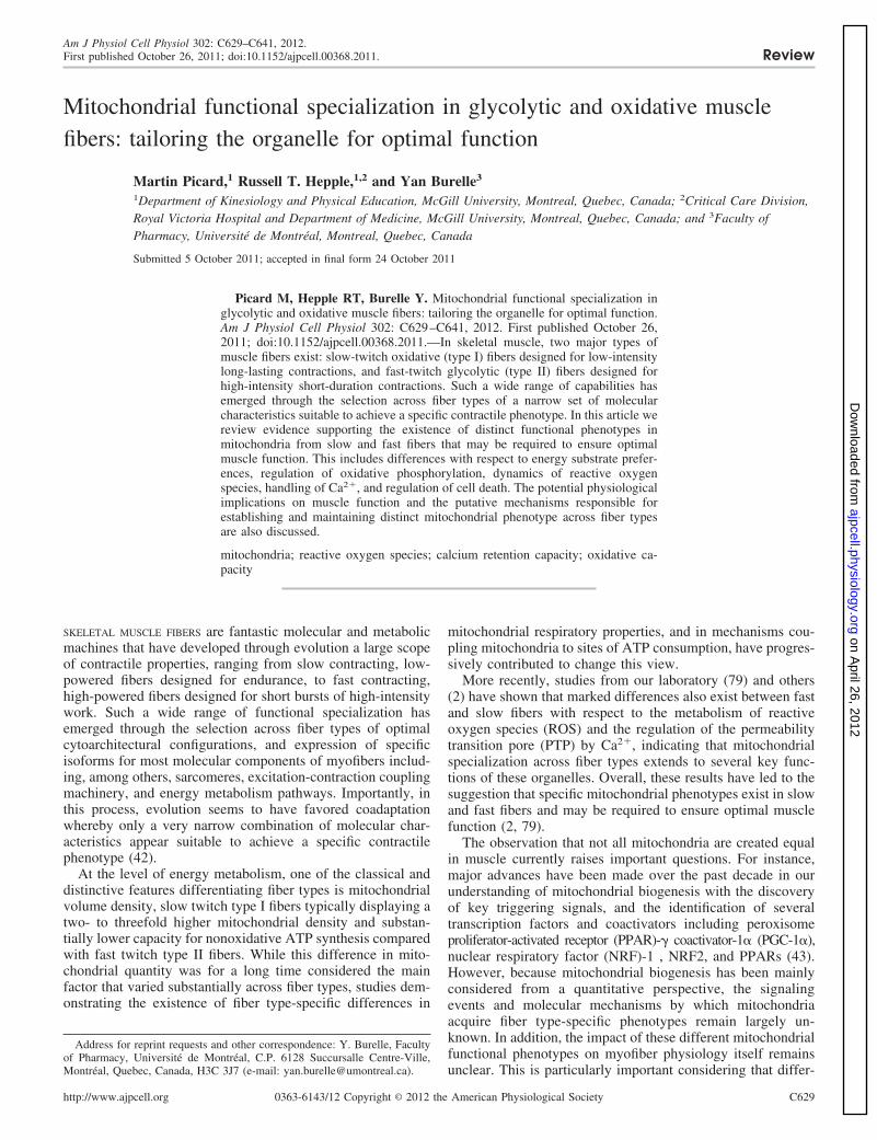
Mitochondrial functional specialization in glycolytic and oxidative musclefibers: tailoring the organelle for optimal function
Martin Picard,1 Russell T. Hepple,1,2 and Yan Burelle3
1Department of Kinesiology and Physical Education, McGill University, Montreal, Quebec, Canada; 2Critical Care Division,Royal Victoria Hospital and Department of Medicine, McGill University, Montreal, Quebec, Canada; and 3Faculty ofPharmacy, Université de Montréal, Montreal, Quebec, Canada
Submitted 5 October 2011; accepted in final form 24 October 2011
Picard M, Hepple RT, Burelle Y. Mitochondrial functional specialization inglycolytic and oxidative muscle fibers: tailoring the organelle for optimal function.Am J Physiol Cell Physiol 302: C629–C641, 2012. First published October 26,2011; doi:10.1152/ajpcell.00368.2011.—In skeletal muscle, two major types ofmuscle fibers exist: slow-twitch oxidative (type I) fibers designed for low-intensitylong-lasting contractions, and fast-twitch glycolytic (type II) fibers designed forhigh-intensity short-duration contractions. Such a wide range of capabilities hasemerged through the selection across fiber types of a narrow set of molecularcharacteristics suitable to achieve a specific contractile phenotype. In this article wereview evidence supporting the existence of distinct functional phenotypes inmitochondria from slow and fast fibers that may be required to ensure optimalmuscle function. This includes differences with respect to energy substrate prefer-ences, regulation of oxidative phosphorylation, dynamics of reactive oxygenspecies, handling of Ca2�, and regulation of cell death. The potential physiologicalimplications on muscle function and the putative mechanisms responsible forestablishing and maintaining distinct mitochondrial phenotype across fiber typesare also discussed.
mitochondria; reactive oxygen species; calcium retention capacity; oxidative ca-pacity
SKELETAL MUSCLE FIBERS are fantastic molecular and metabolicmachines that have developed through evolution a large scopeof contractile properties, ranging from slow contracting, low-powered fibers designed for endurance, to fast contracting,high-powered fibers designed for short bursts of high-intensitywork. Such a wide range of functional specialization hasemerged through the selection across fiber types of optimalcytoarchitectural configurations, and expression of specificisoforms for most molecular components of myofibers includ-ing, among others, sarcomeres, excitation-contraction couplingmachinery, and energy metabolism pathways. Importantly, inthis process, evolution seems to have favored coadaptationwhereby only a very narrow combination of molecular char-acteristics appear suitable to achieve a specific contractilephenotype (42).
At the level of energy metabolism, one of the classical anddistinctive features differentiating fiber types is mitochondrialvolume density, slow twitch type I fibers typically displaying atwo- to threefold higher mitochondrial density and substan-tially lower capacity for nonoxidative ATP synthesis comparedwith fast twitch type II fibers. While this difference in mito-chondrial quantity was for a long time considered the mainfactor that varied substantially across fiber types, studies dem-onstrating the existence of fiber type-specific differences in
mitochondrial respiratory properties, and in mechanisms cou-pling mitochondria to sites of ATP consumption, have progres-sively contributed to change this view.
More recently, studies from our laboratory (79) and others(2) have shown that marked differences also exist between fastand slow fibers with respect to the metabolism of reactiveoxygen species (ROS) and the regulation of the permeabilitytransition pore (PTP) by Ca2�, indicating that mitochondrialspecialization across fiber types extends to several key func-tions of these organelles. Overall, these results have led to thesuggestion that specific mitochondrial phenotypes exist in slowand fast fibers and may be required to ensure optimal musclefunction (2, 79).
The observation that not all mitochondria are created equalin muscle currently raises important questions. For instance,major advances have been made over the past decade in ourunderstanding of mitochondrial biogenesis with the discoveryof key triggering signals, and the identification of severaltranscription factors and coactivators including peroxisomeproliferator-activated receptor (PPAR)-� coactivator-1� (PGC-1�),nuclear respiratory factor (NRF)-1 , NRF2, and PPARs (43).However, because mitochondrial biogenesis has been mainlyconsidered from a quantitative perspective, the signalingevents and molecular mechanisms by which mitochondriaacquire fiber type-specific phenotypes remain largely un-known. In addition, the impact of these different mitochondrialfunctional phenotypes on myofiber physiology itself remainsunclear. This is particularly important considering that differ-
Address for reprint requests and other correspondence: Y. Burelle, Facultyof Pharmacy, Université de Montréal, C.P. 6128 Succursalle Centre-Ville,Montréal, Quebec, Canada, H3C 3J7 (e-mail: [email protected]).
Am J Physiol Cell Physiol 302: C629–C641, 2012.First published October 26, 2011; doi:10.1152/ajpcell.00368.2011. Review
0363-6143/12 Copyright © 2012 the American Physiological Societyhttp://www.ajpcell.org C629
on April 26, 2012
ajpcell.physiology.orgD
ownloaded from
ences in mitochondrial function may influence a number ofcellular variables including cytosolic Ca2�, redox state ofpyridine nucleotide pools, level of reactive oxygen and nitro-gen species, as well as cell death signaling. Finally, the exis-tence of distinct mitochondrial functional phenotypes in slowand fast muscle fibers in normal muscle could have an impor-tant impact on how we judge whether mitochondria are in-volved in muscle dysfunction in a number of pathologicalstates in which changes in fiber type are suspected or known tooccur.
Building on the above-mentioned concept of coadaptation ofmuscle properties, this review will provide an overview of theexperimental evidence currently available to support the exis-tence of mitochondrial functional specialization between fibertypes. We will focus on three functional subcategories forwhich data are available, namely: 1) respiratory properties andregulation of energy exchange; 2) metabolism of ROS; and3) regulation of the PTP, particularly in relation to Ca2�. Foreach of these subsystems, we will discuss the potential phys-iological implications on muscle function, and when possible,the molecular mechanisms that may underlie mitochondrialspecialization. We supplement our discussion of the currentknowledge by suggesting research avenues that will contributeto our expanding understanding of the mechanisms underlyingthe creation and maintenance of specific mitochondrial pheno-types between different muscle fiber types.
Mitochondrial Functional Specialization in Skeletal Muscle
Respiratory properties and coupling to cellular ATPases.Several studies have compared different intrinsic respiratoryproperties of mitochondria in slow and fast skeletal muscleincluding 1) respiratory capacities with various combinationsof substrates; 2) activities of the TCA cycle, �-oxidationpathway, and respiratory chain enzymes; 3) coupling efficiencybetween respiration and phosphorylation, proton conductance,as well as membrane properties; and 4) regulation of respira-tion by ADP and mitochondrial coupling to cellular ATPasesthrough various mechanisms, such as the creatine kinase shut-tle.
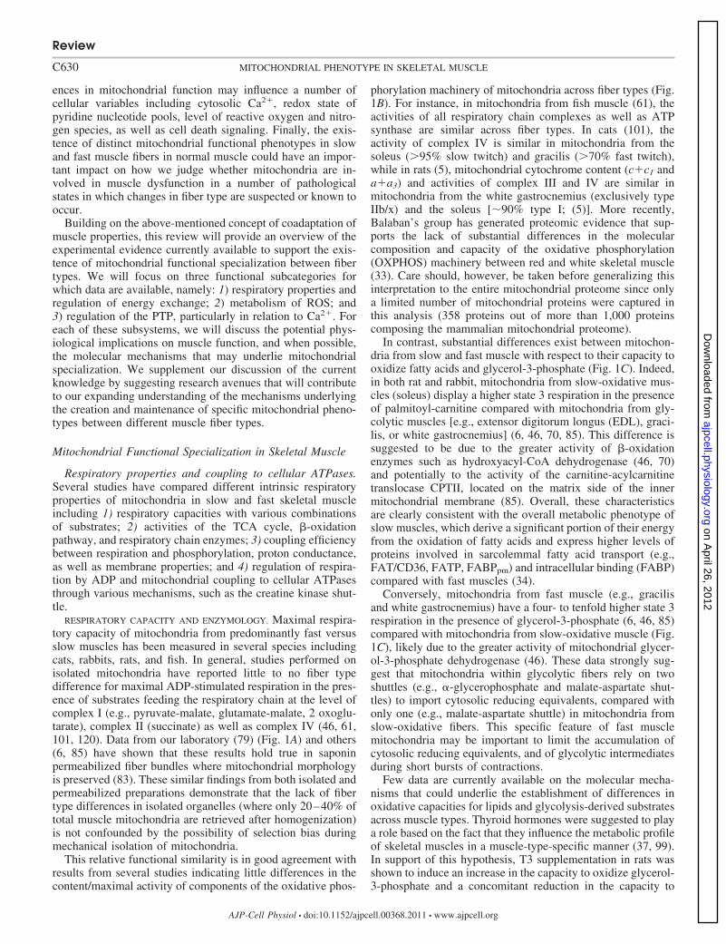
RESPIRATORY CAPACITY AND ENZYMOLOGY. Maximal respira-tory capacity of mitochondria from predominantly fast versusslow muscles has been measured in several species includingcats, rabbits, rats, and fish. In general, studies performed onisolated mitochondria have reported little to no fiber typedifference for maximal ADP-stimulated respiration in the pres-ence of substrates feeding the respiratory chain at the level ofcomplex I (e.g., pyruvate-malate, glutamate-malate, 2 oxoglu-tarate), complex II (succinate) as well as complex IV (46, 61,101, 120). Data from our laboratory (79) (Fig. 1A) and others(6, 85) have shown that these results hold true in saponinpermeabilized fiber bundles where mitochondrial morphologyis preserved (83). These similar findings from both isolated andpermeabilized preparations demonstrate that the lack of fibertype differences in isolated organelles (where only 20–40% oftotal muscle mitochondria are retrieved after homogenization)is not confounded by the possibility of selection bias duringmechanical isolation of mitochondria.
This relative functional similarity is in good agreement withresults from several studies indicating little differences in thecontent/maximal activity of components of the oxidative phos-
phorylation machinery of mitochondria across fiber types (Fig.1B). For instance, in mitochondria from fish muscle (61), theactivities of all respiratory chain complexes as well as ATPsynthase are similar across fiber types. In cats (101), theactivity of complex IV is similar in mitochondria from thesoleus (�95% slow twitch) and gracilis (�70% fast twitch),while in rats (5), mitochondrial cytochrome content (c�c1 anda�a3) and activities of complex III and IV are similar inmitochondria from the white gastrocnemius (exclusively typeIIb/x) and the soleus [�90% type I; (5)]. More recently,Balaban’s group has generated proteomic evidence that sup-ports the lack of substantial differences in the molecularcomposition and capacity of the oxidative phosphorylation(OXPHOS) machinery between red and white skeletal muscle(33). Care should, however, be taken before generalizing thisinterpretation to the entire mitochondrial proteome since onlya limited number of mitochondrial proteins were captured inthis analysis (358 proteins out of more than 1,000 proteinscomposing the mammalian mitochondrial proteome).
In contrast, substantial differences exist between mitochon-dria from slow and fast muscle with respect to their capacity tooxidize fatty acids and glycerol-3-phosphate (Fig. 1C). Indeed,in both rat and rabbit, mitochondria from slow-oxidative mus-cles (soleus) display a higher state 3 respiration in the presenceof palmitoyl-carnitine compared with mitochondria from gly-colytic muscles [e.g., extensor digitorum longus (EDL), graci-lis, or white gastrocnemius] (6, 46, 70, 85). This difference issuggested to be due to the greater activity of �-oxidationenzymes such as hydroxyacyl-CoA dehydrogenase (46, 70)and potentially to the activity of the carnitine-acylcarnitinetranslocase CPTII, located on the matrix side of the innermitochondrial membrane (85). Overall, these characteristicsare clearly consistent with the overall metabolic phenotype ofslow muscles, which derive a significant portion of their energyfrom the oxidation of fatty acids and express higher levels ofproteins involved in sarcolemmal fatty acid transport (e.g.,FAT/CD36, FATP, FABPpm) and intracellular binding (FABP)compared with fast muscles (34).
Conversely, mitochondria from fast muscle (e.g., gracilisand white gastrocnemius) have a four- to tenfold higher state 3respiration in the presence of glycerol-3-phosphate (6, 46, 85)compared with mitochondria from slow-oxidative muscle (Fig.1C), likely due to the greater activity of mitochondrial glycer-ol-3-phosphate dehydrogenase (46). These data strongly sug-gest that mitochondria within glycolytic fibers rely on twoshuttles (e.g., �-glycerophosphate and malate-aspartate shut-tles) to import cytosolic reducing equivalents, compared withonly one (e.g., malate-aspartate shuttle) in mitochondria fromslow-oxidative fibers. This specific feature of fast musclemitochondria may be important to limit the accumulation ofcytosolic reducing equivalents, and of glycolytic intermediatesduring short bursts of contractions.
Few data are currently available on the molecular mecha-nisms that could underlie the establishment of differences inoxidative capacities for lipids and glycolysis-derived substratesacross muscle types. Thyroid hormones were suggested to playa role based on the fact that they influence the metabolic profileof skeletal muscles in a muscle-type-specific manner (37, 99).In support of this hypothesis, T3 supplementation in rats wasshown to induce an increase in the capacity to oxidize glycerol-3-phosphate and a concomitant reduction in the capacity to
Review
C630 MITOCHONDRIAL PHENOTYPE IN SKELETAL MUSCLE
AJP-Cell Physiol • doi:10.1152/ajpcell.00368.2011 • www.ajpcell.org
on April 26, 2012
ajpcell.physiology.orgD
ownloaded from
oxidize octanoyl-carnitine (6), producing the profile observedin fast-glycolytic fibers. However, this effect of T3 was ob-served in slow-oxidative muscles, which express significantamounts of thyroid receptors, but not in fast-glycolytic mus-cles, in which thyroid hormone receptors are less abundant (6).Therefore, it is difficult to explain how physiological levels ofT3 could underlie the difference in substrate-specific oxidativecapacities between slow and fast muscles since the impact ofT3 should, if anything, bring slow and fast mitochondria closertogether in terms of their substrate preference than wouldotherwise exist in the absence of T3.
COUPLING EFFICIENCY, PROTON CONDUCTANCE, AND MEMBRANE
PROPERTIES. Very few studies have investigated whether vari-ations across fiber types exist with respect to coupling effi-ciency of oxidative phosphorylation. This parameter, known asthe P/O ratio, is conventionally determined by measuring theamount of oxygen required to rephosphorylate a knownamount of ADP. It is well established that the P/O ratiodecreases as respiration is progressively reduced from maximalADP-stimulated respiration to submaximal respiration rates(35). The main factor responsible for this phenomenon is the
increasing contribution of the proton leak of the inner mem-brane to respiration as the rate of oxidative phosphorylation isprogressively lowered (15). Early studies comparing P/O ratiosin mitochondria isolated from different skeletal muscle fibersin the rat failed to detect differences when using pyruvate orpalmitoyl-carnitine as substrate (74). In these experiments, P/Ovalues were only measured at maximal respiration rates in thepresence of saturating amounts of ADP, which did not allowexclusion of differences at more physiologically relevant sub-maximal rates of respiration.
However, more recent studies (70) comparing P/O ratiosover the entire range of respiratory capacity in mitochondriaisolated from the rat soleus and the fast EDL also reported nosignificant difference with pyruvate or palmitoyl-carnitine asrespiratory substrates. On the other hand, direct measurementof proton leak kinetics in fish muscle showed that proton leakwas greater in mitochondria from white muscle compared withmitochondria from red muscle (61). Since this difference wasonly apparent when leak values were normalized per unit ofcomplex IV activity, but not when expressed per milligram oftotal mitochondrial proteins, the authors argued that under
Fig. 1. Quantitative and qualitative respiratory parameters measured in permeabilized myofibers. A: mitochondrial respiratory rates normalized for themitochondrial volume marker citrate synthase (CS) under different conditions do not differ between the fast-type glycolytic extensor digitorum longus (EDL)and mixed gastrocnemius (mGas) muscles (blue shading), and the slow-type oxidative soleus (Sol) and adductor longus (AL) muscles (green shading). State 2GM, complex I-driven respiration with glutamate (2 mM) and malate (10 mM); state 3 GM, GM � ADP (2 mM); TMPD, complex IV-driven respiration withtetramethylphenylenediamine (0.5 mM) and ascorbate (5 mM). B: respiratory ratios representing the relative activity of different components of the respiratorychain. State 3 GMS, complex I � II driven respiration with GM and succinate (10 mM). C: maximal respiratory rate with the lipid substrate palmitoyl-carnitine(Palm-carn) is fourfold lower in the glycolytic white gastrocnemius (wGas) muscle than in the oxidative Sol, whereas maximal respiratory rate with the substrateglycerol-3-phosphate (glycerol-3-P) is threefold higher in wGas than in Sol. D: apparent affinity for ADP (Km) is lower in wGas than in Sol. The fast wGas Km
for ADP is not sensitive to the addition of creatine (�Cr, 20 mM), whereas the slow Sol Km for ADP is reduced by 70% with �Cr. Data shown in A and B arefrom Ref. 82. All measurements performed on rat muscle were as in Picard et al. (81, 82) (A and B); Ponsot et al. (85) (C), and Burelle and Hochachka (18)(D). *Statistical significance (P � 0.05) from glycolytic muscles; &statistical significance from no creatine (�Cr) conditions (P � 0.05). P values were obtainedfrom unpaired t-test assuming unequal variance between groups. Data are presented as means SE; n 8–12 per group.
Review
C631MITOCHONDRIAL PHENOTYPE IN SKELETAL MUSCLE
AJP-Cell Physiol • doi:10.1152/ajpcell.00368.2011 • www.ajpcell.org
on April 26, 2012
ajpcell.physiology.orgD
ownloaded from
some circumstances, normalization to a marker of the respira-tory chain capacity may thus be more appropriate than totalprotein, particularly for functions related to the inner mem-brane (61). In addition, this study reported greater membranefluidity in mitochondria from red muscle compared with theircounterparts from white muscle, possibly due to variations inphospholipid profile (e.g., chain length, saturation, cardiolipincontent) (61). However, no information is available on thephospholipid profile in mitochondria across fiber types, and itremains unclear how this could affect the in vivo activity ofmembrane bound proteins. Taken as a whole, these data thussuggest that while fiber type differences may exist with respectto mitochondrial membrane properties and proton leak,whether this results in fiber type differences in mitochondrialcoupling efficiency remains to be demonstrated conclusively.
REGULATION OF RESPIRATION BY ADP AND MITOCHONDRIAL COU-
PLING TO CELLULAR ATPASES. One of the most striking differ-ences between mitochondria from fast-glycolytic and slow-oxidative fibers concerns the sensitivity of respiration to ADP,and the mechanisms coupling mitochondrial ATP supply tosubcellular sites of ATP consumption (53, 57, 93, 96–98).These properties were largely uncovered following the devel-opment of saponin-permeabilized fibers, which allowed studyof mitochondria in a relatively preserved cytoarchitecturalenvironment (58, 93, 98). Using this approach, several studies,including ours, have shown that mitochondria in slow-oxida-tive muscles, such as the heart and the soleus, display anapparent Km (Michaelis-Menten constant) for exogenous ADPin range of 200–500 �M, which is approximately 10-foldhigher than Km values measured in isolated mitochondria (53,57, 96, 97). In contrast, mitochondria within fast-twitch gly-colytic fibers display a Km for ADP between 10 and 30 �M,closer to that observed in isolated mitochondria (Fig. 1D) (53,57, 96, 97). A low permeability of the outer mitochondrialmembrane (OMM) to ADP is suggested as one of the mecha-nisms contributing to the high Km value observed in mitochon-dria from slow-oxidative fibers (53, 57, 96, 97). This is mainlybased on the observation that disruption of the OMM using awell-controlled hypo-osmotic shock lowers the Km for ADP tothe values observed in isolated mitochondria and in mitochon-dria from fast-glycolytic fibers (57). The molecular mecha-nisms underlying fiber type-specific OMM permeability toADP are currently unknown but could involve differences inthe conductance and isoform expression of voltage-dependentanion channel (VDACs), which are responsible for the trans-port of a number of solutes across the MOM including ade-nylates (24, 32, 53, 57, 60, 93, 96, 97). Furthermore, recentevidence demonstrates that mitochondrial affinity for ADP ismodulated by the contractile state of myofibers (76), withcontraction lowering the Km for ADP. This effect was morepronounced in fast fibers, suggesting that different mechanismslinking contractile state and mitochondrial energy exchangemay exist between slow and fast muscles (76).
The other factor likely explaining the low sensitivity ofmitochondria to exogenous ADP in slow-oxidative muscle maybe compartmentalization of energy exchange, which restrictsthe access of exogenous ADP to mitochondria. Indeed, func-tional units, termed intracellular energetic units (ICEUs), havebeen well described in the heart (16, 50, 84, 93, 95, 102), andsome evidence for their existence in the soleus muscle has beenobtained (102). Within these ICEUs, ATP and ADP are focally
released and directly transferred by channeling between mito-chondria on the one hand, and sarcoplasmic reticulum (SR)-Ca2� and myofibrillar (MF-Mg2�) ATPases on the other hand(16, 50, 84, 93, 95, 102). Three lines of evidence, obtained inpermeabilized fibers, have led to this conclusion. First, in thesefibers, SR-Ca2� loading capacity and capacity to relax rigortension are much higher when supported by ATP generated byoxidative phosphorylation compared with exogenous ATPadded to the incubation medium (50). This indicates thatmitochondria-derived ATP has a preferential access to SR-Ca2�/MF-Mg2� ATPases (50). Second, and in line with theseresults, similar mitochondrial respiration rates can be achievedwith 40 times less ADP if ADP is derived from SR-Ca2�/MF-Mg2� ATPase activity, compared with when it is added di-rectly in the incubation medium (95, 102). And third, even inthe presence of a powerful exogenous ADP trap system in theincubation media, ADP produced endogenously by the hydro-lysis of ATP can still stimulate mitochondrial respiration,providing direct evidence for a compartmentalization of energyand a regulatory signal between mitochondria and SR-Ca2�/MF-Mg2� ATPases in slow-oxidative muscle (16, 95, 102).
Currently, the factors involved in this form of compartmen-talization of energy and regulatory signal exchange are unclear.One hypothesis is that it is related to the structural arrangementof mitochondria around myofibrils (72, 84, 95). Indeed, inoxidative muscle fibers, mitochondria appear to be clustered atsites of high ATP demand and are organized into highlyordered elongated structures forming contacts with the SR andhaving extensive branching across the A-band area of thesarcomere where Mg2� ATPases are most abundant (see Fig.3A) (72, 95). This configuration provides the physical proxim-ity between mitochondria and SR-Ca2�/MF-Mg2� ATPasesthat is required to observe ICEUs (72, 84, 93, 95). In contrast,in fast-glycolytic muscle fibers in which mitochondria are lessabundant, mostly located at the level of Z-lines with no transA-band branches (72), the spatial configuration is less compat-ible with the formation of ICEUs (72, 95). In fact, Ventura-Clapier’s group reported that ICEUs were absent in the mousewhite gastrocnemius muscle (51). Moreover, they elegantlydemonstrated that in the white gastrocnemius muscle of micedeficient in the sarcomeric creatine kinase isoform (MM-CK),a compensatory proliferation and spatial reconfiguration ofmitochondria occurs, which coincides with the emergence ofdirect energy channeling within ICEUs (51), thus providingstrong evidence for the role of mitochondrial spatial configu-ration in the development of ICEUs, and more generally onmitochondrial functional specialization across fiber types. Ob-viously, because specialization of mitochondrial energy ex-change across fiber types is intimately linked to the overalldesign of slow and fast fibers, the molecular regulation under-lying this specialization is likely to involve fundamental sig-naling factors with broad impact on myogenesis (71, 94).
Another noticeable difference between mitochondria fromslow-oxidative and fast-glycolytic fibers is the functional cou-pling between mitochondrial ATP production and sites of ATPconsumption through the creatine kinase (CK) system (Fig.1D). In slow-oxidative muscle, mitochondria express sarco-meric mitochondrial CK (sMt-CK), an isoform located in theintermembrane space, which is functionally coupled with theATP/ADP exchanger (ANT) and VDAC channels (115).sMt-CK thus uses ATP produced in the mitochondria to re-
Review
C632 MITOCHONDRIAL PHENOTYPE IN SKELETAL MUSCLE
AJP-Cell Physiol • doi:10.1152/ajpcell.00368.2011 • www.ajpcell.org
on April 26, 2012
ajpcell.physiology.orgD
ownloaded from
generate ADP locally near the ANT, thereby exerting a strongcontrol on oxidative phosphorylation. Together with cytosolicCK isoforms located at the vicinity of several key ATPases(see Ref. 115 for review), sMt-CK also ensures efficient energyand signal transfer through reversible phosphotransfer reac-tions (96, 115). In contrast, this CK shuttle system is nonex-istent in fast-glycolytic muscle, due to the low levels ofsMt-CK (87). However, the mechanisms underlying the fibertype-specific expression of sMt-CK, one of the major factorsresponsible for the presence of CK shuttle in slow muscles, stillremains unknown.
Taken together, the data available thus indicate that majordifferences exist between slow-oxidative and fast-glycolyticmuscle fibers with respect to the mechanisms coupling mito-chondria to sites of ATP consumption. This is likely explain-able by the necessity in oxidative fibers to have efficient energydelivery despite the strong diffusional constraints imposed bythe highly organized and densely packed intracellular environ-ment, contrasting with glycolytic fibers that rely much less onmitochondria for ATP production. Again, however, the signal-ing events and molecular mechanisms underlying this special-ization remain largely unknown.
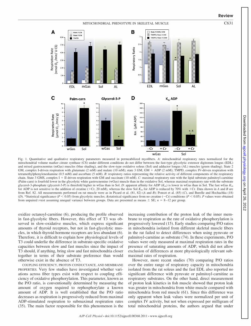
Metabolism of reactive oxygen species. Three studies fromour laboratories (79, 80, 82) and another (2) have demonstratedthe existence of very significant differences in the mitochon-drial metabolism of ROS across fiber types. In these experi-ments, net mitochondrial H2O2 release was measured usingAmplex red both in permeabilized fibers (2, 79, 80, 82) and inisolated mitochondria (79). Results showed that H2O2 releasewas two- to threefold higher from mitochondria in whitegastrocnemius compared with the soleus muscle under basalstate 2 respiration in the presence of complex I or complex IIsubstrates. As a consequence, free radical leak expressed as apercentage of total electron flux through the respiratory chainwas 3.5-fold greater in mitochondria from the white gastroc-nemius compared with that of the soleus muscle (79). Abroader examination of two fast dominant versus two slowdominant muscles confirms the existence of this fiber typedifference in H2O2 release (Fig. 2A) (82).
Currently, the mechanisms underlying this substantial dif-ference in H2O2 release are not fully understood. BecauseAmplex red detects H2O2 that diffuses outside mitochondria,this could be due to 1) a greater production of H2O2, and/or 2) alower endogenous H2O2 scavenging capacity in mitochondriafrom glycolytic muscle compared with mitochondria fromoxidative muscle (Fig. 2, B–D) (see Refs. 14 and 110 forreviews).
H2O2 production in mitochondria is largely determined bysuperoxide production (O2
·�), which occurs mainly at the levelof complexes I and III of the respiratory chain (14, 110).Studies in isolated mitochondria indicate that membrane po-tential (� ) is a key determinant of O2
·� production at thesesites, which increases exponentially in response to small in-creases in � , particularly in the upper range values [i.e.,175–185 mV (39, 55, 107, 112)]. However, as discussed in thesection Coupling efficiency, proton conductance and mem-brane properties, indices of proton leak and direct measures of� suggest that membrane potential is not higher in mitochon-dria from glycolytic muscle, and may in fact tend to be lowerthan in mitochondria from oxidative muscle (61). Although� is a major determinant of O2
·� production, other mecha-
nisms could contribute, including variations in the redox stateof mitochondria (7), the stoichiometry-activity ratios of therespiratory chain complexes (59), and the susceptibility toproton pump slipping (52). However, whether fiber type dif-ferences in these properties exist and translate into differentrates of O2
·� production has not been studied. It should benoted that, although Mn-SOD activity is essential for theconversion of superoxide into H2O2, differences in the activityof this enzyme are unlikely to account for variations in netmitochondrial H2O2 release across fiber types. Indeed, previ-ous work by Van Remmen and colleagues showed that largevariations in Mn-SOD content in transgenic animals (heterozy-gous knock out or overexpression of Mn-SOD) do not translateinto measurable changes in net H2O2 release in skeletal musclemitochondria using the Amplex red system, reflecting the factthat this enzyme is in excess capacity relative to O2
·� produc-tion (48, 66).
In contrast, H2O2 scavenging by endogenous antioxidantsystems was recently shown to have a significant impact onmitochondrial H2O2 release in the Amplex red system (109). Inthis recent study, chemical depletion of GSH in isolated skel-etal muscle mitochondria was shown to increase net H2O2
release two- to threefold. This was observed with respiratorysubstrates and respiratory chain inhibitors targeting differentsites, and yielding different rates of O2
·� production (109).While the main objective of this study was to provide acorrection method to better estimate O2
·� production frommeasurements of H2O2 efflux, the data presented clearly sug-gest that fiber type differences in mitochondrial H2O2 releasecould be attributable to variations in endogenous H2O2 scav-enging capabilities (109). In fact, direct measurements inpermeabilized muscle fibers have shown that the capacity ofmitochondria from fast-glycolytic fibers to scavenge an exog-enous H2O2 load is approximately 40–50% lower comparedwith mitochondria from slow-oxidative fibers (2).
In addition, the activities of important antioxidant enzymesare significantly lower in glycolytic muscle compared withoxidative muscle, even when expressed per unit of citratesynthase activity to take into account differences in mitochon-drial contents (Fig. 2, B–D). This is particularly striking forglutathione peroxidase, the main mitochondrial H2O2 scaveng-ing enzyme, which is on average 88% (range: 77–95) lower inglycolytic compared with oxidative muscles. Taken together,the data available thus suggest that H2O2 buffering capacity permitochondrial unit differs considerably in glycolytic comparedwith oxidative fibers and may account for differences in H2O2
emitting potential.Although the mechanism underlying this difference is un-
clear, it likely involves fiber type differences in the expressionlevel of PGC-1� and PGC-1�. These transcriptional coactiva-tors, in addition to their effect on mitochondrial biogenesis,were shown to regulate the expression level of many ROSdetoxifying enzymes mRNA (SOD1, SOD2, Gpx1, catalase)both at baseline and in response to H2O2 (106), thus allowingto scale the activity of antioxidant systems to the mitochondrialbiomass (Fig. 2E). However, additional mechanisms (i.e., sig-nal amplification in slow muscle; epigenetic silencing in fastmuscles) must exist to account for the net greater abundance ofantioxidant relative to mitochondrial mass (per citrate syn-thase) in slow muscles than in fast muscles.
Review
C633MITOCHONDRIAL PHENOTYPE IN SKELETAL MUSCLE
AJP-Cell Physiol • doi:10.1152/ajpcell.00368.2011 • www.ajpcell.org
on April 26, 2012
ajpcell.physiology.orgD
ownloaded from

Fig. 2. Reactive oxygen species (ROS) metabolism differs between glycolytic EDL and mGas muscles (blue shading), and the slow-type oxidative Sol and ALmuscles (green shading). A: mitochondrial hydrogen peroxide (H2O2) release measured with the Amplex Red system, normalized for citrate synthase, is higherin glycolytic EDL and mGas muscles than in oxidative Sol and AL muscles. B–D: when normalized for mitochondrial content, activity of endogenous antioxidantenzymes manganese and copper-zinc superoxide dismutases (total SOD, U/mg protein) (B), glutathione peroxidase (GPx, �mol·min�1·g protein�1) (C), andcatalase (Cat, K/g protein) (D) is lower in fast glycolytic muscles. AU, arbitrary units. E: overlap among signaling pathways driving mitochondrial biogenesisand antioxidant defenses involves PGC-1�, which coactivates the transcription of both types of genes under the influence of redox-sensitive signaling pathways(106). Data shown in A–D are from Ref. 82. *Statistical significance (P � 0.05) from glycolytic muscles; &statistical significance from Sol muscle (P � 0.05);†statistical significance from EDL muscle (P � 0.05). P values were obtained from unpaired t-test assuming unequal variance between groups. Data are presentedas means SE; n 8 per group.
Review
C634 MITOCHONDRIAL PHENOTYPE IN SKELETAL MUSCLE
AJP-Cell Physiol • doi:10.1152/ajpcell.00368.2011 • www.ajpcell.org
on April 26, 2012
ajpcell.physiology.orgD
ownloaded from
From a physiological perspective, there is a clear rationalefor scaling antioxidant defenses to mitochondrial content. Inoxidative muscle with a large mitochondrial biomass, thisallows protection against oxidative stress (106). Conversely, infast muscle, greater ROS production per mitochondrial unitmay be required to maintain proper redox-dependent signalingdespite low mitochondrial content. In addition, greater capacityto generate mitochondrial ROS may contribute, together withother factors, to trigger adaptive mitochondrial biogenesiswhen glycolytic muscles are recruited more frequently. Al-though strong experimental evidence in support of this hypoth-esis is still lacking, it is nonetheless consistent with data fromseveral recent studies showing that 1) enhanced ROS signalingin muscle from SOD1 knockout mice results in mitochondrialproliferation (47); 2) overexpression of SOD1 in type IIB fibersblocks training-induced mitochondrial biogenesis (65); and3) exogenous antioxidant supplementation in humans bluntsthe benefits of exercise training in terms of insulin sensitivityand response of mitochondrial biogenesis signaling (90, 108).Overall specialization of mitochondrial ROS metabolism,rather than a simple 1:1 scaling between mitochondrial contentand antioxidant capacity, thus appears to be important fornormal function of glycolytic and oxidative fibers.
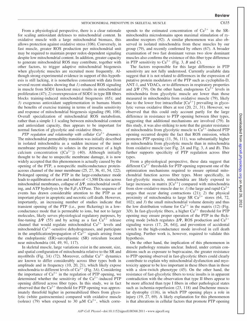
PTP regulation and relationship with cellular Ca2� dynamics.The mitochondrial permeability transition was initially describedin isolated mitochondria as a sudden increase of the innermembrane permeability to solutes in the presence of a highcalcium concentration ([Ca2�]) (40). Although initiallythought to be due to unspecific membrane damage, it is nowwidely accepted that this phenomenon is actually caused by theopening of the PTP, a nonspecific multiconductance protein-aceous channel of the inner membrane (25, 27, 36, 41, 54, 122).Prolonged opening of the PTP in the large-conductance modeleads to equilibration of ions and solutes of �1,500 Da size acrossmitochondrial membranes, collapse of � , mitochondrial swell-ing, and ATP hydrolysis by the F0F1ATPase. This sequence ofevents has drawn considerable attention to the PTP as animportant player in apoptotic and necrotic cell death. However,importantly, an increasing number of studies indicate thattransient opening of the PTP (i.e., pore flickering) in a low-conductance mode that is permeable to ions, but not to largermolecules, likely serves physiological regulatory purposes, byfine-tuning � (55) and by acting as a fast Ca2� releasechannel that would regulate mitochondrial Ca2� levels andmitochondrial Ca2�-sensitive dehydrogenases, and participatein the amplification/propagation of Ca2� signals arising fromthe endoplasmic (ER)-sarcoplasmic (SR) reticulum locatednear mitochondria (44, 49, 91, 117).
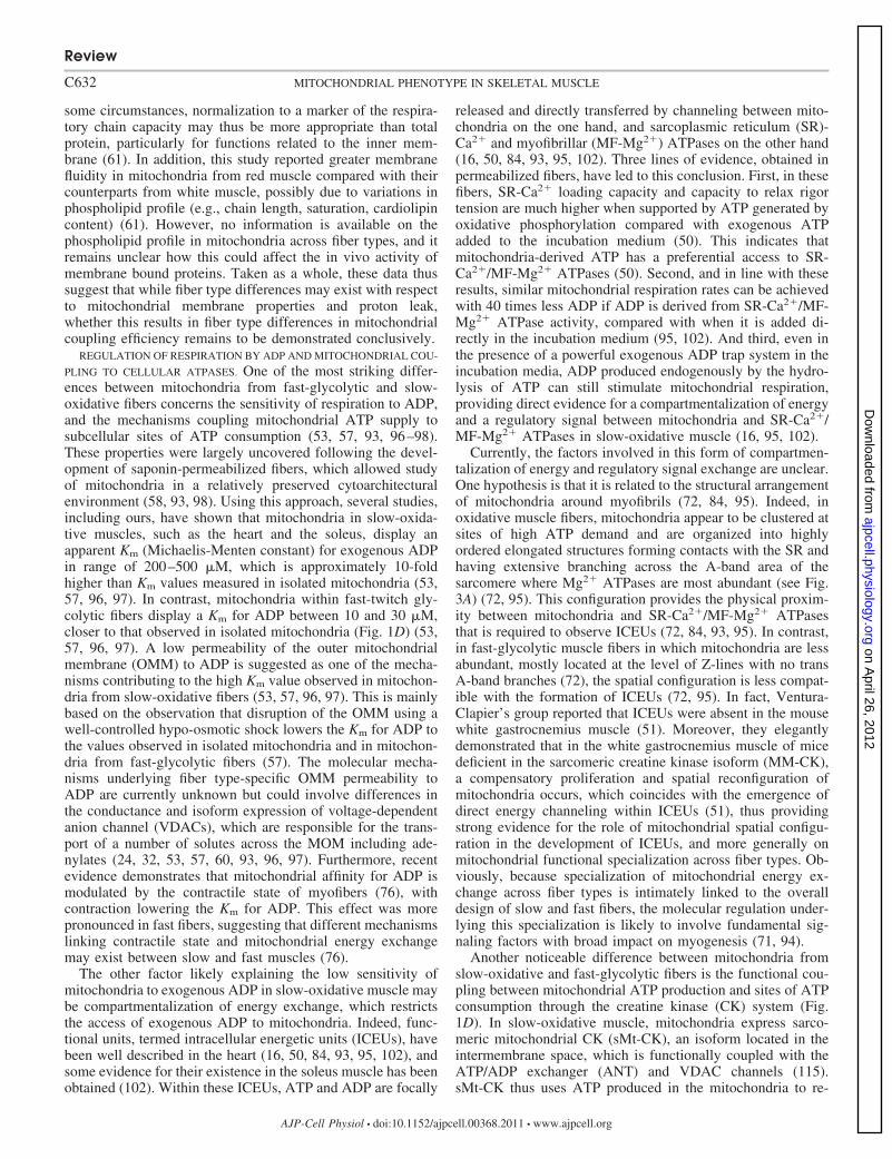
In skeletal muscle, large variations exist in the amount, size,and spatial configuration of mitochondria relative to the SR andmyofibrils (Fig. 3A) (72). Moreover, cellular Ca2� dynamicsare known to differ considerably across fiber types both inamplitude and in frequency (10, 20, 21), which likely exposemitochondria to different levels of Ca2� (Fig. 3A). Consideringthe importance of Ca2� in the regulation of PTP opening, wedetermined whether the sensitivity of the Ca2�-induced PTPopening differed across fiber types. In this study, we in factobserved that the Ca2� threshold for PTP opening was approx-imately threefold higher in permeabilized fibers from glyco-lytic (white gastrocnemius) compared with oxidative muscle(soleus) (79) when exposed to 30 �M Ca2�, which corre-
sponds to the estimated concentration of Ca2� in the SR-mitochondria microdomains upon maximal stimulation of ry-anodine receptors (92). A similar phenomenon was also ob-served in isolated mitochondria from these muscles by ourgroup (79), and recently confirmed by others (67). A broaderexamination of two fast dominant versus two slow dominantmuscles also confirms the existence of this fiber type differencein PTP sensitivity to Ca2� (Fig. 3, B and C).
The factors responsible for this large difference in PTPsensitivity are not fully understood. Currently available datasuggest that it is not related to differences in the expression ofputative protein modulators of the PTP such as cyclophilin-D,ANT-1, and VDACs, or to differences in respiratory propertiesand � (79). On the other hand, endogenous Ca2� levels inmitochondria from glycolytic muscle are lower than thosemeasured in mitochondria from oxidative muscle (79), likelydue to the lower free intracellular [Ca2�] prevailing in glyco-lytic versus oxidative fibers at rest (20, 21, 31). However, weobserved that this could only account partly for the largedifference in resistance to PTP opening between fiber types,suggesting that additional mechanisms are involved (79). Inthis study, it was also striking to note that the greater resistanceof mitochondria from glycolytic muscle to Ca2�-induced PTPopening occurred despite the fact that ROS emission, whichpromotes PTP opening (11, 56, 111), was substantially higherin mitochondria from glycolytic muscle than in mitochondriafrom oxidative muscle (see Fig. 2A and Fig. 3, A and B). Thisfurther supports specificity of PTP regulation across fibertypes.
From a physiological perspective, these data suggest thatdifferent Ca2� thresholds for PTP opening represent one of theoptimization mechanisms required to ensure optimal mito-chondrial function across fiber types. More specifically, infast-glycolytic muscle, mitochondria are likely exposed tolarge increases in matrix [Ca2�] compared with mitochondriafrom slow-oxidative muscle due to: 1) the large and rapid Ca2�
surges that occur during contractions (21); 2) the physicalproximity of mitochondria to large SR Ca2� stores (64, 72,102); and 3) the small mitochondrial volume density and thusthe low distribution volume for Ca2� in the matrix (12) (Fig.3D). In fast-glycolytic fibers, a higher Ca2� threshold for PTPopening may ensure proper operation of the PTP in the flick-ering mode [which regulates � , ROS production and Ca2�
signaling waves (17, 28, 64)], and prevention of accidentalswitch to the high-conductance mode involved in cell deathsignaling. Further work is, however, required to validate thishypothesis.
On the other hand, the implication of this phenomenon inmuscle pathology remains unclear. Indeed, under certain con-ditions such as exposure to bupivacaine, the greater resistanceto PTP opening observed in fast-glycolytic fibers could clearlycontribute to explain why mitochondrial dysfunction and myo-toxicity appear to be less important in these fibers than in thosewith a slow-twitch phenotype (45). On the other hand, theresistance of fast-glycolytic fibers to toxic insults is in apparentcontradiction with the observation that type II fibers appear tobe more affected than type I fibers in other pathological statessuch as ischemia-reperfusion (23, 118) and Duchenne muscu-lar dystrophy (116), in which PTP opening plays a role ininjury (19, 27, 69). A likely explanation for this phenomenonis that alterations in cellular factors that promote PTP opening
Review
C635MITOCHONDRIAL PHENOTYPE IN SKELETAL MUSCLE
AJP-Cell Physiol • doi:10.1152/ajpcell.00368.2011 • www.ajpcell.org
on April 26, 2012
ajpcell.physiology.orgD
ownloaded from

Fig. 3. Considerable differences in cytoarchitecture and intracellular Ca2� transients exist between type I oxidative and type II glycolytic muscle fibers.A: schematic representation of myofibrils from fast type IIb and slow type I fibers. [Ca2�]ic, intracellular calcium concentration. [Adapted from Ogata andYamasaki (72)]. B: mitochondria uptake considerable amounts of Ca2� released from the sarcoplasmic reticulum (SR) during muscle contraction. Excess matrix[Ca2�] triggers the opening of the permeability transition pore (PTP), which has important functional consequences. PTP opening events are classified under twoconductance states: physiological and pathological opening. C: the amount of mitochondrial Ca2� uptake required to trigger PTP opening—calcium retentioncapacity, measured with the Ca2� dye Calcium green—is higher in glycolytic EDL and mGas muscles than in oxidative Sol and AL muscles. D: upon externalCa2� challenge, the time required to trigger opening of the PTP is higher in EDL and mGas than in Sol and AL. Data shown in B and C are from Ref. 82. Alldata were obtained as described in Picard et al. (79, 81). *Statistical significance (P � 0.05) from glycolytic muscles; &statistical significance from Sol muscle(P � 0.05). P values were obtained from unpaired t-test assuming unequal variance between groups. Data are presented as means SE; n 8 per group.
Review
C636 MITOCHONDRIAL PHENOTYPE IN SKELETAL MUSCLE
AJP-Cell Physiol • doi:10.1152/ajpcell.00368.2011 • www.ajpcell.org
on April 26, 2012
ajpcell.physiology.orgD
ownloaded from
are greater in fast fibers than in slow fibers as a result of thesepathological states and overwhelm the capacity of mitochon-dria to resist to permeability transition. Clearly, the involve-ment of mitochondria in cell death depends on the convergenceof several factors, which make it difficult to predict theirinvolvement in disease outcome.
Potential mechanisms underlying mitochondrial functionalspecialization. Very little is known about the signaling mech-anisms that account for the striking phenotypic differencesdescribed above. However, recent findings provide initial in-sights into this process and allow us to speculate about the mostprobable mechanisms responsible for establishing and main-taining distinct mitochondrial phenotypes within fast-glyco-lytic and slow-oxidative muscle fibers.
Before we outline these mechanisms, we must address thefact that mitochondrial functional differences in musclecells of different fiber type could potentially be due todifferent proportions of subsarcolemmal (SS) and intermyo-fibrillar (IMF) mitochondria that populate myofibers. SSmitochondria are densely packed beneath the plasma mem-brane, whereas IMF mitochondria are distributed betweenmyofibrils, as shown in Fig. 3A. Compared with fast fibers,slow fibers contain a similar volume of IMF mitochondria,but contain significantly more SS mitochondria. Interest-ingly, these two geographically different populations ofmitochondria contain different levels of key metabolic en-zymes (30) and have different functional properties (1, 22,73, 77, 103). Compared with IMF, SS mitochondria havebeen shown to have a lower oxidative capacity (22, 73, 77,103), to produce more ROS (1, 22, 103) and to be moresensitive to Ca2�-induced PTP (1). However, slow fiberscontain a greater proportion of SS mitochondria, have thesame oxidative capacity, and produce less ROS than fastfibers, which is inconsistent with the intrinsic properties ofSS relative to IMF mitochondria. Therefore, we concludethat variations in the proportions of SS and IMF mitochon-dria between fiber types do not account for fiber typedifferences in mitochondrial function.
Most of our knowledge regarding mitochondrial biogen-esis—the synthesis of new mitochondria—relates to trans-acting transcription factors [i.e., peroxisome proliferator-activated receptors (PPARs), nuclear respiratory factors(NRF1, NRF2), estrogen-related receptor-�], and coactiva-tors such as PPAR-� coactivator-� and � (PGC-1�, PGC-1�) (38, 88, 100). These nuclear transcriptional elements aremostly known to regulate mitochondrial content (i.e., vol-ume density) in muscle (119). As such, in slow-twitchoxidative muscle where mitochondrial mass is two to threetimes that of fast-twitch glycolytic muscle, PGC-1� isconstitutively expressed at higher levels (62), and higherstill in the tissue that is most dense in mitochondria, theheart (29). This tissue-specific difference in PCG-1� and inPPARs, which regulate the expression of many enzymes offat metabolism (e.g., �-oxidation cycle) (4), may thereforecontribute to explain differences in mitochondrial mass andsubstrate specificity among fiber types. However, for themost part, the discriminating functional differences amongmitochondria from muscles of different fiber type composi-tion illustrated in Figs. 1–3 are unlikely to be solely ex-plained by variations of trans-acting elements.
Instead, epigenetic mechanisms may prove key determi-nants of mitochondrial specialization across fiber types, byfine-tuning sensitivity/responsivity of the nuclear DNA tothe various transcriptional agents mentioned above. Epige-netics refers to a set of heritable but plastic mechanismscapable of stably modulating gene expression in response toenvironmental cues (68, 121). DNA methylation (104) andposttranslational modifications of histones are among themost heavily studied epigenetic mechanisms (86). Thesemolecular mechanisms acting on DNA can efficiently si-lence target nuclear genes, such as specific myosin heavychain subtypes (75). DNA methylation may play a particu-larly important role in muscle fiber differentiation andspecialization, as it does during satellite cell activation(105) and in the differentiation of other tissues from em-bryonic stages (89).
Fiber type-specific responsiveness to given stimuli ortranscription factors and coactivators may likewise be de-termined by specific epigenetic marks. For example, despitethe fact that PGC-1� coregulates both mitochondrial massand antioxidant enzymes (106), the proportions betweenmitochondrial mass and antioxidant enzyme activity are not1:1 among mitochondria from oxidative and glycolytic mus-cles (Fig. 2). Similarly, transcriptional responses to thyroidhormones, as well as their effect on mitochondrial function,differ between slow- and fast-twitch muscles (6), suggestingthat cis-acting epigenetic mechanisms may modulate theability to express important mitochondrial genes (63) in-cluding PGC-1� itself (9). Thus, fiber type-specific epige-netic marks are likely to regulate fiber-type specific pro-teome signatures, including mitochondrial gene expression.
It is noteworthy that several substrates necessary forepigenetic modifications, including s-adenosyl-L-methio-nine (SAM), AcCoA, NAD�, and ATP, are derived frommitochondrial metabolism (114), which may constitute anessential evolutionary mechanism to stably link cellularenergetic demands, mitochondrial function, and the nuclearepigenome. This would ensure an optimal match of myofi-ber function and mitochondrial phenotype. Likewise, fun-damental signaling factors with broad impact on myogenesis(e.g., fluctuations in [Ca2�]) and circadian rhythms (3) maycontribute to the functional specialization of mitochondriaand the establishment of a cytoarchitectural environmentconducive to optimal compartmentalization of energy ex-change. This type of mechanism, whereby epigenetic mod-ifications of the nuclear and mitochondrial genomes estab-lish cell-specific programs based on intracellular cues, maygo a long way in explaining differences in specific mito-chondrial features observed across fiber types. Tools capa-ble of measuring genome-wide methylation profiles (13),proteomics (8, 33), posttranslational modifications of mito-chondrial proteins (26, 78), and metabolomic profiling (113)may prove valuable in deciphering the origin of thesemitochondrial phenotypic variations across cell types.
Summary and Conclusion
In conclusion, while volume density is clearly the mostevident mitochondrial characteristic differentiating oxida-tive from glycolytic fibers, increasing evidence indicatesthat mitochondrial specialization across fiber types is ob-
Review
C637MITOCHONDRIAL PHENOTYPE IN SKELETAL MUSCLE
AJP-Cell Physiol • doi:10.1152/ajpcell.00368.2011 • www.ajpcell.org
on April 26, 2012
ajpcell.physiology.orgD
ownloaded from
served for several key functions of these organelles, includ-ing: 1) capacities to oxidize lipid substrates and glycerol-3-phosphate that reflect the general metabolic orientation offibers; 2) organization of energy exchange mechanismsbetween mitochondria and various cellular ATPases to op-timize energy exchange according to the metabolic profile offibers and their specific cytoarchitectural organization;3) ROS-emitting potential per mitochondrial unit whichdiffers perhaps because of the necessity to balance protec-tion against oxidative stress and maintenance of properROS-mediated cellular signaling; and 4) resistance to Ca2�-induced PTP opening which ensures physiological openingof the PTP without accidental activation of cell death.Overall, these results thus suggest that mitochondrial func-tional specialization exists between fiber types in accor-dance with the principle of coadaptation (42) and that thesephenotypes may be required to ensure optimal muscle func-tion (79). However, the molecular mechanisms that establishand maintain such diverse mitochondrial phenotypes acrossfiber types are still unclear. Deciphering the biologicalmechanisms controlling mitochondrial function in skeletalmuscle and in other cell types should enhance our ability todesign tools and interventions capable of optimizing mito-chondrial function in different situations and across the lifespan.
ACKNOWLEDGMENTS
The authors thank members of the Hepple Lab and the Burelle Lab forassistance in collecting some of the data presented in this paper. We aregrateful to the authors whose work inspired this review, and apologize to thosewhose work could not be cited.
GRANTS
Work presented in this paper was supported by a grant from the NationalScience and Engineering Research Council (NSERC) of Canada to Y. Burelleand by operating grants MOP 57808 and IAO 84673 from the CanadianInstitutes of Health Research (CIHR) to R. T. Hepple. Y. Burelle is a Junior 2Investigator from the Fonds de Recherche en Santé du Québec (FRSQ). M.Picard is a Canadian Institute of Health Research Fellow in Systems Biologyand in Psychosocial Oncology and holds a PhD scholarship from NSERC ofCanada.
DISCLOSURES
No conflicts of interest, financial or otherwise, are declared by the author(s).
AUTHOR CONTRIBUTIONS
Author contributions: M.P., R.T.H., and Y.B. conception and design of theresearch; M.P. performed the experiments; M.P. analyzed the data; M.P. andY.B. interpreted the results of the experiments; M.P. and Y.B. prepared thefigures; M.P. and Y.B. drafted the manuscript; M.P., R.T.H., and Y.B. editedand revised the manuscript; M.P., R.T.H., and Y.B. approved the final versionof the manuscript.
REFERENCES
1. Adhihetty PJ, Ljubicic V, Menzies KJ, Hood DA. Differential suscep-tibility of subsarcolemmal and intermyofibrillar mitochondria to apopto-tic stimuli. Am J Physiol Cell Physiol 289: C994–C1001, 2005.
2. Anderson EJ, Neufer PD. Type II skeletal myofibers possess uniqueproperties that potentiate mitochondrial H2O2 generation. Am J PhysiolCell Physiol 290: C844–C851, 2006.
3. Andrews JL, Zhang X, McCarthy JJ, McDearmon EL, HornbergerTA, Russell B, Campbell KS, Arbogast S, Reid MB, Walker JR,Hogenesch JB, Takahashi JS, Esser KA. CLOCK and BMAL1 regu-late MyoD and are necessary for maintenance of skeletal muscle pheno-type and function. Proc Natl Acad Sci USA 107: 19090–19095, 2010.
4. Arany Z. PGC-1 coactivators and skeletal muscle adaptations in healthand disease. Curr Opin Genet Dev 18: 426–434, 2008.
5. Armstrong RB, Phelps RO. Muscle fiber type composition of the rathindlimb. Am J Anat 171: 259–272, 1984.
6. Bahi L, Garnier A, Fortin D, Serrurier B, Veksler V, Bigard AX,Ventura-Clapier R. Differential effects of thyroid hormones on energymetabolism of rat slow- and fast-twitch muscles. J Cell Physiol 203:589–598, 2005.
7. Balaban RS, Nemoto S, Finkel T. Mitochondria, oxidants, and aging.Cell 120: 483–495, 2005.
8. Balaban RS. Modeling mitochondrial function. Am J Physiol CellPhysiol 291: C1107–C1113, 2006.
9. Barrès R, Osler ME, Yan J, Rune A, Fritz T, Caidahl K, Krook A,Zierath JR. Non-CpG methylation of the PGC-1alpha promoter throughDNMT3B controls mitochondrial density. Cell Metab 10: 189–198,2009.
10. Baylor SM, Hollingworth S. Sarcoplasmic reticulum calcium releasecompared in slow-twitch and fast-twitch fibres of mouse muscle. JPhysiol 551: 125–138, 2003.
11. Bernardi P. Mitochondrial transport of cations: channels, exchangers,and permeability transition. Physiol Rev 79: 1127–1155, 1999.
12. Bianchi K, Vandecasteele G, Carli C, Romagnoli A, Szabadkai G,Rizzuto R. Regulation of Ca2� signalling and Ca2�-mediated cell deathby the transcriptional coactivator PGC-1alpha. Cell Death Differ 13:586–596, 2006.
13. Bock C, Tomazou EM, Brinkman AB, Müller F, Simmer F, Gu H,Jäger N, Gnirke A, Stunnenberg HG, Meissner A. Quantitativecomparison of genome-wide DNA methylation mapping technologies.Nat Biotechnol 28: 1106–1114, 2010.
14. Brand MD, Affourtit C, Esteves TC, Green K, Lambert AJ, Miwa S,Pakay JL, Parker N. Mitochondrial superoxide: production, biologicaleffects, and activation of uncoupling proteins. Free Radic Biol Med 37:755–767, 2004.
15. Brand MD, Chien LF, Ainscow EK, Rolfe DF, Porter RK. The causesand functions of mitochondrial proton leak. Biochim Biophys Acta 1187:132–139, 1994.
16. Braun U, Paju K, Eimre M, Seppet E, Orlova E, Kadaja L, Trum-beckaite S, Gellerich FN, Zierz S, Jockusch H, Seppet EK. Lack ofdystrophin is associated with altered integration of the mitochondria andATPases in slow-twitch muscle cells of MDX mice. Biochim BiophysActa 1505: 258–270, 2001.
17. Brookes PS, Yoon Y, Robotham JL, Anders MW, Sheu SS. Calcium,ATP, and ROS: a mitochondrial love-hate triangle. Am J Physiol CellPhysiol 287: C817–C833, 2004.
18. Burelle Y, Hochachka PW. Endurance training induces muscle-specificchanges in mitochondrial function in skinned muscle fibers. J ApplPhysiol 92: 2429–2438, 2002.
19. Burelle Y, Khairallah M, Ascah A, Allen BG, Deschepper CF, PetrofBJ, Rosiers Des C. Alterations in mitochondrial function as a harbingerof cardiomyopathy: lessons from the dystrophic heart. J Mol Cell Cardiol48: 310–321, 2010.
20. Carroll S, Nicotera P, Pette D. Calcium transients in single fibers oflow-frequency stimulated fast-twitch muscle of rat. Am J Physiol CellPhysiol 277: C1122–C1129, 1999.
21. Carroll SL, Klein MG, Schneider MF. Decay of calcium transientsafter electrical stimulation in rat fast- and slow-twitch skeletal musclefibres. J Physiol 501: 573–588, 1997.
22. Chabi B, Ljubicic V, Menzies KJ, Huang JH, Saleem A, Hood DA.Mitochondrial function and apoptotic susceptibility in aging skeletalmuscle. Aging Cell 7: 2–12, 2008.
23. Chan RK, Austen WG, Ibrahim S, Ding GY, Verna N, HechtmanHB, Moore FD. Reperfusion injury to skeletal muscle affects primarilytype II muscle fibers. J Surg Res 122: 54–60, 2004.
24. Colombini M. Regulation of the mitochondrial outer membrane channel,VDAC. J Bioenerg Biomembr 19: 309–320, 1987.
25. Crompton M, Virji S, Doyle V, Johnson N, Ward JM. The mitochon-drial permeability transition pore. Biochem Soc Symp 66: 167–179, 1999.
26. Deng N, Zhang J, Zong C, Wang Y, Lu H, Yang P, Wang W, YoungGW, Wang Y, Korge P, Lotz C, Doran P, Liem DA, Apweiler R,Weiss JN, Duan H, Ping P. Phosphoproteome analysis reveals regula-tory sites in major pathways of cardiac mitochondria. Mol Cell Proteom-ics 10: M110.000117, 2011.
Review
C638 MITOCHONDRIAL PHENOTYPE IN SKELETAL MUSCLE
AJP-Cell Physiol • doi:10.1152/ajpcell.00368.2011 • www.ajpcell.org
on April 26, 2012
ajpcell.physiology.orgD
ownloaded from
27. Di Lisa F, Bernardi P. Mitochondrial function as a determinant ofrecovery or death in cell response to injury. Mol Cell Biochem 184:379–391, 1998.
28. Duchen MR, Verkhratsky A, Muallem S. Mitochondria and calcium inhealth and disease. Cell Calcium 44: 1–5, 2008.
29. Esterbauer H, Oberkofler H, Krempler F, Patsch W. Human perox-isome proliferator activated receptor gamma coactivator 1 (PPARGC1)gene: cDNA sequence, genomic organization, chromosomal localization,and tissue expression. Genomics 62: 98–102, 1999.
30. Ferreira R, Vitorino R, Alves RMP, Appell HJ, Powers SK, DuarteJA, Amado F. Subsarcolemmal and intermyofibrillar mitochondria pro-teome differences disclose functional specializations in skeletal muscle.Proteomics 10: 3142–3154, 2010.
31. Fryer MW, Stephenson DG. Total and sarcoplasmic reticulum calciumcontents of skinned fibres from rat skeletal muscle. J Physiol 493:357–370, 1996.
32. Gellerich FN, Khuchua ZA, Kuznetsov AV. Influence of the mitochon-drial outer membrane and the binding of creatine kinase to the mitochon-drial inner membrane on the compartmentation of adenine nucleotides inthe intermembrane space of rat heart mitochondria. Biochim BiophysActa 1140: 327–334, 1993.
33. Glancy B, Balaban RS. Protein composition and function of red andwhite skeletal muscle mitochondria. Am J Physiol Cell Physiol 300:C1280–C1290, 2011.
34. Glatz JFC, Schaap FG, Binas B, Bonen A, van der Vusse GJ, LuikenJJFP. Cytoplasmic fatty acid-binding protein facilitates fatty acid utili-zation by skeletal muscle. Acta Physiol Scand 178: 367–371, 2003.
35. Gnaiger E, Méndez G, Hand SC. High phosphorylation efficiency anddepression of uncoupled respiration in mitochondria under hypoxia. ProcNatl Acad Sci USA 97: 11080–11085, 2000.
36. Green DR, Kroemer G. The pathophysiology of mitochondrial celldeath. Science 305: 626–629, 2004.
37. Gustafsson R, Tata JR, Lindberg O, Ernster L. The relationshipbetween the structure and activity of rat skeletal muscle mitochondriaafter thyroidectomy and thyroid hormone treatment. J Cell Biol 26:555–578, 1965.
38. Handschin C, Spiegelman BM. Peroxisome proliferator-activated re-ceptor gamma coactivator 1 coactivators, energy homeostasis, and me-tabolism. Endocr Rev 27: 728–735, 2006.
39. Hansford RG, Hogue BA, Mildaziene V. Dependence of H2O2 forma-tion by rat heart mitochondria on substrate availability and donor age. JBioenerg Biomembr 29: 89–95, 1997.
40. Haworth RA, Hunter DR. The Ca2�-induced membrane transition inmitochondria. II. Nature of the Ca2� trigger site. Arch Biochem Biophys195: 460–467, 1979.
41. Hengartner MO. The biochemistry of apoptosis. Nature 407: 770–776,2000.
42. Hochachka PW. Muscles as Molecular and Metabolic Machines. BocaRaton, FL: CRC, 1994.
43. Hood DA, Irrcher I, Ljubicic V, Joseph AM. Coordination of meta-bolic plasticity in skeletal muscle. J Exp Biol 209: 2265–2275, 2006.
44. Ichas F, Jouaville LS, Mazat JP. Mitochondria are excitable organellescapable of generating and conveying electrical and calcium signals. Cell89: 1145–1153, 1997.
45. Irwin W, Fontaine E, Agnolucci L, Penzo D, Betto R, Bortolotto S,Reggiani C, Salviati G, Bernardi P. Bupivacaine myotoxicity is medi-ated by mitochondria. J Biol Chem 277: 12221–12227, 2002.
46. Jackman MR, Willis WT. Characteristics of mitochondria isolated fromtype I and type IIb skeletal muscle. Am J Physiol Cell Physiol 270:C673–C678, 1996.
47. Jang YC, Lustgarten MS, Liu Y, Müller FL, Bhattacharya A, LiangH, Salmon AB, Brooks SV, Larkin L, Hayworth CR, Richardson A,Van Remmen H. Increased superoxide in vivo accelerates age-associ-ated muscle atrophy through mitochondrial dysfunction and neuromus-cular junction degeneration. FASEB J 24: 1376–1390, 2010.
48. Jang YC, Remmen VH. The mitochondrial theory of aging: insight fromtransgenic and knockout mouse models. Exp Gerontol 44: 256–260,2009.
49. Jouaville LS, Ichas F, Mazat JP. Modulation of cell calcium signals bymitochondria. Mol Cell Biochem 184: 371–376, 1998.
50. Kaasik A, Veksler V, Boehm E, Novotova M, Minajeva A, Ventura-Clapier R. Energetic crosstalk between organelles: architectural integra-tion of energy production and utilization. Circ Res 89: 153–159, 2001.
51. Kaasik A, Veksler V, Boehm E, Novotova M, Ventura-Clapier R.From energy store to energy flux: a study in creatine kinase-deficient fastskeletal muscle. FASEB J 17: 708–710, 2003.
52. Kadenbach B. Intrinsic and extrinsic uncoupling of oxidative phosphor-ylation. Biochim Biophys Acta 1604: 77–94, 2003.
53. Kay L, Li Z, Mericskay M, Olivares J, Tranqui L, Fontaine E, TiivelT, Sikk P, Kaambre T, Samuel JL, Rappaport L, Usson Y, LeverveX, Paulin D, Saks VA. Study of regulation of mitochondrial respirationin vivo. An analysis of influence of ADP diffusion and possible role ofcytoskeleton. Biochim Biophys Acta 1322: 41–59, 1997.
54. Kim JS, He L, Lemasters JJ. Mitochondrial permeability transition: acommon pathway to necrosis and apoptosis. Biochem Biophys ResCommun 304: 463–470, 2003.
55. Korshunov SS, Skulachev VP, Starkov AA. High protonic potentialactuates a mechanism of production of reactive oxygen species inmitochondria. FEBS Lett 416: 15–18, 1997.
56. Kowaltowski AJ, Castilho RF, Vercesi AE. Mitochondrial permeabil-ity transition and oxidative stress. FEBS Lett 495: 12–15, 2001.
57. Kuznetsov AV, Tiivel T, Sikk P, Kaambre T, Kay L, Daneshrad Z,Rossi A, Kadaja L, Peet N, Seppet E, Saks VA. Striking differencesbetween the kinetics of regulation of respiration by ADP in slow-twitchand fast-twitch muscles in vivo. Eur J Biochem 241: 909–915, 1996.
58. Kuznetsov AV, Veksler V, Gellerich FN, Saks V, Margreiter R, KunzWS. Analysis of mitochondrial function in situ in permeabilized musclefibers, tissues and cells. Nat Protoc 3: 965–976, 2008.
59. Kwong LK, Sohal RS. Substrate and site specificity of hydrogenperoxide generation in mouse mitochondria. Arch Biochem Biophys 350:118–126, 1998.
60. Laterveer FD, Nicolay K, Gellerich FN. Experimental evidence fordynamic compartmentation of ADP at the mitochondrial periphery:coupling of mitochondrial adenylate kinase and mitochondrial hexoki-nase with oxidative phosphorylation under conditions mimicking theintracellular colloid osmotic pressure. Mol Cell Biochem 174: 43–51,1997.
61. Leary SC, Lyons CN, Rosenberger AG, Ballantyne JS, Stillman J,Moyes CD. Fiber-type differences in muscle mitochondrial profiles. AmJ Physiol Regul Integr Comp Physiol 285: R817–R826, 2003.
62. Lin J, Wu H, Tarr PT, Zhang CY, Wu Z, Boss O, Michael LF,Puigserver P, Isotani E, Olson EN, Lowell BB, Bassel-Duby R,Spiegelman BM. Transcriptional co-activator PGC-1 alpha drives theformation of slow-twitch muscle fibres. Nature 418: 797–801, 2002.
63. Ling C, Poulsen P, Simonsson S, Rönn T, Holmkvist J, Almgren P,Hagert P, Nilsson E, Mabey AG, Nilsson P, Vaag A, Groop L. Geneticand epigenetic factors are associated with expression of respiratory chaincomponent NDUFB6 in human skeletal muscle. J Clin Invest 117:3427–3435, 2007.
64. Lukyanenko V, Chikando A, Lederer WJ. Mitochondria in cardiomy-ocyte Ca2� signaling. Int J Biochem Cell Biol 41: 1957–1971, 2009.
65. Lustgarten MS, Jang YC, Liu Y, Müller FL, Qi W, Steinhelper M,Brooks SV, Larkin L, Shimizu T, Shirasawa T, McManus LM,Bhattacharya A, Richardson A, Van Remmen H. Conditional knock-out of Mn-SOD targeted to type IIB skeletal muscle fibers increasesoxidative stress and is sufficient to alter aerobic exercise capacity. Am JPhysiol Cell Physiol 297: C1520–C1532, 2009.
66. Mansouri A, Müller FL, Liu Y, Ng R, Faulkner J, Hamilton M,Richardson A, Huang TT, Epstein CJ, Van Remmen H. Alterations inmitochondrial function, hydrogen peroxide release and oxidative damagein mouse hind-limb skeletal muscle during aging. Mech Ageing Dev 127:298–306, 2006.
67. McMillan EM, Quadrilatero J. Differential apoptosis-related proteinexpression, mitochondrial properties, proteolytic enzyme activity, andDNA fragmentation between skeletal muscles. Am J Physiol Regul IntegrComp Physiol 300: R531–R543, 2011.
68. Meaney MJ, Ferguson-Smith AC. Epigenetic regulation of the neuraltranscriptome: the meaning of the marks. Nat Neurosci 13: 1313–1318,2010.
69. Millay DP, Sargent MA, Osinska H, Baines CP, Barton ER, Vuag-niaux G, Sweeney HL, Robbins J, Molkentin JD. Genetic and phar-macologic inhibition of mitochondrial-dependent necrosis attenuatesmuscular dystrophy. Nat Med 14: 442–447, 2008.
70. Mogensen M, Sahlin K. Mitochondrial efficiency in rat skeletal muscle:influence of respiration rate, substrate and muscle type. Acta PhysiolScand 185: 229–236, 2005.
Review
C639MITOCHONDRIAL PHENOTYPE IN SKELETAL MUSCLE
AJP-Cell Physiol • doi:10.1152/ajpcell.00368.2011 • www.ajpcell.org
on April 26, 2012
ajpcell.physiology.orgD
ownloaded from
71. O’Connor RS, Steeds CM, Wiseman RW, Pavlath GK. Phosphocre-atine as an energy source for actin cytoskeletal rearrangements duringmyoblast fusion. J Physiol 586: 2841–2853, 2008.
72. Ogata T, Yamasaki Y. Ultra-high-resolution scanning electron micros-copy of mitochondria and sarcoplasmic reticulum arrangement in humanred, white, and intermediate muscle fibers. Anat Rec 248: 214–223, 1997.
73. Palmer JW, Tandler B, Hoppel CL. Biochemical properties of subsar-colemmal and interfibrillar mitochondria isolated from rat cardiac mus-cle. J Biol Chem 252: 8731–8739, 1977.
74. Pande SV. On rate-controlling factors of long chain fatty acid oxidation.J Biol Chem 246: 5384–5390, 1971.
75. Pandorf CE, Haddad F, Wright C, Bodell PW, Baldwin KM. Differ-ential epigenetic modifications of histones at the myosin heavy chaingenes in fast and slow skeletal muscle fibers and in response to muscleunloading. Am J Physiol Cell Physiol 297: C6–C16, 2009.
76. Perry CGR, Kane DA, Lin CT, Kozy R, Cathey BL, Lark DS, KaneCL, Brophy PM, Gavin TP, Anderson EJ, Neufer PD. Inhibitingmyosin-ATPase reveals a dynamic range of mitochondrial respiratorycontrol in skeletal muscle. Biochem J 437: 215–222, 2011.
77. Philippi M, Sillau AH. Oxidative capacity distribution in skeletalmuscle fibers of the rat. J Exp Biol 189: 1–11, 1994.
78. Phillips D, Aponte AM, Covian R, Balaban RS. Intrinsic protein kinaseactivity in mitochondrial oxidative phosphorylation complexes. Bio-chemistry 50: 2515–2529, 2011.
79. Picard M, Csukly K, Robillard ME, Godin R, Ascah A, Bourcier-Lucas C, Burelle Y. Resistance to Ca2�-induced opening of the perme-ability transition pore differs in mitochondria from glycolytic and oxi-dative muscles. Am J Physiol Regul Integr Comp Physiol 295: R659–R668, 2008.
80. Picard M, Godin R, Sinnreich M, Baril J, Bourbeau J, Perrault H,Taivassalo T, Burelle Y. The mitochondrial phenotype of peripheralmuscle in chronic obstructive pulmonary disease: disuse or dysfunction?Am J Respir Crit Care Med 178: 1040–1047, 2008.
81. Picard M, Ritchie D, Wright KJ, Romestaing C, Thomas MM,Rowan SL, Taivassalo T, Hepple RT. Mitochondrial functional impair-ment with aging is exaggerated in isolated mitochondria compared withpermeabilized myofibers. Aging Cell 9: 1032–1046, 2010.
82. Picard M, Ritchie D, Wright KJ, Thomas MM, Hepple RT. Altera-tions in intrinsic mitochondrial function with aging are fiber type-specificand do not explain differential atrophy between muscles. Aging Cell 6:e18317, 2011.
83. Picard M, Taivassalo T, Gouspillou G, Hepple RT. Mitochondria:isolation, structure and function. J Physiol 589: 4413–4421, 2011.
84. Piquereau J, Novotova M, Fortin D, Garnier A, Ventura-Clapier R,Veksler V, Joubert F. Postnatal development of mouse heart: formationof energetic microdomains. J Physiol 588: 2443–2454, 2010.
85. Ponsot E, Zoll J, N’guessan B, Ribera F, Lampert E, Richard R,Veksler V, Ventura-Clapier R, Mettauer B. Mitochondrial tissuespecificity of substrates utilization in rat cardiac and skeletal muscles. JCell Physiol 203: 479–486, 2005.
86. Portela A, Esteller M. Epigenetic modifications and human disease. NatBiotechnol 28: 1057–1068, 2010.
87. Qin W, Khuchua Z, Boero J, Payne RM, Strauss AW. Oxidativemyocytes of heart and skeletal muscle express abundant sarcomericmitochondrial creatine kinase. Histochem J 31: 357–365, 1999.
88. Rangwala SM, Wang X, Calvo JA, Lindsley L, Zhang Y, Deyneko G,Beaulieu V, Gao J, Turner G, Markovits J. Estrogen-related receptorgamma is a key regulator of muscle mitochondrial activity and oxidativecapacity. J Biol Chem 285: 22619–22629, 2010.
89. Reik W. Stability and flexibility of epigenetic gene regulation in mam-malian development. Nature 447: 425–432, 2007.
90. Ristow M, Zarse K, Oberbach A, Klöting N, Birringer M, KiehntopfM, Stumvoll M, Kahn CR, Bluher M. Antioxidants prevent health-promoting effects of physical exercise in humans. Proc Natl Acad SciUSA 106: 8665–8670, 2009.
91. Rizzuto R, Marchi S, Bonora M, Aguiari P, Bononi A, De Stefani D,Giorgi C, Leo S, Rimessi A, Siviero R, Zecchini E, Pinton P. Ca(2�)transfer from the ER to mitochondria: when, how and why. BiochimBiophys Acta 1787: 1342–1351, 2009.
92. Rizzuto R, Pozzan T. Microdomains of intracellular Ca2�: moleculardeterminants and functional consequences. Physiol Rev 86: 369–408,2006.
93. Saks V, Guzun R, Timohhina N, Tepp K, Varikmaa M, Monge C,Beraud N, Kaambre T, Kuznetsov A, Kadaja L, Eimre M, Seppet E.
Structure-function relationships in feedback regulation of energy fluxesin vivo in health and disease: mitochondrial interactosome. BiochimBiophys Acta 1797: 678–697, 2010.
94. Saks V. The phosphocreatine-creatine kinase system helps to shapemuscle cells and keep them healthy and alive. J Physiol 586: 2817–2818,2008.
95. Saks VA, Kaambre T, Sikk P, Eimre M, Orlova E, Paju K, PiirsooA, Appaix F, Kay L, Regitz-Zagrosek V, Fleck E, Seppet E. Intracel-lular energetic units in red muscle cells. Biochem J 356: 643–657, 2001.
96. Saks VA, Khuchua ZA, Vasilyeva EV, Belikova OYu Kuznetsov AV.Metabolic compartmentation and substrate channelling in muscle cells.Role of coupled creatine kinases in in vivo regulation of cellular respi-ration–a synthesis. Mol Cell Biochem 133–134: 155–192, 1994.
97. Saks VA, Kuznetsov AV, Khuchua ZA, Vasilyeva EV, Belikova JO,Kesvatera T, Tiivel T. Control of cellular respiration in vivo bymitochondrial outer membrane and by creatine kinase. A new speculativehypothesis: possible involvement of mitochondrial-cytoskeleton interac-tions. J Mol Cell Cardiol 27: 625–645, 1995.
98. Saks VA, Veksler VI, Kuznetsov AV, Kay L, Sikk P, Tiivel T,Tranqui L, Olivares J, Winkler K, Wiedemann F, Kunz WS. Perme-abilized cell and skinned fiber techniques in studies of mitochondrialfunction in vivo. Mol Cell Biochem 184: 81–100, 1998.
99. Santos dos RA, Giannocco G, Nunes MT. Thyroid hormone stimulatesmyoglobin expression in soleus and extensorum digitalis longus musclesof rats: concomitant alterations in the activities of Krebs cycle oxidativeenzymes. Thyroid 11: 545–550, 2001.
100. Scarpulla RC. Metabolic control of mitochondrial biogenesis throughthe PGC-1 family regulatory network. Biochim Biophys Acta 1813:1269–1278, 2010.
101. Schwerzmann K, Hoppeler H, Kayar SR, Weibel ER. Oxidativecapacity of muscle and mitochondria: correlation of physiological, bio-chemical, and morphometric characteristics. Proc Natl Acad Sci USA 86:1583–1587, 1989.
102. Seppet EK, Kaambre T, Sikk P, Tiivel T, Vija H, Tonkonogi M,Sahlin K, Kay L, Appaix F, Braun U, Eimre M, Saks VA. Functionalcomplexes of mitochondria with Ca,MgATPases of myofibrils and sar-coplasmic reticulum in muscle cells. Biochim Biophys Acta 1504: 379–395, 2001.
103. Servais S, Couturier K, Koubi H, Rouanet JL, Desplanches D,Sornay-Mayet MH, Sempore B, Lavoie JM, Favier R. Effect ofvoluntary exercise on H2O2 release by subsarcolemmal and intermyofi-brillar mitochondria. Free Radic Biol Med 35: 24–32, 2003.
104. Shock LS, Thakkar PV, Peterson EJ, Moran RG, Taylor SM. DNAmethyltransferase 1, cytosine methylation, and cytosine hydroxymethy-lation in mammalian mitochondria. Proc Natl Acad Sci USA 108:3630–3635, 2011.
105. Sousa-Victor P, Muñoz-Cánoves P, Perdiguero E. Regulation ofskeletal muscle stem cells through epigenetic mechanisms. Toxicol MechMethods 21: 334–342, 2011.
106. St-Pierre J, Drori S, Uldry M, Silvaggi JM, Rhee J, Jäger S,Handschin C, Zheng K, Lin J, Yang W, Simon DK, Bachoo R,Spiegelman BM. Suppression of reactive oxygen species and neurode-generation by the PGC-1 transcriptional coactivators. Cell 127: 397–408,2006.
107. Starkov AA, Fiskum G. Regulation of brain mitochondrial H2O2production by membrane potential and NAD(P)H redox state. J Neuro-chem 86: 1101–1107, 2003.
108. Strobel NA, Peake JM, Matsumoto A, Marsh SA, Coombes JS,Wadley GD. Antioxidant supplementation reduces skeletal muscle mi-tochondrial biogenesis. Med Sci Sports Exerc 43: 1017–1024, 2011.
109. Treberg JR, Quinlan CL, Brand MD. Hydrogen peroxide efflux frommuscle mitochondria underestimates matrix superoxide production–acorrection using glutathione depletion. FEBS J 277: 2766–2778, 2010.
110. Turrens JF. Mitochondrial formation of reactive oxygen species. JPhysiol 552: 335–344, 2003.
111. Vercesi AE, Kowaltowski AJ, Grijalba MT, Meinicke AR, CastilhoRF. The role of reactive oxygen species in mitochondrial permeabilitytransition. Biosci Rep 17: 43–52, 1997.
112. Votyakova TV, Reynolds IJ. DeltaPsi (m)-dependent and -independentproduction of reactive oxygen species by rat brain mitochondria. JNeurochem 79: 266–277, 2001.
113. Wagner BK, Kitami T, Gilbert TJ, Peck D, Ramanathan A,Schreiber SL, Golub TR, Mootha VK. Large-scale chemical dissectionof mitochondrial function. Nat Biotechnol 26: 343–351, 2008.
Review
C640 MITOCHONDRIAL PHENOTYPE IN SKELETAL MUSCLE
AJP-Cell Physiol • doi:10.1152/ajpcell.00368.2011 • www.ajpcell.org
on April 26, 2012
ajpcell.physiology.orgD
ownloaded from
114. Wallace DC, Fan W. Energetics, epigenetics, mitochondrial genetics.Mitochondrion 10: 12–31, 2010.
115. Wallimann T, Wyss M, Brdiczka D, Nicolay K, Eppenberger HM.Intracellular compartmentation, structure and function of creatine kinaseisoenzymes in tissues with high and fluctuating energy demands: the“phosphocreatine circuit” for cellular energy homeostasis. Biochem J281: 21–40, 1992.
116. Webster C, Silberstein L, Hays AP, Blau HM. Fast muscle fibers arepreferentially affected in Duchenne muscular dystrophy. Cell 52: 503–513, 1988.
117. Wieckowski MR, Szabadkai G, Wasilewski M, Pinton P, DuszynskiJ, Rizzuto R. Overexpression of adenine nucleotide translocase reducesCa2� signal transmission between the ER and mitochondria. BiochemBiophys Res Commun 348: 393–399, 2006.
118. Woitaske MD, McCarter RJ. Effects of fiber type on ischemia-reper-fusion injury in mouse skeletal muscle. Plast Reconstr Surg 102: 2052–2063, 1998.
119. Wu Z, Puigserver P, Andersson U, Zhang C, Adelmant G, Mootha V,Troy A, Cinti S, Lowell B, Scarpulla RC, Spiegelman BM. Mecha-nisms controlling mitochondrial biogenesis and respiration through thethermogenic coactivator PGC-1. Cell 98: 115–124, 1999.
120. Yajid F, Mercier JG, Mercier BM, Dubouchaud H, Prefaut C.Effects of 4 wk of hindlimb suspension on skeletal muscle mitochondrialrespiration in rats. J Appl Physiol 84: 479–485, 1998.
121. Zhang TY, Meaney MJ. Epigenetics and the environmental regulation ofthe genome and its function. Annu Rev Psychol 61: 439–466, C1–3, 2010.
122. Zoratti M, Szabò I, De Marchi U. Mitochondrial permeability transitions:how many doors to the house? Biochim Biophys Acta 1706: 40–52, 2005.
Review
C641MITOCHONDRIAL PHENOTYPE IN SKELETAL MUSCLE
AJP-Cell Physiol • doi:10.1152/ajpcell.00368.2011 • www.ajpcell.org
on April 26, 2012
ajpcell.physiology.orgD
ownloaded from