Mitochondrial DNA analysis in primary congenital · PDF fileMitochondrial DNA analysis in...
16
Mitochondrial DNA analysis in primary congenital glaucoma Mukesh Tanwar, 1 Tanuj Dada, 2 Ramanjit Sihota, 2 Rima Dada 1 1 Laboratory for Molecular Reproduction and Genetics, Department of Anatomy, All India Institute of Medical Sciences, Ansari Nagar, New Delhi, India; 2 Dr. R.P. Centre for Ophthalmic Sciences, All India Institute of Medical Sciences, Ansari Nagar, New Delhi, India Purpose: To screen mitochondrial DNA (mtDNA) for nucleotide variations in primary congenital glaucoma (PCG). Methods: The entire coding region of the mitochondrial genome was amplified by polymerase chain reaction from 35 PCG patients and 40 controls. The full mtDNA genome except the D-loop was sequenced. All sequences were analyzed against mitochondrial reference sequence NC_012920. Results: MtDNA sequencing revealed a total of 132 and 58 nucleotide variations in PCG and controls, respectively. Of 132 nucleotide variations, 42 (31.81%) were non-synonymous and 82 (62.12%) were synonymous changes, and 8 were in RNA genes. The highest number of nucleotide variations were recorded in complex I followed by complex IV, then complex V. Eight patients (22.85%) had potentially pathogenic mtDNA nucleotide changes and twenty (57.14%) had mtDNA sequence changes associated with elevated reactive oxygen species (ROS) production. Mitochondria not only constitute the energy-generating system in the cell, but are also critically involved in calcium signaling and apoptosis. Mitochondrial function can be affected by mutations in mitochondrial and nuclear DNA, chemical insults to components of the electron transport chain, and a lack of substrates such as oxygen. Mitochondrial dysfunction results in an excessive generation of free radicals and reduced mitochondrial respiration. Developing trabecular meshwork (TM) is deficient in antioxidant enzymes, and thus is more susceptible to oxidative stress (OS) induced damage. Previous studies have documented certain mtDNA sequence variations associated with elevated ROS levels and OS. Three such changes (G10398A, A12308G, and G13708A) were present in our patients. Elevated ROS may cause OS. This OS may further damage mtDNA and may cause decreased mitochondrial respiration. This may lead to impaired growth, development and differentiation of TM and consequently trabecular-dysgenesis, which is a characteristic feature of PCG. OS affects both TM and retinal ganglion cells (RGCs) and may be involved in the neuronal death affecting the optic nerve in glaucoma. There are several studies which point to mitochondrial dysfunction in different types of glaucoma and critically participate in RGC death. Recent studies also implicate mitochondrial dysfunction-associated OS as a risk factor for glaucoma patients. It has been reported that elevated hydrostatic pressure causes breakdown of the mitochondrial network by mitochondrial fission and induce cristae depletion and cellular ATP reduction in differentiated RGC-5 cells in vitro as well as in vivo. Conclusions: A total of 44 novel mtDNA variations were identified in this study. Non-synonymous mtDNA variations may adversely affect respiratory chain, impair OXPHOS pathway result in low ATP production, high ROS production and impair growth, development and differentiation of TM lead to trabecular-dysgenesis and consequently RGC’s death. Such cases with mtDNA variations and consequent OS may benefit by early diagnosis and prompt management by antioxidant therapy. This may delay OS induced injury to TM and RGCs and hence improve visual prognosis. Glaucomas are a heterogeneous group of eye conditions with manifestation as early as birth to very late age of onset and are among most common cause of blindness worldwide, accounting for 15% of cases. Primary congenital glaucoma (PCG; OMIM 231300; provided in the public domain by the National Centre for Biotechnology Information, Bethesda, MD) is a severe form of glaucoma with manifestation at birth or early childhood. It is characterized by elevated intra-ocular pressure (IOP), and enlarged cornea and globe (buphthalmos) [1]. The only observable anatomic defect in PCG is trabecular- Correspondence to: Dr. Rima Dada, Associate Professor, Laboratory For Molecular Reproduction and Genetics, Department of Anatomy, All India Institute of Medical Sciences, Ansari Nagar, New Delhi, India-110029; Phone: +91-11-26546716; FAX: +91-11-26588663; email: [email protected] dysgenesis. This leads to impaired aqueous drainage, increased intraocular pressure, optic nerve damage, and may consequently lead to partial/permanent visual impairment. Progressive degeneration of retinal ganglion cells (RGCs) and their axons is the primary cause of glaucomatous visual loss. However, many aspects of this blinding disorder are still unclear and current treatment options are not sufficient to block neurodegenerative injury in these patients. PCG is bilateral in 80% cases. The majority of PCG cases present within the first year of life out of which 25% are diagnosed in the neonatal period and in about 60% within first six months of life. The majority of PCG cases are sporadic. PCG is the most common type of pediatric glaucoma and accounts for 55% of pediatric glaucomas. The prevalence of PCG varies across ethnic communities ranging from 1 in 10,000–20,000 in the western populations [2] to 1 in 2,500 Molecular Vision 2010; 16:518-533 <http://www.molvis.org/molvis/v16/a59> Received 14 January 2010 | Accepted 17 March 2010 | Published 24 March 2010 © 2010 Molecular Vision 518
-
Upload
truongdang -
Category
Documents
-
view
218 -
download
2
Transcript of Mitochondrial DNA analysis in primary congenital · PDF fileMitochondrial DNA analysis in...

Mitochondrial DNA analysis in primary congenital glaucoma
Mukesh Tanwar,1 Tanuj Dada,2 Ramanjit Sihota,2 Rima Dada1
1Laboratory for Molecular Reproduction and Genetics, Department of Anatomy, All India Institute of Medical Sciences, AnsariNagar, New Delhi, India; 2Dr. R.P. Centre for Ophthalmic Sciences, All India Institute of Medical Sciences, Ansari Nagar, NewDelhi, India
Purpose: To screen mitochondrial DNA (mtDNA) for nucleotide variations in primary congenital glaucoma (PCG).Methods: The entire coding region of the mitochondrial genome was amplified by polymerase chain reaction from 35PCG patients and 40 controls. The full mtDNA genome except the D-loop was sequenced. All sequences were analyzedagainst mitochondrial reference sequence NC_012920.Results: MtDNA sequencing revealed a total of 132 and 58 nucleotide variations in PCG and controls, respectively. Of132 nucleotide variations, 42 (31.81%) were non-synonymous and 82 (62.12%) were synonymous changes, and 8 werein RNA genes. The highest number of nucleotide variations were recorded in complex I followed by complex IV, thencomplex V. Eight patients (22.85%) had potentially pathogenic mtDNA nucleotide changes and twenty (57.14%) hadmtDNA sequence changes associated with elevated reactive oxygen species (ROS) production. Mitochondria not onlyconstitute the energy-generating system in the cell, but are also critically involved in calcium signaling and apoptosis.Mitochondrial function can be affected by mutations in mitochondrial and nuclear DNA, chemical insults to componentsof the electron transport chain, and a lack of substrates such as oxygen. Mitochondrial dysfunction results in an excessivegeneration of free radicals and reduced mitochondrial respiration. Developing trabecular meshwork (TM) is deficient inantioxidant enzymes, and thus is more susceptible to oxidative stress (OS) induced damage. Previous studies havedocumented certain mtDNA sequence variations associated with elevated ROS levels and OS. Three such changes(G10398A, A12308G, and G13708A) were present in our patients. Elevated ROS may cause OS. This OS may furtherdamage mtDNA and may cause decreased mitochondrial respiration. This may lead to impaired growth, development anddifferentiation of TM and consequently trabecular-dysgenesis, which is a characteristic feature of PCG. OS affects bothTM and retinal ganglion cells (RGCs) and may be involved in the neuronal death affecting the optic nerve in glaucoma.There are several studies which point to mitochondrial dysfunction in different types of glaucoma and critically participatein RGC death. Recent studies also implicate mitochondrial dysfunction-associated OS as a risk factor for glaucomapatients. It has been reported that elevated hydrostatic pressure causes breakdown of the mitochondrial network bymitochondrial fission and induce cristae depletion and cellular ATP reduction in differentiated RGC-5 cells in vitro aswell as in vivo.Conclusions: A total of 44 novel mtDNA variations were identified in this study. Non-synonymous mtDNA variationsmay adversely affect respiratory chain, impair OXPHOS pathway result in low ATP production, high ROS productionand impair growth, development and differentiation of TM lead to trabecular-dysgenesis and consequently RGC’s death.Such cases with mtDNA variations and consequent OS may benefit by early diagnosis and prompt management byantioxidant therapy. This may delay OS induced injury to TM and RGCs and hence improve visual prognosis.
Glaucomas are a heterogeneous group of eye conditionswith manifestation as early as birth to very late age of onsetand are among most common cause of blindness worldwide,accounting for 15% of cases. Primary congenital glaucoma(PCG; OMIM 231300; provided in the public domain by theNational Centre for Biotechnology Information, Bethesda,MD) is a severe form of glaucoma with manifestation at birthor early childhood. It is characterized by elevated intra-ocularpressure (IOP), and enlarged cornea and globe (buphthalmos)[1]. The only observable anatomic defect in PCG is trabecular-
Correspondence to: Dr. Rima Dada, Associate Professor, LaboratoryFor Molecular Reproduction and Genetics, Department of Anatomy,All India Institute of Medical Sciences, Ansari Nagar, New Delhi,India-110029; Phone: +91-11-26546716; FAX: +91-11-26588663;email: [email protected]
dysgenesis. This leads to impaired aqueous drainage,increased intraocular pressure, optic nerve damage, and mayconsequently lead to partial/permanent visual impairment.Progressive degeneration of retinal ganglion cells (RGCs) andtheir axons is the primary cause of glaucomatous visual loss.However, many aspects of this blinding disorder are stillunclear and current treatment options are not sufficient toblock neurodegenerative injury in these patients.
PCG is bilateral in 80% cases. The majority of PCG casespresent within the first year of life out of which 25% arediagnosed in the neonatal period and in about 60% within firstsix months of life. The majority of PCG cases are sporadic.PCG is the most common type of pediatric glaucoma andaccounts for 55% of pediatric glaucomas. The prevalence ofPCG varies across ethnic communities ranging from 1 in10,000–20,000 in the western populations [2] to 1 in 2,500
Molecular Vision 2010; 16:518-533 <http://www.molvis.org/molvis/v16/a59>Received 14 January 2010 | Accepted 17 March 2010 | Published 24 March 2010
© 2010 Molecular Vision
518
and 1 in 1,250 in the Saudi Arabian population [3] and Gypsypopulation of Slovakia [2], and 1 in 3,300 in Andhra Pradesh,India [4]. Early and reliable diagnosis of this disease is vital,so that appropriate and prompt treatment is initiated. This canimprove the visual outcome and prevent visual loss.
Three genetic loci: GLC3A at 2p21, GLC3B at 1p36, andGLC3C at 14q24.3-q31.1 have been mapped for PCG [3,5,6]. Mutations in CYP1B1 (GLC3A locus) have been found inPCG patients from different populations [3,7-10] It isestimated that all known loci/genes of glaucoma account forthe minority of total cases of glaucoma, and thus, many othergenes remain to be identified.
The role of mitochondrial DNA (mtDNA) mutations andoxidative stress (OS) has been reported in primary open angleglaucoma (POAG) [11,12]. Recent studies reported anincreased frequency of mtDNA sequence changes in primaryopen angle glaucoma (POAG), primary angle closureglaucoma (PACG), and pseudoexfoliation glaucoma (PEG)compared to controls [11,13,14]. Therefore this study wasplanned with the aim to screen PCG cases for mitochondrialDNA variations.
METHODSClinical examination and selection of cases: Primarycongenital glaucoma cases (n=35) presenting at the Dr. R. P.Centre for Ophthalmic Sciences (AIIMS, New Delhi, India),were enrolled for this study, after ethical approval of theInstitutional Review Board (IRB00006862; All India Instituteof Medical Sciences, New Delhi, India). The diagnosisinvolved clinical ocular and systemic examination. Inclusioncriteria of the patients were: increased corneal diameter(>12.0 mm), raised IOP (>21 mmHg) with presence/absenceof Haab’s striae, and optic disc changes (where examinationwas possible). Symptoms of epiphora and photophobia wereadditional inclusion factors. The age of onset ranged frombirth to 3 years. All patients with a history of bloodtransfusion, TORCH (Toxoplasmosis; Rubella;Cytomegalovirus; Herpes Simplex Virus) infection, and drugintake in the mother during pregnancy were excluded.Glaucoma cases other than PCG were also excluded. Detailedfamily history of ocular or other hereditary disorders up tothree generations were taken, and pedigree charts wereconstructed. Forty ethnically matched normal individualswithout any ocular disorders with IOP<20mmHg and cornealdiameter <12×12mm were enrolled as controls.Sample collection and DNA isolation: Peripheral bloodsample was collected from patients and controls byvenipuncture after informed consent. Blood samples werecollected in EDTA (EDTA) vaccutainers and stored in −80 °C(°C) until DNA isolation. DNA was isolated from whole bloodusing the phenol-chloroform method.Polymerase chain reaction (PCR) amplification and sequenceanalysis of the mitochondrial DNA coding region: The whole
mitochondrial genome was amplified in all patients andcontrols using 24 pairs of primers [15]. PCR amplificationsfor all primer sets were performed in a 40 μl volumecontaining 1.0 μl of 20 μM stock solution for each primer, 100ng of genomic DNA, 1 unit of Taq polymerase (BangloreGenei, Bengaluru, Karnataka, India), 0.1 mM of each dNTP,4 μl of 10× PCR buffer (with 15 mM MgCl2), by means of 30cycles of amplification, each consisting of 30 s denaturationat 94 °C, 30 s annealing at 56 °C and 1 min extension at 72 °C.Finally, and extension for 5 min at 72 °C was performed.Amplified PCR products were purified using a gel/PCR DNAfragments extraction kit (catalog number DF100; GeneaidBiotech Ltd., Sijhih City, Taiwan). Purified PCR productswere sent for sequencing to MCLAB (Molecular CloningLaboratories, South San Francisco, CA). The full mtDNAgenome was sequenced except D-loop as D-loop is a hyper-variable region. All fragments were sequenced in bothforward and reverse direction for confirmation. All sequencevariants from both PCG patients and controls were comparedto Human Mitochondrial reference sequence NC_012920provided by the National Center for BiotechnologyInformation (NCBI) using ClustalW2 (multiple sequencealignment program for DNA; European Molecular BiologyLaboratory (EMBL) – European Bioinformatics Institute(EBI).Prediction of pathogenecity: For prediction of pathogeniccharacteristics of all non-synonymous mtDNA changes twohomology based programs PolyPhen (PolymorphismPhenotyping) and SIFT (Sorting Intolerant From Tolerant)analysis tool were used. PolyPhen structurally analyzes anamino acid polymorphism and predicts whether that aminoacid change is likely to be deleterious to protein function[16-18]. The prediction is based on the position-specificindependent counts (PSIC) score derived from multiplesequence alignments of observations in case of functionaldomain of protein and Predicted hydrophobic andtransmembrane (PHAT) matrix element difference in case oftransmembrane region of protein. PolyPhen scores of >2.0indicate the polymorphism is probably damaging to proteinfunction. Scores of 1.5–2.0 are possibly damaging, and scoresof <1.5 are likely benign. SIFT is a sequence homology-basedtool that sorts intolerant from tolerant amino acid substitutionsand predicts whether an amino acid substitution in a proteinwill have a phenotypic effect [19-22]. SIFT is based on thepremise that protein evolution is correlated with proteinfunction. Positions important for function should beconserved in an alignment of the protein family, whereasunimportant positions should appear diverse in an alignment.Positions with normalized probabilities less than 0.05 arepredicted to be deleterious and, those greater than or equal to0.05 are predicted to be tolerated.Statistical analysis: Pearson χ2/Fisher’s exact test was appliedto make a comparison between two groups (cases versuscontrols). P-values less than 0.05 were considered as
Molecular Vision 2010; 16:518-533 <http://www.molvis.org/molvis/v16/a59> © 2010 Molecular Vision
519
significant. All tests were done using SPSS software forwindows (version 11.5; SPSS Inc., Chicago, IL).
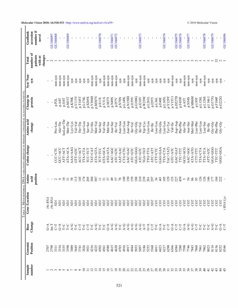
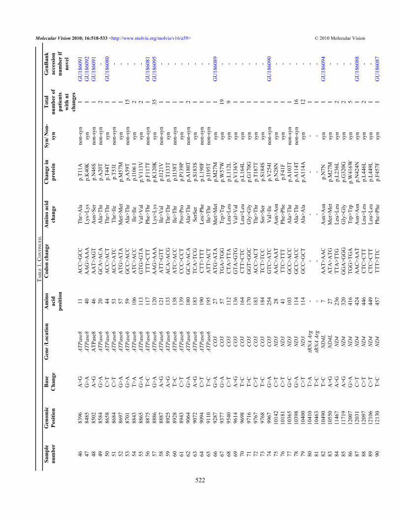
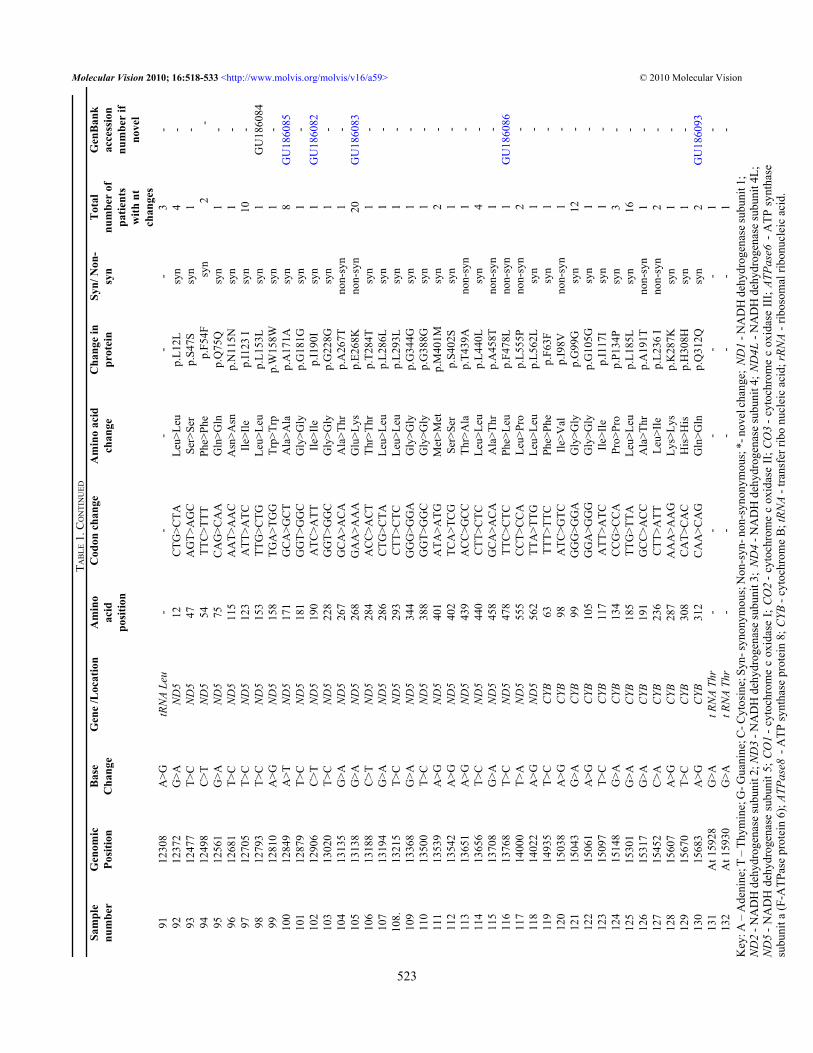
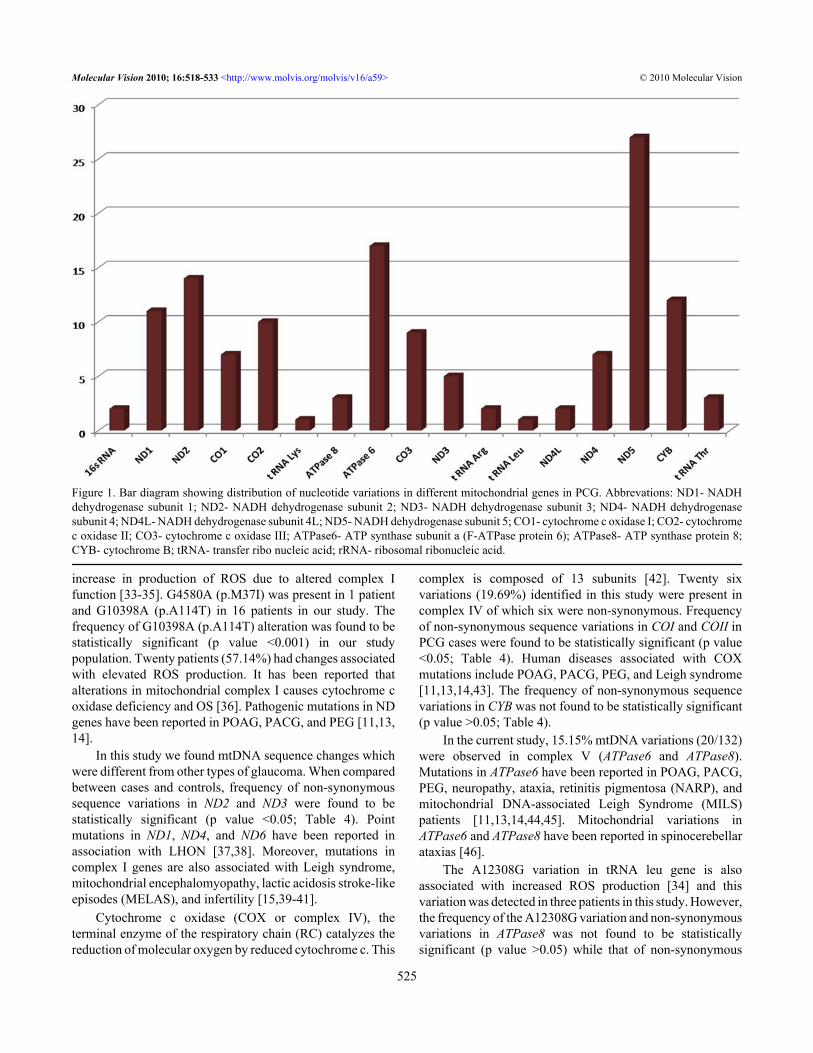
RESULTSMtDNA sequencing following whole genome amplificationof mitochondrial DNA revealed a total of 132 nucleotidevariations (Table 1) in PCG patients and 58 in controls (Table2). Of the 132 nucleotide variations, 42 (31.81%) were non-synonymous, 82 (62.18%) were synonymous changes, and 8were in RNA genes. In total, 23.48% (31/132) variations werenovel out of which 41.93% (13/31) were non-synonymous(Table 1). A total of 66/132 (50.00%) variations wereobserved in complex I, 12/132 (9.09%) in complex III, 26/132(19.69%) in complex IV, and 20/132 (15.15%) were incomplex V (Figure 1). Out of the total variations reported,complex I had 31.81% (21/66) non-synonymous basechanges, complex III had 25.00% (3/12), complex IV had23.07% (6/26), and complex V had 55.00% (11/20) non-synonymous base changes. Of 58 variations in the controls,14 were non-synonymous changes.
Two non-synonymous changes (p.W239C in ND2 andp.A20T in ATPase6) were present both in cases as well ascontrols. The remaining 40 non-synonymous changes werelimited to PCG cases only. All novel variations from patientsand controls were submitted to the GenBank database andaccession numbers were obtained (Table 1 and Table 2).
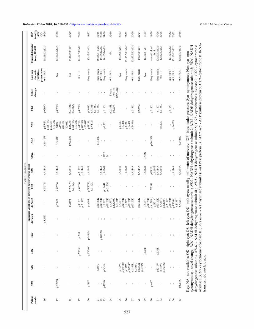
SIFT and PolyPhen analysis of all non-synonymouschanges from cases and controls revealed five pathogenicchanges (p.P2L, p.I10T, p.M31T in ND1 protein and p.M37I,p.W239C in ND2 protein). Eight patients (22.85%) werepositive for either of these pathologic mtDNA nucleotidechanges. Clinical features of patients and mtDNA variationsidentified in this study have been tabulated (Table 3).
DISCUSSIONThe human mtDNA is a 16,569-base pair double-stranded,compact, circular molecule which lacks histones and iswithout introns. MtDNA has several overlapping genes andincomplete termination codons. It contains 37 genes whichregulate oxidative phosphorylation (OXPHOS). Of these, 24are needed for mtDNA translation (2 rRNAs [rRNAs] and 22tRNAs [tRNAs]), and 13 encode subunits of the respiratorychain: seven subunits of complex I (ND1, 2, 3, 4, 4L, 5, and6 [ND stands for NADH dehydrogenase]), one subunit ofcomplex III (cytochrome b), three subunits of cytochrome coxidase (COX I, II, and III), and two subunits of ATP synthase(ATPase6 and ATPase8; Figure 2).
MtDNA mutates 10 times more frequently as comparedto nuclear DNA due to its proximity to the electron transportchain (ETC) and lack of histones and other protective proteinsand has very basic repair mechanism [23]. Mitochondria areessential for ATP production by OXPHOS and are susceptibleto oxidative damage because reactive oxygen species (ROS)
damage mitochondrial enzymes directly and altermitochondrial membrane permeability leading to cell death[24]. Most studies suggest that the majority of intracellularROS produced by non-phagocytic cells are derived frommitochondria [25,26]. Thus mitochondria are both source andtarget of free radicals.
Several human diseases have been associated withmtDNA mutations, indicating that dysfunction of thecomponents of oxidative phosphorylation encoded by themitochondrial genome can be deleterious [27]. Abnormalitiesin mtDNA have proven to be associated with leber’shereditary optic neuropathy (LHON) [28], POAG,pseudoexfoliation glaucoma (PEG), primary angle closureglaucoma (PACG), other spontaneous optic neuropathies[11,13,14,29], and male infertility [15].
In this study we screened 35 PCG cases for mtDNAvariations. We found 42 non-synonymous mtDNA variationsin PCG patients in different mitochondrial genes. The highestnumber of nucleotide variations were recorded in complex I,followed by complex IV and then complex V. Eight patients(22.85%) were found to be positive for pathogenic changeswhile in PEG patients this was 10.30% [14].
Complex I is responsible for pumping of protons (H+)from the matrix to the inter-membrane space in associationwith complex III and IV. Although the mitochondrial ETC isvery effective in the reduction of oxygen to water, there is aconstant “leak” of electrons from the ETC to oxygen and thisresults in the formation of superoxide anions. It is generallyagreed that there are two main sites in the respiratory chainwhere superoxide anions are generated viz. complex I andcomplex III [30,31]. Dismutation of superoxide anionsproduces hydrogen peroxide as a secondary product, and inthe presence of transition metals this can be converted to ahighly reactive hydroxyl radical that can readily oxidizeproteins, lipids, carbohydrates, DNA, and RNA [32]. Fiftypercent nucleotide variations identified in our study were incomplex I.
SIFT and PolyPhen analysis of missense changes showedthat p.P2L, p.I10T, and p.M31T in ND1 protein and p.M37Iand p.W239C in ND2 protein were deleterious to proteinfunction. PHAT (predicted hydrophobic and transmembrane)score difference of p.P2L and p.I10T was >2 and PSIC scoreof p.M31T was >2. PSIC score of p.M37I and W239C was >2and >1.5, respectively. All these changes (p.P2L, p.I10T, andp.M31T in ND1 and p.M37I and p.W239C in ND2) had SIFTscores <0.05 and were predicted to be deleterious. Pathogenicvariants p.P2L, p.I10T, and p.M31T were present in 3 cases(1 case each) while p.M37I and p.W239C were present in oneand four cases, respectively. However, frequency of thepathogenic variants (p.P2L, p.I10T, and p.M31T in ND1 andp.M37I and p.W239C in ND2) was not found to be statisticallysignificant (p value >0.05) in our study population.
Recent studies have shown that G4580A (p.M37I) inND2 and G10398A (p.A114T) in ND3 are associated with an
Molecular Vision 2010; 16:518-533 <http://www.molvis.org/molvis/v16/a59> © 2010 Molecular Vision
520
TAB
LE 1
. MIT
OC
HO
ND
RIA
L D
NA
VA
RIA
TIO
NS O
BSE
RV
ED IN
PRIM
AR
Y C
ON
GEN
ITA
L G
LAU
CO
MA
PATI
ENTS
.Sa
mpl
enu
mbe
rG
enom
icPo
sitio
nB
ase
Cha
nge
Gen
e /L
ocat
ion
Am
ino
acid
posi
tion
Cod
on c
hang
eA
min
o ac
idch
ange
Cha
nge
inpr
otei
nSy
n/ N
on-
syn
Tot
alnu
mbe
r of
patie
nts
with
nt
chan
ges
Gen
Ban
kac
cess
ion
num
ber
ifno
vel
127
07G
>A16
s RN
A-
--
--
1-
227
90In
s T16
s RN
A-
--
--
2G
U18
6097
333
11C
>TN
D1
2C
CC
>CTC
Pro>
Leu
p.P2
Lno
n-sy
n1
GU
1860
684
3316
G>A
ND
14
GC
C>A
CC
Ala
>Thr
p.A
4Tno
n-sy
n1
-5
3335
T>C
ND
110
ATT
>AC
TIs
oLeu
>Thr
p.I1
0Tno
n-sy
n1
-6
3398
T>C
ND
131
ATA
>AC
AM
et>T
hrp.
M31
Tno
n-sy
n2
GU
1860
697
3480
A>G
ND
158
AA
A>A
AG
Lys>
Lys
p.K
58K
syn
2-
836
45T>
CN
D1
113
GTT
>GTC
Val
>Val
p.V
113V
syn
1-
937
41C
>TN
D1
145
AC
C>A
CT
Thr>
Thr
p.T1
45T
syn
1-
1038
26T>
CN
D1
174
TTA
>CTA
Leu>
Leu
p.L1
74L
syn
1-
1139
21C
>TN
D1
205
TCC
>TC
TSe
r>Se
rp.
S205
Ssy
n2
-12
4216
T>C
ND
130
4TA
T>C
AT
Tyr>
His
p.Y
304H
non-
syn
1-
1342
25A
>GN
D1
307
ATA
>GTA
Met
>Val
p.M
307V
non-
syn
1-
1445
02T>
CN
D2
11TC
T>TC
CSe
r>Se
rp.
S11S
syn
1G
U18
6070
1545
61T>
CN
D2
31G
TA>G
CA
Val
>Ala
p.V
31A
non-
syn
1-
1645
80G
>AN
D2
37A
TG>A
TAM
et>I
sop.
M37
Ino
n-sy
n1
-17
4615
A>G
ND
249
AA
C>A
GC
Asn
>Ser
p.N
49S
non-
syn
1G
U18
6071
1846
38A
>GN
D2
57A
TC>G
TCIle
>Val
p.I5
7Vno
n-sy
n3
GU
1860
7219
4703
T>C
ND
278
AA
T>A
AC
Asn
>Asn
p.N
78N
syn
1-
2049
16A
>GN
D2
149
CTA
>CTG
Leu>
Leu
p.L1
49L
syn
1-
2149
17A
>GN
D2
150
AA
C>G
AC
Asn
>Asp
p.N
150D
non-
syn
1-
2249
44A
>GN
D2
159
ATC
>GTC
Ile>V
alp.
I159
Vno
n-sy
n1
-23
5033
A>G
ND
218
8G
GA
>GG
GG
ly>G
lyp.
G18
8Gsy
n1
-24
5097
A>G
ND
221
0A
TC>G
TCIle
>Val
p.I2
10V
non-
syn
1G
U18
6073
2551
86A
>TN
D2
239
TGA
>TG
TTr
p>C
ysp.
W23
9Cno
n-sy
n4
-26
5252
G>A
ND
226
1TT
G>T
TALe
u>Le
up.
L261
Lsy
n1
-27
5360
C>T
ND
229
7A
TC>A
TTIle
>Ile
p.I2
97I
syn
1-
2860
11T>
CC
O1
36C
TT>C
TCLe
u>Le
up.
L36L
syn
1-
2960
23G
>AC
O1
4OG
AG
>GA
AG
lu>G
lup.
E40E
syn
1-
3062
17T>
CC
O1
105
TTA
>CTA
Leu>
Leu
p.L1
05L
syn
1G
U18
6074
3162
90C
>TC
O1
129
TAC
>TA
T)Ty
r>Ty
rp.
Y12
9Ysy
n3
-32
6366
G>A
CO
115
5G
TC>A
TCV
al>I
lep.
V15
5 I
non-
syn
3-
3365
84C
>TC
O1
227
GA
C>G
AT
Asp
>Asp
p.D
227D
syn
1G
U18
6075
3472
80C
>TC
O1
459
TTC
>TTT
Phe>
Phe
p.F4
59F
syn
2G
U18
6076
3575
98G
>AC
O2
5G
CG
>AC
GA
la>T
hrp.
A5T
non-
syn
1-
3677
46A
>GC
O2
54A
AC
>AG
CA
sn>S
erp.
N54
Sno
n-sy
n1
GU
1860
7737
7843
A>G
CO
286
ATA
>ATG
Met
>Met
p.M
86M
syn
3-
3878
68C
>TC
O2
95C
TT>T
TTLe
u>Ph
ep.
L95F
non-
syn
2-
3979
61T>
CC
O2
126
TTA
>CTA
Leu>
Leu
p.L1
26L
syn
1-
4079
62T>
CC
O2
126
TTA
>TC
ALe
u>Se
rp.
L126
Sno
n-sy
n1
GU
1860
7841
8023
T>C
CO
214
6A
TT>A
TCIle
>Ile
p.I 1
46 I
syn
1-
4281
16A
>GC
O2
177
GG
A>G
GG
Gly
>Gly
p.G
177G
syn
1G
U18
6079
4381
36T>
CC
O2
184
TTC
>TTT
Phe>
Phe
p.F1
84F
syn
32-
4482
52G
>AC
O2
222
GG
G>G
GA
Gly
>Gly
p.G
222G
syn
2-
4583
46C
>Tt R
NA
Lys
--
--
-1
GU
1860
96
Molecular Vision 2010; 16:518-533 <http://www.molvis.org/molvis/v16/a59> © 2010 Molecular Vision
521
TAB
LE 1
. CO
NTI
NU
ED.
Sam
ple
num
ber
Gen
omic
Posi
tion
Bas
eC
hang
eG
ene
/Loc
atio
nA
min
oac
idpo
sitio
n
Cod
on c
hang
eA
min
o ac
idch
ange
Cha
nge
inpr
otei
nSy
n/ N
on-
syn
Tot
alnu
mbe
r of
patie
nts
with
nt
chan
ges
Gen
Ban
kac
cess
ion
num
ber
ifno
vel
4683
96A
>GAT
Pase
811
AC
C>G
CC
Thr>
Ala
p.T1
1Ano
n-sy
n1
GU
1860
9147
8485
G>A
ATPa
se8
40A
AG
>AA
ALy
s>Ly
sp.
K40
Ksy
n1
GU
1860
9248
8502
A>G
ATP
ase8
46A
AT>
AG
TA
sn>S
erp.
N46
Sno
n-sy
n1
GU
1860
9149
8584
G>A
ATPa
se6
20G
CA
>AC
AA
la>T
hrp.
A20
Tno
n-sy
n2
-50
8658
C>T
ATPa
se6
44A
CC
>AC
TTh
r>Th
rp.
T44T
syn
1G
U18
6080
5186
84C
>TAT
Pase
653
AC
C>A
TCTh
r>Ile
p.T5
3Ino
n-sy
n1
-52
8697
G>A
ATPa
se6
57A
TG>A
TAM
et>M
etp.
M57
Msy
n1
-53
8701
G>A
ATPa
se6
59G
CC
>AC
CA
la>T
hrp.
A59
Tno
n-sy
n15
-54
8843
T>A
ATPa
se6
106
ATC
>AC
CIle
>Ile
p.I1
06 I
syn
2-
5588
65G
>AAT
Pase
611
3G
TG>G
TAV
al>V
alp.
V11
3Vsy
n2
-56
8875
T>C
ATPa
se6
117
TTT>
CTT
Phe>
Thr
p.F1
17T
non-
syn
1G
U18
6081
5788
86G
>AAT
Pase
612
0A
AG
>AA
ALy
s>Ly
sp.
K12
0Ksy
n35
GU
1860
9558
8887
A>G
ATPa
se6
121
ATT
>GTT
Ile>V
alp.
I121
Vno
n-sy
n1
-59
8925
A>G
ATPa
se6
133
AC
A>A
CG
Thr>
Thr
p.T1
33T
syn
1-
6089
28T>
CAT
Pase
613
8A
TC>A
CC
Ile>T
hrp.
I138
Tno
n-sy
n1
-61
8943
C>T
ATPa
se6
139
CC
C>C
CT
Pro>
Pro
p.P1
39P
syn
1-
6290
64G
>AAT
Pase
618
0G
CA
>AC
AA
la>T
hrp.
A18
0Tno
n-sy
n2
-63
9072
A>G
ATPa
se6
183
TCA
>TC
GSe
rSer
p.S1
83S
syn
1-
6490
94C
>TAT
Pase
619
0C
TT>T
TTLe
u>Ph
ep.
L190
Fno
n-sy
n1
-65
9110
T>C
ATPa
se6
195
ATT
>AC
TIle
>Thr
p.I1
95T
non-
syn
1-
6692
87G
>AC
O3
27A
TG>A
TAM
et>M
etp.
M27
Msy
n1
GU
1860
8967
9377
G>A
CO
357
TGA
>TG
GTr
p>Tr
pp.
W57
Wsy
n19
-68
9540
C>T
CO
311
2C
TA>T
TALe
u>Le
up.
L112
Lsy
n9
-69
9614
A>G
CO
313
6G
TA>G
TGV
al>V
alp.
V13
6Vsy
n1
-70
9698
T>C
CO
316
4C
TT>C
TCLe
u>Le
up.
L164
Lsy
n1
-71
9716
T>C
CO
317
0G
GT>
GG
CG
ly>G
lyp.
G17
0Gsy
n1
-72
9767
C>T
CO
318
3A
CC
>AC
TTh
r>Th
rp.
T187
Tsy
n1
-73
9768
T>C
CO
318
4TC
T>TC
CSe
r>Se
rp.
S184
Ssy
n1
-74
9967
G>A
CO
325
4G
TC>A
TCV
al>I
lep.
V25
4Ino
n-sy
n1
GU
1860
9075
1014
2C
>TN
D3
28A
AC
>AA
TA
sn>A
snp.
N28
Nsy
n1
-76
1018
1C
>TN
D3
41TT
C>T
TTPh
e>Ph
ep.
F41F
syn
1-
7710
365
G>C
ND
310
3G
CC
>AC
CA
la>T
hrp.
A10
3Tno
n-sy
n1
-78
1039
8G
>AN
D3
114
GC
C>A
CC
Ala
>Thr
p.A
114T
non-
syn
16-
7910
400
C>T
ND
311
4G
CC
>GC
TA
la>A
lap.
A11
4Asy
n12
-80
1041
0T>
AtR
NA
Arg
--
--
-1
-81
1046
3T>
CtR
NA
Arg
--
--
-1
-82
1049
0T>
CN
D4L
7A
AT>
AA
CA
sn>A
snp.
N7N
syn
1G
U18
6094
8310
550
A>G
ND
4L27
ATA
>ATG
Met
>Met
p.M
27M
syn
1-
8411
467
A>G
ND
423
6TT
A>T
TGLe
u>Le
up.
L236
Lsy
n2
-85
1171
9A
>GN
D4
320
GG
A>G
GG
Gly
>Gly
p.G
320G
syn
2-
8612
007
G>A
ND
441
6TG
G>T
GA
Trp>
Trp
p.W
416W
syn
5-
8712
031
C>T
ND
442
4A
AC
>AA
TA
sn>A
snp.
N42
4Nsy
n1
GU
1860
8888
1209
7C
>TN
D4
446
CTC
>CTT
Leu>
Leu
p.L4
46L
syn
2-
8912
106
C>T
ND
444
9C
TC>C
TTLe
u>Le
up.
L449
Lsy
n1
-90
1213
0T>
CN
D4
457
TTT>
TTC
Phe>
Phe
p.F4
57F
syn
1G
U18
6087
Molecular Vision 2010; 16:518-533 <http://www.molvis.org/molvis/v16/a59> © 2010 Molecular Vision
522
TAB
LE 1
. CO
NTI
NU
ED
Sam
ple
num
ber
Gen
omic
Posi
tion
Bas
eC
hang
eG
ene
/Loc
atio
nA
min
oac
idpo
sitio
n
Cod
on c
hang
eA
min
o ac
idch
ange
Cha
nge
inpr
otei
nSy
n/ N
on-
syn
Tot
alnu
mbe
r of
patie
nts
with
nt
chan
ges
Gen
Ban
kac
cess
ion
num
ber
ifno
vel
9112
308
A>G
tRN
A Le
u-
--
--
3-
9212
372
G>A
ND
512
CTG
>CTA
Leu>
Leu
p.L1
2Lsy
n4
-93
1247
7T>
CN
D5
47A
GT>
AG
CSe
r>Se
rp.
S47S
syn
1-
9412
498
C>T
ND
554
TTC
>TTT
Phe>
Phe
p.F5
4Fsy
n2
-95
1256
1G
>AN
D5
75C
AG
>CA
AG
ln>G
lnp.
Q75
Qsy
n1
-96
1268
1T>
CN
D5
115
AA
T>A
AC
Asn
>Asn
p.N
115N
syn
1-
9712
705
T>C
ND
512
3A
TT>A
TCIle
>Ile
p.I1
23 I
syn
10-
9812
793
T>C
ND
515
3TT
G>C
TGLe
u>Le
up.
L153
Lsy
n1
GU
1860
8499
1281
0A
>GN
D5
158
TGA
>TG
GTr
p>Tr
pp.
W15
8Wsy
n1
-10
012
849
A>T
ND
517
1G
CA
>GC
TA
la>A
lap.
A17
1Asy
n8
GU
1860
8510
112
879
T>C
ND
518
1G
GT>
GG
CG
ly>G
lyp.
G18
1Gsy
n1
-10
212
906
C>T
ND
519
0A
TC>A
TTIle
>Ile
p.I1
90I
syn
1G
U18
6082
103
1302
0T>
CN
D5
228
GG
T>G
GC
Gly
>Gly
p.G
228G
syn
1-
104
1313
5G
>AN
D5
267
GC
A>A
CA
Ala
>Thr
p.A
267T
non-
syn
1-
105
1313
8G
>AN
D5
268
GA
A>A
AA
Glu
>Lys
p.E2
68K
non-
syn
20G
U18
6083
106
1318
8C
>TN
D5
284
AC
C>A
CT
Thr>
Thr
p.T2
84T
syn
1-
107
1319
4G
>AN
D5
286
CTG
>CTA
Leu>
Leu
p.L2
86L
syn
1-
108.
1321
5T>
CN
D5
293
CTT
>CTC
Leu>
Leu
p.L2
93L
syn
1-
109
1336
8G
>AN
D5
344
GG
G>G
GA
Gly
>Gly
p.G
344G
syn
1-
110
1350
0T>
CN
D5
388
GG
T>G
GC
Gly
>Gly
p.G
388G
syn
1-
111
1353
9A
>GN
D5
401
ATA
>ATG
Met
>Met
p.M
401M
syn
2-
112
1354
2A
>GN
D5
402
TCA
>TC
GSe
r>Se
rp.
S402
Ssy
n1
-11
313
651
A>G
ND
543
9A
CC
>GC
CTh
r>A
lap.
T439
Ano
n-sy
n1
-11
413
656
T>C
ND
544
0C
TT>C
TCLe
u>Le
up.
L440
Lsy
n4
-11
513
708
G>A
ND
545
8G
CA
>AC
AA
la>T
hrp.
A45
8Tno
n-sy
n1
-11
613
768
T>C
ND
547
8TT
C>C
TCPh
e>Le
up.
F478
Lno
n-sy
n1
GU
1860
8611
714
000
T>A
ND
555
5C
CT>
CC
ALe
u>Pr
op.
L555
Pno
n-sy
n2
-11
814
022
A>G
ND
556
2TT
A>T
TGLe
u>Le
up.
L562
Lsy
n1
-11
914
935
T>C
CYB
63TT
T>TT
CPh
e>Ph
ep.
F63F
syn
1-
120
1503
8A
>GC
YB98
ATC
>GTC
Ile>V
alp.
I98V
non-
syn
1-
121
1504
3G
>AC
YB99
GG
G>G
GA
Gly
>Gly
p.G
99G
syn
12-
122
1506
1A
>GC
YB10
5G
GA
>GG
GG
ly>G
lyp.
G10
5Gsy
n1
-12
315
097
T>C
CYB
117
ATT
>ATC
Ile>I
lep.
I117
Isy
n1
-12
415
148
G>A
CYB
134
CC
G>C
CA
Pro>
Pro
p.P1
34P
syn
3-
125
1530
1G
>AC
YB18
5TT
G>T
TALe
u>Le
up.
L185
Lsy
n16
-12
615
317
G>A
CYB
191
GC
C>A
CC
Ala
>Thr
p.A
191T
non-
syn
1-
127
1545
2C
>AC
YB23
6C
TT>A
TTLe
u>Ile
p.L2
36 I
non-
syn
2-
128
1560
7A
>GC
YB28
7A
AA
>AA
GLy
s>Ly
sp.
K28
7Ksy
n1
-12
915
670
T>C
CYB
308
CA
T>C
AC
His
>His
p.H
308H
syn
1-
130
1568
3A
>GC
YB31
2C
AA
>CA
GG
ln>G
lnp.
Q31
2Qsy
n2
GU
1860
9313
1A
t 159
28G
>At R
NA
Thr
--
--
-1
-13
2A
t 159
30G
>At R
NA
Thr
--
--
-1
-K
ey: A
– A
deni
ne; T
– T
hym
ine;
G- G
uani
ne; C
- Cyt
osin
e; S
yn- s
ynon
ymou
s; N
on-s
yn- n
on-s
ynon
ymou
s; *
- nov
el ch
ange
; N
D1
- NA
DH
deh
ydro
gena
se su
buni
t 1;
ND
2- N
AD
H d
ehyd
roge
nase
subu
nit 2
; ND
3- N
AD
H d
ehyd
roge
nase
subu
nit 3
; N
D4
- NA
DH
deh
ydro
gena
se su
buni
t 4; N
D4L
- NA
DH
deh
ydro
gena
se su
buni
t 4L;
ND
5- N
AD
H d
ehyd
roge
nase
sub
unit
5; C
O1
- cyt
ochr
ome
c ox
idas
e I;
CO
2- c
ytoc
hrom
e c
oxid
ase
II; C
O3
- cyt
ochr
ome
c ox
idas
e II
I; AT
Pase
6- A
TP s
ynth
ase
subu
nit a
(F-A
TPas
e pr
otei
n 6)
; ATP
ase8
- ATP
synt
hase
pro
tein
8; C
YB- c
ytoc
hrom
e B
; tRN
A- t
rans
fer r
ibo
nucl
eic
acid
; rRN
A- r
ibos
omal
ribo
nucl
eic
acid
.
Molecular Vision 2010; 16:518-533 <http://www.molvis.org/molvis/v16/a59> © 2010 Molecular Vision
523
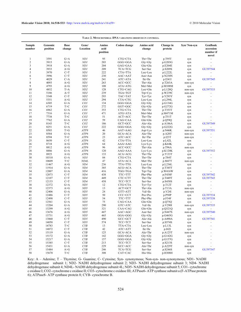
TABLE 2. MITOCHONDRIAL DNA VARIATIONS OBSERVED IN CONTROLS.
Samplenumber
Genomicposition
Basechange
Gene/Location
Aminoacid
position
Codon change Amino acidchange
Change inprotein
Syn/ Non-syn GenBankaccessionnumber if
novel1 3591 G>A ND1 95 CTG>CTA Thr>Thr p.T95T syn -2 3915 G>A ND1 203 GGG>GGA Gly>Gly p.G203G syn -3 3918 G>A ND1 204 GAG>GAA Glu>Glu p.E204E syn -4 3933 A>G ND1 209 TCA>TCG Ser>Ser p.S209S syn GU3975445 3970 C>T ND1 222 CTA>TTA Leu>Leu p.L222L syn -6 3996 C>T ND1 230 AAC>AAT Asn>Asn p.N230N syn -7 4029 C>A ND1 241 ATC>ATA Ile>Ile p.I241I syn GU3975458 4093 A>G ND1 263 ACC>GCC Thr>Ala p.T263A non-syn -9 4793 A>G ND2 108 ATA>ATG Met>Met p.M108M syn -
10 4852 T>A ND2 128 CTG>CAG Leu>Gln p.L128Q non-syn GU39753311 5186 A>T ND2 239 TGA>TGT Trp>Cys p.W239C non-syn -12 5348 C>T ND2 293 TAC>TAT Tyr>Tyr p.Y293Y syn -13 5351 A>G ND2 294 CTA>CTG Leu>Leu p.L294L syn -14 6305 G>A CO1 134 GGG>GGA Gly>Gly p.G134G syn -15 6719 T>C CO1 272 GGT>GGC Gly>Gly p.G272G syn -16 6962 G>A CO1 353 CTG>CTA Thr>Thr p.T353T syn -17 7316 G>A CO1 471 ATG>ATA Met>Met p.M471M syn –18 7738 T>C CO2 51 ACT>ACC Thr>Thr p.T51T syn -19 7762 G>A CO2 59 CAG>CAA Gln>Gln p.Q59Q syn -20 8143 T>C CO2 186 GCT>GCC Ala>Ala p.A186A syn GU39754921 8251 G>A CO2 222 GGG>GGA Gly>Gly p.G222G syn -22 8503 T>G ATP8 46 AAT>AAG Asp>Lys p.N46K non-syn GU39755123 8584 G>A ATP6 20 GCA>ACA Ala>Thr p.A20T non-syn -24 8594 T>C ATP6 23 ATC>ACC Ile>Thr p.I23T non-syn -25 8650 C>T ATP6 42 CTA>TTA Leu>Leu p.L42L syn -26 8718 A>G ATP6 64 AAA>AAG Lys>Lys p.K64K syn -27 8812 A>G ATP6 96 ACC>GCC Thr>Ala p.T96A non-syn -28 8886 G>A ATP6 120 AAG>AAA Lys>Lys p.K120K syn GU39755229 8925 A>G ATP6 133 ACA>ACG Thr>Thr p.T133T syn -30 10310 G>A ND3 84 CTG>CTA Thr>Thr p.T84T syn -31 10609 T>C ND4L 47 ATA>ACA Met>Thr p.M47T non-syn -32 11467 A>G ND4 236 TTA>TTG Leu>Leu p.L236L syn -33 11914 G>A ND4 385 ACG>ACA Thr>Thr p.T385T syn -34 12007 G>A ND4 416 TGG>TGA Trp>Trp p.W416W syn -35 12073 C>T ND4 438 TTC>TTT Phe>Phe p.F438F syn GU39754236 12107 C>T ND4 449 CTC>CTT Thr>Thr p.T449T syn GU39754337 12133 C>T ND4 458 TCC>TCT Ser>Ser p.S458S syn -38 12372 G>A ND5 12 CTG>CTA Tyr>Tyr p.T12T syn -39 12373 A>G ND5 13 ACT>GCT Thr>Ala p.T13A non-syn -40 12406 G>A ND5 24 GTT>ATT Val>Ile p.V24I non-syn -41 12486 C>T ND5 50 CCC>CCT Pro>Pro p.P50P syn GU39753442 12498 C>T ND5 54 TTC>TTT Phe>Phe p.F54F syn GU39753843 12561 G>A ND5 75 CAG>CAA Gln>Gln p.Q75Q syn -44 13204 G>A ND5 290 GTC>ATC Val>Ile p.V290I non-syn GU39753545 13299 A>G ND5 321 CAA>CAG Gln>Gln p.Q321Q syn -46 13676 A>G ND5 447 AAC>AGC Asn>Ser p.N447S non-syn GU39754047 13731 A>G ND5 465 GGA>GGG Gly>Gly p.G465G syn -48 13860 C>T ND5 490 GCC>GCT Ala>Ala p.A490A syn GU39754149 14058 C>T ND5 574 TCC>TCT Ser>Ser p.S574S syn -50 14783 T>C CYB 13 TTA>CTA Leu>Leu p.L13L syn -51 14872 C>T CYB 42 ATC>ATT Ile>Ile p.I42I syn -52 15119 G>A CYB 125 GCA>ACA Ala>Thr p.A125T non-syn -53 15172 G>A CYB 142 GGG>GGA Gly>Gly p.G142G syn -54 15217 G>A CYB 157 GGG>GGA Gly>Gly p.G157G syn -55 15385 C>T CYB 213 TCC>TCT Ser>Ser p.S213S syn -56 15431 G>A CYB 229 GCC>ACC Ala>Thr p.A229T non-syn -57 15484 A>G CYB 246 TCA>TCG Ser>Ser p.S246S syn GU39754758 15670 T>C CYB 308 CAT>CAC His>His p.H308H syn -
Key: A – Adenine; T – Thymine; G- Guanine; C- Cytosine; Syn- synonymous; Non-syn- non-synonymous; ND1- NADHdehydrogenase subunit 1; ND2- NADH dehydrogenase subunit 2; ND3- NADH dehydrogenase subunit 3; ND4- NADHdehydrogenase subunit 4; ND4L- NADH dehydrogenase subunit 4L; ND5- NADH dehydrogenase subunit 5; CO1- cytochromec oxidase I; CO2- cytochrome c oxidase II; CO3- cytochrome c oxidase III; ATPase6- ATP synthase subunit a (F-ATPase protein6); ATPase8- ATP synthase protein 8; CYB- cytochrome B.
Molecular Vision 2010; 16:518-533 <http://www.molvis.org/molvis/v16/a59> © 2010 Molecular Vision
524
increase in production of ROS due to altered complex Ifunction [33-35]. G4580A (p.M37I) was present in 1 patientand G10398A (p.A114T) in 16 patients in our study. Thefrequency of G10398A (p.A114T) alteration was found to bestatistically significant (p value <0.001) in our studypopulation. Twenty patients (57.14%) had changes associatedwith elevated ROS production. It has been reported thatalterations in mitochondrial complex I causes cytochrome coxidase deficiency and OS [36]. Pathogenic mutations in NDgenes have been reported in POAG, PACG, and PEG [11,13,14].
In this study we found mtDNA sequence changes whichwere different from other types of glaucoma. When comparedbetween cases and controls, frequency of non-synonymoussequence variations in ND2 and ND3 were found to bestatistically significant (p value <0.05; Table 4). Pointmutations in ND1, ND4, and ND6 have been reported inassociation with LHON [37,38]. Moreover, mutations incomplex I genes are also associated with Leigh syndrome,mitochondrial encephalomyopathy, lactic acidosis stroke-likeepisodes (MELAS), and infertility [15,39-41].
Cytochrome c oxidase (COX or complex IV), theterminal enzyme of the respiratory chain (RC) catalyzes thereduction of molecular oxygen by reduced cytochrome c. This
complex is composed of 13 subunits [42]. Twenty sixvariations (19.69%) identified in this study were present incomplex IV of which six were non-synonymous. Frequencyof non-synonymous sequence variations in COI and COII inPCG cases were found to be statistically significant (p value<0.05; Table 4). Human diseases associated with COXmutations include POAG, PACG, PEG, and Leigh syndrome[11,13,14,43]. The frequency of non-synonymous sequencevariations in CYB was not found to be statistically significant(p value >0.05; Table 4).
In the current study, 15.15% mtDNA variations (20/132)were observed in complex V (ATPase6 and ATPase8).Mutations in ATPase6 have been reported in POAG, PACG,PEG, neuropathy, ataxia, retinitis pigmentosa (NARP), andmitochondrial DNA-associated Leigh Syndrome (MILS)patients [11,13,14,44,45]. Mitochondrial variations inATPase6 and ATPase8 have been reported in spinocerebellarataxias [46].
The A12308G variation in tRNA leu gene is alsoassociated with increased ROS production [34] and thisvariation was detected in three patients in this study. However,the frequency of the A12308G variation and non-synonymousvariations in ATPase8 was not found to be statisticallysignificant (p value >0.05) while that of non-synonymous
Figure 1. Bar diagram showing distribution of nucleotide variations in different mitochondrial genes in PCG. Abbrevations: ND1- NADHdehydrogenase subunit 1; ND2- NADH dehydrogenase subunit 2; ND3- NADH dehydrogenase subunit 3; ND4- NADH dehydrogenasesubunit 4; ND4L- NADH dehydrogenase subunit 4L; ND5- NADH dehydrogenase subunit 5; CO1- cytochrome c oxidase I; CO2- cytochromec oxidase II; CO3- cytochrome c oxidase III; ATPase6- ATP synthase subunit a (F-ATPase protein 6); ATPase8- ATP synthase protein 8;CYB- cytochrome B; tRNA- transfer ribo nucleic acid; rRNA- ribosomal ribonucleic acid.
Molecular Vision 2010; 16:518-533 <http://www.molvis.org/molvis/v16/a59> © 2010 Molecular Vision
525

TAB
LE 3
. MIT
OC
HO
ND
RIA
L D
NA
VA
RIA
TIO
NS A
ND
CLI
NIC
AL
FEA
TUR
ES O
F PR
IMA
RY
CO
NG
ENIT
AL
GLA
UC
OM
A PA
TIEN
TS.
Seq
uenc
e va
riat
ions
in d
iffer
ent m
itoch
ondr
ial g
enes
Patie
ntnu
mbe
rN
D1
ND
2CO
1CO
2AT
Pase
8AT
Pase
6CO
3N
D3
ND
4LN
D4
ND
5CY
BO
ther
chan
ges
Las
t cup
disc
rat
io(O
S/O
D) a
tpr
esen
tatio
n
Cor
neal
dia
met
er(m
m) O
S/O
DIO
P(m
mH
g) O
S/O
D1
––
p.Y
129Y
;p.
F459
Fp.
M86
M;
p.L9
5F–
p.A
20T;
p.A
59T
p.W
57W
;p.
L112
Lp.
A11
4T-
–p.
I123
I;p.
E268
K;
p.M
401M
;p.
L555
P
p.P1
34P;
p.L1
85L
Ins.
of T
at
2790
_91
(16s
RN
A)
Tota
lcu
ppin
g15
x15/
15x1
5.5
40/3
0
2–
–p.
Y12
9Y;
p.F4
59F
p.M
86M
;p.
L95F
–p.
A20
T;p.
A59
Tp.
W57
W;
p.L1
12L
p.A
114T
-–
p.I1
23I;
p.E2
68K
;p.
M40
1M;
p.L5
55P
p.P1
34P;
p.L1
85L
Ins.
of T
at
2790
_91
(16s
RN
A)
0.8:
1/0.
8:1
13x1
3/13
x13
36/4
0
3p.
M31
Tp.
V15
5 I
–p.
V11
3Vp.
W57
Wp.
A11
4A-
p.W
416W
p.E2
68K
p.G
99G
-H
azy
MED
IA15
x14/
15x1
526
/38
4–
–p.
D22
7DG
222G
–I1
95T
W57
WA
114A
-p.
W41
6Wp.
E26
8Kp.
A19
1T;
p.Q
312Q
-0.
4:1/
0.4:
112
x11/
12x1
222
/24
5–
––
p.N
54S
–p.
A59
Tp.
W57
W;
p.L1
12L
p.A
114T
-p.
I190
I;p.
E268
Kp.
G99
G;
p.L1
85L
-0.
7:1/
0.7:
114
.5x1
5/15
x15
28/2
8
6p.
Y30
4H;
p.M
307V
p.N
150T
––
p.M
57M
p.W
57W
;p.
L112
Lp.
A11
4T-
–p.
I123
I;p.
E268
K;
p.G
344G
p.L2
36I;
p.K
287Q
-0.
5:1/
0.5:
112
x13/
12x1
322
/23
7–
––
p.I1
46I;
p.F1
84F
–p.
A59
T;p.
P139
Pp.
M27
M;
p.W
57W
;p.
L112
L;p.
T187
T
p.A
114T
-p.
L236
Lp.
I123
I;p.
E268
Kp.
F63F
;p.
G99
G;
p.G
105G
;p.
L185
L
A>G
at
1230
8tR
NA
Leu
0.4:
1/0.
4:1
12x1
3/11
x11.
538
/14
8–
––
p.G
177G
––
p.W
57W
p.A
114A
-–
p.Q
75Q
;p.
E268
Kp.
G99
GG
>A a
t15
928
(tR
NA
Thr
)
Haz
y m
edia
NA
23/2
5
9p.
L174
L;p.
S205
S–
––
––
p.W
57W
p.A
114A
-–
p.I1
23I;
p.V
172I
;p.
E268
K;
p.L2
93L
p.G
99G
C>T
at
8346
(tR
NA
Lys
)
NA
12x1
2/12
x12
22/2
2
10p.
V11
3V;
p.T1
45T
p.S1
1S;
p.I2
97I
L126
S;F1
84F
p.I1
06I;
p.T5
3 I
p.W
57W
;p.
L112
Lp.
N28
N;
p.A
114T
-p.
L236
Lp.
I123
I;p.
E268
K;
p.G
388G
–A
>G a
t12
308
tRN
A L
eu
NA
NA
23/2
4
11–
p.W
239C
p.L1
05T
––
p.A
59T;
p.L1
90F
p.W
57W
;p.
L112
L;p.
V13
6V
p.A
114T
-p.
L449
Lp.
I123
I;p.
L153
L;p.
E268
K;
p.L2
86L
p.L1
85L
G>A
at
2707
(16s
RN
A)
; A>G
at
1230
8tR
NA
Leu
;G
>A a
t15
930
tRN
A A
rg
NA
14x1
4/14
x15
26/2
2
12p.
P2L
p.N
49S
––
–A
59T;
I106
I–
p.A
114T
-p.
G32
0Gp.
N11
5N;
p.I1
23I;
p.E2
68K
;p.
L440
L;p.
A45
8T;
p.F4
78L
p.L1
85L
-N
A14
x14.
5/14
x14.
432
/32
13p.
M31
T–
p.V
155
I–
–p.
V11
3Vp.
W57
Wp.
A11
4A-
p.W
416W
p.A
171A
;E2
68K
p.G
99G
-H
azy
med
ia14
x14/
14x1
431
/30
14–
––
––
p.S1
83S
p.W
57W
p.A
114A
-p.
W41
6Wp.
A17
1A;
p.E2
68K
;p.
T284
T
p.G
99G
-H
azy
med
iaN
A25
/24
15–
p.L2
61L
–p.
L126
Lp.
T11A
;p.
N46
S–
p.W
57W
;p.
S184
Sp.
A11
4A-
–p.
A17
1A;
p.E2
68K
p.G
99G
;p.
H30
8H-
0.6:
1/0.
6:1
14.5
x14/
13.5
x13
32/3
2
Molecular Vision 2010; 16:518-533 <http://www.molvis.org/molvis/v16/a59> © 2010 Molecular Vision
526
TAB
LE 3
. CO
NTI
NU
ED.
Se
quen
ce v
aria
tions
in d
i�er
ent m
itoch
ondr
ial g
enes
Patie
ntnu
mbe
rN
D1
ND
2C
O1
CO
2A
TPas
e8A
TPas
e6C
O3
ND
3N
D4L
ND
4N
D5
CYB
Oth
erch
ange
sL
ast c
updi
sc r
atio
(OS/
OD
) at
pres
enta
tion
Cor
neal
dia
met
er(m
m) O
S/O
DIO
P(m
mH
g) O
S/O
D16
––
––
p.K
40K
–p.
W57
Wp.
A11
4A-
p.W
416W
p.F5
4F;
p.A
171A
;p.
A26
7T;
p.E2
68K
p.G
99G
-0.
4:1/
0.5:
111
x11/
12x1
2.5
18/2
6
17p.
S205
S–
––
–p.
T44T
p.W
57W
p.A
114A
-p.
F457
FS4
7S;
A17
1A;
G18
1G;
E268
K
p.G
99G
-N
A14
x14/
14x1
4.5
30/2
8
18–
––
––
p.A
59T
p.W
57W
;p.
L112
Lp.
A11
4T-
p.G
320G
p.I1
23I;
p.A
171A
;p.
E268
K
p.L1
85L;
p.Q
312Q
-N
A14
.5x1
4/14
x14
20/2
0
19–
–p.
V15
5 I
p.A
5T–
p.F1
7T;
p.A
180T
p.W
57W
p.A
103T
;p.
A11
4A-
–p.
A17
1A;
p.G
228G
;p.
E268
K
p.G
99G
-0.
5:1
112
x12.
5/12
x12
22/2
2
20p.
I10T
–p.
Y12
9Yp.
M86
M–
p.A
59T
p.W
57W
;p.
L112
Lp.
A11
4T-
–p.
I123
I;p.
V17
2Ip.
I98V
;p.
P134
P;p.
L185
L
-H
azy
med
ia12
x13/
13x1
318
/37
21–
p.I5
9V–
––
p.K
120K
––
-p.
L446
L–
p.L1
85L
-H
azy
med
ia15
x16/
11.5
x12
32/1
522
––
–p.
G22
2G–
p.K
120K
p.A
114A
--
–-
0.7:
1/0.
5:1
15x1
5/16
x16
28/2
823
p.K
58K
p.V
31A
––
p.A
59T;
p.K
120K
p.L1
64L;
p.G
170G
p.A
114T
p.M
27M
-p.
L12L
p.L1
85L
-H
azy
med
ia14
x15/
14x1
534
/36
24–
––
–p.
A59
T;p.
K12
0K;
p.P1
34P
--
-–
p.L1
85L;
p.L2
36I
T>A
at
1041
1 (t
RN
A A
rg)
0.7:
1/0.
7:1
NA
32/1
0
25–
p.I5
7V;
p.W
239C
––
–p.
A59
T;p.
K12
0K-
p.A
114T
--
p.L1
2L;
p.L4
40L
–-
NA
14x1
5/14
x15
22/2
2
26–
p.I5
7V;
p.W
239C
––
–p.
A59
T;p.
K12
0K-
p.A
114T
--
p.L1
2L;
p.L4
40L
–-
Haz
y m
edia
13x1
3.5/
15x1
4.5
22/2
2
27–
p.L1
49L;
p.G
188G
––
–p.
K12
0K-
p.A
114A
--
p.T4
39A
p.L1
85L
-0.
8:1/
0.8:
113
x13.
5/13
x13.
520
/20
28–
p.M
37I;
p.N
78N
––
–p.
K12
0K-
p.A
114A
--
–p.
G99
G-
hazy
med
ia14
x14/
14x1
422
/24
29–
–p.
E40E
––
p.A
59T;
p.K
120K
-p.
A11
4Tp.
N7N
-–
–-
NA
14x1
4/11
x11
18/2
2
30p.
A4T
––
––
p.K
120K
;p.
I121
VV
254I
p.F4
1F;
p.A
114T
-p.
N42
4N–
p.L1
85L
-H
azy
med
iaco
rnea
l ulc
er/
14x1
418
/20
31–
p.I2
10V
p.L3
6L–
–p.
K12
0K-
p.A
114A
--
–p.
I117
I-
Haz
y m
edia
12x1
0/12
x12.
540
/26
32–
p.I5
7V;
p.W
239C
––
–p.
A59
T;p.
K12
0K;
p.A
180T
-p.
A11
4T-
-p.
L12L
p.L1
85L
-0.
8:1
112
x12/
12x1
222
/26
33–
––
––
p.K
120K
-–
--
–p.
L185
L-
0.8:
1/0.
9:1
14x1
4/14
x14
26/2
434
––
–-
-p.
K12
0K;
p.T1
33T
-p.
A11
4A-
-p.
S402
S–
-0.
5:1/
0.5:
112
x12/
12x1
2.5
20/2
2
35p.
K58
K–
––
–p.
K12
0K
-p.
A11
4A-
p.L4
46L
––
–0.
5:1/
0.3:
113
x13/
13x1
3.5
24/1
6
Key
: NA
- not
ava
ilabl
e; O
D- r
ight
eye
; OS-
left
eye;
OU
- bot
h ey
es; m
mH
g- m
ilim
eter
of m
ercu
ry; I
OP-
intra
ocu
lar p
ress
ure;
Syn
- syn
onym
ous;
Non
-syn
- non
-sy
nony
mou
s; -
nove
l cha
nge;
ND
1- N
AD
H d
ehyd
roge
nase
subu
nit 1
; ND
2- N
AD
H d
ehyd
roge
nase
subu
nit 2
; ND
3- N
AD
H d
ehyd
roge
nase
subu
nit 3
; ND
4- N
AD
Hde
hydr
ogen
ase
subu
nit 4
; ND
4L- N
AD
H d
ehyd
roge
nase
sub
unit
4L;
ND
5- N
AD
H d
ehyd
roge
nase
sub
unit
5; C
O1
- cyt
ochr
ome
c ox
idas
e I;
CO
2- c
ytoc
hrom
e c
oxid
ase I
I; C
O3
- cyt
ochr
ome c
oxi
dase
III;
ATPa
se6
- ATP
synt
hase
subu
nit a
(F-A
TPas
e pro
tein
6) ;
ATP
ase8
- ATP
synt
hase
pro
tein
8; C
YB- c
ytoc
hrom
e B; t
RN
A-
trans
fer r
ibo
nucl
eic
acid
.
Molecular Vision 2010; 16:518-533 <http://www.molvis.org/molvis/v16/a59> © 2010 Molecular Vision
527

variations in ATPase6 and others (16s RNA, tRNA), as shownin Table 4, were statistically significant (p value <0.05).
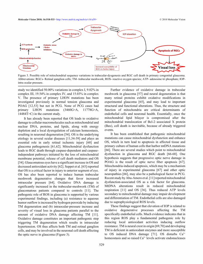
Non-synonymous mitochondrial variations adverselyaffect oxidative phosphorylation resulting in decreasedmitochondrial respiration and increased free radical (FR)production [47]. Thus, we hypothesize that mtDNA variationswith resultant lower ATP levels may impair growth,development, and differentiation of TM and result intrabecular-dysgenesis (Figure 3); a characteristic feature ofPCG. Trabecular-dysgenesis leads to impairment in aqueousdrainage hence causes elevation in IOP. ROS levels mayincrease to supraphysiological levels in TM endothelial cellsand due to low ATP levels these cells are unable to eliminatethe reactive oxygen intermediates (ROI). MtDNA mutationsare also associated with optic neuropathies like LHON [38],NARP [48,49] or Leigh syndrome [50]. The mechanisms by
which mitochondrial abnormalities may place the optic nerveat risk remain uncertain [51].
Distribution of high number of mitochondria in the opticnerve head reflects the high energy requirement of the humanoptic nerve head. Neurons, because of their high energyrequirement, are heavily dependent on mitochondria forsurvival [52]. Mitochondria not only constitute an energy-generating system, but are also critically involved in calciumsignaling and apoptosis. Mitochondrial function is impairedby mutations in mitochondrial and nuclear DNA, chemicalinsults to components of the electron transport chain, and alack of substrates such as oxygen. The latter is relevant totissue hypoxia that is believed to be present in theglaucomatous retina and optic nerve head either primarily orsecondary to elevated IOP. Any malfunction of themitochondrial electron transport chain results in excessivegeneration of free radicals and low ATP production. In our
Figure 2. Schematic representation of components of the OXPHOS pathway localized in inner mitochondrial membrane.
TABLE 4. WITH P-VALUE AND RELATIVE RISK AT 95% CONFIDENCE INTERVAL BY USING PEARSON χ2/FISHER’S EXACT TEST FOR NON-SYNONYMOUSSEQUENCE VARIATIONS IN DIFFERENT MITOCHONDRIAL GENES IN PCG AND CONTROLS.
Gene name Cases (n=35) Controls (n=40) p-value Relative risk at 95%confidence interval
ND1 6 (17.14%) 2 (5.0%) 0.136 1.73 (1.07–2.81)ND2 10 (28.57%) 2 (5.0%) 0.005 2.10 (1.41–3.12)ND3 17 (48.60%) 0 (0%) <0.001 3.22 (2.19–4.72)ND4 0 (0%) 0 (0%) —- —–
ND4L 0 (0%) 2 (5.0%) 0.495 1.92 (1.54–2.39)ND5 21 (60.00%) 4 (10.0%) <0.001 3.00 (1.86–4.83)CO1 4 (11.42%) 0 (0%) 0.043 2.29 (1.75–2.98)CO2 5 (14.30%) 0 (0%) 0.019 2.33 (1.78–3.05)CO3 1 (2.90%) 0 (0%) 0.467 2.17 (1.70–2.78)CYB 4 (11.42%) 8 (20.00%) 0.360 0.57 (0.18–1.73)
ATPase6 18 (51.42%) 7 (17.50%) 0.002 2.12 (1.34–3.34)ATPase8 1 (2.85%) 1 (2.5) 1.00 1.07 (0.26–4.40)Others 8 (22.85%) 0 (0%) 0.001 2.48 (1.85–3.32)
Abbrevations: ND1 - NADH dehydrogenase subunit 1; ND2 - NADH dehydrogenase subunit 2; ND3 - NADH dehydrogenasesubunit 3; ND4 - NADH dehydrogenase subunit 4; ND4L - NADH dehydrogenase subunit 4L; ND5 - NADH dehydrogenasesubunit 5; CO1 - cytochrome c oxidase I; CO2 - cytochrome c oxidase II; CO3 - cytochrome c oxidase III; ATPase6 - ATP synthasesubunit a (F-ATPase protein 6); ATPase8 - ATP synthase protein 8; CYB - cytochrome B; tRNA- transfer ribo nucleic acid;rRNA- ribosomal ribonucleic acid.
Molecular Vision 2010; 16:518-533 <http://www.molvis.org/molvis/v16/a59> © 2010 Molecular Vision
528
study we identified 50.00% variations in complex I, 9.02% incomplex III, 19.54% in complex IV, and 15.03% in complexV. The presence of primary LHON mutations has beeninvestigated previously in normal tension glaucoma andPOAG [12,53] but not in PCG. None of PCG cases hadprimary LHON mutations (3460G>A, 11778G>A,14484T>C) in the current study.
It has already been reported that OS leads to oxidativedamage to cellular macromolecules such as mitochondrial andnuclear DNA, proteins, and lipids, along with energydepletion and a local dysregulation of calcium homeostasis,resulting in neuronal degeneration [54]. OS is the underlyingetiology in several ocular diseases [11,54-59] and plays anessential role in early retinal ischemic injury [60] andglaucoma pathogenesis [61,62]. Mitochondrial dysfunctionleads to RGC death through caspase-dependent and caspase-independent pathways initiated by the loss of mitochondrialmembrane potential, release of cell death mediators and OS[54]. Glaucomatous eyes have a significant increase in OS anddecreased antioxidant activity [62]. Seppet et al. [63] reportedthat OS is a critical factor in injury to anterior segment of eye.OS has also been reported to induce human trabecularmeshwork degenerative changes that favor increasedintraocular pressure [64]. Oxidative DNA damage issignificantly increased in the trabecular-meshwork (TM) ofglaucomatous patients compared to controls [11]. Thepathogenic role of ROS in glaucoma is supported by variousexperimental findings, including (a) resistance to aqueoushumor outflow is increased by hydrogen peroxide by inducingTM degeneration and (b) intraocular-pressure increase andseverity of visual loss in glaucoma patients parallel to theamount of oxidative DNA damage affecting TM [11].Oxidative damage constitutes an important pathogenic steptriggering TM degeneration which results in intraocularhypertension. OS thus affects both TM and retinal ganglioncells, and may be involved in the neuronal cell death affectingthe optic nerve in glaucoma (Figure 3).
Further evidence of oxidative damage in trabecularmeshwork in glaucoma [57] and neural degeneration is thatmany retinal proteins exhibit oxidative modifications inexperimental glaucoma [65], and may lead to importantstructural and functional alterations. Thus, the structure andfunction of mitochondria are critical determinants ofendothelial cells and neuronal health. Essentially, once themitochondrial lipid bilayer is compromised after themitochondrial translocation of Bcl-2–associated X protein(Bax), cell death is inevitable, because of already triggeredevents.
It has been established that pathogenic mitochondrialmutations can cause mitochondrial dysfunction and enhanceOS, which in turn lead to apoptosis in affected tissue andprimary culture of human cells that harbor mtDNA mutations[66]. There are several studies which point to mitochondrialdysfunction in glaucoma and RGC death [66-68]. Onehypothesis suggests that progressive optic nerve damage inPOAG is the result of optic nerve fiber apoptosis [67].Mitochondria-induced apoptosis, which may be a mechanismof injury in experimental glaucoma [67] and other opticneuropathies [66], may also be a pathological factor in PCG.Recent study by Abu-Amero et al. [11] reported mitochondrialdysfunction-associated OS as a risk factor for glaucoma.MtDNA alterations result in reduced mitochondrialrespiration [11] and OS [36]. Thus reduced ATP levelssecondary to mitochondrial damage may impair developmentand differentiation of TM. Endothelial cells are also damageddue to supraphysiological ROS levels.
These findings suggest that elevation of IOP is related tooxidative degenerative processes affecting the TMspecifically endothelial cells. Much evidence indicates that inthis region ROS play a fundamental pathogenic role byreducing local antioxidant activities inducing outflowresistance. TM is neural crest in origin [69,70] and developingTM is deficient in antioxidant enzymes and more susceptibleto OS induced DNA damage [71]. OS disturbs Ca2+
homeostasis and so raised Ca2+ levels activate endonucleases
Figure 3. Possible role of mitochondrial sequence variations in trabecular-dysgenesis and RGC cell death in primary congenital glaucoma.Abbrevations: RGCs- Retinal ganglion cells; TM- trabecular meshwork; ROS- reactive oxygen species; ATP- adenosine tri phosphate; IOP-intra ocular pressure.
Molecular Vision 2010; 16:518-533 <http://www.molvis.org/molvis/v16/a59> © 2010 Molecular Vision
529

which cause nuclear DNA damage [63]. OS, early indevelopment and/or throughout life could precipitate bothmetabolic and anatomic sequelae that cause trabeculardysgenesis and ultimately optic nerve damage in PCG.
Elevated IOP is a characteristic feature of glaucoma andan important risk factor for optic nerve damage [72].However, the precise relationship between among elevatedIOP, glaucomatous optic nerve (ON) damage, and retinalganglion cell death are poorly understood. Growing evidenceindicates that mitochondrial structural and functionaldynamics play an important role in cell and animalphysiology. Imbalance in the control of mitochondrial fusionand fission dramatically alters overall mitochondrialmorphology [73-76]. Elevated IOP in glaucoma inducesreduction of cytochrome c oxidase (COX) activity,mitochondrial fission, mitochondrial matrix swelling, cristaedepletion, triggers release of optic nerve atrophy type-I(OPA1), and induces subsequent apoptotic cell death indifferentiated RGC-5 cells [77,78] (Figure 4). Similarfindings were also confirmed in a mouse model [79].
In summary, frequency of mtDNA sequence variations inPCG was significantly higher as compared to controls. Fivepathogenic changes (3 in ND1 and 2 in ND2) and 3 otherchanges (G10398A, A12308G, and G13708A) associatedwith elevated ROS were present in our patients. Non-synonymous mtDNA alterations may lead to mitochondrialdysfunction which leads to reduced mitochondrial respiration,OS, damage to mtDNA, altered mitochondrial morphology,alterations in mitochondrial fission and fusion, and ultimatelycell’s demise. OS impairs development and differentiation oftrabecular meshwork that favor increased intraocular pressurein PCG and consequently RGC death.
This study describes mtDNA sequence variations in arelatively small number of patients with PCG of north Indianethnic origin. However, these results should be confirmed inother populations. Knowledge of mtDNA mutations and/ormitochondrial dysfunction in PCG may lead to a betterunderstanding of glaucoma pathophysiology [80]. Novel
approaches are now available for studying mitochondrialdisease in the eye, and a novel in vitro treatment has alreadybeen devised for the metabolic defect of at least one mtDNAmutation in LHON [81]. PCG cases with mtDNA variationsand consequent OS may benefit by early diagnosis and promptmanagement with antioxidant therapy.
Conclusion: A total of 44 novel mtDNA variations wereidentified in current study. MtDNA variations adversely affectrespiratory chain, impair the OXPHOS pathway resulting inlow ATP production, and impair growth, development, anddifferentiation of TM. Mitochondrial DNA variations alsolead to increased ROS production, oxidative injury to TM andRGCs. Thus, early diagnosis of mitochondrial DNAvariations and prompt antioxidant administration may delayOS induced injury to TM and RGCs and hence improve visualprognosis.
ACKNOWLEDGMENTSThe authors would like to thank the families of the patientsfor their cooperation without this study was not possible. Theauthors also thank to Dr. Guresh Kumar, Department of Bio-Statistics for statistical analysis. The author Mukesh Tanwarthanks University Grants Commission (UGC), Govt. of Indiafor providing Senior Research Fellowship (SRF).
REFERENCES1. Sarfarazi M, Stoilov I. Molecular genetics of primary congenital
glaucoma. Eye 2000; 14:422-8. [PMID: 11026969]2. Gencik A, Gencikova A, Ferak V. Population genetical aspects
of primary congenital glaucoma. I. Incidence, prevalence,gene frequency, and age of onset. Hum Genet 1982;61:193-7. [PMID: 7173860]
3. Stoilov I, Akarsu AN, Sarfarazi M. Identification of threedifferent truncating mutations in cytochrome P4501B1(CYP1B1) as the principal cause of primary congenitalglaucoma (Buphthalmos) in families linked to the GLC3Alocus on chromosome 2p21. Hum Mol Genet 1997;6:641-7. [PMID: 9097971]
4. Dandona L, Williams JD, Williams BC, Rao GN.Populationbased assessment of childhood blindness in
Figure 4. Role of elevated IOP in RGCs death in glaucoma. Abbrevations: RGCs- Retinal ganglion cells; IOP- intra ocular pressure; OPA1-optic nerve atrophy type-I.
Molecular Vision 2010; 16:518-533 <http://www.molvis.org/molvis/v16/a59> © 2010 Molecular Vision
530
http://www.ncbi.nlm.nih.gov/entrez/query.fcgi?cmd=Retrieve&db=PubMed&dopt=abstract&list_uids=7173860
southern India. Arch Ophthalmol 1998; 116:545-6. [PMID:9565065]
5. Akarsu AN, Turacli ME, Aktan SG, Barsoum-Homsy M,Chevrette L, Sayli BS, Sarfarazi M. A second locus (GLC3B)for primary congenital glaucoma (buphthalmos) maps to the1p36 region. Hum Mol Genet 1996; 5:1199-203. [PMID:8842741]
6. Stoilov IR, Sarfarazi M. The third genetic locus (GLC3C) forprimary congenital glaucoma (PCG) maps to chromosome14q24.3. ARVO Annual Meeting; 2002 May 5–10; FortLauderdale (FL).
7. Tanwar M, Dada T, Sihota R, Yadav U, Das TK, Dada R.Mutation spectrum of CYP1B1 in North Indian congenitalglaucoma patients. Mol Vis 2009; 15:1200-9. [PMID:19536304]
8. Panicker SG, Reddy AB, Mandal AK, Ahmed N, NagarajaramHA, Hasnain SE, Balasubramanian D. Identification of novelmutations causing familial primary congenital glaucoma inIndian pedigrees. Invest Ophthalmol Vis Sci 2002;43:1358-66. [PMID: 11980847]
9. Mashima Y, Suzuki Y, Sergeev Y, Ohtake Y, Tanino T, KimuraI, Miyata H, Aihara M, Tanihara H, Inatani M, Azuma N,Iwata T, Araie M. Novel Cytochrome P4501B1 (CYP1B1)gene mutations in Japanese patients with Primary CongenitalGlaucoma. Invest Ophthalmol Vis Sci 2001; 42:2211-6.[PMID: 11527932]
10. Bejjani BA, Lewis RA, Tomey KF, Anderson KL, Dueker DK,Jabak M, Astle WF, Otterud B, Leppert M, Lupski JR.Mutations in CYP1B1, the gene for cytochrome P4501B1, arethe predominant cause of primary congenital glaucoma inSaudi Arabia. Am J Hum Genet 1998; 62:325-33. [PMID:9463332]
11. Abu-Amero KK, Morales J, Bosley TM. Mitochondrialabnormalities in patients with primary open-angle glaucoma.Invest Ophthalmol Vis Sci 2006; 47:2533-41. [PMID:16723467]
12. Izzotti A, Bagnis A, Sacca SC. The role of oxidative stress inglaucoma. Mutat Res 2006; 612:105-14. [PMID: 16413223]
13. Abu-Amero KK, Morales J, Osman MN, Bosley TM. Nuclearand mitochondrial analysis of patients with primary angle-closure glaucoma. Invest Ophthalmol Vis Sci 2007;48:5591-6. [PMID: 18055808]
14. Abu-Amero KK, Bosley TM, Morales J. Analysis of nuclearand mitochondrial genes in patients with pseudoexfoliationglaucoma. Mol Vis 2008; 14:29-36. [PMID: 18246027]
15. Kumar R, Venkatesh S, Kumar M, Tanwar M, Shasmsi MB,Kumar R, Gupta NP, Sharma RK, Talwar P, Dada R.Oxidative stress and sperm mitochondrial DNA mutation inidiopathic oligoasthenozoospermic men. Indian J BiochemBiophys 2009; 46:172-7. [PMID: 19517995]
16. Sunyaev S, Ramensky V, Koch I, Lathe W III. Kondrashiv,Bork P. Prediction of deleterious human alleles. Hum MolGenet 2001; 10:591-7. [PMID: 11230178]
17. Ramensky V, Bork P, Sunyaev S. Human non-synonymousSNPs: server and survey. Nucleic Acids Res 2002;30:3894-900. [PMID: 12202775]
18. Sunyaev S, Ramensky V, Bork P. Towards a structural basis ofhuman non-synonymous single nucleotide polymorphisms.Trends Genet 2000; 16:198-200. [PMID: 10782110]
19. Kumar P, Henikoff S, Ng PC. Predicting the effects of codingnon-synonymous variants on protein function using the SIFTalgorithm. Nat Protoc 2009; 4:1073-81. [PMID: 19561590]
20. Ng PC, Henikoff S. SIFT: predicting amino acid changes thataffect protein function. Nucleic Acids Res 2003; 31:3812-4.[PMID: 12824425]
21. George Priya Doss C, Rajasekaran R, Sudandiradoss C,Ramanathan K, Purohit R, Sethumadhavan R. A novelcomputational and structural analysis of nsSNPs in CFTRgene. Genomic Med 2008; 2:23-32. [PMID: 18716917]
22. Gale CP, Grant PJ. The characterisation and functional analysisof the human glyoxalase-1 gene using methods ofbioinformatics. Gene 2004; 340:251-60. [PMID: 15475166]
23. Brown WM, George M Jr, Wilson AC. Rapid evolution ofanimal mitochondrial DNA. Proc Natl Acad Sci USA 1979;76:1967-71. [PMID: 109836]
24. Richter C. Oxidative stress, mitochondria, and apoptosis. RestorNeurol Neurosci 1998; 12:59-62. [PMID: 12671298]
25. Finkel T. Redox-dependent signal transduction. FEBS Lett2000; 476:52-4. [PMID: 10878249]
26. Sastre J, Pallarado FV, Garcia de la Asuncion J, Vina J.Mitochondria, oxidative stress and aging. Free Radic Res2000; 32:189-98. [PMID: 10730818]
27. Wallace DC. Diseases of mitochondrial DNA. Annu RevBiochem 1992; 61:1175-212. [PMID: 1497308]
28. Abu-Amero KK, Bosley TM. Mitochondrial abnormalities inpatients with LHON-like optic neuropathies. InvestOphthalmol Vis Sci 2006; 47:4211-20. [PMID: 17003408]
29. Bosley TM, Constantinescu CS, Tench CR, Abu-Amero KK.Mitochondrial changes in leukocytes of patients with opticneuritis. Mol Vis 2007; 13:1516-28. [PMID: 17893651]
30. Turrens JF, Boveris A. Generation of superoxide anion by theNADH dehydrogenase of bovine heart mitochondria.Biochem J 1980; 191:421-7. [PMID: 6263247]
31. Turrens JF, Alexandre A, Lehninger AL. Ubisemiquinone is theelectron donor for superoxide formation by complex III ofheart mitochondria. Arch Biochem Biophys 1985;237:408-14. [PMID: 2983613]
32. Richter C, Gogvadze V, Laffranchi R, Schlapbach R, SchweizerM, Suter M, Walter P, Yaffee M. Oxidants in mitochondria:from physiology to diseases. Biochim Biophys Acta 1995;1271:67-74. [PMID: 7599228]
33. van der Walt JM, Nicodemus KK, Martin ER, Scott WK, NanceMA, Watts RL, Hubble JP, Haines JL, Koller WC, Lyons K,Pahwa R, Stern MB, Colcher A, Hiner BC, Jankovic J, OndoWG, Allen FH Jr, Goetz CG, Small GW, Mastaglia F, StajichJM, McLaurin AC, Middleton LT, Scott BL, Schmechel DE,Pericak-Vance MA, Vance JM. Mitochondrialpolymorphisms significantly reduce the risk of Parkinsondisease. Am J Hum Genet 2003; 72:804-11. [PMID:12618962]
34. Ross OA, McCormack R, Curran MD, Dugid RA, Barnett YA,Rea IM, Middleton D. Mitochondrial DNA polymorphism:its role in longevity of the Irish population. Exp Gerontol2001; 36:1161-78. [PMID: 11404057]
35. Bhat A, Koul A, Sharma S, Rai E. Bukhari SIAm Dhar MK,Bamezai RN. The possible role of 10398A and 16189Cvariants in providing susceptibility to T2DM in two northIndian populations: a replicative study. Hum Genet 2007;120:821-6. [PMID: 17066297]
Molecular Vision 2010; 16:518-533 <http://www.molvis.org/molvis/v16/a59> © 2010 Molecular Vision
531
http://www.ncbi.nlm.nih.gov/entrez/query.fcgi?cmd=Retrieve&db=PubMed&dopt=abstract&list_uids=9565065
http://www.ncbi.nlm.nih.gov/entrez/query.fcgi?cmd=Retrieve&db=PubMed&dopt=abstract&list_uids=9565065
http://www.ncbi.nlm.nih.gov/entrez/query.fcgi?cmd=Retrieve&db=PubMed&dopt=abstract&list_uids=8842741
http://www.ncbi.nlm.nih.gov/entrez/query.fcgi?cmd=Retrieve&db=PubMed&dopt=abstract&list_uids=8842741
http://www.ncbi.nlm.nih.gov/entrez/query.fcgi?cmd=Retrieve&db=PubMed&dopt=abstract&list_uids=9463332
http://www.ncbi.nlm.nih.gov/entrez/query.fcgi?cmd=Retrieve&db=PubMed&dopt=abstract&list_uids=9463332
http://www.ncbi.nlm.nih.gov/entrez/query.fcgi?cmd=Retrieve&db=PubMed&dopt=abstract&list_uids=1497308
http://www.ncbi.nlm.nih.gov/entrez/query.fcgi?cmd=Retrieve&db=PubMed&dopt=abstract&list_uids=6263247
http://www.ncbi.nlm.nih.gov/entrez/query.fcgi?cmd=Retrieve&db=PubMed&dopt=abstract&list_uids=2983613
36. Grad LI, Lemire BD. Mitochondrial complex I mutations inCaenorhabditis elegans produce cytochrome c oxidasedeficiency, oxidative stress and vitamin-responsive lacticacidosis. Hum Mol Genet 2004; 13:303-14. [PMID:14662656]
37. Huoponen K, Vilkki J, Aula P, Nikoskelainen EK, SavontausML. A new mtDNA mutation associated with Leberhereditary optic neuroretinopathy. Am J Hum Genet 1991;48:1147-53. [PMID: 1674640]
38. Johns DR, Neufeld MJ, Park RD. An ND6 mitochondrial DNAmutation associated with Leber’s hereditary optic neuropathy.Biochem Biophys Res Commun 1992; 187:1551-7. [PMID:1417830]
39. Morris AA, Leonard JV, Brown GK, Bidouki SK, Bindoff LA,Woodward CE, Harding AE, Lake BD, Harding BN, FarrellMA, Bell JE, Mirakhur M, Turnbull DM. Deficiency ofrespiratory chain complex I is a common cause of Leighdisease. Ann Neurol 1996; 40:25-30. [PMID: 8687187]
40. Loeffen J, Smeitink J, Triepels R, Smeets R, Schuelke M,Sengers R, Trijbels F, Hamel B, Mullaart R, van den HeuvelL. The first nuclear-encoded complex I mutation in a patientwith Leigh syndrome. Am J Hum Genet 1998;63:1598-608. [PMID: 9837812]
41. Lim BC, Park JD, Hwang H, Kim KJ, Hwang YS, Chae JH,Cheon JE, Kim IO, Lee R, Moon HK. Mutations in NDsubunits of complex I are an important genetic cause ofchildhood mitochondrial encephalopathies. J Child Neurol2009; 24:828-32. [PMID: 19617458]
42. Shoubridge EA. Cytochrome c oxidase deficiency. Am J MedGenet 2001; 106:46-52. [PMID: 11579424]
43. Diaz F. Cytochrome c oxidase deficiency: patients and animalmodels. Biochim Biophys Acta 2010; 1802:100-10. [PMID:19682572]
44. Rojo A, Campos Y, Sánchez JM, Bonaventura I, Aguilar M,García A, González L, Rey MJ, Arenas J, Olivé M, Ferrer I.NARP-MILS syndrome caused by 8993 T>G mitochondrialDNA mutation: a clinical, genetic and neuropathologicalstudy. Acta Neuropathol 2006; 111:610-6. [PMID:16525806]
45. Kucharczyk R, Rak M, di Rago JP. Biochemical consequencesin yeast of the human mitochondrial DNA 8993T>C mutationin the ATPase6 gene found in NARP/MILS patients. BiochimBiophys Acta 2009; 1793:817-24. [PMID: 19269308]
46. Safaei S, Houshmand M, Banoei MM, Panahi MS, Nafisi S,Parivar K, Rostami M, Shariati P. Mitochondrial tRNALeu/Lys and ATPase 6/8 gene variations in spinocerebellarataxias. Neurodegener Dis 2009; 6:16-22. [PMID: 19066432]
47. St. John JC, Cooke ID, Barratt CLR. Mitochondrial mutationsand male infertility. Nat Med 1997; 3:124-5. [PMID:9018221]
48. Holt IJ, Harding AE, Petty RKH, Morgan-Hughes JA. A newmitochondrial disease associated with mitochondrial DNAheteroplasmy. Am J Hum Genet 1990; 46:428-33. [PMID:2137962]
49. Puddu P, Barboni P, Mantovani V, Montagna P, Cerullo A,Bragliani M, Molinotti C, Caramazza R. Retinitispigmentosa, ataxia, and mental retardation associated withmitochondrial DNA mutation in an Italian family. Br JOphthalmol 1993; 77:84-8. [PMID: 8435424]
50. Tatuch Y, Christodoulou J, Feigenbaum A, Clarke JTR,Wherret J, Smith C, Rudd N, Petrova-Benedict R, RobinsonBH. Heteroplasmic mtDNA mutation (T—-G) at 8993 cancause Leigh disease when the percentage of abnormal mtDNAis high. Am J Hum Genet 1992; 50:852-8. [PMID: 1550128]
51. Howell N. LHON and other optic nerve atrophies: themitochondrial connection. Dev Ophthalmol 2003;37:94-108. [PMID: 12876832]
52. Barron MJ, Griffiths P, Turnbull DM, Bates D, Nichols P. Thedistributions of mitochondria a sodium channels reflect thespecific energy requirements and conduction properties of thehuman optic nerve head. Br J Ophthalmol 2004; 88:286-90.[PMID: 14736793]
53. Opial D, Boehnke M, Tadesse S, Lietz-Partzsch A, Flammer J,Munier F, Mermoud A, Hirano M, Flückiger F, Mojon DS.Leber’s hereditary optic neuropathy mitochondrial DNAmutations in normal-tension glaucoma. Graefes Arch ClinExp Ophthalmol 2001; 239:437-40. [PMID: 11561792]
54. Tezel G, Yang X. Caspase-independent component of retinalganglion cell death, in vitro. Invest Ophthalmol Vis Sci 2004;45:4049-59. [PMID: 15505055]
55. Verdejo C, Marco P, Renau-Piqueras J, Pinazo-Duran MD.Lipid peroxidation in proliferative vitreoretinopathies. Eye1999; 13:183-8. [PMID: 10450379]
56. Meagher EA, FitzGerald GA. Indices of lipid peroxidation invivo: strengths and limitations. Free Radic Biol Med 2000;28:1745-50. [PMID: 10946216]
57. Elejalde Guerra JI. Oxidative stress, diseases and antioxidanttreatment. An Med Interna 2001; 18:326-35. [PMID:11503582]
58. Head KA. Natural therapies for ocular disorders, part two:cataracts and glaucoma. Altern Med Rev 2001; 6:141-66.[PMID: 11302779]
59. Zoric L. Parameters of oxidative stress in the lens, aqueoushumor and blood in patients with diabetes and senile cataracts.Srp Arh Celok Lek 2003; 131:137-42. [PMID: 14608877]
60. Kuriyama H, Waki M, Nakagawa M, Tsuda M. Involvement ofoxygen free radicals in experimental retinal ischemia and theselective vulnerability of retinal damage. Ophthalmic Res2001; 33:196-202. [PMID: 11464071]
61. Kortuem K, Geiger LK, Levin LA. Differential susceptibilityof retinal ganglion cells to reactive oxygen species. InvestOphthalmol Vis Sci 2000; 41:3176-82. [PMID: 10967081]
62. Zanon-Moreno V, Marco-Ventura P, Lleo-Perez A, Pons-Vazquez S, Garcia-Medina JJ, Vinuesa-Silva I, Moreno-Nadal MA, Pinazo-Duran MD. Oxidative stress in primaryopen-angle glaucoma. J Glaucoma 2008; 17:263-8. [PMID:18552610]
63. Seppet E, Gruno M, Peetsalu A, Gizatullina Z, Nguyen HP,Vielhaber S, Wussling MHP, Trumbeckaite S, ArandarcikaiteO, Jerzembeck D, Sonnabend M, Jegorov K, Zierz S,Striggow F, Gellerich FN. Mitochondria and energeticdepression in cell pathophysiology. Int J Mol Sci 2009;10:2252-03. [PMID: 19564950]
64. Saccà SC, Izzotti A, Rossi P, Traverso C. Glaucomatousoutflow pathway and oxidative stress. Exp Eye Res 2007;84:389-99. [PMID: 17196589]
65. Tezel G, Yang X, Cai J. Proteomic identification of oxidativelymodified retinal proteins in a chronic pressure-induced rat
Molecular Vision 2010; 16:518-533 <http://www.molvis.org/molvis/v16/a59> © 2010 Molecular Vision
532
http://www.ncbi.nlm.nih.gov/entrez/query.fcgi?cmd=Retrieve&db=PubMed&dopt=abstract&list_uids=1674640
http://www.ncbi.nlm.nih.gov/entrez/query.fcgi?cmd=Retrieve&db=PubMed&dopt=abstract&list_uids=1417830
http://www.ncbi.nlm.nih.gov/entrez/query.fcgi?cmd=Retrieve&db=PubMed&dopt=abstract&list_uids=1417830
http://www.ncbi.nlm.nih.gov/entrez/query.fcgi?cmd=Retrieve&db=PubMed&dopt=abstract&list_uids=8687187
http://www.ncbi.nlm.nih.gov/entrez/query.fcgi?cmd=Retrieve&db=PubMed&dopt=abstract&list_uids=9837812
http://www.ncbi.nlm.nih.gov/entrez/query.fcgi?cmd=Retrieve&db=PubMed&dopt=abstract&list_uids=9018221
http://www.ncbi.nlm.nih.gov/entrez/query.fcgi?cmd=Retrieve&db=PubMed&dopt=abstract&list_uids=9018221
http://www.ncbi.nlm.nih.gov/entrez/query.fcgi?cmd=Retrieve&db=PubMed&dopt=abstract&list_uids=2137962
http://www.ncbi.nlm.nih.gov/entrez/query.fcgi?cmd=Retrieve&db=PubMed&dopt=abstract&list_uids=2137962
http://www.ncbi.nlm.nih.gov/entrez/query.fcgi?cmd=Retrieve&db=PubMed&dopt=abstract&list_uids=8435424

model of glaucoma. Invest Ophthalmol Vis Sci 2005;46:3177-87. [PMID: 16123417]
66. Tezel G, Yang X. Caspase-independent component of retinalganglion cell death, in vitro. Invest Ophthalmol Vis Sci 2004;45:4049-59. [PMID: 15505055]
67. Tatton WG, Chalmers-Redman RM, Sud A, Podos SM, MittagTW. Maintaining mitochondrial membrane impermeability.an opportunity for new therapy in glaucoma? SurvOphthalmol 2001; 45:S277-83. [PMID: 11377449]
68. Mittag TW, Danias J, Pohorenec G, Yuan HM, Burakgazi E,Chalmers-Redman R, Podos SM, Tatton WG. Retinal damageafter 3 to 4 months of elevated intraocular pressure in a ratglaucoma model. Invest Ophthalmol Vis Sci 2000;41:3451-9. [PMID: 11006238]
69. Johnston MC, Noden DM, Hazelton RD, Coulombre JL,Coulombre AJ. Origins of avian ocular and periocular tissues.Exp Eye Res 1979; 29:27-43. [PMID: 510425]
70. Le Douarin N. The Neural Crest. New York: CambridgeUniversity Press; 1982.
71. Sadler TW. Langman’sMedical Embryology. 8th ed.Philadelphia, PA: Lippincott Williams & Wilkins; 2000.
72. Weinreb RN, Khaw PT. Primary open-angle glaucoma. Lancet2004; 363:1711-20. [PMID: 15158634]
73. Chen H, Chan DC. Emerging functions of mammalianmitochondrial fusion and fission. Hum Mol Genet 2005;14:R283-9. [PMID: 16244327]
74. Yaffe MP. The machinery of mitochondrial inheritance andbehavior. Science 1999; 283:1493-7. [PMID: 10066164]
75. Bossy-Wetzel E, Barsoum MJ, Godzik A, Schwarzenbacher R,Lipton SA. Mitochondrial fission in apoptosis,
neurodegeneration and aging. Curr Opin Cell Biol 2003;15:706-16. [PMID: 14644195]
76. Youle RJ, Karbowski M. Mitochondrial fission in apoptosis.Nat Rev Mol Cell Biol 2005; 6:657-63. [PMID: 16025099]
77. Ju WK, Liu Q, Kim KY, Crowston JG, Lindsey JD, AgarwalN, Ellisman MH, Perkins GA, Weinreb RN. Elevatedhydrostatic pressure triggers mitochondrial fission anddecreases cellular ATP in differentiated RGC-5 cells. InvestOphthalmol Vis Sci 2007; 48:2145-51. [PMID: 17460273]
78. Ju WK, Kim KY, Lindsey JD, Angert M, Patel A, Scott RT, LiuQ, Crowston JG, Ellisman MH, Perkins GA, Weinreb RN.Elevated hydrostatic pressure triggers release of OPA1 andcytochrome C, and induces apoptotic cell death indifferentiated RGC-5 cells. Mol Vis 2009; 15:120-34.[PMID: 19169378]
79. Ju WK, Kim KY, Lindsey JD, Angert M, Duong-Polk KX, ScotRT, Kim JJ, Kukhmazov I, Ellisman MH, Perkins GA,Weinreb RN. Intraocular Pressure Elevation InducesMitochondrial Fission and Triggers OPA1 Release inGlaucomatous Optic Nerve. Invest Ophthalmol Vis Sci 2008;49:4903-11. [PMID: 18469184]
80. Carelli V, Ross-Cisneros FN, Sadun AA. Mitochondrialdysfunction as a cause of optic neuropathies. Prog Retin EyeRes 2004; 23:53-89. [PMID: 14766317]
81. Guy J, Qi X, Pallotti F, Schon EA, Manfredi G, Carelli V,Martinuzzi A, Hauswirth WW, Lewin AS. Rescue of amitochondrial deficiency causing Leber Hereditary OpticNeuropathy. Ann Neurol 2002; 52:534-42. [PMID:12402249]
Molecular Vision 2010; 16:518-533 <http://www.molvis.org/molvis/v16/a59> © 2010 Molecular Vision
The print version of this article was created on 19 March 2010. This reflects all typographical corrections and errata to the articlethrough that date. Details of any changes may be found in the online version of the article.
533