Mitochondrial and endoplasmic reticulum calcium...
11
Cell Calcium 69 (2018) 62–72 Contents lists available at ScienceDirect Cell Calcium jou rn al hom epage: www.elsevier.com/locate/ceca Review Mitochondrial and endoplasmic reticulum calcium homeostasis and cell death Saverio Marchi a , Simone Patergnani a , Sonia Missiroli a , Giampaolo Morciano a , Alessandro Rimessi a , Mariusz R. Wieckowski b , Carlotta Giorgi a , Paolo Pinton a,∗ a Dept. of Morphology, Surgery and Experimental Medicine, Section of Pathology, Oncology and Experimental Biology, Laboratory for Technologies of Advanced Therapies (LTTA), University of Ferrara, Ferrara, Italy b Dept. of Biochemistry, Nencki Institute of Experimental Biology, Warsaw, Poland a r t i c l e i n f o Article history: Received 21 March 2017 Received in revised form 4 May 2017 Accepted 4 May 2017 Available online 5 May 2017 Keywords: Endoplasmic reticulum Mitochondria Mitochondria associated membranes (MAMs) Calcium ROS ER-mitochondria contact sites Ca 2+ transfer Cell death Apoptosis Oncogenes Tumor suppressors a b s t r a c t The endoplasmic reticulum (ER) and mitochondria cannot be considered as static structures, as they intimately communicate, forming very dynamic platforms termed mitochondria-associated membranes (MAMs). In particular, the ER transmits proper Ca 2+ signals to mitochondria, which decode them into specific inputs to regulate essential functions, including metabolism, energy production and apoptosis. Here, we will describe the different molecular players involved in the transfer of Ca 2+ ions from the ER lumen to the mitochondrial matrix and how modifications in both ER-mitochondria contact sites and Ca 2+ signaling can alter the cell death execution program. © 2017 Elsevier Ltd. All rights reserved. Contents 1. Introduction . . . . . . . . . . . . . . . . . . . . . . . . . . . . . . . . . . . . . . . . . . . . . . . . . . . . . . . . . . . . . . . . . . . . . . . . . . . . . . . . . . . . . . . . . . . . . . . . . . . . . . . . . . . . . . . . . . . . . . . . . . . . . . . . . . . . . . . . . . . . . 62 2. ER calcium players and cell death . . . . . . . . . . . . . . . . . . . . . . . . . . . . . . . . . . . . . . . . . . . . . . . . . . . . . . . . . . . . . . . . . . . . . . . . . . . . . . . . . . . . . . . . . . . . . . . . . . . . . . . . . . . . . . . . . . . . . . 64 3. Mitochondrial calcium players and cell death . . . . . . . . . . . . . . . . . . . . . . . . . . . . . . . . . . . . . . . . . . . . . . . . . . . . . . . . . . . . . . . . . . . . . . . . . . . . . . . . . . . . . . . . . . . . . . . . . . . . . . . . . 66 4. Concluding remarks . . . . . . . . . . . . . . . . . . . . . . . . . . . . . . . . . . . . . . . . . . . . . . . . . . . . . . . . . . . . . . . . . . . . . . . . . . . . . . . . . . . . . . . . . . . . . . . . . . . . . . . . . . . . . . . . . . . . . . . . . . . . . . . . . . . . .68 Acknowledgements . . . . . . . . . . . . . . . . . . . . . . . . . . . . . . . . . . . . . . . . . . . . . . . . . . . . . . . . . . . . . . . . . . . . . . . . . . . . . . . . . . . . . . . . . . . . . . . . . . . . . . . . . . . . . . . . . . . . . . . . . . . . . . . . . . . . .69 References . . . . . . . . . . . . . . . . . . . . . . . . . . . . . . . . . . . . . . . . . . . . . . . . . . . . . . . . . . . . . . . . . . . . . . . . . . . . . . . . . . . . . . . . . . . . . . . . . . . . . . . . . . . . . . . . . . . . . . . . . . . . . . . . . . . . . . . . . . . . . . 69 1. Introduction Mitochondria are not only the energy powerhouse of the cell but also a major hub for cellular Ca 2+ signaling crucial for cell life and death [1–3]. Indeed, mitochondria play a pivotal role in cell fate due in large part to their participation in the dynamic regulation ∗ Corresponding author. E-mail address: [email protected] (P. Pinton). of cellular Ca 2+ . Under physiological conditions, the accumulation of Ca 2+ in mitochondria stimulates oxidative metabolism through the modulation of Ca 2+ -sensitive dehydrogenases and metabolite carriers [4–6]. Direct measurements of intracellular ATP levels have confirmed this notion, since agonist-dependent changes in the bulk mitochondrial Ca 2+ accumulation correlate with enhanced mito- chondrial, and then cytosolic, ATP concentrations [7]. The electrochemical gradient known as the mitochondrial mem- brane potential is used by the F1FO-ATP synthase to run the https://doi.org/10.1016/j.ceca.2017.05.003 0143-4160/© 2017 Elsevier Ltd. All rights reserved.
Transcript of Mitochondrial and endoplasmic reticulum calcium...
R
Mc
SAa
Ab
a
ARRAA
KEMM(CRECCAOT
C
1
add
h0
Cell Calcium 69 (2018) 62–72
Contents lists available at ScienceDirect
Cell Calcium
jou rn al hom epage: www.elsev ier .com/ locate /ceca
eview
itochondrial and endoplasmic reticulum calcium homeostasis andell death
averio Marchia, Simone Patergnania, Sonia Missiroli a, Giampaolo Morcianoa,lessandro Rimessia, Mariusz R. Wieckowskib, Carlotta Giorgia, Paolo Pintona,∗
Dept. of Morphology, Surgery and Experimental Medicine, Section of Pathology, Oncology and Experimental Biology, Laboratory for Technologies ofdvanced Therapies (LTTA), University of Ferrara, Ferrara, ItalyDept. of Biochemistry, Nencki Institute of Experimental Biology, Warsaw, Poland
r t i c l e i n f o
rticle history:eceived 21 March 2017eceived in revised form 4 May 2017ccepted 4 May 2017vailable online 5 May 2017
eywords:ndoplasmic reticulumitochondriaitochondria associated membranes
MAMs)alcium
a b s t r a c t
The endoplasmic reticulum (ER) and mitochondria cannot be considered as static structures, as theyintimately communicate, forming very dynamic platforms termed mitochondria-associated membranes(MAMs). In particular, the ER transmits proper Ca2+ signals to mitochondria, which decode them intospecific inputs to regulate essential functions, including metabolism, energy production and apoptosis.Here, we will describe the different molecular players involved in the transfer of Ca2+ ions from the ERlumen to the mitochondrial matrix and how modifications in both ER-mitochondria contact sites andCa2+ signaling can alter the cell death execution program.
© 2017 Elsevier Ltd. All rights reserved.
OSR-mitochondria contact sitesa2+ transferell deathpoptosisncogenes
umor suppressorsontents
1. Introduction . . . . . . . . . . . . . . . . . . . . . . . . . . . . . . . . . . . . . . . . . . . . . . . . . . . . . . . . . . . . . . . . . . . . . . . . . . . . . . . . . . . . . . . . . . . . . . . . . . . . . . . . . . . . . . . . . . . . . . . . . . . . . . . . . . . . . . . . . . . . . 622. ER calcium players and cell death . . . . . . . . . . . . . . . . . . . . . . . . . . . . . . . . . . . . . . . . . . . . . . . . . . . . . . . . . . . . . . . . . . . . . . . . . . . . . . . . . . . . . . . . . . . . . . . . . . . . . . . . . . . . . . . . . . . . . . 643. Mitochondrial calcium players and cell death . . . . . . . . . . . . . . . . . . . . . . . . . . . . . . . . . . . . . . . . . . . . . . . . . . . . . . . . . . . . . . . . . . . . . . . . . . . . . . . . . . . . . . . . . . . . . . . . . . . . . . . . . 664. Concluding remarks. . . . . . . . . . . . . . . . . . . . . . . . . . . . . . . . . . . . . . . . . . . . . . . . . . . . . . . . . . . . . . . . . . . . . . . . . . . . . . . . . . . . . . . . . . . . . . . . . . . . . . . . . . . . . . . . . . . . . . . . . . . . . . . . . . . . .68
Acknowledgements . . . . . . . . . . . . . . . . . . . . . . . . . . . . . . . . . . . . . . . . . . . . . . . . . . . . . . . . . . . . . . . . . . . . . . . . . . . . . . . . . . . . . . . . . . . . . . . . . . . . . . . . . . . . . . . . . . . . . . . . . . . . . . . . . . . . .69References . . . . . . . . . . . . . . . . . . . . . . . . . . . . . . . . . . . . . . . . . . . . . . . . . . . . . . . . . . . . . . . . . . . . . . . . . . . . . . . . . . . . . . . . . . . . . . . . . . . . . . . . . . . . . . . . . . . . . . . . . . . . . . . . . . . . . . . . . . . . . . 69
. Introduction
of cellular Ca2+. Under physiological conditions, the accumulation2+
Mitochondria are not only the energy powerhouse of the cell butlso a major hub for cellular Ca2+ signaling crucial for cell life andeath [1–3]. Indeed, mitochondria play a pivotal role in cell fateue in large part to their participation in the dynamic regulation
∗ Corresponding author.E-mail address: [email protected] (P. Pinton).
ttps://doi.org/10.1016/j.ceca.2017.05.003143-4160/© 2017 Elsevier Ltd. All rights reserved.
of Ca in mitochondria stimulates oxidative metabolism throughthe modulation of Ca2+-sensitive dehydrogenases and metabolitecarriers [4–6]. Direct measurements of intracellular ATP levels haveconfirmed this notion, since agonist-dependent changes in the bulk
mitochondrial Ca2+ accumulation correlate with enhanced mito-chondrial, and then cytosolic, ATP concentrations [7].The electrochemical gradient known as the mitochondrial mem-brane potential is used by the F1FO-ATP synthase to run the
Calciu
etdftciCdrtedtTostbccmmttia1d
irta
Fz
S. Marchi et al. / Cell
ndergonic reaction of ADP phosphorylation and by Ca2+ to enterhe mitochondrial matrix according to its electrochemical gra-ient [8]. The first studies performed in the 1960s and 1970sunctionally characterized an electrophoretic uptake mechanism,oday molecularly identified and named the mitochondrial cal-ium uniporter (MCU) complex, that allows rapid uptake of Ca2+
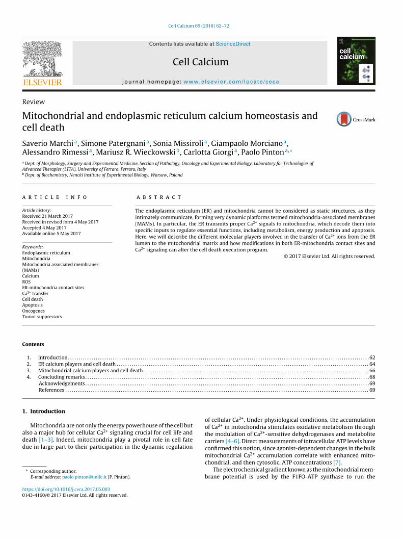
n “energized” (healthy) mitochondria [9]. The low affinity fora2+ of the MCU system is reconciled by the fact that mitochon-ria are not exposed to the bulk cytosolic concentration rise butather to high Ca2+ microdomains generated in the proximity ofhe endoplasmic reticulum (ER) [10]. The ER lumen is consid-red to be the major intracellular Ca2+ storage compartment, andepletion of the ER Ca2+ content is followed by rapid accumula-ion inside the mitochondrial matrix through the uniporter system.he close apposition between the ER and mitochondria consistsf mitochondrial reticular and/or branched networks in the cyto-ol that intimately and dynamically interact with the ER network;hese connections are termed mitochondria-associated mem-ranes (MAMs) [11] (Fig. 1). These contacts between organellesan be readily observed by light and electron microscopy in manyell types, and it is estimated that there are approximately 100itochondrion-ER contacts in a yeast cell [12,13], while approxi-ately 20% of the mitochondrial surface is found in close proximity
o the ER in mammalian cells [10]. Tomography analysis has shownhat tight or loose tethers could adjoin the two organelles, depend-ng on the nature of the peripheral ER [14]. Indeed, both Pellegrinind Nabi labs have recently proposed that smooth ER is apposed at0 nm with mitochondria, whereas rough ER localizes at a 50 nmistance with the mitochondrial outer membrane [15,16].
The main effectors of the ER Ca2+ release machinery are thenositol 1,4,5-trisphosphate (IP3) receptors (IP3R) and ryanodine
eceptors (RyRs) [17]. IP3Rs are ligand-gated channels that facili-ate the release of Ca2+ from ER stores in response to the binding ofgonists to cell surface receptors and the production of the secondig. 1. Immunofluorescence analysis of mitochondrial (green) and Endoplasmic Reticuloomed and the contact points between the two organelles have been indicated with blu
m 69 (2018) 62–72 63
messenger IP3 [18,19]. The Ca2+ released through IP3Rs is trans-ferred to the mitochondrial intermembrane space by a class ofmitochondrial porins known as voltage-dependent anion channels(VDACs), which form very abundant and large voltage-gated poresin the outer mitochondrial membrane at the ER-mitochondria con-tacts [20]. The tightening of the ER-mitochondria connections isrelevant to cell death, revealing an unexpected dependence ofcell function and survival on the maintenance of proper spacingbetween the ER and mitochondria [21,22]. Impaired Ca2+ handlingcan lead to matrix Ca2+ overload and activation of a high conduc-tance pore, the so-called “mitochondrial permeability transitionpore” (mPTP) [23–25]. Mitochondrial Ca2+ overload has long beenknown to be a critical event in the bioenergetic crisis associatedwith cell death by necrosis (a prototypical example is the exci-totoxicity of neurons) and acts as a critical sensitizing signal inthe intrinsic apoptosis pathways [26]. Treatment with apoptoticstimuli, such as C2-ceramide, causes a release of Ca2+ from the ER,inducing dramatic changes in mitochondrial morphology. Indeed,the mitochondrial Ca2+ overload results in dramatic alterations inmitochondrial functions, including decreased ATP production andincreased generation of reactive oxygen species (ROS) [27–29]. Ca2+
and ROS are the most important triggers for mPTP opening, actingin living cells in conjunction with a variety of pathological chal-lenges [30]. Its opening induces mitochondrial swelling, and theselarge-scale alterations of organelle morphology allow the releaseof caspase cofactors into the cytosol. It has been proposed that theCa2+-mediated generation of ROS required for cell death inductionderived from a massive binding of mitochondrial Ca2+ to cardi-olipin, with consequent disintegration of respiratory chain complexII and oxidative stress [31].
Interestingly, a dynamic interplay between Ca2+ and hydrogen
peroxide really occurs at the ER-mitochondria contact sites [32].The ER-mitochondria Ca2+ transfer stimulates H2O2 mobilizationfrom mitochondrial cristae to MAMs, generating specific H2O2 nan-um (red) compartments of human fibroblasts. The color-merged image has beene circles. MAMs: mitochondria associated membranes.

6 Calciu
oCinha
s(aneisftr
dpmdiiT2ffhaCam
2
dcs
cCls(sSibsmiagiatoctcaad
4 S. Marchi et al. / Cell
domains that, in turn, sensitize the ER Ca2+ release to maintaina2+ oscillations [32]. These data reveal that the ER-mitochondrial
nterface accommodates much higher concentrations of key sig-aling molecules compared to those found in the bulk cytosol,ighlighting its role as a molecular platform for the decoding of
wide range of danger signals.The ER–mitochondria contacts have been also linked to ER
tress-mediated cell death and the Unfolding Protein ResponseUPR). Indeed, a variety of ER co-factors and chaperons are enrichedt MAMs [33]. Moreover, during the early phases of ER stress, theumber of ER-mitochondria connections significantly increases,nsuring mitochondrial Ca2+ uptake and ATP production, whichn turn represent the bioenergetics basis for the adaption to suchtressful condition [34]. On the other hand, alterations in MAMsunctional properties induce ER stress and the UPR [35]. Thus,he crosstalk between ER and mitochondria could regulate stressesponses and the UPR at different levels.
The crucial role played by Ca2+ signals in the regulation of celleath and apoptosis was confirmed in the early 2000s, when twoioneer studies demonstrated that Ca2+ transfer from the ER toitochondria was required for the initiation of programmed cell
eath by some apoptotic stimuli [36,37]. These observations weren turn based on several studies indicating that the ER Ca2+ contents a key determinant of cell sensitivity to apoptotic stress [38,39].hereafter, the cancer-related properties of members of the Bcl-/Bax family were associated with the manipulation of Ca2+ transferrom the ER to mitochondria to regulate cell death [40,41]. Over theollowing years, a wide range of oncogenes and tumor suppressorsave been described to require Ca2+ transfer to exert their pro- ornti-apoptotic functions. In this review, we will discuss the role ofa2+ transport systems at the ER and mitochondria in cell deathnd survival and describe the different molecular pathways thatodulate apoptosis onset by targeting the Ca2+ machinery.
. ER calcium players and cell death
Considering the pivotal role of the Ca2+ ion in regulating celleath processes, it is not surprising that ER pumps and channelsontrolling Ca2+ signaling are primary contributors to cell fate deci-ions in physiological and pathological conditions [42] (Fig. 2).
Two primary ER-resident proteins are involved in such pro-esses. The first are the SERCA (sarco/endoplasmic-reticuluma2+-ATPase) type Ca2+ pumps, which maintain correct ER Ca2+
evels by actively pumping Ca2+ into the ER from the intracellularpace. Notably, SERCAs are present as three different SERCA genesATP2A1, ATPA2 and ATP2A3), and each of them generate diverseplice variants. The most ubiquitous pumps are the SERCA2b andERCA2c isoforms, while less expressed is SERCA3, which is presentn six isoforms. SERCA proteins also exhibit tissue-specific distri-ution; for example, SERCA1a and 1b are primarily found in thekeletal muscle [43]. Of these isoforms, the most important andost well studied is SERCA2b, as it displays the highest Ca2+ affin-
ty, and its correct functioning is crucial for Ca2+ uptake in the ERnd the regulation of cell death mechanisms. Indeed, heterozy-ous deletion of the gene encoding SERCA2 in transgenic micenduced squamous cell carcinomas of the upper digestive tract [44],nd SERCA2b mRNA expression dramatically decreased in highlyumorigenic thyroid cells [45]. Accordingly, forced overexpressionf SERCA not only augmented ER Ca2+ levels but also sensitizedells when exposed to pro-apoptotic stimuli [46]. Interestingly,wo SERCA1 splice variants, which encode for truncated proteins
haracterized by only one (instead of seven) Ca2+-binding domain,re associated with a reduction in the ER Ca2+ steady state levelnd an increase in ER Ca2+ leakage, culminating in apoptotic celleath [46]. Moreover, overexpresson of truncated SERCA1 isoformm 69 (2018) 62–72
predisposes to ER stress and amplifies the apoptotic response byincreasing the ER-mitochondria contact points and mitochondrialCa2+ accumulation [47].
Due to the critical role of the SERCA pump in mediating Ca2+-related apoptosis, it could represent an attractive target for anumber of proteins regulating cell death. One example is the tumorsuppressor p53. This protein also localizes to ER sites, where itinteracts with SERCA and exerts its pro-apoptotic functions by reg-ulating the prompt Ca2+ transfer into mitochondria [48,49]. Duringcell death stimuli, p53 accumulates at the ER-mitochondria con-tacts, where it lowers the oxidation state of SERCA pumps andenhances toxic Ca2+ signaling events [48].
Numerous other proteins are known to affect SERCA activity.The most recently discovered ones are the ER-luminal protein disul-fide isomerase TMX1 and the redox-sensitive protein SEPN1. TMX1is reported to inhibit SERCA2b, thereby blocking Ca2+ transfer andmitochondria bioenergetics. Interestingly, the lower mitochondrialactivity ascribed to the TMX1-dependent control of Ca2+ exacer-bates the “Warburg effect”, favoring tumor growth [50]. In contrastto TMX1 activity, SEPN1 enhances the ER Ca2+ uptake activity ofSERCA. Specifically, a redox-active form of SEPN1 interacts withSERCA at ER-mitochondria contact sites, consequently regulatingER Ca2+ levels [51]. Furthermore, SEPN1 counteracts oxidative dam-age, which occurs during ER stress and triggers the UPR. In fact,following UPR induction, ERO1 activity is boosted and the amountof ROS increases, which in turn inactivates SERCA by convertingits luminal thiols into ROS-mediated oxidized derivatives. SEPN1is able to reduce these molecules and restore SERCA activity [51].Notably, SEPN1 is associated with several diseases, particularlywith myopathies characterized by a loss of function and death ofmuscular cells [52].
The hypothesis developed from these experimental observa-tions is that SERCA down-regulation minimizes the apoptoticresponse by reducing Ca2+ transfer, whereas increased SERCA pumpactivity is associated with cell death. However, SERCA2 expressionpositively correlates with colorectal tumor size and metastasis [53].Moreover, modulation of SERCA has been identified as a valuabletherapeutic strategy for inhibiting Notch1 in Notch-driven cancers[54]. The inhibition of SERCA activities by exposure to thapsigargintriggered ER stress-induced apoptosis, which initially manifests asan acute inhibition of protein synthesis and cell cycle arrest and,if sustained, promotes apoptosis. The role played by Ca2+ in thethapsigargin-mediated cell death process has now been clarified:the rapid and complete inhibition of SERCAs evokes a massive Ca2+
release from the ER through the IP3R, and the rise in intracellularCa2+ levels is the key event that causes cell death. Thus, it has tobe the availability of ER Ca2+, and not the absolute amounts of it,which determines whether the ER is able to use the cation to triggercell death.
The Ca2+-dependent pro-apoptotic features of thapsigargin havebeen used to produce the so-called “smart bomb” drug for prostatecancer [55,56]. Because the cytotoxicity of thapsigargin is not celltype specific, significantly damaging even normal cells, thapsigar-gin was combined with a targeting peptide that is a substrate of theserine/protease prostate-specific antigen (PSA). This compound hasbeen shown to be selectively toxic against PSA-producing prostatecancer cells. Moreover, substantial tumor regression of a panel ofhuman cancer xenografts in vivo was induced by a thapsigarginanalog conjugated with the proteolytic enzyme prostate-specificmembrane antigen (PSMA), which, despite its name, is not spe-cific for prostate and is also found in the neovascular tissue of alarge number of cancers [57]. This prodrug has been termed mip-
sagargin (G-202) and is currently being tested in a phase II clinicaltrial [58]. Notably, it has been recently showed that resveratrol andits derivate piceatannol induce cancer cell-specific cell death by
S. Marchi et al. / Cell Calcium 69 (2018) 62–72 65
focus
r[
tae((raeprIn6IbsTIfbpfp
Fig. 2. Ca2+ handling at the ER-mitochondria interface:
educing SERCA activity and enhancing ER-mitochondria tethering59].
The second family of proteins indispensable for correct main-enance of optimal ER Ca2+ levels are divided in two sub-groupsnd are the major Ca2+ release channels expressed on thendo/sarcoplasmic reticulum (ER/SR): the ryanodine receptorsRyRs) and inositol 1,4,5-trisphosphate receptors (IP3Rs) [60]Fig. 2). RyRs are mostly expressed in the SR (commonly used toefer to the entire ER in smooth and striated myocytes), while IP3Rsre large conductance non-selective cation channels ubiquitouslyxpressed in all cell types. The link between RyRs and cell death isoorly understood, although a very recent work reported a crucialole for RyRs in regulating autophagy [61]. By contrast, the role ofP3Rs in the regulation of cell fate is largely studied. The IP3R chan-els exist in three different isoforms that display approximately0–80% homology in their amino acid sequence (IP3R1, IP3R2 and
P3R3) and are all activated by the second messenger IP3, Ca2+, Ca2+-inding proteins, ATP, thiol modification and phosphorylation byeveral proteins, including oncogenes and tumor suppressors [62].hese isoforms have distinct but overlapping expression patterns.n fact, most cell types express at least two or even all three iso-orms. However, predominant expression of a specific isoform haseen demonstrated in some cell types. For example, IP3R3 is the
rincipal isoform expressed in most cultured cell types, while iso-orm 1 is the predominant isoform in neuronal cells, and IP3R2 isredominant in muscle and liver cells [63].on the ER Ca2+ homeostasis. See text for further details.
As described in the Introduction Section, the primary role ofIP3Rs is to allow the transfer of Ca2+ ions from the ER to the intra-cellular stores, particularly inside mitochondria, where Ca2+ playscrucial roles in the control of cell death mechanisms. It has beendemonstrated that the regulation of cell fate by Ca2+ transfer fromthe ER to mitochondria is primarily controlled by a specific IP3Risoform. Indeed, changes in the expression levels and/or phos-phorylation of IP3R3 ultimately resulted in increased or decreasedsusceptibility to cell death. Primary evidence of the particular type3 features were observed in cells with IP3R3 silencing, where apop-totic levels appeared significantly reduced [64,65]. In addition, theimportance of IP3R3 in the regulation of cell fate was demonstratedby several studies showing that several oncogenes and tumor sup-pressors exerted their effects by regulating IP3R3 expression andactivity [66,67]. Classical examples of this group are the oncogeneAKT and the tumor suppressor PTEN, which regulate apoptosis bycontrolling ER Ca2+ release [68–70]. PTEN interacts with IP3Rs,counteracting the Akt-mediated inhibition of ER Ca2+ efflux in aphosphatase-dependent way and thus protecting from cell death[68]. Similar observations were observed for the tumor suppres-sor promyelocytic leukemia protein (PML) [71]. Interestingly, ourgroup recently demonstrated that PML also affects IP3R3 activity toregulate the autophagic response [72]. This occurs because proper
2+
Ca transfer from the ER to mitochondria is required to respond tocellular energy demands by promoting mitochondrial metabolism.Since autophagy is highly dependent on energy supply, depletion ofIP3Rs (and thus reduced Ca2+ transfer) lowered the mitochondrial
6 Calciu
mIetgsmitaiattctdiiBipNlpm
ri(atcsatpcirsiWt
rnuBeaaEsrnpbdatwcodu
6 S. Marchi et al. / Cell
etabolism and ATP production, thereby triggering autophagy.n fact, IP3R-null cell lines have elevated autophagy, and stablexpression of IP3R3 completely restored basal autophagy levels inhese IP3R-null cells [73]. Based on this seminal work, Foskett’sroup recently proposed that tumorigenic cell lines relied on con-titutive IP3R-mediated Ca2+ transfer to mitochondria to sustainitochondrial functions, and inhibition of IP3R by xestospongin B
nduced massive death in cancer cells, whereas their normal coun-erparts were spared [74]. Thus, upon some stressful conditions,brogated ER-mitochondrial Ca2+ transfer can promote, rather thannhibit, cell death, as previously observed during neuronal ER stressnd in Huntington’s disease model mice [75]. Anyway, targetinghe ER-mitochondria Ca2+ connection might represent an effec-ive approach for re-establishing the apoptosis sensitivity of tumorells. This is the strategy that was used to develop the small pep-ide BIRD-2 (Bcl-2-IP3 receptor disruptor-2), which triggers celleath in a wide range of SCLC (small cell lung cancer) lines through
ts binding to the BH4 domain of Bcl-2 [76,77], thereby prevent-ng Bcl-2 interaction with IP3Rs [78]. These findings suggest thatIRD-2 induces Ca2+-mediated apoptosis, as previously reported
n lymphoid malignancies [79,80] and in diffuse large B-cell lym-homa (DL-BCL) cell lines containing elevated Bcl-2 levels [81].otably, it has been recently proposed that the Bcl-2 pharmaco-
ogical inhibitor ABT737 synergize with cisplatin in reducing therogression of human ovarian cancer xenografts by increasing ER-itochondrial Ca2+ transfer [82].Although SERCAs and IP3Rs represent the main targets for the
egulation of Ca2+-dependent apoptosis, there are a number ofntraluminal Ca2+-binding proteins, such as calreticulin, calnexinCNX), and GRP78/BiP, which are able to modulate Ca2+ signalingnd apoptosis independently of ER channels and pumps. Amonghem, the most well studied and characterized is the ER chaperonealreticulin. Overexpression of this protein results in augmentedensitivity to apoptosis [83]. It has been suggested that such pro-poptotic effects may be due to the subsequent increase of Ca2+ inhe ER, which is thus available for the Ca2+-dependent apoptosisrocess. Accordingly, calreticulin-KO cells are resistant to inducedell death [39]. In addition, it has been demonstrated that duringmmunogenic cell death upon treatment with anthracyclines, cal-eticulin translocates with ERp57 to the cell surface, and this eventeems to lead to immunogenic presentation to T-cells, provok-ng the activation of immune response and subsequent apoptosis.
hen the interaction between calreticulin and ERp57 is disrupted,he immune response is lost [84,85].
Appropriate Ca2+ transfer to mitochondria also requires the cor-ect distance between the two organelles, as well as an adequateumber of connections. Therefore, disruption of ER-mitochondrianits could result in altered Ca2+ signaling and apoptosis. NOGO-, a critical regulator of the tubular structure of the ER, is highlyxpressed during pulmonary arterial hypertension, a disease char-cterized by excessive proliferation of vascular cells and lack ofpoptosis [86]. NOGO-B induction increased the space between theR and mitochondria, lowered the intracellular Ca2+ content anduppressed mitochondria-dependent apoptosis [86]. Again, twoecent papers emphasized that the status of ER-mitochondria con-ections could be critical to the proper execution of the apoptoticrocess. The first study reported that the 21 kDa protein encodedy the FATE1 gene decreases sensitivity to mitochondrial Ca2+-ependent apoptotic stimuli and to chemotherapeutic drugs indrenocortical carcinoma (ACC) and in other cancer types [87]. Fur-her studies are needed to determine whether this protein interactsith one of the main channels involved in the maintenance of the
orrect ER Ca2+ levels, but FATE1 is implicated in the regulationf Ca2+-dependent apoptosis by modulating the ER-mitochondriaistance. In the second work, Bonneau et al. proposed a novel andnexpected role for IP3R binding protein released with inositol
m 69 (2018) 62–72
1,4,5-trisphosphate (IRBIT) [88]. IRBIT was discovered in 2003 [89]and has been described as a genuine suppressor of IP3R functions bycompeting with IP3 when IRBIT is phosphorylated at multiple sites[90]. However, upon apoptotic stimulations, IRBIT is dephosphory-lated, inducing cell death by two main mechanisms: i) IRBIT movesfrom MAMs to the cytoplasm together with Bcl2l10, a homolog ofBcl-2, thereby inhibiting its antiapoptotic functions; and ii) IRBITcontributes to the formation and stabilization of ER-mitochondriacontact points, allowing efficient mitochondrial Ca2+ uptake andcell death induction [88].
3. Mitochondrial calcium players and cell death
Ca2+ ions released from the ER are able to bypass the outer mito-chondrial membrane (OMM) and reach the intermembrane spacethrough VDAC channels (Fig. 3). Three different VDAC isoformsare expressed in almost all mammalian tissues, showing compara-ble channel properties, although they have opposite effects on cellsensitivity to apoptotic challenges that involve mitochondrial Ca2+
loading. VDAC1, at the ER-mitochondria juxtapositions, selectivelyinteracts with IP3R3, thereby potentiating the transfer of low-amplitude apoptotic Ca2+ signals to mitochondria [91]. At MAMs,GRP75 permits this interaction; small interfering RNA (siRNA)silencing of GRP75 abolishes the functional coupling between IP3Rand VDAC1, reducing mitochondrial Ca2+ uptake in response toagonist stimulation [92].
The serine/threonine kinase AKT, often up-regulated in can-cer, promotes VDAC1 association with hexokinase 2 (HK2) throughphosphorylation events [93,94]. This association occurs at MAMsand enables HK2 to phosphorylate glucose using the ATP exitingfrom mitochondria through VDAC1, thereby stimulating glycolysis.Conversely, upon inhibition of Akt, HK2 dissociates from VDAC1,causing VDAC1 closure and increased mitochondrial membranepotential [93].
It has recently been shown that mTORC2 is physically asso-ciated with MAMs and that mTORC2-Akt signaling mediatesMAMs integrity and function [95]. mTORC2 localizes at the ER-mitochondria interface in a growth factor-stimulated manner,where it phosphorylates and activates Akt, which in turn phos-phorylates the MAMs resident proteins PACS2, IP3R, and HK2 toregulate MAMs integrity, Ca2+, and energy metabolism, respec-tively [95]. Notably, a recent study reported that myeloid cellleukemia factor 1 (Mcl-1), interacting with VDAC1, promoteslung cancer cell migration, thereby increasing mitochondrial Ca2+
uptake and ROS generation [96]. Also, the anti-apoptotic memberof Bcl-2 family, Bcl-xL, interacts with VDAC1 and protects from celldeath stimuli by limiting the transfer of Ca2+ signals to mitochon-dria [97,98].
The well-defined role of VDAC1 in triggering apoptosis hasencouraged the development of therapeutic strategies based onlimiting VDAC1-related cell death activities to treat differentdiseases associated with enhanced apoptosis, such as neurodegen-erative diseases. A high-throughput screening approach revealedthat the compounds VBIT-3 and VBIT-4 displayed strong antiapop-totic activity by preventing VDAC1 oligomerization, detachment ofhexokinase bound to mitochondria and disruption of intracellularCa2+ levels, thus restoring normal mitochondrial functions [99].
Unlike isoform 1, VDAC2 appears to have an antiapoptotic role,interacting with Bcl-xS and leading to release of Bak [100], whereasVDAC3 seems to have no significant influence on apoptosis [91].VDAC2 interacts specifically with Bak. Indeed, cells deficient in
VDAC2 were more susceptible to apoptotic death, and, accordingly,overexpression of VDAC2 selectively prevented Bak activation andinhibited the mitochondrial apoptotic pathway [101]. Interestingly,VDAC2 depletion shifts the localization of Bak from mitochondria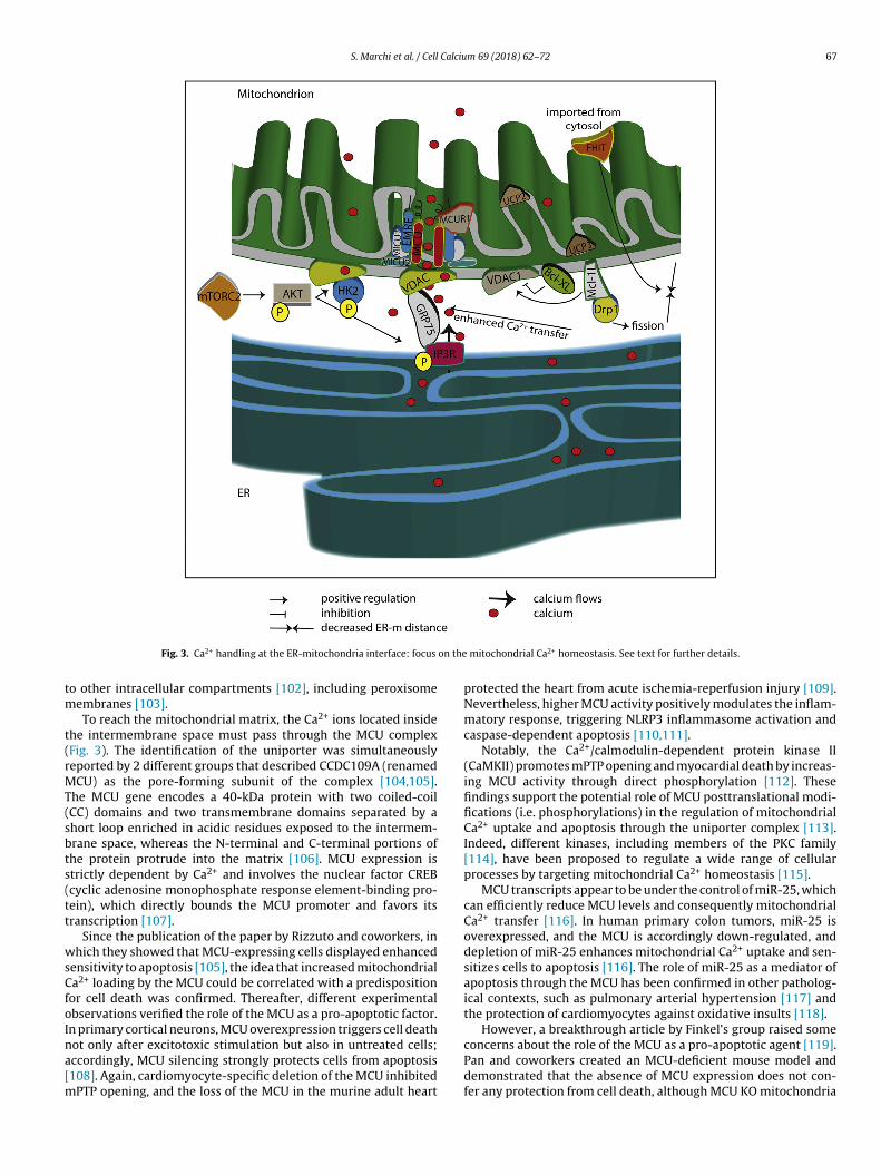
S. Marchi et al. / Cell Calcium 69 (2018) 62–72 67
on the
tm
t(rMT(sbts(tt
wsCfoIna[m
Fig. 3. Ca2+ handling at the ER-mitochondria interface: focus
o other intracellular compartments [102], including peroxisomeembranes [103].To reach the mitochondrial matrix, the Ca2+ ions located inside
he intermembrane space must pass through the MCU complexFig. 3). The identification of the uniporter was simultaneouslyeported by 2 different groups that described CCDC109A (renamedCU) as the pore-forming subunit of the complex [104,105].
he MCU gene encodes a 40-kDa protein with two coiled-coilCC) domains and two transmembrane domains separated by ahort loop enriched in acidic residues exposed to the intermem-rane space, whereas the N-terminal and C-terminal portions ofhe protein protrude into the matrix [106]. MCU expression istrictly dependent by Ca2+ and involves the nuclear factor CREBcyclic adenosine monophosphate response element-binding pro-ein), which directly bounds the MCU promoter and favors itsranscription [107].
Since the publication of the paper by Rizzuto and coworkers, inhich they showed that MCU-expressing cells displayed enhanced
ensitivity to apoptosis [105], the idea that increased mitochondriala2+ loading by the MCU could be correlated with a predispositionor cell death was confirmed. Thereafter, different experimentalbservations verified the role of the MCU as a pro-apoptotic factor.n primary cortical neurons, MCU overexpression triggers cell death
ot only after excitotoxic stimulation but also in untreated cells;ccordingly, MCU silencing strongly protects cells from apoptosis108]. Again, cardiomyocyte-specific deletion of the MCU inhibitedPTP opening, and the loss of the MCU in the murine adult heart
mitochondrial Ca2+ homeostasis. See text for further details.
protected the heart from acute ischemia-reperfusion injury [109].Nevertheless, higher MCU activity positively modulates the inflam-matory response, triggering NLRP3 inflammasome activation andcaspase-dependent apoptosis [110,111].
Notably, the Ca2+/calmodulin-dependent protein kinase II(CaMKII) promotes mPTP opening and myocardial death by increas-ing MCU activity through direct phosphorylation [112]. Thesefindings support the potential role of MCU posttranslational modi-fications (i.e. phosphorylations) in the regulation of mitochondrialCa2+ uptake and apoptosis through the uniporter complex [113].Indeed, different kinases, including members of the PKC family[114], have been proposed to regulate a wide range of cellularprocesses by targeting mitochondrial Ca2+ homeostasis [115].
MCU transcripts appear to be under the control of miR-25, whichcan efficiently reduce MCU levels and consequently mitochondrialCa2+ transfer [116]. In human primary colon tumors, miR-25 isoverexpressed, and the MCU is accordingly down-regulated, anddepletion of miR-25 enhances mitochondrial Ca2+ uptake and sen-sitizes cells to apoptosis [116]. The role of miR-25 as a mediator ofapoptosis through the MCU has been confirmed in other patholog-ical contexts, such as pulmonary arterial hypertension [117] andthe protection of cardiomyocytes against oxidative insults [118].
However, a breakthrough article by Finkel’s group raised some
concerns about the role of the MCU as a pro-apoptotic agent [119].Pan and coworkers created an MCU-deficient mouse model anddemonstrated that the absence of MCU expression does not con-fer any protection from cell death, although MCU KO mitochondria
6 Calciu
sisepa(ma[
bkMtiaecmdtiMte[lht
arCtdtia
piCbddotC
rEidHmrifCtcott
8 S. Marchi et al. / Cell
howed no detectable Ca2+- induced mPTP opening [119]. Interest-ngly, in some cellular settings (i.e., breast cancer cell lines), MCUeems to have no or antiapoptotic functions [120,121]. These data,legantly discussed in [122], suggested the existence of alternativeathways for Ca2+ uptake that might regulate apoptosis in a mech-nism unrelated to the MCU. Indeed, uncoupling proteins 2 and 3UCP2 and UCP3) and SLC25A23 have been described to modulate
itochondrial Ca2+ accumulation and apoptosis through a mech-nism that might be at least in part independent from the MCU123,124].
To date, two other membrane proteins, MCUb and EMRE, haveeen shown to be part of the Ca2+-permeable pore, whereas theey regulators of the whole complex are MICU1 and its paralogICU2, as well as the MCU partner MCUR1 [125–128]. Among
hem, MICU1 is the most studied, and several findings have linkedts altered activity to variations in cell death response. MICU1cts as a gatekeeper of the uniporter complex, preventing Ca2+
ntry under resting conditions and activating the channel at highytosolic Ca2+ concentrations, thereby stimulating Ca2+ entry in theitochondrial matrix [129–132]. The down-regulation of MICU1
ramatically elevates Ca2+ content under basal conditions, boostinghe production of mitochondrial ROS, particularly the superox-de anion, which sensitize cells to apoptosis [130,131]. Moreover,
ICU1 loss promotes chronic Ca2+ entry via the MCU, leadingo oxidative damage and impaired migration in human aorticndothelial cells as well as diminished vascular integrity in vivo133]. Importantly, MICU1 is critical for liver regeneration, sinceiver-specific MICU1 knock-down mice exhibited suppression ofepatocyte proliferation and massive necrosis after partial hepa-ectomy [134].
MCUR1 is a 40-kDa protein localized at the IMM that inter-cts with the MCU, but not with MICU1 [135]; MCUR1 silencingesults in a dramatic inhibition of agonist-induced mitochondriala2+ uptake [135]. Although the role of MCUR1 as a regulator ofhe MCU complex is still debated [136,137], it has been recentlyescribed that MCUR1 expression in Drosophila regulates the Ca2+
hreshold necessary for the permeability transition [138]. Interest-ngly, depletion of MCUR1 resulted in a cellular bioenergetic crisisnd induction of the autophagic process [139].
Another aspect that plays an essential role in the control of apo-tosis is the remodeling of the mitochondrial network, which could
nfluence the capacity of mitochondria to receive and decode thea2+ death signals from the ER. In particular, changes in the num-er of so-called “Ca2+ hot spots” could affect Ca2+ signaling and celleath. This is the case for the pro-apoptotic protein fragile histi-ine triad (FHIT); acting at the mitochondrial compartment, FHITverexpression enhances the number of ER–mitochondria connec-ions, leading to increased mitochondrial Ca2+ accumulation anda2+-dependent apoptosis [140].
Mitofusin 2 (MFN2), a component of the mitochondrial networkemodeling machinery, has been associated with the formation ofR-mitochondria contacts [141,142]. Ablation or silencing of MFN2n fibroblasts and HeLa cells reduces the efficiency of mitochon-rial Ca2+ uptake in response to stimuli that generate IP3 [143].owever, Filadi and colleagues have raised some doubts about thisodel, demonstrating that MFN2 ablation or silencing increases,
ather than diminishes, the close juxtaposition between the twontracellular structures and strengthens the efficacy of Ca2+ trans-er from the ER to mitochondria, sensitizing cells to mitochondriala2+ overload-dependent death [144]. Very recently, in support ofhe original mechanism of action proposed for MFN2, Naon et al.onfirmed that acute MFN2 ablation reduces mitochondrial uptake
f Ca2+ released from the ER, due to the higher distance betweenhe organelles [145]. Thus, the role of MFN2 as an ER-mitochondrialethering factor is still controversial, with different pieces of evi-m 69 (2018) 62–72
dence supporting either the classical model [146] or the revisedone [15,147]. Notably, mitochondrial MFN2, but not ER-associatedMFN2, interacts with the mitochondrial ubiquitin ligase MITOL,mediating the addition of lysine 63-linked polyubiquitin chainsto MFN2 but not its proteasomal degradation [148]. MITOL regu-lates MFN2 localization and MAMs formation, playing a critical rolein neuronal function and survival, which is also illustrated by thereduction in ER Ca2+ transfer that occurs in MITOL-deficient cells[148].
In addition to the ER-mitochondria structural connection, fusionand fission events regulating the shape of the organelles dras-tically influence mitochondrial Ca2+ responses and consequentlycell death. Specifically, it has been proposed that mitochondrialfission factors constitute essential components of cell death sig-naling pathways [149]. By promoting mitochondrial division, thepro-fission factor dynamin-related protein-1 (Drp-1) inhibits thepropagation of Ca2+ signals and protects against Ca2+-mediatedcell death [150]. In addition, the anti-apoptotic variant of Mcl-1 has been demonstrated to control mitochondrial dynamicsby promoting Drp-1-mediated mitochondrial fission, preventingmitochondrial hyperpolarization and Ca2+ uptake [151,152].
Interestingly, it has been demonstrated that the mitochon-drial fission factor Fis1 is able to transmit apoptotic signals frommitochondria to the ER by interacting with Bap31 at the ER andfacilitating its cleavage into the pro-apoptotic p20Bap31 [153].Both proteins constitute a molecular scaffold for the recruitmentand activation of procaspase-8, with a concomitant increase in Ca2+
release from the ER and consequent Ca2+ accumulation in mito-chondria [153].
On the other hand, IF1, the endogenous ATPase inhibitor, pre-serves mitochondrial ultrastructure and limits apoptosis [154].Down-regulation of IF1 induces higher cytochrome c release, whichin turn activates the IP3R, an early event that occurs during apo-ptosis [155], resulting in the amplification of the apoptotic signal[154]. These findings suggest that modifications of both molecularinteractions and mitochondrial architecture could emit danger sig-nals that are decoded by the ER, which responds by eliciting Ca2+
waves and consequently enhances apoptosis.
4. Concluding remarks
Although the distance between the ER and the matrix side ofthe inner mitochondrial membrane is extremely low, less than 50nanometers, the journey that Ca2+ ions must take to reach theirmitochondrial destination requires several regulatory steps andmolecular checkpoints. Alterations in just one of these control-ling pathways result in dramatic metabolic or apoptotic defects,revealing the importance of proper ER-mitochondria Ca2+ transferto maintain the physiological status of the cell. Indeed, different dis-eases have been linked to remodeling of MAMs functions, such ascancer [66] and neurodegeneration [156]. Enhanced understand-ing of the molecular machinery that governs Ca2+ signaling hasprovided opportunities to develop specific Ca2+-based therapeuticapproaches. Moreover, the capacity to pharmacologically modulateCa2+ channels and transport indicates that when Ca2+ signals aredeeply involved in a precise pathological route, a specific Ca2+ chan-nel or pump could be a valuable drug target. Notably, targeting Ca2+
signaling could also be considered as a potential adjuvant therapy,especially to counteract tumor growth and metastatic spread. Thisis the case of carboxyamidotriazole (CAI), a non-voltage-operatedcalcium channel blocker, which has been used in clinical trials in
combination with radiation therapy for the treatment of newlydiagnosed glioblastoma multiforme (for further details, see https://clinicaltrials.gov/). Hence, increasing our knowledge regarding themolecular aspects and functions of different Ca2+ players is the only
Calciu
wa
A
pURtTfctg
R
S. Marchi et al. / Cell
ay to define the best use of Ca2+-based pharmacological agents tomeliorate patient outcomes.
cknowledgements
PP is grateful to Camilla degli Scrovegni for continuous sup-ort. PP is supported by the Italian Ministry of Education,niversity and Research (COFIN no. 20129JLHSY 002, FIRB no.BAP11FXBC 002, and Futuro in Ricerca no. RBFR10EGVP 001),he Italian Cystic Fibrosis Research Foundation (19/2014) andelethon (GGP15219/B). PP and CG are supported by local fundsrom the University of Ferrara and the Italian Association for Can-er Research (IG-18624 and MFAG-13521). MRW is supported byhe National Science Centre, Poland (grant 2014/15/B/NZ1/00490),rant W100/HFSC/2011, and HFSP grant RGP0027/2011.
eferences
[1] R. Rizzuto, D. De Stefani, A. Raffaello, C. Mammucari, Mitochondria assensors and regulators of calcium signalling, Nat. Rev. Mol. Cell Biol. 13(2012) 566–578.
[2] C. Giorgi, F. Baldassari, A. Bononi, M. Bonora, E. De Marchi, S. Marchi, S.Missiroli, S. Patergnani, A. Rimessi, J.M. Suski, M.R. Wieckowski, P. Pinton,Mitochondrial Ca(2+) and apoptosis, Cell Calcium 52 (2012) 36–43.
[3] A. Rimessi, C. Giorgi, P. Pinton, R. Rizzuto, The versatility of mitochondrialcalcium signals: from stimulation of cell metabolism to induction of celldeath, Biochim. Biophys. Acta 1777 (2008) 808–816.
[4] M. Bonora, S. Patergnani, A. Rimessi, E. De Marchi, J.M. Suski, A. Bononi, C.Giorgi, S. Marchi, S. Missiroli, F. Poletti, M.R. Wieckowski, P. Pinton, ATPsynthesis and storage, Purinergic Signal. 8 (2012) 343–357.
[5] R.M. Denton, P.J. Randle, B.R. Martin, Stimulation by calcium ions of pyruvatedehydrogenase phosphate phosphatase, Biochem. J. 128 (1972) 161–163.
[6] J.G. McCormack, A.P. Halestrap, R.M. Denton, Role of calcium ions inregulation of mammalian intramitochondrial metabolism, Physiol. Rev. 70(1990) 391–425.
[7] L.S. Jouaville, P. Pinton, C. Bastianutto, G.A. Rutter, R. Rizzuto, Regulation ofmitochondrial ATP synthesis by calcium: evidence for a long-termmetabolic priming, Proc. Natl. Acad. Sci. U. S. A. 96 (1999) 13807–13812.
[8] G.S. Williams, L. Boyman, W.J. Lederer, Mitochondrial calcium and theregulation of metabolism in the heart, J. Mol. Cell. Cardiol. 78 (2015) 35–45.
[9] I. Drago, P. Pizzo, T. Pozzan, After half a century mitochondrial calcium in-and efflux machineries reveal themselves, EMBO J. 30 (2011) 4119–4125.
[10] R. Rizzuto, P. Pinton, W. Carrington, F.S. Fay, K.E. Fogarty, L.M. Lifshitz, R.A.Tuft, T. Pozzan, Close contacts with the endoplasmic reticulum asdeterminants of mitochondrial Ca2+ responses, Science 280 (1998)1763–1766.
[11] C. Giorgi, S. Missiroli, S. Patergnani, J. Duszynski, M.R. Wieckowski, P. Pinton,Mitochondria-associated membranes: composition, molecular mechanisms,and physiopathological implications, Antioxid. Redox Signal. 22 (2015)995–1019.
[12] G. Achleitner, B. Gaigg, A. Krasser, E. Kainersdorfer, S.D. Kohlwein, A.Perktold, G. Zellnig, G. Daum, Association between the endoplasmicreticulum and mitochondria of yeast facilitates interorganelle transport ofphospholipids through membrane contact, Eur. J. Biochem. 264 (1999)545–553.
[13] J.E. Vance, Phospholipid synthesis in a membrane fraction associated withmitochondria, J. Biol. Chem. 265 (1990) 7248–7256.
[14] G. Csordas, C. Renken, P. Varnai, L. Walter, D. Weaver, K.F. Buttle, T. Balla,C.A. Mannella, G. Hajnoczky, Structural and functional features andsignificance of the physical linkage between ER and mitochondria, J. CellBiol. 174 (2006) 915–921.
[15] P.T. Wang, P.O. Garcin, M. Fu, M. Masoudi, P. St-Pierre, N. Pante, I.R. Nabi,Distinct mechanisms controlling rough and smooth endoplasmic reticulumcontacts with mitochondria, J. Cell Sci. 128 (2015) 2759–2765.
[16] M. Giacomello, L. Pellegrini, The coming of age of the mitochondria-ERcontact: a matter of thickness, Cell Death Differ. 23 (2016) 1417–1427.
[17] A.R. Marks, Intracellular calcium-release channels: regulators of cell life anddeath, Am. J. Physiol. 272 (1997) H597–605.
[18] S. Patel, S.K. Joseph, A.P. Thomas, Molecular properties of inositol1,4,5-trisphosphate receptors, Cell Calcium 25 (1999) 247–264.
[19] D.B. van Rossum, R.L. Patterson, K. Kiselyov, D. Boehning, R.K. Barrow, D.L.Gill, S.H. Snyder, Agonist-induced Ca2+ entry determined by inositol1,4,5-trisphosphate recognition, Proc. Natl. Acad. Sci. U. S. A. 101 (2004)2323–2327.
[20] V.D. De Pinto, F. Palmieri, Transmembrane arrangement of mitochondrialporin or voltage-dependent anion channel (VDAC), J. Bioenerg. Biomembr.24 (1992) 21–26.
[21] D. Naon, L. Scorrano, At the right distance: ER-mitochondria juxtaposition incell life and death, Biochim. Biophys. Acta 1843 (2014) 2184–2194.
m 69 (2018) 62–72 69
[22] H. Ivanova, M. Kerkhofs, R.M. La Rovere, G. Bultynck, Endoplasmicreticulum-mitochondrial Ca2+ fluxes underlying cancer cell survival, Front.Oncol. 7 (2017) 70.
[23] H.K. Baumgartner, J.V. Gerasimenko, C. Thorne, P. Ferdek, T. Pozzan, A.V.Tepikin, O.H. Petersen, R. Sutton, A.J. Watson, O.V. Gerasimenko, Calciumelevation in mitochondria is the main Ca2+ requirement for mitochondrialpermeability transition pore (mPTP) opening, J. Biol. Chem. 284 (2009)20796–20803.
[24] M. Bonora, M.R. Wieckowski, C. Chinopoulos, O. Kepp, G. Kroemer, L.Galluzzi, P. Pinton, Molecular mechanisms of cell death: central implicationof ATP synthase in mitochondrial permeability transition, Oncogene 34(2015) 1475–1486.
[25] G. Morciano, C. Giorgi, M. Bonora, S. Punzetti, R. Pavasini, M.R. Wieckowski,G. Campo, P. Pinton, Molecular identity of the mitochondrial permeabilitytransition pore and its role in ischemia-reperfusion injury, J. Mol. Cell.Cardiol. 78 (2015) 142–153.
[26] A. Danese, S. Patergnani, M. Bonora, M.R. Wieckowski, M. Previati, C. Giorgi,P. Pinton, Calcium regulates cell death in cancer: roles of the mitochondriaand mitochondria-associated membranes (MAMs), Biochim. Biophys. Acta(2017).
[27] D.R. Green, G. Kroemer, The pathophysiology of mitochondrial cell death,Science 305 (2004) 626–629.
[28] A. Rimessi, M. Previati, F. Nigro, M.R. Wieckowski, P. Pinton, Mitochondrialreactive oxygen species and inflammation: molecular mechanisms, diseasesand promising therapies, Int. J. Biochem. Cell Biol. 81 (2016) 281–293.
[29] M. Wasilewski, L. Scorrano, The changing shape of mitochondrial apoptosis,ABBV Trends Endocrinol. Metab. 20 (2009) 287–294.
[30] M. Bonora, P. Pinton, The mitochondrial permeability transition pore andcancer: molecular mechanisms involved in cell death, Front. Oncol. 4 (2014)302.
[31] M.S. Hwang, C.T. Schwall, E. Pazarentzos, C. Datler, N.N. Alder, S. Grimm,Mitochondrial Ca(2+) influx targets cardiolipin to disintegrate respiratorychain complex II for cell death induction, Cell Death Differ. 21 (2014)1733–1745.
[32] D.M. Booth, B. Enyedi, M. Geiszt, P. Varnai, G. Hajnoczky, Redoxnanodomains are induced by and control calcium signaling at theER-mitochondrial interface, Mol. Cell 63 (2016) 240–248.
[33] S. Paillusson, R. Stoica, P. Gomez-Suaga, D.H. Lau, S. Mueller, T. Miller, C.C.Miller, There’s something wrong with my MAM; the ER-mitochondria axisand neurodegenerative diseases, Trends Neurosci. 39 (2016) 146–157.
[34] R. Bravo, J.M. Vicencio, V. Parra, R. Troncoso, J.P. Munoz, M. Bui, C. Quiroga,A.E. Rodriguez, H.E. Verdejo, J. Ferreira, M. Iglewski, M. Chiong, T. Simmen,A. Zorzano, J.A. Hill, B.A. Rothermel, G. Szabadkai, S. Lavandero, IncreasedER-mitochondrial coupling promotes mitochondrial respiration andbioenergetics during early phases of ER stress, J. Cell Sci. 124 (2011)2143–2152.
[35] M. Bui, S.Y. Gilady, R.E. Fitzsimmons, M.D. Benson, E.M. Lynes, K. Gesson,N.M. Alto, S. Strack, J.D. Scott, T. Simmen, Rab32 modulates apoptosis onsetand mitochondria-associated membrane (MAM) properties, J. Biol. Chem.285 (2010) 31590–31602.
[36] P. Pinton, D. Ferrari, E. Rapizzi, F. Di Virgilio, T. Pozzan, R. Rizzuto, The Ca2+concentration of the endoplasmic reticulum is a key determinant ofceramide-induced apoptosis: significance for the molecular mechanism ofBcl-2 action, EMBO J. 20 (2001) 2690–2701.
[37] L. Scorrano, S.A. Oakes, J.T. Opferman, E.H. Cheng, M.D. Sorcinelli, T. Pozzan,S.J. Korsmeyer, BAX and BAK regulation of endoplasmic reticulum Ca2+: acontrol point for apoptosis, Science 300 (2003) 135–139.
[38] P. Pinton, D. Ferrari, P. Magalhaes, K. Schulze-Osthoff, F. Di Virgilio, T.Pozzan, R. Rizzuto, Reduced loading of intracellular Ca(2+) stores anddownregulation of capacitative Ca(2+) influx in Bcl-2-overexpressing cells, J.Cell Biol. 148 (2000) 857–862.
[39] K. Nakamura, E. Bossy-Wetzel, K. Burns, M.P. Fadel, M. Lozyk, I.S. Goping, M.Opas, R.C. Bleackley, D.R. Green, M. Michalak, Changes in endoplasmicreticulum luminal environment affect cell sensitivity to apoptosis, J. CellBiol. 150 (2000) 731–740.
[40] L.K. Nutt, A. Pataer, J. Pahler, B. Fang, J. Roth, D.J. McConkey, S.G. Swisher,Bax and Bak promote apoptosis by modulating endoplasmic reticular andmitochondrial Ca2+ stores, J. Biol. Chem. 277 (2002) 9219–9225.
[41] M. Chami, A. Prandini, M. Campanella, P. Pinton, G. Szabadkai, J.C. Reed, R.Rizzuto, Bcl-2 and Bax exert opposing effects on Ca2+ signaling, which donot depend on their putative pore-forming region, J. Biol. Chem. 279 (2004)54581–54589.
[42] D. Mekahli, G. Bultynck, J.B. Parys, H. De Smedt, L. Missiaen,Endoplasmic-reticulum calcium depletion and disease, Cold Spring Harb.Perspect Biol. 3 (2011).
[43] R. Bravo-Sagua, A.E. Rodriguez, J. Kuzmicic, T. Gutierrez, C. Lopez-Crisosto, C.Quiroga, J. Diaz-Elizondo, M. Chiong, T.G. Gillette, B.A. Rothermel, S.Lavandero, Cell death and survival through the endoplasmicreticulum-mitochondrial axis, Curr. Mol. Med. 13 (2013) 317–329.
[44] L.H. Liu, G.P. Boivin, V. Prasad, M. Periasamy, G.E. Shull, Squamous celltumors in mice heterozygous for a null allele of Atp2a2, encoding the
sarco(endo)plasmic reticulum Ca2+-ATPase isoform 2 Ca2+ pump, J. Biol.Chem. 276 (2001) 26737–26740.[45] F. Pacifico, L. Ulianich, S. De Micheli, S. Treglia, A. Leonardi, P. Vito, S.Formisano, E. Consiglio, B. Di Jeso, The expression of the sarco/endoplasmic

7 Calciu
0 S. Marchi et al. / Cellreticulum Ca2+-ATPases in thyroid and its down-regulation followingneoplastic transformation, J. Mol. Endocrinol. 30 (2003) 399–409.
[46] M. Chami, D. Gozuacik, D. Lagorce, M. Brini, P. Falson, G. Peaucellier, P.Pinton, H. Lecoeur, M.L. Gougeon, M. le Maire, R. Rizzuto, C. Brechot, P.Paterlini-Brechot, SERCA1 truncated proteins unable to pump calciumreduce the endoplasmic reticulum calcium concentration and induceapoptosis, J. Cell Biol. 153 (2001) 1301–1314.
[47] M. Chami, B. Oules, G. Szabadkai, R. Tacine, R. Rizzuto, P. Paterlini-Brechot,Role of SERCA1 truncated isoform in the proapoptotic calcium transfer fromER to mitochondria during ER stress, Mol. Cell 32 (2008) 641–651.
[48] C. Giorgi, M. Bonora, G. Sorrentino, S. Missiroli, F. Poletti, J.M. Suski, F.Galindo Ramirez, R. Rizzuto, F. Di Virgilio, E. Zito, P.P. Pandolfi, M.R.Wieckowski, F. Mammano, G. Del Sal, P. Pinton, p53 at the endoplasmicreticulum regulates apoptosis in a Ca2+-dependent manner, Proc. Natl.Acad. Sci. U. S. A. 112 (2015) 1779–1784.
[49] C. Giorgi, M. Bonora, S. Missiroli, F. Poletti, F.G. Ramirez, G. Morciano, C.Morganti, P.P. Pandolfi, F. Mammano, P. Pinton, Intravital imaging revealsp53-dependent cancer cell death induced by phototherapy via calciumsignaling, Oncotarget 6 (2015) 1435–1445.
[50] A. Raturi, T. Gutierrez, C. Ortiz-Sandoval, A. Ruangkittisakul, M.S.Herrera-Cruz, J.P. Rockley, K. Gesson, D. Ourdev, P.H. Lou, E. Lucchinetti, N.Tahbaz, M. Zaugg, S. Baksh, K. Ballanyi, T. Simmen, TMX1 determines cancercell metabolism as a thiol-based modulator of ER-mitochondria Ca2+ flux, J.Cell Biol. 214 (2016) 433–444.
[51] M. Marino, T. Stoilova, C. Giorgi, A. Bachi, A. Cattaneo, A. Auricchio, P. Pinton,E. Zito, SEPN1 an endoplasmic reticulum-localized selenoprotein linked toskeletal muscle pathology, counteracts hyperoxidation by means ofredox-regulating SERCA2 pump activity, Hum. Mol. Genet. 24 (2015)1843–1855.
[52] A. Lescure, M. Rederstorff, A. Krol, P. Guicheney, V. Allamand, Selenoproteinfunction and muscle disease, Biochim. Biophys. Acta 1790 (2009)1569–1574.
[53] L. Fan, A. Li, W. Li, P. Cai, B. Yang, M. Zhang, Y. Gu, Y. Shu, Y. Sun, Y. Shen, X.Wu, G. Hu, Q. Xu, Novel role of Sarco/endoplasmic reticulum calcium ATPase2 in development of colorectal cancer and its regulation by F36, a curcuminanalog, Biomed. Pharmacother. 68 (2014) 1141–1148.
[54] G. Roti, A. Carlton, K.N. Ross, M. Markstein, K. Pajcini, A.H. Su, N. Perrimon,W.S. Pear, A.L. Kung, S.C. Blacklow, J.C. Aster, K. Stegmaier, Complementarygenomic screens identify SERCA as a therapeutic target in NOTCH1 mutatedcancer, Cancer Cell 23 (2013) 390–405.
[55] S.R. Denmeade, C.M. Jakobsen, S. Janssen, S.R. Khan, E.S. Garrett, H. Lilja, S.B.Christensen, J.T. Isaacs, Prostate-specific antigen-activated thapsigarginprodrug as targeted therapy for prostate cancer, J. Natl. Cancer Inst. 95(2003) 990–1000.
[56] S.R. Denmeade, J.T. Isaacs, The SERCA pump as a therapeutic target: makinga smart bomb for prostate cancer, Cancer Biol. Ther. 4 (2005) 14–22.
[57] S.R. Denmeade, A.M. Mhaka, D.M. Rosen, W.N. Brennen, S. Dalrymple, I.Dach, C. Olesen, B. Gurel, A.M. Demarzo, G. Wilding, M.A. Carducci, C.A.Dionne, J.V. Moller, P. Nissen, S.B. Christensen, J.T. Isaacs, Engineering aprostate-specific membrane antigen-activated tumor endothelial cellprodrug for cancer therapy, Sci. Transl. Med. 4 (2012) 140ra186.
[58] N.T. Doan, E.S. Paulsen, P. Sehgal, J.V. Moller, P. Nissen, S.R. Denmeade, J.T.Isaacs, C.A. Dionne, S.B. Christensen, Targeting thapsigargin towards tumors,Steroids 97 (2015) 2–7.
[59] C.T. Madreiter-Sokolowski, B. Gottschalk, W. Parichatikanond, E. Eroglu, C.Klec, M. Waldeck-Weiermair, R. Malli, W.F. Graier, Resveratrol specificallykills cancer cells by a devastating increase in the Ca2+ coupling between thegreatly tethered endoplasmic reticulum and mitochondria, Cell. Physiol.Biochem. 39 (2016) 1404–1420.
[60] G. Santulli, A.R. Marks, Essential roles of intracellular calcium releasechannels in muscle, brain, metabolism, and aging, Curr. Mol. Pharmacol. 8(2015) 206–222.
[61] T. Vervliet, I. Pintelon, K. Welkenhuyzen, M.D. Bootman, H. Bannai, K.Mikoshiba, W. Martinet, N. Nadif Kasri, J.B. Parys, G. Bultynck, Basalryanodine receptor activity suppresses autophagic flux, Biochem.Pharmacol. 132 (2017) 133–142.
[62] H. Ivanova, T. Vervliet, L. Missiaen, J.B. Parys, H. De Smedt, G. Bultynck,Inositol 1,4,5-trisphosphate receptor-isoform diversity in cell death andsurvival, Biochim. Biophys. Acta 1843 (2014) 2164–2183.
[63] J.K. Foskett, C. White, K.H. Cheung, D.O. Mak, Inositol trisphosphate receptorCa2+ release channels, Physiol. Rev. 87 (2007) 593–658.
[64] C.C. Mendes, D.A. Gomes, M. Thompson, N.C. Souto, T.S. Goes, A.M. Goes,M.A. Rodrigues, M.V. Gomez, M.H. Nathanson, M.F. Leite, The type III inositol1,4,5-trisphosphate receptor preferentially transmits apoptotic Ca2+ signalsinto mitochondria, J. Biol. Chem. 280 (2005) 40892–40900.
[65] S. Blackshaw, A. Sawa, A.H. Sharp, C.A. Ross, S.H. Snyder, A.A. Khan, Type 3inositol 1,4,5-trisphosphate receptor modulates cell death, FASEB J. 14(2000) 1375–1379.
[66] S. Marchi, C. Giorgi, M. Oparka, J. Duszynski, M.R. Wieckowski, P. Pinton,Oncogenic and oncosuppressive signal transduction atmitochondria-associated endoplasmic reticulum membranes, Mol. Cell
Oncol. 1 (2014) e956469.[67] M. Bittremieux, J.B. Parys, P. Pinton, G. Bultynck, ER functions of oncogenesand tumor suppressors: modulators of intracellular Ca(2+) signaling,Biochim. Biophys. Acta 1863 (2016) 1364–1378.
m 69 (2018) 62–72
[68] A. Bononi, M. Bonora, S. Marchi, S. Missiroli, F. Poletti, C. Giorgi, P.P. Pandolfi,P. Pinton, Identification of PTEN at the ER and MAMs and its regulation ofCa(2+) signaling and apoptosis in a protein phosphatase-dependent manner,Cell Death Differ. 20 (2013) 1631–1643.
[69] T. Szado, V. Vanderheyden, J.B. Parys, H. De Smedt, K. Rietdorf, L. Kotelevets,E. Chastre, F. Khan, U. Landegren, O. Soderberg, M.D. Bootman, H.L. Roderick,Phosphorylation of inositol 1,4,5-trisphosphate receptors by protein kinaseB/Akt inhibits Ca2+ release and apoptosis, Proc. Natl. Acad. Sci. U. S. A. 105(2008) 2427–2432.
[70] S. Marchi, M. Marinello, A. Bononi, M. Bonora, C. Giorgi, A. Rimessi, P. Pinton,Selective modulation of subtype III IP(3)R by Akt regulates ER Ca(2)(+)release and apoptosis, Cell. Death. Dis. 3 (2012) e304.
[71] C. Giorgi, K. Ito, H.K. Lin, C. Santangelo, M.R. Wieckowski, M. Lebiedzinska, A.Bononi, M. Bonora, J. Duszynski, R. Bernardi, R. Rizzuto, C. Tacchetti, P.Pinton, P.P. Pandolfi, PML regulates apoptosis at endoplasmic reticulum bymodulating calcium release, Science 330 (2010) 1247–1251.
[72] S. Missiroli, M. Bonora, S. Patergnani, F. Poletti, M. Perrone, R. Gafa, E. Magri,A. Raimondi, G. Lanza, C. Tacchetti, G. Kroemer, P.P. Pandolfi, P. Pinton, C.Giorgi, PML at mitochondria-associated membranes is critical for therepression of autophagy and cancer development, Cell Rep. 16 (2016)2415–2427.
[73] C. Cardenas, R.A. Miller, I. Smith, T. Bui, J. Molgo, M. Muller, H. Vais, K.H.Cheung, J. Yang, I. Parker, C.B. Thompson, M.J. Birnbaum, K.R. Hallows, J.K.Foskett, Essential regulation of cell bioenergetics by constitutive InsP3receptor Ca2+ transfer to mitochondria, Cell 142 (2010) 270–283.
[74] C. Cardenas, M. Muller, A. McNeal, A. Lovy, F. Jana, G. Bustos, F. Urra, N.Smith, J. Molgo, J.A. Diehl, T.W. Ridky, J.K. Foskett, Selective vulnerability ofcancer cells by inhibition of Ca(2+) transfer from endoplasmic reticulum tomitochondria, Cell Rep. 14 (2016) 2313–2324.
[75] T. Higo, K. Hamada, C. Hisatsune, N. Nukina, T. Hashikawa, M. Hattori, T.Nakamura, K. Mikoshiba, Mechanism of ER stress-induced brain damage byIP(3) receptor, Neuron 68 (2010) 865–878.
[76] Y.P. Rong, G. Bultynck, A.S. Aromolaran, F. Zhong, J.B. Parys, H. De Smedt,G.A. Mignery, H.L. Roderick, M.D. Bootman, C.W. Distelhorst, The BH4domain of Bcl-2 inhibits ER calcium release and apoptosis by binding theregulatory and coupling domain of the IP3 receptor, Proc. Natl. Acad. Sci. U.S. A. 106 (2009) 14397–14402.
[77] G. Monaco, E. Decrock, H. Akl, R. Ponsaerts, T. Vervliet, T. Luyten, M. DeMaeyer, L. Missiaen, C.W. Distelhorst, H. De Smedt, J.B. Parys, L. Leybaert, G.Bultynck, Selective regulation of IP3-receptor-mediated Ca2+ signaling andapoptosis by the BH4 domain of Bcl-2 versus Bcl-Xl, Cell Death Differ. 19(2012) 295–309.
[78] E.F. Greenberg, K.S. McColl, F. Zhong, G. Wildey, A. Dowlati, C.W. Distelhorst,Synergistic killing of human small cell lung cancer cells by the Bcl-2-inositol1,4,5-trisphosphate receptor disruptor BIRD-2 and the BH3-mimeticABT-263, Cell. Death. Dis. 6 (2015) e2034.
[79] F. Zhong, M.W. Harr, G. Bultynck, G. Monaco, J.B. Parys, H. De Smedt, Y.P.Rong, J.K. Molitoris, M. Lam, C. Ryder, S. Matsuyama, C.W. Distelhorst,Induction of Ca(2)+-driven apoptosis in chronic lymphocytic leukemia cellsby peptide-mediated disruption of Bcl-2-IP3 receptor interaction, Blood 117(2011) 2924–2934.
[80] H. Akl, R.M. La Rovere, A. Janssens, P. Vandenberghe, J.B. Parys, G. Bultynck,HA14-1 potentiates apoptosis in B-cell cancer cells sensitive to a peptidedisrupting IP 3 receptor/Bcl-2 complexes, Int. J. Dev. Biol. 59 (2015) 391–398.
[81] H. Akl, G. Monaco, R. La Rovere, K. Welkenhuyzen, S. Kiviluoto, T. Vervliet, J.Molgo, C.W. Distelhorst, L. Missiaen, K. Mikoshiba, J.B. Parys, H. De Smedt, G.Bultynck, IP3R2 levels dictate the apoptotic sensitivity of diffuse large B-celllymphoma cells to an IP3R-derived peptide targeting the BH4 domain ofBcl-2, Cell. Death. Dis. 4 (2013) e632.
[82] Q. Xie, J. Su, B. Jiao, L. Shen, L. Ma, X. Qu, C. Yu, X. Jiang, Y. Xu, L. Sun, ABT737reverses cisplatin resistance by regulating ER-mitochondria Ca2+ signaltransduction in human ovarian cancer cells, Int. J. Oncol. 49 (2016)2507–2519.
[83] S. Lim, W. Chang, B.K. Lee, H. Song, J.H. Hong, S. Lee, B.W. Song, H.J. Kim, M.J.Cha, Y. Jang, N. Chung, S.Y. Choi, K.C. Hwang, Enhanced calreticulinexpression promotes calcium-dependent apoptosis in postnatalcardiomyocytes, Mol. Cells 25 (2008) 390–396.
[84] T. Panaretakis, N. Joza, N. Modjtahedi, A. Tesniere, I. Vitale, M. Durchschlag,G.M. Fimia, O. Kepp, M. Piacentini, K.U. Froehlich, P. van Endert, L. Zitvogel,F. Madeo, G. Kroemer, The co-translocation of ERp57 and calreticulindetermines the immunogenicity of cell death, Cell Death Differ. 15 (2008)1499–1509.
[85] M. Obeid, A. Tesniere, T. Panaretakis, R. Tufi, N. Joza, P. van Endert, F.Ghiringhelli, L. Apetoh, N. Chaput, C. Flament, E. Ullrich, S. de Botton, L.Zitvogel, G. Kroemer, Ecto-calreticulin in immunogenic chemotherapy,Immunol. Rev. 220 (2007) 22–34.
[86] G. Sutendra, P. Dromparis, P. Wright, S. Bonnet, A. Haromy, Z. Hao, M.S.McMurtry, M. Michalak, J.E. Vance, W.C. Sessa, E.D. Michelakis, The role ofNogo and the mitochondria-endoplasmic reticulum unit in pulmonaryhypertension, Sci. Transl. Med. 3 (2011) 88ra55.
[87] M. Doghman-Bouguerra, V. Granatiero, S. Sbiera, I. Sbiera, S. Lacas-Gervais,
F. Brau, M. Fassnacht, R. Rizzuto, E. Lalli, FATE1 antagonizes calcium- anddrug-induced apoptosis by uncoupling ER and mitochondria, EMBO Rep. 17(2016) 1264–1280.
Calciu
[
[
[
[
[
[
[
[
[
[
[
[
S. Marchi et al. / Cell
[88] B. Bonneau, H. Ando, K. Kawaai, M. Hirose, H. Takahashi-Iwanaga, K.Mikoshiba, IRBIT controls apoptosis by interacting with the Bcl-2 homolog,Bcl2l10, and by promoting ER-mitochondria contact, Elife 5 (2016).
[89] H. Ando, A. Mizutani, T. Matsu-ura, K. Mikoshiba, IRBIT, a novel inositol1,4,5-trisphosphate (IP3) receptor-binding protein, is released from the IP3receptor upon IP3 binding to the receptor, J. Biol. Chem. 278 (2003)10602–10612.
[90] H. Ando, A. Mizutani, H. Kiefer, D. Tsuzurugi, T. Michikawa, K. Mikoshiba,IRBIT suppresses IP3 receptor activity by competing with IP3 for thecommon binding site on the IP3 receptor, Mol. Cell 22 (2006) 795–806.
[91] D. De Stefani, A. Bononi, A. Romagnoli, A. Messina, V. De Pinto, P. Pinton, R.Rizzuto, VDAC1 selectively transfers apoptotic Ca2+ signals to mitochondria,Cell Death Differ. 19 (2012) 267–273.
[92] G. Szabadkai, K. Bianchi, P. Varnai, D. De Stefani, M.R. Wieckowski, D.Cavagna, A.I. Nagy, T. Balla, R. Rizzuto, Chaperone-mediated coupling ofendoplasmic reticulum and mitochondrial Ca2+ channels, J. Cell Biol. 175(2006) 901–911.
[93] K. Gottlob, N. Majewski, S. Kennedy, E. Kandel, R.B. Robey, N. Hay, Inhibitionof early apoptotic events by Akt/PKB is dependent on the first committedstep of glycolysis and mitochondrial hexokinase, Genes Dev. 15 (2001)1406–1418.
[94] S. Miyamoto, A.N. Murphy, J.H. Brown, Akt mediates mitochondrialprotection in cardiomyocytes through phosphorylation of mitochondrialhexokinase-II, Cell Death Differ. 15 (2008) 521–529.
[95] C. Betz, D. Stracka, C. Prescianotto-Baschong, M. Frieden, N. Demaurex, M.N.Hall, Feature Article: mTOR complex 2-Akt signaling atmitochondria-associated endoplasmic reticulum membranes (MAM)regulates mitochondrial physiology, Proc. Natl. Acad. Sci. U. S. A. 110 (2013)12526–12534.
[96] H. Huang, K. Shah, N.A. Bradbury, C. Li, C. White, Mcl-1 promotes lungcancer cell migration by directly interacting with VDAC to increasemitochondrial Ca2+ uptake and reactive oxygen species generation, Cell.Death. Dis. 5 (2014) e1482.
[97] N. Arbel, D. Ben-Hail, V. Shoshan-Barmatz, Mediation of the antiapoptoticactivity of Bcl-xL protein upon interaction with VDAC1 protein, J. Biol.Chem. 287 (2012) 23152–23161.
[98] G. Monaco, E. Decrock, N. Arbel, A.R. van Vliet, R.M. La Rovere, H. De Smedt,J.B. Parys, P. Agostinis, L. Leybaert, V. Shoshan-Barmatz, G. Bultynck, TheBH4 domain of anti-apoptotic Bcl-XL but not that of the related Bcl-2, limitsthe voltage-dependent anion channel 1 (VDAC1)-mediated transfer ofpro-apoptotic Ca2+ signals to mitochondria, J. Biol. Chem. 290 (2015)9150–9161.
[99] D. Ben-Hail, R. Begas-Shvartz, M. Shalev, A. Shteinfer-Kuzmine, A. Gruzman,S. Reina, V. De Pinto, V. Shoshan-Barmatz, Novel compounds targeting themitochondrial protein VDAC1 inhibit apoptosis and protect againstmitochondrial dysfunction, J. Biol. Chem. 291 (2016) 24986–25003.
100] M. Plotz, B. Gillissen, A.M. Hossini, P.T. Daniel, J. Eberle, Disruption of theVDAC2-Bak interaction by Bcl-x(S) mediates efficient induction of apoptosisin melanoma cells, Cell Death Differ. 19 (2012) 1928–1938.
101] E.H. Cheng, T.V. Sheiko, J.K. Fisher, W.J. Craigen, S.J. Korsmeyer, VDAC2inhibits BAK activation and mitochondrial apoptosis, Science 301 (2003)513–517.
102] J. Lauterwasser, F. Todt, R.M. Zerbes, T.N. Nguyen, W. Craigen, M. Lazarou, M.van der Laan, F. Edlich, The porin VDAC2 is the mitochondrial platform forBax retrotranslocation, Sci. Rep. 6 (2016) 32994.
103] K.I. Hosoi, N. Miyata, S. Mukai, S. Furuki, K. Okumoto, E.H. Cheng, Y. Fujiki,The VDAC2-BAK axis regulates peroxisomal membrane permeability, J. CellBiol. 216 (2017) 709–722.
104] J.M. Baughman, F. Perocchi, H.S. Girgis, M. Plovanich, C.A. Belcher-Timme, Y.Sancak, X.R. Bao, L. Strittmatter, O. Goldberger, R.L. Bogorad, V. Koteliansky,V.K. Mootha, Integrative genomics identifies MCU as an essential componentof the mitochondrial calcium uniporter, Nature 476 (2011) 341–345.
105] D. De Stefani, A. Raffaello, E. Teardo, I. Szabo, R. Rizzuto, A forty-kilodaltonprotein of the inner membrane is the mitochondrial calcium uniporter,Nature 476 (2011) 336–340.
106] K. Oxenoid, Y. Dong, C. Cao, T. Cui, Y. Sancak, A.L. Markhard, Z. Grabarek, L.Kong, Z. Liu, B. Ouyang, Y. Cong, V.K. Mootha, J.J. Chou, Architecture of themitochondrial calcium uniporter, Nature 533 (2016) 269–273.
107] S. Shanmughapriya, S. Rajan, N.E. Hoffman, X. Zhang, S. Guo, J.E. Kolesar, K.J.Hines, J. Ragheb, N.R. Jog, R. Caricchio, Y. Baba, Y. Zhou, B.A. Kaufman, J.Y.Cheung, T. Kurosaki, D.L. Gill, M. Madesh, Ca2+ signals regulatemitochondrial metabolism by stimulating CREB-mediated expression of themitochondrial Ca2+ uniporter gene MCU, Sci. Signal. 8 (2015) ra23.
108] J. Qiu, Y.W. Tan, A.M. Hagenston, M.A. Martel, N. Kneisel, P.A. Skehel, D.J.Wyllie, H. Bading, G.E. Hardingham, Mitochondrial calcium uniporter Mcucontrols excitotoxicity and is transcriptionally repressed by neuroprotectivenuclear calcium signals, Nat. Commun. 4 (2013) 2034.
109] J.Q. Kwong, X. Lu, R.N. Correll, J.A. Schwanekamp, R.J. Vagnozzi, M.A. Sargent,A.J. York, J. Zhang, D.M. Bers, J.D. Molkentin, The mitochondrial calciumuniporter selectively matches metabolic output to acute contractile stress inthe heart, Cell Rep. 12 (2015) 15–22.
110] K. Triantafilou, T.R. Hughes, M. Triantafilou, B.P. Morgan, The complementmembrane attack complex triggers intracellular Ca2+ fluxes leading toNLRP3 inflammasome activation, J. Cell Sci. 126 (2013) 2903–2913.
111] A. Rimessi, V. Bezzerri, S. Patergnani, S. Marchi, G. Cabrini, P. Pinton,Mitochondrial Ca2+-dependent NLRP3 activation exacerbates the
m 69 (2018) 62–72 71
Pseudomonas aeruginosa-driven inflammatory response in cystic fibrosis,Nat. Commun. 6 (2015) 6201.
[112] M.L. Joiner, O.M. Koval, J. Li, B.J. He, C. Allamargot, Z. Gao, E.D. Luczak, D.D.Hall, B.D. Fink, B. Chen, J. Yang, S.A. Moore, T.D. Scholz, S. Strack, P.J. Mohler,W.I. Sivitz, L.S. Song, M.E. Anderson, CaMKII determines mitochondrialstress responses in heart, Nature 491 (2012) 269–273.
[113] B.S. Jhun, J. Mishra, S. Monaco, D. Fu, W. Jiang, S.S. Sheu, J. O-Uchi, Themitochondrial Ca2+ uniporter: regulation by auxiliary subunits and signaltransduction pathways, Am. J. Physiol. Cell Physiol. 311 (2016) C67–80.
[114] P. Pinton, S. Leo, M.R. Wieckowski, G. Di Benedetto, R. Rizzuto, Long-termmodulation of mitochondrial Ca2+ signals by protein kinase C isozymes, J.Cell Biol. 165 (2004) 223–232.
[115] A. Bononi, C. Agnoletto, E. De Marchi, S. Marchi, S. Patergnani, M. Bonora, C.Giorgi, S. Missiroli, F. Poletti, A. Rimessi, P. Pinton, Protein kinases andphosphatases in the control of cell fate, Enzyme Res. 2011 (2011) 329098.
[116] S. Marchi, L. Lupini, S. Patergnani, A. Rimessi, S. Missiroli, M. Bonora, A.Bononi, F. Corra, C. Giorgi, E. De Marchi, F. Poletti, R. Gafa, G. Lanza, M.Negrini, R. Rizzuto, P. Pinton, Downregulation of the mitochondrial calciumuniporter by cancer-related miR-25, Curr. Biol. 23 (2013) 58–63.
[117] Z. Hong, K.H. Chen, A. DasGupta, F. Potus, K. Dunham-Snary, S. Bonnet, L.Tian, J. Fu, S. Breuils-Bonnet, S. Provencher, D. Wu, J. Mewburn, M.L.Ormiston, S.L. Archer, MicroRNA-138 and microRNA-25 down-regulatemitochondrial calcium uniporter, causing the pulmonary arterialhypertension cancer phenotype, Am. J. Respir. Crit. Care Med. 195 (2017)515–529.
[118] L. Pan, B.J. Huang, X.E. Ma, S.Y. Wang, J. Feng, F. Lv, Y. Liu, C.M. Li, D.D. Liang,J. Li, L. Xu, Y.H. Chen, MiR-25 protects cardiomyocytes against oxidativedamage by targeting the mitochondrial calcium uniporter, Int. J. Mol. Sci. 16(2015) 5420–5433.
[119] X. Pan, J. Liu, T. Nguyen, C. Liu, J. Sun, Y. Teng, M.M. Fergusson, I.I. Rovira, M.Allen, D.A. Springer, A.M. Aponte, M. Gucek, R.S. Balaban, E. Murphy, T.Finkel, The physiological role of mitochondrial calcium revealed by micelacking the mitochondrial calcium uniporter, Nat. Cell Biol. 15 (2013)1464–1472.
[120] D.D. Hall, Y. Wu, F.E. Domann, D.R. Spitz, M.E. Anderson, Mitochondrialcalcium uniporter activity is dispensable for MDA-MB-231 breast carcinomacell survival, PLoS One 9 (2014) e96866.
[121] M.C. Curry, A.A. Peters, P.A. Kenny, S.J. Roberts-Thomson, G.R. Monteith,Mitochondrial calcium uniporter silencing potentiates caspase-independentcell death in MDA-MB-231 breast cancer cells, Biochem. Biophys. Res.Commun. 434 (2013) 695–700.
[122] D. Pendin, E. Greotti, T. Pozzan, The elusive importance of being amitochondrial Ca(2+) uniporter, Cell Calcium 55 (2014) 139–145.
[123] M. Trenker, R. Malli, I. Fertschai, S. Levak-Frank, W.F. Graier, Uncouplingproteins 2 and 3 are fundamental for mitochondrial Ca2+ uniport, Nat. CellBiol. 9 (2007) 445–452.
[124] N.E. Hoffman, H.C. Chandramoorthy, S. Shanmughapriya, X.Q. Zhang, S.Vallem, P.J. Doonan, K. Malliankaraman, S. Guo, S. Rajan, J.W. Elrod, W.J.Koch, J.Y. Cheung, M. Madesh, SLC25A23 augments mitochondrial Ca(2)(+)uptake, interacts with MCU, and induces oxidative stress-mediated celldeath, Mol. Biol. Cell 25 (2014) 936–947.
[125] D. De Stefani, M. Patron, R. Rizzuto, Structure and function of themitochondrial calcium uniporter complex, Biochim. Biophys. Acta 1853(2015) 2006–2011.
[126] S. Marchi, P. Pinton, The mitochondrial calcium uniporter complex:molecular components, structure and physiopathological implications, J.Physiol. 592 (2014) 829–839.
[127] K.J. Kamer, V.K. Mootha, The molecular era of the mitochondrial calciumuniporter, Nat. Rev. Mol. Cell Biol. 16 (2015) 545–553.
[128] J.K. Foskett, B. Philipson, The mitochondrial Ca(2+) uniporter complex, J.Mol. Cell. Cardiol. 78 (2015) 3–8.
[129] L. Wang, X. Yang, S. Li, Z. Wang, Y. Liu, J. Feng, Y. Zhu, Y. Shen, Structural andmechanistic insights into MICU1 regulation of mitochondrial calciumuptake, EMBO J. 33 (2014) 594–604.
[130] K. Mallilankaraman, P. Doonan, C. Cardenas, H.C. Chandramoorthy, M.Muller, R. Miller, N.E. Hoffman, R.K. Gandhirajan, J. Molgo, M.J. Birnbaum,B.S. Rothberg, D.O. Mak, J.K. Foskett, M. Madesh, MICU1 is an essentialgatekeeper for MCU-mediated mitochondrial Ca(2+) uptake that regulatescell survival, Cell 151 (2012) 630–644.
[131] G. Csordas, T. Golenar, E.L. Seifert, K.J. Kamer, Y. Sancak, F. Perocchi, C.Moffat, D. Weaver, S. de la Fuente Perez, R. Bogorad, V. Koteliansky, J.Adijanto, V.K. Mootha, G. Hajnoczky, MICU1 controls both the threshold andcooperative activation of the mitochondrial Ca(2)(+) uniporter, Cell Metab.17 (2013) 976–987.
[132] J. Matesanz-Isabel, J. Arias-del-Val, P. Alvarez-Illera, R.I. Fonteriz, M.Montero, J. Alvarez, Functional roles of MICU1 and MICU2 in mitochondrialCa(2+) uptake, Biochim. Biophys. Acta 1858 (2016) 1110–1117.
[133] N.E. Hoffman, H.C. Chandramoorthy, S. Shamugapriya, X. Zhang, S. Rajan, K.Mallilankaraman, R.K. Gandhirajan, R.J. Vagnozzi, L.M. Ferrer, K.Sreekrishnanilayam, K. Natarajaseenivasan, S. Vallem, T. Force, E.T. Choi, J.Y.Cheung, M. Madesh, MICU1 motifs define mitochondrial calcium uniporter
binding and activity, Cell Rep. 5 (2013) 1576–1588.[134] A.N. Antony, M. Paillard, C. Moffat, E. Juskeviciute, J. Correnti, B. Bolon, E.Rubin, G. Csordas, E.L. Seifert, J.B. Hoek, G. Hajnoczky, MICU1 regulation ofmitochondrial Ca(2+) uptake dictates survival and tissue regeneration, Nat.Commun. 7 (2016) 10955.
7 Calciu
amplifying calcium-dependent apoptosis, Nat. Cell Biol. 5 (2003)1051–1061.
[156] M. Krols, G. Bultynck, S. Janssens, ER-Mitochondria contact sites: a new
2 S. Marchi et al. / Cell
[135] K. Mallilankaraman, C. Cardenas, P.J. Doonan, H.C. Chandramoorthy, K.M.Irrinki, T. Golenar, G. Csordas, P. Madireddi, J. Yang, M. Muller, R. Miller, J.E.Kolesar, J. Molgo, B. Kaufman, G. Hajnoczky, J.K. Foskett, M. Madesh, MCUR1is an essential component of mitochondrial Ca2+ uptake that regulatescellular metabolism, Nat. Cell Biol. 14 (2012) 1336–1343.
[136] H. Vais, J.E. Tanis, M. Muller, R. Payne, K. Mallilankaraman, J.K. Foskett,MCUR1, CCDC90A is a regulator of the mitochondrial calcium uniporter, CellMetab. 22 (2015) 533–535.
[137] V. Paupe, J. Prudent, E.P. Dassa, O.Z. Rendon, E.A. Shoubridge, CCDC90A(MCUR1) is a cytochrome c oxidase assembly factor and not a regulator ofthe mitochondrial calcium uniporter, Cell Metab. 21 (2015) 109–116.
[138] D. Chaudhuri, D.J. Artiga, S.A. Abiria, D.E. Clapham, Mitochondrial calciumuniporter regulator 1 (MCUR1) regulates the calcium threshold for themitochondrial permeability transition, Proc. Natl. Acad. Sci. U. S. A. 113(2016) E1872–1880.
[139] D. Tomar, Z. Dong, S. Shanmughapriya, D.A. Koch, T. Thomas, N.E. Hoffman,S.A. Timbalia, S.J. Goldman, S.L. Breves, D.P. Corbally, N. Nemani, J.P.Fairweather, A.R. Cutri, X. Zhang, J. Song, F. Jana, J. Huang, C. Barrero, J.E.Rabinowitz, T.S. Luongo, S.M. Schumacher, M.E. Rockman, A. Dietrich, S.Merali, J. Caplan, P. Stathopulos, R.S. Ahima, J.Y. Cheung, S.R. Houser, W.J.Koch, V. Patel, V.M. Gohil, J.W. Elrod, S. Rajan, M. Madesh, MCUR1 is ascaffold factor for the MCU complex function and promotes mitochondrialbioenergetics, Cell Rep. 15 (2016) 1673–1685.
[140] A. Rimessi, S. Marchi, C. Fotino, A. Romagnoli, K. Huebner, C.M. Croce, P.Pinton, R. Rizzuto, Intramitochondrial calcium regulation by the FHIT geneproduct sensitizes to apoptosis, Proc. Natl. Acad. Sci. U. S. A. 106 (2009)12753–12758.
[141] A. Raturi, T. Simmen, Where the endoplasmic reticulum and themitochondrion tie the knot: the mitochondria-associated membrane(MAM), Biochim. Biophys. Acta 1833 (2013) 213–224.
[142] T. Klecker, S. Bockler, B. Westermann, Making connections: interorganellecontacts orchestrate mitochondrial behavior, Trends Cell Biol. 24 (2014)537–545.
[143] O.M. de Brito, L. Scorrano, Mitofusin 2 tethers endoplasmic reticulum tomitochondria, Nature 456 (2008) 605–610.
[144] R. Filadi, E. Greotti, G. Turacchio, A. Luini, T. Pozzan, P. Pizzo, Mitofusin 2ablation increases endoplasmic reticulum-mitochondria coupling, Proc.
Natl. Acad. Sci. U. S. A. 112 (2015) E2174–2181.[145] D. Naon, M. Zaninello, M. Giacomello, T. Varanita, F. Grespi, S.Lakshminaranayan, A. Serafini, M. Semenzato, S. Herkenne, M.I.Hernandez-Alvarez, A. Zorzano, D. De Stefani, G.W. Dorn, 2nd, L. Scorrano,Critical reappraisal confirms that Mitofusin 2 is an endoplasmic
m 69 (2018) 62–72
reticulum-mitochondria tether, Proc. Natl. Acad. Sci. U. S. A. 113 (2016)11249–11254.
[146] M. Schneeberger, M.O. Dietrich, D. Sebastian, M. Imbernon, C. Castano, A.Garcia, Y. Esteban, A. Gonzalez-Franquesa, I.C. Rodriguez, A. Bortolozzi, P.M.Garcia-Roves, R. Gomis, R. Nogueiras, T.L. Horvath, A. Zorzano, M. Claret,Mitofusin 2 in POMC neurons connects ER stress with leptin resistance andenergy imbalance, Cell 155 (2013) 172–187.
[147] P. Cosson, A. Marchetti, M. Ravazzola, L. Orci, Mitofusin-2 independentjuxtaposition of endoplasmic reticulum and mitochondria: anultrastructural study, PLoS One 7 (2012) e46293.
[148] A. Sugiura, S. Nagashima, T. Tokuyama, T. Amo, Y. Matsuki, S. Ishido, Y. Kudo,H.M. McBride, T. Fukuda, N. Matsushita, R. Inatome, S. Yanagi, MITOLregulates endoplasmic reticulum-mitochondria contacts via Mitofusin2,Mol. Cell 51 (2013) 20–34.
[149] R.J. Youle, A.M. van der Bliek, Mitochondrial fission, fusion, and stress,Science 337 (2012) 1062–1065.
[150] G. Szabadkai, A.M. Simoni, M. Chami, M.R. Wieckowski, R.J. Youle, R. Rizzuto,Drp-1-dependent division of the mitochondrial network blocksintraorganellar Ca2+ waves and protects against Ca2+-mediated apoptosis,Mol. Cell 16 (2004) 59–68.
[151] G. Morciano, C. Giorgi, D. Balestra, S. Marchi, D. Perrone, M. Pinotti, P. Pinton,Mcl-1 involvement in mitochondrial dynamics is associated with apoptoticcell death, Mol. Biol. Cell 27 (2016) 20–34.
[152] G. Morciano, G. Pedriali, L. Sbano, T. Iannitti, C. Giorgi, P. Pinton, Intersectionof mitochondrial fission and fusion machinery with apoptotic pathways:role of Mcl-1, Biol. Cell. 108 (2016) 279–293.
[153] R. Iwasawa, A.L. Mahul-Mellier, C. Datler, E. Pazarentzos, S. Grimm, Fis1 andBap31 bridge the mitochondria-ER interface to establish a platform forapoptosis induction, EMBO J. 30 (2011) 556–568.
[154] D. Faccenda, C.H. Tan, A. Seraphim, M.R. Duchen, M. Campanella, IF1 limitsthe apoptotic-signalling cascade by preventing mitochondrial remodelling,Cell Death Differ. 20 (2013) 686–697.
[155] D. Boehning, R.L. Patterson, L. Sedaghat, N.O. Glebova, T. Kurosaki, S.H.Snyder, Cytochrome c binds to inositol (1,4,5) trisphosphate receptors,
regulator of cellular calcium flux comes into play, J. Cell Biol. 214 (2016)367–370.


![ROC RyR - Columbia University€¦ · RyR VDAC SERCA SERCA NCE PMCA mM [Ca] 100 nM [Ca] mM [Ca] mM [Ca] 2 Sarcoplasmic Reticulum (SR) / T Tubule System. 3. 4 Twitch Summation Tetanus.](https://static.fdocuments.in/doc/165x107/5ec3efeb3ae4ef235843ba2f/roc-ryr-columbia-ryr-vdac-serca-serca-nce-pmca-mm-ca-100-nm-ca-mm-ca-mm.jpg)