Ch.16 Knockout Mice as a Tool to the Understanding of Diabetes Mellitus 2004. 9.14.
A sketch of the central nervous system and its origins
G. E. Schneider 2009Part 4: Development and differentiation, spinal level
MIT 9.14 Class 8
CNS structure at the spinal level; autonomic and enteric nervous systems
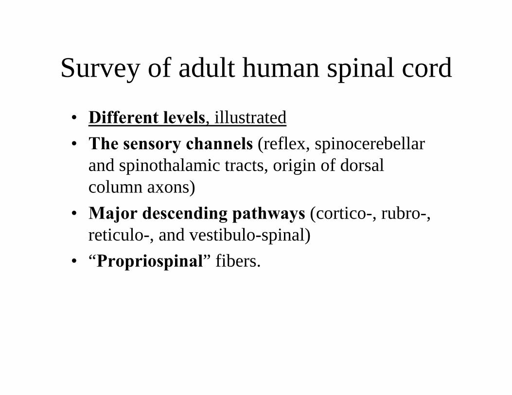
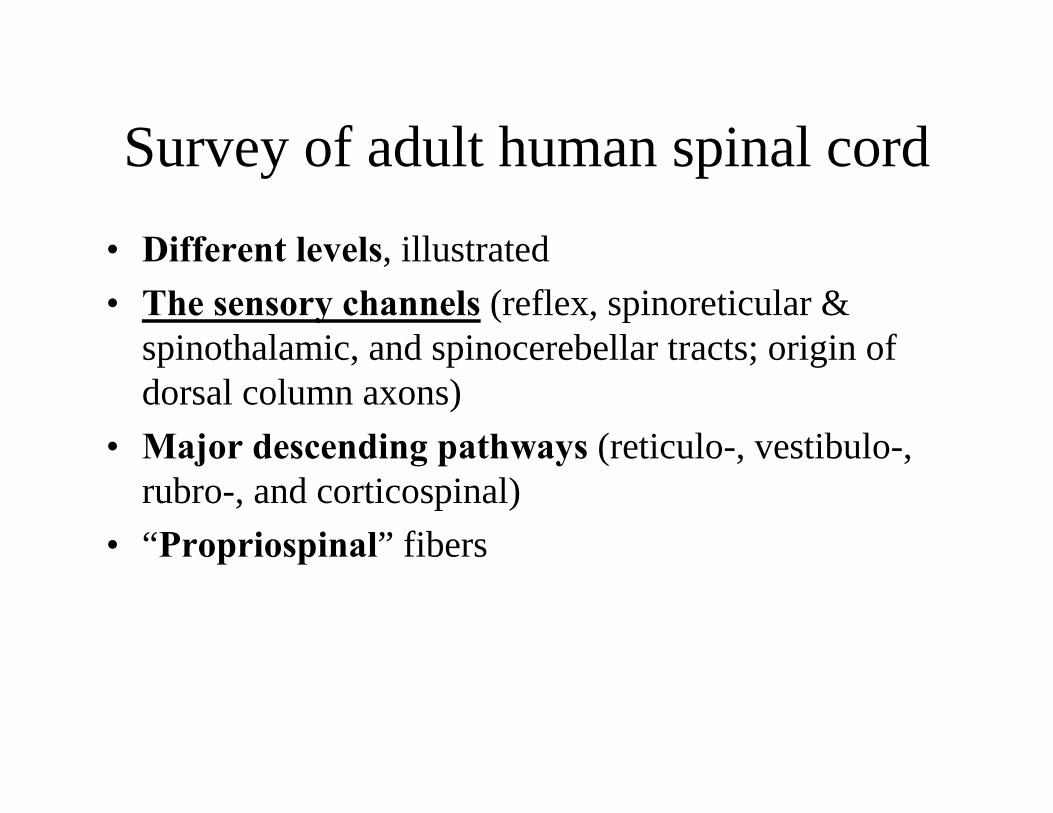
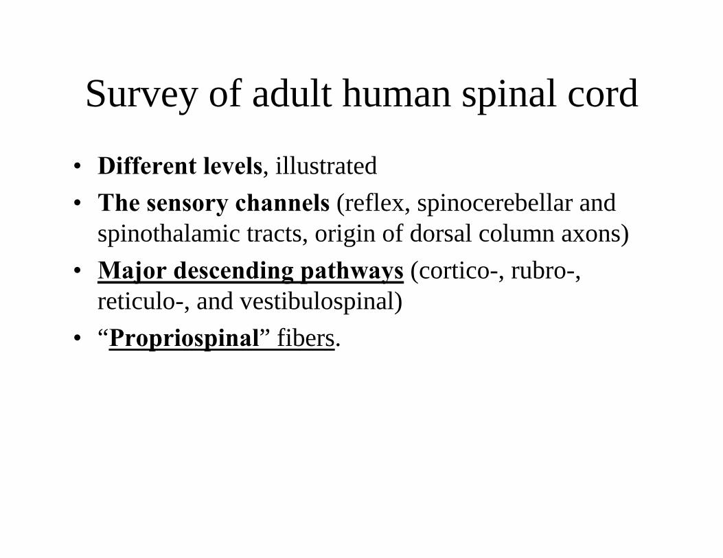
Survey of adult human spinal cord
• Different levels, illustrated • The sensory channels (reflex, spinocerebellar
and spinothalamic tracts, origin of dorsalcolumn axons)
• Major descending pathways (cortico-, rubro-, reticulo-, and vestibulo-spinal)
• “Propriospinal” fibers.

Left: Internal
structure ofspinal cord
Right:
Levels, rostral to caudal
INTERNAL STRUCTURE OF THE SPINAL CORD
Dorsal RootFibers
Fasc.Gracilis
Nuc cornucommissuralis PosteriorSubstantia Gelatinosa
Nuc. Posteromarginalis
Zone of LissauerLat. Corticospinal Tr.
Nuc. Proprius Cornu DorsalisNuc. ReticularisNuc. Dorsalis (of Clarke)
Ant. Spinocerebellar Tr.Lat. Spinothalamic Tr.
Nuc. Motorii LateralisFasc. Proprius
Ant. Spinothalamic Tr.Vestibulospinal Tr.
Nuc. Motorii MedialisVentral Root Fibers
Internal Structure
Spinal Cord Segment C1
Segment C5
Segment C8
Segment T2
Segment T10
Segment L1
Segment L4
Segment S4
III
IIIIV
V
VI
VII
VIIIIX
IXIX
IX
X M
Figure by MIT OpenCourseWare.
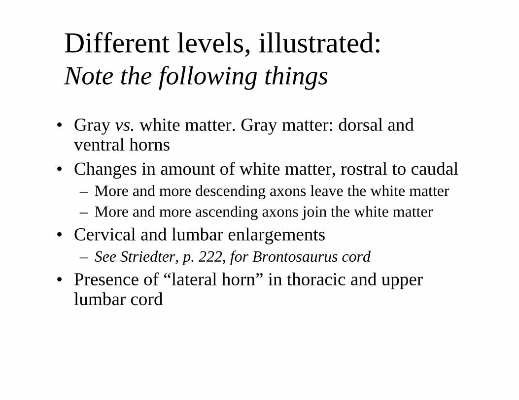
Different levels, illustrated: Note the following things
• Gray vs. white matter. Gray matter: dorsal and ventral horns
• Changes in amount of white matter, rostral to caudal– More and more descending axons leave the white matter – More and more ascending axons join the white matter
• Cervical and lumbar enlargements – See Striedter, p. 222, for Brontosaurus cord
• Presence of “lateral horn” in thoracic and upper lumbar cord
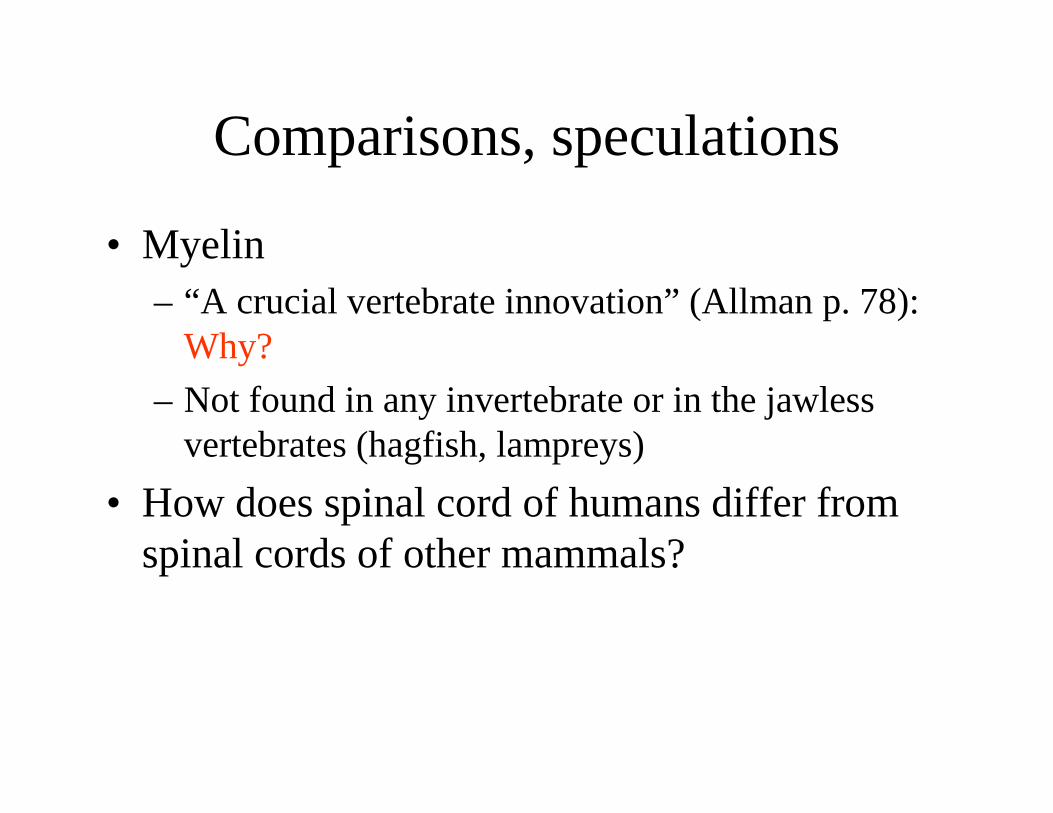
Comparisons, speculations
• Myelin – “A crucial vertebrate innovation” (Allman p. 78):
Why? – Not found in any invertebrate or in the jawless
vertebrates (hagfish, lampreys) • How does spinal cord of humans differ from
spinal cords of other mammals?

Spinal cord cross sectionLumbar level
Note the laminae of Rexed. See Per Brodal, p. 80f.
Figure by MIT OpenCourseWare.
Zona Terminalis
Substantia Gelatinosa
IIIIII
IV
V
VI
VII
VIIIIX
Drawing based on cell - body stain
Note the laminae ofRexed.
Survey of adult human spinal cord
• Different levels, illustrated • The sensory channels (reflex, spinoreticular &
spinothalamic, and spinocerebellar tracts; origin of dorsal column axons)
• Major descending pathways (reticulo-, vestibulo-, rubro-, and corticospinal)
• “Propriospinal” fibers

Termination of Dorsal Root Fibers
Again, note the laminae of Rexed.

Adult spinal cord, schematic frontal section:reflex and lemniscal channels
Spinothalamic tract
Dorsal columns
Dorsal root fibers of various sizes
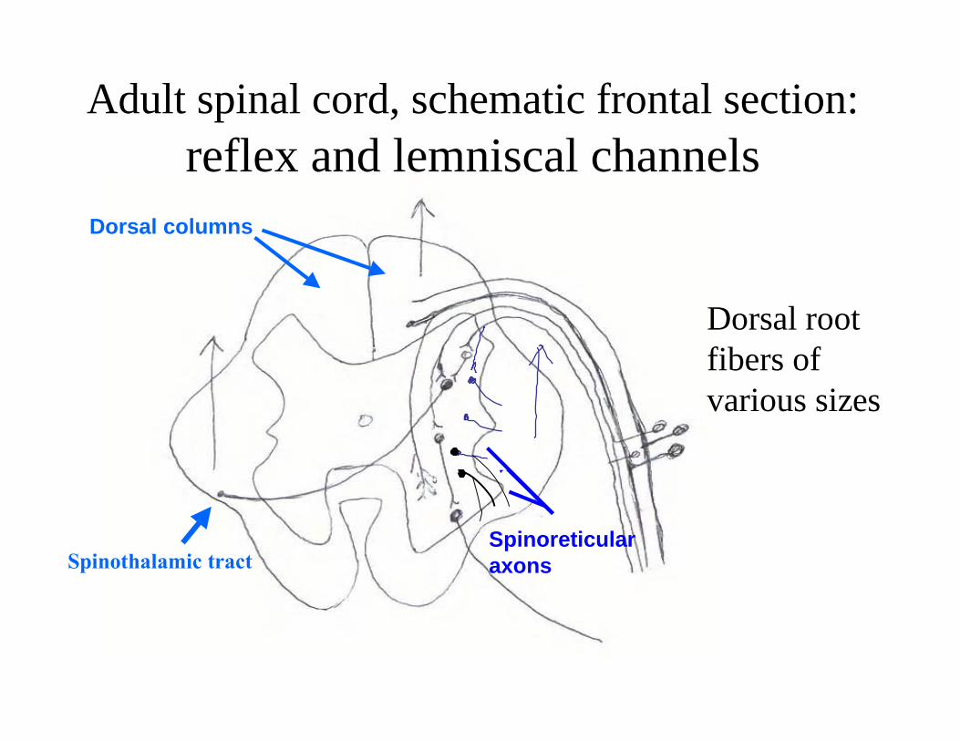
Adult spinal cord, schematic frontal section:reflex and lemniscal channels
Dorsal root fibers of various sizes
Spinothalamic tract
Dorsal columns
Spinoreticular axons
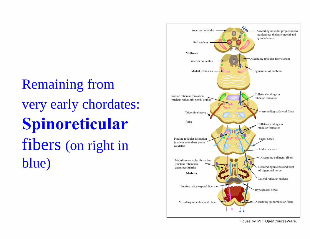
Remaining from very early chordates: Spinoreticular fibers (on right in blue)
Ascending reticular projections tointralaminar thalamic nuclei andhypothalamus
Superior colliculus
Red nucleus
Midbrain
interior colliculus
Medial lemniscus Tegmentum of midbrain
Collateral endings inreticular formation
Ascending collateral fibers
Collateral endings inreticular formation
Facial nerve
Abducens nerve
Ascending collateral fibers
Pontine reticular formation(nucleus reticularis pontiscaudalis)
Medullory reticular formation(nucleus reticularisgigantocellularis)
Medulla
Pontine reticulospinal fibers
Medullary reticulospinal fibers Ascending spinoreticular fibers
Hypoglossal nerve
Lateral reticular nucleus
Descending nucleus and tractof trigeminal nerve
Pontine reticular formation(nucleus reticuloris pontis oralis)
Trigeminal nerve
Pons
Ascending reticular fiber system
Figure by MIT OpenCourseWare.
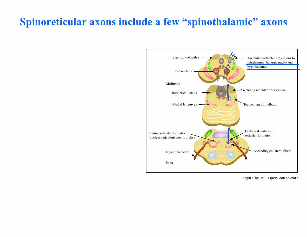
Spinoreticular axons include a few “spinothalamic” axons
Ascending reticular projections tointralaminar thalamic nuclei andhypothalamus
Superior colliculus
Red nucleus
Midbrain
interior colliculus
Medial lemniscus Tegmentum of midbrain
Collateral endings inreticular formation
Ascending collateral fibers
Pontine reticular formation(nucleus reticuloris pontis oralis)
Trigeminal nerve
Pons
Ascending reticular fiber system
Figure by MIT OpenCourseWare.
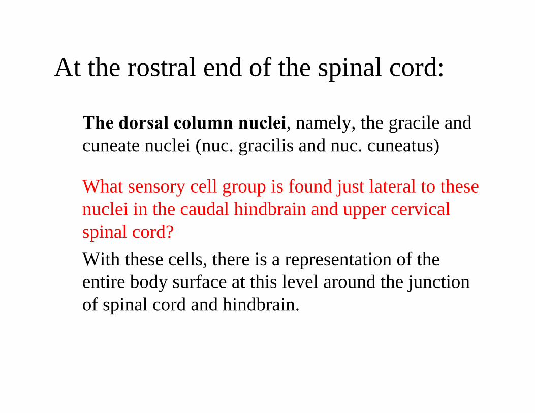
At the rostral end of the spinal cord:
The dorsal column nuclei, namely, the gracile and cuneate nuclei (nuc. gracilis and nuc. cuneatus)
What sensory cell group is found just lateral to these nuclei in the caudal hindbrain and upper cervical spinal cord? With these cells, there is a representation of the entire body surface at this level around the junction of spinal cord and hindbrain.
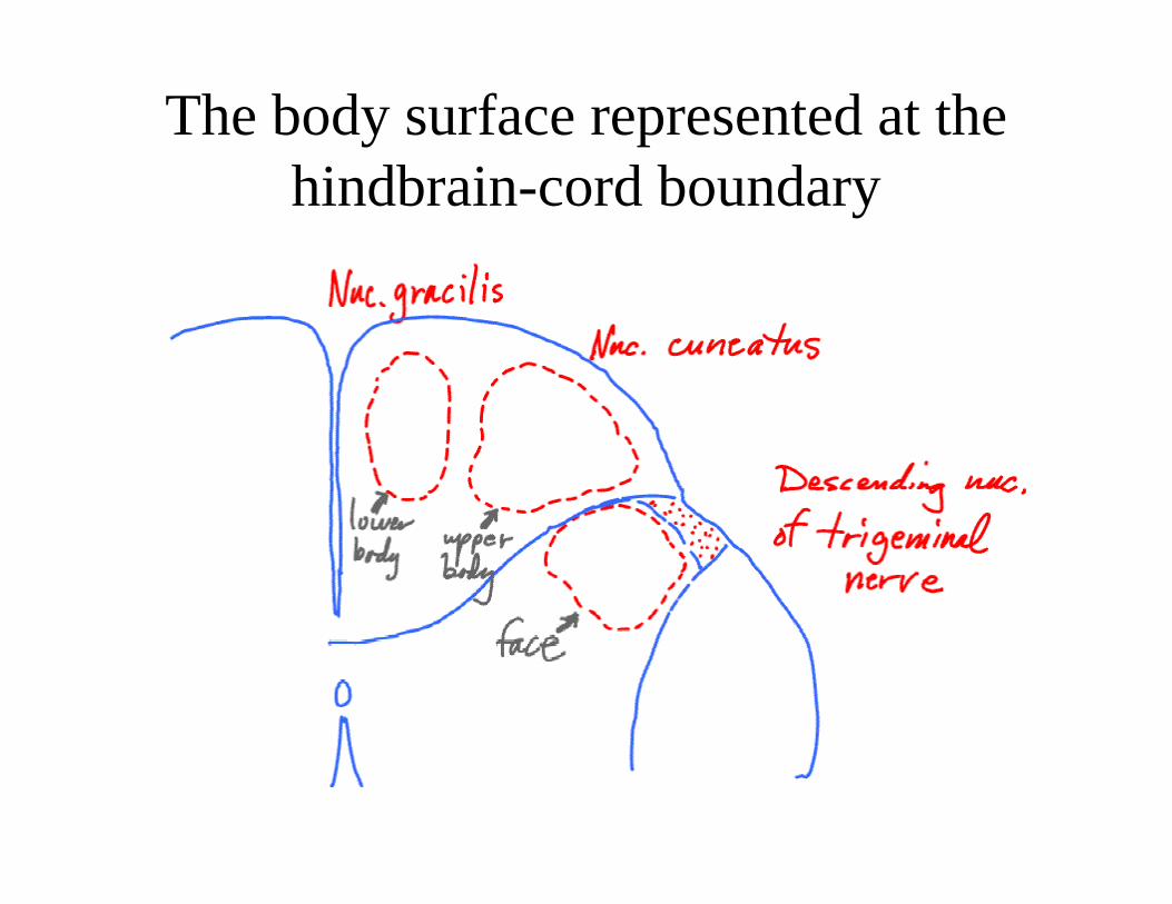
The body surface represented at the hindbrain-cord boundary

Cerebellar channel: Clarke’s Column, illustrated at 5th thoracic segment (Nauta & Feirtag, Fig. 64)
t ino jlexmpo cn ofoInr ewnts from lovememo
unk rlimbs & t
Figure by MIT OpenCourseWare.
Survey of adult human spinal cord
• Different levels, illustrated • The sensory channels (reflex, spinocerebellar and
spinothalamic tracts, origin of dorsal column axons) • Major descending pathways (cortico-, rubro-,
reticulo-, and vestibulospinal) • “Propriospinal” fibers.
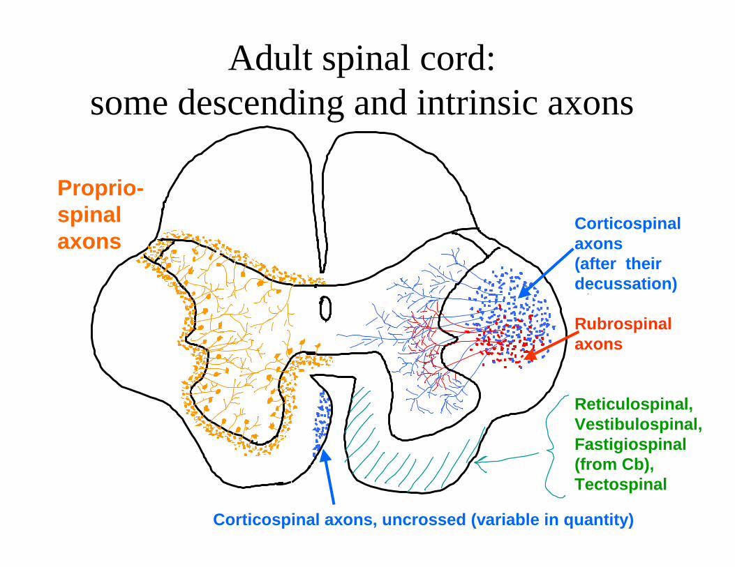
Adult spinal cord:some descending and intrinsic axons
Proprio-spinal Corticospinal axons axons
(after their decussation)
Rubrospinal axons
Reticulospinal, Vestibulospinal, Fastigiospinal (from Cb), Tectospinal
Corticospinal axons, uncrossed (variable in quantity)
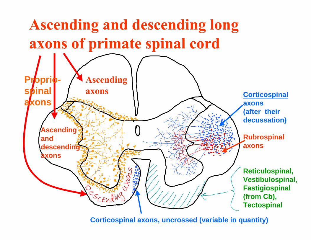
Ascending and descending long axons of primate spinal cord
Proprio- Ascending spinal axons axons
Ascending and descending axons
Corticospinal axons (after their decussation)
Rubrospinal axons
Reticulospinal, Vestibulospinal, Fastigiospinal (from Cb), Tectospinal
Corticospinal axons, uncrossed (variable in quantity)

Intermission: The ventricular system; the meninges and glia
• Remember: the origins of the ventricle in the formation of the neural tube
• The importance of the cerebrospinal fluid in the mature CNS:– Nutrients
– Fluid balance regulation via specific cell regions
– Also a communication medium (because of chemical secretions into it and diffusion from it)
• Where the fluid is made and how it flows:
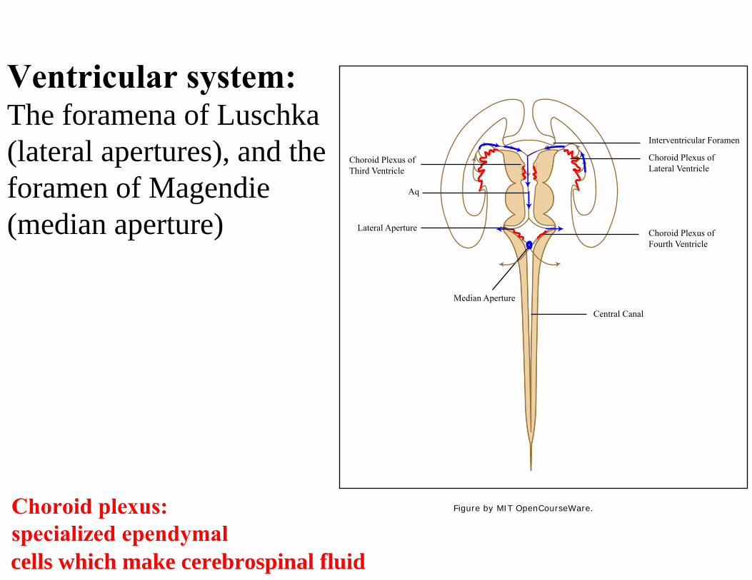
Ventricular system: The foramena of Luschka (lateral apertures), and the foramen of Magendie (median aperture)
Choroid plexus: specialized ependymalcells which make cerebrospinal fluid
Interventricular Foramen
Choroid Plexus of Lateral Ventricle
Choroid Plexus ofFourth Ventricle
Central Canal
Median Aperture
Lateral Aperture
Aq
Choroid Plexus of Third Ventricle
Figure by MIT OpenCourseWare.
The Meninges
1. Define "dura mater" and "pia mater": meaning of the Latin terms, and basic anatomy.
2. Define "arachnoid membrane" and "subarachnoid space".
See Nauta & Feirtag, ch. 10; also P. Brodal, ch. 1, and other texts
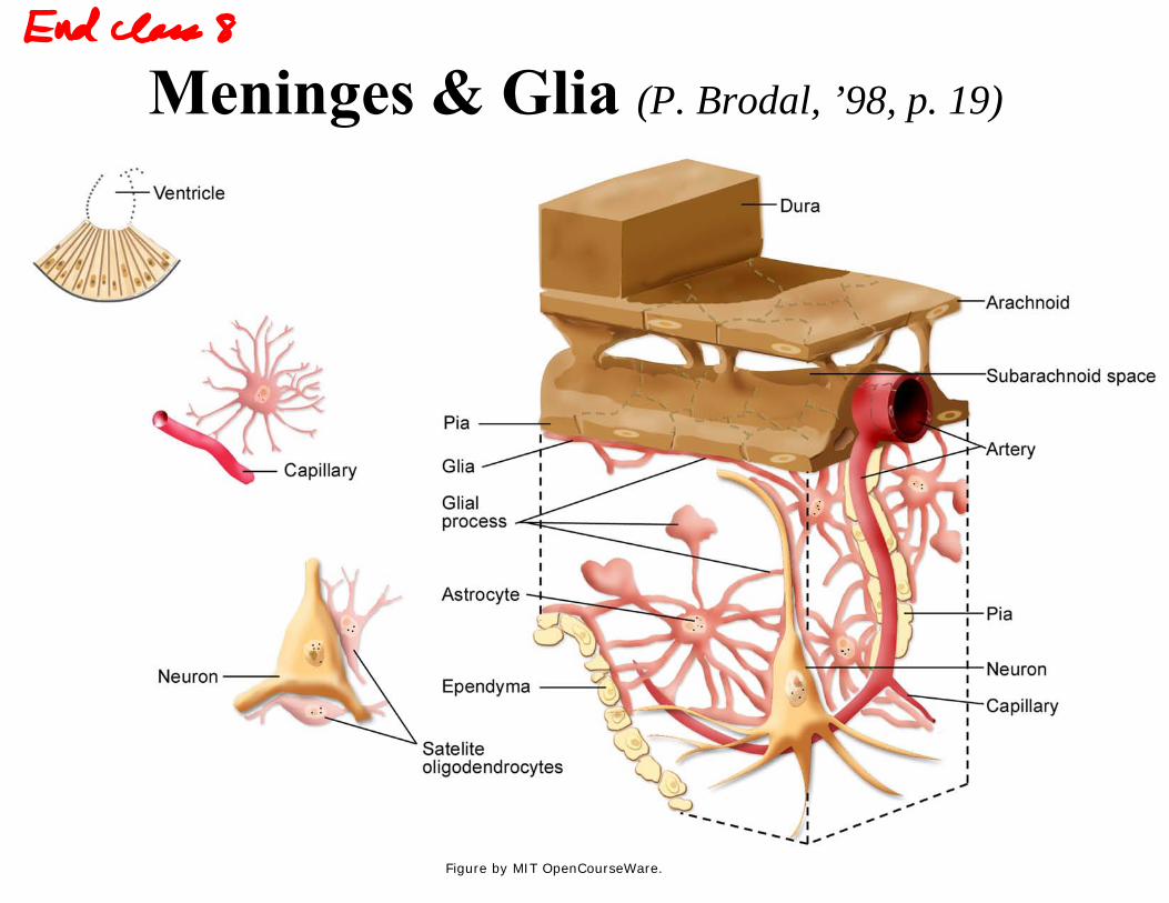
Meninges & Glia (P. Brodal, ’98, p. 19)
Figure by MIT OpenCourseWare.
Picture taken with transmission electron microscope (EM): Astroctyes, pial cells, subarachnoid space(Peters, Palay & Webster, 1976)
SS = subarachnoid space PM = pial membrane Col = collagen fibers SM = smooth muscle GL = glia limitans (astrocyte processes) B = basal lamina As = astrocyte arrows, lower fig: attachment points
Figures removed due to copyright restrictions.
Autonomic nervous system (ANS)
• Overview of functions • Schematic overview of structure • Formation of sympathetic ganglia from the
neural crest • Sympathetic innervation pattern (thoracico-
lumbar system) • Parasympathetic innervation (cranio-sacral
system); dual innervation of smooth muscles and glands.
• Chemical mediation at synapses (first discovered by Otto Loewi in 1921).

Important functions of some autonomic pathways
PARASYMP. FUNCTIONS SYMPATHETIC FUNCTIONS GLAND, TISSUE
Constricts pupil (miosis) Dilates pupil (mydriasis) Iris
Stimulates secretion Little effect on secretion Lacrimal gland
Secretion reduced; less watery Secretion increased; watery Salivary glands
Little effectStimulates secretion (ACh) Sweat glands Constricts Dilates the lumen Lungs, bronchi
Slows heart rate Speeds heart rate; increased Heart ventricular contraction
Stimulates motility, secretions Inhibits motility & secretions Stomach, intestines Relaxes Constricts except with very Anal sphincters
intense activation Vasodilation, engorgement of Orgastic contraction of ductus Sex organs erectile tissue deferens, seminal vesicle, prostatic or
uterine muscles; vasoconstriction
Contracts bladder, relaxes Relaxes wall of bladder; Urinary bladder sphincter, promotes emptying constricts internal sphincter,
inhibits emptying

Important functions of some autonomic pathways
GLAND, TISSUE SYMPATHETIC FUNCTIONS PARASYMP. FUNCTIONS
Adrenal medulla Stimulates secretion Little or no effect Blood vessels, trunk & extremities
Constricts ---
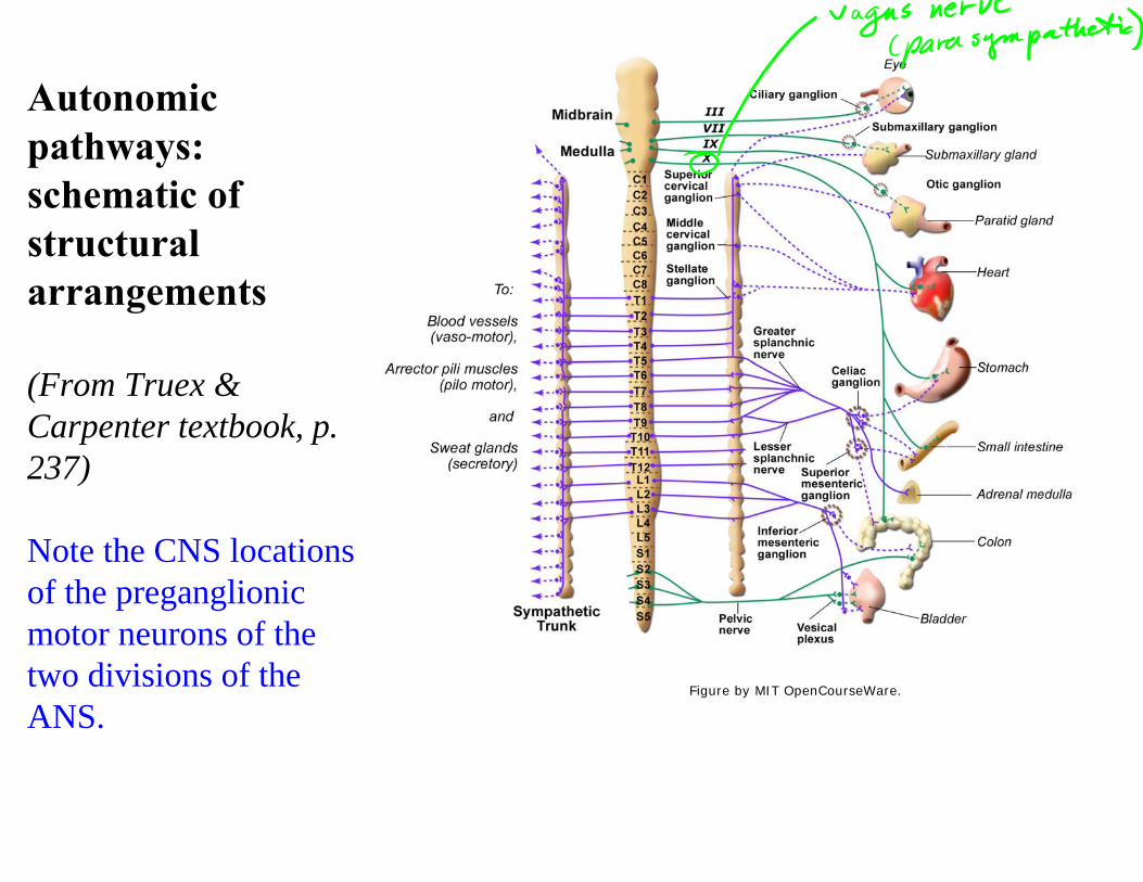
Autonomic pathways: schematic of structural arrangements
(From Truex & Carpenter textbook, p.
s
237)
Note the CNS locationof the preganglionic motor neurons of the two divisions of the ANS.
Figure by MIT OpenCourseWare.
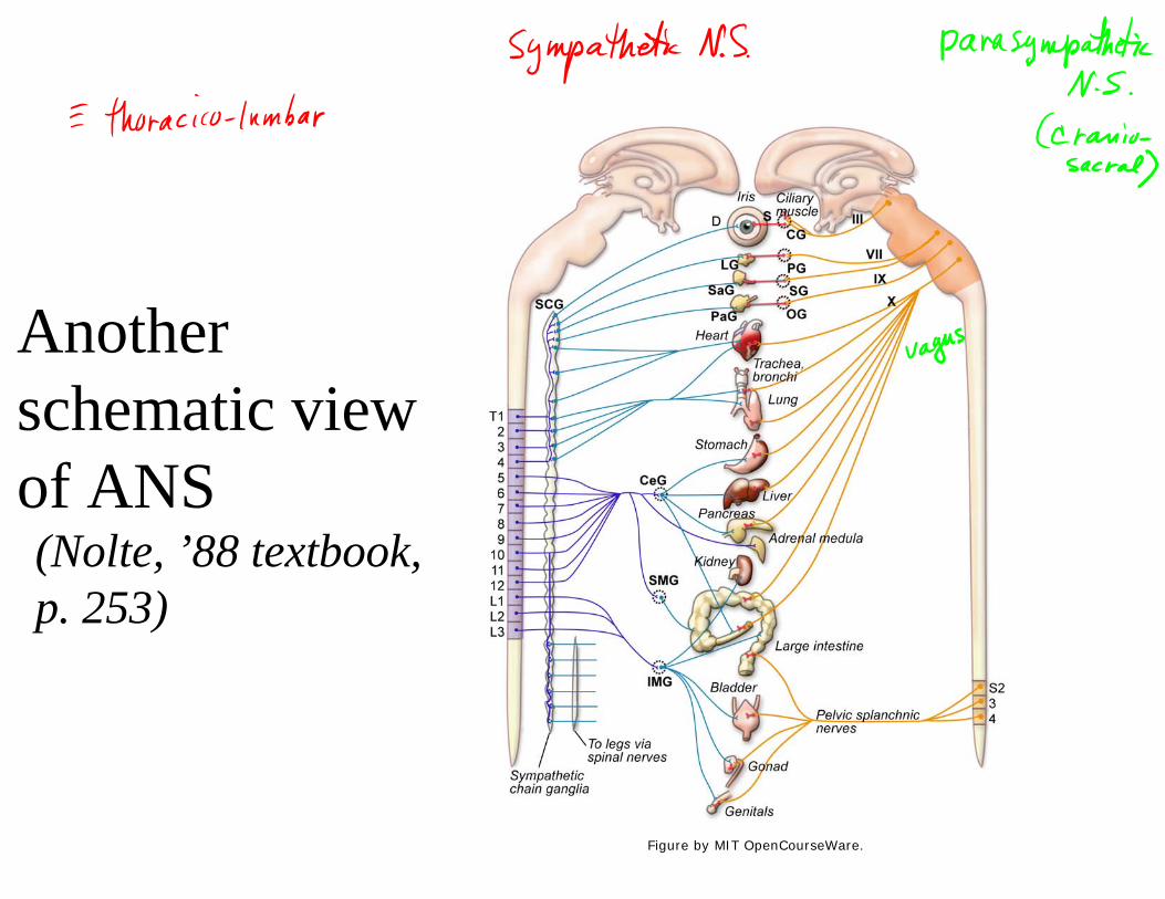
Another schematic view of ANS (Nolte, ’88 textbook, p. 253)
Figure by MIT OpenCourseWare.
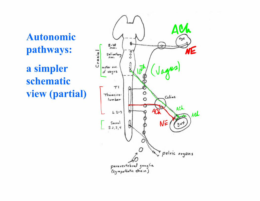
Autonomic pathways:
a simpler schematic view (partial)
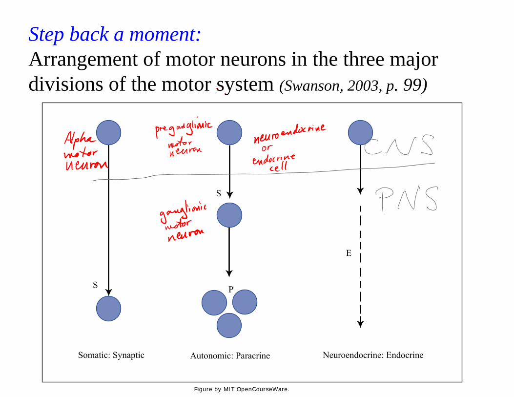
Step back a moment: Arrangement of motor neurons in the three major divisions of the motor system (Swanson, 2003, p. 99)
Somatic: Synaptic Autonomic: Paracrine Neuroendocrine: Endocrine
S
E
S P
Figure by MIT OpenCourseWare.
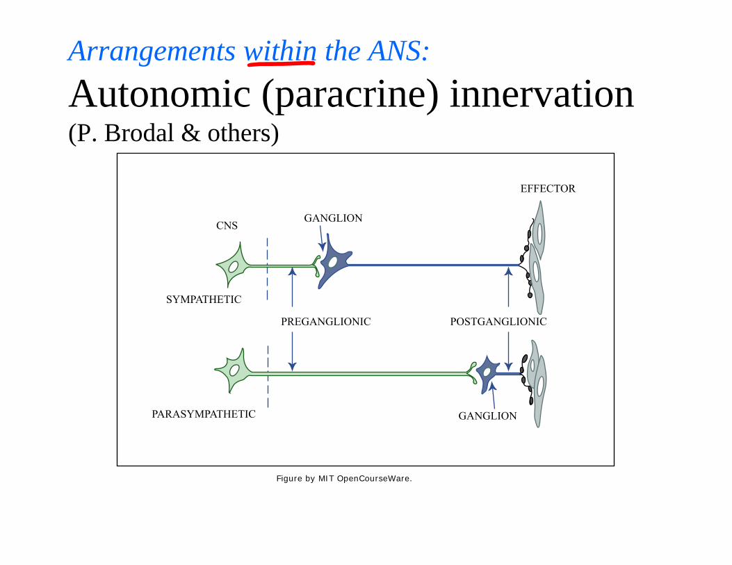
Arrangements within the ANS: Autonomic (paracrine) innervation(P. Brodal & others)
GANGLION
GANGLION
PREGANGLIONIC POSTGANGLIONIC
CNS
EFFECTOR
SYMPATHETIC
PARASYMPATHETIC
Figure by MIT OpenCourseWare.
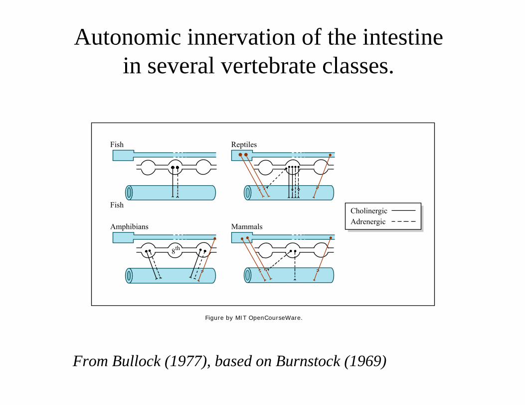
Autonomic innervation of the intestine in several vertebrate classes.
From Bullock (1977), based on Burnstock (1969)
Fish
Fish
Reptiles
Amphibians Mammals
CholinergicAdrenergic
8th
Figure by MIT OpenCourseWare.
More on the ANS next time
MIT OpenCourseWare http://ocw.mit.edu
9.14 Brain Structure and Its Origins Spring 2009
For information about citing these materials or our Terms of Use, visit: http://ocw.mit.edu/terms.