mirVana™ miRNA Detection Kit · mirVana™ miRNA Detection Kit 2 I.B. Reagents Provided with the...
39
mirVana™ miRNA Detection Kit (Part Number AM1552) Instruction Manual I. Introduction . . . . . . . . . . . . . . . . . . . . . . . . . . . . . . . . . . . . . . . . . . . . . . . . . . . . . . . 1 A. Background and Product Description B. Reagents Provided with the Kit and Storage C. Materials Not Provided with the Kit D. Related Products Available from Applied Biosystems II. mirVana™ miRNA Detection Kit Instructions . . . . . . . . . . . . . . . . . . . . . . . . . . . 5 A. Sample RNA B. Radiolabeled RNA Probe Preparation and Amount C. Hybridization of Probe and Sample RNA D. RNase Digestion of Hybridized Probe and Sample RNA E. Separation and Detection of Protected Fragments F. Experimental Setup Examples III. Troubleshooting . . . . . . . . . . . . . . . . . . . . . . . . . . . . . . . . . . . . . . . . . . . . . . . . . . 16 A. Running the Positive Control Reaction B. Pilot Experiment for Assay Optimization C. Troubleshooting Faint or Absent Protected Fragment Bands D. Protected Fragments that are Smeared or Consist of a Ladder of Bands E. Full Length Probe Is Seen in All Lanes F. Aberrant, Pointed or Smeared Bands G. Reasons for Legitimate but Unexpected Bands on Autoradiographs IV. Additional Procedures . . . . . . . . . . . . . . . . . . . . . . . . . . . . . . . . . . . . . . . . . . . . . 26 A. Preparation of Antisense RNA Probes B. Gel Purification of Probe C. Removal of Free Ribonucleotides from Probe Preparations D. Quantitation of Target RNA E. Calculating the Concentration of Unlabeled RNA from a Spectrophotometer Reading F. Gel Analysis of Small RNA Transcripts or RNA Oligonucleotides G. Additional Recipes V. Appendix . . . . . . . . . . . . . . . . . . . . . . . . . . . . . . . . . . . . . . . . . . . . . . . . . . . . . . . . 36 A. References B. Safety Information C. Quality Control
Transcript of mirVana™ miRNA Detection Kit · mirVana™ miRNA Detection Kit 2 I.B. Reagents Provided with the...

mirVana™ miRNA Detection Kit
(Part Number AM1552)
Instruction Manual
I. Introduction. . . . . . . . . . . . . . . . . . . . . . . . . . . . . . . . . . . . . . . . . . . . . . . . . . . . . . . 1
A. Background and Product DescriptionB. Reagents Provided with the Kit and StorageC. Materials Not Provided with the KitD. Related Products Available from Applied Biosystems
II. mirVana™ miRNA Detection Kit Instructions . . . . . . . . . . . . . . . . . . . . . . . . . . . 5
A. Sample RNAB. Radiolabeled RNA Probe Preparation and AmountC. Hybridization of Probe and Sample RNAD. RNase Digestion of Hybridized Probe and Sample RNAE. Separation and Detection of Protected FragmentsF. Experimental Setup Examples
III. Troubleshooting . . . . . . . . . . . . . . . . . . . . . . . . . . . . . . . . . . . . . . . . . . . . . . . . . . 16
A. Running the Positive Control ReactionB. Pilot Experiment for Assay OptimizationC. Troubleshooting Faint or Absent Protected Fragment BandsD. Protected Fragments that are Smeared or Consist of a Ladder of BandsE. Full Length Probe Is Seen in All LanesF. Aberrant, Pointed or Smeared BandsG. Reasons for Legitimate but Unexpected Bands on Autoradiographs
IV. Additional Procedures . . . . . . . . . . . . . . . . . . . . . . . . . . . . . . . . . . . . . . . . . . . . . 26
A. Preparation of Antisense RNA ProbesB. Gel Purification of ProbeC. Removal of Free Ribonucleotides from Probe PreparationsD. Quantitation of Target RNAE. Calculating the Concentration of Unlabeled RNA from a Spectrophotometer ReadingF. Gel Analysis of Small RNA Transcripts or RNA OligonucleotidesG. Additional Recipes
V. Appendix . . . . . . . . . . . . . . . . . . . . . . . . . . . . . . . . . . . . . . . . . . . . . . . . . . . . . . . . 36
A. ReferencesB. Safety InformationC. Quality Control

Manual 1552M Revision B Revision Date: April 15, 2008
For research use only. Not for use in diagnostic procedures.
Information in this document is subject to change without notice. Applied Biosystems assumes no responsibil-ity for any errors that may appear in this document.
Applied Biosystems disclaims all warranties with respect to this document, expressed or implied, including butnot limited to those of merchantability or fitness for a particular purpose. In no event shall Applied Biosystemsbe liable, whether in contract, tort, warranty, or under any statute or on any other basis for special, incidental,indirect, punitive, multiple or consequential damages in connection with or arising from this document,including but not limited to the use thereof.
Literature Citation: When describing a procedure for publication using this product, please refer to it as themirVana™ miRNA Detection Kit.
If a paper that cites one of Ambion’s products is published in a research journal, the author(s) may receive afree Ambion T-shirt by sending in the completed form at the back of this instruction manual, along with acopy of the paper.
Warranty and Liability: Applied Biosystems is committed to delivering superior product quality and perfor-mance, supported by industry-leading global service and technical support teams. Warranty information forthe accompanying consumable product is available at www.ambion.com/info/warranty in “Limited Warrantyfor Consumables,” which is subject to the exclusions, conditions, exceptions, and limitations set forth underthe caption “EXCLUSIONS, CONDITIONS, EXCEPTIONS, AND LIMITATIONS” in the full warrantystatement. Please contact Applied Biosystems if you have any questions about our warranties or would likeinformation about post-warranty support.
Patents and Licensing Notifications: The mirVana™ miRNA Detection Kit is covered by US patent5422241.
Trademarks: Applied Biosystems, AB (Design), Ambion, FirstChoice, MAXIscript and RNaseZap are regis-tered trademarks, and mirVana, Decade and GlycoBlue are trademarks of Applera Corporation or its subsidiar-ies in the US and/or certain other countries. All other trademarks are the sole property of their respectiveowners.
© 2008 Ambion, Inc. All Rights Reserved.

I.A. Background and Product Description
Introduction
1
I. Introduction
A. Background and Product Description
Procedure Overview The mirVana™ miRNA Detection procedure is fast and takes place in asingle microfuge tube. Sample RNA containing the target RNA(s) ofinterest is simply mixed with one or several radiolabeled antisense RNAprobes and hybridization buffer. After heat denaturation, the mixture isincubated at 42°C to hybridize the probes to the complementary RNAmolecules in the experimental RNA samples. After hybridization, unhy-bridized RNA species (from the sample) and excess RNA probes areremoved by a rapid ribonuclease digestion step. Finally, radiolabeledprobe protected from RNase digestion by hybridization to the target RNAis recovered using Ambion® patented single step technology for simulta-neous ribonuclease inactivation and nucleic acid precipitation. Radiola-beled protected RNA probe fragments are then analyzed on a denaturingpolyacrylamide gel. Because no transfer to solid support is required andbecause the hybridization is performed in solution, the procedure ensuresa sensitive and linear detection signal after autoradiography.
The mirVana miRNA Detection Kit includes a control dsDNA tem-plate and Probe Elution Buffer to transcribe and gel purify a 32 nt anti-sense RNA probe. When used in a mirVana miRNA Detection reactionwith the provided control Mouse Kidney Total RNA, this probe gener-ates a 22 bp protected fragment specific for miR-16 miRNA(Lagos-Quintana et al. 2001).
High sensitivity The kit contains a 2X Hybridization Buffer that was developed withshort antisense probes (typically 19–35 target-specific bases) to provideoptimal sensitivity and specificity for detection of short RNA moleculessuch as siRNA or miRNA. Compared to hybridization protocols thatrely on RNA bound to a solid support (i.e. traditional Northern blots),small RNA molecules are detected more readily and quantitated moreaccurately using a solution hybridization procedure. Quantitative anal-yses can be performed in solution with as little as 10–50 ng of totalRNA to detect attomole (10–18 mole) amounts of target RNA.
Versatility Several radiolabeled probes have been successfully used with the mir-Vana miRNA Detection Kit to analyze the expression of miRNA,siRNA, small nuclear RNA (snRNA), and messenger RNA (mRNA).Another advantage of the assay over Northern blotting is the potentialto simultaneously detect several tiny RNAs of the same size or both tinyRNA and longer RNA species (e.g. siRNA and target messenger RNA)in the same experimental sample.

mirVana™ miRNA Detection Kit
I.B. Reagents Provided with the Kit and Storage2
Simplicity The Ambion mirVana miRNA Detection Kit is designed to avoid someof the problems associated with ribonuclease protection assays, and tooffer simplicity while still allowing flexibility for experimental optimiza-tion. The procedure differs from standard protocols in several respectsproviding greater sensitivity and specificity, as well as being faster andeasier to use. The provided 2X Hybridization Buffer streamlines theprocedure as the probe and sample RNA do not need to be co-precipi-tated or dried down prior to resuspension in hybridization buffer. Also,no proteinase K digestion or phenol-chloroform extraction steps arerequired. This means that the entire procedure can be conducted in asingle tube, reducing hands-on time and variability between experimen-tal samples.
B. Reagents Provided with the Kit and Storage
The mirVana miRNA Detection Kit should be stored at –20°C in anon-frost-free freezer.
The mirVana miRNA Detection Kit provides reagents for 100 assays.The kit also contains control dsDNA template for 10 transcription reac-tions, and Probe Elution Buffer for up to 40 gel purifications.
C. Materials Not Provided with the Kit
Radiolabeled RNA probe DNA template and reagents for preparing radiolabeled RNAprobe—the Ambion mirVana™ miRNA Probe Construction Kit wasdeveloped specifically for production of RNA probes for use in miRNAand siRNA studies (see section IV.A starting on page 26 for instructionson probe preparation).
Amount Component Storage
1 mL 2X Hybridization Buffer –20°C
300 μL RNase A/T1 Solution –20°C
500 μL Yeast RNA (5 mg/mL) –20°C
17 mL RNase Digestion Buffer –20°C
22.5 mL RNase Inactivation/PPT Solution –20°C
1.4 mL Gel Loading Buffer II –20°C
20 μL Mouse Kidney Total RNA (0.5 mg/mL)
–20°C
10 μL Control Template (10 μM) –20°C
8 mL Probe Elution Buffer –20°C
1.75 mL Nuclease-free Water any temp*
* Store at –20°C, 4°C or room temp

I.D. Related Products Available from Applied Biosystems
Introduction
3
General laboratory
equipment and supplies
• RNase-free 1.5 mL or 0.5 mL polypropylene microfuge tubes,adjustable pipettors and RNase-free tips
• ACS grade 100% ethanol• Constant temperature incubator (42°C) and heat block (95–100°C)• Microcentrifuge capable of at least 10,000 X g
Apparatus and reagents for preparing and running denaturing acryla-mide gels (high quality urea, acrylamide and bis-acrylamide,Tris-borate-EDTA buffer, ammonium persulfate, TEMED)
D. Related Products Available from Applied Biosystems
miRNA Certified FirstChoice®
Total RNASee web or print catalog for P/Ns
All of Ambion's high quality total RNA from normal human, mouse, and rattissue is prepared by methods that quantitatively recover microRNAs. Theentire line of FirstChoice Total RNAs are free of DNA and shown to be intactby stringent quality control standards.
MAXIscript® KitsP/N AM1308–AM1326
MAXIscript Kits are designed for synthesis of high specific-activity RNAprobes with specific activities reaching 1 x 10⁹ cpm/μg in just 10 minutes.MAXIscript Kits are available for DNA templates containing T7, T3, andSP6 promoters.
mirVana™ miRNA Probe
Construction KitP/N AM1550
This kit is designed to produce short (<100 nt) labeled RNA transcripts foruse in hybridization assays to detect small RNAs, including miRNA andsiRNA. The kit supplies reagents for both transcription template preparationand RNA probe synthesis. Radiolabeled probes made with the kit are ideal foruse with the mirVana miRNA Detection Kit.
mirVana™ Probe & Marker KitP/N AM1554
The mirVana Probe & Marker Kit is an end labeling kit designed for makingshort radiolabeled probes, and low molecular weight markers for studiesinvolving microRNAs. It can be used with synthetic RNA or DNA oligonu-cleotides to prepare labeled probes, and the kit also provides reagents to pre-pare small radiolabeled RNA size markers (Decade™ Markers), andsingle-nucleotide RNA ladders. Rapid cleanup reagents are included to pre-pare the reaction products for various downstream application.
mirVana™ miRNA Isolation
KitP/N AM1560
The mirVana miRNA Isolation Kit (patent pending) is designed especially forthe isolation of small RNAs, such as microRNA (miRNA), small interferingRNA (siRNA), and small nuclear RNA (snRNA), from tissues and cells. Thekit uses a fast and efficient glass fiber filter (GFF) based procedure to isolatetotal RNA ranging in size from kilobases down to 10-mers. It also includes aprocedure to enrich the population of RNAs that are 200 bases and smaller,which enhances the sensitivity of small RNA detection by solution hybridiza-tion and Northern blot analysis.
Decade™ MarkersP/N AM7778
The Decade Marker System is a set of reagents to prepare radiolabeled lowmolecular weight RNA markers: from 10–100 nt in 10 nt increments. Theuser supplies only [γ-32P]ATP to end label a single, gel purified RNA tran-script which is then cleaved into the 10 molecular weight markers in a simple5 minute reaction.

mirVana™ miRNA Detection Kit
I.D. Related Products Available from Applied Biosystems4
RNase-free Tubes & TipsSee web or print catalog for P/Ns
Ambion RNase-free tubes and tips are available in most commonly used sizesand styles. They are guaranteed RNase- and DNase-free. See our latest catalogor our website (www.ambion.com/prod/tubes) for specific information.
RNaseZap® SolutionP/N AM9780, AM9782, AM9784
RNaseZap RNase Decontamination Solution is simply sprayed, poured, orwiped onto surfaces to instantly inactivate RNases. Rinsing twice with dis-tilled water will eliminate all traces of RNase and RNaseZap Solution.

II.A. Sample RNA
mirVana™ miRNA Detection Kit Instructions
5
II. mirVana™ miRNA Detection Kit Instructions
A. Sample RNA
Standard RNA isolation procedures involving RNA-binding glass fiberfilters must not be used to prepare the experimental RNA samplesbecause these methods will not quantitatively recover RNA smaller than200 nt. We recommend using the Ambion® mirVana™ miRNA IsolationKit to purify representative total RNA populations or to isolate controlfractions specifically enriched or depleted for small RNA species. Whenworking with total RNA purchased from commercial suppliers, be sureto inquire about the method used to isolate the RNA. Many of AmbionFirstChoice® Total RNA products from mouse, rat and human tissueshave been validated for miRNA research. A list is available on our website, or by calling our Technical Services Department.
www.ambion.com/techlib/resources/miRNA/index.html
B. Radiolabeled RNA Probe Preparation and Amount
Only the probe is visualized at the end of the mirVana miRNA Detec-tion procedure; thus it is critically important that very high qualityprobes are used in the procedure. We recommend using highspecific-activity 32P-labeled antisense RNA probes prepared by in vitrotranscription, or 5' labeling.
In vitro transcribed RNA The mirVana miRNA Probe Construction Kit is ideal for thisapplication—it was developed specifically to quickly prepare short,high specific activity, antisense RNA probes of any sequence. RNAprobes made by in vitro transcription should be gel purified to removetranscripts that are shorter than the full-length probe, because theseshorter products may cause “background” smears and/or spurious pro-tected bands in the assay.
5' end-labeled RNA
oligonucleotides
Alternatively gel- or HPLC-purified chemically synthesized RNA oligo-nucleotides can be 5' end labeled and purified with the mirVana Probe& Marker Kit. Because they include only a single radiolabeled nucle-otide per molecule, probes generated with this procedure are typically oflower specific activity than in vitro transcribed probes, but they will notrequire gel purification after the labeling reaction. More informationabout probe design and preparation can be found in sectionIV. Additional Procedures on page 26.
mirVana™ miRNA Detection Kit
II.C. Hybridization of Probe and Sample RNA6
Use a 3–10 fold molar excess
of probe over target
For quantitative detection of the target RNA, it is important that thelabeled probe be present in a 3–10 fold molar excess over the target RNAin the hybridization reaction. In most cases 1–5 x 10⁴ cpm of high specificactivity probe will meet this requirement. See section III.B on page 17 forinstructions on optimizing the mirVana miRNA Detection assay.
C. Hybridization of Probe and Sample RNA
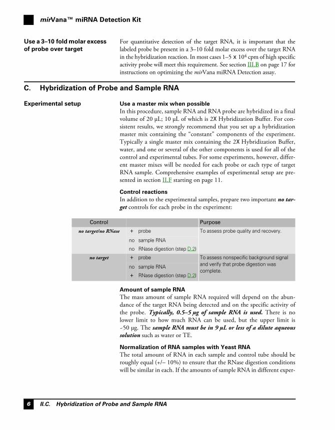
Experimental setup Use a master mix when possible
In this procedure, sample RNA and RNA probe are hybridized in a finalvolume of 20 μL; 10 μL of which is 2X Hybridization Buffer. For con-sistent results, we strongly recommend that you set up a hybridizationmaster mix containing the “constant” components of the experiment.Typically a single master mix containing the 2X Hybridization Buffer,water, and one or several of the other components is used for all of thecontrol and experimental tubes. For some experiments, however, differ-ent master mixes will be needed for each probe or each type of targetRNA sample. Comprehensive examples of experimental setup are pre-sented in section II.F starting on page 11.
Control reactions
In addition to the experimental samples, prepare two important no tar-get controls for each probe in the experiment:
Amount of sample RNA
The mass amount of sample RNA required will depend on the abun-dance of the target RNA being detected and on the specific activity ofthe probe. Typically, 0.5–5 μg of sample RNA is used. There is nolower limit to how much RNA can be used, but the upper limit is~50 μg. The sample RNA must be in 9 μL or less of a dilute aqueoussolution such as water or TE.
Normalization of RNA samples with Yeast RNA
The total amount of RNA in each sample and control tube should beroughly equal (+/– 10%) to ensure that the RNase digestion conditionswill be similar in each. If the amounts of sample RNA in different exper-
Control Purpose
no target/no RNase + probe To assess probe quality and recovery.
no sample RNA
no RNase digestion (step D.2)
no target + probe To assess nonspecific background signal and verify that probe digestion was complete.
no sample RNA
+ RNase digestion (step D.2)

II.C. Hybridization of Probe and Sample RNA
mirVana™ miRNA Detection Kit Instructions
7
IMPORTANT
imental samples varies, use the Yeast RNA (provided with the kit) toadjust the total amount of RNA in each hybridization reaction, includ-ing controls, up to 4–5 μg.
1. Assemble the
hybridization reactions at
room temp
a. Thaw the following kit components and reagents at room
temp
• 2X Hybridization Buffer• Yeast RNA• Nuclease-free Water• Experimental sample RNA(s)• Radiolabeled probe(s)Briefly vortex each tube; keep the Yeast RNA and experimental sam-ple RNA on ice. If probes are in Probe Elution Buffer keep at roomtemp (if probes are in dilute aqueous solution keep them on ice).
b. In an RNase-free microcentrifuge tube prepare a master mix
for hybridization based on the following final component
amounts per tube, and mix thoroughly:
The 2X Hybridization Buffer is very
dense, so be sure to blend the
master mix thoroughly once all the
components are assembled. After
dispensing the master mix into each
tube containing the variable
components, mix thoroughly as
well. Incomplete mixing often
results in aberrant or irreproducible
results.
For optimal mixing of the hybridization reactions, we recommendthat you dispense the variable component(s) that are not in the mas-ter mix into each tube first. Then add the master mix to each tubeand mix thoroughly by pipetting the mixture up and down severaltime or by vortexing 5–10 sec. Centrifuge the tube briefly to collectthe reaction mixture at the bottom of the tube.
c. Set up two no target control tubes for each probe
For each different probe used, include two no target control tubescontaining the following:• the same amount of labeled probe used for the experimental tubes
in step 1.b• Yeast RNA equal to the total amount of RNA (sample RNA +
Yeast RNA) in each experimental tube• 10 μL of 2X Hybridization Buffer• Nuclease-free Water to 20 μL
Amount Component
10 μL 2X Hybridization Buffer
1–5 x 10⁴ cpm Labeled RNA probe
0.5–5 μg Sample RNA
to 5 μg Yeast RNA
to 20 μL Nuclease-free Water

mirVana™ miRNA Detection Kit
II.D. RNase Digestion of Hybridized Probe and Sample RNA8
IMPORTANT
2. Incubate 3 min at
95–100°C
This 3 min heat treatment at 95–100°C denatures the RNA to reducethe effect of secondary structure on hybridization in the next step.Include all the experimental and control samples in this step and thenext step.
3. Incubate 2 hr to overnight
at 42°C
Incubate reactions at 42°C to hybridize probe to its complement in theexperimental sample RNA. To minimize or eliminate condensationaround the tops of the tubes during hybridization, they should betightly capped and incubated preferably in a cabinet-type incubator.Alternatively, submerge the tubes in a water bath or in a water-filledheat block.The hybridization time can be as short as 1 hr for relatively abundanttarget RNA such as miR-16 in mouse kidney (see Figure 4 on page 15).Hybridization times that yield very intense protected fragment signalsmay be reduced in subsequent experiments. For accurate quantitation,however, hybridization reactions must go essentially to completion.
Keep the reactions at hybridization
temperature until adding the RNase
mixture in step D.2 below.
The temperature of hybridization can also be optimized for certainRNAs. Hybridization temperatures up to 52°C are sometimes beneficialfor highly structured target RNA or reactions containing multipleprobes.
D. RNase Digestion of Hybridized Probe and Sample RNA
NOTE
The RNase Digestion Buffer contains GlycoBlue™ coprecipitant to facilitate
precipitation of protected probes and removal of the precipitation solution.
1. Prepare a working
dilution of RNase A/T1
Solution in RNase
Digestion Buffer
a. Thaw a bottle of RNase Digestion Buffer, vortex well, and remove150 μL X the number of assay tubes to a fresh tube.
b. Gently vortex the RNase A/RNase T1 Solution, spin briefly, and addRNase A/RNase T1 Solution to the RNase Digestion Buffer.We recommend using a 1:100 dilution of RNase A/RNase T1 inRNase Digestion Buffer for initial experiments; however, the RNaseconcentration may need to be adjusted according to the target RNAand/or the amount of sample RNA used. (Section III on page 16contains optimization and troubleshooting information. Also seeFigure 4 on page 15.)
c. Gently vortex and spin briefly to mix the components thoroughly.
II.D. RNase Digestion of Hybridized Probe and Sample RNA
mirVana™ miRNA Detection Kit Instructions
9
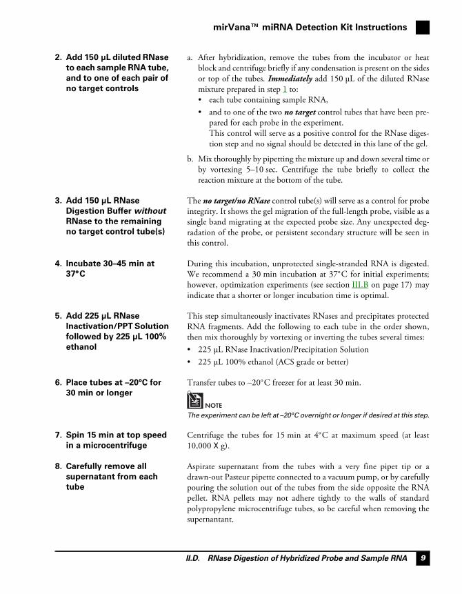
2. Add 150 μL diluted RNase
to each sample RNA tube,
and to one of each pair of
no target controls
a. After hybridization, remove the tubes from the incubator or heatblock and centrifuge briefly if any condensation is present on the sidesor top of the tubes. Immediately add 150 μL of the diluted RNasemixture prepared in step 1 to:• each tube containing sample RNA,• and to one of the two no target control tubes that have been pre-
pared for each probe in the experiment.This control will serve as a positive control for the RNase diges-tion step and no signal should be detected in this lane of the gel.
b. Mix thoroughly by pipetting the mixture up and down several time orby vortexing 5–10 sec. Centrifuge the tube briefly to collect thereaction mixture at the bottom of the tube.
3. Add 150 μL RNase
Digestion Buffer without
RNase to the remaining
no target control tube(s)
The no target/no RNase control tube(s) will serve as a control for probeintegrity. It shows the gel migration of the full-length probe, visible as asingle band migrating at the expected probe size. Any unexpected deg-radation of the probe, or persistent secondary structure will be seen inthis control.
4. Incubate 30–45 min at
37°C
During this incubation, unprotected single-stranded RNA is digested.We recommend a 30 min incubation at 37°C for initial experiments;however, optimization experiments (see section III.B on page 17) mayindicate that a shorter or longer incubation time is optimal.
5. Add 225 μL RNase
Inactivation/PPT Solution
followed by 225 μL 100%
ethanol
This step simultaneously inactivates RNases and precipitates protectedRNA fragments. Add the following to each tube in the order shown,then mix thoroughly by vortexing or inverting the tubes several times:• 225 μL RNase Inactivation/Precipitation Solution• 225 μL 100% ethanol (ACS grade or better)
6. Place tubes at –20°C for
30 min or longer
Transfer tubes to –20°C freezer for at least 30 min.
NOTE
The experiment can be left at –20°C overnight or longer if desired at this step.
7. Spin 15 min at top speed
in a microcentrifuge
Centrifuge the tubes for 15 min at 4°C at maximum speed (at least10,000 X g).
8. Carefully remove all
supernatant from each
tube
Aspirate supernatant from the tubes with a very fine pipet tip or adrawn-out Pasteur pipette connected to a vacuum pump, or by carefullypouring the solution out of the tubes from the side opposite the RNApellet. RNA pellets may not adhere tightly to the walls of standardpolypropylene microcentrifuge tubes, so be careful when removing thesupernantant.
mirVana™ miRNA Detection Kit
II.E. Separation and Detection of Protected Fragments10
NOTE
Do not remove the residual fluid by
vacuum-drying, because the salt
present in RNase Inactivation/
Precipitation Solution will cause
aberrant migration of the protected
fragment during electrophoresis.
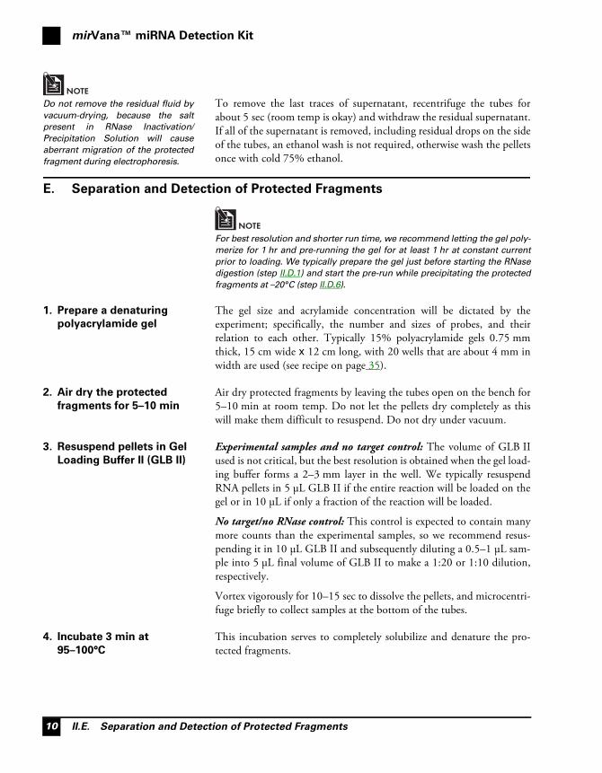
To remove the last traces of supernatant, recentrifuge the tubes forabout 5 sec (room temp is okay) and withdraw the residual supernatant.If all of the supernatant is removed, including residual drops on the sideof the tubes, an ethanol wash is not required, otherwise wash the pelletsonce with cold 75% ethanol.
E. Separation and Detection of Protected Fragments
NOTE
For best resolution and shorter run time, we recommend letting the gel poly-
merize for 1 hr and pre-running the gel for at least 1 hr at constant current
prior to loading. We typically prepare the gel just before starting the RNase
digestion (step II.D.1) and start the pre-run while precipitating the protected
fragments at –20°C (step II.D.6).
1. Prepare a denaturing
polyacrylamide gel
The gel size and acrylamide concentration will be dictated by theexperiment; specifically, the number and sizes of probes, and theirrelation to each other. Typically 15% polyacrylamide gels 0.75 mmthick, 15 cm wide x 12 cm long, with 20 wells that are about 4 mm inwidth are used (see recipe on page 35).
2. Air dry the protected
fragments for 5–10 min
Air dry protected fragments by leaving the tubes open on the bench for5–10 min at room temp. Do not let the pellets dry completely as thiswill make them difficult to resuspend. Do not dry under vacuum.
3. Resuspend pellets in Gel
Loading Buffer II (GLB II)
Experimental samples and no target control: The volume of GLB IIused is not critical, but the best resolution is obtained when the gel load-ing buffer forms a 2–3 mm layer in the well. We typically resuspendRNA pellets in 5 μL GLB II if the entire reaction will be loaded on thegel or in 10 μL if only a fraction of the reaction will be loaded.
No target/no RNase control: This control is expected to contain manymore counts than the experimental samples, so we recommend resus-pending it in 10 μL GLB II and subsequently diluting a 0.5–1 μL sam-ple into 5 μL final volume of GLB II to make a 1:20 or 1:10 dilution,respectively.
Vortex vigorously for 10–15 sec to dissolve the pellets, and microcentri-fuge briefly to collect samples at the bottom of the tubes.
4. Incubate 3 min at
95–100°C
This incubation serves to completely solubilize and denature the pro-tected fragments.

II.F. Experimental Setup Examples
mirVana™ miRNA Detection Kit Instructions
11
5. Load the samples and run
the gel
Rinse the urea out of the wells of the gel, and immediately load eachsample. Load equal volumes of sample and controls onto the gel; afterdilution an equal volume of the no target/no RNase control will repre-sent ~5–10% of the counts in the experimental samples so that itdoesn’t obscure the signal in adjacent lanes.
It is helpful to have size markers on the gel; single-stranded RNA mark-ers such as Ambion Decade Markers are the most accurate, butdouble-stranded DNA markers can be used if it is not critical to knowthe exact size of the products.
12 x 15 cm gels can be run at 10–30 mAmp constant current. The posi-tion of bromophenol blue and xylene cyanol in 15% gels corresponds toapproximately 10 and 30 nt, respectively. Run gels until the leading dyeband (bromophenol blue) is near the middle of the gel (~6 cm). The runtime is about 30 min, although it will vary depending on the length ofthe gel and the percentage of acrylamide.
6. Detect radiolabeled
protected fragments by
autoradiography
Remove at least one glass plate and cover the gel with plastic wrap.Expose the gel face to X-ray film for 1 hr to overnight with an intensify-ing screen at –80°C. The gel can be re-exposed several times if necessaryafter allowing it to warm up to room temp and wiping off condensationmoisture. The gel should be stored at –80° or –20°C if not re-exposedimmediately. It is not necessary to dry standard 0.75 mm thick gels forautoradiography.
F. Experimental Setup Examples
Below are shown four representative experiments performed with themirVana miRNA Detection Kit. This information provides examples ofexperimental setups and actual data obtained with the kit.
1. Multiple probes with a
single experimental RNA
sample
In this experiment four different probes are tested with 1 μg MouseKidney Total RNA. To bring the RNA amount to 4 μg, 3 μg of YeastRNA is added to each experimental sample. For each of the four probes,a no target control and a no target/no RNase control are set up contain-ing 4 μg of Yeast RNA (for a total of 8 control reactions).
To set up this experiment two master mixes (experimental and no targetcontrol) containing all of the components except the probes are mixedthoroughly. 10% extra is included to account for pipetting error.
No target Master Mix Experimental Master Mix
Component Per tube For 8 controls Per tube For 4 samples
2X Hybridization Buffer 10 μL 88 μL 10 μL 44 μL
Yeast RNA (5 mg/mL) 0.8 μL 7 μL 0.6 μL 2.6 μL
Mouse Kidney RNA (0.5 mg/mL) – – – – 2 μL 8.8 μL
Nuclease-free Water to 18 μL 63.4 μL to 18 μL 23.8 μL
mirVana™ miRNA Detection Kit
II.F. Experimental Setup Examples12
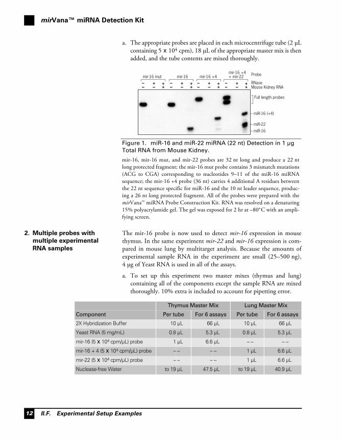
a. The appropriate probes are placed in each microcentrifuge tube (2 μLcontaining 5 x 10⁴ cpm), 18 μL of the appropriate master mix is thenadded, and the tube contents are mixed thoroughly.
2. Multiple probes with
multiple experimental
RNA samples
The mir-16 probe is now used to detect mir-16 expression in mousethymus. In the same experiment mir-22 and mir-16 expression is com-pared in mouse lung by multitarget analysis. Because the amounts ofexperimental sample RNA in the experiment are small (25–500 ng),4 μg of Yeast RNA is used in all of the assays.
a. To set up this experiment two master mixes (thymus and lung)containing all of the components except the sample RNA are mixedthoroughly. 10% extra is included to account for pipetting error.
Figure 1. miR-16 and miR-22 miRNA (22 nt) Detection in 1 μg
Total RNA from Mouse Kidney.
mir-16, mir-16 mut, and mir-22 probes are 32 nt long and produce a 22 ntlong protected fragment; the mir-16 mut probe contains 3 mismatch mutations(ACG to CGA) corresponding to nucleotides 9–11 of the miR-16 miRNAsequence; the mir-16 +4 probe (36 nt) carries 4 additional A residues betweenthe 22 nt sequence specific for miR-16 and the 10 nt leader sequence, produc-ing a 26 nt long protected fragment. All of the probes were prepared with themirVana™ miRNA Probe Construction Kit. RNA was resolved on a denaturing15% polyacrylamide gel. The gel was exposed for 2 hr at –80°C with an ampli-fying screen.
mir-16 +4+ mir-22
–– ++– +
–– ++– +
–– ++– +
–– ++– +
mir-16 +4mir-16mir-16 mut
Mouse Kidney RNARNase
Probe
– miR-22
– miR-16
– miR-16 (+4)
Full length probes
Thymus Master Mix Lung Master Mix
Component Per tube For 6 assays Per tube For 6 assays
2X Hybridization Buffer 10 μL 66 μL 10 μL 66 μL
Yeast RNA (5 mg/mL) 0.8 μL 5.3 μL 0.8 μL 5.3 μL
mir-16 (5 x 10⁴ cpm/μL) probe 1 μL 6.6 μL – – – –
mir-16 + 4 (5 x 10⁴ cpm/μL) probe – – – – 1 μL 6.6 μL
mir-22 (5 x 10⁴ cpm/μL) probe – – – – 1 μL 6.6 μL
Nuclease-free Water to 19 μL 47.5 μL to 19 μL 40.9 μL

II.F. Experimental Setup Examples
mirVana™ miRNA Detection Kit Instructions
13
b. 1 μL of RNase-free water (for the no target/no RNase control) or1 μL of the appropriate total RNA diluted to contain 25, 50, 100,250, or 500 ng RNA are placed in each microcentrifuge tube. Then19 μL of the Thymus or Lung Master Mix is added, and the tubecontents are mixed thoroughly.
3. A single probe with
multiple experimental
RNA samples
In this experiment mir-16 probe is used to analyze mir-16 expression in10 different mouse tissues. 4 μg of Yeast RNA is used for each of the twono target controls (not shown in the figure) and to complement the1 μg of sample RNA in the experimental reactions.
a. To set up this experiment one master mix containing all of thecomponents except the experimental sample RNA is mixedthoroughly. One Master Mix for 13 tubes is needed for thisexperiment (12 hybridization reactions plus ~10% for pipettingerror).
Figure 2. Analysis of miR-16 or miR-22 miRNA Expression in
Mouse Thymus & Lung
miR-16 and miR-22 miRNA were detected in 500, 250, 100, 50, or 25 ng ofAmbion® FirstChoice® Total RNA from mouse thymus (mir-16 probe) ormouse lung (mir-16 +4 and mir-22 probes). The gel was exposed for 3.5 hr.
No targ
et/no
RNas
e
500 n
g
250 n
g
100 n
g
50 ng
25 ng
No targ
et/no
RNas
e
500 n
g
250 n
g
100 n
g
50 ng
25 ng
– miR-22miR-16 –
– miR-16 +4
Full length probes
Master Mix
Component Per tube For 13 assays
2X Hybridization Buffer 10 μL 130 μL
Yeast RNA (5 mg/mL) 0.8 μL 10.4 μL
mir-16 (5 x 10⁴ cpm/μL) probe 1 μL 13 μL
Nuclease-free Water to 19 μL 93.6 μL

mirVana™ miRNA Detection Kit
II.F. Experimental Setup Examples14
b. 1 μL of RNase-free water (for the no target controls) or 1 μL of theappropriate total RNA diluted to contain 1 μg RNA are placed ineach microcentrifuge tube. Then 19 μL of Master Mix is added, andthe tube contents are mixed thoroughly.
4. A single probe and
sample RNA, different
reaction parameters
The mir-16 probe and Mouse Kidney Total RNA are used to test differ-ent parameters of the mirVana miRNA Detection procedure.
Testing reaction conditions
In the first part of the experiment, nine variations of hybridization timeand RNase digestion conditions are tested with 1 μg of sample RNA.Thus a master mix containing all of the components is prepared (plus10% for pipetting error), and 20 μL is dispensed into each tube.
Testing sample RNA quantity
The second part of the experiment compares different amounts of sam-ple RNA, and includes the two no target controls.
Figure 3. miR-16 miRNA Expression Across Mouse Tissues
miR-16 miRNA was detected in 1 μg of Ambion® FirstChoice® Total RNAfrom 10 different mouse tissues using the mirVana™ miRNA Detection Kit(bottom panel). The gel was exposed for 2 hr. The same variation of mir-16expression across mouse tissues was demonstrated by Northern blot analysisusing the same high specific activity mir-16 probe (top panel, 2 day exposure).
Brain
Heart
Kidne
y
Liver
Lung
Splee
n
Thym
us
Testi
cle
Ovary
Embry
o
– miR-16
– miR-16mirVana™ miRNA Detection Kit
Northern blot analysis
Reaction Condition Master Mix
Component Per tube For 9 samples
2X Hybridization Buffer 10 μL 100 μL
Mouse Kidney Total RNA (0.5 mg/mL) 2 μL 20 μL
mir-16 probe (5 x 10⁴ cpm/μL) 1 μL 10 μL
Yeast RNA (5 mg/mL) 0.6 μL 6 μL
Nuclease-free Water to 20 μL 64 μL
II.F. Experimental Setup Examples
mirVana™ miRNA Detection Kit Instructions
15
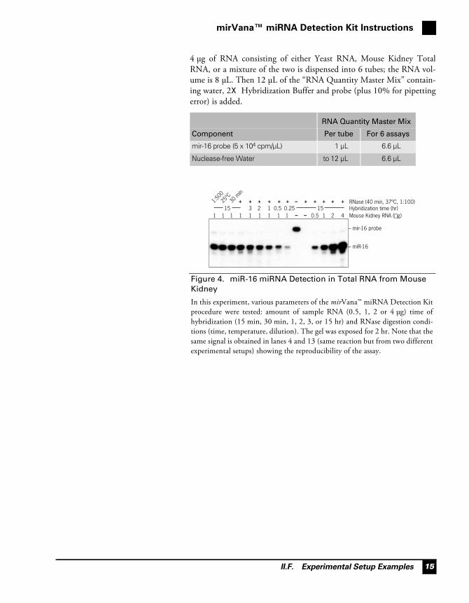
4 μg of RNA consisting of either Yeast RNA, Mouse Kidney TotalRNA, or a mixture of the two is dispensed into 6 tubes; the RNA vol-ume is 8 μL. Then 12 μL of the “RNA Quantity Master Mix” contain-ing water, 2X Hybridization Buffer and probe (plus 10% for pipettingerror) is added.
RNA Quantity Master Mix
Component Per tube For 6 assays
mir-16 probe (5 x 10⁴ cpm/μL) 1 μL 6.6 μL
Nuclease-free Water to 12 μL 6.6 μL
Figure 4. miR-16 miRNA Detection in Total RNA from Mouse
Kidney
In this experiment, various parameters of the mirVana™ miRNA Detection Kitprocedure were tested: amount of sample RNA (0.5, 1, 2 or 4 μg) time ofhybridization (15 min, 30 min, 1, 2, 3, or 15 hr) and RNase digestion condi-tions (time, temperature, dilution). The gel was exposed for 2 hr. Note that thesame signal is obtained in lanes 4 and 13 (same reaction but from two differentexperimental setups) showing the reproducibility of the assay.
+++++++++ + +–
– –1 1 1 1 13 2 1 0.5 0.2515 15
RNase (40 min, 37ºC, 1:100)Hybridization time (hr)
30 m
in
25ºC
1:500
1 1 1 1 1 2 4 Mouse Kidney RNA (µg)0.5
– mir-16 probe
– miR-16
mirVana™ miRNA Detection Kit
III.A. Running the Positive Control Reaction16
III. Troubleshooting
A. Running the Positive Control Reaction
The positive control reaction included with the mirVana miRNADetection Kit consists of Mouse Kidney Total RNA and a DNA tem-plate (Control Template) for transcription of an antisense RNA probespecific for miR-16 miRNA.
1. Probe preparation To synthesize a radiolabeled positive control probe, use 1 μL of theControl Template in a 20 μL in vitro T7 RNA Polymerase transcriptionreaction containing at least 3 μM [α-32P]UTP (5 μL of 800 Ci/mmol,10 mCi/mL) and up to 5 μM unlabeled UTP. The lower the concentra-tion of unlabeled nucleotide, the higher the specific activity of the tran-script, and thus the greater the sensitivity of the assay. For maximumsensitivity, do not add any unlabeled form of the limiting nucleotide.Only T7 RNA polymerase can be used in the transcription reactionwith this template. Treat the control probe with DNase to remove theDNA template, and gel purify it (section IV.B on page 29) for use in thecontrol mirVana miRNA Detection reaction.
The size of the antisense mir-16 RNA probe from the Control Tem-plate is 32 nt, and the protected mir-16 fragment is 22 nt.
NOTE
The control probe can also be used in Northern blot experiments (see
Figure 3 on page 14).
2. Assay setup Use about 5 x 10⁴ cpm of probe with different amounts of Mouse Kid-ney RNA, e.g. 2, 4, and 8 μL corresponding to 1, 2, and 4 μg, respec-tively. Bring the final amount of RNA in each tube to 4 μg by addingYeast RNA. Include two no target control hybridization reactions con-taining the same amount of probe and 4 μg Yeast RNA. Follow the pro-tocol outlined in section II. mirVana™ miRNA Detection Kit Instructions;hybridize overnight at 42°C, use RNase A/RNase T1 Mix at a 1:100 dilu-tion for 30 min at 37°C and analyze on a denaturing 15% polyacrylamidegel, loading only 10% of the no target/no RNase control.
3. Expected results Protected fragments of 22 nt should be seen in the reactions containingMouse Kidney RNA, and the protected fragments should be moreintense with increasing amounts of Mouse Kidney RNA. The no targetcontrol lane should have no signal, and the no target/no RNase laneshould show a band corresponding to full length probe (32 nt). Anexample of a control experiment using the provided Mouse Kidney TotalRNA and probe made from the Control Template is shown in Figure 4on page 15.

III.B. Pilot Experiment for Assay Optimization
Troubleshooting
17
B. Pilot Experiment for Assay Optimization
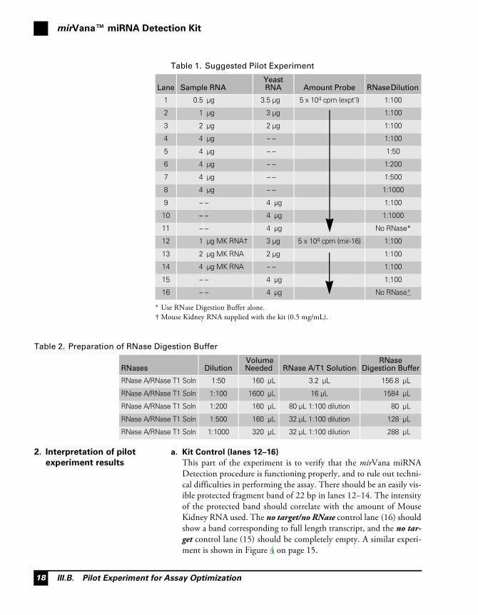
1. Pilot experiment setup The following experiment is suggested to help new users determine theappropriate amounts of sample RNA and probe to use when the abun-dance of the target RNA is unknown. Carry out the assay with a range ofinput sample RNA, using a constant amount of high specific activityprobe. Also, to optimize conditions for the RNase digestion, vary thedilution of the RNase A/T1 Solution in RNase Digestion Buffer. Youmay also want to test additional variables such as the hybridization tem-perature (25–52°C), the hybridization time (1–24 hr), the probeamount (1–10 x 104 cpm), the RNase digestion incubation time(15–45 min), or the RNase digestion incubation temperature (roomtemp to 37°C). If sample RNA is in short supply, these experiments canbe scaled back to conserve sample RNA. The details of the pilot experi-ment are shown in Table 1, and instructions for making the RNaseDigestion Buffers are shown in Table 2 on page 18.a. Probe
For maximum sensitivity, prepare high specific activity experimentaland control probes, i.e. labeling reactions should not contain anyunlabeled limiting nucleotide. For more detailed information onprobe synthesis and purification, see section IV.A, IV.B, and IV.C.
b. Molecular size markers
To assess the size of the probes and protected fragments we recom-mend using 10–50 nt molecular size markers. Ambion Decade™Markers (included in the mirVana Probe & Marker Kit) will gener-ate a 10 nt ladder using polynucleotide kinase and [γ–32P]ATP. Thekit also contains reagents to prepare a single nucleotide ladder froma 5' end radiolabeled RNA probe.
c. General instructions for the pilot experiment
Follow the protocol in section II. mirVana™ miRNA Detection KitInstructions on page 5. Prepare two Master Mixes containing eitheryour experimental probe or the mir−16 probe made from the Con-trol Template, Hybridization Buffer, and Nuclease-free Water. Dis-pense the RNA into your assay tubes, and then add the appropriateMaster Mix. Run only 10% of the no target/no RNase controls(tubes 11 and 16), to prevent overexposure of the autoradiograph.Expose the gel initially for 3 hr with an intensifying screen at –80°C.
mirVana™ miRNA Detection Kit
III.B. Pilot Experiment for Assay Optimization18
2. Interpretation of pilot
experiment results
a. Kit Control (lanes 12–16)
This part of the experiment is to verify that the mirVana miRNADetection procedure is functioning properly, and to rule out techni-cal difficulties in performing the assay. There should be an easily vis-ible protected fragment band of 22 bp in lanes 12–14. The intensityof the protected band should correlate with the amount of MouseKidney RNA used. The no target/no RNase control lane (16) shouldshow a band corresponding to full length transcript, and the no tar-get control lane (15) should be completely empty. A similar experi-ment is shown in Figure 4 on page 15.
Table 1. Suggested Pilot Experiment
Lane Sample RNAYeast RNA Amount Probe RNase Dilution
1 0.5 μg 3.5 μg 5 x 10⁴ cpm (expt’l) 1:100
2 1 μg 3 μg 1:100
3 2 μg 2 μg 1:100
4 4 μg – – 1:100
5 4 μg – – 1:50
6 4 μg – – 1:200
7 4 μg – – 1:500
8 4 μg – – 1:1000
9 – – 4 μg 1:100
10 – – 4 μg 1:1000
11 – – 4 μg No RNase*
* Use RNase Digestion Buffer alone.
12 1 μg MK RNA†
† Mouse Kidney RNA supplied with the kit (0.5 mg/mL).
3 μg 5 x 10⁴ cpm (mir-16) 1:100
13 2 μg MK RNA 2 μg 1:100
14 4 μg MK RNA – – 1:100
15 – – 4 μg 1:100
16 – – 4 μg No RNase*
Table 2. Preparation of RNase Digestion Buffer
RNases Dilution Volume Needed RNase A/T1 Solution
RNase Digestion Buffer
RNase A/RNase T1 Soln 1:50 160 μL 3.2 μL 156.8 μL
RNase A/RNase T1 Soln 1:100 1600 μL 16 μL 1584 μL
RNase A/RNase T1 Soln 1:200 160 μL 80 μL 1:100 dilution 80 μL
RNase A/RNase T1 Soln 1:500 160 μL 32 μL 1:100 dilution 128 μL
RNase A/RNase T1 Soln 1:1000 320 μL 32 μL 1:100 dilution 288 μL

III.C. Troubleshooting Faint or Absent Protected Fragment Bands
Troubleshooting
19
b. Experimental no target controls (lanes 9–11)
The no target/no RNase control lane (11) should show a band cor-responding to the full length probe. Since probe molecules that havebeen protected by RNA in the sample will produce the data in anRPA, it is essential that this lane shows a single band of the expectedsize. If the autoradiograph shows severe degradation of probe in thislane, then ribonuclease contamination of your tubes, pipette tips, ora component of the kit should be suspected.The two no target control lanes (9 & 10) should be empty. Lane 10was digested with the lowest amount of RNase used in the experi-ment. This reaction will show whether the 1:1000 dilution of RNaseused in tube 8 is sufficient to degrade all of the probe.
c. Increasing sample RNA (lanes 1–4)
The autoradiograph should show a band of the expected protectedfragment size from the samples in Tubes 1 to 4. If the intensity of themajor protected fragment in the autoradiograph increases withincreasing input RNA, then the probe was present in molar excessover the target RNA in all the reactions. For accurate quantitation ofRNA levels, it is essential that the probe is present in molar excesscompared to the target and that hybridization goes to completion. If the signal is very weak or absent, even in the reaction with thehighest amount of input RNA, the target RNA may be very rare; seesection III.C.2 on page 20 for suggestions.
d. Different concentrations of RNase (lanes 5–8)
The ribonuclease concentration was varied to optimize the sig-nal-to-noise ratio in the assay. Most probe/target RNA combinationswill give acceptable, even identical, results when digested withRNase concentrations that vary over at least a 10-fold range. Sometemplates, however, may require fine-tuning of RNase digestionconditions for optimal results.
C. Troubleshooting Faint or Absent Protected Fragment Bands
If the mirVana miRNA Detection assay autoradiograph does not showthe expected results (i.e. no band of the predicted size representing themajor protected fragment is present), but the probe is intact (as assessedby the no target/no RNase control), consider the following suggestionsfor troubleshooting the procedure.
1. The target is not present
in the sample
It is possible that absence of signal is a legitimate result, and that theRNA of interest is not expressed or was not efficiently recovered duringRNA sample preparation. To confirm this possibility, repeat the assayalong with an assay of a separate sample RNA prep known to containdetectable levels of the target RNA. If this is not possible, increasingamounts of chemically synthesized or in vitro transcribed sense strandRNA can be added to RNA samples to serve as a positive control. This

mirVana™ miRNA Detection Kit
III.C. Troubleshooting Faint or Absent Protected Fragment Bands20
strategy will also show the limit of sensitivity of the assay for a particularprobe. For more information on using synthetic sense strand RNA insolution hybridization, see section IV.D. Quantitation of Target RNA onpage 32 or refer to Technical Bulletin #165—available at:
www.ambion.com/techlib/tb/tb_165.html
2. Increase the sensitivity of
the assay
a. Use a probe with a higher specific activity.
Probe specific activity is determined by the specific activity of theradiolabeled nucleotide, and its proportion to the correspondingunlabeled nucleotide in the labeling reaction. The highest specificactivity is obtained by using only radiolabeled nucleotide in the tran-scription reaction and none of the unlabeled form. See sectionIV.A.3. Making radiolabeled RNA probes on page 27 for more infor-mation on probe synthesis.
b. Use more target RNA.
Another way to increase the sensitivity of the assay is to use moresample RNA – as much as about 50 μg per hybridization reactioncan be used. Fractions enriched in small RNA species with the mir-Vana miRNA Isolation Kit procedure can also be used instead oftotal RNA to increase the amount of small RNA target (e.g. miRNAor siRNA).
3. Check the probe design Transcription of the “wrong strand” from the template DNA in theprobe synthesis reaction will produce a probe that cannot hybridize tothe target RNA. In order to make an antisense probe (target-comple-mentary) from cloned sequences, plasmid template should be linearizedon the 5' side of the gene or portion of gene. PCR templates should bedesigned with the phage polymerase promoter on the 3' side of thedesired probe sequence. If using the mirVana miRNA Probe Construc-tion Kit, the DNA oligonucleotide template must have the samesequence as the sense target RNA except that U residues are replacedwith T’s. The 8 nt sequence 5'-CCTGTCTC-3', complementary tothe T7 Promoter Primer provided with the kit, must also be added tothe 3' end of the DNA oligonucleotide sequence.
4. Suboptimal hybridization
temperature
In some cases it is better to hybridize at temperatures higher than 42°C.This can be helpful in multiple probe assays, and may serve to preventprobe:probe interactions.
5. Overdigestion with
RNase
Overdigestion with RNase is rarely seen using a 1:100 dilution ofRNase A/T1 Solution. Evidence for overdigestion would be smearing ofthe signal below the expected position of the protected fragment in anexperiment where the no target/no RNase control lane did not showsmearing.

III.D. Protected Fragments that are Smeared or Consist of a Ladder of Bands
Troubleshooting
21
IMPORTANT
D. Protected Fragments that are Smeared or Consist of a Ladder of Bands
1. No target/no RNase
control lane is not
smeared
a. Excess probe
Decreasing the probe amount or its specific activity may improve thesignal-to-noise ratio and help to eliminate unwanted background.While quantitative results require that the probe be present in molarexcess over the target RNA, a large excess of probe can result in highbackground.
b. Gel quality and sample volume
For best resolution, we recommend letting the gel polymerize for atleast 1 hr and pre-running the gel for at least 1 hr at constant currentprior to loading. It is also critical to rinse the urea out of the wellsimmediately before loading. Finally, overloading gel wells reducesproduct resolution and may affect data quality. Wells that are0.4–0.6 cm wide work well for loading the 5–10 μL samples fromthis procedure. The best resolution is obtained when the gel loadingbuffer forms a 2–3 mm layer in the well.
c. Heterogeneity in probe length
This is usually due to premature termination of transcription duringthe probe synthesis reaction, or to radiolytic, enzymatic, or nonspe-cific degradation of the probe. Since these probe fragments can stillhybridize to their complements in the sample RNA, protection offragments that are shorter than expected may be seen (Zinn, et al.1983). To prevent this type of background we strongly recommendthat the probe be gel-purified, stored at –80°C or –20°C, and usedwithin a few days of synthesis for best results. The lower the specificactivity of the probe, the less prone it will be to radiolytic decay, andthe longer it can be used with satisfactory results. As an assessment ofprobe quality, the no target/no RNase control lane should show aband representing mainly full-length probe.
d. Suboptimal RNase digestion conditions
Vary the concentration of RNase A/RNase T1 Solution in theRNase digestion step over a range of about 50 to 100 fold, for exam-ple by using RNase A/RNase T1 Solution diluted 1:50, 1:150,1:500, 1:1000, 1:2000. Background smearing may be reduced byfine tuning of the RNase digestion.
When the RNase concentration is
decreased, the yeast RNA control
reaction should be digested with the
lowest concentration (highest
dilution) of RNase used, to assure
validity of the experimental results.
Underdigestion will usually leave leftover full length probe in the notarget control lane (no target/+RNase). The experimental sampleswill show smearing or a ladder of bands between the full lengthprobe and the protected fragment.
mirVana™ miRNA Detection Kit
III.D. Protected Fragments that are Smeared or Consist of a Ladder of Bands22
Overdigestion by RNase will be evident as a smear or ladder of bandsbelow the expected size of the protected fragment. Using theRNase A/RNase T1 Solution at a 1:100 dilution rarely results inoverdigestion of the hybrids.
e. Local “breathing”
Depending on the sequence of the double-stranded RNA hybridformed by the probe and target RNA, “breathing” (local denatur-ation) may occur, which can result in partial cleavage of the pro-tected probe:RNA target hybrid. With internally labeled probe (i.e.prepared by in vitro transcription) this will produce a shorter discreteband(s) or a ladder of bands shorter than the expected size of the pro-tected fragment but will not affect the overall interpretation of thedata. With 5' end-labeled probes however, breathing may result in acomplete loss of signal. If this problem is suspected, the incubationtemperature of the RNase digestion reaction can be lowered to roomtemp or 16°C.
f. Mismatches between probe and target RNA
See III.G.3. There are mismatches between probe and target RNA onpage 25.
g. Degradation of target RNA
The mirVana miRNA Detection assay is relatively insensitive toRNA degradation. However, a badly degraded sample RNA prepara-tion may contain species capable of protecting only part of theprobe. The longer the probe, the less target RNA degradation can betolerated. If degradation of target RNA is suspected to be a problem,the integrity of the sample RNA can be assessed by electrophoresison a 1% denaturing formaldehyde or glyoxal based agarose gel(smearing of the ribosomal RNA bands indicates degradation).Alternatively, the sample RNA can be analyzed by capillary electro-phoresis using an Agilent 2100 bioanalyzer. A 28S:18S rRNA peakratio at or near 2:1 indicates that the RNA is intact. Another way tocheck the integrity of the sample RNA is to use the provided controlprobe to detect miR-16 miRNA in your RNA sample. This probehas been successfully used to detect miR-16 in total RNA from vari-ous cell lines, and from human, rat, and mouse tissues.There is one report suggesting that autoclaving of siliconized tubesresulted in a high pH residue which degrades RNA.
2. Smear or ladder is
also seen in the
no target/no RNase
control lane
Poor results seen in the context of a degraded probe indicate problemswith probe synthesis, purification, or stability. Check the recommenda-tions for probe synthesis and purification in section IV.A and IV.B.
Smeared signal that starts at the expected position of protected fragmentand continues down can be caused by degradation of the probe. This ismost often from radiolysis, but it can also be due to RNase contaminationof the probe solution, your tubes, pipette tips, or a component of the kit.
III.E. Full Length Probe Is Seen in All Lanes
Troubleshooting
23
E. Full Length Probe Is Seen in All Lanes
There are several possible causes for this result. In each case, full-lengthprobe, or band(s) slightly shorter than the probe, are seen in the experi-mental sample lane and also in the no target control lane (no tar-get/+RNase).
Nonspecific protected bands may be observed in some cases along withthe expected target-specific protected fragment. These bands are of lowintensity, and may vary in size, but generally are easily distinguishablefrom the expected band on a gel. They will not affect analysis of theresults, and can essentially be ignored.
The relative intensity of the full length probe band compared to the pro-tected fragment band, as well as the presence or absence of smearingbetween the two, will provide clues to which of the following problemsis occurring.
1. RNases were completely
inactive or were not
added
The full length probe band would be very strong in this case, and therewould be no band at the expected position of the protected fragment.The RNases can be checked for activity by comparing on a gelRNase-treated RNA with RNA incubated in RNase Digestion Bufferwithout nuclease. The RNase dilution and the amount of RNA usedshould mimic the experimental conditions. If all of the sample RNA isnot degraded to very low molecular weight fragments, there could be aproblem with the RNase A/T1 Solution.
2. Too much probe was
added to the reaction
If too much probe is used, there may be a faint band of undigestedprobe left. If this is the case, the protected fragment band will usually bemore intense than this full-length probe band, and both bands will bedistinct, with no smearing between the two. We generally recommendusing no more than 1–10 x 104 cpm of high specific activity probe forup to 10 μg of RNA sample. A pilot experiment using different amountsof input RNA can be done to determine the amount of probe necessaryfor probe to be present in molar excess over target.
3. Residual DNA
transcription template is
protecting the probe from
RNase digestion
The experimental data in this case will look much like that described inthe preceding section, a faint full-length probe band in all lanes [includ-ing the no target control lane (no target/+RNase)], and a protected frag-ment band in the experimental sample lanes, with little if any smearingbetween the bands. To avoid this, we always recommend treating invitro transcription reactions with DNase I, and gel purifying probe foruse in this procedure.
mirVana™ miRNA Detection Kit
III.F. Aberrant, Pointed or Smeared Bands24
4. The RNase did not
completely degrade
unprotected probe
Incomplete RNase digestion is indicated if there is a full-length probeband followed by a smear of material that ends where the protected frag-ment is expected to migrate. This rarely occurs when theRNase A/RNase T1 Solution is used at 1:100, but if it does, use a lessdilute RNase solution in subsequent experiments (e.g. 1:50).
5. Problem with probe
design
The probe should be longer than the protected fragment so that anobvious difference in size is seen between the full-length undigestedprobe and the protected fragment. This additional nontarget-specificsequence must be cleavable by RNase A and/or T1 (RNases A and T1do not cleave 3' of A residues). For detailed information about probedesign and preparation see section IV.A on page 26.
F. Aberrant, Pointed or Smeared Bands
1. Residual salt in the RNA
pellet
If the supernatant from the final precipitation step is not completelyremoved, salts from the precipitation mixture will be concentrated andmay lead to aberrant migration (“tunneling”) of the protected fragment.We recommend briefly respinning the final pellet after the supernatantis removed, and then thoroughly aspirating any residual supernatantusing a very fine-tipped pipet. Alternatively pellets can be washed with75% ethanol.
2. Gel quality and sample
volume
See section D.1.b on page 21.
3. Radioactive material that
fails to migrate into the
gel
It is unclear what causes this problem; it may be the result of several dif-ferent factors. Probes that have not been gel purified experience this“hang up in the well” phenomenon more frequently than gel purifiedprobes. Phenol/chloroform extraction of probes that were not gel puri-fied tends to diminish the amount of material left in the wells. Anothercontributing factor seems to be residues left in microcentrifuge tubesfrom the manufacturing process. Autoclaving of siliconized tubesappears to exacerbate this problem.
G. Reasons for Legitimate but Unexpected Bands on Autoradiographs
Due to the extreme sensitivity of the mirVana miRNA Detection assay,probe-related sequences in the sample RNA that might go undetectedby other methods may be detected with this assay. Typically, these extrabands are less intense than the primary protected fragment, and theymay permit additional valid conclusions to be made concerning thenature of the expression or the processing of the RNA(s) being studied.Extra bands, however, can also be artifactual; thus interpretation of thedata requires caution and knowledge of the various factors that can con-tribute to the pattern of fragments seen in the assay.

III.G. Reasons for Legitimate but Unexpected Bands on Autoradiographs
Troubleshooting
25
1. Probe hybridizes to target
RNA with differing but
related sequence
If the probe is complementary to an RNA that is a member of a familyof related genes, and more than one member of the family is present inthe sample RNA, then the probe may cross-hybridize to the RNA fromthe related gene(s). In some cases, the related RNA will have multiplemismatches with the probe, resulting in protected fragments that areshorter than those seen with the completely homologous RNA. Multi-ple mismatches in short probes can also result in a complete loss of sig-nal (see for example mir-16 mut probe in Figure 1 on page 12). This canbe advantageous for distinguishing between miRNAs with sequencesimilarity which run as a single band on Northern blots. Some investi-gators will want to use higher concentrations of RNase to maximize rec-ognition of small differences between the probe and sample RNA, whileothers will want to use lower concentrations of RNase to minimizeprobe cleavage at mismatched positions.
2. Probe detects
heterogeneity in
processing of RNA
transcripts
Depending on the design of the probe and the experimental RNA sam-ple, probes may be capable of protecting fragments of different sizes thatresult from heterogeneity in the target RNA. Such heterogeneity couldresult from differential transcription initiation or termination sites orfrom the presence of immature RNA species in the RNA sample (e.g.precursor siRNAs or miRNAs).
3. There are mismatches
between probe and target
RNA
If the probe is not completely homologous to the target RNA in thesample RNA mixture, total or partial cleavage of the probe-target RNAhybrid may occur at the position of the mismatch. The extent of RNasecleavage at mismatch positions depends on the nature of the mismatch(e.g. A-C mismatches are cleaved more frequently than are G-U mis-matches), and on the RNase digestion conditions. While ribonucleaseprotection assays are capable of detecting some single-base mismatches,other mismatches will not be cleaved (Myers et al. 1985). If the mis-matches consist of more than single-nucleotide differences, they will becleaved much more efficiently. Because the procedure uses short probes,multiple mismatches in a probe may also result in a complete loss of tar-get RNA protection. For example, three consecutive mismatches in themir-16 mut probe (Figure 1 on page 12) completely abolish the detec-tion of miR-16 miRNA in Mouse Kidney Total RNA. Cleavage at mis-match positions can often be minimized by decreasing the RNaseconcentration and/or the incubation time and temperature duringRNase digestion.
mirVana™ miRNA Detection Kit
IV.A. Preparation of Antisense RNA Probes26
IV. Additional Procedures
A. Preparation of Antisense RNA Probes
Because only the probe is visualized at the end of the mirVana miRNADetection procedure, the success of the experiment is largely dependenton the quality of the probe. Below are recommendations for probedesign and preparation.
1. Probe size The mirVana miRNA Detection procedure is optimized for use withshort antisense RNA probes. Typically these probes contain a 19–35 ntsequence that is the complement of the target RNAs of interest and3–10 nt or more of nonspecific sequence that will not hybridize withthe target RNA. The probe size must be within the effective separationrange of the gel that will be used to visualize the results (12–15% poly-acrylamide gels). The shorter the probe, the more tolerant the assay is topartially degraded sample RNA. On the other hand, the shorter theprobe, the less sensitive the assay will be and high specific activity probesmust be used.
Including sequence in the probe that does not hybridize to the target willresult in probes that are longer than the protected fragment so that anobvious difference in size is seen between the full-length undigestedprobe and the protected fragment after RNase digestion. The shift insize from full-length to a smaller protected fragment helps to validatethat RNA from the sample is protecting the probe and is not an artifact.With short probes, a shift of 3–4 nt or more is enough for efficient res-olution on 15% polyacrylamide gels.
2. Probe sequence The nonspecific sequence in the probe must be cleavable by RNase Aand/or T1 since those are the RNases used in the procedure. RNase Acleaves 3' of U and C residues; RNase T1 cleaves 3' of G residues. Thus,adding a stretch of 3–4 A residues to the probe between the target-spe-cific sequence and the nonspecific sequence will increase the size of theprotected fragment regardless of the target size. This is useful for simul-taneously detecting several targets of the same size (e.g. mir-16 +4, seeFigure 1 on page 12, Figure 3 on page 14, and the example below).Adding 3–4 A residues typically increases protected fragment sizeenough for good resolution of bands on denaturing 15% acrylamidegels (i.e. for multitarget detection of miRNA or siRNA).
In vitro transcribed probes
In general, probes prepared by in vitro transcription are designed toinclude a 5' non-target-specific sequence. The mirVana miRNA ProbeConstruction Kit procedure is optimized to quickly prepare dsDNA forin vitro transcription with synthesized RNA transcripts carrying the

IV.A. Preparation of Antisense RNA Probes
Additional Procedures
27
leader sequence 5'-GGGAGACAGG-3'. Note that this sequence is cleav-able by RNases A and T1 and does not contain any U residues. It hasbeen reported that the presence of the limiting nucleotide in thesequence within 12 bases of the RNA polymerase promoter results inincreased premature termination (Ling et al. 1989). Using [α-32P]UTPto prepare radiolabeled RNA probe, this design helps to ensurefull-length probe synthesis. Furthermore both probe and protected frag-ment will have the same specific activity.The dsDNA template included in the mirVana miRNA Detection Kitwill generate a 32 nt antisense RNA probe upon transcription with T7RNA polymerase. This probe is specific for miR-16 miRNA(Lagos-Quintana et al. 2001) and also includes the 5'-GGGAGACAGG-3'nonspecific sequence.
End-labeled antisense RNA oligonucleotides
Antisense, 5' end radiolabeled probes must include the non-target-spe-cific sequence at their 3' end because if the nonspecific sequence were atthe 5' end, it would be removed during the RNase digestion. The posi-tive control probe provided with the mirVana Probe & Marker Kit isspecific for miR-16 miRNA and carries the sequence 5'-CCAGAG-3' (seesequence below).
Example probe and target sequences
Sequence elements that are not derived from the target sequence areunderlined.• The (22 nt) miR-16 miRNA sequence is:
5'-UAGCAGCACGUAAAUAUUGGCG-3'
• The sequence of the (32 nt) mir-16 probe transcribed from the mir-Vana miRNA Detection Kit Control Template is:3'-AUCGUCGUGCAUUUAUAACCGCGGACAGAGGG-5'
• The sequence of the mirVana Probe & Marker Kit (28 nt) mir-16control probe is:3'-GAGACCAUCGUCGUGCAUUUAUAACCGC-5'
• Either probe will generate the same protected, (22 nt) miR-16 spe-cific, RNA sequence:3'-AUCGUCGUGCAUUUAUAACCGC-5'
• The sequence of the mirVana miRNA Probe Construction Kit(36 nt) mir-16 +4 control probe is:3'-AUCGUCGUGCAUUUAUAACCGCAAAAGGACAGAGGG-5'
• With this probe, the protected, (26 nt) miR-16 specific, RNAsequence will be 4 nt longer:3'-AUCGUCGUGCAUUUAUAACCGCAAAA-5'
3. Making radiolabeled RNA
probes
a. In vitro transcription
Phage RNA polymerases are widely used for the in vitro synthesis ofRNA transcripts from DNA templates by run-off transcription. Thetemplate must have a double-stranded 19–23 base promoter

mirVana™ miRNA Detection Kit
IV.A. Preparation of Antisense RNA Probes28
upstream of the sequence to be transcribed. The template is thenmixed with RNA polymerase, rNTPs, and transcription buffer, andthe reaction mixture is incubated for 10 min to 1 hour at 37°C. Invitro transcription kits are available from Ambion (MAXIscriptKits). Templates for in vitro transcription can be quickly preparedfrom a short, inexpensive, desalted DNA oligonucleotide using themirVana miRNA Probe Construction Kit; the kit also includesreagents for in vitro transcription with T7 RNA polymerase.For reasons of practicality, the labeled NTP (generally [α-32P]UTPor CTP, at about 800 Ci/mmol and 10 mCi/mL or greater) is usu-ally present in the transcription reaction at a limiting concentration,and is therefore referred to as the “limiting nucleotide.” (Note thatthe “limiting NTP” can be a mixture of both the labeled and unla-beled form of that NTP). The greater the concentration of limitingnucleotide, the higher the RNA yield (and proportion of full-lengthtranscripts), but if unlabeled nucleotide is used to increase the limit-ing nucleotide concentration, it will lower the specific activity of thetranscript.In general, probes should be synthesized at the lowest specific activ-ity which will give the sensitivity required to detect a particular tar-get. When making probe to detect an unknown amount of targetsequence, start with a maximum specific activity transcription reac-tion containing no unlabeled limiting nucleotide. If a strong hybrid-ization signal is seen using this probe, the specific activity can bereduced in subsequent experiments by adding unlabeled limitingnucleotide to about 5–10 μM. Reducing probe specific activity willreduce assay sensitivity, but these probes will have a longer shelf life(due to less radiolytic decay), and they may also exhibit less nonspe-cific hybridization.Occasionally problems may be encountered in obtaining good yieldsof full-length transcripts. In general this problem stems from thelimiting concentration of labeled nucleotides. Because of the highcost of these nucleotides, transcription reactions are generally run atconcentrations of labeled nucleotide well below the Km of the phagepolymerases. Thus, for synthesis of full-length high specific activityprobes there is a trade-off between using labeled nucleotide at a lowconcentration to obtain a high specific activity and adding sufficientamount of the limiting nucleotide to achieve synthesis of reasonableamounts of full-length transcripts.
b. 5' end labeling
Antisense RNA probes prepared by chemical synthesis can also beused with the mirVana miRNA Detection Kit. These probes are5' end labeled by a phosphorylation reaction. In general, 1–10 pmolof gel- or HPLC-purified RNA oligonucleotide are incubated at37°C for 1 hour with T4 polynucleotide kinase (PNK), kinase buffer

IV.B. Gel Purification of Probe
Additional Procedures
29
and [γ-32P]ATP (6000 or 7000 Ci/mmol, 10–150 mCi/mL).Because only one radioactive phosphate is transferred per moleculeof RNA probe, the reaction is performed in the absence of unlabeledATP to ensure the highest specific activity possible. For optimallabeling of RNA probes we recommend the Ambion mirVana Probe& Marker Kit. This kit is optimized to efficiently 5' end label bothDNA and RNA oligonucleotides and also provides reagents for therapid removal of unincorporated nucleotides and preparation ofsmall RNA ladders.
4. After the labeling reaction • Probes prepared by in vitro transcription should be treated withDNase I to remove the DNA template. Yield and specific activity ofthe radiolabeled transcript should be determined; complete instruc-tions for this are provided in the mirVana miRNA Probe Construc-tion protocol—follow the link on our web site at:www.ambion.com/catalog/CatNum.php?1550
• For accurate quantitation of radiolabeled probes by scintillationcounting, remove unincorporated nucleotides (see section IV.C onpage 31).
• Probes prepared by in vitro transcription should also be gel purifiedto remove transcripts that are shorter than the full-length product(see section IV.B on page 29). The quality and efficient recovery ofradiolabeled probes can be quickly assessed by denaturing polyacry-lamide gel analysis (see section IV.F on page 34).
B. Gel Purification of Probe
Gel purification of probes prepared by in vitro transcription is recom-mended to separate full-length transcripts from prematurely terminatedtranscription products as well as from unincorporated nucleotides. Thisprocedure can be performed directly after DNase treatment of the tran-scription reaction or after removal of the free ribonucleotides with spincolumn or ethanol precipitation (see section IV.C).
This technique can also be used to gel purify 5' end-labeled probes,unlabeled transcripts or chemically synthesized RNA oligonucleotidesto be used as control sense target RNA or competitor unlabeled anti-sense probe.
1. Separation of the
transcription products on
a gel
Add an equal volume of Gel Loading Buffer II to the DNase-treatedtranscription reaction.
Heat for 3 min at 95–100°C and load all or part of the transcriptionreaction into the freshly-rinsed wells of a 0.75 mm thick denaturingpolyacrylamide gel (see recipe in section IV.G.2 on page 35). A12% polyacrylamide gel is typically used for probes 29–45 nt long (theposition of bromophenol blue and xylene cyanol in 12% gels corre-

mirVana™ miRNA Detection Kit
IV.B. Gel Purification of Probe30
IMPORTANT
sponds approximately to 15 and 40 nt, respectively). Run the gel at10–25 mAmp until the bromophenol blue reaches the bottom of thegel.
NOTE
If possible, use a “preparative scale” comb that will form large capacity wells.
Alternatively load the material into several smaller wells, e.g. an entire
DNase I treated transcription reaction (21 μL plus 21 μL Gel Loading Buffer II)
can be loaded in 2 standard 10 x 5 x 0.75 mm wells.
2. Excise the gel fragment
containing the full-length
transcript
When the full-length transcript is visualized by
autoradiography:
After electrophoresis remove one glass plate from the gel, cover the gelwith plastic wrap, and expose it to x-ray film for 30 sec to several min-utes; the exposure time will depend on the specific activity of the probe.The goal is to get an autoradiograph with a faint or “grey” signal so thata small discrete gel fragment can be excised. Glow-in-the-dark stickersare the easiest way to orient the film with the gel to cut out the band.Once exposed, develop the film and identify the full-length labeled tran-script; it is usually the most slowly migrating, most intense band on theautoradiograph. Now, align the exposed x-ray film with the gel, and cutout the area of the gel that contains the full-length labeled probe with arazor blade or scalpel, and transfer it to an RNase-free microfuge tube.Remove the smallest possible fragment of gel that contains thefull-length probe.
If the transcription reaction was not
treated with DNase I the most slowly
migrating band will correspond to
the undigested DNA template. This
band is ~20 nt longer than the
expected RNA probe and is not
radioactive.
When the full-length transcript is visualized by ethidium
bromide staining:
After electrophoresis stain the gel for 5–10 min in a 0.5–1 μg/mL solu-tion of ethidium bromide in 1X TBE. Wash the gel 2 times for 2 min in1X TBE and visualize RNA using a UV transilluminator protected witha plastic wrap. The full-length transcript is usually the most slowlymigrating, most intense band on the gel. Excise the smallest gel slicepossible containing the full-length transcript with a clean scalpel orrazor blade and transfer it to an RNase-free microfuge tube.
3. Elute the RNA from
acrylamide gel slice
To elute the full-length transcript, add 100–150 μL Probe ElutionBuffer to the gel slice and incubate at 37°C for 30 min. Transfer theProbe Elution Buffer, which contains the eluted RNA, to a cleanmicrofuge tube, and repeat with 50–100 μL of Probe Elution Buffer.Pool the two elution fractions and determine the cpm/μL of the recov-ered RNA by scintillation counting using 1–2 μL of the eluted material.
With this elution procedure about 50% of the RNA will usually berecovered—enough to set up many hybridization reactions. Sincelonger RNA molecules elute more slowly from the gel we recommend

IV.C. Removal of Free Ribonucleotides from Probe Preparations
Additional Procedures
31
increasing the elution time for transcripts longer than 40 nt to at least1 hr. For convenience, or to maximize recovery of RNA from the gel,incubation can be carried out overnight with ~200 μL or more ProbeElution Buffer. The Probe Elution Buffer contains EDTA and SDSwhich will inactivate low levels of nuclease contamination.
4. Storage of RNA transcript Eluted RNA can be stored at –20°C in the Probe Elution Buffer up to10 days. Alternatively probes can be precipitated and resuspended innuclease-free water or TE. Since the Probe Elution Buffer already con-tains 0.5 M ammonium acetate simply add a carrier, such as yeast RNAor glycogen, and 3 volumes of 100% ethanol and follow steps 4–8 of theprocedure described in section IV.C below.
C. Removal of Free Ribonucleotides from Probe Preparations
There are a number of ways to remove free nucleotides from transcrip-tion reactions, including spin columns (such as with the mirVana Probe& Marker Kit), gel purification on a denaturing polyacrylamide gel ortwo successive ammonium acetate/ethanol precipitations. Here we pro-vide a protocol for ammonium acetate/ethanol precipitation.
IMPORTANT
Always add 5 μg of yeast RNA or other carrier (such as Ambion GlycoBlue™
Coprecipitant) prior to precipitating RNA. Due to the low mass and small size
of the transcription products, adding a carrier will greatly improve recovery.
1. Add RNase-free water to the transcription or labeling reaction tobring the volume to 50 μL.
2. Add 5 μL 5 M ammonium acetate and a carrier reagent.3. Add 3 volumes of 100% ethanol and vortex to mix.4. Chill the solution at –20°C for 30 min or longer.5. Spin for >15 min at maximum speed in a 4°C microcentrifuge.6. Carefully remove the supernatant and discard (dispose of radioactive
supernatants appropriately). 7. Wash the pellet by adding 200 μL cold 75% ethanol and repeating
steps 5 and 6. Allow to air dry for 5 min.8. Resuspend the pellet in 20–50 μL of nuclease-free water or TE
(10 mM Tris-HCl pH 7, 0.1 mM EDTA).9. For complete removal of unincorporated ribonucleotides repeat
steps 1–7.
Storage
Unlabeled RNA should be stored in aliquots at –70°C. The currentlyused aliquot can be stored at –20°C. The RNA should be stable for atleast 6 months at –70°C in the absence of RNase contamination.

mirVana™ miRNA Detection Kit
IV.D. Quantitation of Target RNA32
Isotopically labeled RNA should be stored at –20°C until use. Due tohigh specific activity of the probe, radiolytic degradation will occur. It istherefore advisable to use the probe within 1 week after synthesis, or togel purify the remaining full-length probe a second time just before use.
D. Quantitation of Target RNA
Absolute quantitation using
a standard curve
Several different procedures exist for determining the abundance of aparticular target RNA in a heterogeneous RNA mixture. The preferredapproach is based on construction of a standard curve using knownamounts of in vitro transcribed or chemically synthesized sense strandRNA in a mirVana miRNA Detection reaction. Hybridization reactionscontaining various amounts of the experimental RNA mixture are ana-lyzed in conjunction with the reactions used to generate the standardcurve. The intensity of probe fragments protected by the differentamounts of sample RNA is compared to the standard curve to estimatethe absolute amount of the protecting RNA species in the sample RNA.The assay can be quantitated using a phosphorimager, by densitometricscanning of an autoradiograph, or by excising the regions of the gel thatcontain the protected fragments and scintillation counting (Hersheyand Krause 1989, Roller et al. 1989). Alternatively, a standard curve canbe constructed by TCA precipitating and counting the protected frag-ments (Lee and Costlow 1987). For accurate quantitation, it is criticalthat hybridization reactions are performed in the presence of an excessof labeled antisense probe, and that they proceed to completion.
Relative quantitation with
multiprobe assays
The relative abundance of protecting RNA species in an experimentaltotal RNA sample can also be estimated by a multiprobe assay withprobes of known specific activity. For example in the experimentsshown in section II.F, the mir-16, mir-16 +4 and mir-22 antisenseprobes all possess 7 U residues, none of which are present in the leadersequence they share 5'-GGGAGACAGG-3' (the mir-16 +4 also contains 4additional A residues). Therefore all of the probes and protected frag-ments have the same specific activity and their relative intensity can bedirectly compared, i.e. mir-22 has roughly the same level of expressionas mir-16 in mouse lung, but miR-22 is about 2 fold more abundantthan miR-16 in mouse kidney.
Relative quantitation with
housekeeping gene probes
The same approach can be extended to well characterized, constitutivelyexpressed, housekeeping RNA species (e.g. GAPDH mRNA). To dothis, design a reference probe to generate short protected RNA frag-ments slightly longer than the experimental protected fragment withwhich it will be compared. This can be accomplished by extending thesequence of the probe complementary to the target, or by adding severalA residues to the probe (see section IV.A for probe design suggestions).

IV.E. Calculating the Concentration of Unlabeled RNA from a Spectrophotometer Reading
Additional Procedures
33
The sequence of the reference target should be carefully chosen to haveroughly the same base content as the experimental target RNA—mostimportantly, the number of A residues in the sequence will dictate thespecific activity of probes prepared by in vitro transcription with[α-32P]UTP. For linear detection of both protected fragments by auto-radiography, the level of expression of the reference and target RNAshould be in the same range. Alternatively the specific activity of one orboth probes can be modified or another probe sequence can be chosen.
Conversion of molar or mass amounts of protecting RNA in the totalRNA sample into number of target RNA per cell requires knowledge ofthe number of cells used to prepare the sample RNA. Detailed descrip-tion for the quantitation of cell number can be found in the followingreferences: Lee and Costlow (1987), and Rymaszewski et al. (1990).
E. Calculating the Concentration of Unlabeled RNA from a
Spectrophotometer Reading
IMPORTANT
If calculating the concentration of in vitro transcribed RNA by A260, DNA and
free nucleotides must first be removed.
Dilute the sample 1:50 to 1:500 in water to bring the concentration intothe linear range of the spectrophotometer. Quantitate by measuringA260 units. Multiply the reading by the dilution factor to give A260.
The RNA concentration can be calculated with either of the followingequations:
Where:• ε = extinction coefficient (L x mol-1 x cm-1)• l = path length (cm): All modern spectrophotometers have a path
length of 1 cm.• M = molecular weight (g/mol)• C = concentration
Precise quantitation
For accurate quantitation of small RNA transcript, we recommendusing the exact molecular weight and extinction coefficient for eachRNA sequence. A web-based calculator for calculating molecularweights and extinction coefficients is available at the following address:
www.ambion.com/techlib/misc/RNA_calculator.html
C (mol/L)= A260/(ε x l)
C (μg/mL) =A260 x M x 1000 =ε x l
x A260
1000 x M
ε x l

mirVana™ miRNA Detection Kit
IV.F. Gel Analysis of Small RNA Transcripts or RNA Oligonucleotides34
Approximate quantitation
Because for short oligonucleotides (<80 nt), 1000 X M/ε X l ≈ 33, theapproximate concentration can be determined with the following for-mula:
C (μg/mL) ≈ 33 x A260 [Use C (μg/mL) ≈ 40 x A260 for long ssRNA, andC (μg/mL) ≈ 50 x A260 for long dsDNA.]
F. Gel Analysis of Small RNA Transcripts or RNA Oligonucleotides
The best way to quickly analyze small RNA transcripts or RNA oligonu-cleotides is to run an aliquot of the prep on a denaturing 12 or 15%polyacrylamide gel (see section IV.G on page 35 for recipes). The pro-cedure below can be used to analyze radiolabeled or unlabeled RNAtranscripts before or after gel purification, to check the quality of achemically synthesized RNA oligonucleotide or to validate its integrityafter a labeling reaction and removal of free nucleotides.1. Mix an aliquot of the RNA with Gel Loading Buffer II in a final vol-
ume of 5–10 μL.• use 1–5% of a transcription or labeling reaction for radiolabeled
RNA• use ≥10% of the transcription reaction for unlabeled RNA• use 50–200 pmol for RNA oligonucleotides (e.g. 0.5–2 μL of a
100 μM solution)2. Heat sample for 3 min at 95–100°C3. Load the sample on a denaturing 12 or 15% polyacrylamide gel and
electrophorese at 20–25 mAmp.4. Stop electrophoresis when the bromophenol blue dye front has
migrated to the bottom of the gel.5. Expose to X-ray film for one to several minutes for radiolabeled tran-
scripts or stain the gel as described below for unlabeled RNA.6. Soak the gel for 5 min in a 0.5–1 μg/mL solution of ethidium bro-
mide in 1X TBE.7. Wash the gel for 2–5 min in 1X TBE.8. Visualize the RNA using a UV transilluminator.

IV.G. Additional Recipes
Additional Procedures
35
G. Additional Recipes
1. 10X TBE TBE is generally used at 1X final concentration for preparing gelsand/or for gel running buffer.
IMPORTANT
Do not treat TBE with diethylpyrocarbonate (DEPC).
Dissolve with stirring in about 850 mL nuclease-free water. Adjust thefinal volume to 1 L.
Alternatively, Ambion offers nuclease-free solutions of 10X TBE(P/N AM9863, AM9865) and ready-to-resuspend powdered 10X TBEpackets (P/N AM9864). Both are made from of ultrapure molecularbiology grade reagents.
2. Denaturing acrylamide
gel mixes
The following instructions are to prepare 15 mL of gel mix with theindicated percentage of acrylamide and 8 M urea. 15 mL is enough gelsolution for one 13 x 15 cm x 0.75 mm gel. Ambion offers reagents foracrylamide gel preparation; see our latest catalog or our web site(www.ambion.com) for specific information.
Concentration Component for 1 L
0.9 M Tris base 109 g
0.9 M Boric Acid 55 g
20 mM 0.5 M EDTA solution 40 mL
Amount
12% gel 15% gel Component
7.2 g 7.2 g Urea
1.5 mL 1.5 mL 10X TBE
4.5 mL 5.6 mL 40% acrylamide (acryl:bis acryl = 19:1)
Adjust to 15 mL with nuclease-free water
Stir to mix, then add:
75 μL 75 μL 10% ammonium persulfate
15 μL 15 μL TEMED
Mix briefly after adding the last 2 ingredients, and pour gel immediately.
mirVana™ miRNA Detection Kit
V.A. References36
V. Appendix
A. References
Hershey AD and Krause JE (1989) Molecular Characterization of a Functional cDNA Encoding the Rat Sub-stance P Receptor. Science 247: 960–962.
Lagos-Quintana M, Rauhut R, Lendeckel W, and Tuschl T. (2001) Identification of novel genes coding forsmall expressed RNAs. Science. 294(5543):853–8.
Lee JJ and Costlow NA (1987) A Molecular Titration Assay to Measure Transcript Prevalence Levels. Meth.Enzyme 152: 633–648.
Ling M, Risman SS, Klement JF, McGraw N, and McAllister WT (1989) Abortive Initiation by BacteriophageT3 and T7 RNA Polymerases under Conditions of Limiting Substrate. Nucl. Acids Res. 17: 1605–1618.
Myers RM, Larin Z, and Maniatis T (1985) Detection of Single Base Substitutions by Ribonuclease Cleavageat Mismatches in RNA:DNA Duplexes. Science 230: 1242–1246
Roller RJ, Kinloch RA, Hiraoka BY, Li SS-L, and Wassarman PM (1989) Gene Expression During Mamma-lian Oogenesis and Early Embryogenesis: Quantification of three messenger RNAs Abundant in Fully GrownMouse Oocytes. Development 106: 251–261.
Rymaszewski Z, Abplanalp WA, Cohen RM, and Chomezynski P (1990) Estimation of Cellular DNA Contentin Cell Lysates Suitable for RNA Isolation. Anal Bioch 188: 91–96.
Zinn K, DiMaio D, and Maniatis T (1983) Identification of Two Distinct Regulatory Regions Adjacent to theHuman-Interferon Gene. Cell 34: 865–879.
B. Safety Information
The MSDS for any chemical supplied by Applied Biosystems orAmbion is available to you free 24 hours a day.
IMPORTANT
For the MSDSs of chemicals not distributed by Applied Biosystems or
Ambion, contact the chemical manufacturer.
To obtain Material Safety
Data Sheets
• Material Safety Data Sheets (MSDSs) can be printed or downloadedfrom product-specific links on our website at the following address:www.ambion.com/techlib/msds
• Alternatively, e-mail your request to:[email protected]. Specify the cata-log or part number(s) of the product(s), and we will e-mail the asso-ciated MSDSs unless you specify a preference for fax delivery.
• For customers without access to the internet or fax, our technical ser-vice department can fulfill MSDS requests placed by telephone orpostal mail. (Requests for postal delivery require 1–2 weeks for pro-cessing.)

V.C. Quality Control
Appendix
37
Chemical safety guidelines To minimize the hazards of chemicals:• Read and understand the Material Safety Data Sheets (MSDS) pro-
vided by the chemical manufacturer before you store, handle, orwork with any chemicals or hazardous materials.
• Minimize contact with chemicals. Wear appropriate personal protec-tive equipment when handling chemicals (for example, safety glasses,gloves, or protective clothing). For additional safety guidelines, con-sult the MSDS.
• Minimize the inhalation of chemicals. Do not leave chemical con-tainers open. Use only with adequate ventilation (for example, fumehood). For additional safety guidelines, consult the MSDS.
• Check regularly for chemical leaks or spills. If a leak or spill occurs,follow the manufacturer’s cleanup procedures as recommended onthe MSDS.
• Comply with all local, state/provincial, or national laws and regula-tions related to chemical storage, handling, and disposal.
C. Quality Control
Functional Analysis Each component is tested in a functional RPA following the protocol.The Mouse Kidney Total RNA is used as sample and the probe is tran-scribed from the Control Template.
Nuclease testing Relevant kit components are tested in the following nuclease assays:
RNase activity
Meets or exceeds specification when a sample is incubated with 25 nglabeled RNA and analyzed by PAGE.
Nonspecific endonuclease activity
Meets or exceeds specification when a sample is incubated with 300 ngsupercoiled plasmid DNA and analyzed by agarose gel electrophoresis.
Exonuclease activity
Meets or exceeds specification when a sample is incubated with 40 nglabeled Sau3A fragments of pUC19, and analyzed by PAGE.
Protease testing Meets or exceeds specification when a sample is incubated with 1 μgprotease substrate and analyzed by fluorescence.