miR-331-3pRegulatesERBB-2ExpressionandAndrogen ... ·...
10
miR-331-3p Regulates ERBB-2 Expression and Androgen Receptor Signaling in Prostate Cancer * □ S Received for publication, June 4, 2009, and in revised form, July 4, 2009 Published, JBC Papers in Press, July 7, 2009, DOI 10.1074/jbc.M109.030098 Michael R. Epis 1 , Keith M. Giles 1 , Andrew Barker, Tulene S. Kendrick, and Peter J. Leedman 2 From the Laboratory for Cancer Medicine, University of Western Australia Center for Medical Research, Western Australian Institute for Medical Research, and the School of Medicine and Pharmacology, The University of Western Australia, Perth, Western Australia 6000, Australia MicroRNAs (miRNAs) are short, non-coding RNAs that reg- ulate gene expression and are aberrantly expressed in human cancer. The ERBB-2 tyrosine kinase receptor is frequently over- expressed in prostate cancer and is associated with disease progression and poor survival. We have identified two spe- cific miR-331-3p target sites within the ERBB-2 mRNA 3-untranslated region and show that miR-331-3p expression is decreased in prostate cancer tissue relative to normal adja- cent prostate tissue. Transfection of multiple prostate cancer cell lines with miR-331-3p reduced ERBB-2 mRNA and pro- tein expression and blocked downstream phosphatidylinosi- tol 3-kinase/AKT signaling. Furthermore, miR-331-3p trans- fection blocked the androgen receptor signaling pathway in prostate cancer cells, reducing activity of an androgen-stim- ulated prostate-specific antigen promoter and blocking pros- tate-specific antigen expression. Our findings provide insight into the regulation of ERBB-2 expression in cancer and sug- gest that miR-331-3p has the capacity to regulate signaling pathways critical to the development and progression of prostate cancer cells. Prostate cancer (PCa) 3 is the second leading cause of cancer death among men in the United States. In 2007, 218,890 new cases and 27,050 prostate cancer-related deaths had occurred in the United States (1). Initially, prostate tumors express the androgen receptor (AR) and are dependent on androgens for their growth, providing the basis of androgen ablation therapy; however, some of these tumors will eventually recur in an androgen-independent form, with a significantly worse prog- nosis. There are no effective therapies for androgen-indepen- dent prostate cancer. Overexpression of the human epidermal growth factor receptor 2 (ERBB-2/HER2) and activation of its downstream signaling cascades, including the PI3K/AKT path- way, which promotes cell proliferation, metastasis, apoptosis resistance, and angiogenesis, has been identified in a range of tumors, including those of the breast, prostate, and pancreas (2). Studies in patients with localized PCa have shown that ERBB-2 expression is significantly associated with disease pro- gression, metastasis, and survival (3). In addition, it is thought that elevated ERBB-2 expression and AKT signaling facilitates the development of androgen-independent PCa by activating AR signaling in the absence of androgen (4 –7). These studies emphasize the important functional role of ERBB-2 and its sig- naling pathways in the progression of PCa, in part through interactions with AR signaling, and highlight its potential as a therapeutic target. MicroRNAs (miRNAs) are a class of short, endogenous, non-coding RNA molecules that bind with imperfect comple- mentarity to the 3-untranslated regions (3-UTRs) of target mRNAs, causing translational repression or message degrada- tion (8, 9). MiRNAs have important roles in normal cellular development and function (10, 11), and altered expression of miRNAs is associated with cancer (12). Many miRNA genes are located at fragile genomic regions that are amplified, deleted, or rearranged in cancer (13), whereas aberrant expression of other miRNAs in cancer can be attributed to alterations in miRNA biogenesis or miRNA promoter methylation (14 –16), or to transcription factors such as MYC and p53 that directly regu- late miRNA transcription (17, 18). It has been suggested that some miRNAs may act as oncogenes or tumor suppressor genes (12, 19). For example, decreased expression of the let-7 miRNAs is associated with RAS oncogene overexpression and reduced survival in non-small cell lung cancer (20, 21), whereas increased miR-21 expression in a range of cancers, including those of the breast, prostate, lung, colon, pancreas, and stomach (22), is associated with reduced apoptosis, che- moresistance, and increased tumor growth (23). Several studies have reported aberrant patterns of miRNA expres- sion in PCa (24, 25), whereas others have implicated specific miRNAs in the development of androgen-independent PCa. These include miR-125b, which regulates expression of the pro-apoptotic factor Bak1 and promotes androgen-indepen- dent growth (26) and miR-146a, which regulates expression of the oncogenic ROCK1 kinase and modulates tumorigenic- ity (27). Interestingly, the tumor suppressor miRNAs miR- 125a and miR-125b (22) have been shown to directly regulate * This work was supported by the National Health and Medical Research Council of Australia and the Cancer Council of Western Australia. □ S The on-line version of this article (available at http://www.jbc.org) contains supplemental Figs. S1–S3. 1 Co-first authors. 2 To whom correspondence should be addressed: Level 6, MRF Bldg., Rear 50 Murray St., Perth, WA 6000, Australia. Fax: 618-9224-0322; E-mail: [email protected]. 3 The abbreviations used are: PCa, prostate cancer; AR, androgen receptor; miRNA, microRNA; pri, primary; qRT-PCR, quantitative reverse tran- scriptase-PCR; UTR, untranslated region; NAT, normal adjacent tissue; DHT, dihydrotestosterone; LNA, locked nucleic acid; miR-NC, negative control miRNA; CMV, cytomegalovirus; GAPDH, glyceraldehyde-3-phosphate dehydrogenase; PSA, prostate-specific antigen; PI3K, phosphatidylinositol 3-kinase; BisTris, 2-[bis(2-hydroxyethyl)amino]-2-(hydroxymethyl)pro- pane-1,3-diol; PTEN, phosphatase and tensin homolog. THE JOURNAL OF BIOLOGICAL CHEMISTRY VOL. 284, NO. 37, pp. 24696 –24704, September 11, 2009 © 2009 by The American Society for Biochemistry and Molecular Biology, Inc. Printed in the U.S.A. 24696 JOURNAL OF BIOLOGICAL CHEMISTRY VOLUME 284 • NUMBER 37 • SEPTEMBER 11, 2009 by guest on June 24, 2018 http://www.jbc.org/ Downloaded from
Transcript of miR-331-3pRegulatesERBB-2ExpressionandAndrogen ... ·...

miR-331-3p Regulates ERBB-2 Expression and AndrogenReceptor Signaling in Prostate Cancer*□S
Received for publication, June 4, 2009, and in revised form, July 4, 2009 Published, JBC Papers in Press, July 7, 2009, DOI 10.1074/jbc.M109.030098
Michael R. Epis1, Keith M. Giles1, Andrew Barker, Tulene S. Kendrick, and Peter J. Leedman2
From the Laboratory for Cancer Medicine, University of Western Australia Center for Medical Research, Western Australian Institute forMedical Research, and the School of Medicine and Pharmacology, The University of Western Australia,Perth, Western Australia 6000, Australia
MicroRNAs (miRNAs) are short, non-coding RNAs that reg-ulate gene expression and are aberrantly expressed in humancancer. The ERBB-2 tyrosine kinase receptor is frequently over-expressed in prostate cancer and is associated with diseaseprogression and poor survival. We have identified two spe-cific miR-331-3p target sites within the ERBB-2 mRNA3�-untranslated region and show that miR-331-3p expressionis decreased in prostate cancer tissue relative to normal adja-cent prostate tissue. Transfection of multiple prostate cancercell lines with miR-331-3p reduced ERBB-2 mRNA and pro-tein expression and blocked downstream phosphatidylinosi-tol 3-kinase/AKT signaling. Furthermore, miR-331-3p trans-fection blocked the androgen receptor signaling pathway inprostate cancer cells, reducing activity of an androgen-stim-ulated prostate-specific antigen promoter and blocking pros-tate-specific antigen expression. Our findings provide insightinto the regulation of ERBB-2 expression in cancer and sug-gest that miR-331-3p has the capacity to regulate signalingpathways critical to the development and progression ofprostate cancer cells.
Prostate cancer (PCa)3 is the second leading cause of cancerdeath among men in the United States. In 2007, 218,890 newcases and 27,050 prostate cancer-related deaths had occurredin the United States (1). Initially, prostate tumors express theandrogen receptor (AR) and are dependent on androgens fortheir growth, providing the basis of androgen ablation therapy;however, some of these tumors will eventually recur in anandrogen-independent form, with a significantly worse prog-nosis. There are no effective therapies for androgen-indepen-dent prostate cancer. Overexpression of the human epidermal
growth factor receptor 2 (ERBB-2/HER2) and activation of itsdownstream signaling cascades, including the PI3K/AKT path-way, which promotes cell proliferation, metastasis, apoptosisresistance, and angiogenesis, has been identified in a range oftumors, including those of the breast, prostate, and pancreas(2). Studies in patients with localized PCa have shown thatERBB-2 expression is significantly associated with disease pro-gression, metastasis, and survival (3). In addition, it is thoughtthat elevated ERBB-2 expression and AKT signaling facilitatesthe development of androgen-independent PCa by activatingAR signaling in the absence of androgen (4–7). These studiesemphasize the important functional role of ERBB-2 and its sig-naling pathways in the progression of PCa, in part throughinteractions with AR signaling, and highlight its potential as atherapeutic target.MicroRNAs (miRNAs) are a class of short, endogenous,
non-coding RNA molecules that bind with imperfect comple-mentarity to the 3�-untranslated regions (3�-UTRs) of targetmRNAs, causing translational repression or message degrada-tion (8, 9). MiRNAs have important roles in normal cellulardevelopment and function (10, 11), and altered expression ofmiRNAs is associated with cancer (12).ManymiRNA genes arelocated at fragile genomic regions that are amplified, deleted, orrearranged in cancer (13), whereas aberrant expression of othermiRNAs in cancer can be attributed to alterations in miRNAbiogenesis or miRNA promoter methylation (14–16), or totranscription factors such as MYC and p53 that directly regu-late miRNA transcription (17, 18). It has been suggested thatsome miRNAs may act as oncogenes or tumor suppressorgenes (12, 19). For example, decreased expression of the let-7miRNAs is associated with RAS oncogene overexpressionand reduced survival in non-small cell lung cancer (20, 21),whereas increased miR-21 expression in a range of cancers,including those of the breast, prostate, lung, colon, pancreas,and stomach (22), is associated with reduced apoptosis, che-moresistance, and increased tumor growth (23). Severalstudies have reported aberrant patterns of miRNA expres-sion in PCa (24, 25), whereas others have implicated specificmiRNAs in the development of androgen-independent PCa.These include miR-125b, which regulates expression of thepro-apoptotic factor Bak1 and promotes androgen-indepen-dent growth (26) and miR-146a, which regulates expressionof the oncogenic ROCK1 kinase andmodulates tumorigenic-ity (27). Interestingly, the tumor suppressor miRNAs miR-125a andmiR-125b (22) have been shown to directly regulate
* This work was supported by the National Health and Medical ResearchCouncil of Australia and the Cancer Council of Western Australia.
□S The on-line version of this article (available at http://www.jbc.org) containssupplemental Figs. S1–S3.
1 Co-first authors.2 To whom correspondence should be addressed: Level 6, MRF Bldg., Rear 50
Murray St., Perth, WA 6000, Australia. Fax: 618-9224-0322; E-mail:[email protected].
3 The abbreviations used are: PCa, prostate cancer; AR, androgen receptor;miRNA, microRNA; pri, primary; qRT-PCR, quantitative reverse tran-scriptase-PCR; UTR, untranslated region; NAT, normal adjacent tissue; DHT,dihydrotestosterone; LNA, locked nucleic acid; miR-NC, negative controlmiRNA; CMV, cytomegalovirus; GAPDH, glyceraldehyde-3-phosphatedehydrogenase; PSA, prostate-specific antigen; PI3K, phosphatidylinositol3-kinase; BisTris, 2-[bis(2-hydroxyethyl)amino]-2-(hydroxymethyl)pro-pane-1,3-diol; PTEN, phosphatase and tensin homolog.
THE JOURNAL OF BIOLOGICAL CHEMISTRY VOL. 284, NO. 37, pp. 24696 –24704, September 11, 2009© 2009 by The American Society for Biochemistry and Molecular Biology, Inc. Printed in the U.S.A.
24696 JOURNAL OF BIOLOGICAL CHEMISTRY VOLUME 284 • NUMBER 37 • SEPTEMBER 11, 2009
by guest on June 24, 2018http://w
ww
.jbc.org/D
ownloaded from

ERBB-2 expression in breast cancer (28), but not in PCa (26).To date it has been unclear whether miRNAs regulateERBB-2 expression in PCa.In this study, we show that miR-331-3p directly regulates
ERBB-2 mRNA and protein expression in multiple PCa celllines via two specific ERBB-2 3�-UTR target binding sites.We found that miR-331-3p expression is down-regulated inERBB-2-overexpressing PCa tissue relative to normal adja-cent prostate tissue, and that miR-331-3p reduces down-
stream ERBB-2 signaling via phosphorylated AKT in PCacells. Furthermore, miR-331-3p blocked the AR signalingpathway by reducing transcriptional activity and expressionof prostate-specific antigen (PSA), an AR target gene. Ourdata show a new mechanism by which reduced miR-331-3pexpression in PCa promotes elevated ERBB-2 expression andsignaling; through cross-talk this facilitates AR signalingand has implications for the development, progression, andtreatment of PCa.
FIGURE 1. Identification of two specific miR-331-3p target sites within the ERBB-2 mRNA 3�-UTR. A, schematic representation of the ERBB-2 mRNA withtwo 3�-UTR miR-331-3p binding sites (A and B) predicted by TargetScan. The miR-331-3p seed sequence is underlined. B, sequence alignment of the predictedERBB-2 3�-UTR miR-331-3p target sites showing conservation between human, mouse, rat, and dog. The miR-331-3p seed sequence (CCAGGGG) is shown inbold and underlined, and conserved nucleotides are shaded. Stars indicate nucleotides conserved across all four species.
Regulation of ERBB-2 Gene Expression in PCa by miR-331-3p
SEPTEMBER 11, 2009 • VOLUME 284 • NUMBER 37 JOURNAL OF BIOLOGICAL CHEMISTRY 24697
by guest on June 24, 2018http://w
ww
.jbc.org/D
ownloaded from

EXPERIMENTAL PROCEDURES
Cell Culture, miRNA Precursors, and LNA Inhibitors, andNormal/Tumor Tissue RNA—LNCaP-FGC, CWR-22RV1, andDU145 cell lines were obtained from the American Type Cul-ture Collection (ATCC) and cultured at 37 °C in 5% CO2 withDulbecco’s modified Eagle’s medium supplemented with 10%fetal bovine serum. For ligand treatments, cells were starvedovernight in 1% charcoal-stripped serum and stimulated withheregulin (�-subunit, Sigma; 50 ng/ml for 20 min), or DHTand/or bicalutamide at 10 nM and 10 �M, respectively, for24 h. Synthetic miRNA precursor molecules corresponding tohuman miR-331-3p (precursor miR miRNA product IDPM10881) and a negative control miRNA (miR-NC; precursormiRmiRNA negative control number 1, product ID AM17110)were obtained from Ambion. Synthetic LNA precursor mol-ecules corresponding to hsa-miR-331-3p (miRCURY knock-down number 138573-00) and a negative control scramble-LNA (miRCURY knockdown number 199002-00) wereobtained from Exiqon. Total RNA from normal adjacent tis-sue (NAT) and prostate tumor were purchased fromAmbion(FirstChoice; product ID AM7288, acinar adenocarcinoma,moderately differentiated, stage II, T2N0M0, Gleason score6 (3 � 3)).Luciferase Plasmid Construction—pmiR-REPORT-miR-331-
3p-targetwas generated by ligating annealedDNAoligonucleo-tides corresponding to a perfect hsa-miR-331-3p target site(forward, 5�-CAA CAA AAT CAC TAG TCT TCC A-3� andreverse, 5�-TGG AAG ACT AGT GAT TTT GTT G-3�) to
unique SpeI and HindIII sites thatwere inserted 3� of the luciferaseopen reading frame of pmiR-REPORT (Promega) firefly lucifer-ase reporter vector. Full-lengthERBB-2 3�-UTR reporter plasmidwas generated by cloning the PCR-amplified, full-length ERBB-2 3�-UTR (nucleotides 4006–4624 ofGenBankTM accession number NM_004448) into the pmiR-REPORTluciferase plasmid backbone(Ambion). Wild type and mutantERBB-2 target reporter plasmidspmiR-REPORT-ERBB-2-A and -Bwere generated by cloning annealedoligonucleotides corresponding tonucleotides 4093–4115 and 4513–4535, respectively, of ERBB-2(GenBank accession numberNM_004448) mRNA 3�-UTR intoSpeI and HindIII sites in pmiR-REPORT. Mutant vectors con-tainedmutations in themiR-331-3pseed binding regions. Oligonucleo-tide sequences were: target A wildtype, 5�-ACT AGTGCC CTC CGACCA CTT CCA GGG GAA AGCTT; target A mutant, 5�-ACT AGTGCC CTC CGA CCA CTT CGA
CGC GAA AGC TT; target B wild type, 5�-ACT AGT AGATGA AAT AAA GAC CCA GGG GGA AGC TT; and target Bmutant, 5�-ACT AGT AGA TGA AAT AAA GAG CGA CGCGGA AGC TT. Mutated bases are underlined. All plasmidDNA sequences were verified by DNA sequencing.Transfections and Luciferase Assays—Cells were seeded 24 h
prior to transfection and transfected using Lipofectamine 2000(Invitrogen) with miRNA precursor molecules at final concen-trations ranging from 1 to 30 nM. Cells were harvested at 12–24h (for RNA extraction) or 3 days (for protein extraction). Forreporter gene assays, cells were seeded in 24-well plates and co-transfected using Lipofectamine 2000 (Invitrogen) with 100 ng offirefly luciferase reporter DNA and either 20 ng of pRL-CMV or100 ng of pRL-thymidine kinase Renilla luciferase reporter DNAas a transfection control. Cell lysates were assayed for firefly andRenilla luciferase activities 24 h after transfection using the DualLuciferase Reporter Assay System (Promega) and a FluostarOPTIMA luminometer (BMG Labtech), and firefly luciferaseactivities normalized to Renilla luciferase activities.RNAExtraction and RT-PCR—Total RNAwas extracted from
cell lineswithTRIzol reagent (Invitrogen) and treatedwithDNaseI (Promega) to eliminate contaminating genomic DNA. For qRT-PCR analysis of ERBB-2, GAPDH, and pri-miR-331-3p expres-sion, 125 ng of total RNA was reverse transcribed to cDNA withrandomhexamers andThermoscript (Invitrogen). Real-timePCRfor ERBB-2,GAPDH, and pri-miR-331-3p was performed using aCorbett 3000 RotorGene instrument (Corbett Research) withPlatinum� SYBR� Green (Invitrogen) and ERBB-2 and GAPDH
FIGURE 2. miR-331-3p regulates ERBB-2 expression in prostate cancer cell lines. A, immunoblotting detec-tion of ERBB-2 and �-actin expression using protein extracts harvested from LNCaP, 22RV1, and DU145 cells 3days after transfection with miR-331-3p or the miR-NC precursor. B, qRT-PCR analysis of ERBB-2 mRNA expres-sion in LNCaP, 22RV1, and DU145 cells 24 h after transfection with miR-331-3p or miR-NC. ERBB-2 RNA expres-sion was normalized to GAPDH RNA expression, and is shown as a ratio of miR-331-3p-transfected cells tomiR-NC-transfected cells using the 2���CT method and GenEx statistical software. Data are representative ofthree independent experiments. Asterisk indicates a significant difference from miR-NC-transfected controlcells (p � 0.03). Error bars represent confidence intervals (CI � 0.95).
Regulation of ERBB-2 Gene Expression in PCa by miR-331-3p
24698 JOURNAL OF BIOLOGICAL CHEMISTRY VOLUME 284 • NUMBER 37 • SEPTEMBER 11, 2009
by guest on June 24, 2018http://w
ww
.jbc.org/D
ownloaded from

primers from Primer Bank (29) and pri-miR-331-3p primers (30):ERBB-2-F, 5�-TGA CACCTAGCGGAGCGAT-3�; ERBB-2-R,5�-GGG GGA TGT GTT TTC CCT CAA-3�; GAPDH-F,5�-ATG GGG AAG GTG AAG GTC G-3�; GAPDH-R, 5�-GGG GTC ATT GAT GGC AAC AAT A-3�; miR-331-3p-F,5�-GAGCTGAAAGCACTCCCAA-3�;miR-331-3p-R,5�-CACACT CTT GAT GTT CCA GGA-3�. Expression of ERBB-2 orpri-miR-331 RNA relative to GAPDH mRNA was determinedusing the 2���CTmethod (31). For analysis ofmiR-331-3p expres-sion by qRT-PCR, reverse transcription and PCRwere carried outusing TaqMan miRNA assay kits (Applied Biosystems) for hsa-miR-331 (part number 4373046), U44 small nuclear RNA (partnumber 4373384), and U6 small nuclear RNA (part number4373381) with a Corbett 3000 RotorGene thermocycler (CorbettResearch) according to themanufacturer’s instructions.Western Blotting—Cytoplasmic protein extracts were pre-
pared as described (32), resolved on NuPAGE 4–12% BisTrisgels or NuPAGE 10% BisTris gels (Invitrogen), and transferredto polyvinylidene difluoride membranes (Roche). Membraneswere blocked in 5% skimmilk/Tris-Buffered-Saline Tween andprobed with anti-�-actin mouse monoclonal antibody(1:10000, Abcam ab6276-100), anti-ERBB-2 (CB-11) mouse
monoclonal antibody (1:1000,Abcam ab8054-1), anti-phospho-ERBB-2 rabbit monoclonal anti-body (1:1000, Abcam ab47755-100),anti-AKT rabbit monoclonal anti-body (1:1000, Cell SignalingTechnol-ogy number 9272), anti-phospho-AKT (Ser-473) rabbit monoclonalantibody (1:500, Cell SignalingTechnology number 4060), anti-AR(H-280) rabbit monoclonal anti-body (1:1000, Santa Cruz Biotech-nology sc-13062), or anti-PSA rab-bit polyclonal antibody (1:1000,DakoCytomation A0562). Second-ary horseradish peroxidase-linkedanti-mouse IgG (NA931V) and anti-rabbit IgG (NA934V) antibodieswere used at 1:10000 (GE Health-care), prior to detection with ECLPlus detection reagent and ECL-Hy-perfilm (GE Healthcare).Statistical Analysis—Statistical
analysis of qRT-PCR data wereperformed using GenEx software(MultiD). All analyses were per-formed at a minimum confidenceinterval of 95% (CI � 0.95) and nor-mality of data were confirmed by theKolmogorov-Smirnov test (KS test).
RESULTS
The ERBB-2 3�-UTR ContainsTwo Putative Target Sites formiR-331-3p—TargetScan (33) anal-ysis (release 5: December 2008) pre-
dicted that the ERBB-2 3�-UTR contains two putative miR-331-3p binding sites at nucleotide 4108–4115 (labeledA) and asecond site at nucleotide 4528–4534 (labeledB, Fig. 1A). TheAtarget site has a context score of 97% on TargetScan, the B sitehas a lower score of 88%, and is less well conserved within theseed region (34). However, both miR-331-3p sites had somedegree of sequence conservation between human, mouse, rat,and dog (Fig. 1B). Although there were three other miRNAswith two predicted putative binding sites in the ERBB-2mRNA3�-UTR (miR-1197, miR-1207-5p, and miR-1252) the Tar-getScan context score for each site was significantly lower thanfor both of the miR-331-3p sites (70 and 27, 42 and 54, and 35and 41%, respectively).miR-331-3p Down-regulates ERBB-2 Gene Expression in
Human Prostate Cancer Cells—Given our interest in ERBB-2expression and signaling in PCa, we investigated the potentialfor miR-331-3p to regulate ERBB-2 expression in three differ-ent PCa cell lines (LNCaP, AR�; 22RV1, AR�; and DU145,AR).We found lowmiR-331-3p expression across the cell lines,with the highest levels in DU145 cells and lower levels in thetwo AR� cell lines (supplemental Fig. S1A). ERBB-2 mRNAexpression was inversely correlated with miR-331-3p expres-
FIGURE 3. miR-331-3p expression is reduced in prostate tumor relative to normal adjacent tissue and isinversely correlated with ERBB-2 mRNA expression. A, qRT-PCR analysis of the pri-miR-331-3p expression innormal adjacent prostate tissue (NAT) RNA versus prostate tumor (T) RNA. Total RNA was reverse transcribed andmiR-331-3p expression determined by qRT-PCR. Data were normalized to GAPDH expression and relative tumormiR-331-3p expression was calculated. Asterisk indicates a significant difference between pri-miR-331-3p expres-sion in NAT versus tumor (p�0.0001). B, qRT-PCR analysis for mature miR-331-3p in NAT versus tumor. Total RNA wasreverse transcribed and miR-331-3p expression determined by the TaqMan miRNA qRT-PCR assay. Data were nor-malized to U44 and U6 small nuclear RNA expression and relative miR-331-3p expression was calculated. Asteriskindicates a significant difference between mature miR-331-3p expression in tumor versus NAT (p � 0.00001). C, qRT-PCR analysis of ERBB-2 mRNA expression in NAT versus tumor RNA. Total RNA was reverse transcribed and ERBB-2and GAPDH expression determined by qRT-PCR. Data were normalized to GAPDH RNA expression and tumor ERBB-2was expressed relative to NAT ERBB-2. Asterisk indicates a significant difference between ERBB-2 expression in tumorversus NAT (p � 0.0001). Error bars are as described in the legend to Fig. 2.
Regulation of ERBB-2 Gene Expression in PCa by miR-331-3p
SEPTEMBER 11, 2009 • VOLUME 284 • NUMBER 37 JOURNAL OF BIOLOGICAL CHEMISTRY 24699
by guest on June 24, 2018http://w
ww
.jbc.org/D
ownloaded from

Regulation of ERBB-2 Gene Expression in PCa by miR-331-3p
24700 JOURNAL OF BIOLOGICAL CHEMISTRY VOLUME 284 • NUMBER 37 • SEPTEMBER 11, 2009
by guest on June 24, 2018http://w
ww
.jbc.org/D
ownloaded from
sion, as DU145 cells express the least, whereas LNCaP and22RV1 cells contain higher levels of ERBB-2 mRNA (supple-mental Fig. S1B). To evaluate the effects ofmiR-331-3p on eachof these cell lines, we transfected either a negative control ormiR-331-3p precursor into the cells and determined the level ofERBB-2 gene expression at 72 h post-transfection. Comparedwith a negative control miRNA precursor, miR-331-3p signifi-cantly down-regulated expression of both ERBB-2 protein andRNA in each of the PCa cell lines, independent ofAR status (Fig.2, A and B).We next examined the expression of miR-331-3p in PCa ver-
sus normal adjacent tissue. The expression of miR-331-3p wassubstantially lower in the tumor tissue compared with normalprostate, for both miR-331-3p, as well as the pri-miR-331-3ptranscript (Fig. 3, A and B). We also noted elevated ERBB-2expression in the same tumor sample relative to normal adja-cent tissue (Fig. 3C), suggesting a reciprocal relationshipbetween the levels of miR-331-3p and ERBB-2 in human PCa.The 3�-UTR of ERBB-2 Is a Direct Target of miR-331-3p in
Prostate Cancer Cells—TargetScan analysis suggested thatthe 3�-UTR of ERBB-2 would be a direct target for miR-331-3p, via one or both predicted binding sites. To test thishypothesis, we generated miR-331-3p perfect target andERBB-2 3�-UTR reporter constructs (Fig. 4A). PCa cells(22RV1) were transfected with either a negative controlmiRNA (miR-NC) or miR-331-3p precursor to up-regulatemiR-331-3p expression, or a negative control or miR-331-3pantagonist (locked nucleic acid-modified oligonucleotide,LNA-anti-miR) to block miR-331-3p activity. In 22RV1 cellsco-transfected with miR-331-3p precursor and a perfectmiR-331-3p target site reporter, there was a significantreduction in reporter activity, compared with cells with themiR-NC precursor (Fig. 4A). Similarly, with the full-lengthERBB-2 3�-UTR, the miR-331-3p precursor induced a signif-icant decrease in reporter activity. In contrast, cells trans-fected with LNA-miR-331-3p showed an increase in reporteractivity, which was not evident in LNA NC-transfected cells(Fig. 4A).
Wenext determined the contribution of each of the twomiR-331-3p sites to the regulation of reporter activity. Using themiR-331-3p perfect target reporter, and wild type ERBB-23�-UTR site A and B reporter constructs, we found that therewas a similar contribution to reporter activity from bothsites with miR-331-3p transfection, when compared withtransfection with an unrelated miRNA (supplemental Fig.S2). Furthermore, smaller 3�-UTR ERBB-2 constructs weretested (see Fig. 4B). In 22RV1 cells transfected withmutant Aand B site reporters, the effect of miR-331-3p was elimi-nated, consistent with miR-331-3p binding directly to eachof the two target sites (Fig. 4B).
Taken together, these data confirm that each of the twomiR-331-3p target sites in theERBB-2 3�-UTR is a direct and specifictarget for miR-331-3p, and that both sites contribute to theregulation of ERBB-2 expression by miR-331-3p. These effectsare observed in a range of human PCa cell lines, independent ofAR status.MiR-331-3p Regulates ERBB-2 and AR Signaling in Prostate
Cancer Cells—To evaluate the effects of miR-331-3p on PCacell signaling, we initially treated 22RV1, DU145, and LNCaPcells with heregulin to confirm activation of the ERBB-2 path-way. In 22RV1, DU145, and LNCaP cells treated with heregulinfor 20 min, transfection with the miR-331-3p precursor signif-icantly reduced the level of phosphorylated ERBB-2 (Fig. 5Aand supplemental Fig. S3,A and B). Furthermore, we examinedthe effects of miR-331-3p on phosphorylated AKT levels inprostate cancer cells to determine whether miR-331-3p couldregulate the PI3K/AKT signaling pathway downstream ofERBB-2. Cells transfected with miR-331-3p had substantiallylower phosphorylated AKT levels, consistent with miR-331-3pregulating the PI3K/AKT kinase pathway.It has been proposed that elevated levels of ERBB-2 expres-
sion and PI3K/AKT signaling promote the progression of pros-tate cancer by activating the AR signaling pathway. Cross-talkbetween the ERBB-2 and AR pathways can activate transcrip-tion of AR pathway genes, even in the absence of androgenstimulation (4, 6). To investigate whethermiR-331-3pmodifiesAR signaling via its blockade of ERBB-2 signaling, we trans-fected LNCaP cells with miR-331-3p or the miR-NC precursorand, after 24 h, stimulated the cells with DHT, with or withoutbicalutamide, an AR antagonist. As expected, DHT inducedPSA protein expression and this effect was blocked by eitherbicalutamide or miR-331-3p treatment (Fig. 5B), suggestingthatmiR-331-3p repressesAR signaling indirectly by regulatingERBB-2 expression and signaling, because miR-331-3p did notregulate AR expression directly (Fig. 5A), and the AR mRNA3�-UTR does not contain the predicted miR-331-3p target sites(data not shown).To define the mechanism by which miR-331-3p blocks AR
signaling, we co-transfected LNCaP cells with a DHT-respon-sive PSA-LUC reporter construct and either miR-331-3p ormiR-NC (Fig. 5C). Reporter activity was increased followingDHT treatment, and this effect was significantly reduced bymiR-331-3p transfection. Furthermore, the combination ofmiR-331-3p and bicalutamide was more effective at reducingDHT-induced PSA-LUC reporter activity than either treat-ment alone.Taken together, these data suggest that by regulating ERBB-2
expression and PI3K/AKT signaling, miR-331-3p can regulateAR signaling in PCa. miR-331-3p blocked DHT-induced PSAexpression and PSA-LUC reporter gene activity in LNCaP cells
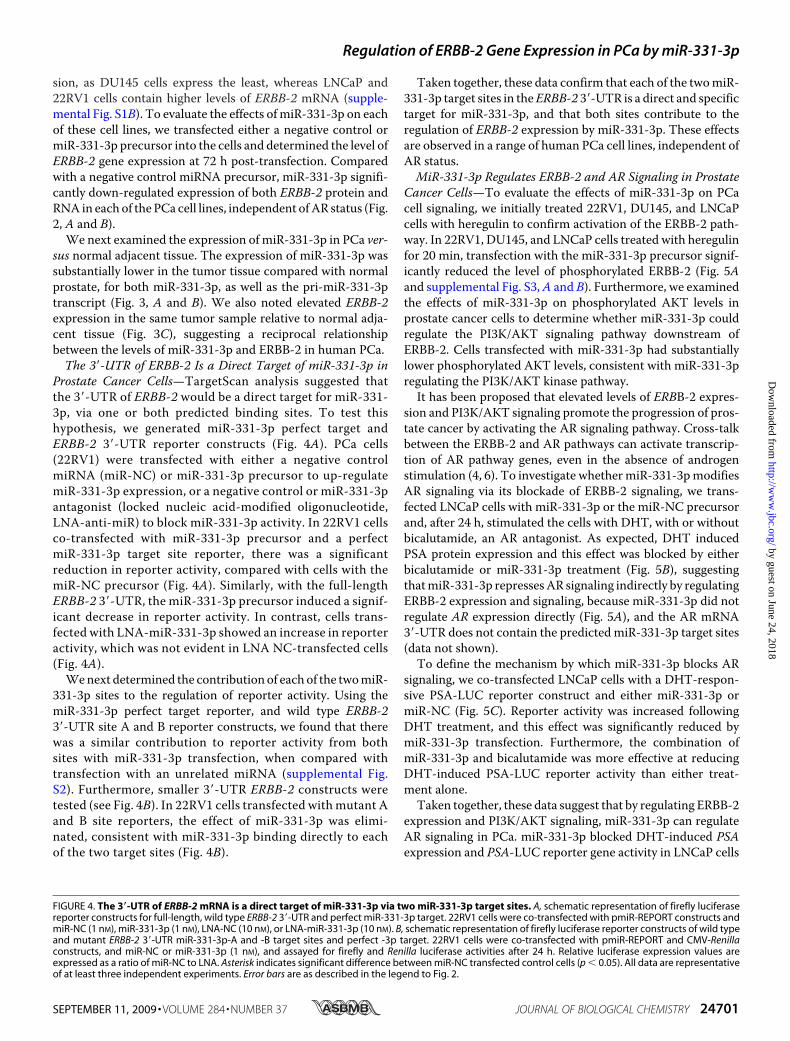
FIGURE 4. The 3�-UTR of ERBB-2 mRNA is a direct target of miR-331-3p via two miR-331-3p target sites. A, schematic representation of firefly luciferasereporter constructs for full-length, wild type ERBB-2 3�-UTR and perfect miR-331-3p target. 22RV1 cells were co-transfected with pmiR-REPORT constructs andmiR-NC (1 nM), miR-331-3p (1 nM), LNA-NC (10 nM), or LNA-miR-331-3p (10 nM). B, schematic representation of firefly luciferase reporter constructs of wild typeand mutant ERBB-2 3�-UTR miR-331-3p-A and -B target sites and perfect -3p target. 22RV1 cells were co-transfected with pmiR-REPORT and CMV-Renillaconstructs, and miR-NC or miR-331-3p (1 nM), and assayed for firefly and Renilla luciferase activities after 24 h. Relative luciferase expression values areexpressed as a ratio of miR-NC to LNA. Asterisk indicates significant difference between miR-NC transfected control cells (p � 0.05). All data are representativeof at least three independent experiments. Error bars are as described in the legend to Fig. 2.
Regulation of ERBB-2 Gene Expression in PCa by miR-331-3p
SEPTEMBER 11, 2009 • VOLUME 284 • NUMBER 37 JOURNAL OF BIOLOGICAL CHEMISTRY 24701
by guest on June 24, 2018http://w
ww
.jbc.org/D
ownloaded from

but it did not alter AR expression,suggesting that miR-331-3p indi-rectly reduces transcription andexpression of AR pathway targetgenes such as PSA via cross-talkbetween ERBB-2 and AR signalingpathways.
DISCUSSION
We have demonstrated that theERBB-2 mRNA 3�-UTR containstwo specific, direct miR-331-3p tar-get sites, and that miR-331-3pdown-regulates ERBB-2mRNA andprotein expression and signalingin multiple PCa cell lines. Theobserved reduction in ERBB-2mRNA levels by miR-331-3p is con-sistent with recent reports indicat-ing that manymiRNAs regulate tar-get gene expression by promotingmRNA decay (35, 36). miR-331-3pexpression is decreased in ERBB-2overexpressing PCa tissue relativeto normal adjacent tissue, suggest-ing a role for miR-331-3p in thedevelopment and progression ofthis disease. Furthermore, we haveshown that miR-331-3p blocked ARsignaling in PCa cells, without reduc-ing AR expression, by decreasingandrogen-induced PSA promoteractivity and PSA protein expression.These data suggest that loss of miR-331-3p expression could promote theincreasedERBB-2 expression and sig-naling seen inmany prostate cancers,and that thismaypromote theARsig-naling pathway.Although ERBB-2 gene amplifi-
cation is common in ERBB-2 over-expressing breast tumors, it is a veryrare event in PCa (37), suggestingthat post-transcriptional mecha-nisms, such as regulation by miR-NAs, may determine its expressionin this disease. Recent evidence sug-gests that expression of the ERBBreceptor family in cancer cells canbe mediated in part by miRNAs (28,38, 39). There is poor conservationof the two ERBB-2 3�-UTR miR-331-3p target sites (Fig. 1B,A andB)between human and rodent. How-ever, TargetScan analysis (33) indi-cates that these sites are among thehighest ranking predicted miR-331-3p target sites, with context
FIGURE 5. miR-331-3p decreases ERBB-2 protein expression and signaling, and blocks PSA expression andpromoter activity in LNCaP cells. A, LNCaP cells were transfected with miR-NC or miR-331-3p (30 nM) for 48 h andserum starved for 24 h thereafter, followed by stimulation � heregulin (HRG; 50 ng/ml) for 20 min. Cell lysates wereanalyzed for total ERBB-2, phospho-ERBB-2, AR, total AKT, and phospho-AKT expression by immunoblotting. B,LNCaP cells were transfected with miR-331-3p for 48 h and treated � DHT (10 nM) and � bicalutamide (10 �M). TotalPSA expression was determined by immunoblotting. C, LNCaP cells were co-transfected with a PSA-luciferase vector(PSA-LUC) and thymidine kinase-Renilla vector and with miR-NC or miR-331-3p (1 nM). Relative luciferase expression(firefly normalized to Renilla) values are expressed as a ratio of miR-NC-transfected cells (�S.D.). Asterisk indicates signifi-cantdifferencebetweenmiR-NCtransfectedcontrolcells(p�0.05).Errorbarsrepresent confidence intervals (CI�0.95).
Regulation of ERBB-2 Gene Expression in PCa by miR-331-3p
24702 JOURNAL OF BIOLOGICAL CHEMISTRY VOLUME 284 • NUMBER 37 • SEPTEMBER 11, 2009
by guest on June 24, 2018http://w
ww
.jbc.org/D
ownloaded from

score percentiles of 97 and 88%, respectively, when taking intoaccount criteria that are associated with miRNA target sitefunctionality, such as 3� compensatory base pairing, local AUsequence content, and position within the 3�-UTR (34). Ourreporter gene assays indicate that target sites A and B contrib-ute to the direct repression of ERBB-2 expression by miR-331-3p despite their weak evolutionary conservation. This isconsistent with our previous findings with miR-7 and the epi-dermal growth factor receptor, wheremiR-7 targets two poorlyconserved sites within the epidermal growth factor receptor3�-UTR to regulate epidermal growth factor receptor expres-sion and signaling (39), emphasizing that strong miRNA targetsite conservation between species is not essential for a site to befunctional. Importantly, mutation of each ERBB-2 3�-UTRmiR-331-3p target site seed binding region impaired repressionof reporter gene activity by miR-331-3p, whereas in contrast,transfection with unrelated miRNA did not repress reporteractivity. This confirmed the direct and specific interaction ofmiR-331-3p with each ERBB-2mRNA 3�-UTR site.
Anti-androgen therapy (bicalutamide (CasodexTM)) iswidely used for locally advanced PCa; however, after an initialresponse to treatment some tumors recur in an androgen-inde-pendent form. The molecular mechanisms underlying andro-gen independence are not fully understood, however, elevatedERBB-2 expression and signaling is thought to promote theprogression of PCa from an androgen-dependent to an andro-gen-independent state by producing constitutive activation ofAR signaling despite androgen withdrawal or blockade (4, 6).This may be achieved by enhanced recruitment of the AR toandrogen-responsive promoters (40). Thus, ERBB-2 hasemerged as a therapeutic target in PCa, with preclinical andclinical studies investigating the efficacy of anti-ERBB-2mono-clonal antibodies (e.g. trastuzumab or pertuzumab) for thetreatment of the disease (41–44). These studies have yieldeddisappointing results with little or no anti-tumor activityobserved. However, these trials have involved patients withadvanced PCa and patients have not been stratified or recruitedaccording to tumor ERBB-2 expression, suggesting that betterselection may be required in future studies (37). It is possiblethat blocking ERBB-2 signaling will hinder the progression ofPCa to androgen independence, or that ERBB-2 blockade willrestore androgen dependence, suggesting that co-administra-tion of an ERBB-2 inhibitor with an anti-androgen (e.g. bicalu-tamide) might have clinical benefits (45). Our transfectionstudies are consistent with this hypothesis, with the combina-tion of miR-331-3p and bicalutamide giving stronger repres-sion of androgen-stimulated PSA promoter activity than eithermiR-331-3p or bicalutamide alone (Figs. 5C and 6). It has alsobeen suggested that resistance to second line therapies, includ-ing ERBB-2 inhibitors, may result from loss of the tumor sup-pressor PTEN and subsequent activation of the PI3K/AKTpathway, a common event in advanced PCa that is associatedwith androgen independence (46–48). Indeed, PTEN loss isassociated with trastuzumab resistance in breast cancer (49).Furthermore, bicalutamide monotherapy is associated withincreased expression of ERBB-2 and phosphorylated AKT andreduced expression of PTEN in PCa (5). Interestingly, our worksuggests that miR-331-3p has the capacity to block PI3K/AKT
signaling in PCa cells independent of their PTEN status(LNCaP, PTEN-negative; 22Rv1 and DU145, PTEN-positive)(50), possibly by targeting other molecules involved in thispathway downstream of PTEN, and thus miR-331-3p could beused to overcome the inherent resistance of PTEN-negativeprostate cancers to anti-ERBB-2 and other second line thera-pies (e.g. chemotherapy).
Our study is the first to implicatemiR-331-3p in PCa. Dereg-ulated expression of miRNAs in cancer is often associated withgain or loss of chromosomal regions, with many miRNA geneslocated in fragile sites that are frequently altered in cancer (51–53). Alternatively, aberrant miRNA expression can occurthrough epigenetic mechanisms or by defects in miRNA bio-genesis (14–16). We observed decreased expression of bothprimary and mature miR-331-3p in PCa tissue relative to nor-mal adjacent prostate tissue (Fig. 3, A and B), suggesting thatreduced miR-331-3p expression in PCa cells does not resultfromabnormal processing ofmiR-331-3p. The humanmiR-331gene is located at 12q22, a chromosomal region that is not com-monly altered in PCa. One possible explanation for decreasedmiR-331-3p expression in PCa is that there is reduced miR-331-3p gene transcription, resulting in less primary, and there-fore mature, miR-331-3p being produced. Other studies havereported altered transcription of miRNA genes in cancer (17,54). In this regard, it will be interesting to characterize themiR-331-3p promoter and its activity in PCa cells.In summary, we have identified ERBB-2 as a direct and spe-
cific target of miR-331-3p in PCa cells. miR-331-3p expressionis reduced in PCa and promotes elevated ERBB-2 expressionand signaling, which increases AR signaling. Transfection ofPCa cell lines with miR-331-3p reduced ERBB-2 expression,PI3K/AKT signaling, and blocked AR signaling. This suggeststhat miR-331-3p has the capacity to regulate activity of critical
FIGURE 6. miR-331-3p blocks AR signaling via inhibition of ERBB-2expression and AKT activity in prostate cancer cells. AR antagonists suchas bicalutamide bind to the AR and prevent its activation and expression ofAR target genes, such as PSA. Nevertheless, AR signaling may persist in pros-tate cancer cells despite AR blockade, in part via increased expression of theERBB-2 receptor tyrosine kinase and subsequent activation of the PI3K/AKTpathway, which causes AR phosphorylation and promotes expression of ARtarget genes. miR-331-3p directly targets the ERBB-2 mRNA 3�-UTR to regu-late ERBB-2 protein expression, thereby reducing PI3K/AKT signaling and ARsignaling. The combination of an AR antagonist (bicalutamide) and miR-331-3p effectively blocks AR signaling (PSA expression and PSA promoteractivity) in LNCaP prostate cancer cells. (�) indicates activation step of path-way and (�) indicates inhibition of pathway component.
Regulation of ERBB-2 Gene Expression in PCa by miR-331-3p
SEPTEMBER 11, 2009 • VOLUME 284 • NUMBER 37 JOURNAL OF BIOLOGICAL CHEMISTRY 24703
by guest on June 24, 2018http://w
ww
.jbc.org/D
ownloaded from

signaling pathways in PCa cells. Ongoing studies will investi-gate the potential for miR-331-3p to mediate cross-talkbetween the AR and ERBB-2 signaling pathways and modulatethe sensitivity of PCa cells to anti-androgen and anti-ERBB-2therapies.
Acknowledgments—We thank Dr. Andrew Thomson, Dianne Bev-eridge, and Kavitha Iyer for helpful discussions regarding thismanuscript.
REFERENCES1. Jemal, A., Siegel, R.,Ward, E.,Murray, T., Xu, J., andThun,M. J. (2007)CA
Cancer J. Clin. 57, 43–662. Vernimmen, D., Gueders, M., Pisvin, S., Delvenne, P., and Winkler, R.
(2003) Br. J. Cancer 89, 899–9063. Isharwal, S., Miller, M. C., Epstein, J. I., Mangold, L. A., Humphreys, E.,
Partin, A. W., and Veltri, R. W. (2008) Int. J. Cancer 123, 2636–26434. Craft, N., Shostak, Y., Carey, M., and Sawyers, C. L. (1999) Nat. Med. 5,
280–2855. Festuccia, C., Gravina, G. L., Muzi, P., Pomante, R., Ventura, L., Vessella,
R. L., Vicentini, C., and Bologna, M. (2007) Endocr. Relat. Cancer 14,601–611
6. Mellinghoff, I. K., Vivanco, I., Kwon, A., Tran, C., Wongvipat, J., and Saw-yers, C. L. (2004) Cancer Cell 6, 517–527
7. Shi, X. B., Ma, A. H., Tepper, C. G., Xia, L., Gregg, J. P., Gandour-Edwards,R., Mack, P. C., Kung, H. J., and deVere White, R. W. (2004) Prostate 60,257–271
8. Bartel, D. P. (2004) Cell 116, 281–2979. Humphreys, D. T.,Westman, B. J., Martin, D. I., and Preiss, T. (2005) Proc.
Natl. Acad. Sci. U.S.A. 102, 16961–1696610. Chen, J. F., Mandel, E. M., Thomson, J. M., Wu, Q., Callis, T. E., Ham-
mond, S. M., Conlon, F. L., and Wang, D. Z. (2006) Nat. Genet. 38,228–233
11. Cheng, A. M., Byrom, M. W., Shelton, J., and Ford, L. P. (2005) NucleicAcids Res. 33, 1290–1297
12. Zhang, B., Pan, X., Cobb, G. P., and Anderson, T. A. (2007)Dev. Biol. 302,1–12
13. Calin, G. A., Sevignani, C., Dumitru, C. D., Hyslop, T., Noch, E., Yen-damuri, S., Shimizu, M., Rattan, S., Bullrich, F., Negrini, M., and Croce,C. M. (2004) Proc. Natl. Acad. Sci. U.S.A. 101, 2999–3004
14. Bueno, M. J., Perez de Castro, I., Gomez de Cedron, M., Santos, J., Calin,G. A., Cigudosa, J. C., Croce, C. M., Fernandez-Piqueras, J., and Malum-bres, M. (2008) Cancer Cell 13, 496–506
15. Schmittgen, T. D. (2008) J. Cell Mol. Med. 12, 1811–181916. Viswanathan, S. R., Daley, G. Q., and Gregory, R. I. (2008) Science 320,
97–10017. Chang, T. C., Yu, D., Lee, Y. S., Wentzel, E. A., Arking, D. E., West, K. M.,
Dang, C. V., Thomas-Tikhonenko, A., and Mendell, J. T. (2008) Nat.Genet. 40, 43–50
18. He,L.,He,X., Lim,L. P., deStanchina, E.,Xuan,Z., Liang,Y.,Xue,W.,Zender,L., Magnus, J., Ridzon, D., Jackson, A. L., Linsley, P. S., Chen, C., Lowe, S.W.,Cleary, M. A., and Hannon, G. J. (2007)Nature 447, 1130–1134
19. Esquela-Kerscher, A., and Slack, F. J. (2006) Nat. Rev. Cancer 6, 259–26920. Johnson, S. M., Grosshans, H., Shingara, J., Byrom, M., Jarvis, R., Cheng,
A., Labourier, E., Reinert, K. L., Brown, D., and Slack, F. J. (2005) Cell 120,635–647
21. Takamizawa, J., Konishi, H., Yanagisawa, K., Tomida, S., Osada, H., En-doh, H., Harano, T., Yatabe, Y., Nagino, M., Nimura, Y., Mitsudomi, T.,and Takahashi, T. (2004) Cancer Res. 64, 3753–3756
22. Volinia, S., Calin, G. A., Liu, C. G., Ambs, S., Cimmino, A., Petrocca, F.,Visone, R., Iorio, M., Roldo, C., Ferracin, M., Prueitt, R. L., Yanaihara, N.,Lanza, G., Scarpa, A., Vecchione, A., Negrini, M., Harris, C. C., and Croce,C. M. (2006) Proc. Natl. Acad. Sci. U.S.A. 103, 2257–2261
23. Si,M. L., Zhu, S.,Wu,H., Lu, Z.,Wu, F., andMo, Y. Y. (2007)Oncogene 26,
2799–280324. Ambs, S., Prueitt, R. L., Yi, M., Hudson, R. S., Howe, T. M., Petrocca, F.,
Wallace, T. A., Liu, C. G., Volinia, S., Calin, G. A., Yfantis, H. G., Stephens,R. M., and Croce, C. M. (2008) Cancer Res. 68, 6162–6170
25. Ozen,M., Creighton, C. J., Ozdemir,M., and Ittmann,M. (2008)Oncogene27, 1788–1793
26. Shi, X. B., Tepper, C. G., and White, R. W. (2008) J. Cell. Mol. Med. 12,1456–1465
27. Lin, S. L., Chiang, A., Chang, D., and Ying, S. Y. (2008) RNA 14, 417–42428. Scott, G. K., Goga, A., Bhaumik, D., Berger, C. E., Sullivan, C. S., and Benz,
C. C. (2007) J. Biol. Chem. 282, 1479–148629. Wang, X., and Seed, B. (2003) Nucleic Acids Res. 31, e15430. Jiang, J., Lee, E. J., Gusev, Y., and Schmittgen, T. D. (2005) Nucleic Acids
Res. 33, 5394–540331. Livak, K. J., and Schmittgen, T. D. (2001)Methods 25, 402–40832. Giles, K. M., Daly, J. M., Beveridge, D. J., Thomson, A. M., Voon, D. C.,
Furneaux, H. M., Jazayeri, J. A., and Leedman, P. J. (2003) J. Biol. Chem.278, 2937–2946
33. Lewis, B. P., Burge, C. B., and Bartel, D. P. (2005) Cell 120, 15–2034. Grimson, A., Farh, K. K., Johnston, W. K., Garrett-Engele, P., Lim, L. P.,
and Bartel, D. P. (2007)Mol. Cell 27, 91–10535. Filipowicz, W., Jaskiewicz, L., Kolb, F. A., and Pillai, R. S. (2005) Curr.
Opin. Struct. Biol. 15, 331–34136. Liu, T., Papagiannakopoulos, T., Puskar, K., Qi, S., Santiago, F., Clay, W.,
Lao, K., Lee, Y., Nelson, S. F., Kornblum, H. I., Doyle, F., Petzold, L., Shrai-man, B., and Kosik, K. S. (2007) PLoS ONE 2, e804
37. Solit, D. B., and Rosen, N. (2007) J. Clin. Oncol. 25, 241–24338. Kefas, B., Godlewski, J., Comeau, L., Li, Y., Abounader, R., Hawkinson,M.,
Lee, J., Fine, H., Chiocca, E. A., Lawler, S., and Purow, B. (2008)Cancer Res.68, 3566–3572
39. Webster, R. J., Giles, K. M., Price, K. J., Zhang, P. M., Mattick, J. S., andLeedman, P. J. (2009) J. Biol. Chem. 284, 5731–5741
40. Liu, Y., Majumder, S., McCall, W., Sartor, C. I., Mohler, J. L., Gregory,C. W., Earp, H. S., and Whang, Y. E. (2005) Cancer Res. 65, 3404–3409
41. Bradbury, J. (2007) Lancet Oncol. 8, 28742. Albanell, J., Codony, J., Rovira, A., Mellado, B., and Gascon, P. (2003)Adv.
Exp. Med. Biol. 532, 253–26843. Small, E. J., Bok, R., Reese, D. M., Sudilovsky, D., and Frohlich, M. (2001)
Semin. Oncol. 28, Suppl. 15, 71–7644. Bianco, A. R. (2004) J. Chemother. 16, Suppl. 4, 52–5445. Gravina, G. L., Festuccia, C., Millimaggi, D., Tombolini, V., Dolo, V., Vi-
centini, C., and Bologna, M. (2009) Urology 74, 452–45746. Malik, S. N., Brattain,M., Ghosh, P.M., Troyer, D. A., Prihoda, T., Bedolla,
R., and Kreisberg, J. I. (2002) Clin. Cancer Res. 8, 1168–117147. Murillo, H., Huang, H., Schmidt, L. J., Smith, D. I., and Tindall, D. J. (2001)
Endocrinology 142, 4795–480548. Whang, Y. E.,Wu,X., Suzuki,H., Reiter, R. E., Tran, C., Vessella, R. L., Said,
J. W., Isaacs, W. B., and Sawyers, C. L. (1998) Proc. Natl. Acad. Sci. U.S.A.95, 5246–5250
49. Nagata, Y., Lan, K. H., Zhou, X., Tan, M., Esteva, F. J., Sahin, A. A., Klos,K. S., Li, P., Monia, B. P., Nguyen, N. T., Hortobagyi, G. N., Hung, M. C.,and Yu, D. (2004) Cancer Cell 6, 117–127
50. Festuccia, C., Gravina, G. L., D’Alessandro, A. M., Millimaggi, D., DiRocco, C., Dolo, V., Ricevuto, E., Vicentini, C., and Bologna,M. (2008) Int.J. Oncol. 33, 381–388
51. Calin, G. A., Dumitru, C. D., Shimizu, M., Bichi, R., Zupo, S., Noch, E.,Aldler, H., Rattan, S., Keating, M., Rai, K., Rassenti, L., Kipps, T., Negrini,M., Bullrich, F., and Croce, C. M. (2002) Proc. Natl. Acad. Sci. U.S.A. 99,15524–15529
52. Selcuklu, S. D., Yakicier, M. C., and Erson, A. E. (2009) Cancer Genet.Cytogenet. 189, 15–23
53. Wei, J. S., Song, Y. K., Durinck, S., Chen, Q. R., Cheuk, A. T., Tsang, P.,Zhang, Q., Thiele, C. J., Slack, A., Shohet, J., and Khan, J. (2008)Oncogene27, 5204–5213
54. Reddy, S. D., Ohshiro, K., Rayala, S. K., and Kumar, R. (2008) Cancer Res.68, 8195–8200
Regulation of ERBB-2 Gene Expression in PCa by miR-331-3p
24704 JOURNAL OF BIOLOGICAL CHEMISTRY VOLUME 284 • NUMBER 37 • SEPTEMBER 11, 2009
by guest on June 24, 2018http://w
ww
.jbc.org/D
ownloaded from

LeedmanMichael R. Epis, Keith M. Giles, Andrew Barker, Tulene S. Kendrick and Peter J.
Prostate CancermiR-331-3p Regulates ERBB-2 Expression and Androgen Receptor Signaling in
doi: 10.1074/jbc.M109.030098 originally published online July 7, 20092009, 284:24696-24704.J. Biol. Chem.
10.1074/jbc.M109.030098Access the most updated version of this article at doi:
Alerts:
When a correction for this article is posted•
When this article is cited•
to choose from all of JBC's e-mail alertsClick here
Supplemental material:
http://www.jbc.org/content/suppl/2009/07/07/M109.030098.DC1
http://www.jbc.org/content/284/37/24696.full.html#ref-list-1
This article cites 54 references, 18 of which can be accessed free at
by guest on June 24, 2018http://w
ww
.jbc.org/D
ownloaded from