miR-181d/MALT1 regulatory axis attenuates … · miR-181d/MALT1 regulatory axis attenuates...
9
Original Article miR-181d/MALT1 regulatory axis attenuates mesenchymal phenotype through NF-kB pathways in glioblastoma Fan Yang a, b, c, d, 1 , Xing Liu a, b, c, d, 1 , Yanwei Liu a, b, c, d , Yuqing Liu b, d , Chuanbao Zhang a, b, c, d , Zheng Wang b, d , Tao Jiang a, b, c, d, ** , Yongzhi Wang a, b, c, d, * a Department of Neurosurgery, Beijing Tiantan Hospital, Capital Medical University, Beijing, 100050, China b Beijing Neurosurgical Institute, Capital Medical University, Beijing, 100050, China c China National Clinical Research Center for Neurological Diseases, Beijing, 100050, China d Chinese Glioma Cooperative Group (CGCG), China article info Article history: Received 15 February 2017 Received in revised form 27 February 2017 Accepted 2 March 2017 Keywords: Glioma Non-coding RNA Mesenchymal transition Transcriptome subtype NF-kB signaling abstract The mesenchymal (MES) subtype of glioblastoma (GBM) indicated a more malignant phenotype and worse prognosis compared with their proneural (PN) counterpart. The plasticity between PN and MES transcriptome signatures provided an approach for clinical intervention. However, few miRNAs have been identified to participate in the shift between subtypes. Here, we utilized transcriptomic data and experimental evidences to prove that miR-181d was a novel regulator of NFkB signaling pathway by directly repressing MALT1, leading to induced PN markers and reduced MES genes. Functionally, ectopic expression of miR-181d suppressed GBM cell proliferation, colony formation and anchor-independent growth, as well as migration, invasion and tube formation. Moreover, miR-181d overexpression increased radio- and chemo-sensitivity for GBM cells. Rescue of MALT1 could partially reverse the effects of miR-181d in GBM malignant behaviors. Clinically, miR-181d could serve as a prognostic indicator for GBM patients. Taken together, we concluded that loss of miR-181d contributes to aggressive biological processes associated with MES phenotype via NFkB signaling, which broaden our insights into the un- derlying mechanisms in subtype transition and miRNA-based tailored medicine for GBM management. © 2017 Elsevier B.V. All rights reserved. Introduction Glioblastoma (GBM; World Health Organization grade IV), the most common and lethal primary brain tumor, carries an invariably poor clinical outcome in adults [1]. Despite treatment involving surgery, ionizing radiation, and chemotherapy with the alkylating agent temozolomide (TMZ), patients with GBM only have an average survival of slightly more than 1 year after the initial diagnosis [1,2]. The unfavorable prognosis is largely attributed to the highly proliferative, invasive and therapeutic resistant nature of glioma. Hence, an understanding of the biomolecular mechanisms involved in glioma malignant behaviors is urgently needed. To comprehensively explore the molecular mechanism of GBM, many groups have turned to high-throughput profiling researches [3]. Although inconsistencies were observed across various clas- sification schemes, the mesenchymal (MES) and proneural (PN) subtypes were robust and generally consistent [4e6]. GBM pa- tients in the MES subtype exhibit radio- and chemo-resistant properties, increased invasiveness, reduced cell stiffness, and relatively worse prognosis than PN tumors [7]. Moreover, PN tu- mors were proved to give rise to MES recurrences, indicative of a MES transition during glioma progression [5]. Following studies further confirmed the occurrence of MES shift in GBM and iden- tified several master transcription factors, including NFkB, STAT3, were responsible for this procedure [7,8]. Considering the malig- nant behaviors of MES GBMs, clarifying the mechanistic basis activating MES phenotype may aid in the development of tailored therapy for GBM patients. Abbreviations: GBM, glioblastoma; MES, mesenchymal subtype; PN, proneural subtype; TMZ, temozolomide; NC, negative control; FACS, fluorescence-activated cell sorting; GO, Gene Ontology; GSEA, gene set enrichment analysis; GSVA, gene set variation analysis. * Corresponding author. Department of Neurosurgery, Beijing Tiantan Hospital, Capital Medical University, No. 6 TiantanXili, Dongcheng District, Beijing, 100050, China. ** Corresponding author. Beijing Neurosurgical Institute, Capital Medical University, No. 6 TiantanXili, Dongcheng District, Beijing, 100050, China. E-mail addresses: [email protected] (T. Jiang), [email protected] (Y. Wang). 1 Fan Yang and Xing Liu contributed equally to this work. Contents lists available at ScienceDirect Cancer Letters journal homepage: www.elsevier.com/locate/canlet http://dx.doi.org/10.1016/j.canlet.2017.03.002 0304-3835/© 2017 Elsevier B.V. All rights reserved. Cancer Letters 396 (2017) 1e9 Downloaded for Anonymous User (n/a) at Capital University of Medical Sciences from ClinicalKey.com by Elsevier on May 31, 2017. For personal use only. No other uses without permission. Copyright ©2017. Elsevier Inc. All rights reserved.
Transcript of miR-181d/MALT1 regulatory axis attenuates … · miR-181d/MALT1 regulatory axis attenuates...
lable at ScienceDirect
Cancer Letters 396 (2017) 1e9
Contents lists avai
Cancer Letters
journal homepage: www.elsevier .com/locate/canlet
Original Article
miR-181d/MALT1 regulatory axis attenuates mesenchymal phenotypethrough NF-kB pathways in glioblastoma
Fan Yang a, b, c, d, 1, Xing Liu a, b, c, d, 1, Yanwei Liu a, b, c, d, Yuqing Liu b, d,Chuanbao Zhang a, b, c, d, Zheng Wang b, d, Tao Jiang a, b, c, d, **, Yongzhi Wang a, b, c, d, *
a Department of Neurosurgery, Beijing Tiantan Hospital, Capital Medical University, Beijing, 100050, Chinab Beijing Neurosurgical Institute, Capital Medical University, Beijing, 100050, Chinac China National Clinical Research Center for Neurological Diseases, Beijing, 100050, Chinad Chinese Glioma Cooperative Group (CGCG), China
a r t i c l e i n f o
Article history:Received 15 February 2017Received in revised form27 February 2017Accepted 2 March 2017
Keywords:GliomaNon-coding RNAMesenchymal transitionTranscriptome subtypeNF-kB signaling
Abbreviations: GBM, glioblastoma; MES, mesenchsubtype; TMZ, temozolomide; NC, negative control;cell sorting; GO, Gene Ontology; GSEA, gene set enriset variation analysis.* Corresponding author. Department of Neurosurg
Capital Medical University, No. 6 TiantanXili, DongchChina.** Corresponding author. Beijing NeurosurgicalUniversity, No. 6 TiantanXili, Dongcheng District, Beij
E-mail addresses: [email protected] (T.(Y. Wang).
1 Fan Yang and Xing Liu contributed equally to this
http://dx.doi.org/10.1016/j.canlet.2017.03.0020304-3835/© 2017 Elsevier B.V. All rights reserved.
Downloaded for Anonymous UserFor personal use only
a b s t r a c t
The mesenchymal (MES) subtype of glioblastoma (GBM) indicated a more malignant phenotype andworse prognosis compared with their proneural (PN) counterpart. The plasticity between PN and MEStranscriptome signatures provided an approach for clinical intervention. However, few miRNAs havebeen identified to participate in the shift between subtypes. Here, we utilized transcriptomic data andexperimental evidences to prove that miR-181d was a novel regulator of NFkB signaling pathway bydirectly repressing MALT1, leading to induced PN markers and reduced MES genes. Functionally, ectopicexpression of miR-181d suppressed GBM cell proliferation, colony formation and anchor-independentgrowth, as well as migration, invasion and tube formation. Moreover, miR-181d overexpressionincreased radio- and chemo-sensitivity for GBM cells. Rescue of MALT1 could partially reverse the effectsof miR-181d in GBM malignant behaviors. Clinically, miR-181d could serve as a prognostic indicator forGBM patients. Taken together, we concluded that loss of miR-181d contributes to aggressive biologicalprocesses associated with MES phenotype via NFkB signaling, which broaden our insights into the un-derlying mechanisms in subtype transition and miRNA-based tailored medicine for GBM management.
© 2017 Elsevier B.V. All rights reserved.
Introduction
Glioblastoma (GBM; World Health Organization grade IV), themost common and lethal primary brain tumor, carries an invariablypoor clinical outcome in adults [1]. Despite treatment involvingsurgery, ionizing radiation, and chemotherapy with the alkylatingagent temozolomide (TMZ), patients with GBM only have anaverage survival of slightly more than 1 year after the initial
ymal subtype; PN, proneuralFACS, fluorescence-activatedchment analysis; GSVA, gene
ery, Beijing Tiantan Hospital,eng District, Beijing, 100050,
Institute, Capital Medicaling, 100050, China.
Jiang), [email protected]
work.
(n/a) at Capital University of Medica. No other uses without permission. C
diagnosis [1,2]. The unfavorable prognosis is largely attributed tothe highly proliferative, invasive and therapeutic resistant nature ofglioma. Hence, an understanding of the biomolecular mechanismsinvolved in glioma malignant behaviors is urgently needed.
To comprehensively explore the molecular mechanism of GBM,many groups have turned to high-throughput profiling researches[3]. Although inconsistencies were observed across various clas-sification schemes, the mesenchymal (MES) and proneural (PN)subtypes were robust and generally consistent [4e6]. GBM pa-tients in the MES subtype exhibit radio- and chemo-resistantproperties, increased invasiveness, reduced cell stiffness, andrelatively worse prognosis than PN tumors [7]. Moreover, PN tu-mors were proved to give rise to MES recurrences, indicative of aMES transition during glioma progression [5]. Following studiesfurther confirmed the occurrence of MES shift in GBM and iden-tified several master transcription factors, including NFkB, STAT3,were responsible for this procedure [7,8]. Considering the malig-nant behaviors of MES GBMs, clarifying the mechanistic basisactivating MES phenotype may aid in the development of tailoredtherapy for GBM patients.
l Sciences from ClinicalKey.com by Elsevier on May 31, 2017.opyright ©2017. Elsevier Inc. All rights reserved.
F. Yang et al. / Cancer Letters 396 (2017) 1e92
MicroRNAs (miRNAs) are endogenous small noncoding RNAs,which could act as regulators of gene expression by post-transcriptionally reducing target mRNAs with partial complemen-tarity in their 30-untranslated regions (UTRs) [9]. A spectrum ofmiRNAs have been demonstrated to be altered in multiple cellularprocesses including proliferation, apoptosis, differentiation,migration and angiogenesis, affecting the hallmarks of tumorigenicprocesses [10,11]. Similarly, several miRNAs have been found toparticipate in the pathogenesis of GBM, including oncogenic miR-21 and tumor-suppressive miR-181 family [12e14]. Besides of theclassification and prognostic significance, these miRNAs could alsoserve as biomarker for glioma diagnosis and therapeutics [15]. Inthe present study, we aimed to investigate the expression andfunction of miR-181d in both glioma patients and cell lines. Bycomprehensively analyzing the miRNA/mRNA profiles, we revealedthat miR-181d was highly expressed in PN subtype and could serveas a prognostic indicator for glioma patients. In vitro and in vivoexperiments both demonstrated that miR-181d restrained themalignancy of GBM cells via its direct target mucosa associatedlymphoid tissue lymphoma translocation gene 1 (MALT1) and NFkBsignaling pathway. Notably, NFkB pathway inhibition by miR-181dinhibited the expression of MES signature, which will increase thesensitization of GBM cells to chemo- and radio-therapy. Collec-tively, our results will help us comprehend the underlying mech-anisms of miR-181d in glioma and provided an alternative miRNA-based approach for glioma precision oncology.
Materials and methods
Tissue specimens
The mRNA/miRNA profiles and relevant clinical parameters were obtained fromthe Chinese Glioma Genome Atlas (CGGA, www.cgga.org) and the Cancer GenomeAtlas (TCGA) database (http://cancergenome.nih.gov). Human glioma samples usedin the present study were confirmed by pathologist according to WHO criteria [16].This studywas approved by the Ethics Committee of Beijing Tiantan Hospital, CapitalMedical University, and written informed consents were obtained from all patients.
Cell culture and establishment of TMZ-resistant cell line
The human GBM cell lines LN229, U87MG, SNB19 and U251 were obtained fromthe Institute of Biochemistry and Cell Biology, Chinese Academy of Science. Cellswere grown in DMEM (Dulbecco's modified Eagle's medium, Gibco) containing 10%of fetal bovine serum (FBS, Gibco). Human umbilical vein endothelial cell (HUVEC)were obtained from Harbin Medical University and cultured in endothelial cell basalmedium supplemented with 1% endothelial cell growth supplement and 5% FBS. Toestablish the TMZ-resistant cells, the U87MG parental cells were first maintained ata low dose of TMZ (5 mM, SigmaeAldrich) and then successively exposed to incre-mental doses of TMZ (10 mM, 20 mM, 40 mM, 80 mM). Each time, the surviving cellswere maintained until a normal rate of growth was obtained. TMZ-resistant U87MGcells with more than 20 passages were maintained at corresponding doses and usedfor subsequent experiments [17]. Cells were maintained at 37 �C under a humidifiedatmosphere containing 5% CO2.
Lentivirus preparation
The lentiviral packaging kit was purchased from Open GeneCopoeia. Lentiviruscarrying hsa-miR-181d or hsa-miR-negative control (NC) was packaged in 293T cellsand concentrated from the supernatant, as instructed by themanufacturer's manual.Stable cell lines were established by infecting lentivirus into U87MG, LN229, SNB19and U251 followed by puromycin (2 mg/ml) selection. These established stable celllines were maintained in DMEM containing 10% FBS and puromycin (0.5 mg/ml) forfurther experiments.
RNA extraction and quantitative real-time polymerase chain reaction (qRT-PCR)
Total RNA from frozen tumor samples was extracted using the Trizol reagent(Ambion). RNA concentration and quality were measured using the ND-1000spectrophotometer (NanoDrop Technologies). qRT-PCR was performed with 7500fast (Applied Biosystems) and used to analyze the expression of miRNA andmRNA inhuman glioma samples and cell lines. Each cDNA sample was run as triplicates.Expression of U6 was used as an endogenous control for miR-181d, while GAPDHwas used as an internal control for normalization and quantification of mRNAexpression (MALT1, IL-6, Olig2, PDGFRa, CD44, Vimentin). All primer sequences arelisted in Supplementary Table 1.
Downloaded for Anonymous User (n/a) at Capital University of MedFor personal use only. No other uses without permissio
Protein extraction and western blot
Protein were extracted using RIPA buffer according to the manufacturer's pro-tocol. Expression of proteinwas confirmed by MALT1 (rabbit, 1:1500, ABclonal), P65(rabbit, 1:1500, Cell signaling technology), phoshorylated-P65 (p-P65; rabbit,1:1500, Cell signaling technology), CD44 (mouse, 1:2000, Cell signaling technology),Vimentin (rabbit, 1:1000, Cell signaling technology), Olig2 (rabbit, 1:1500, Abcam),PDGFRa (rabbit, 1:1000, Abcam) and GAPDH (mouse, 1:5000, Proteintech), followedby incubation with appropriate correlated HRP-conjugated secondary antibody.Representative images from 2 or 3 independent experiments are shown.
Luciferase reporter assay
To determine whether miR-181d directly binds to the MALT1 30-UTR, dualluciferase reporter assays were performed. The pmirGLO (Promega) vector was usedto construct MALT1 30-UTR containing reporter. In brief, 293T and U87MG cellsseeded in 96-well plates (8� 103 cells/well) were co-transfected withMALT130-UTRreporters, miR-181d plasmid or control miRNA. Similarly, to determine the NFkBpathway transcriptional activity, a reporter containing NFkB response element wasused (pGL4.32[luc2P/NFkB-RE/Hypro] Vector, Promega). The plasmids were trans-fected into cells treated with miR-181d plasmid or negative control, and an internalcontrol vector expressing Renilla luciferase (Promega). At 48 h after transfection,cells were lysed and subjected to luciferase assays using the Dual-Luciferase Re-porter Assay System (Promega). Firefly luciferase activity was normalized to Renillaluciferase values.
Cell proliferation assay
Cell proliferation was conducted using the Cell-Counting Kit 8 (CCK8; DojindoLaboratories) according to themanufacturer's instructions. After planting cells in 96-well microtiter plates (Corning Costar) at 1.0 � 103/well, 10 mL of CCK8 was added toeach well (medium 100 mL) at the time of harvest. A half-hour after adding CCK8,cellular viability was determined by measuring the absorbance of the converted dyeat 450 nm.
Colony formation and soft agar assay
Previously established stable cell lines were planted in 6-well microtiter plates(Corning Costar) at 500/well. The assay should be stopped when the colonies areclearly visible even without looking under the microscope. Cell colony were fixedwith 4% paraformaldehyde and observed by staining with 0.1% crystal violet. For softagar assay, GBM cells (8 � 102 cells/well) were suspended in 1.5 mL complete me-dium supplemented with 0.7% agarose (Gibco). The cells were placed in 6-well platecontaining 1.5 mL complete medium (double concentration) and agarose (1.2%) onthe bottom layer. The plates were incubated at 37 �C with 5% CO2 for 20 days. Eachexperiment was performed at least three times.
Apoptosis detection and transwell migration/invasion assays
The apoptosis assay was tested in U87MG and LN229 cells after transfectionusing the Annexin Alexa Fluor647/PI (4A Biotech) and analyzed by fluorescence-activated cell sorting (FACS). The cells were harvested by trypsinization andwashed once with phosphate buffer saline (PBS). To measure cell migration, 8-mmpore size culture inserts (Transwell; Costar) were placed into the wells of 24-wellculture plates, separating the upper and the lower chambers. In the lower cham-ber, 600 mL of DMEM/F12 containing 10% FBS was added. Then, serum-free mediumcontaining 3 � 104 cells were added to the upper chamber for migration assays,whereas 1 � 105 cells were used for Matrigel invasion assays. Incubation at 37 �Cwith 5% CO2, the number of cells that hadmigrated through the pores was quantifiedby counting 5 independent visual fields under the microscope (Zeiss). Cellmorphology was fixed with 4% paraformaldehyde and observed by staining with0.1% crystal violet. Each experiment was performed at least three times.
Tube formation assays
Tube formation assays were performed on growth-factor-reduced (GFR)Matrigel Matrix (BD Biosciences) and visualized using either phase contrast mi-croscopy. HUVECs (2 � 105/mL) were cultured in 96-well plate with GBM cell su-pernatant that were collected from previously established stable cell lines,respectively. Then putted the 96-well plate into incubator (37 �C, 5% CO2) andvisualized by microscope after 8 h.
Determination of interleukin 6 (IL-6) in supernatant of cultured GBM cells
Human IL-6 quantikine ELISA kits (R&D systems) were used to determine thelevel of corresponding protein. Briefly, 100 mL of assay diluent and 100 mL of stan-dard, control or sample were pipetted to cover with plate sealer and incubate atroom temperature for 2 h. Then, aspirate each well and add 200 mL of Conjugate toincubate at room temperature for another 2 h on the shaker. Afterwards, 200 mLSubstrate Solutionwere added and kept from the exposure of light for 30min beforethe addition of Stop Solution. The plate was read at 450 nm within 30 min. Astandard curve was drawn to calculate the protein concentrations for each sample.
ical Sciences from ClinicalKey.com by Elsevier on May 31, 2017.n. Copyright ©2017. Elsevier Inc. All rights reserved.
F. Yang et al. / Cancer Letters 396 (2017) 1e9 3
Nude mouse tumor intracranial model
BALB/c female athymic micewere implanted in the brainwith established stablecell U87MG-miR-181d and U87MG-negative control. Briefly, mice were anes-thetized with 5% chloral hydrate and cells were implanted using cranial guidescrews as previously described [15]. A TJ-4A Syringe Pump Controller and micro-infusion syringe pump (1 mL/min) were used to implant 1� 106 cells into the brain ofmice. After 40 days, mice brains' tumors were detected by MRI for animal. Then,mice were sacrificed simultaneously and brains were extracted and fixed in 4%paraformaldehyde for 24 h, embedded in paraffin, and sectioned into 5 mm slices forimmunohistochemistry.
Statistical analysis
Statistical analyses were performed using GraphPad Prism, version 6.0 and SPSS,version 16.0. Heat maps were constructed by Gene Cluster 3.0 and Gene Tree Viewsoftware. KaplaneMeier survival analysis was used to estimate the survival distri-butions. Then, the log-rank test was applied to assess the statistical significancebetween stratified survival groups. Differential genes between high- and low-expression groups were selected using significance analysis of microarray (SAM)algorithm. The miR-181d negatively associated genes were identified as R < �0.4using Pearson correlation analyses. Gene Ontology (GO) analysis was performedusing DAVID (http://david.abcc.ncifcrf.gov/) [18]. Gene set enrichment analysis(GSEA) was performed using GSEA software downloaded from the Broad Institute(www.broadinstitute.org/gsea) [19]. Gene set variation analysis (GSVA) wasanalyzed using the GSVA package [20] of R. miRWalk 2.0 [21] andmiRanda [22] wereutilized for miR-181d potential targets screening. All differences were consideredstatistically significant at the level of two-side P < 0.05.
Results
Aberrant expression and prognostic significance of miR-181d forpatients with GBMs
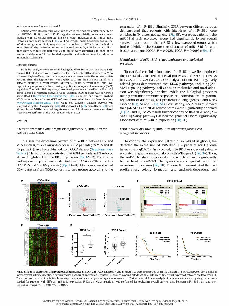
To assess the expression pattern of miR-181d between PN andMES subclass, miRNA array data for 45 GBM patients (35MES and 10PNpatients) have been obtained fromCGGAdataset (SupplementaryTable 2). The results demonstrated that GBM patients in PN subtypeshowed high-level of miR-181d expression (Fig. 1AeD). The consis-tent expression pattern was validated using TCGA miRNA array data(177 MES and 106 PN patients) (Fig. 1AeD). Afterwards, we dividedGBM patients from TCGA cohort into two groups according to the
Fig. 1. miR-181d expression and prognostic significance in CGGA and TCGA datasets. A anmesenchymal subtypes identified by significance analysis of microarray algorithm. C. VolcanThe expression pattern of miR-181d between proneural and mesenchymal subtypes were coapplied for patients with different miR-181d expression. F. KaplaneMeier algorithm wasexpression groups. **, P < 0.01, ***, P < 0.001.
Downloaded for Anonymous User (n/a) at Capital University of MedicaFor personal use only. No other uses without permission. C
expression of miR-181d. Similarly, GSEA between different groupsdemonstrated that patients with high-level of miR-181d wereenriched inPN-associated gene set (Fig.1E).Moreover, patients in themiR-181d high-expressed group had significantly longer overallsurvival than those in the miR-181d low-expressed group, whichfurther highlight the suppressive character of miR-181d for glio-blastoma patients (CGGA, P ¼ 0.0028; TCGA, P ¼ 0.0095) (Fig. 1F).
Identification of miR-181d related pathways and biologicalprocesses
To clarify the cellular functions of miR-181d, we first exploredthe miR-181d associated biological processes and KEGG pathwaysin TCGA and CGGA datasets. GO analyses of miR-181d negativelyrelated genes demonstrated that KEGG pathways, including JAK-STAT signaling pathway, cell adhesion molecules and focal adhe-sion was significantly enriched, while the biological processesmainly contained immune response, cell adhesion, cell migration,regulation of apoptosis, cell proliferation, angiogenesis and NFkBcascade (Fig. 2A and B, Fig. S1). Consistently, GSEA results showedthat JAK-STAT and NFkB related terms were significantly enriched(Fig. 2C and D). GSVA results further confirmed that NFkB and JAK-STAT signaling pathways associated gene sets were significantlyassociated with miR-181d expression (Fig. 2E).
Ectopic overexpression of miR-181d suppresses glioma cellmalignant behaviors
To confirm the expression pattern of miR-181d in glioma, wedetected the expression of miR-181d in a panel of adult gliomatissues using qRT-PCR. As expected, miR-181d was gradually down-regulated in glioma samples along withWHO grade (Fig. 3A). Then,the miR-181d stable expressed cells, which showed significantlyhigher level of miR-181d NC group, were subjected to furtherexperimental analyses (Fig. 3B). The results demonstrated that cellproliferation, colony formation and anchor-independent cell
d B. Heatmaps were constructed using the differential miRNAs between proneural ando plot indicated that miR-181d were differential expressed between the two group. D.mpared. E. Gene set enrichment analysis of proneural and mesenchymal gene sets wasperformed for evaluating overall survival time between miR-181d high- and low-
l Sciences from ClinicalKey.com by Elsevier on May 31, 2017.opyright ©2017. Elsevier Inc. All rights reserved.
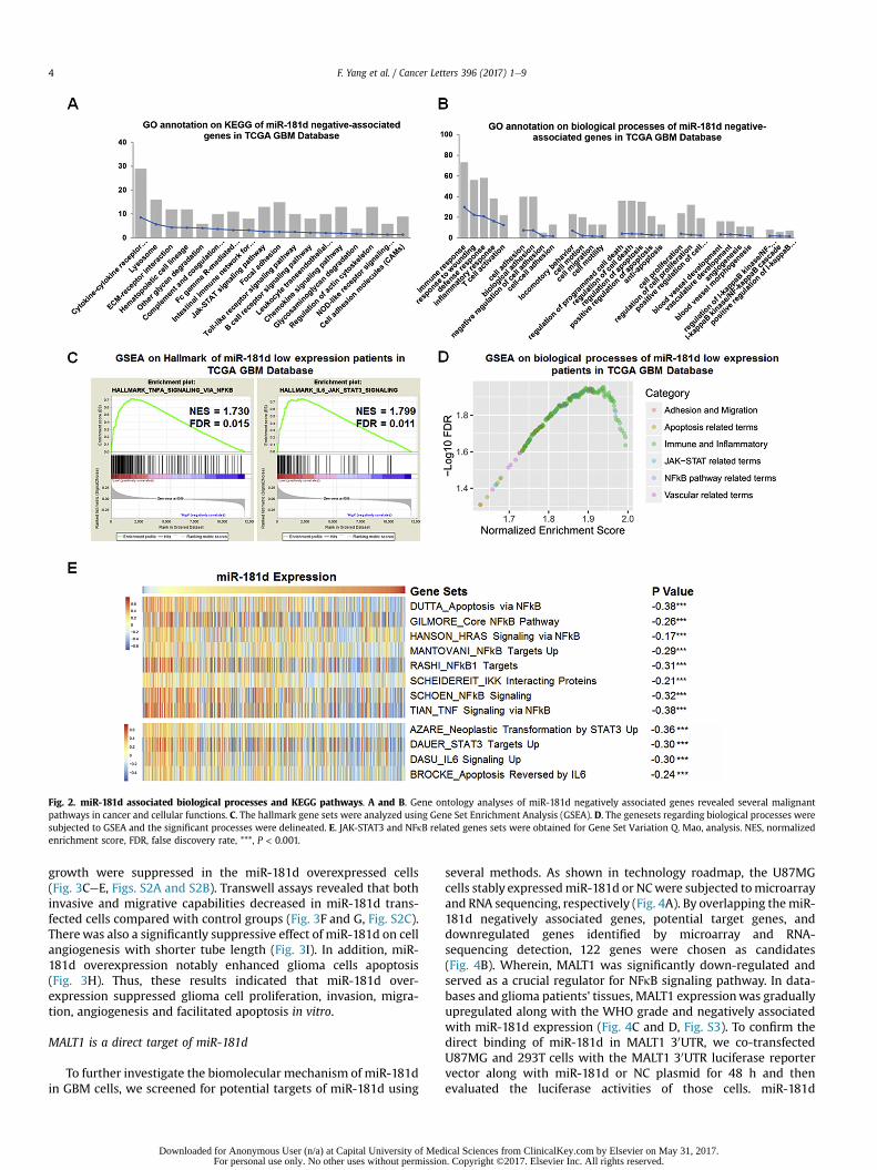
Fig. 2. miR-181d associated biological processes and KEGG pathways. A and B. Gene ontology analyses of miR-181d negatively associated genes revealed several malignantpathways in cancer and cellular functions. C. The hallmark gene sets were analyzed using Gene Set Enrichment Analysis (GSEA). D. The genesets regarding biological processes weresubjected to GSEA and the significant processes were delineated. E. JAK-STAT3 and NFkB related genes sets were obtained for Gene Set Variation Q. Mao, analysis. NES, normalizedenrichment score, FDR, false discovery rate, ***, P < 0.001.
F. Yang et al. / Cancer Letters 396 (2017) 1e94
growth were suppressed in the miR-181d overexpressed cells(Fig. 3CeE, Figs. S2A and S2B). Transwell assays revealed that bothinvasive and migrative capabilities decreased in miR-181d trans-fected cells compared with control groups (Fig. 3F and G, Fig. S2C).Therewas also a significantly suppressive effect of miR-181d on cellangiogenesis with shorter tube length (Fig. 3I). In addition, miR-181d overexpression notably enhanced glioma cells apoptosis(Fig. 3H). Thus, these results indicated that miR-181d over-expression suppressed glioma cell proliferation, invasion, migra-tion, angiogenesis and facilitated apoptosis in vitro.
MALT1 is a direct target of miR-181d
To further investigate the biomolecular mechanism of miR-181din GBM cells, we screened for potential targets of miR-181d using
Downloaded for Anonymous User (n/a) at Capital University of MedFor personal use only. No other uses without permissio
several methods. As shown in technology roadmap, the U87MGcells stably expressedmiR-181d or NCwere subjected tomicroarrayand RNA sequencing, respectively (Fig. 4A). By overlapping themiR-181d negatively associated genes, potential target genes, anddownregulated genes identified by microarray and RNA-sequencing detection, 122 genes were chosen as candidates(Fig. 4B). Wherein, MALT1 was significantly down-regulated andserved as a crucial regulator for NFkB signaling pathway. In data-bases and glioma patients' tissues, MALT1 expressionwas graduallyupregulated along with the WHO grade and negatively associatedwith miR-181d expression (Fig. 4C and D, Fig. S3). To confirm thedirect binding of miR-181d in MALT1 30UTR, we co-transfectedU87MG and 293T cells with the MALT1 30UTR luciferase reportervector along with miR-181d or NC plasmid for 48 h and thenevaluated the luciferase activities of those cells. miR-181d
ical Sciences from ClinicalKey.com by Elsevier on May 31, 2017.n. Copyright ©2017. Elsevier Inc. All rights reserved.
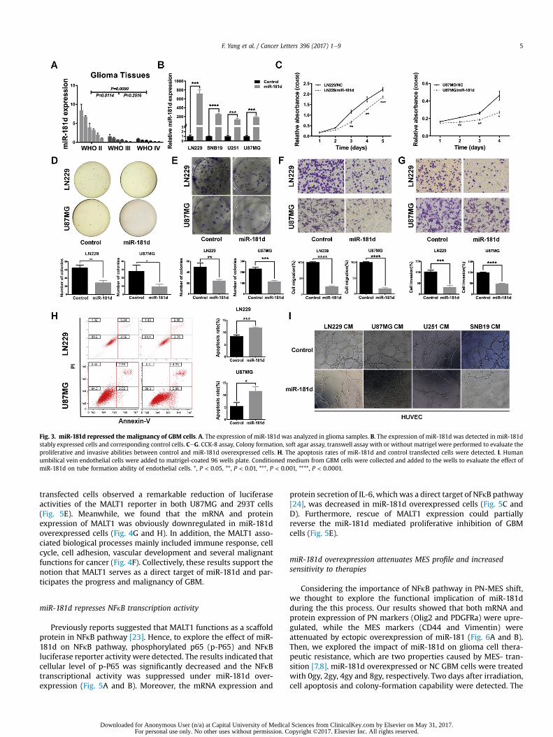
Fig. 3. miR-181d repressed the malignancy of GBM cells. A. The expression of miR-181d was analyzed in glioma samples. B. The expression of miR-181d was detected in miR-181dstably expressed cells and corresponding control cells. CeG. CCK-8 assay, Colony formation, soft agar assay, transwell assay with or without matrigel were performed to evaluate theproliferative and invasive abilities between control and miR-181d overexpressed cells. H. The apoptosis rates of miR-181d and control transfected cells were detected. I. Humanumbilical vein endothelial cells were added to matrigel-coated 96 wells plate. Conditioned medium from GBM cells were collected and added to the wells to evaluate the effect ofmiR-181d on tube formation ability of endothelial cells. *, P < 0.05, **, P < 0.01, ***, P < 0.001, ****, P < 0.0001.
F. Yang et al. / Cancer Letters 396 (2017) 1e9 5
transfected cells observed a remarkable reduction of luciferaseactivities of the MALT1 reporter in both U87MG and 293T cells(Fig. 5E). Meanwhile, we found that the mRNA and proteinexpression of MALT1 was obviously downregulated in miR-181doverexpressed cells (Fig. 4G and H). In addition, the MALT1 asso-ciated biological processes mainly included immune response, cellcycle, cell adhesion, vascular development and several malignantfunctions for cancer (Fig. 4F). Collectively, these results support thenotion that MALT1 serves as a direct target of miR-181d and par-ticipates the progress and malignancy of GBM.
miR-181d represses NFkB transcription activity
Previously reports suggested that MALT1 functions as a scaffoldprotein in NFkB pathway [23]. Hence, to explore the effect of miR-181d on NFkB pathway, phosphorylated p65 (p-P65) and NFkBluciferase reporter activity were detected. The results indicated thatcellular level of p-P65 was significantly decreased and the NFkBtranscriptional activity was suppressed under miR-181d over-expression (Fig. 5A and B). Moreover, the mRNA expression and
Downloaded for Anonymous User (n/a) at Capital University of MedicaFor personal use only. No other uses without permission. C
protein secretion of IL-6, which was a direct target of NFkB pathway[24], was decreased in miR-181d overexpressed cells (Fig. 5C andD). Furthermore, rescue of MALT1 expression could partiallyreverse the miR-181d mediated proliferative inhibition of GBMcells (Fig. 5E).
miR-181d overexpression attenuates MES profile and increasedsensitivity to therapies
Considering the importance of NFkB pathway in PN-MES shift,we thought to explore the functional implication of miR-181dduring the this process. Our results showed that both mRNA andprotein expression of PN markers (Olig2 and PDGFRa) were upre-gulated, while the MES markers (CD44 and Vimentin) wereattenuated by ectopic overexpression of miR-181 (Fig. 6A and B).Then, we explored the impact of miR-181d on glioma cell thera-peutic resistance, which are two properties caused by MES- tran-sition [7,8]. miR-181d overexpressed or NC GBM cells were treatedwith 0gy, 2gy, 4gy and 8gy, respectively. Two days after irradiation,cell apoptosis and colony-formation capability were detected. The
l Sciences from ClinicalKey.com by Elsevier on May 31, 2017.opyright ©2017. Elsevier Inc. All rights reserved.
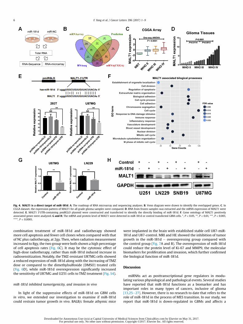
Fig. 4. MALT1 is a direct target of miR-181d. A. The roadmap of RNA microarray and sequencing analyses. B. Venn diagram were drawn to identify the overlapped genes. C. InCGGA dataset, the expression pattern of MALT1 for all grade glioma samples were compared. D. RNA from frozen samples was extracted and the mRNA expression of MALT1 weredetected. E. MALT1 30UTR-containing pmiRGLO plasmid were constructed and transfected to identify the directly binding of miR-181d. F. Gene ontology of MALT1 positivelyassociated genes were analyzed. G and H. The mRNA and protein level of MALT1 were detected in miR-181d or control transfected GBM cells. *, P < 0.05, **, P < 0.01, ***, P < 0.001,****, P < 0.0001.
F. Yang et al. / Cancer Letters 396 (2017) 1e96
combination treatment of miR-181d and radiotherapy showedmore cell apoptosis and fewer cell clones when compared with thatof NC plus radiotherapy, at 2gy. Then, when radiationmeasurementincreased to 8gy, the two groupwere both shown a high percentageof cell apoptosis rates (Fig. 6C). It may be the cytotoxic effect ofhigh-dose radiotherapy, rather than miR-181d induced increase inradiosensitization. Notably, the TMZ-resistant U87MG cells showeda reduced expression of miR-181d along with the increasing of TMZdose or compared to the dimethylsulfoxide (DMSO) treated cells(Fig. 6D), while miR-181d overexpression significantly increasedthe sensitivity of U87MG and U251 cells to TMZ treatment (Fig. S4).
miR-181d inhibited tumorigenicity, and invasion in vivo
In light of the suppressive effects of miR-181d on GBM cellsin vitro, we extended our investigation to examine if miR-181dcould restrain tumor growth in vivo. BALB/c female athymic mice
Downloaded for Anonymous User (n/a) at Capital University of MedFor personal use only. No other uses without permissio
were implanted in the brain with established stable cell U87-miR-181d and U87-control. MRI and HE showed the inhibition of tumorgrowth in the miR-181d e overexpressing group compared withthe control group (Fig. 7A and B). The overexpression of miR-181dcould reduce the protein level of Ki-67 and MMP9, the molecularbiomarkers for proliferation and invasion, which further confirmedthe biological function of miR-181d.
Discussion
miRNAs act as posttranscriptional gene regulators in modu-lating various physiological and pathological events. Several studieshave reported that miR-181d functions as a biomarker and hasimportant roles in many types of cancers, inclusive of glioma[13,25e27]. However, there is no research to date that refers to therole of miR-181d in the process of MES transition. In our study, wereport that miR-181d is down-regulated in GBMs and affects a
ical Sciences from ClinicalKey.com by Elsevier on May 31, 2017.n. Copyright ©2017. Elsevier Inc. All rights reserved.
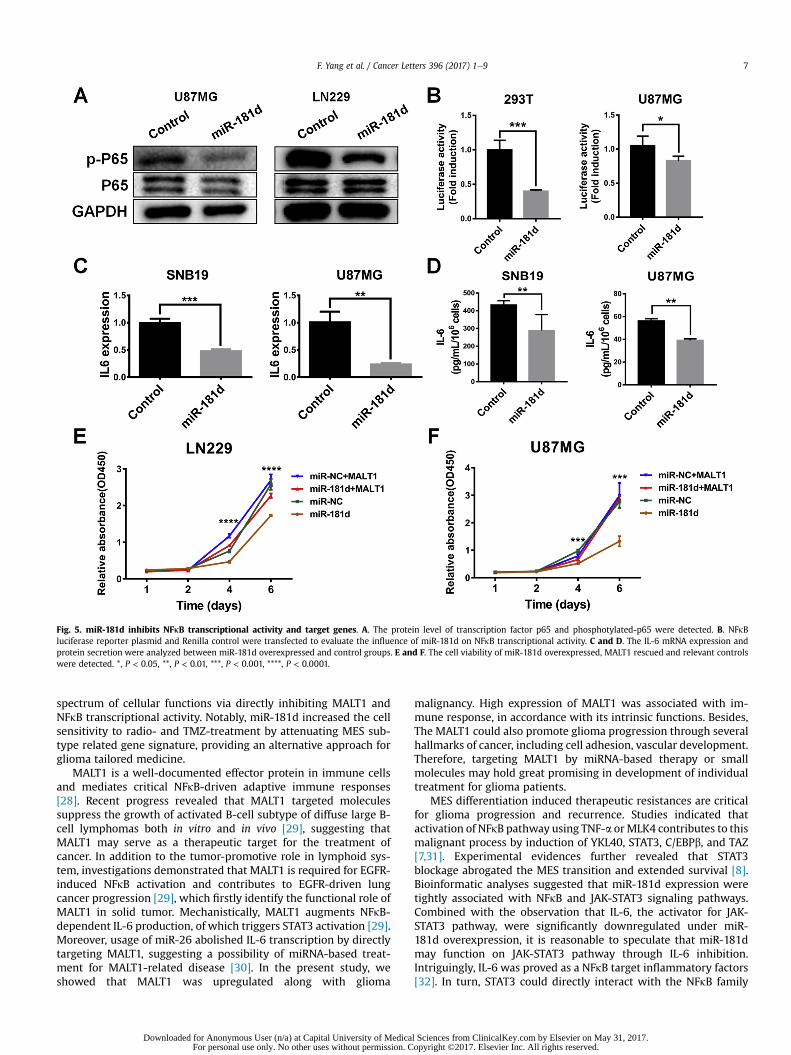
Fig. 5. miR-181d inhibits NFkB transcriptional activity and target genes. A. The protein level of transcription factor p65 and phosphotylated-p65 were detected. B. NFkBluciferase reporter plasmid and Renilla control were transfected to evaluate the influence of miR-181d on NFkB transcriptional activity. C and D. The IL-6 mRNA expression andprotein secretion were analyzed between miR-181d overexpressed and control groups. E and F. The cell viability of miR-181d overexpressed, MALT1 rescued and relevant controlswere detected. *, P < 0.05, **, P < 0.01, ***, P < 0.001, ****, P < 0.0001.
F. Yang et al. / Cancer Letters 396 (2017) 1e9 7
spectrum of cellular functions via directly inhibiting MALT1 andNFkB transcriptional activity. Notably, miR-181d increased the cellsensitivity to radio- and TMZ-treatment by attenuating MES sub-type related gene signature, providing an alternative approach forglioma tailored medicine.
MALT1 is a well-documented effector protein in immune cellsand mediates critical NFkB-driven adaptive immune responses[28]. Recent progress revealed that MALT1 targeted moleculessuppress the growth of activated B-cell subtype of diffuse large B-cell lymphomas both in vitro and in vivo [29], suggesting thatMALT1 may serve as a therapeutic target for the treatment ofcancer. In addition to the tumor-promotive role in lymphoid sys-tem, investigations demonstrated that MALT1 is required for EGFR-induced NFkB activation and contributes to EGFR-driven lungcancer progression [29], which firstly identify the functional role ofMALT1 in solid tumor. Mechanistically, MALT1 augments NFkB-dependent IL-6 production, of which triggers STAT3 activation [29].Moreover, usage of miR-26 abolished IL-6 transcription by directlytargeting MALT1, suggesting a possibility of miRNA-based treat-ment for MALT1-related disease [30]. In the present study, weshowed that MALT1 was upregulated along with glioma
Downloaded for Anonymous User (n/a) at Capital University of MedicaFor personal use only. No other uses without permission. C
malignancy. High expression of MALT1 was associated with im-mune response, in accordance with its intrinsic functions. Besides,The MALT1 could also promote glioma progression through severalhallmarks of cancer, including cell adhesion, vascular development.Therefore, targeting MALT1 by miRNA-based therapy or smallmolecules may hold great promising in development of individualtreatment for glioma patients.
MES differentiation induced therapeutic resistances are criticalfor glioma progression and recurrence. Studies indicated thatactivation of NFkB pathway using TNF-a orMLK4 contributes to thismalignant process by induction of YKL40, STAT3, C/EBPb, and TAZ[7,31]. Experimental evidences further revealed that STAT3blockage abrogated the MES transition and extended survival [8].Bioinformatic analyses suggested that miR-181d expression weretightly associated with NFkB and JAK-STAT3 signaling pathways.Combined with the observation that IL-6, the activator for JAK-STAT3 pathway, were significantly downregulated under miR-181d overexpression, it is reasonable to speculate that miR-181dmay function on JAK-STAT3 pathway through IL-6 inhibition.Intriguingly, IL-6 was proved as a NFkB target inflammatory factors[32]. In turn, STAT3 could directly interact with the NFkB family
l Sciences from ClinicalKey.com by Elsevier on May 31, 2017.opyright ©2017. Elsevier Inc. All rights reserved.
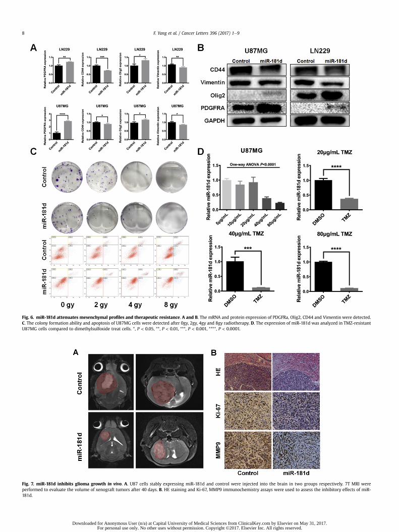
Fig. 6. miR-181d attenuates mesenchymal profiles and therapeutic resistance. A and B. The mRNA and protein expression of PDGFRa, Olig2, CD44 and Vimentin were detected.C. The colony formation ability and apoptosis of U87MG cells were detected after 0gy, 2gy, 4gy and 8gy radiotherapy. D. The expression of miR-181d was analyzed in TMZ-resistantU87MG cells compared to dimethylsulfoxide treat cells. *, P < 0.05, **, P < 0.01, ***, P < 0.001, ****, P < 0.0001.
Fig. 7. miR-181d inhibits glioma growth in vivo. A. U87 cells stably expressing miR-181d and control were injected into the brain in two groups respectively. 7T MRI wereperformed to evaluate the volume of xenograft tumors after 40 days. B. HE staining and Ki-67, MMP9 immunochemistry assays were used to assess the inhibitory effects of miR-181d.
F. Yang et al. / Cancer Letters 396 (2017) 1e98
Downloaded for Anonymous User (n/a) at Capital University of Medical Sciences from ClinicalKey.com by Elsevier on May 31, 2017.For personal use only. No other uses without permission. Copyright ©2017. Elsevier Inc. All rights reserved.
F. Yang et al. / Cancer Letters 396 (2017) 1e9 9
member RELA, thereby contributing to constitutive NFkB activation[33]. This reinforced crosstalk might boost downstream biologicaleffects caused by NFkB and STAT3 pathways. Experimental evi-dence is need for clarifying the latent relationship between miR-181d and STAT3 pathways.
In summary, our results thoroughly show that miR-181d acts asa tumor suppressor by targeting MALT1 to restrain numeroustumor-related malignancy. We also demonstrated that miR-181doverexpression confers increased therapeutic sensitization toGBM cells. Our results highlight the regulatory mechanisms ofmiRNA implicated in MES differentiation and could offer moreinspiration to miRNA-based glioma therapy.
Funding
This work was supported by the National Natural ScienceFoundation of China (81402052); the Beijing Nova Program(Z16110004916082); the Beijing Postdoctoral Research Foundation(2016ZZ-37); the National Key Research and Development Plan (No.2016YFC0902500); the Beijing Science and Technology Plan (No.Z131100006113018, Z141100000214009); the Capital MedicalDevelopment Research Fund (2016-1-1072) and the ResearchSpecial Fund for Public Welfare Industry of Heath (No. 201402008).
Conflict of interest statement
None declared.
Appendix A. Supplementary data
Supplementary data related to this article can be found at http://dx.doi.org/10.1016/j.canlet.2017.03.002.
References
[1] T. Jiang, Y. Mao, W. Ma, Q. Mao, Y. You, X. Yang, et al., CGCG clinical practiceguidelines for the management of adult diffuse gliomas, Cancer Lett. 375 (2)(2016) 263e273.
[2] Q.T. Ostrom, H. Gittleman, J. Fulop, M. Liu, R. Blanda, C. Kromer, et al., CBTRUSstatistical report: primary brain and central nervous system tumors diagnosedin the United States in 2008e2012, Neuro Oncol. 17 (Suppl. 4) (2015)iv1eiv62.
[3] Y. Wang, T. Jiang, Understanding high grade glioma: molecular mechanism,therapy and comprehensive management, Cancer Lett. 331 (2013) 139e146.
[4] R.G. Verhaak, K.A. Hoadley, E. Purdom, V. Wang, Y. Qi, M.D. Wilkerson, et al.,Integrated genomic analysis identifies clinically relevant subtypes of glio-blastoma characterized by abnormalities in PDGFRA, IDH1, EGFR, and NF1,Cancer Cell. 17 (2010) 98e110.
[5] H.S. Phillips, S. Kharbanda, R. Chen, W.F. Forrest, R.H. Soriano, T.D. Wu, et al.,Molecular subclasses of high-grade glioma predict prognosis, delineate apattern of disease progression, and resemble stages in neurogenesis, CancerCell. 9 (2006) 157e173.
[6] A. Li, J. Walling, S. Ahn, Y. Kotliarov, Q. Su, M. Quezado, et al., Unsupervisedanalysis of transcriptomic profiles reveals six glioma subtypes, Cancer Res. 69(2009) 2091e2099.
[7] K.P. Bhat, V. Balasubramaniyan, B. Vaillant, R. Ezhilarasan, K. Hummelink,F. Hollingsworth, et al., Mesenchymal differentiation mediated by NF-kB pro-motes radiation resistance in glioblastoma, Cancer Cell. 24 (2013) 331e346.
[8] J. Lau, S. Ilkhanizadeh, S. Wang, Y.A. Miroshnikova, N.A. Salvatierra, R.A. Wong,et al., STAT3 blockade inhibits radiation-induced malignant progression inglioma, Cancer Res. 75 (2015) 4302e4311.
[9] S.C. Janga, S. Vallabhaneni, MicroRNAs as post-transcriptional machines andtheir interplay with cellular networks, Adv. Exp. Med. Biol. 722 (2011) 59e74.
Downloaded for Anonymous User (n/a) at Capital University of MedicaFor personal use only. No other uses without permission. C
[10] M.B. Irmak-Yazicioglu, Mechanisms of microRNA deregulation and microRNAtargets in gastric cancer, Oncol. Res. Treat. 39 (2016) 136e139.
[11] M. Hemmatzadeh, H. Mohammadi, F. Jadidi-Niaragh, F. Asghari, M. Yousefi,The role of oncomirs in the pathogenesis and treatment of breast cancer,Biomed. Pharmacother. 78 (2016) 129e139.
[12] J.A. Chan, A.M. Krichevsky, K.S. Kosik, MicroRNA-21 is an antiapoptotic factorin human glioblastoma cells, Cancer Res. 65 (2005) 6029e6033.
[13] W. Zhang, J. Zhang, K. Hoadley, D. Kushwaha, V. Ramakrishnan, S. Li, et al.,miR-181d: a predictive glioblastoma biomarker that downregulates MGMTexpression, Neuro Oncol. 14 (2012) 712e719.
[14] S. Yin, W. Du, F. Wang, B. Han, Y. Cui, D. Yang, et al., MicroRNA-326 sensitizeshuman glioblastoma cells to curcumin via the SHH/GLI1 signaling pathway,Cancer Biol. Ther. (2016) 0.
[15] Z. Shi, J. Zhang, X. Qian, L. Han, K. Zhang, L. Chen, et al., AC1MMYR2, an in-hibitor of dicer-mediated biogenesis of Oncomir miR-21, reverses epithelial-mesenchymal transition and suppresses tumor growth and progression,Cancer Res. 73 (2013) 5519e5531.
[16] D.N. Louis, A. Perry, G. Reifenberger, D.A. von, D. Figarella-Branger,W.K. Cavenee, et al., The 2016 World Health Organization classification oftumors of the central nervous system: a summary, Acta Neuropathol. 131(2016) 803e820.
[17] T. Ashizawa, Y. Akiyama, H. Miyata, A. Iizuka, M. Komiyama, A. Kume, et al.,Effect of the STAT3 inhibitor STX-0119 on the proliferation of atemozolomide-resistant glioblastoma cell line, Int. J. Oncol. 45 (2014)411e418.
[18] d.W. Huang, B.T. Sherman, R.A. Lempicki, Systematic and integrative analysisof large gene lists using DAVID bioinformatics resources, Nat. Protoc. 4 (2009)44e57.
[19] A. Subramanian, P. Tamayo, V.K. Mootha, S. Mukherjee, B.L. Ebert,M.A. Gillette, et al., Gene set enrichment analysis: a knowledge-basedapproach for interpreting genome-wide expression profiles, Proc. Natl.Acad. Sci. U.S.A. 102 (2005) 15545e15550.
[20] S. H€anzelmann, R. Castelo, J. Guinney, GSVA: gene set variation analysis formicroarray and RNA-seq data, BMC Bioinforma. 14 (2013) 7.
[21] H. Dweep, N. Gretz, miRWalk2.0: a comprehensive atlas of microRNA-targetinteractions, Nat. Methods 12 (2015) 697.
[22] D. Betel, A. Koppal, P. Agius, C. Sander, C. Leslie, Comprehensive modeling ofmicroRNA targets predicts functional non-conserved and non-canonical sites,Genome Biol. 11 (2010) R90.
[23] L.R. Klei, D. Hu, R. Panek, D.N. Alfano, R.E. Bridwell, K.M. Bailey, et al., MALT1protease activation triggers acute disruption of endothelial barrier integrityvia CYLD cleavage, Cell Rep. 17 (2016) 221e232.
[24] G. Malaponte, S.S. Signorelli, V. Bevelacqua, J. Polesel, M. Taborelli, C. Guarneri,et al., Increased levels of NF-kb-dependent markers in cancer-associated deepvenous thrombosis, PLoS One 10 (2015) e0132496.
[25] X.F. Wang, Z.M. Shi, X.R. Wang, L. Cao, Y.Y. Wang, J.X. Zhang, et al., MiR-181dacts as a tumor suppressor in glioma by targeting K-ras and Bcl-2, J. CancerRes. Clin. Oncol. 138 (2012) 573e584.
[26] R. Li, X. Li, S. Ning, J. Ye, L. Han, C. Kang, et al., Identification of a core miRNA-pathway regulatory network in glioma by therapeutically targeting miR-181d,miR-21, miR-23b, b-Catenin, CBP, and STAT3, PLoS One 9 (2014) e101903.
[27] R. Su, H.S. Lin, X.H. Zhang, X.L. Yin, H.M. Ning, B. Liu, et al., MiR-181 family:regulators of myeloid differentiation and acute myeloid leukemia as well aspotential therapeutic targets, Oncogene 34 (2015) 3226e3239.
[28] M. Jaworski, B.J. Marsland, J. Gehrig, W. Held, S. Favre, S.A. Luther, et al., Malt1protease inactivation efficiently dampens immune responses but causesspontaneous autoimmunity, EMBO J. 33 (2014) 2765e2781.
[29] L. Font�an, A. Melnick, Molecular pathways: targeting MALT1 paracaspaseactivity in lymphoma, Clin. Cancer Res. 19 (2013) 6662e6668.
[30] C.Y. Chen, J.T. Chang, Y.F. Ho, A.B. Shyu, MiR-26 down-regulates TNF-a/NF-kBsignalling and IL-6 expression by silencing HMGA1 and MALT1, Nucleic AcidsRes. 44 (2016) 3772e3787.
[31] S.H. Kim, R. Ezhilarasan, E. Phillips, D. Gallego-Perez, A. Sparks, D. Taylor, et al.,Serine/threonine kinase MLK4 determines mesenchymal identity in gliomastem cells in an NF-kB-dependent manner, Cancer Cell. 29 (2016) 201e213.
[32] S. Grivennikov, E. Karin, J. Terzic, D. Mucida, G.Y. Yu, S. Vallabhapurapu, et al.,IL-6 and Stat3 are required for survival of intestinal epithelial cells anddevelopment of colitis-associated cancer, Cancer Cell. 15 (2009) 103e113.
[33] H. Lee, A. Herrmann, J.H. Deng, M. Kujawski, G. Niu, Z. Li, et al., Persistentlyactivated Stat3 maintains constitutive NF-kappaB activity in tumors, CancerCell. 15 (2009) 283e293.
l Sciences from ClinicalKey.com by Elsevier on May 31, 2017.opyright ©2017. Elsevier Inc. All rights reserved.